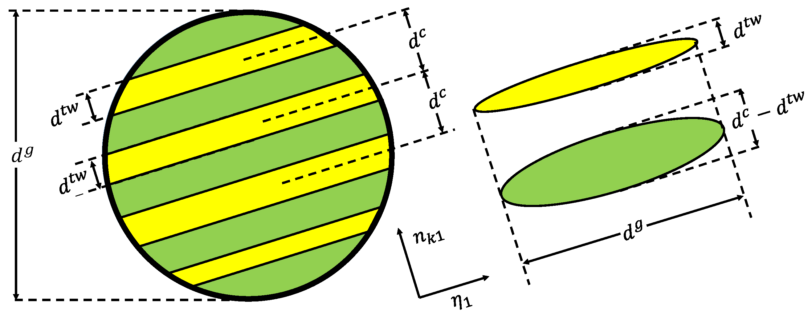
Figure 1.
Representation of the orientation of the twins and the parent matrix with their distinctive dimensions employed in the formulation of the composite grain (CG) model [
19].
Figure 1.
Representation of the orientation of the twins and the parent matrix with their distinctive dimensions employed in the formulation of the composite grain (CG) model [
19].
Figure 2.
Illustration of the twin boundary propagating in the parent matrix instigating the dislocation transmutation mechanism and the subsequent latent hardening.
Figure 2.
Illustration of the twin boundary propagating in the parent matrix instigating the dislocation transmutation mechanism and the subsequent latent hardening.
Figure 3.
Illustration of orientations of test specimens extracted from rolled AZ31 magnesium alloy sheet. (a) Dimensions of the AZ31 magnesium compression test specimens extracted from ND and RD orientations. (b) Mini dog bone tension specimen cut in ND and the grip designed for the mini sample to conduct the test. (c); Round dog bone tension specimen in RD.
Figure 4.
Initial texture for AZ31 obtained through neutron diffraction at LANL presented in and pole figure. X-axis represents TD, Y-axis represents RD and Z-axis represents ND.
Figure 4.
Initial texture for AZ31 obtained through neutron diffraction at LANL presented in and pole figure. X-axis represents TD, Y-axis represents RD and Z-axis represents ND.
Figure 5.
Stress–strain data obtained from mechanical testing AZ31 magnesium. Figures (a,c,e) show compression and tension results for the test conducted at 0.001 s strain rate and temperature of 293 K, 373 K, and 473 K respectively, and figures (b,d,f) show compression and tension test results for 0.1 s strain rate and temperatures of 293 K, 373 K, and 473 K, respectively.
Figure 6.
Stress–strain data comparison at temperatures of 293 K, 373 K, and 473 K for same loading condition and strain rate 0.001 s. Figures (a,c) show compression results and (b,d) show tension results.
Figure 6.
Stress–strain data comparison at temperatures of 293 K, 373 K, and 473 K for same loading condition and strain rate 0.001 s. Figures (a,c) show compression results and (b,d) show tension results.
Figure 7.
Stress–strain data comparison at temperatures of 293 K, 373 K, and 473 K for same loading condition and strain rate 0.1 s. Figures (a,c) show compression results and (b,d) show tension results.
Figure 7.
Stress–strain data comparison at temperatures of 293 K, 373 K, and 473 K for same loading condition and strain rate 0.1 s. Figures (a,c) show compression results and (b,d) show tension results.
Figure 8.
Dimensions of a flat dog bone specimen obtained from a 2 mm thin sheet of ZEK100, designed using ASTM standards and Gleeble sample drawing. The sample was cut in 0° (RD), 30°, 60°, and 90° (TD) orientations.
Figure 8.
Dimensions of a flat dog bone specimen obtained from a 2 mm thin sheet of ZEK100, designed using ASTM standards and Gleeble sample drawing. The sample was cut in 0° (RD), 30°, 60°, and 90° (TD) orientations.
Figure 9.
Initial texture for ZEK100 obtained using X-ray diffraction (XRD) scan presented in and pole figure. X-axis in the plot is TD and Y-axis is RD.
Figure 9.
Initial texture for ZEK100 obtained using X-ray diffraction (XRD) scan presented in and pole figure. X-axis in the plot is TD and Y-axis is RD.
Figure 10.
Stress–strain data comparison at temperatures of 293 K (a), 373 K (b), and 473 K (c) for same loading condition and strain rate 0.001 s.
Figure 10.
Stress–strain data comparison at temperatures of 293 K (a), 373 K (b), and 473 K (c) for same loading condition and strain rate 0.001 s.
Figure 11.
Stress–strain response comparing the experimental and simulation in Compression and tension at the strain rate of 0.001 s. (a) RD at 293 K (b) ND 293 K (c) RD at 373 K (d) ND at 373 K (e) RD at 473 K (f) ND at 473 K.
Figure 11.
Stress–strain response comparing the experimental and simulation in Compression and tension at the strain rate of 0.001 s. (a) RD at 293 K (b) ND 293 K (c) RD at 373 K (d) ND at 373 K (e) RD at 473 K (f) ND at 473 K.
Figure 12.
Stress–strain response comparing the experimental and simulation in Compression and tension at the strain rate of 0.1 s. (a) RD at 293 K (b) ND 293 K (c) RD at 373 K (d) ND at 373 K (e) RD at 473 K (f) ND at 473 K.
Figure 12.
Stress–strain response comparing the experimental and simulation in Compression and tension at the strain rate of 0.1 s. (a) RD at 293 K (b) ND 293 K (c) RD at 373 K (d) ND at 373 K (e) RD at 473 K (f) ND at 473 K.
Figure 13.
Activity of slip and twin within the parent (a,c,e,g) and primary twin (b,d,f,g) obtained from the simulations of AZ31 at room temperature (293 K) and 0.001 s strain-rate. Subplots (a–d) show compression activity and (e–h) show tension activity. RD is in-plane and ND is thru-thickness.
Figure 13.
Activity of slip and twin within the parent (a,c,e,g) and primary twin (b,d,f,g) obtained from the simulations of AZ31 at room temperature (293 K) and 0.001 s strain-rate. Subplots (a–d) show compression activity and (e–h) show tension activity. RD is in-plane and ND is thru-thickness.
Figure 14.
Texture pole figure for {0 0 0 1} and at failure obtained from simulation for tension and compression in both ND and RD at room temperature and 0.001 s strain rate. The X-axis represents TD, Y-axis represents RD and Z-axis represents ND.
Figure 14.
Texture pole figure for {0 0 0 1} and at failure obtained from simulation for tension and compression in both ND and RD at room temperature and 0.001 s strain rate. The X-axis represents TD, Y-axis represents RD and Z-axis represents ND.
Figure 15.
Stress–strain response of ZEK100 in tension and the simulation data fit achieved using the VPSC dislocation density model. Subplots (a–c) are tests and simulations conducted at 293 K, 373 K, and 473 K, respectively.
Figure 15.
Stress–strain response of ZEK100 in tension and the simulation data fit achieved using the VPSC dislocation density model. Subplots (a–c) are tests and simulations conducted at 293 K, 373 K, and 473 K, respectively.
Figure 16.
Activity of slip and twin within the parent (a,c,e,g) and primary twin (b,d,f,g) obtained from the simulations of ZEK100 at room temperature (293 K) and 0.001 s strain-rate. Subplots (a–d) show compression activity and (e–h) show tension activity. Each row represents activity for 0°, 30°, 60°, and 90°, respectively.
Figure 17.
Pole figures in and representing texture evolution for ZEK100 at room temperature and 0.001 s strain rate in tension. Plots (a,c,e,g) are undeformed texture and (b,d,f,h) are deformed texture from simulations strained in 0°, 30°, 60° and 90°, respectively.
Figure 17.
Pole figures in and representing texture evolution for ZEK100 at room temperature and 0.001 s strain rate in tension. Plots (a,c,e,g) are undeformed texture and (b,d,f,h) are deformed texture from simulations strained in 0°, 30°, 60° and 90°, respectively.
Table 1.
Matrix of tests used for fitting and testing crystal plasticity model. (*: Tests performed).
Table 1.
Matrix of tests used for fitting and testing crystal plasticity model. (*: Tests performed).
|
Magnesium AZ31
|
|---|
|
Strain Rate
|
Orientation
|
293 K
|
373 K
|
473 K
|
|---|
|
Tension
|
Compression
|
Tension
|
Compression
|
Tension
|
Compression
|
|---|
| 0.001 | RD | * | * | * | * | * | * |
| ND | * | * | * | * | * | * |
| 0.1 | RD | * | * | * | * | * | * |
| ND | * | * | * | * | * | * |
| Magnesium ZEK100 |
| Strain Rate | Orientation | 293 K | 373 K | 473 K |
| Tension | Compression | Tension | Compression | Tension | Compression |
| 0.001 | RD | * | − | * | − | * | − |
| 30° to RD | * | − | * | − | * | − |
| 60° to RD | * | − | * | − | * | − |
| TD | * | − | * | − | * | − |
Table 2.
Slip and Twinning modes for magnesium used in VPSC simulation.
Table 2.
Slip and Twinning modes for magnesium used in VPSC simulation.
|
Symbol
|
Mode
|
Crystallography
|
No. of Systems
|
b (nm)
|
|---|
| Prismatic | | 3 | 0.321 |
| Basal | | 3 | 0.321 |
| Pyramidal | | 6 | 0.612 |
| Tensile Twin | | 6 | 0.138 |
| Compression Twin | | 6 | 0.0924 |
Table 3.
Prismatic, basal, and pyramidal dislocation density hardening parameters for magnesium AZ31.
Table 3.
Prismatic, basal, and pyramidal dislocation density hardening parameters for magnesium AZ31.
|
Parameters
|
Prismatic
|
Basal
|
Pyramidal
|
|---|
| | | |
| | | |
| | | |
| (MPa) | | | |
| (MPa) (293 K) | 84 | 26 | 130 |
| (MPa) (373 K) | 75 | 24 | 110 |
| (MPa) (473 K) | 30 | 15 | 45 |
| 0 | 0 | 0 |
| X | | 0.9 | 0.9 |
| 0 | 0 | 0 |
| 0 | 0 | 0 |
Table 4.
Tension and compression twin dislocation density hardening parameters for magnesium AZ31.
Table 4.
Tension and compression twin dislocation density hardening parameters for magnesium AZ31.
|
Parameters
|
Tension Twin
|
Compression Twin
|
|---|
| (MPa) (293 K) | 31 | 225 |
| (MPa) (293 K) | 5 | 225 |
| (MPa) (373 K) | 31 | 200 |
| (MPa) (373 K) | 5 | 200 |
| (MPa) (473 K) | 31 | 120 |
| (MPa) (473 K) | 5 | 120 |
Table 5.
Prismatic, basal, and pyramidal dislocation density hardening parameters for magnesium ZEK100.
Table 5.
Prismatic, basal, and pyramidal dislocation density hardening parameters for magnesium ZEK100.
|
Parameters
|
Prismatic
|
Basal
|
Pyramidal
|
|---|
|
|
|
|
|
|
|
|
|
|
|
|
|
|
|
| (MPa) |
|
|
|
| (MPa) (293 K) | 136 | 56 | 200 |
| (MPa) (373 K) | 75 | 27 | 80 |
| (MPa) (473 K) | 45 | 15 | 50 |
|
| 0 | 0 | 0 |
| X |
| 0.9 | 0.9 |
|
| 0 | 0 | 0 |
|
| 0 | 0 | 0 |
Table 6.
Tension and compression twin dislocation density hardening parameters for magnesium ZEK100.
Table 6.
Tension and compression twin dislocation density hardening parameters for magnesium ZEK100.
|
Parameters
|
Tension Twin
|
Compression Twin
|
|---|
| (MPa) (293 K) | 50 | 250 |
| (MPa) (293 K) | 25 | 250 |
| (MPa) (373 K) | 50 | 100 |
| (MPa) (373 K) | 25 | 100 |
| (MPa) (473 K) | 50 | 60 |
| (MPa) (473 K) | 25 | 60 |

 ,
, 


