Article

Development of Cerium Oxide/Chitosan/Graphene Oxide Nanocomposite: An Investigation toward Its Biological Applications under In Vitro Conditions

1
Department of Electronics and Communication Engineering, Rajalakshmi Engineering College, Thandalam 602 105, Tamil Nadu, India
2
Department of Chemistry, College of Science, King Saud University, P.O. Box 2455, Riyadh 11451, Saudi Arabia
3
School of Chemical Engineering, Yeungnam University, Gyeongsan 38541, Republic of Korea
*
Authors to whom correspondence should be addressed.
Crystals 2023, 13(9), 1393; https://doi.org/10.3390/cryst13091393
Submission received: 18 August 2023 / Revised: 13 September 2023 / Accepted: 15 September 2023 / Published: 19 September 2023
(This article belongs to the Section Hybrid and Composite Crystalline Materials)

Abstract

:
A ternary nanocomposite was prepared using cerium oxide, chitosan, and graphene oxide (CeO2/CS/GO) using a simple and cost-effective wet chemical method. The physicochemical properties of the developed ternary nanocomposite were examined using X-ray diffraction, Fourier transform infrared spectroscopy, scanning electron microscopy with energy-dispersive X-ray spectroscopy, and ultraviolet-visible spectroscopy. Furthermore, the therapeutic behavior of the developed CeO2/CS/GO composite was assessed using anti-bacterial, anti-fungal, and anti-cancer assays. For Escherichia coli, Staphylococcus aureus, and Salmonella species, 750 µg/mL of the CeO2/CS/GO composite showed effective anti-bacterial activity, with a zone of inhibition of 9 mm. Additionally, the CeO2/CS/GO composite’s anti-fungal activity against Aspergillus niger was studied. The anti-cancer properties of different concentrations of the CeO2/CS/GO composite were assessed on MCF-7 cells, and 18.8% of cells were found to be viable at the maximum concentration of 1000 µg/mL CeO2/CS/GO and 46.37% at 125 µg/mL. The results of the hemolysis assay performed using human red blood cells and various concentrations of the CeO2/CS/GO composite indicated that the nanocomposite possesses biological properties. Overall, it can act as a therapeutic platform for breast cancer, bacterial and fungal infections.

1. Introduction

Nanotechnology is an interdisciplinary domain that focuses on providing solutions to diverse fields such as healthcare, pharmacy, biophysical modeling, biological engineering, and genetics [1]. The ability to apply surface-functionalized small-scale structures with novel features and the potential to control or manipulate organisms at the molecular or nanoscale level are the main areas of focus in contemporary nanotechnology. This will pave the way for the assessment of nanoplatforms as therapeutic agents [2]. Breast cancer is the most prevalent type of cancer and a major cause of mortality for women over 35 years of age. Nanotechnology enables therapeutic platforms on the nanoscale to selectively target and treat tumors. Validation of the developed nanoplatform as a therapeutic agent against breast cancer cell lines must be performed under both in vivo and in vitro conditions [3]. Several researchers have been interested in developing cerium oxide-based nanoplatforms (CeO2 NPs) because of their distinct physicochemical properties, which play a vital role in their biological properties [4]. For example, the potential redox abilities of CeO2 nanoparticles are responsible for their excellent antioxidant properties, which has been confirmed both in vitro and in vivo [5]. In particular, CeO2 exhibits interchangeable oxidation states of cerium (3+ and 4+ states) on the surface of the nanoparticle, wherein small CeO2 NPs with larger surface areas possess a large number of 3+ sites [6]. These mixed valence states of Ce lead to the generation of oxygen vacancies and subsequently defects. In addition, owing to the adoption of fluorite crystal structures, CeO2 NPs have been used in several fascinating fields, including biomedicine. However, the wider application of CeO2 has been mainly inhibited by the aggregation of NPs, which is mainly addressed by modifying the surface of CeO2 NPs using various materials, including carbonaceous substances and polymers, which effectively prevent aggregation and improve biocompatibility [7].
In this context, graphene has attracted significant attention owing to its excellent properties. Graphene is a two-dimensional sp2-bonded carbon material with a honeycomb lattice structure. It is a fundamental component of graphitic substances and has attracted considerable interest owing to its unique properties. Graphene has a large surface-to-volume ratio, excellent tensile strength, chemical durability, and good thermal conductivity [8]. Graphene oxide (GO), an oxygenated derivative of graphene, harbors a large number of oxygen-containing groups on both the surface and edges of GO. These groups not only enhance the solubility and biocompatibility of materials but also collectively enhance the electronic and redox properties of the resulting composite materials [9]. According to Pang et al., the physicochemical characteristics of GO, such as size and length, surface characteristics, and aggregation factors, are primarily responsible for its anti-bacterial activity [10]. These factors affect the manner in which cells die after the deposition of bacteria onto the nanomaterials, which involves membrane breakdown once they touch the sharp edges of GO, leakage of cell contents into the environment, and ultimately cell death [11].
Moreover, CeO2 has often been combined with other materials, particularly biomaterials, to fabricate multi-component composites with adequate sites for cell attachment and mechanical support [12]. For example, various biopolymers, including gelatin, collagen, alginate, and chitosan (CS), have been used to prepare CeO2-based multicomponent composites with potential biological properties [13]. Chitosan is a linear polymer that resembles cellulose and has distinct chemical and biological properties as it contains amine and hydroxyl groups in its structure [14].
Owing to its outstanding biocompatibility, biodegradability, low toxicity, and inherent anti-bacterial activity, CS, a natural substance, has been extensively used in biomedical applications [15,16]. Due to their unique characteristics, such as high solubility and biocompatibility, antioxidant power, antimicrobial and anti-inflammatory properties, nanocomposites made from non-mineral nanoparticles loaded with natural biopolymers like chitosan are very effective for treating wound healing, cancer, inflammatory diseases, and cardiovascular conditions [17,18]. Furthermore, the anti-cancer activity of CS against hepatocellular carcinoma is mediated by a decrease in mitochondrial membrane potential, an increase in membrane lipid peroxidation, and neutralization of cell surface charge [19].
In a study, chitosan-coated cerium oxide (CS/CeO2 NCs) derived from aqueous extracts of tea polyphenols were developed using a green synthesis route. With zones of inhibition of 13.5 ± 0.2 and 11.7 ± 0.2 mm, respectively, the developed CS/CeO2 NCs showed outstanding antibacterial and antifungal activity against foodborne pathogens such as Escherichia coli, Staphylococcus aureus, and Botrytis cinerea. The findings showed that biosynthesized CS/CeO2 NCs had the potential to be used as powerful antibacterial agents [20]. Chitosan-functionalized 2D layered MoS2 hybrid nanocomposites were developed and subjected to in vitro assessment for their antibacterial and anticancer activities. The chitosan-MoS2 hybrid nanocomposite exhibited zones of inhibition against S. aureus at 22, 28, and 32 mm and against E. coli at 26, 30, and 35 mm in the antibacterial application. The Chitosan-MoS2 hybrid nanocomposites demonstrated the most substantial MCF-7 cell inhibition in the anticancer investigation, with a maximum cell inhibition of 65.45% at 100 g/mL−1 [21]. In another study, ciprofloxacin (CIP)-loaded CeO2-CS hybrid prepared by a green approach was used to test the antibiotic sensitivity of 350 mastitis-positive milk samples [20]. The binary hybrid demonstrated increased antibacterial activity at lower MIC (8 μg/mL) compared to that of free CIP drug alone. Very recently, CeO2-CS hybrid was also used to prepare food packaging films which demonstrated excellent strong antimicrobial activity against E. coli and L. monocytogenes. These reports affirm the application of chitosan-based nanoceria composites to act as effective antibacterial and anticancer agents.
In the present study, a CeO2/CS/GO ternary nanocomposite was developed using an optimized synthesis route considering the biopotential of the independent nanoparticles. The crystalline nature of CeO2/CS/GO was confirmed using X-ray diffraction (XRD), and the surface morphology and weight percentage of the elemental compositions were assessed using scanning electron microscopy (SEM) with energy-dispersive X-ray (EDAX). The bonded functional groups were identified using Fourier transform infrared (FT-IR) spectroscopy, and the absorbance of the CeO2/CS/GO surface was measured using UV-vis-spectroscopy. The developed CeO2/CS/GO ternary nanocomposite was subjected to anti-cancer, anti-bacterial, antifungal, and hemolysis assay studies to confirm its biological potential (Scheme 1). The best anti-cancer activity was recorded when the MCF-7 cell line was treated with 125 g/mL of the developed CeO2/CS/GO ternary nanocomposite, and the percentage of live cells was found to be 46.37%. The effective anti-bacterial activity was observed at the maximum concentration (1000 g/mL) of the CeO2/CS/GO ternary nanocomposite, and the zone of inhibition was found to be 10 mm for Proteus, Salmonella, Escherichia coli, and Staphylococcus species. Similarly, the zone of inhibition recorded at 1000 g/mL of the CeO2/CS/GO ternary nanocomposite with Aspergillus species was found to be 4 mm. The absorbance peak was 0.711 for 250 g/disc, which was the effective response recorded in the hemolysis assay.

2. Materials and Methods

2.1. Materials

All chemicals and solvents used in this study were of analytical grade. Cerous nitrate hexahydrate (CeN3O9.6H2O), graphite powder, sodium nitrate, and sodium hydroxide were purchased from Fisher Science (India). The Alsever solution, which contains water, 2% dextrose, 0.8% sodium citrate, 0.05% citric acid, and 0.42% sodium chloride, as well as isosaline (0.85%, pH 7.2), isosaline (10% v/v), phosphate buffer (0.15 M, pH 7.3), and hyaline (0.36%) were purchased from Sigma. The XRD pattern of the developed CeO2/CS/GO ternary nanocomposite was analyzed using a RIGAKU mini Flux 2C (Japan). The specific cross-functional groups in the CeO2/CS/GO nanocomposites were determined using FT-IR spectroscopy (JASCO FT-IR-6300, Hachioji, Tokyo, Japan). UV-vis spectroscopy (Model no: DS11, Labman Scientific Instruments, Chennai, Tamil Nadu, India) was used to determine the absorbance of the nanomaterials. The surface of the developed nanostructure was analyzed using a FESEM (SUPRA 55, Carl Zeiss NTS GmBH, Oberkochen, Germany). A Metzer-inverted confocal microscope (METZ-796, Mathura, Uttar Pradesh, India) was used to obtain confocal images of the MCF-7 cell lines.

2.2. Preparation of CeO2 NPs

CeO2 was prepared at room temperature using a simple wet chemical procedure. CeN3O9·6H2O (0.1 M) was dispersed in 100 mL of distilled water. Subsequently, 50 mL of 0.5 M NaOH solution was added dropwise to 100 mL cerous nitrate hexahydrate and subjected to constant stirring. The resulting mixture was further stirred continuously for 2 h, resulting in a yellow solution that confirmed the formation of CeO2 NPs. The obtained CeO2 NPs were cleaned using distilled water and then dried at 150 °C and annealed at 350 °C.

2.3. Preparation of GO NPs

Concentrated H2SO4 (46 mL) was mixed with 2 g of graphite powder at 0 °C for 5 min. After the addition of NaNO3 (1 g), the mixture was agitated for 5 min. Subsequently, potassium permanganate (6 g) was gently added to the prepared mixture, which was then agitated for 30 min at 35 °C. H2O (96 mL) was then added dropwise to the above mixture and the temperature was increased to 98 °C. Upon continuous stirring for 10 min, the initial black color of the mixture changed to reddish-brown, which confirmed the formation of GO NPs.

2.4. Preparation of Chitosan Nanoparticles (CS NPs)

One liter of 1 M HCl was added to demineralize 100 g prawn shells. The reaction was performed for 2–6 h at room temperature and agitated at 250 rpm. The pretreated shells were filtered and washed thoroughly with distilled water of pH 7.0. They were then bleached for 10 min in ethanol and dried in a 70 °C oven for completing the demineralization. The evaporated shells were soaked in distilled water. NaOH (1 M) was added to the shells at a solid/liquid ratio of 1:10. The mixture was reacted with continuous stirring for 3 h at 80 °C, filtered, and rinsed with pure water such that the pH became acidic. The shells were thoroughly cleaned and bleached for 10 min in ethanol, and the resulting chitin was dried at 70 °C, which completed the deproteinization process. Chitin was deacetylated by reacting it with 12.5 M NaOH at a solid/liquid ratio of 1:15 (g/mL). An ultra-freezer was used to chill the reaction mixture and freeze it for 24 h at −83 °C. The reaction was then continued with agitation at 250 rpm for 4 or 6 h, while the mixture’s temperature was raised to 115 °C. The chitosan obtained was filtered and washed with pure water to attain a neutral pH at 70 °C.

2.5. Preparation of the CeO2/CS/GO Ternary Nanocomposite

A total of 1 g of the developed CeO2 NPs was diluted in 100 mL distilled water and ultrasonicated for 30 min. Subsequently, 1 g GO and 0.5 g CS were individually ultrasonicated for approximately 2 h each. Finally, the two liquids were combined to form a nanocomposite solution. The mixture was blended and agitated for 2 h to produce a homogeneous solution. Finally, the materials were allowed to settle and were washed thrice with distilled water. The CeO2/CS/GO ternary nanocomposite thus obtained in a (1:0.5:1) ratio was the optimized concentration at which polymer nanocomposites with exceptional structural properties were affirmed using XRD analysis. Here, the concentration of CS was reduced to one-half in comparison with CeO2 NPs and GO NPs due to its inherent anti-microbial property. The key objective behind this present investigation is to assess the anti-cancer, anti-bacterial, anti-fungal, and hemolysis biopotentials of the developed CeO2/CS/GO ternary nanocomposite by fixing equal ratios for CeO2 and GO NPs and reducing to one-half the ratio of CS which is more dominant in its biological properties.

2.6. In Vitro Anti-Bacterial Assay Using the CeO2/CS/GO Ternary Nanocomposite

A turbidity standard (0.5 MacFarland tube) was used to standardize 1 × 10⁶ CFU/mL of the inoculums. Wooden applicators with adhering cotton wool swabs were prepared. They were sterilized in tin culture tubes and paper using dry heat or an autoclave. The assay was performed using the gelatin agar disc diffusion technique. Petri plates were set with Muller–Hinton agar (MHA) media, and the bacterial suspensions were added using sterilized cotton swabs dipped in the inoculum. A total of 201 of the standard antibiotic (ampicillin) and sterile samples (in disc form) were added to the MHA plates. The surfaces of the plates were incubated at 37 °C for 24 h. The specimen size was used to determine the effectiveness of the antibacterial agent.

2.7. In Vitro Anti-Fungal Assay Using the CeO2/CS/GO Ternary Nanocomposite

Standard samples were maintained at 4 °C on a Sabouraud’s dextrose agar (SDA) medium. The stock cells were transferred to laboratory tubes containing Sabouraud dextrose broth and incubated for 48 h at ambient temperature to obtain viable cultures for testing. The test was performed using the agar disc diffusion method. The anti-fungal efficacies of the compounds were assessed on an SDA medium using the disc-based dissemination technique. SDA was poured on the Petri plate. After the growth medium had set, the inoculate was spread using a sterilized brush moistened with the fungus solution. The samples were then placed on the surface of the plate. Amphotericin B was used as the control. The prepared plates were incubated at 37 °C for 24 h, after which the width of the resistance area was used to gauge the anti-fungal activity.

2.8. In Vitro MTT Assay Using the CeO2/CS/GO Ternary Nanocomposite

The MCF-7 cell line was obtained from the King Institute (Guindy, Chennai). The cells were maintained in a minimal essential medium supplemented with 10% fetal bovine serum (FBS; Cistron Laboratories, Coimbatore, Tamil Nadu, India), penicillin (100 U/mL), and streptomycin (100 μg/mL) in a humidified atmosphere of 50 μg/mL CO2 at 37 °C. Trypsin, methylthiazolyldiphenyl-tetrazolium bromide (MTT), and dimethyl sulfoxide (DMSO) were purchased from Sisco Research Laboratory Chemicals (Mumbai, India). All other chemicals and reagents were obtained from Sigma-Aldrich, Mumbai, India.
After the cells reached confluence, different concentrations of serially diluted samples were added and incubated for 24 h. After incubation, the samples were removed from the wells and washed with phosphate-buffered saline (pH 7.4) or MEM without serum. Then, 100 µL/well (5 mg/mL) of 0.5% MTT was added and incubated for 4 h. Subsequently, 1 mL DMSO was added to each well. The absorbance was measured at 570 nm using a UV-visible spectrophotometer, and DMSO was used as the blank [22]. The concentration required for IC50 was determined graphically. The percentage cell viability was calculated using the following formula:
% Cell viability = OD value of treated cells/OD value of control cells × 100

2.9. In Vitro Hemolysis Assay Using the CeO2/CS/GO Ternary Nanocomposite

The hemolysis assay study was performed using the HRBC membrane stabilization technique. The outermost layer of HRBCs is destroyed when exposed to hazardous substances, such as hypotonic fluid, causing hemolysis and breakdown of hemoglobin. As the elements of the HRBC membrane are comparable to those found in lymphocytes, the suppression of the HRBC envelope rupture is used as a proxy for a substance’s hemolysis capabilities. Diclofenac sodium was used as the standard. Blood samples were collected from healthy individuals. Equal volumes of sterile Alsever solution, an anticoagulant, and the collected blood were combined. The packet cells were rinsed with saline (0.85%, pH 7.2) and isosaline (10% v/v) to prepare a solution after centrifuging at 3000 rpm for 5 min. Consequently, the HRBCs were lysed. The experimental mixture contained phosphate buffer (0.15 M, pH 7.4), hyposaline (0.36%), HRBC suspension (0.5 mL), and different quantities of the sample (200, 400, 600, 800, and 1000 g/mL). Double-distilled water (2 mL) was used to prepare a control sample instead of hyposaline. The mixture was incubated at 37 °C for 10 min. Percentage hemolysis was calculated as follows:
P e r c e n t a g e   o f   h a e m o l y s i s = 100 O D   o f   t r e a t e d   s a m p l e   O D   o f   c o n t r o l × 100

3. Results

3.1. XRD Analysis

Initially, to analyze the biological potential of the cerium oxide/chitosan/graphene oxide (CeO2/CS/GO), the nanocomposite was prepared in a 1:0.5:1 ratio. In one of the previous reports, 40 mg of chitosan was added to 6 g of CeO2 NPs, and the resultant CeCh-NPs was subjected to cytotoxicity assessment with the A549 cell line and human foreskin fibroblasts (HFF) cell line. Apoptotic study using flow cytometry, AO/PI stain assay, and antioxidant assay were performed. CeCh-NPs’ selective inhibitory impact on A549 cells (IC50: 50.65 g/mL) in comparison with HFF cells (IC50: 131.108) was shown during the evaluation of the cytotoxic effects of CeCh-NPs. This work affirms the cytotoxicity effects of CeCh-NPs with a very minimum concentration of chitosan NP combined with a higher concentration of CeO2 NPs on cancer cell lines [23]. The purpose of choosing this particular composition was to obtain a highly crystalline composite with superior biological potential. In our previous study, it was revealed that an equivalent composition of a graphene derivative with metal or metal oxide nanoparticles yielded good biological properties of the resulting composite [24]. Thus, the equivalent composition of both CeO2 and GO was selected. On the other hand, a small amount of chitosan was selected (0.5 g, instead of 1 g in the composite), as the purpose was to test the biological properties of the main contributors, i.e., CeO2 and GO. Moreover, it was highly likely that a higher amount of chitosan might compromise the crystallinity of the composite [25] while processing the sample. Figure 1 shows the XRD pattern of the CeO2/CS/GO ternary nanocomposite, which indicates the formation of the nanocomposite. In this composite, the XRD reflections of CeO2 nanoparticles are indexed and compared with (JCPDS card no: 34-0394), which represents face-centered cubic (fcc)-structure CeO2 nanoparticles. The diffraction peaks appear at ~28.9° (111), ~33.08° (200), 47.47° (220), and 56.36° (311), which suggests the presence of crystalline CeO2. Notably, some of the XRD peaks are missing, which are possibly suppressed by the high-intensity XRD peak at (111). Apart from these reflections, the chitosan exhibits two peaks: the first peak appears at 2θ = 10.75°, which is only slightly visible and suppressed by another high-intensity peak of chitosan, which is present at 2θ = 20.5°. These peaks correspond to the crystalline structure of chitosan. On the other hand, the characteristic peak of GO also appears and overlaps with the chitosan peak at 2θ = 10.74°, which corresponds to the (001) plane of GO. Furthermore, the XRD analyses of the chitosan and GO and CeO2 nanoparticles are provided in the supplementary file (Figures S1–S3).

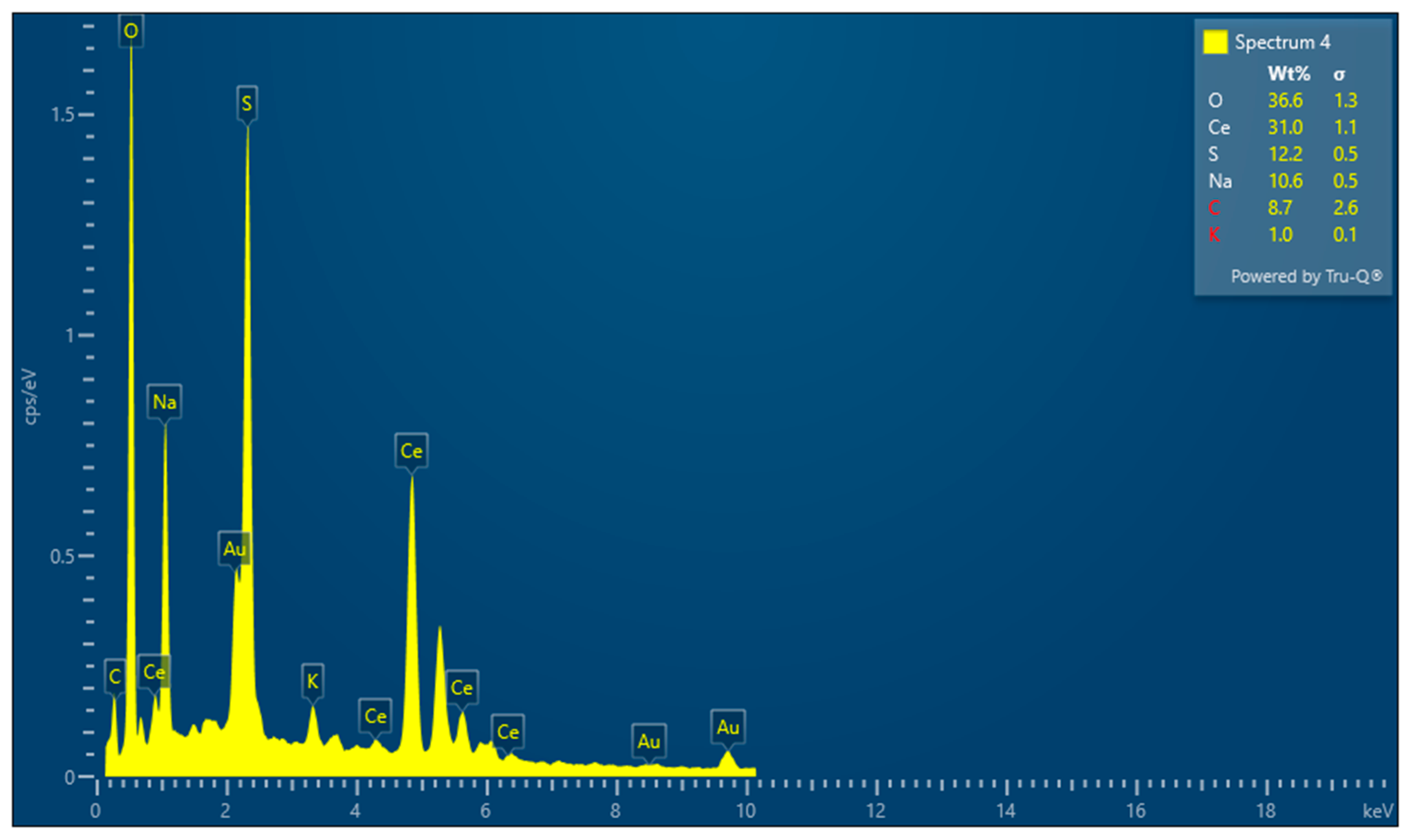
3.2. Surface Morphology
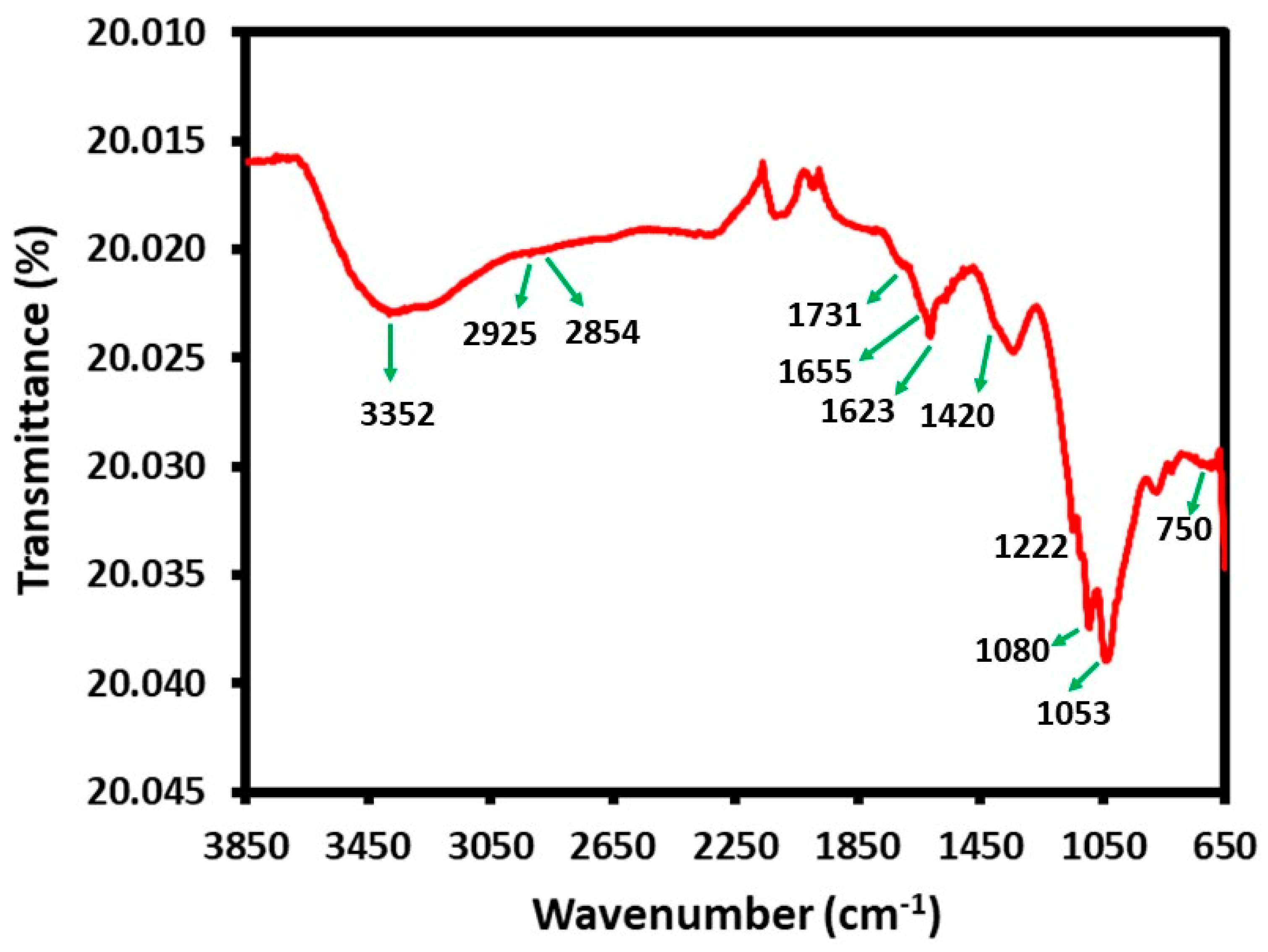
The surface morphology of the CeO2/CS/GO ternary nanocomposite is shown in Figure 2a,b and its elemental composition is presented in Figure 3 and Table S1, respectively. Figure 3 depicts the elemental compositions of the CeO2/CS/GO ternary nanocomposites, which included O-36.6, Ce-31.0, S-12.2, and C-8.7, etc. Figure 2a,b affirm the non-agglomerated surface of the developed CeO2/CS/GO ternary nanocomposite. This surface topography improves the biopotential of the developed CeO2/CS/GO ternary nanocomposite, and the same is addressed in the below sections.

3.3. FT-IR Analysis

The presence of different functional groups and their bonding in the developed CeO2/CS/GO ternary nanocomposite were confirmed by recording the FT-IR spectrum of the sample (3500–1000 cm–1), as shown in Figure 4. In this spectrum, the FT-IR peaks of GO can be located at different positions, such as the peaks of hydrogen-bonded O–H (3100–3600 cm−1), C=O (1731 cm−1), C=C (1623 cm−1), and C–O (1222 cm−1, 1053 cm−1). On the other hand, FT-IR peaks of chitosan in the same spectrum can be identified at 3410 cm −1 (O–H and N–H), 2925 cm −1, 2854 cm −1 (aliphatic C–H), 1655 cm −1 (bending vibration N–H), 1420 cm −1 (C–N), and 1080 cm −1 (C–O). Whereas, FTIR spectra of CeO2 exhibit different peak positions, such as CeO2 having a surface hydroxyl group (OH) band located in the region of 3200–3600 cm−1, and carbonate adsorbates on the surface of CeO2 located by characteristic peaks in the region of 1400–1500 cm−1, corresponding to the stretching vibrations of carbonate ions. Furthermore, the fingerprint region (below 1500 cm−1) contains smaller peaks and features that are specific to the CeO2 nanoparticle structure. These can provide additional information about the particle’s crystalline phase and purity. The band at 750 cm−1 can be attributed to the Ce–O stretching vibrations which ultimately confirm the formation of nanocomposites involving three different components such as GO, chitosan, and CeO2 nanoparticles. Furthermore, the FT-IR spectra of the chitosan and GO and CeO2 nanoparticles are provided in the supplementary file (Figures S4–S6).

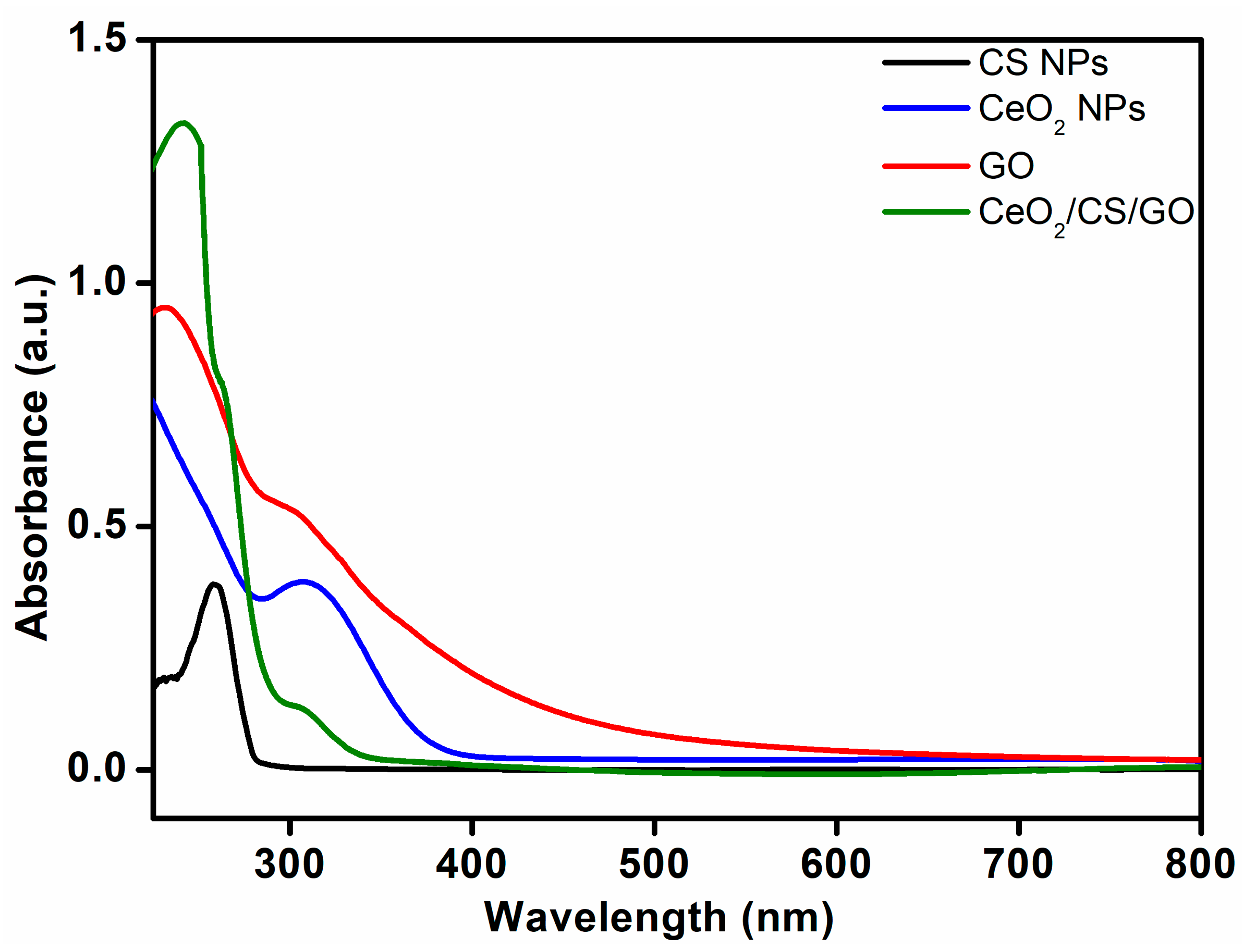
3.4. UV-Vis Analysis

The UV spectrum of CS NP, CeO2 NP, GO, and the CeO2/CS/GO ternary nanocomposite is shown in Figure 5. This spectrum demonstrates the presence of three different peaks at ~230, ~250, and ~300 nm. Typically, the absorbance peak of CeO2 NPs can be observed at a wavelength of ~303 nm, which corresponds to the characteristic absorption peak of Ce4+ of CeO2 NPs, which in this case appears at ~300 nm [26]. Similarly, the absorption spectrum of GO consists of two absorption bands which are usually centered at 234 nm is assigned to the π-π* transition of aromatic C–C bonds of graphene lattice, whereas a shoulder band at 298 nm corresponds to the n-π* transition of C=O bond [27]. However, in this study, both these characteristic peaks of GO are overlapped with the absorption peaks of CeO2 NPs and thus cannot be clearly differentiated. Whereas, the absorption peak at ~250 nm can be attributed to the presence of chitosan, and thus the presence of absorption peaks representing all the components of the composite clearly suggests the formation of CeO2/CS/GO ternary nanocomposites.

3.5. Anti-Bacterial Assay

In this study, the CeO2/CS/GO ternary nanocomposite was prepared in a single-step reaction which was further used to perform biological experiments including anti-bacterial and anti-fungal studies. Notably, such type of ternary composite is rarely used to explore biological potential; however, binary composites involving only chitosan and CeO2 has been reported in the literature [28]. For instance, Senthilkumar et al. prepared a hybrid involving chitosan and cerium oxide nanoparticles using natural Sidaacuta leaf extract [29]. This binary composite at the concentration of 50 μL exhibited considerable morphological changes against E. coli and B. subtilis organisms due to the direct interaction between chitosan-CeO2 nanoparticles. Very recently, Appu et al. prepared CS/CeO2 using aqueous extracts of tea polyphenols. This composite showed an excellent antibacterial and antifungal efficacy against foodborne pathogens including E. coli, S. aureus, and B. cinerea with zones of inhibition of 13.5 ± 0.2 and 11.7 ± 0.2 mm, respectively. However, in the current study, we also included GO which is known to possess significant biological potential, which is reflected in the current ternary composite.
The developed CeO2/CS/GO ternary nanocomposite was incubated with various bacteria, including Proteus, Salmonella, E. coli, Staphylococcus, and Bacillus species, using the agar disc diffusion method. The zone of inhibition (ZOI) was 10 mm for all bacteria, excluding Bacillus species, at 1000 g/disc of CeO2/CS/GO nanocomposite. At a minimum concentration of 500 µg/disc of CeO2/CS/GO nanocomposite, the zone of inhibition (ZOI) was 8 mm for Salmonella and E. coli, 7 mm for Staphylococcus species, and 5 mm for Proteus and Bacillus species, as shown in Table 1 and Figure 6.
In previous reports, lanthanum-doped zinc oxide nanomaterials were incubated with bacteria such as P. mirabilis, S. typhi, S. aureus, and B. subtilis. At the maximum concentration of 0.07 µg/mL, the zone of inhibition was 22 mm for P. mirabilis, 7 mm for S. typhi, and 0 mm for S. aureus and B. subtilis [30]. Similarly, the anti-bacterial properties of titanium dioxide nanoparticles were analyzed using S. aureus, Serratia marcescens, and E. coli. The zone of inhibition for a maximum concentration of 1000 g/disc was 5 mm for S. aureus and S. marcens, and 3 mm for E. coli [31]. Typically, nanomaterials including nanocomposites exert anti-bacterial mechanisms through various means, including the direct physical interaction of highly sharp edges of a variety of nanomaterials with the corresponding biological entities including membranes of the bacterial cell walls [32]. The second possible mechanism of antibacterial activity of nanomaterials is the generation of reactive oxygen species (ROS), including hydroxyl radicals, superoxide anion, and hydrogen peroxide etc., by the inducing ability of nanoparticles [33,34]. Nanomaterials are known to induce oxidative stress which potentially influences intracellular calcium concentrations, activates transcription factors, and modulates cytokine production via the generation of free radicals [35]. Apart from these, the third possible mechanism is the trapping of bacteria within the aggregated nanomaterials. This mechanism is particularly suitable for layered materials such as graphene derivatives, carbon nitride, etc. [36]. The other mechanisms are the generation of oxidative stress, interruption of the glycolysis process of the bacteria, release of metal ions, contribution to the generation/explosion of nanobubbles, etc. [11,37,38,39]. In this study, the as-prepared CeO2/CS/GO ternary nanocomposite may exert antibacterial properties by the combination of both ROS generation and the physical damage to the membranes of the bacterial cell wall. These mechanisms can be attributed to the CeO2 and graphene oxide in the composite, as CeO2 is typically known to exert antibacterial activity by ROS generation, while the sharp edges of GO are known to cause physical damage to the bacterial cell wall [40,41]. CeO2 NPs’ antibacterial efficacy in the absence of light depends on concentration. We can observe an expansion of the zone of inhibition as the nanoparticle concentration is raised. The conversion of Ce4+ to Ce3+ results in oxidative stress on the bacterial membrane as well. CeO2 NPs are electrostatically drawn to the bacterial cell membrane and adhere there. They experience oxidative stress as a result of the acquisition of electrons through Ce4+ to Ce3+ conversion. Since gram-positive bacteria have a strong cell wall comprised of peptidoglycan that is difficult to penetrate, gram-negative bacteria are more vulnerable to antibacterial activity than gram-positive bacteria [42].

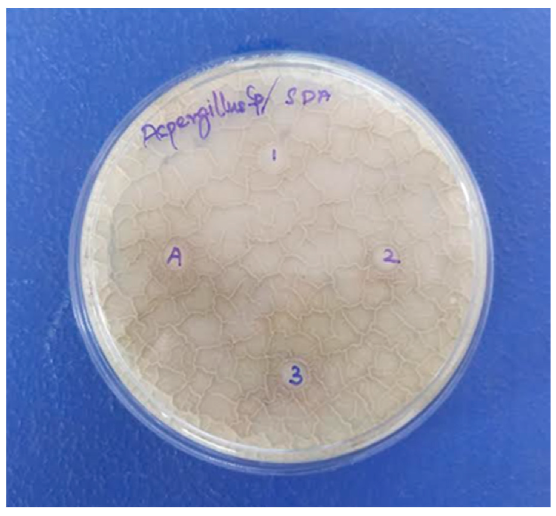
3.6. Anti-Fungal Assay

The antifungal activity of the developed CeO2/CS/GO ternary nanocomposite was analyzed against A. niger using the agar disc diffusion method, as shown in Figure 7. At the maximum concentration of 1000 µg/disc of CeO2/CS/GO nanocomposite, the ZOI was recorded to be 4 mm. In contrast, at a minimum concentration of 500 µg/disc of CeO2/CS/GO nanocomposite, the ZOI was 2 mm (Figure 7). Table 2 presents the ZOI values recorded upon incubating A. niger with different nanocomposites. Chitosan may have hindered the integrity of protein biosynthesis and cell surface architecture, which may have had an impact on the growth and development of Aspergillus ochraceus. These results have implications for the use of chitosan as an alternative method of fungi pathogen control [43]. In a study, maximum microbial inhibition was demonstrated by CeO2/GO as a result of the improved photocatalytic activity of cerium oxide and graphene oxide. The transport of photoradicals to the graphene oxide is aided by the light’s excitation of cerium nanoparticles attached to graphene sheets. The balance between adsorption and desorption was upset, making it easy for more charged particles to pass across the molecular matrix. Additionally, layer-structured GO aided in the equally distributed nanoparticle distribution, which may have improved photocatalytic activity [44,45].

3.7. Hemolysis Assay

HRBCs were used for assessing the hemolysis activity of the developed CeO2/GO/CS ternary nanocomposite. Absorbance was recorded upon the interaction of HRBCs with various concentrations of the developed NC. The percentage hemolysis was 0.5 at 1000 µg/disc; at 500 µg/disc, it was 0.696, while it was 0.711 at 250 µg/disc and 125 µg/disc, and 0.622 for the blank (Table S2, Supplementary File). In previous studies on anti-inflammatory responses for copper nanoparticles using HRBCs, percentage hemolysis was recorded to be 0.246 at 200 µg/mL, while it was 0.649 at the lower concentration of 50 µg/mL [50]. Similarly, when silver nanoparticles were analyzed for anti-inflammatory response, the percentage of hemolysis was recorded to be 0.18 for 800 µg/mL and 0.54 for 200 µg/mL [51]. These observations affirm the hemolysis activity of HRBCs. As a part of future work, advanced studies will be carried out to affirm the biopotential of the CeO2/CS/GO ternary nanocomposite toward the treatment of blood-borne disease.

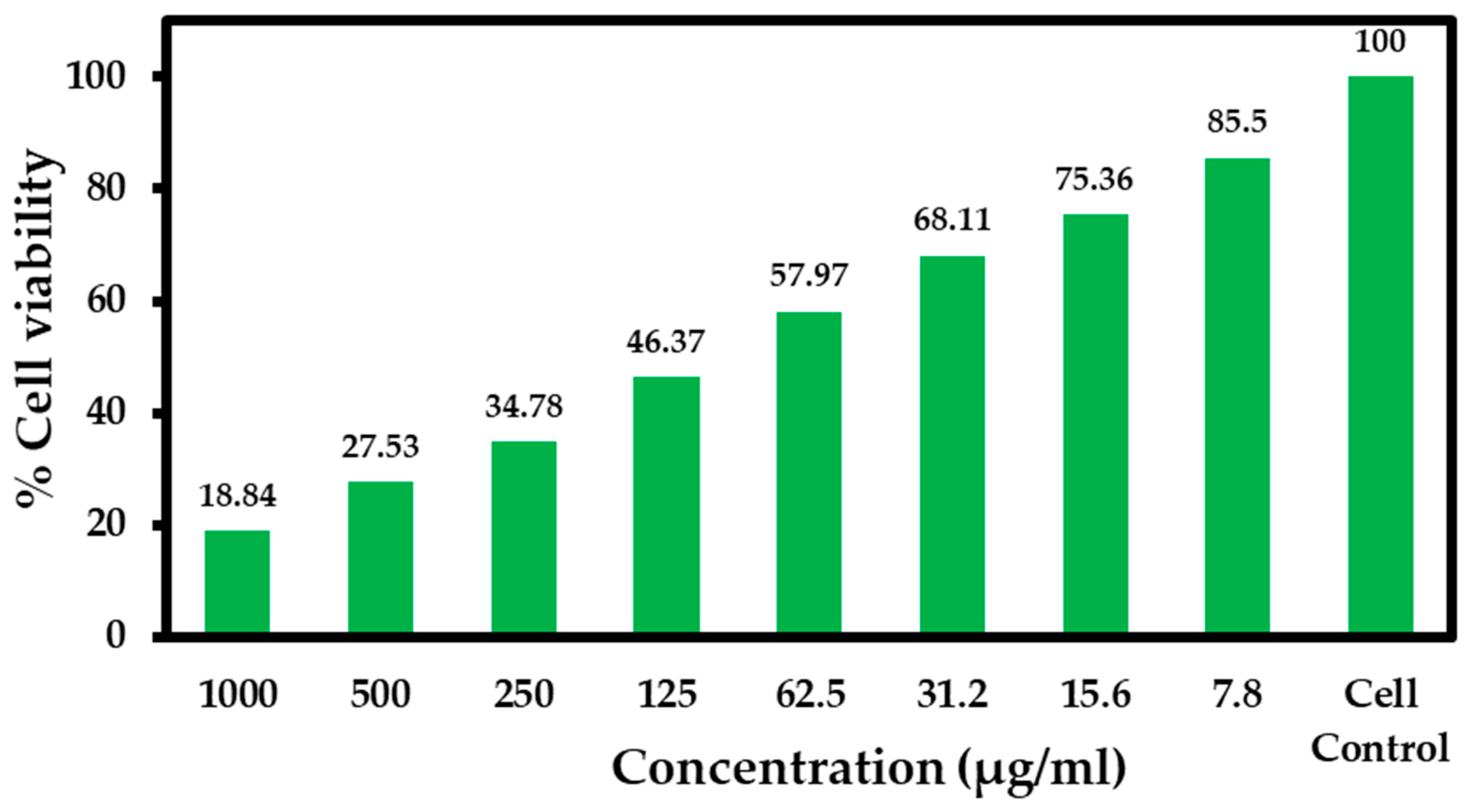
3.8. Anti-Cancer Activity of the CeO2/CS/GO Ternary Nanocomposite Using MTT Assay

The culture medium containing a minimum of 100,000 MCF-7 cells was incubated with varying concentrations of the CeO2/CS/GO ternary nanocomposite, and the percentage of live cells was recorded as shown in Table 3. Here, IC50 concentration is defined as the concentration of the CeO2/CS/GO ternary nanocomposite for which nearly 50% of MCF-7 cancer cells remain dead. Varying concentrations (1000, 500, 250, 125, 62.5, 31.5, 15.6, and 7.8 µg/mL) of the developed CeO2/CS/GO nanocomposite were used to assess its anti-cancer property against the MCF-7 cell line. The IC50 value was estimated to be 125 µg/mL using Equation (1). The quantitative assessment of the anti-cancer activity of the developed CeO2/CS/GO nanocomposites at various concentrations is presented in Figure 8. Qualitative assessments were performed from the record of the biochemical interactions of the CeO2/CS/GO nanocomposite with MCF-7 cells in the culture medium, as presented in Figure 9. As the concentration of the CeO2/CS/GO nanocomposite increased, the number of cell deaths also increased, which is well presented using various concentrations such as 1000 µg/mL, 250 µg/mL, 125 µg/mL, and 62.5 µg/mL, respectively. Cell death is revealed by the rupture of cells, which leads to a reduction in cell size and a dip in the quantity of cells present in the culture medium, as shown in Figure 9. The %live cells was 18.84 at the maximum concentration of 1000 µg/mL and 85.5 at the minimum concentration of 7.8 µg/mL, respectively. Table 4 presents the mean value plus or minus the standard deviation obtained from the record of the anti-cancer activity of the CeO2/CS/GO nanocomposite at various concentrations with the MCF-7 cell line three times. For the human foreskin fibroblasts (HFF) and lung cell line (A549), the IC50 of ceria-loaded chitosan nanoparticles (CeCh-NPs) was reported to be 50.65 and 131.108 μg/mL, respectively [28]. According to a previous report, the median chitosan–cerium oxide nanoparticle concentrations against AGS, A459, PC3, and HFF cells were about 156.02, 169.1, 155.8, and 307.5 µg/mL, respectively [52]. In a study, after 48 h of incubation with varying concentrations of CeNPs (10 to 100 µg/mL), it was discovered that the cytotoxic effect of CeNPs on HaCaT (normal cell lines) and MCF-7 (cancer cell lines) suspended in Dulbecco’s phosphate buffered saline (DPBS), with a pH of 7.4, is transient, dose-dependent, and depends on the type of cell lines [53]. In another study, a new series of CeO2NPs having different structures and morphologies such as nickel (Ni)-doped CeO2, hollow CeO2 (hCeO2), and hollow CeO2/silicon dioxide (SiO2) (hCeO2/SiO2) NPs were synthesized. Additionally, the results showed that hCeO2/SiO2NPs had a lower half maximum inhibitory concentration (IC50) (18.625 g/mL) than other synthetic drugs. When compared with tumoral cells (HT-29), all four chemicals had a much lower cytotoxic effect on non-cancerous cells (HFFF2). In comparison with the previous reports, the results obtained from the present investigation demonstrate the excellent anti-cancer activity of the developed CeO2/CS/GO nanocomposite against MCF-7 cells.

4. Conclusions

A CeO2/CS/GO ternary nanocomposite was prepared using a cost-effective wet chemical procedure. The physicochemical properties of the developed CeO2/CS/GO nanocomposite were confirmed using XRD spectroscopy, SEM with EDAX, FT-IR spectroscopy, and UV-vis spectroscopy. The biological potential of the developed CeO2/CS/GO nanocomposite was confirmed by performing in vitro anti-bacterial, anti-fungal, anti-cancer, and hemolysis studies. Based on our observations, we affirm inherent anti-cancer activity with the MCF-7 cell line; anti-fungal activity with Aspergillus niger; anti-bacterial activity with E. coli, Bacillus species, Salmonella species, Proteus species, and staphylococcus species; and hemolysis response with HRBCs. As a part of future work, quantitative and qualitative studies can be conducted with the developed CeO2/CS/GO nanocomposite through its interaction with other types of cancer cell lines, bacterial cell lines, and fungal cell lines. Furthermore, an anti-cancer drug can be introduced in the developed CeO2/CS/GO nanocomposite, and its efficacy as a drug delivery platform can also be assessed.

Supplementary Materials

The following are available online at https://www.mdpi.com/article/10.3390/cryst13091393/s1. Figure S1: XRD analysis of Chitosan; Figure S2: XRD analysis of GO; Figure S3: XRD analysis of CeO2; Figure S4: FT-IR analysis of Chitosan; Figure S5: FT-IR analysis of GO; Figure S6: FT-IR analysis of CeO2; Figure S7: Hemolysis assay using the CeO2/CS/GO ternary nanocomposite; Table S1: Elemental composition of the CeO2/CS/GO ternary nanocomposite; Table S2: Hemolysis assay response upon interaction of CeO2/CS/GO ternary nanocomposite with HRBCs.

Author Contributions

Conceptualization, J.S.; methodology, J.S. and S.P.; validation, J.S. and S.P.; formal analysis, M.R.S., M.K. (Mujeeb Khan) and B.S.; investigation, J.S. and S.P.; resources, J.S. and M.R.S.; data curation, S.P., M.R.S., M.K. (Merajuddin Khan) and B.S.; writing—original draft preparation, J.S., S.P. and M.R.S.; writing—review and editing, J.S., M.R.S., M.K. (Merajuddin Khan) and M.K. (Mujeeb Khan); supervision, J.S.; project administration, J.S.; funding acquisition, M.R.S. All authors have read and agreed to the published version of the manuscript.

Funding

This research was funded by Researchers Supporting Project number (RSPD2023R665), King Saud University, Riyadh, Saudi Arabia.

Institutional Review Board Statement

Not applicable.

Informed Consent Statement

Not applicable.

Data Availability Statement

Data contained within the article.

Acknowledgments

The authors acknowledge the funding from Researchers Supporting Project number (RSPD2023R665), King Saud University, Riyadh, Saudi Arabia.

Conflicts of Interest

The authors declare no conflict of interest.

References

  1. Saeedi, M.; Vahidi, O.; Moghbeli, M.; Ahmadi, S.; Asadnia, M.; Akhavan, O.; Seidi, F.; Rabiee, M.; Saeb, M.R.; Webster, T.J. Customizing nano-chitosan for sustainable drug delivery. J. Control Release 2022, 350, 175–192. [Google Scholar] [CrossRef]
  2. Emilian Leucuta, S. Nanotechnology for delivery of drugs and biomedical applications. Curr. Clin. Pharmacol. 2010, 5, 257–280. [Google Scholar] [CrossRef] [PubMed]
  3. Dai, X.; Cheng, H.; Bai, Z.; Li, J. Breast cancer cell line classification and its relevance with breast tumor subtyping. J. Cancer 2017, 8, 3131. [Google Scholar] [CrossRef] [PubMed]
  4. Maleki, P.; Nemati, F.; Gholoobi, A.; Hashemzadeh, A.; Sabouri, Z.; Darroudi, M. Green facile synthesis of silver-doped cerium oxide nanoparticles and investigation of their cytotoxicity and antibacterial activity. Inorg. Chem. Commun. 2021, 131, 108762. [Google Scholar] [CrossRef]
  5. Dhall, A.; Self, W. Cerium oxide nanoparticles: A brief review of their synthesis methods and biomedical applications. Antioxidants 2018, 7, 97. [Google Scholar] [CrossRef]
  6. Tsunekawa, S.; Sivamohan, R.; Ohsuna, T.; Takahashi, H.; Tohji, K. Ultraviolet absorption spectra of CeO2 nano-particles. In Materials Science Forum; Trans Tech Publications Ltd.: Wollerau, Switzerland, 1999; pp. 439–445. [Google Scholar]
  7. Saranya, J.; Sreeja, B.; Padmalaya, G.; Radha, S.; Manikandan, T. Ultrasonic assisted cerium oxide/graphene oxide hybrid: Preparation, anti-proliferative, apoptotic induction and G2/M cell cycle arrest in HeLa cell lines. J. Inorg. Organomet. Polym. Mater. 2020, 30, 2666–2676. [Google Scholar] [CrossRef]
  8. Çiplak, Z.; Yildiz, N.; Çalimli, A. Investigation of graphene/Ag nanocomposites synthesis parameters for two different synthesis methods. Fuller. Nanotub. Carbon Nanostructures 2015, 23, 361–370. [Google Scholar] [CrossRef]
  9. Saranya, J.; Saminathan, P.; Ankireddy, S.R.; Shaik, M.R.; Khan, M.; Khan, M.; Shaik, B. Cerium Oxide/Graphene Oxide Hybrid: Synthesis, Characterization, and Evaluation of Anticancer Activity in a Breast Cancer Cell Line (MCF-7). Biomedicines 2023, 11, 531. [Google Scholar] [CrossRef]
  10. Pang, L.; Dai, C.; Bi, L.; Guo, Z.; Fan, J. Biosafety and antibacterial ability of graphene and graphene oxide in vitro and in vivo. Nanoscale Res. Lett. 2017, 12, 564. [Google Scholar] [CrossRef]
  11. Liu, S.; Zeng, T.H.; Hofmann, M.; Burcombe, E.; Wei, J.; Jiang, R.; Kong, J.; Chen, Y. Antibacterial activity of graphite, graphite oxide, graphene oxide, and reduced graphene oxide: Membrane and oxidative stress. ACS Nano 2011, 5, 6971–6980. [Google Scholar] [CrossRef]
  12. Kayani, F.B.; Rafique, S.; Akram, R.; Hussain, M.; Raja, K.; Khan, J.S. Fabrication of novel chitosan@Ag/CeO2 hybrid nanocomposites for the study of antibacterial activity. Phys. E Low-Dimens. Syst. Nanostructures 2023, 149, 115683. [Google Scholar] [CrossRef]
  13. Panda, P.K.; Dash, P.; Yang, J.-M.; Chang, Y.-H. Development of chitosan, graphene oxide, and cerium oxide composite blended films: Structural, physical, and functional properties. Cellulose 2022, 29, 2399–2411. [Google Scholar] [CrossRef]
  14. Mali, S.; Nune, K.; Misra, R. Biomimetic nanostructured hydroxyapatite coatings on metallic implant materials. Mater. Technol. 2016, 31, 782–790. [Google Scholar] [CrossRef]
  15. Pimenta, J.A.; Zaparolli, D.; Pécora, J.D.; Cruz-Filho, A.M. Chitosan: Effect of a new chelating agent on the microhardness of root dentin. Braz. Dent. J. 2012, 23, 212–217. [Google Scholar] [CrossRef] [PubMed]
  16. Alimirzaei, F.; Vasheghani-Farahani, E.; Ghiaseddin, A.; Soleimani, M. pH-Sensitive Chitosan Hydrogel with Instant Gelation for Myocardial Regeneration. J. Tissue Sci. Eng. 2017, 8, 3. [Google Scholar]
  17. de Sousa Victor, R.; Marcelo da Cunha Santos, A.; Viana de Sousa, B.; de Araújo Neves, G.; Navarro de Lima Santana, L.; Rodrigues Menezes, R. A review on Chitosan’s uses as biomaterial: Tissue engineering, drug delivery systems and cancer treatment. Materials 2020, 13, 4995. [Google Scholar]
  18. Anjum, S.; Arora, A.; Alam, M.; Gupta, B. Development of antimicrobial and scar preventive chitosan hydrogel wound dressings. Int. J. Pharm. 2016, 508, 92–101. [Google Scholar] [CrossRef]
  19. Zaboon, M.H.; Saleh, A.A.; Al-Lami, H.S. Synthesis, characterization and cytotoxicity investigation of chitosan-amino acid derivatives nanoparticles in human breast cancer cell lines. J. Mex. Chem. Soc. 2021, 65, 178–188. [Google Scholar] [CrossRef]
  20. Appu, M.; Wu, H.; Chen, H.; Huang, J. Tea polyphenols mediated biogenic synthesis of chitosan-coated cerium oxide (CS/CeO2) nanocomposites and their potent antimicrobial capabilities. Environ. Sci. Pollut. Res. 2023, 30, 42575–42586. [Google Scholar] [CrossRef]
  21. Kasinathan, K.; Murugesan, B.; Pandian, N.; Mahalingam, S.; Selvaraj, B.; Marimuthu, K. Synthesis of biogenic chitosan-functionalized 2D layered MoS2 hybrid nanocomposite and its performance in pharmaceutical applications: In-Vitro antibacterial and anticancer activity. Int. J. Biol. Macromol. 2020, 149, 1019–1033. [Google Scholar] [CrossRef]
  22. Mosmann, T. Rapid colorimetric assay for cellular growth and survival: Application to proliferation and cytotoxicity assays. J. Immunol. Methods 1983, 65, 55–63. [Google Scholar] [CrossRef] [PubMed]
  23. Abbasi, N.; Homayouni Tabrizi, M.; Ardalan, T.; Roumi, S. Cerium oxide nanoparticles-loaded on chitosan for the investigation of anticancer properties. Mater. Technol. 2022, 37, 1439–1449. [Google Scholar] [CrossRef]
  24. Adil, S.F.; Shaik, M.R.; Nasr, F.A.; Alqahtani, A.S.; Ahmed, M.Z.; Qamar, W.; Kuniyil, M.; Almutairi, A.; Alwarthan, A.; Siddiqui, M.R.H. Enhanced apoptosis by functionalized highly reduced graphene oxide and gold nanocomposites in MCF-7 breast cancer cells. ACS Omega 2021, 6, 15147–15155. [Google Scholar] [CrossRef] [PubMed]
  25. Ioelovich, M. Crystallinity and hydrophility of chitin and chitosan. J. Chem. 2014, 3, 7–14. [Google Scholar]
  26. Nurhasanah, I.; Safitri, W.; Arifin, Z.; Subagio, A.; Windarti, T. Antioxidant Activity and Dose Enhancement Factor of CeO2 Nanoparticles Synthesized by Precipitation Method; IOP Conference Series: Materials Science and Engineering; IOP Publishing Ltd.: Bristol, UK, 2018; p. 012031. [Google Scholar]
  27. Wang, S.; Zhang, Y.; Ma, H.-L.; Zhang, Q.; Xu, W.; Peng, J.; Li, J.; Yu, Z.-Z.; Zhai, M. Ionic-liquid-assisted facile synthesis of silver nanoparticle-reduced graphene oxide hybrids by gamma irradiation. Carbon 2013, 55, 245–252. [Google Scholar] [CrossRef]
  28. Sanmugam, A.; Abbishek, S.; Kumar, S.L.; Sairam, A.B.; Palem, V.V.; Kumar, R.S.; Almansour, A.I.; Arumugam, N.; Vikraman, D. Synthesis of chitosan based reduced graphene oxide-CeO2 nanocomposites for drug delivery and antibacterial applications. J. Mech. Behav. Biomed. Mater. 2023, 145, 106033. [Google Scholar] [CrossRef]
  29. Senthilkumar, R.; Bhuvaneshwari, V.; Ranjithkumar, R.; Sathiyavimal, S.; Malayaman, V.; Chandarshekar, B. Synthesis, characterization and antibacterial activity of hybrid chitosan-cerium oxide nanoparticles: As a bionanomaterials. Int. J. Biol. Macromol. 2017, 104, 1746–1752. [Google Scholar] [CrossRef]
  30. Manikandan, A.; Manikandan, E.; Meenatchi, B.; Vadivel, S.; Jaganathan, S.; Ladchumananandasivam, R.; Henini, M.; Maaza, M.; Aanand, J.S. Rare earth element (REE) lanthanum doped zinc oxide (La: ZnO) nanomaterials: Synthesis structural optical and antibacterial studies. J. Alloys Compd. 2017, 723, 1155–1161. [Google Scholar] [CrossRef]
  31. Gudata, L.; Saka, A.; Tesfaye, J.L.; Shanmugam, R.; Dwarampudi, L.P.; Nagaprasad, N.; Stalin, B.; Krishnaraj, R. Investigation of TiO2 nanoparticles using leaf extracts of Lippia adoensis (Kusaayee) for antibacterial activity. J. Nanomater. 2022, 2022, 3881763. [Google Scholar] [CrossRef]
  32. Akhavan, O.; Ghaderi, E. Toxicity of graphene and graphene oxide nanowalls against bacteria. ACS Nano 2010, 4, 5731–5736. [Google Scholar] [CrossRef]
  33. Dutta, T.; Sarkar, R.; Pakhira, B.; Ghosh, S.; Sarkar, R.; Barui, A.; Sarkar, S. ROS generation by reduced graphene oxide (rGO) induced by visible light showing antibacterial activity: Comparison with graphene oxide (GO). RSC Adv. 2015, 5, 80192–80195. [Google Scholar] [CrossRef]
  34. Lakshmi Prasanna, V.; Vijayaraghavan, R. Insight into the mechanism of antibacterial activity of ZnO: Surface defects mediated reactive oxygen species even in the dark. Langmuir 2015, 31, 9155–9162. [Google Scholar] [CrossRef]
  35. Canaparo, R.; Foglietta, F.; Limongi, T.; Serpe, L. Biomedical applications of reactive oxygen species generation by metal nanoparticles. Materials 2020, 14, 53. [Google Scholar] [CrossRef] [PubMed]
  36. Akhavan, O.; Ghaderi, E.; Esfandiar, A. Wrapping bacteria by graphene nanosheets for isolation from environment, reactivation by sonication, and inactivation by near-infrared irradiation. J. Phys. Chem. B 2011, 115, 6279–6288. [Google Scholar] [CrossRef] [PubMed]
  37. Akhavan, O.; Ghaderi, E. Escherichia coli bacteria reduce graphene oxide to bactericidal graphene in a self-limiting manner. Carbon 2012, 50, 1853–1860. [Google Scholar] [CrossRef]
  38. Wang, Y.-W.; Cao, A.; Jiang, Y.; Zhang, X.; Liu, J.-H.; Liu, Y.; Wang, H. Superior antibacterial activity of zinc oxide/graphene oxide composites originating from high zinc concentration localized around bacteria. ACS Appl. Mater. Interfaces 2014, 6, 2791–2798. [Google Scholar] [CrossRef] [PubMed]
  39. Jannesari, M.; Akhavan, O.; Hosseini, H.R.M.; Bakhshi, B. Oxygen-rich graphene/ZnO2-Ag nanoframeworks with pH-switchable catalase/peroxidase activity as O2 nanobubble-self generator for bacterial inactivation. J. Colloid Interface Sci. 2023, 637, 237–250. [Google Scholar] [CrossRef]
  40. Zhang, M.; Zhang, C.; Zhai, X.; Luo, F.; Du, Y.; Yan, C. Antibacterial mechanism and activity of cerium oxide nanoparticles. Sci. China Mater. 2019, 62, 1727–1739. [Google Scholar] [CrossRef]
  41. Nanda, S.S.; Yi, D.K.; Kim, K. Study of antibacterial mechanism of graphene oxide using Raman spectroscopy. Sci. Rep. 2016, 6, 28443. [Google Scholar] [CrossRef]
  42. Sawai, J. Quantitative evaluation of antibacterial activities of metallic oxide powders (ZnO, MgO and CaO) by conductimetric assay. J. Microbiol. Methods 2003, 54, 177–182. [Google Scholar] [CrossRef]
  43. Meng, D.; Garba, B.; Ren, Y.; Yao, M.; Xia, X.; Li, M.; Wang, Y. Antifungal activity of chitosan against Aspergillus ochraceus and its possible mechanisms of action. Int. J. Biol. Macromol. 2020, 158, 1063–1070. [Google Scholar] [CrossRef] [PubMed]
  44. Egorova, M.N.; Tarasova, L.A.; Vasilieva, F.D.; Akhremenko, Y.A.; Smagulova, S.A. Antimicrobial activity of graphene oxide sheets. In Proceedings of the AIP Conference Proceedings, Yakutsk, Russia, 18–23 June 2018; AIP Publishing: Long Island, NY, USA. [Google Scholar]
  45. Cao, M.; Wang, P.; Ao, Y.; Wang, C.; Hou, J.; Qian, J. Visible light activated photocatalytic degradation of tetracycline by a magnetically separable composite photocatalyst: Graphene oxide/magnetite/cerium-doped titania. J. Colloid Interface Sci. 2016, 467, 129–139. [Google Scholar] [CrossRef] [PubMed]
  46. Safaei, M.; Taran, M. Fabrication, characterization, and antifungal activity of sodium hyaluronate-TiO2 bionanocomposite against Aspergillus niger. Mater. Lett. 2017, 207, 113–116. [Google Scholar] [CrossRef]
  47. Hasanin, M.; Hashem, A.H.; Lashin, I.; Hassan, S.A. In vitro improvement and rooting of banana plantlets using antifungal nanocomposite based on myco-synthesized copper oxide nanoparticles and starch. Biomass Convers. Biorefin. 2021, 13, 8865–8875. [Google Scholar] [CrossRef]
  48. Roopan, S.M.; Priya, D.D.; Shanavas, S.; Acevedo, R.; Al-Dhabi, N.A.; Arasu, M.V. CuO/C nanocomposite: Synthesis and optimization using sucrose as carbon source and its antifungal activity. Mater. Sci. Eng. C 2019, 101, 404–414. [Google Scholar] [CrossRef]
  49. Sahraee, S.; Milani, J.M.; Ghanbarzadeh, B.; Hamishehkar, H. Physicochemical and antifungal properties of bio-nanocomposite film based on gelatin-chitin nanoparticles. Int. J. Biol. Macromol. 2017, 97, 373–381. [Google Scholar] [CrossRef]
  50. Anitha, P.; Sakthivel, P. Microwave Assisted Synthesis and Characterization of Silver Nanoparticles Using Citrullus Lanatus Leaf Extract and Its Anti-Inflammatory Activity Against Human Blood Cells. Int. J. Adv. Eng. Nano Technol. 2016, 3, 3. [Google Scholar]
  51. Rajeshkumar, S.; Menon, S.; Ponnanikajamideen, M.; Ali, D.; Arunachalam, K. Anti-inflammatory and antimicrobial potential of Cissus quadrangularis-assisted copper oxide nanoparticles. J. Nanomater. 2021, 2021, 5742981. [Google Scholar] [CrossRef]
  52. Kermani, G.; Karimi, E.; Tabrizi, M.H. Hybrid nanoarchitectonics of chitosan-cerium oxide nanoparticles for anticancer potentials. J. Inorg. Organomet. Polym. Mater. 2022, 32, 2591–2599. [Google Scholar] [CrossRef]
  53. Yang, K.; Wan, J.; Zhang, S.; Zhang, Y.; Lee, S.-T.; Liu, Z. In vivo pharmacokinetics, long-term biodistribution, and toxicology of PEGylated graphene in mice. ACS Nano 2011, 5, 516–522. [Google Scholar] [CrossRef]
  54. Jermy, R.; Ravinayagam, V.; Alamoudi, W.; Almohazey, D.; Elanthikkal, S.; Dafalla, H.; Rehman, S.; Chandrasekar, G.; Baykal, A. Tuning pH sensitive chitosan and cisplatin over spinel ferrite/silica nanocomposite for anticancer activity in MCF-7 cell line. J. Drug Deliv. Sci. Technol. 2020, 57, 101711. [Google Scholar] [CrossRef]
  55. Doghish, A.S.; El-Sayyad, G.S.; Sallam, A.-A.M.; Khalil, W.F.; El Rouby, W.M. Graphene oxide and its nanocomposites with EDTA or chitosan induce apoptosis in MCF-7 human breast cancer. RSC Adv. 2021, 11, 29052–29064. [Google Scholar] [CrossRef] [PubMed]
  56. Atif, M.; Iqbal, S.; Fakhar-E-Alam, M.; Ismail, M.; Mansoor, Q.; Mughal, L.; Aziz, M.H.; Hanif, A.; Farooq, W. Manganese-doped cerium oxide nanocomposite induced photodynamic therapy in MCF-7 cancer cells and antibacterial activity. Biomed Res. Int. 2019, 2019, 7156828. [Google Scholar] [CrossRef] [PubMed]
Scheme 1. Schematic representation of CeO2/CS/GO ternary nanocomposite and its biological activity.
Crystals 13 01393 sch001
Figure 1. XRD peaks of the CeO2/CS/GO ternary nanocomposite.
Crystals 13 01393 g001
Figure 2. (a,b) SEM images of the CeO2/CS/GO ternary nanocomposite.
Crystals 13 01393 g002
Figure 3. EDAX peaks of the CeO2/CS/GO ternary nanocomposite.
Crystals 13 01393 g003
Figure 4. FT-IR analysis of the CeO2/CS/GO ternary nanocomposite.
Crystals 13 01393 g004
Figure 5. UV analysis of the CeO2 NPs, CS NPs, GO, and CeO2/CS/GO ternary nanocomposite.
Crystals 13 01393 g005
Figure 6. Anti-bacterial activity of CeO2/CS/GO ternary nanocomposite. E. coli, Bacillus species, Salmonella species, Proteus species, and Staphylococcus species. ((1) CeO2/CS/GO ternary nanocomposite (1000 µg/disc), (2) CeO2/CS/GO ternary nanocomposite (750 µg/disc), (3) CeO2/CS/GO ternary nanocomposite (500 µg/disc), and (A) Ampicillin (antibiotic)).
Crystals 13 01393 g006
Figure 7. Anti-fungal activity of CeO2/CS/GO ternary nanocomposite on Aspergillus niger.
Crystals 13 01393 g007
Figure 8. Percentage cell viability of the MCF-7 cell line after treatment with the CeO2/CS/GO nanocomposite using MTT assay.
Crystals 13 01393 g008
Figure 9. Microscopic images containing untreated MCF-7 cells and the treated behavior of the CeO2/CS/GO with MCF-7 cell line. All the images were acquired with an objective lens of 20×  magnification. Scale bars show 20 μm (ac) and 10 μm (d,e).
Crystals 13 01393 g009
Table 1. Anti-bacterial activity of CeO2/CS/GO NPs against various bacteria.
S. No.MicroorganismsProteus Species
(ZOI in mm)
Salmonella
Species
(ZOI in mm)
E. coli
Species
(ZOI in mm)
Staphylococcus
Species
(ZOI in mm)
Bacillus
Species
(ZOI in mm)
1CeO2/CS/GO ternary nanocomposite (1000 µg/disc)101010108
2CeO2/CS/GO ternary nanocomposite (750 µg/disc)79996
3CeO2/CS/GO ternary nanocomposite (500 µg/disc)58875
AAmpicillin (antibiotic)1520151513
Table 2. Comparative analysis of different NPs against Aspergillus niger.
S. No.Maximum Concentration of Nanocomposite
(1000 µg/disc)
Aspergillus Species
(ZOI in mm)
References
1Sodium hydranate titanium dioxide bio-nanocomposite13.3[46]
2Polymer nanocomposite15[47]
3CuO/C nanocomposite12[48]
4Gelatin chitin nanocomposite10[49]
5CeO2/GO/CS ternary nanocomposite10This work
Table 3. Anti-cancer activity of different NPs and hybrids against MCF-7 cell lines.
S. No.Nanocomposite% Live Cells at 1000 µg/mLReferences
1CeO2/GO nanocomposite19.44[9]
2Cu Fe2O4/HYPS nanocomposite21.23[54]
3GO/CS nanocomposite23.8[55]
4Mn/CeO2 nanocomposite22.58[56]
5CeO2/GO/CS ternary
nanocomposite
18.84This work
Table 4. Anticancer activity of CeO2/CS/GO ternary nanocomposite on MCF-7 cell line using MTT assay.
S. NoConcentration (µg/mL)Cell Viability (% Live Cells)
1.100018.84 ± 0.48
2.50027.53 ± 0.31
3.25034.78 ± 0.35
4.12546.37 ± 0.12
5.62.557.97 ± 0.26
6.31.268.11 ± 0.27
7.15.675.36 ± 1.19
8.7.885.50 ± 0.34
9.Cell control100 ± 0.45
Mean value ± Standard Deviation (Three replicates).
Disclaimer/Publisher’s Note: The statements, opinions and data contained in all publications are solely those of the individual author(s) and contributor(s) and not of MDPI and/or the editor(s). MDPI and/or the editor(s) disclaim responsibility for any injury to people or property resulting from any ideas, methods, instructions or products referred to in the content.

