Article

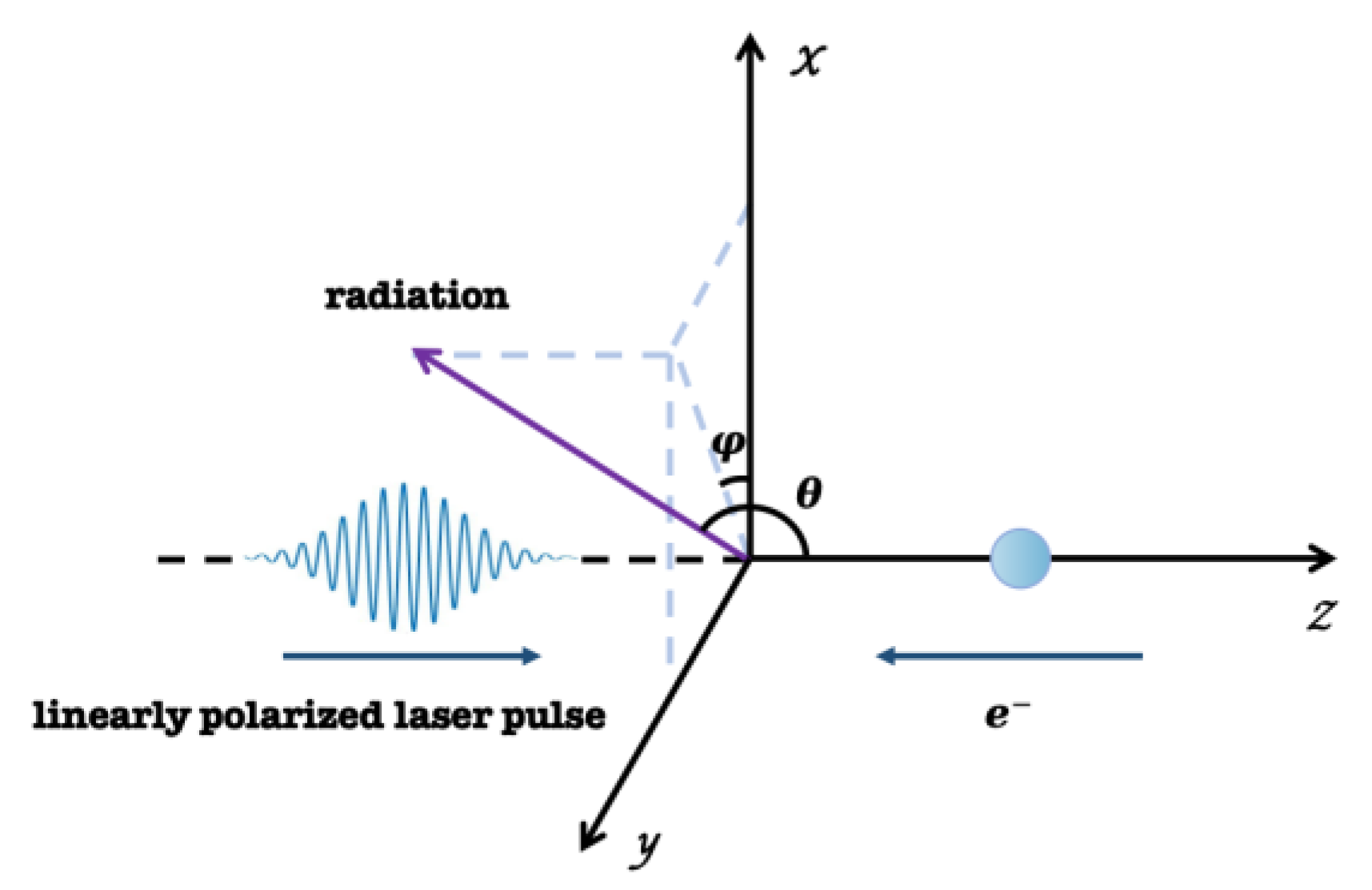
Highly Collimated Monochromatic X-rays Generated by Collision of High-Energy Electrons with Tightly Focused Linearly Polarized Laser Pulse

1
Bell Honors School, Nanjing University of Posts and Telecommunications, Nanjing 210023, China
2
School of Communications and Information Engineering, Nanjing University of Posts and Telecommunications, Nanjing 210023, China
3
School of Biomedical Engineering, University of Science and Technology of China, Suzhou 215004, China
4
College of Science, Nanjing University of Posts and Telecommunications, Nanjing 210023, China
*
Author to whom correspondence should be addressed.
Crystals 2024, 14(1), 13; https://doi.org/10.3390/cryst14010013
Submission received: 4 December 2023 / Revised: 17 December 2023 / Accepted: 20 December 2023 / Published: 22 December 2023

Abstract

:
This article delves into the generation and modulation process of X-rays as high-energy photon sources. Using the principles of classical electrodynamics, this study enables nonrelativistic short pulse lasers to collide with high-energy electrons while the collision center is away from the focal point. This scattering method may produce X-rays with good collimation and monochromaticity, and it progressively approaches inverse Thomson scattering. We studied and analyzed the effects of different electron characteristics and laser parameter settings on the high-energy angular distribution and spectrum of X-rays, especially the setting of the collision center and initial electron velocity, as well as the setting of laser intensity and pulse width. Linear polarized laser pulses with relativistic intensity can generate discrete supercontinuum X-rays with spectral distortion. In addition, the relationships between electronic and laser properties and radiation energy were also studied. Our research can provide valuable insights for manipulating collimated or distorted, monochromatic, or tunable X-rays, as well as understanding their properties.

1. Introduction

Relativistic electrons colliding with the electromagnetic field produced by powerful lasers causes Thomson and Compton scattering, which is frequently accompanied by a Doppler frequency shift. Inverse Thomson and Compton scattering happens when electromagnetic waves interact with relativistic electrons. The essential elements of an all-optical X-ray source that can generate electron beams for laser plasma accelerators (LPAs) can be constructed based on the inverse Thomson scattering (ITS) mechanism. The spectrum issue of nonlinear inverse Thomson scattering in laser fields has drawn a lot of interest lately [1,2,3,4,5,6].
A high-energy photon source (HEPS) is one of the major scientific engineering projects and a fourth-generation Synchrotron light source with the highest brightness in the world [7,8]. HEPS has led to the progress of a series of disciplines, including ultrafast biology, materials science, nonlinear Quantum electrodynamics and nuclear spectroscopy [9,10,11,12,13,14,15,16,17]. At present, a high-energy photon source exists in large acceleration facilities in the form of a X-ray free-electron laser or Synchrotron. It is worth noting that nonlinear inverse Thomson scattering (NITS) can generate collimated radiation with enormous energy in a relatively controlled set. The relativistic electrons needed for this technology and NITS are correlated to some extent. When the initial direction of electron motion is opposite to the direction of laser propagation, NITS occurs. NITS generates high-order harmonic radiation with collimation and monochromaticity; and in some cases, it generates X-rays and γ -rays [18,19,20,21,22].
Previous work has explored NITS and radiation characteristics in many cases. Chang et al. studied the characteristics and modulation of spatial radiation generated by the collision of high-energy electrons and circularly polarized laser pulses [23,24]. Investigators at National Institute of Advanced Industrial Science and Technology (NIIST) discussed the γ -ray vortices emitted from Nonlinear Inverse Thomson Scattering [25]. Yoshitaka Taira et al. explored the spatial distribution of high-order harmonics generated by NITS from the perspective of astrophysics [1]. To delve deeper into the generation and modulation of X-rays as high-energy photon sources, this study used tightly focused laser pulses to collide with single high-energy electrons. Previous work has placed greater emphasis on the impact of changes in laser parameters on radiation characteristics, while lacking analysis based on changes in electronic properties. The radiation energy and spectral distribution from different angles are also lacking. This article will conduct research based on the overlooked points mentioned above.
In this paper, we investigate the properties of NITS radiation when a tightly focused linearly polarized laser pulse collides with a single high-energy electron. The influence of collision characteristics (collision center and initial energy of electron) and laser characteristics (laser intensity and pulse width) on the angular distribution of radiation energy and harmonic characteristics (i.e., spectrum) is studied. The parameters selected in this paper are currently relatively easily and stably obtained in experiments. By choosing these characteristics, high-energy electrons and linearly polarized lasers can collide in experiments to produce high-energy X-rays with good collimation and monochromaticity. This can provide a theoretical foundation and simulation basis for the experiment. Under the framework of classical electrodynamics, analytical expressions for the laws of electron motion and radiation have been derived. Through numerical simulation, the angular distribution and spectrum of radiant energy under different conditions are obtained. The characteristics of radiation energy, distribution mode and harmonic frequency are analyzed. Research has found that at appropriate collision positions, the single electron with high initial energy collides with nonrelativistic short pulse lasers, producing high-energy X-rays with good collimation and monochromaticity. This is of great significance for promoting the construction and development of HEPS and obtaining better (highly collimated, monochromatic, or supercontinuum) X-ray sources.

2. Theory and Formula

The schematic diagram of reverse Thomson scattering occurring when a single electron collides with a linearly polarized laser is shown in Figure 1.
The paraxial approximation is no longer applicable for intensely concentrated lasers, and higher-order field effects must be considered. The following components of the electric field can be obtained from a linearly polarized vector potential along + x , which models the beam [26].
E x = E S 0 + ϵ 2 ξ 2 S 2 ρ 4 S 3 4 + ϵ 4 S 2 8 ρ 2 S 3 4 ρ 2 ρ 2 16 ξ 2 S 4 16 ρ 4 ρ 2 + 2 ξ 2 S 5 8 + ρ 8 S 6 32
E y = E ξ v ϵ 2 S 2 + ϵ 4 ρ 2 S 4 ρ 4 S 5 4
E z = E ξ ϵ C 1 + ϵ 3 C 2 2 + ρ 2 C 3 ρ 4 C 4 4 + ϵ 5 3 C 3 8 3 ρ 2 C 4 8 + 17 ρ 4 C 5 16 3 ρ 6 C 5 16 3 ρ 6 C 6 8 + ρ 8 C 7 32
The magnetic field’s elements are organized similarly and are listed below.
B x = 0
B y = E S 0 + ϵ 2 ρ 2 S 2 2 ρ 4 S 3 4 + ϵ 4 S 2 8 + ρ 2 S 3 4 + 5 ρ 4 S 4 16 ρ 6 S 5 4 + ρ 8 S 6 32
B z = E v ϵ C 1 + ϵ 3 C 2 2 + ρ 2 C 3 2 ρ 4 C 4 4 + ϵ 5 3 C 3 8 + 3 ρ 2 C 4 8 + 3 ρ 4 C 5 16 8 ρ 6 C 6 4 + ρ 8 C 7 32
Since we approximate the tightly focused beam to be a long cylinder, its cross section has a circular shape with a radius of b 0 . Based on this, it follows that the cross section through any point z on an axis has a circular cross section with radius b ( z ) = b 0 1 + z / z r 2 , defined by Rayleigh length z r = k b 0 2 / 2 , and k equal to 2 π / λ . The laser can only be kept strong at the focus spot since it has the lowest laser radius and the highest laser intensity. In addition, the light spot’s radius steadily fluctuates within the Rayleigh length and has a value of around b 0 . ϵ = b 0 / z r is the diffraction angle.
Based on the description, in the Equations (1)–(6), ξ = x / b 0 , v = y / b 0 , and E = E 0 b 0 b e x p η 2 L 2 r 2 b 2 ; E 0 = k A 0 . L in stands for the pulse width, which is related to beam duration. The laser’s duration is τ , L = τ c .
a 0 = e A 0 m c 2
S n = b 0 b n s i n ξ + n ξ G , n = 0,1 , 2
C n = b 0 b n c o s ξ + n ξ G
where ξ 0 is a constant phase in this formula, and ξ = ξ 0 + ξ p ξ R + ξ G . The wavefront curvature phase is referred to as ξ R = k r 2 / 2 R , while the plane wave phase is ξ p = η = ω t k z . ξ G = tan 1 z / z r is the Guoy phase associated with the fact that a Gaussian beam undergoes a total phase change in π as z changes from to + . R z = z + z r 2 / z is the radius of curvature of a wavefront that intersects the beam axis at the position z .
The subsequent definitions apply to several of the formula’s coefficients. k = ω / c , r 2 = x 2 + y 2 and ρ = r / b 0 . These equations were created using a vector potential with an amplitude A 0 and a frequency ω . Each of the fields satisfies Maxwell’s equations E plus terms of order ϵ 6 .
The model established in this paper only designs a single electron. We discuss the dynamics of the electron, of mass m , and of charge e by numerically solving the equations,
d p d t = e E + u × B , d γ d t = e c u · E
where p is the momentum, which is equal to γ m c · u , the energy γ = γ 0 m c 2 , the Lorentz factor γ 0 = 1 u 2 1 / 2 . u is the velocity, normalized by the speed of light c in vacuum. The peak field intensity I 0 will be expressed in terms of a 0 = e E 0 / m c ω , where I 0 λ 2 1.375 × 10 18 a 0 2 W / c m 2 μ m 2 .
In the space coordinate system, the radiation direction vector can be written as n = sin θ cos φ , sin θ sin φ , cos θ , which is like Spherical coordinate system. θ represents the angle to the laser movement direction and φ means the deflection azimuth angle on the plane perpendicular to the origin point.
The radiation spectrum and power of a single electron cannot be neglected in this paper and the equations are expressed below.
d P t d Ω = n × n u × d t u 2 1 u · n 6 t
d W d Ω = + n × n u × d t u 2 1 u · n 6 d t
where the radiated power at time t is normalized by e 2 ω 2 / 4 π c per unit solid angle. Ω is the solid angle, and the above equations represent the radiation power of the n th harmonic in the unit solid angle. d t u indicates the electron’s acceleration. As a result, the electromagnetic radiation may be estimated once the electron’s mobility state is known. Additionally, t = t + s 0 n · r , where t is the retard time or time of the electron. The distance s 0 between the observer and the origin and the electron’s position vector, r , are given. The buildup of radiation energy over each solid angle plays a crucial role in the radiant power over time.
By substituting the expressions of the laser electric field and magnetic field (Equations (1)~(6)) into Equation (10), the position, velocity, and acceleration of the electron motion process can be obtained, and the integration can be performed to obtain:
d 2 I d ω d Ω = + e i s t n · r n × n u × d t u 1 u · n 2 t d t 2
d 2 I d ω d Ω = 2 ε 0 s 0 2 E a 2
where s = ω / ω 0 , ω is the frequency of the scattering radiation. The energy radiated per unit solid angle per unit frequency interval normalized by e 2 / 4 π 2 c . Finally, the radiated energy spectrum is given by Equation (14). ε 0 is the vacuum permittivity. In this equation, E a represents the vector fields of the scattered radiation.

3. Numerical Results

The incident laser’s initial phase is zero in the theoretical modeling of electric Thomson scattering in pulses with linear polarization. Without a tiny incline in any direction, the electron strikes the laser immediately. The angle between the direction of the incident laser’s propagation and the strongest electron radiation direction under the influence of a light field is called the θ angle. This theoretical simulation centered between 178° and 180° to enable more precise and detailed investigation on a small scale. The observation azimuth angle φ has a range from 0 to 360 degrees. This discussion uses a laser with a 3 λ 0 beam focus radius. The Rayleigh length for our laser is z r = b 0 2 / 2 , and the laser’s wavelength λ 0 is set at 1 μ m . We set the initial position of the electron before colliding with the laser to z i . The collision center position z c , is the place with the largest electron motion amplitude. The energy at the angle of greatest radiant energy is referred to as the maximum radiant energy. High-energy electrons can be obtained through linear accelerator acceleration, vacuum intense laser acceleration, laser plasma wake field accelerator, etc. [27,28,29].
z i and z c have a moderately positive association, as depicted in Figure 2. The selection of the initial position of electrons will affect the position of the center of laser electron collision. The collision center is always found in the opposite direction of the electron’s initial position, and the distance between the two is essentially set. By altering the electrons’ initial position, we will indirectly discuss the effect of the collision center on the NITS features in the discussion that follows.

3.1. Radiation Energy Angular Distribution

Figure 3 displays the angular distribution of X-ray electron radiation energy at various collision centers when a linearly polarized laser collides with electrons. Take discrete values of the initial electron energy and collision position simultaneously to observe the trend of energy angular distribution under the influence of both. We only analyze the scenario where z c 0 occurs since we discovered that the angular distribution of radiant energy is roughly symmetric regarding z c , that is, when z c is the same and X-rays have the same angular distribution of radiant energy. The resultant X-ray collimation improves, that is, the energy distribution is more concentrated, as the collision center moves further away from the origin and the angle between the strongest electron radiation direction and the incident laser’s propagation direction decreases. Here, we set the electron’s initial location at z i . The change in z i will govern the offset of z c , it could have been inferred from the study above. Figure 3a,d,g, where the radiation energy is normalized, shows this phenomenon intuitively. With increasing distance from the focal point, the laser pulse’s vector potential decays, which significantly lessens the driving power of transverse momentum on electrons. As a result, the axial direction has a higher concentration of X-ray radiation energy.
The angular distribution of radiation energy produced by collisions of high-energy electrons with various initially energies and laser pulses with the same parameters is also shown in Figure 3. When the initial position of the electron remains unchanged and only the initial velocity of the electron is changed, the measured radiation angle range narrows and the radiation collimation gets superior as the electron’s starting energy rises. More intuitive numerical results have been presented in Table 1. At θ = 180 ° , the most radiant energy is always concentrated. The longitudinal motion of electrons will be inhibited by stronger longitudinal ponderomotive force, thereby reducing the value of γ . In comparison to changes in the transverse direction, longitudinal alterations in electron mobility state are far more pronounced. As a result, the direct back direction experiences a concentration of radiation. The longitudinal radiation impact and radiation concentration increase with increasing electron starting energy.
Transverse pondermotive forces’ driving influence on electrons will become more apparent as laser intensity increases due to a rise in the vector potential gradient of the laser field. According to Figure 4, as the laser intensity coefficient a 0 rises, the radiation angle range also increases. The angular distribution of radiant energy under the nonrelativistic intensity ( a 0 1 ) always exhibits a unimodal mode with good collimation, meaning that the energy declines from the forward direction and the rear direction as in other directions. The single peak structure of radiation is disrupted when the laser intensity reaches relativistic intensity ( a 0 > 1 ), and the collimation reduces. When the laser parameters exceed the critical value, the angular distribution of radiant energy is replaced by a flat cone, which causes the resulting X-ray to be warped and lose collimation. By this time, the radiation brought on by the electron’s lateral oscillation has already overtaken that brought on by the suppression of longitudinal momentum. This result is brought about by an increase in vector potential gradient brought about by a rise in laser intensity, according to the electron motion formula. Consequently, X-rays with nonrelativistic intensity have superior collimation. More intuitive numerical results have been presented in Table 2.
It is important to remain cognizant that X-ray collimation is independent of pulse width. As seen in Figure 4, the radiation angle range does not vary as the pulse width does. More specifically, the energy conversion of radiation generated by long pulses is more uniform at various angles θ . As shown in Figure 4j, under the influence of short pulses, when sin θ cos φ is positive, there is a significant depression in the radiation energy image, which is far from the smoothness shown in Figure 4l. At the same time, the electron motion driven by the long pulse has more cycles of the quiver motion, and the attosecond pulses emitted by the tight collision quiver make up the gap between different angles. In this investigation, the intensity of NITS for a single electron is rather low. As a result, in actual manufacturing operations, γ and X-rays are frequently produced using electron beams created by a huge number of electrons. The length of the electron beam in this instance governs the NITS pulse duration [30,31,32].

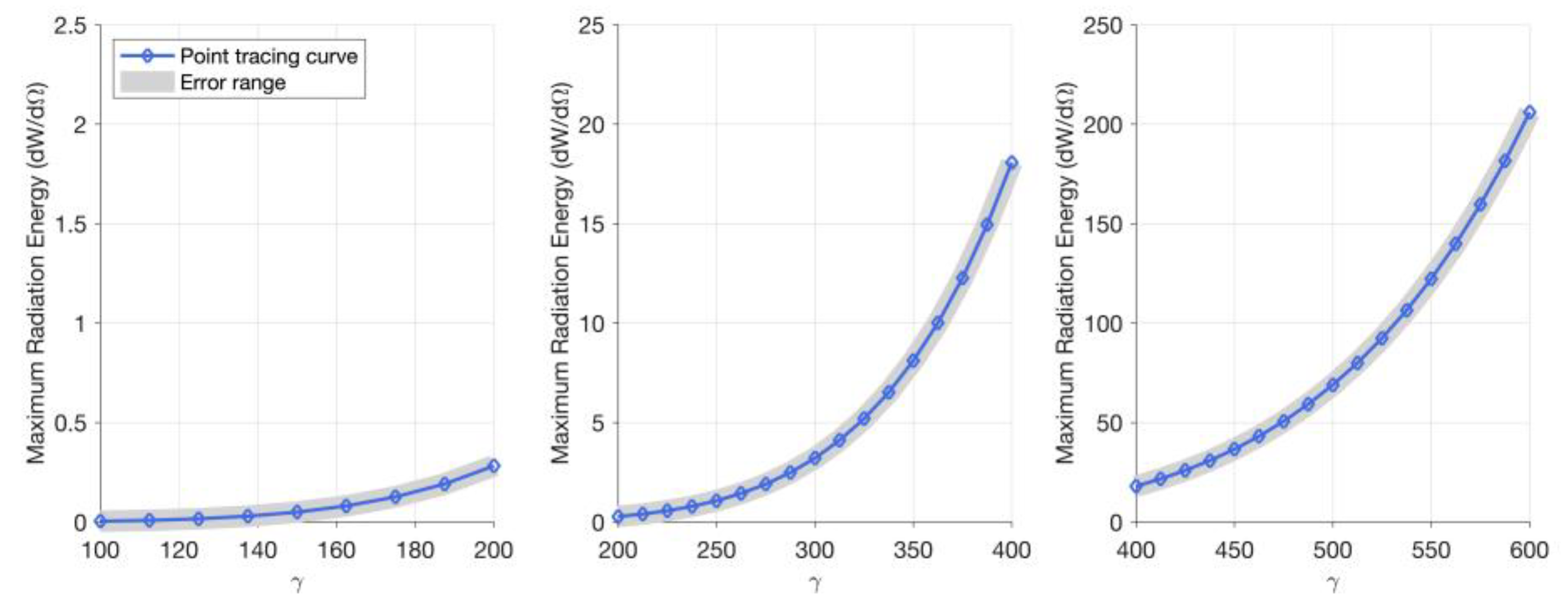
3.2. Maximum Radiation Energy

The X-ray with the collision center position is approximately symmetric regarding z c = 0 , as illustrated in Figure 5. The maximum radiation energy exhibits a diminishing pattern as the collision center increasingly travels away from z c = 0 reaching its peak energy point at z c = 0 . The collision center and the electrons’ initial energy can both have an impact on the resulting X-ray’s energy intensity in NITS collision characteristics. Large-quality collimation and high-energy X-ray can be obtained when the initial energy of the electron is large, and the collision center is not far from z c = 0 .
As the curve in Figure 6 illustrates, when the γ value is at the highest level ( γ = 400 ~ 600 ), the initial energy of electrons has a significant impact on the maximum radiation energy of X-rays. The radiation energy expression indicated a positive correlation between the radiation energy and the initial electron velocity. However, the nonlinear link between the two is weak when the electrons’ initially velocity is low. Figure 6 shows that the nonlinear relationship between γ and maximum radiation energy steadily strengthens with increasing starting electron velocity.
As seen in Figure 7, the maximum radiant energy of an X-ray grows as the laser’s intensity rises, and the two exhibit a positive association. The value of electron motion and lateral acceleration will both have an impact on the state of electron motion with a change in a 0 . On the one hand, a larger longitudinal momentum reduces γ by suppressing the longitudinal motion of electrons. On the other side, the transverse ponderomotive force that propels electron motion will become stronger and oscillate more violently, resulting in more transverse acceleration and a corresponding rise in radiant energy. In contrast to the “jump point” phenomenon that the radiation energy changes with the laser intensity under the circular polarization laser drive discovered by Chang et al. [17], the radiation energy does not change abruptly under the linear polarization laser drive of different intensities. The method of extending the laser duration can be utilized to address the issue of highly collimated high-energy X-rays produced by nonrelativistic lasers since the pulse width has no effect on the monochromatic collimation of radiation but can somewhat affect the radiation energy.

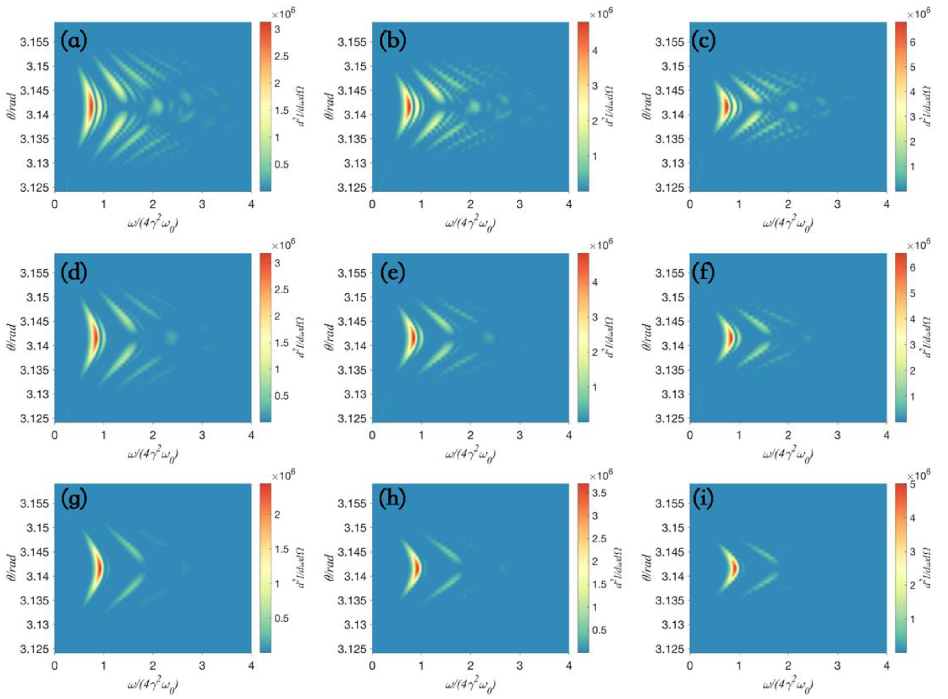
3.3. The Harmonic Spectrum

As the distance from the collision center z c grows, it can be observed from Figure 8 that the laser pulse vector potential decays, which causes the electronic oscillation to diminish. To explore the relationship between frequency spectrum and fundamental frequency more clearly, the frequency bands of the harmonic spectrum shown in Figure 8 and Figure 9 are normalized using 4 γ 2 ω 0 .
The bandwidth of scattered light narrows when the nonlinear Doppler frequency shift caused by the interaction of the laser and electron is inhibited. It is essential to consider how to balance elements includes monochromaticity, collimation, and energy to produce X-rays that satisfy application requirements.
The harmonic spectrum and angular distribution produced by collisions of high-energy electrons with various initial energies and laser pulses at various locations are shown in Figure 8. The radiation spectrum indicates that the angle range of the energy radiated per unit solid angle per unit frequency concentration continually reduces as the initial energy of electrons grows and the initial position deviates. The collimation of scattered X-rays is greatly improved. Meanwhile, the intensity of harmonic components depends on the amplitude of electron motion. The increase in the above two parameters lead to a decrease in the amplitude of electron motion, resulting in a gradual weakening of harmonic energy. Moreover, the changes in the above parameters have an impact on the number of oscillations in the spectrum. Therefore, the number of interference fringes in the radiation spectrum is related to the initial energy of electrons.
When a 0 < 1 , the increase in laser intensity indicates that the pondermotive force is more powerfully driving high-energy electrons. The spectral redshift results from the significant acceleration’s enhancement of the nonlinear Doppler effect. For greater intensity lasers, the axial velocity of the electrons varies more sharply because of the increased intensity of mass dynamics, producing a more pronounced broadening effect. As depicted in Figure 9, the X-ray spectrum displays a discrete supercontinuum when the laser intensity reaches relativistic intensity. Although complex and chaotic, the harmonic spatial distribution of X-rays. Whether under a nonrelativistic laser or a relativistic laser shock, the harmonic radiation energy increases as the laser intensity increases.
Electrodynamic processes that include long pulse laser and electron interaction are extremely intricate. Figure 9 shows that as the pulse width grows, so does the number of oscillations in the spectrum. According to the spectral stripes in Figure 9, the spectral bandwidth is narrower the longer the pulse width. Although under the influence of long pulses (Figure 9f,i,l), discrete ripples can still be observed in some spectra (within the range of horizontal coordinate values of 3~4), their actual values are very small and difficult to observe during the experimental process. Significant nonlinear compression characteristics can be observed in the NITS spectra. In particular, the spectrum tends to be compressed at higher frequencies. In the meantime, as pulse width grows, more interference fringes appear. This is owing to the numerous, diverse attosecond pulses that are produced by the numerous complex electrodynamic processes that lengthy pulses trigger.
In the case of NITS, the spectrum’s energy is always greater when θ = π than it is at other angles. As the laser pulse width increases, the focusing ability of radiation becomes less effective. When using long pulses, the monochromaticity of radiation is better. This is because when the pulse duration of the laser is longer, the spectral bandwidth of the laser is narrower. An increase in radiant energy is also present during this procedure. From this, it can be inferred that lowering the pulse width is a useful method for obtaining high-energy, high monochromatic X-ray lines. When a 0 1 , it is possible to achieve the adjustable discrete supercontinuum spectrum by appropriately adjusting the pulse width to produce X-rays.

4. Conclusions

The X-ray generated by the collision of a high-energy single electron with a tightly focused laser pulse is carefully studied in this study based on the acquisition of a high-energy photon source (HEPS). Using the classical electrodynamics principles, analytical formulas for the energy and spectrum of X-rays were developed, and numerical simulations were carried out. Good collimation and monochromaticity X-rays are produced by nonrelativistic Thomson scattering, whereas tunable (supercontinuum) X-rays are produced by a relativistic intensity laser. Long pulses can produce discrete supercontinuum spectra, whereas short pulses can help create X-ray lines with superior monochromaticity. A positive correlation between relativistic intensity and nonrelativistic intensity will be visible in the change in radiation energy with laser intensity. The initial electron energy has a certain impact on the collimation and frequency domain characteristics of X-rays. At the same time, the initial position of electrons will also affect the process of interaction between electrons and laser pulses. Regarding z c = 0 , the change in X-ray radiation properties is symmetric. The radiation energy is at its highest at z c = 0 . The better the radiation collimation, the larger the value of z c and the starting value of electron. It follows that higher collimated monochromatic X-rays can be produced by high-energy collisions that occur far from z c = 0 . Our finding has valuable implications for astronomical observation, biology (such as the treatment of cancer), and advancing the development of HEPS.

Author Contributions

Conceptualization, M.Z. and Y.T.; methodology, Y.T.; software, M.Z. and Y.Z.; validation, Y.C.; formal analysis, M.Z.; investigation, M.Z.; resources, Y.T.; data curation, Y.C.; writing—original draft preparation, M.Z.; writing—review and editing, M.Z. and Y.Z.; visualization, M.Z. and Y.Z.; supervision, Y.T.; project administration, Y.T.; funding acquisition, Y.T. All authors have read and agreed to the published version of the manuscript.

Funding

National Natural Sciences Foundation of China under Grant No. 10947170/A05 and No. 11104291; Natural Science Foundation of Nanjing University of Posts and Telecommunications under Grant No. NY221098; Jiangsu Qing Lan Project and STITP Project under Grant No. 202310293062Z.

Data Availability Statement

Data are contained within the article.

Acknowledgments

We are grateful to Nanjing University of Posts and Telecommunications for providing us with a research base and computing power support for our research.

Conflicts of Interest

The authors declare no conflict of interest.

References

  1. Yoshitaka, T.; Masahiro, K. Generation of Optical Vortices by Nonlinear Inverse Thomson Scattering at Arbitrary Angle Interactions. Astrophys. J. 2018, 860, 45. [Google Scholar]
  2. Vais, O.E.; Bochkarev, S.G.; Bychenkov, V.Y. Nonlinear Thomson scattering of a relativistically strong tightly focused ultrashort laser pulse. Plasma Phys. Rep. 2016, 42, 818–833. [Google Scholar] [CrossRef]
  3. Ramsey, D.; Malaca, B.; Piazza, A.D.; Formanek, M.; Palastro, J.P. Nonlinear thomson scattering with ponderomotive control. Phys. Rev. E 2021, 105, 065201. [Google Scholar] [CrossRef] [PubMed]
  4. Borovskiy, A.V.; Galkin, A.L. Saturation of electron emission with increasing ultrarelativistic intensity of the Gaussian laser pulse. Laser Phys. Lett. 2021, 18, 066002. [Google Scholar] [CrossRef]
  5. Petrillo, V.; Bacci, A.; Curatolo, C.; Drebot, I.; Giribono, A.; Maroli, C. Polarization of x-gamma radiation produced by a thomson and compton inverse scattering. Phys. Rev. Spec. Top.—Accel. Beams 2015, 18, 110701. [Google Scholar] [CrossRef]
  6. Kalmykov, S.Y.; Davoine, X.; Ghebregziabher, I.; Shadwick, B.A. Customizable electron beams from optically controlled laser plasma acceleration for γ-ray sources based on inverse Thomson scattering. Nucl. Inst. Methods Phys. Res. A 2016, 829, 52–57. [Google Scholar] [CrossRef]
  7. Jiao, Y.; Pan, W. High Energy Photon Source. High Power Laser Part. Beams 2022, 34, 14–20. [Google Scholar]
  8. Jiao, Y. High Energy Photon Source. Mod. Phys. 2022, 34, 38–43. [Google Scholar]
  9. Han, C.; Long, F.; Gao, Y.; Wei, P.; Liu, Y.; Guo, X.; Chen, S.; Du, Y. Development of corrector magnet power supply for high energy photon source. Radiat. Detect. Technol. Methods 2022, 6, 470–478. [Google Scholar]
  10. Sun, S.; Lu, H.; Li, X.; Zhang, X.; Yang, Y.; Sun, Y.; Zhang, L.; Li, Y.; Li, Z.; Chen, W.; et al. Mechanical design of a cryogenic Delta–Knot undulator for high energy photon source. Radiat. Detect. Technol. Methods 2021, 5, 122–127. [Google Scholar] [CrossRef]
  11. Dong, Y. High Energy Photon Source (HEPS): A High-performance Light Source for Biological Sciences. C.; The Biophysical Society of China, Institute of Biophysics, CAS: Beijing, China, 2019. [Google Scholar]
  12. Yang, Y.; Li, X.; Lu, H.; Huang, Y.; Sun, S.; Sun, Y.; Li, Y.; Zhang, L.; Zhao, S.; Chen, W.; et al. A new practical design of a merged APPLE-Knot undulator for High Energy Photon Source. Nucl. Inst. Methods Phys. Res. A 2022, 1034, 166816. [Google Scholar] [CrossRef]
  13. He, J.; Sui, Y.; Li, Y.; Ma, H.; Du, Y.; Wang, X.; Yue, J.; Cao, J. Beam Position Monitor Characterization for the High Energy Photon Source Synchrotron. Symmetry 2023, 15, 660. [Google Scholar] [CrossRef]
  14. Chi, Z.; Du, Y.; Huang, W.; Tang, C. Linearly polarized X-ray fluorescence computed tomography based on a Thomson scattering light source: A Monte Carlo study. Synchrotron Radiat. 2020, 27, 737–745. [Google Scholar] [CrossRef] [PubMed]
  15. Suortti, P.; Thomlinson, W. Medical applications of synchrotron radiation. Phys. Med. Biol. 2003, 48, R1–R35. [Google Scholar] [CrossRef] [PubMed]
  16. Taira, Y.; Hayakawa, T.; Katoh, M. Gamma-ray vortices from nonlinear inverse Thomson scattering of circularly polarized light. Sci. Rep. 2017, 7, 5018. [Google Scholar] [CrossRef] [PubMed]
  17. Lee, K.; Chung, S.; Park, S.; Jeong, Y.; Kim, D. Effects of high-order fields of a tightly focused laser pulse on relativistic nonlinear Thomson scattered radiation by a relativistic electron. EPL Europhys. Lett. 2010, 89, 64006. [Google Scholar] [CrossRef]
  18. Luo, W.; Yu, T.; Chen, M.; Song, Y.; Zhu, Z.; Ma, Y.; Zhuo, H. Generation of bright attosecond x-ray pulse trains via Thomson scattering from laser-plasma accelerators. Opt. Express 2014, 22, 32098–32106. [Google Scholar] [CrossRef] [PubMed]
  19. Tan, F.; Zhu, B.; Han, D.; Xin, J.-T.; Zhao, Z.-Q.; Cao, L.-F.; Gu, Y.-Q.; Zhang, B.-H. Numerical simulation for all-optical Thomson scattering X-ray source. Chin. Phys. B 2014, 23, 253–259. [Google Scholar] [CrossRef]
  20. Pogorelsky, I.; Ben-Zvi, I.; Hirose, T.; Kashiwagi, S.; Yakimenko, V.; Kusche, K.; Siddons, P.; Skaritka, J.; Kumita, T.; Tsunemi, A.; et al. Demonstration of 8 × 10¹⁸ photons/second peaked at 1.8 å in a relativistic Thomson scattering experiment. Phys. Rev. Spec. Top. Beams 2000, 3, 090702. [Google Scholar] [CrossRef]
  21. Liu, Y.Y.; Salamin, Y.I.; Dou, Z.-K.; Xu, Z.-F.; Li, J.-X. Vortex γ rays from scattering laser bullets off ultrarelativistic electrons. Opt. Lett. 2020, 45, 395–398. [Google Scholar] [CrossRef]
  22. Sakai, I.; Aoki, T.; Dobashi, K.; Fukuda, M.; Higurashi, A.; Hirose, T.; Iimura, T.; Kurihara, Y.; Okugi, T.; Omori, T.; et al. Production of high brightness γ rays through backscattering of laser photons on high-energy electrons. Phys. Rev. Spec. Top. Beams 2003, 6, 091001. [Google Scholar] [CrossRef]
  23. Chang, Y.; Zeng, Z.; Wang, C.; Long, Z.; Tian, Y. Modulation of high-energy γ -rays by collision of an ultra-high-energy electron with a tightly focused circularly polarized laser pulse. Appl. Opt. 2022, 61, 6038–6045. [Google Scholar] [CrossRef] [PubMed]
  24. Chang, Y.; Wang, C.; Wang, Y.; Long, Z.; Zeng, Z.; Tian, Y. Collimation and monochromaticity of γ-rays generated by high-energy electron colliding with tightly focused circularly polarized laser with varied intensities. Laser Phys. Lett. 2022, 19, 065301. [Google Scholar] [CrossRef]
  25. Investigators at National Institute of Advanced Industrial Science and Technology (NIAIST) Discuss Findings in Applied Materials (Ammonia-free Epitaxy of Single-crystal Inn Using a Plasmaintegrated Gas-injection Module). Electronics Newsweekly. 2022.
  26. Salamin, Y.I.; Keitel, C.H. Electron Acceleration by a Tightly Focused Laser Beam. Phys. Rev. Lett. 2002, 88, 9. [Google Scholar] [CrossRef] [PubMed]
  27. Vaziri, M.; Golshani, M.; Sohaily, S.; Bahrampour, A. Electron acceleration by linearly polarized twisted laser pulse with narrow divergence. Phys. Plasmas 2015, 22, 033118. [Google Scholar] [CrossRef]
  28. Akhyani, M.; Jahangiri, F.; Niknam, A.R.; Massudi, R. Optimizing chirped laser pulse parameters for electron acceleration in vacuum. Appl. Phys. 2015, 118, 183106. [Google Scholar] [CrossRef]
  29. Cai, X.-M.; Zhao, J.-Y.; Lin, Q.; Luo, J.-L. Electron acceleration by sub cycle pulsed focused vector beams. JOSA B 2016, 33, 158–164. [Google Scholar] [CrossRef]
  30. Lan, P.; Lu, P.; Cao, W.; Wang, X. Attosecond and zeptosecond x-ray pulses via nonlinear Thomson backscattering. Phys. Rev. E Stat. Nonlinear Soft Matter Phys. 2005, 72, 066501. [Google Scholar] [CrossRef]
  31. Kobzak, S.; Weidenmüller, H.A.; Pálffy, A. Laser-nucleus interactions in the sudden regime. Phys. Rev. C 2021, 103, 044616. [Google Scholar] [CrossRef]
  32. Dunning, D.J.; Mcneil BW, J.; Thompson, N.R. Towards Zeptosecond-Scale Pulses from X-ray Free-Electron Lasers. Phys. Procedia 2013, 52, 62–67. [Google Scholar] [CrossRef]
Figure 1. Schematic diagram of inverse Thomson scattering controlled by linearly polarized laser.
Crystals 14 00013 g001
Figure 2. The effect of initial electron position on the laser electron collision center.
Crystals 14 00013 g002
Figure 3. The energy angular distribution of X-rays generated by the collision of a single electron with different collision center positions and initial γ values with a 0 = 1 , L = 3 λ 0 linearly polarized laser pulses. The radiation energy values at each position are normalized. The observed θ range is [ 178 ° , 180 ° ] , E = d W / d Ω . (a) z i = 10 λ 0 , γ = 100 , (b) z i = 10 λ 0 , γ = 125 , (c) z i = 10 λ 0 , γ = 150 , (d) z i = 30 λ 0 , γ = 100 , (e) z i = 30 λ 0 , γ = 125 , (f) z i = 30 λ 0 , γ = 150 , (g) z i = 50 λ 0 , γ = 100 , (h) z i = 50 λ 0 , γ = 125 , and (i) z i = 50 λ 0 , γ = 150 .
Crystals 14 00013 g003
Figure 4. The energy angular distribution of X-rays produced by linearly polarized lasers with different intensities and different pulse widths. The initial γ value of the electron is 200. The radiation energy values at each position are normalized. The observed θ range is [178°, 180°]. E = d W / d Ω . (a) a 0 = 0.5 , L = λ 0 , (b) a 0 = 0.5 , L = 3 λ 0 , (c) a 0 = 0.5 , L = 9 λ 0 , (d) a 0 = 1 , L = I , (e) a 0 = 1 , L = 3 λ 0 , (f) a 0 = 1 , L = 9 λ 0 , (g) a 0 = 3 , L = λ 0 , (h) a 0 = 3 , L = 3 λ 0 , (i) a 0 = 3 , L = 9 λ 0 , (j) a 0 = 5 , L = λ 0 , (k) a 0 = 5 , L = 3 λ 0 , and (l) a 0 = 5 , L = 9 λ 0 .
Crystals 14 00013 g004
Figure 5. Maximum radiant energy curve of X-ray under the influence of different γ values ( a 0 = 1 , L = 3 λ 0 ).
Crystals 14 00013 g005
Figure 6. The maximum radiation energy curve of X-rays and its error range at a 0 = 1 , L = 3 λ 0 , z i = 0 .
Crystals 14 00013 g006
Figure 7. Maximum radiant energy of X-ray under the influence of different laser intensities and pulse widths.
Crystals 14 00013 g007
Figure 8. The spatial distribution of the harmonic energy of X-rays produced by the collision of a single electron with different collision center positions and initial γ values with a 0 = 1 , L = 3 λ 0 linearly polarized laser pulses. The value of harmonic energy at each position is represented by color. Different colors represent different d 2 I / d ω d Ω . (a) z i = 10 λ 0 , γ = 100 , (b) z i = 10 λ 0 , γ = 125 , (c) z i = 10 λ 0 , γ = 150 , (d) z i = 30 λ 0 , γ = 100 , (e) z i = 30 λ 0 , γ = 125 , (f) z i = 30 λ 0 , γ = 150 , (g) z i = 50 λ 0 , γ = 100 , (h) z i = 50 λ 0 , γ = 125 , and (i) z i = 50 λ 0 , γ = 150 .
Crystals 14 00013 g008
Figure 9. The spatial distribution of the harmonic energy of X-rays produced by linearly polarized lasers with different intensities and different pulse widths. The initial γ value of the electron is 200. The value of harmonic energy at each position is represented by color. Different colors represent different d 2 I / d ω d Ω . The intensity and pulse width of each image are: (a) a 0 = 0.5 , L = λ 0 , (b) a 0 = 0.5 , L = 3 λ 0 , (c) a 0 = 0.5 , L = 9 λ 0 , (d) a 0 = 1 , L = λ 0 , (e) a 0 = 1 , L = 3 λ 0 , (f) a 0 = 1 , L = 9 λ 0 , (g) a 0 = 3 , L = λ 0 , (h) a 0 = 3 , L = 3 λ 0 , (i) a 0 = 3 , L = 9 λ 0 , (j) a 0 = 5 , L = λ 0 , (k) a 0 = 5 , L = 3 λ 0 , and (l) a 0 = 5 , L = 9 λ 0 .
Crystals 14 00013 g009
Table 1. The nonzero range of radiation energy in the sin θ cos φ direction for each subgraph in Figure 3.
Label on Figure 3(a)(b)(c)(d)(e)(f)(g)(h)(i)
Δ sin θ cos φ 0.03080.02560.02160.03070.02520.02100.03060.02440.0204
Table 2. The nonzero range of radiation energy in the sin θ cos φ direction for each subgraph in Figure 4.
Label on Figure 4(a)(b)(c)(d)(e)(f)(g)(h)(i)(j)(k)(l)
Δ sin θ cos φ 0.01130.01140.01100.01470.01490.01440.03080.03270.03040.04780.05170.0476
Disclaimer/Publisher’s Note: The statements, opinions and data contained in all publications are solely those of the individual author(s) and contributor(s) and not of MDPI and/or the editor(s). MDPI and/or the editor(s) disclaim responsibility for any injury to people or property resulting from any ideas, methods, instructions or products referred to in the content.

