1. Introduction
Cyanuric acid (henceforth, CYH; singly deprotonated form CY
−) (CO·NH)
3 is a heterocyclic compound with a sturdy, robust and relatively unreactive planar six-membered ring molecule, which nevertheless has had a diverse role to play in chemistry, particularly with regard to commercial and industrial use, for many years. It has featured in products ranging from flame retardants [
1] and plant growth feed [
2] to a catalyst for NO
x removal from exhaust gases [
3], lavatory cleaning blocks [
4], and an additive to stabilise free chlorine in swimming pools [
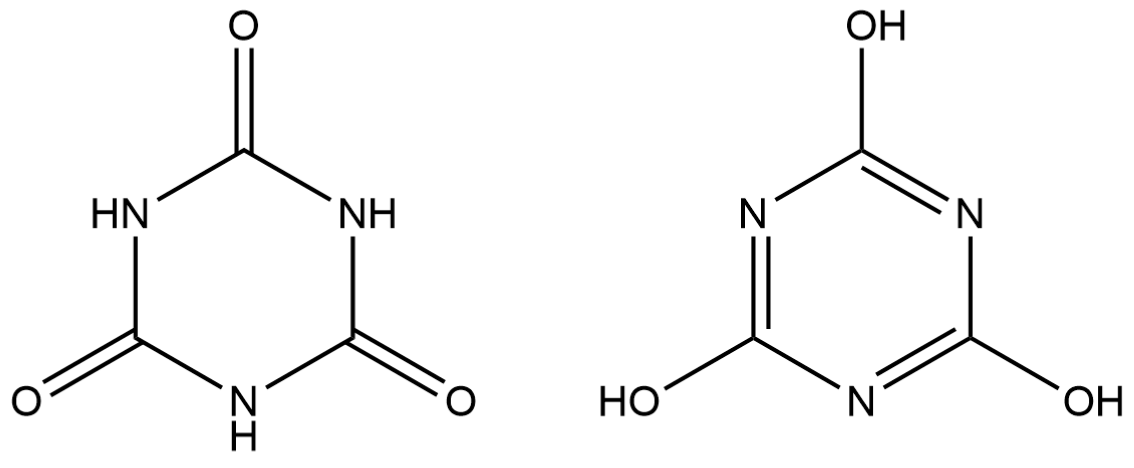
5]. It exists predominantly in the triketo form in solution [
6] and in crystal structures (see
Scheme 1). The current release of the Cambridge Structural Database (CSD; version 5.45, November 2023) [
7] contains almost 90 structures (some of them independent studies of the same materials) in which CYH is a neutral molecule on its own or cocrystallised with solvent molecules, other neutral molecules, or ionic salts, and they are all in the triketo form with three carbonyl groups and three N–H bonds. It should be noted that this form is sometimes referred to as isocyanuric acid despite its preponderance over the alternative trihydroxy form (
Scheme 1). Lists of the CSD REFCODEs (structural identifiers) for the database searches described in this section are available as
Supplementary Materials.
The planar threefold symmetrical shape of the CYH molecule, together with the availability of three donors and three acceptors for hydrogen bonds, make it an ideal candidate for supramolecular chemistry involving extended hydrogen bonding patterns. It almost always forms one- or two-dimensional hydrogen-bonded arrays [
8], and there has been a particular focus on the CYH–melamine system. These two molecules are complementary hydrogen-bonding partners with matching symmetry, forming a very strongly bonded structure with a rosette-like motif [
9] that has one of the most stable non-covalent solid-state structures known [
10,
11]. Indeed, such is the strength of the hydrogen bonding, some 29 kJ mol
−1 per hydrogen bond [
11], that it is frequently difficult to characterise the products by single-crystal X-ray diffraction, since crystal growth is typically rapid and the crystals are rather insoluble once formed, so other techniques such as solid-state NMR are important. Hydrogen-bonding motifs for CYH cocrystals are so reliable that it is possible to predict, and therefore design, the structural outcome of a particular reaction, something that has been particularly well exploited in the CYH–melamine system by varying substituents on the two components [
10,
11,
12,
13,
14,
15,
16,
17,
18,
19,
20] or by adding further cocrystal components [
21].
Cyanuric acid is a mildly acidic compound with p
Ka values of 6.88, 11.40, and 13.5 for the mono-, di-, and trianionic forms, respectively [
22]. While single deprotonation is easily achieved and double deprotonation can be achieved by careful control of the reaction equilibrium, triple deprotonation is much more uncommon. Only the singly deprotonated form CY
− is found in salts with organic cations, of which there are 23 known examples in the CSD. Conversely, the CYH molecule is singly and/or doubly protonated in hydrogen phosphate [
23], 5-amino-2,4,6-triiodobenzene-1,3-dicarboxylate [
24], and chloride salts [
25]; these cations adopt the trihydroxy form with only one or two nitrogen atoms protonated.
CYH serves as a ligand to transition, post-transition, and lanthanide metals in all three deprotonated forms. In the majority (approximately 70%) of the almost 100 structures of such complexes in the CSD (a relatively small number for such a small and versatile ligand), coordination to the metal centre is exclusively through nitrogen, though coordination through oxygen is also found in multidentate and bridging modes of coordination. Some of these complexes also contain alkali or alkaline earth metal cations, which are coordinated by the oxygen atoms of the cyanurate ligands.
There are just over 40 CSD entries for structures containing one or more alkali metals and deprotonated forms of CYH but no transition, post-transition, or lanthanide metals; a few of these also contain an alkaline earth metal. Here, in contrast to the transition metal complexes, coordination is mainly through oxygen, with weaker interactions through nitrogen in some cases.
A total of 31 CSD entries are formulated as salts or complexes of CYH anions with alkaline earth metals and no other metals: 3 for Mg, 4 for Ca, 14 for Sr, 7 for Ba, and 3 with two different metals. Several of these are duplicate determinations of the same structure, so there are just over 20 distinct structures. Again, coordination is principally through oxygen, with an increasing tendency to include bonding also through nitrogen as the periodic group of elements is descended and the ionic radius of the metal increases to accommodate higher coordination numbers.
We have conducted a systematic structural study of complexes of CYH with the alkaline earth metals, prepared from aqueous solution with no ligands available other than water and the neutral and anionic forms of CYH, to investigate the interplay and competition of coordination and hydrogen bonding with particular reference to the trend in ionic radii in the series from Mg through Ca and Sr to Ba. We find in each case an empirical formula M(CY)2(H2O)n with two singly-deprotonated CY− anions acting as ligands and/or uncoordinated counterions and between two and eight coordinated and uncoordinated water molecules. The coordination number increases down the series: Mg 6, Ca 7, Sr 8, Ba 10.
[Mg(H2O)6][CY]2·2H2O (1)
{[Ca(CY)(H2O)3][CY]}∞ (2)
{[Sr(CY)2(H2O)4]}∞ (3)
{[Ba(CY)2(H2O)2]}∞ (4)
Although these structures have been reported previously by others [
26,
27,
28,
29,
30], those studies have been directed primarily at properties such as optical anisotropy, second harmonic generation, and corrosion inhibition, and the structural trends have not generally been noted. In the case of the strontium complex
3, the two previously reported structures [
28,
29] had inappropriate (and different) treatments of their hydrogen atoms, as discussed below. Correct assignment of hydrogen atom positions is particularly important for the interpretation of hydrogen bonding, and this is aided by low-temperature X-ray diffraction data collection as employed in the current investigation, whereas all the previous structures were determined from room-temperature data.
2. Materials and Methods
All reagents were reagent-grade commercial products from Aldrich (Gillingham, UK) and Avocado (Altrincham, UK) and were used as received. Laboratory-grade distilled water was the only solvent for reactions. Elemental analyses were carried out by the Newcastle University analytical services unit and indicate the chemical purity of the products. Access to suitable X-ray powder diffraction facilities was not available at the time of the experiments to confirm phase purity, but the formation of concomitant polymorphs seems unlikely, given that what appear to be complexes with the same structures were obtained by other researchers using similar conditions and only for the strontium complex has a polymorph been reported, prepared in a mixed product by hydrothermal synthesis [
27].
Synthesis of the magnesium complex (1), {[Mg(H2O)6][CY]2·2H2O}∞: 0.258 g (2 mmol) CYH was dissolved in 50 cm3 of boiling water. A total of 0.484 g (1 mmol) of solid Mg(CO3)·Mg(OH)2·5H2O was added and the mixture was boiled for 15 min, then filtered hot. As the filtrate cooled, a mass of small crystals of 1 formed, which nevertheless were suitable for X-ray diffraction. The crystals were not air-stable and gradually lost water, as shown in the CHN analysis. Yield was 52% (0.219 g). Elemental analysis data obtained correspond to C6H14MgN6O11, i.e., with the loss of three molecules of water per formula unit. Calc (%): C, 19.45; H, 3.81; N, 22.68. Found (%): C, 19.12; H, 3.88; N, 22.15.
Synthesis of the calcium complex (2), {[Ca(CY)(H2O)3][CY]}∞: 0.258 g CYH was dissolved in 50 cm3 boiling water to which was added 0.074 g (1 mmol) of solid Ca(OH)2. After five minutes of boiling, all solid material had dissolved, so the reaction flask was stoppered and set aside. After 3–4 h of cooling, crystals of 2 had formed. Yield was 44% (0.237 g). Calc (%): C, 20.58; H, 2.87; N, 23.99. Found (%): C, 20.32; H, 2.83; N, 23.24.
Synthesis of the strontium complex (3), {[Sr(CY)2(H2O)4]}∞: 0.262 g (2 mmol) CYH and 0.147 g (1 mmol) SrCO3 were boiled for several minutes in 30 cm3 water until the solids had dissolved. The reaction flask was then sealed and set aside to cool undisturbed. A small crop of white crystals of 3 formed overnight. Yield was 10% (0.043 g). Calc (%): C, 17.35; H, 2.79; N, 20.23. Found (%); C, 17.43; H, 2.73; N, 19.36.
Synthesis of the barium complex (4), {[Ba(CY)2(H2O)2]}∞: 0.258 g (1 mmol) CYH was dissolved in 40 cm3 boiling water. Solid Ba(OH)2 (0.315 g, 1 mmol) was added and the mixture was boiled until around 20 cm3 remained, then the hot solution was filtered. Colourless crystals of 4 formed after standing undisturbed for three days at room temperature. Yield was 10% (0.034 g). Calc (%): C, 16.78; H, 1.88; N, 19.57. Found (%): C, 16.54; H, 2.50; N, 19.10.
X-ray crystallography: crystallographic experimental parameters for all four compounds are summarised in
Table 1. Data for all compounds were collected on a KappaCCD diffractometer (Nonius, Delft, The Netherlands) using graphite-monochromated Mo-
Kα radiation (λ = 0.71073 Å) at 150K. The treatment of hydrogen atoms in structure refinements is described below for each structure, as this is an important facet of understanding the role of hydrogen bonding in the crystal structures. Software used: data collection with Nonius COLLECT [
31]; cell determination with DIRAX [
32]; data reduction with EvalCCD [
33]; absorption correction by symmetry-equivalent and repeated reflections using SADABS [
34]; structure solution by direct methods using SIR2002 (
1 and
2) [
35] and SHELXTL (
3 and
4) [
36]; refinement by full-matrix least-squares on all unique
F2 values using SHELXL-2019/2 [
37]. Structure figures were produced using SHELXTL, DIAMOND–3 [
38], and Mercury 1.4 [
39]. The structures have been deposited at the Cambridge Crystallographic Centre (CCDC), with deposition numbers given in
Table 1; the data may be obtained at
https://www.ccdc.cam.ac.uk/structures/ free of charge (accessed on 17 January 2024).
3. Results and Discussion
All four complexes, prepared by simple reactions of cyanuric acid and alkaline earth hydroxides or carbonates in boiling water and obtained as colourless crystals, have the empirical formula M(CY)2(H2O)n (n = 8 for Mg, 3 for Ca, 4 for Sr, 2 for Ba). All contain singly deprotonated CY− anions in the triketo form as ligands and/or uncoordinated counterions, and all water molecules are coordinated to metal ions in each case, either in terminal or bridging mode, except that the magnesium complex also contains uncoordinated water molecules. Hydrogen bonding is a major feature of all the structures; cyanurate N–H and water O–H serve as donors, while the acceptors are O atoms of water and CY− as well as deprotonated N atoms. The four structures differ in other details, as described below. In each case, the asymmetric unit contains two CY− anions; for consistency and convenience in making structural comparisons, atoms in these anions are numbered 1–3 and 4–6, while the O atoms of water molecules are numbered from 7 upwards.
3.1. Compound 1, [Mg(H2O)6][CY]2·2H2O
There are only two previously reported structures of complexes of magnesium with cyanuric acid and no other metals; one of them has been the subject of two independent studies [
26,
27] and is the same as our compound
1, while the other, prepared hydrothermally, has the same ionic components but no uncoordinated water molecules [
27]. The main structural feature of compound
1 is that CY
− does not coordinate to the Mg
2+ centre at all, but serves as a counterion to the [Mg(OH
2)
6]
2+ cation. The two anions in the asymmetric unit lie in general positions, while there are two crystallographically independent cations, each located on an inversion centre. In addition, two uncoordinated water molecules occupy general positions in the asymmetric unit. This lack of coordination of organic ligands is a common observation in aqueous magnesium chemistry; similar behaviour is found for other small-ring molecules structurally similar to cyanuric acid, such as pyromellitic acid [
40], phthalic acid [
41], 5-nitrobarbituric acid [
42], and the trithio analogue of cyanuric acid [
43].
All the H atoms in this structure could be located readily in difference maps. Two water molecules, one coordinated (O9) and one uncoordinated (O13), which lie within hydrogen-bonding distance of each other, were found to have one H atom ordered and the other disordered over two positions with relative occupancy 0.56:0.44(3); the disordered H atoms were refined with a common isotropic displacement parameter for each pair, and otherwise H atoms were refined with individual unconstrained isotropic displacement parameters. Water O–H bond lengths were restrained to 0.84(1) Å and H–O–H angles restrained to be equal without a specified value. Nitrogen-bound H atoms were refined freely. All N–H and O–H bonds serve as hydrogen bond donors; the acceptors for N–H...O hydrogen bonds are carbonyl O atoms. All the carbonyl O atoms accept two hydrogen bonds, and the two deprotonated N atoms accept hydrogen bonds from water molecules (one coordinated, one uncoordinated). The two uncoordinated water molecules each donate and accept two hydrogen bonds. In each cation, two inversion-related
trans-coordinated water molecules accept a hydrogen bond, while the other four water molecules do not serve as acceptors. All hydrogen bonds are approximately linear. Hydrogen bond geometry is given in
Table 2.
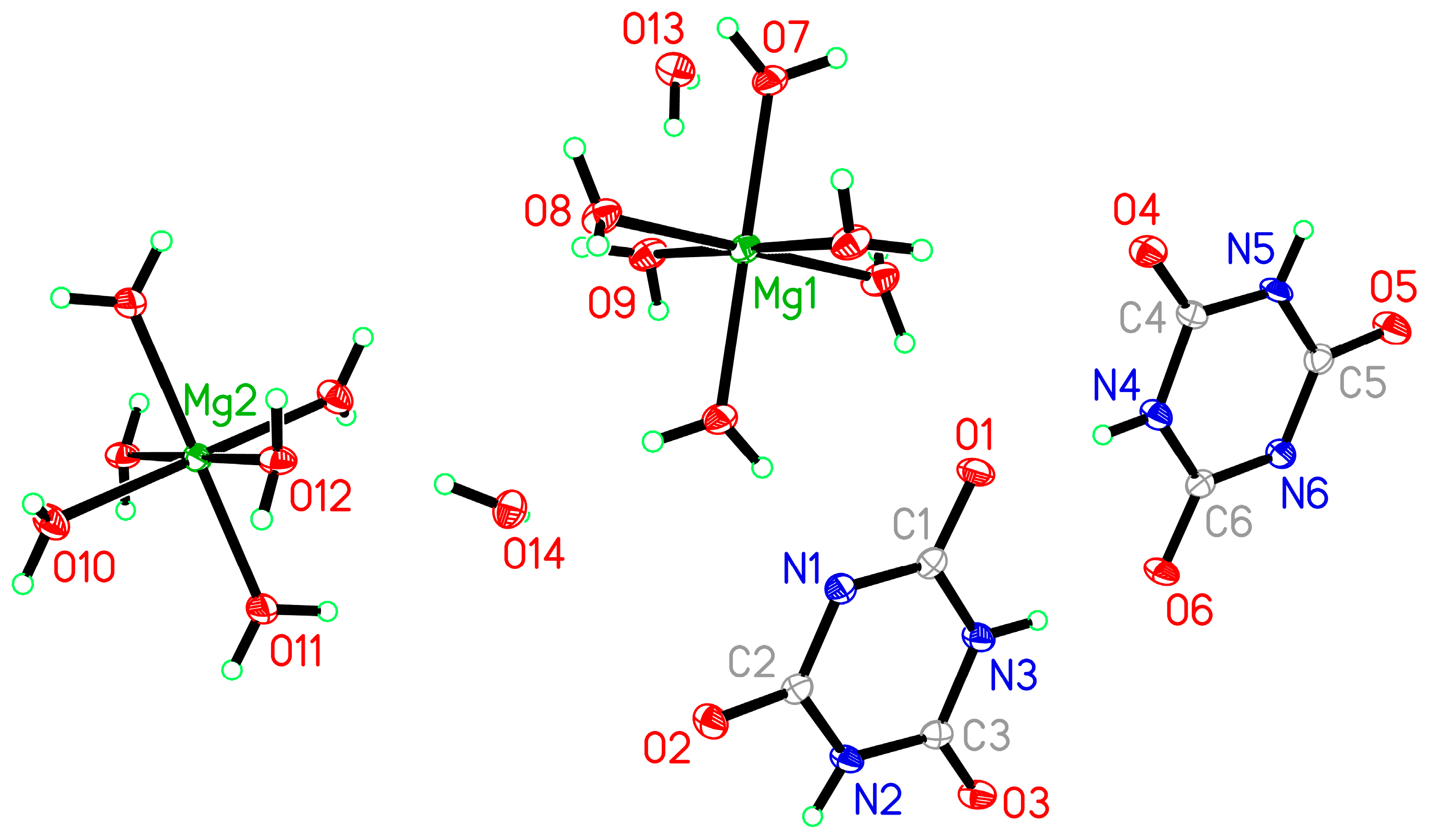
Magnesium coordination is essentially regular octahedral with Mg–O bond lengths in the range 2.0284(17)–2.1058(16) Å and all
cis angles within 3.1° of 90°. The internal geometry of the planar CY
− anions is unexceptional. The asymmetric unit is shown in
Figure 1, together with additional aqua ligands to complete the coordination of magnesium.
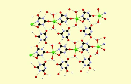
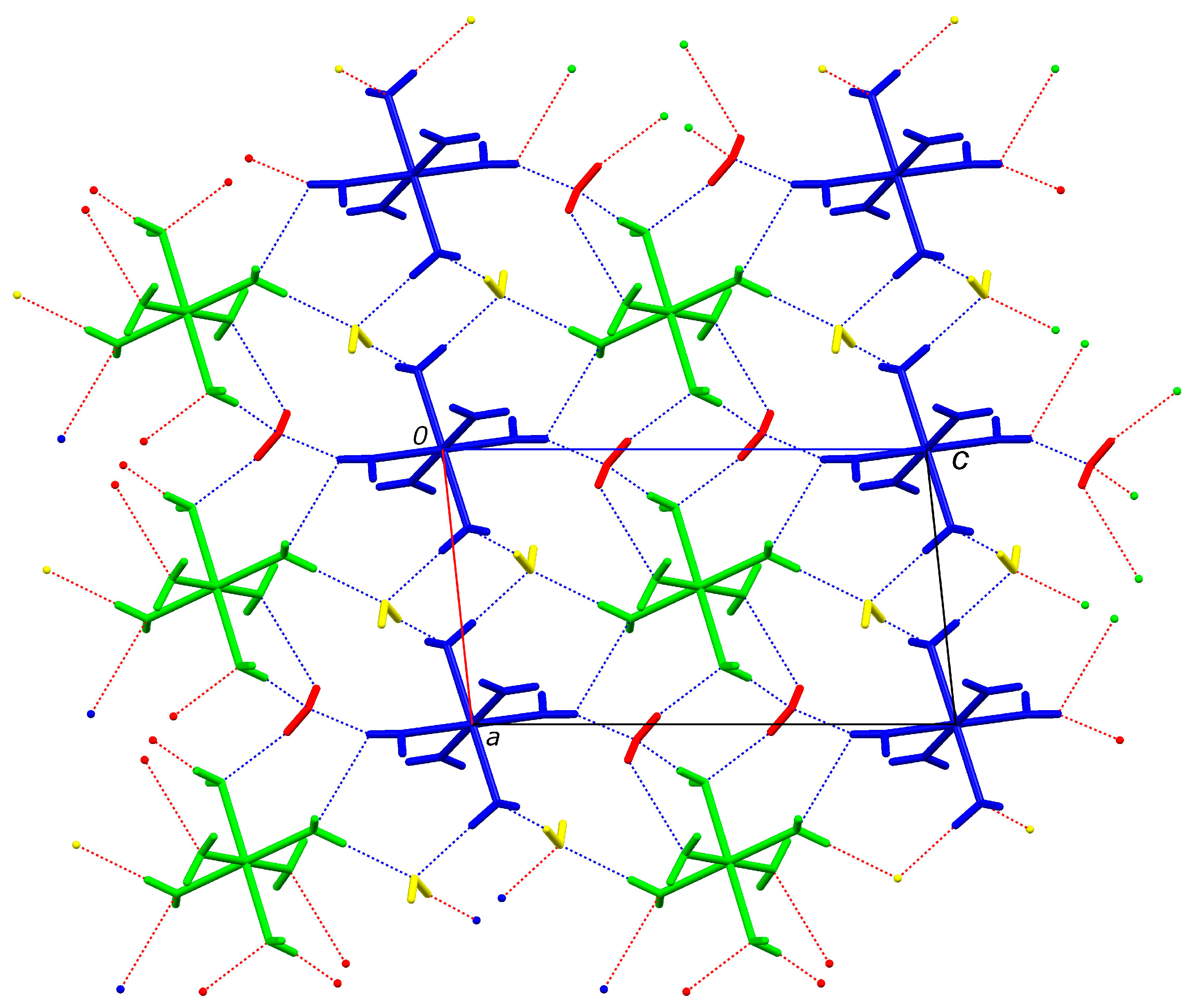
The extensive three-dimensional hydrogen bonding pattern can best be described in three stages of construction. In the first two stages, the cations and uncoordinated water molecules form a layer in the
ac plane (101), as shown in
Figure 2.
The four crystallographically independent species in
Figure 2 are coloured differently, with the same colour used for symmetry-equivalent species. The sheet consists of alternating chains, parallel to the short
a axis, of hexaaquamagnesium(II) ions linked by hydrogen bonding to one of the water molecules. The chains consist of only one type of hexaaqua ion, linked to its symmetry-equivalents by one type of water molecule; thus the blue [Mg(H
2O)
6]
2+ ions are linked to each other exclusively by the yellow water molecules, and the green [Mg(H
2O)
6]
2+ ions are linked to each other exclusively by the red water molecules, giving the first imagined stage of construction. In the second stage, additional yellow–green and red–blue hydrogen bonding in the
c axis direction links these chains together to give the sheet structure. The disorder identified in coordinated O9 and uncoordinated O13 water molecules is found in the components of this structure shown in green and red. This represents the two alternative directions (clockwise and anti-clockwise when viewed from one side) of the rings formed by two coordinated and two uncoordinated water molecules and does not affect the rest of the hydrogen bonding.
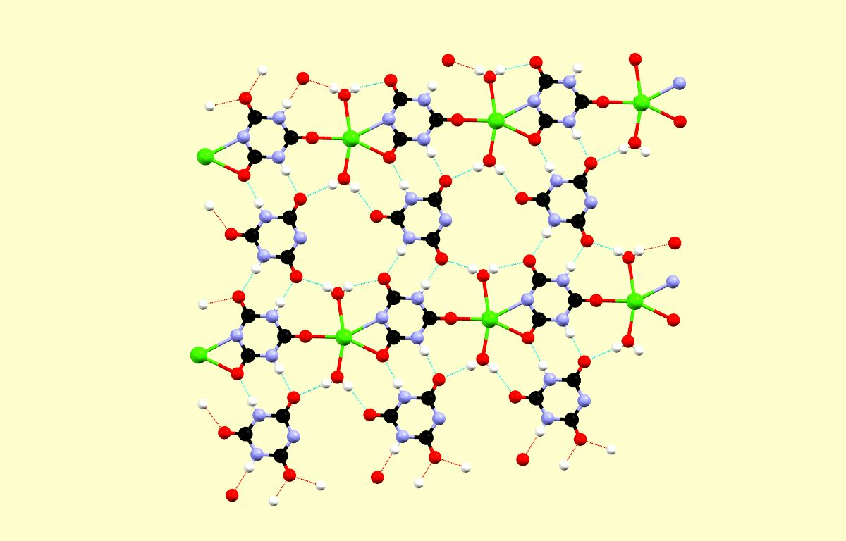
The cyanurate anions form hydrogen-bonded ribbons parallel to the
c axis, and each ribbon consists of the two crystallographically independent cyanurate anions alternating. These ribbons are sandwiched between the cation/water sheets, with which they share more hydrogen bonds, including the O–H...N hydrogen bonds between water molecules and deprotonated N atoms (
Figure 3).
3.2. Compound 2, {[Ca(CY)(H2O)3][CY]}∞
There are four previous reports of calcium complexes of cyanuric acid with no other metals present. One contains doubly deprotonated cyanuric acid with a deceptively simple 1:1 stoichiometry, together with, empirically, 3.5 water molecules per metal ion, but with a complex polymeric network structure in which the anion serves both as a ligand and as an uncoordinated counterion [
29]; the structure has some features similar to that of compound
2 despite the different degrees of deprotonation. A calcium(II) complex of CY
− originally described, improbably, as also containing CYH and uncoordinated OH
− anions, but with no H atoms included in the structural model [
44], has been reinvestigated and more appropriately formulated as [Ca(CY)(H
2O)
6](CY)·H
2O [
45], with calcium coordinated by six aqua ligands and one carbonyl O atom of CY
−. The other reported structure is the same as that of our compound
2 [
27], but with little structural information given other than identifying the basic coordination of Ca(II), the main interest being in the optical and other physical properties of this and other cyanurate complexes.
Figure 4 shows the asymmetric unit, augmented by one extra CY
− ligand, one bridging water molecule, and one Ca
2+ cation to complete the calcium coordination and the environment of the symmetry-related bridging water molecules. All atoms lie in general positions, and the asymmetric unit contains one calcium(II) ion, three coordinated water molecules, and two CY
− anions, one of which is coordinated and the other not. All H atoms in this structure were refined freely with individual isotropic displacement parameters and with no constraints or restraints.
The calcium centre, larger than its magnesium congener, can accommodate two cyanurate ligands in its coordination, but these are symmetry-related; the second CY
− in the empirical formula serves as a charge-balancing counterion. The cyanurate ligand bridges two calcium ions, bonding to one in monodentate fashion through one of its carbonyl O atoms O1 and to the other in chelating bidentate mode through carbonyl O3 and the deprotonated N atom N2 with a narrow bite angle. Two aqua ligands are terminal, while two symmetry-equivalent bridging aqua ligands connect calcium with a second cation to give a centrosymmetric four-membered Ca
2O
2 ring. This arrangement gives a coordination number of 7, with a geometry that is approximately octahedral if the chelating group is regarded as occupying a single coordination site and pentagonal bipyramidal if these two coordinating atoms are considered separately, with the aqua ligands O7 and O9 occupying the axial positions. The coordination geometry (bond lengths and angles) is given in
Table 3.
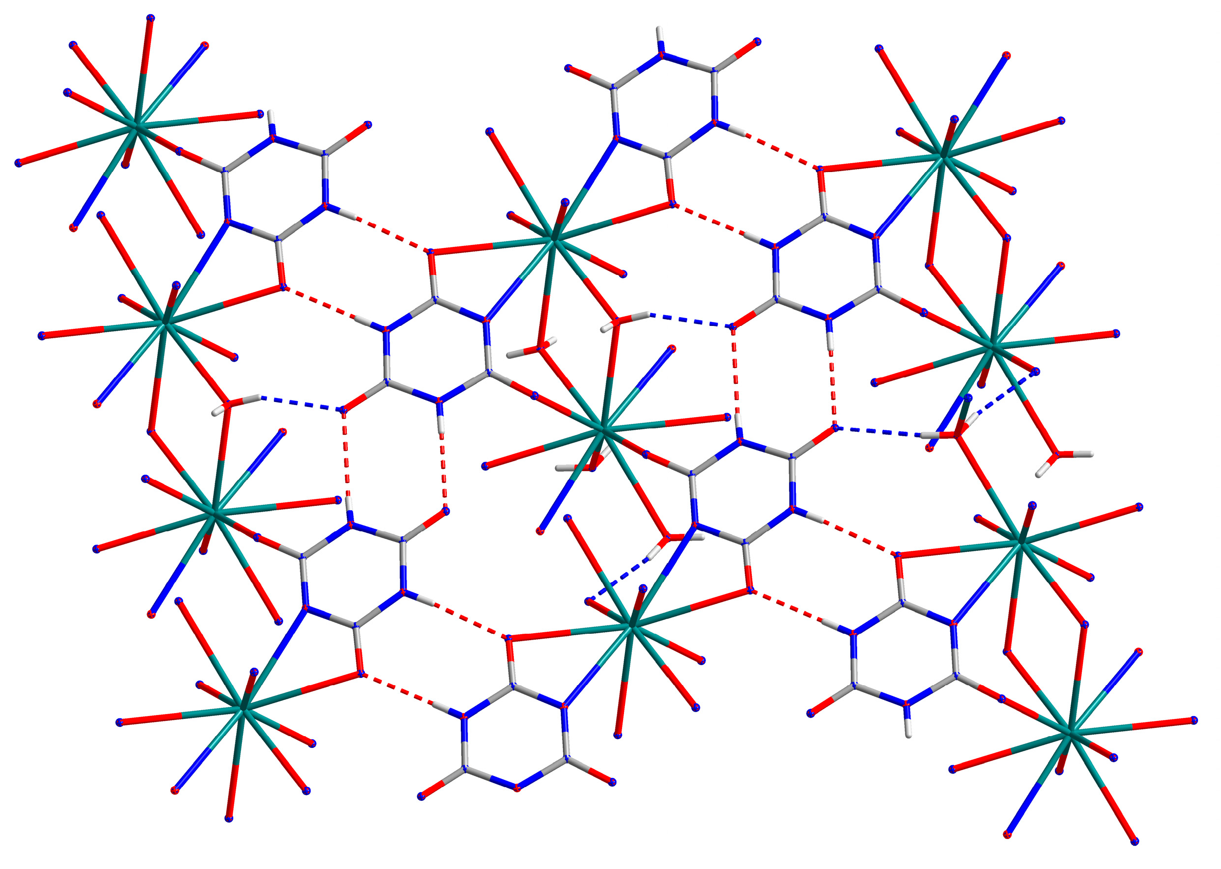
Bridging of calcium centres by the CY− ligands generates a polymeric chain running parallel to the b axis, and pairs of these chains are bonded together through the bridging O9 aqua ligands.
If terminal O7 and the O9 aqua bridges are ignored, the coordinated and uncoordinated cyanurate anions are essentially coplanar with the equatorial plane of the pentagonal bipyramidal coordination. Thus, the uncoordinated anions link polymer chains together by hydrogen bonding to give sheets parallel to the (
01) plane, as shown in
Figure 5.
The ribbons of hydrogen-bonded CY
− anions running horizontally in
Figure 5 are the same as those seen in the structure of compound
1, though there they are all uncoordinated (the two crystallographically independent anions alternating) while, here, they are alternating coordinated and uncoordinated anions. In both cases, the hydrogen-bond motif is
R22(8) in standard notation [
46], the most common motif found in the hydrogen bonding of structures containing cyanuric acid and its derivatives. The hydrogen bonding geometry of compound
2 is listed in
Table 4.
Bridging of calcium centres through pairs of O9 aqua ligands combines pairs of adjacent hydrogen-bonded sheets into double sheets with an interplanar separation of about 3.2 Å, such that overlapping cyanurates are appropriately positioned for π-π stacking interactions to enhance the stability of the crystal structure.
Finally, the terminally bonded aqua ligand O7 forms hydrogen bonds to O5 and N6 in two different uncoordinated cyanurate anions in the next double sheet, thereby providing the hydrogen bonding in the third dimension for this multi-faceted structure. All N–H and O–H bonds serve as donors in hydrogen bonds; of the two deprotonated N atoms, N2 coordinates to Ca and is not involved in hydrogen bonding, while N6 is the acceptor of an O–H...N hydrogen bond. Carbonyl O atoms not coordinated to Ca accept two hydrogen bonds each, O3 accepts one, and O1 none. No water molecules serve as hydrogen bond acceptors in this structure.
3.3. Compound 3, {[Sr(CY)2(H2O)4]}∞
Of the alkaline earth metals, strontium has the richest reported structural chemistry of the cyanurate complexes, with no fewer than 14 entries in the CSD. Of these, eight contain doubly or triply deprotonated cyanuric acid derived from hydrothermal, high-temperature solid-state, or other conditions more forcing than straightforward reactions in aqueous solution [
27,
29,
47,
48]. One also contains as ligands oxalate and a cyanurate-derived oxime [
49]. There are two structural forms of the same stoichiometry for Sr(CY)
2(H
2O)
2, one of which has been reported twice, obtained by hydrothermal synthesis [
27,
50]; these have a layer structure with different stacking arrangements and with structural features combining those found in compounds
2 and
3. There are two previous reports of what is undoubtedly compound
3 [
28,
29], but they have what appear to be incorrectly assigned H atoms, as discussed below, as a consequence of undetected disorder.
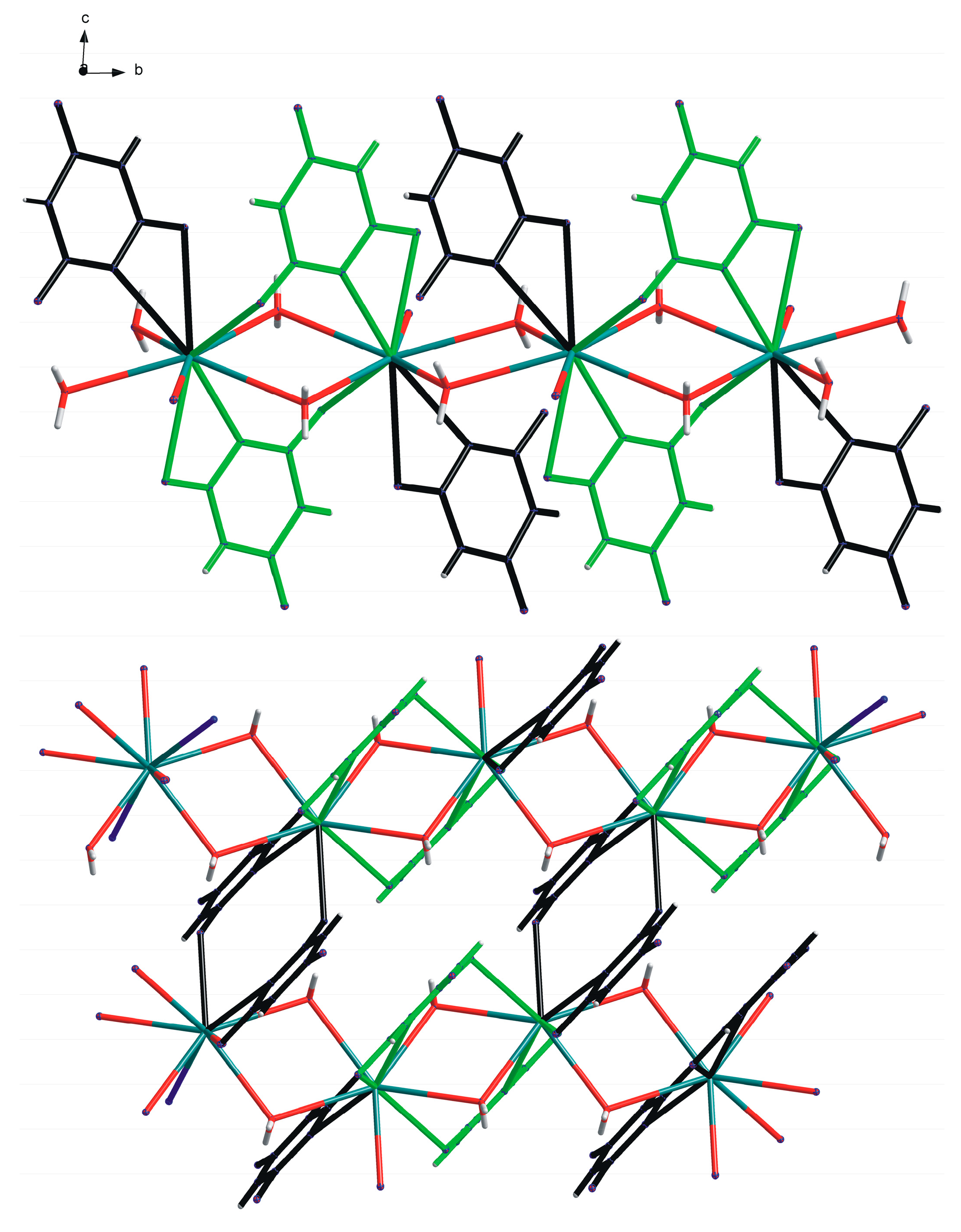
In the structure of compound
3, both CY
− anions and all the water molecules are coordinated to Sr. All aqua ligands are terminal. Each cyanurate ligand coordinates through two of its three O atoms to different metal centres and so serves as a simple bridging ligand; in contrast to compound
2, all N atoms remain uncoordinated. Strontium has coordination number 8, continuing the increase from Mg (6) through Ca (7). All four aqua ligands lie on one side of Sr, with the four bridging CY
− ligands all on the other side. Pairs of inversion-related ligands link the metal ions together in a polymeric chain parallel to the
a axis. A section of this chain is shown in
Figure 6, with the non-H atoms of the asymmetric unit labelled, and the coordination geometry is given in
Table 5.
Successive pairs of bridging ligands along the polymer chain are separated by approximately 3.3 Å and overlap each other, indicating a degree of π-π stacking within the chain.
The structural role of H atoms and hydrogen bonding in compound
3 requires the recognition of some minor disorder in the crystal structure. The cyanurate ligands with atom numbers 4–6 are fully ordered, along with water molecules 8 and 10 and the strontium ions; N5 is deprotonated, while H atoms are retained on N4 and N6. The close approach of two cyanurate ligands with atom numbers 1–3 across an inversion centre includes an N3...N3a distance of only 2.967(8) Å. These two N atoms, which are symmetry-equivalent, cannot both carry an H atom, nor can they both be deprotonated; this separation is appropriate for an N–H...N hydrogen bond, which must be disordered across the inversion centre. In this ligand, N1 clearly retains its H atom, forming an N–H...O hydrogen bond to a carbonyl O atom, so there must be a 50% disordered H atom bonded also to N2. In turn, this leads to the disorder of two orientations of the aqua ligands O9 and O7, the disorder of these ligands being correlated. Evidence of this pattern of disorder comes from the presence and relative sizes of difference electron density peaks close to N3, N2, O7 (3 peaks), and O9 (3 peaks) when H atoms are not included in the structural model as well as from the sensible geometry of the assigned disordered hydrogen bonds. We believe this disorder has been overlooked in the two previous reports of the structure of compound
3 [
28,
29]. One [
29] has a full-occupancy H atom attached to N3 (our numbering), which leads to an H...H distance of only 1.46 Å—this would be even shorter, but the refinement with a restrained N–H distance of 0.99 Å takes the H atom markedly out of the ligand plane, which is not to be expected. The other [
28] has this N atom deprotonated despite the N...N distance of 2.973 Å, while N2 (our numbering) has a full-occupancy H atom; the H atoms in this reported structure were refined freely with no constraints or restraints, and we note that H2 and the aqua H atoms we have identified as disordered have the highest isotropic displacement parameters, consistent with overlooked disorder, while the largest difference electron density peaks reported in the files deposited in the CSD include positions corresponding to omitted disordered H atoms on N3 and aqua ligands.
The geometry of hydrogen bonding in structure 3 is given in
Table 6. This includes alternative arrangements for hydrogen bonding involving N2, O7, and O9.
All hydrogen bonds involving the CY
− ligand with atom numbers 4–6 (including N–H...O hydrogen bonds linking symmetry-inequivalent ligands in neighbouring polymer chains) and aqua ligands O8 and O10 are ordered. Those involving N2, N3, and the aqua ligands O7 and O9, indicated with * in
Table 6, are subject to correlated disorder. Coplanar pairs of cyanurate ligands 4–6 within the same polymer chain are connected by hydrogen bonds in an
R22(8) motif, and each of these ligands forms pairs of N–H...O hydrogen bonds with a cyanurate ligand 1–3 in an adjacent polymer chain to give the same motif. The ribbons of hydrogen-bonded cyanurate anions are finite, with only four members in this structure, however, unlike the polymeric ribbons in the structures of compounds
1 and
2. The hydrogen bonding is shown in
Figure 7; this presents only one component of the disordered pattern and has all H3 positions occupied, so the N–H...N hydrogen bond is also not shown. Hydrogen bonds donated by aqua ligands provide the three-dimensional connectivity of the structure.
3.4. Compound 4, {[Ba(CY)2(H2O)2]}∞
Previous structural reports of barium complexes of cyanuric acid include two containing triply deprotonated anions [
51,
52] and one with both singly and doubly deprotonated anions [
29]. The structure of compound
4 has been reported three times: for one of these [
53], only limited information is available through the CSD; another [
30] gives extensive details and is fully consistent with our own results; the third [
27] incorrectly describes barium as being eight-coordinate with no mention of the two Ba–N bonds involved in the chelate character of the cyanurate ligands.
As in compound
3, all potential ligands are coordinated to the metal centre in compound
4, with no counterions or uncoordinated water molecules. The trend already seen in the sequence Mg, Ca, Sr of cyanurate displacing water from the coordination sphere is continued with Ba, which is ten-coordinate. Four of the bonds from Ba are to water molecules, all of which function as bridges; four are to carbonyl O atoms; and two are to N atoms, the CY
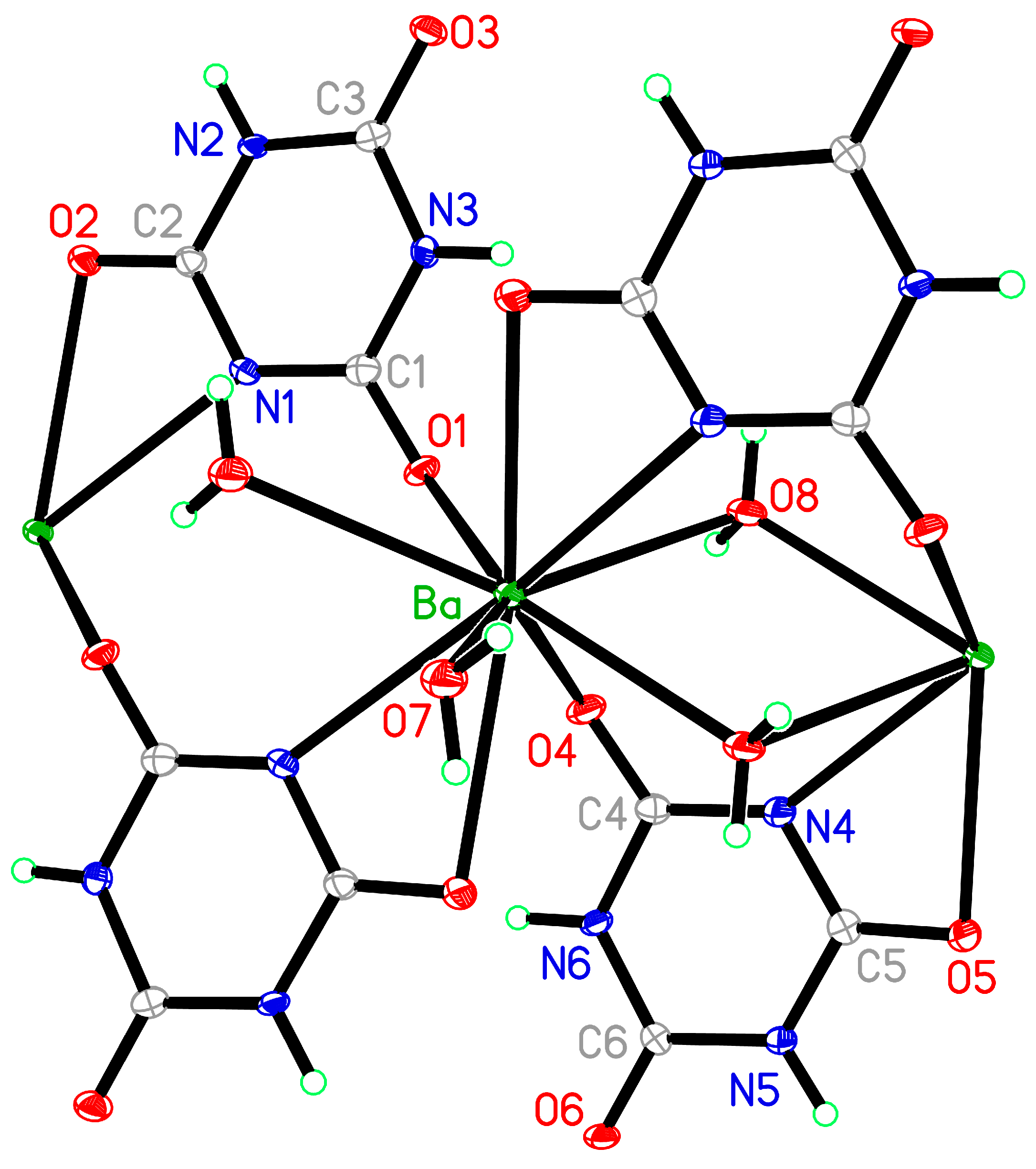
− anions each coordinating to two metal centres, one in monodentate fashion through O and the other in bidentate mode through N and O. The coordination geometry of Ba is given in
Table 7 and illustrated in
Figure 8.
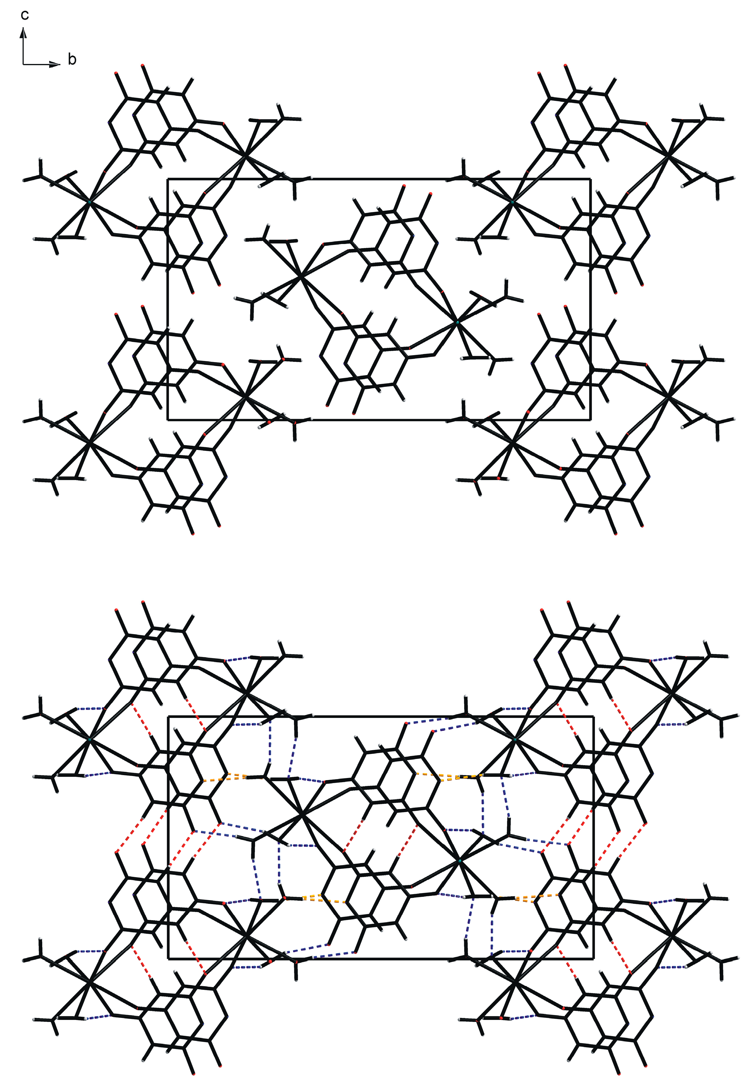
Coordination in this structure generates a sheet network (2D), unlike the chain polymers (1D) of compounds
2 and
3. The barium ions and water molecules together generate a polymeric chain of corner-connected centrosymmetric Ba
2O
2 parallelograms running parallel to the
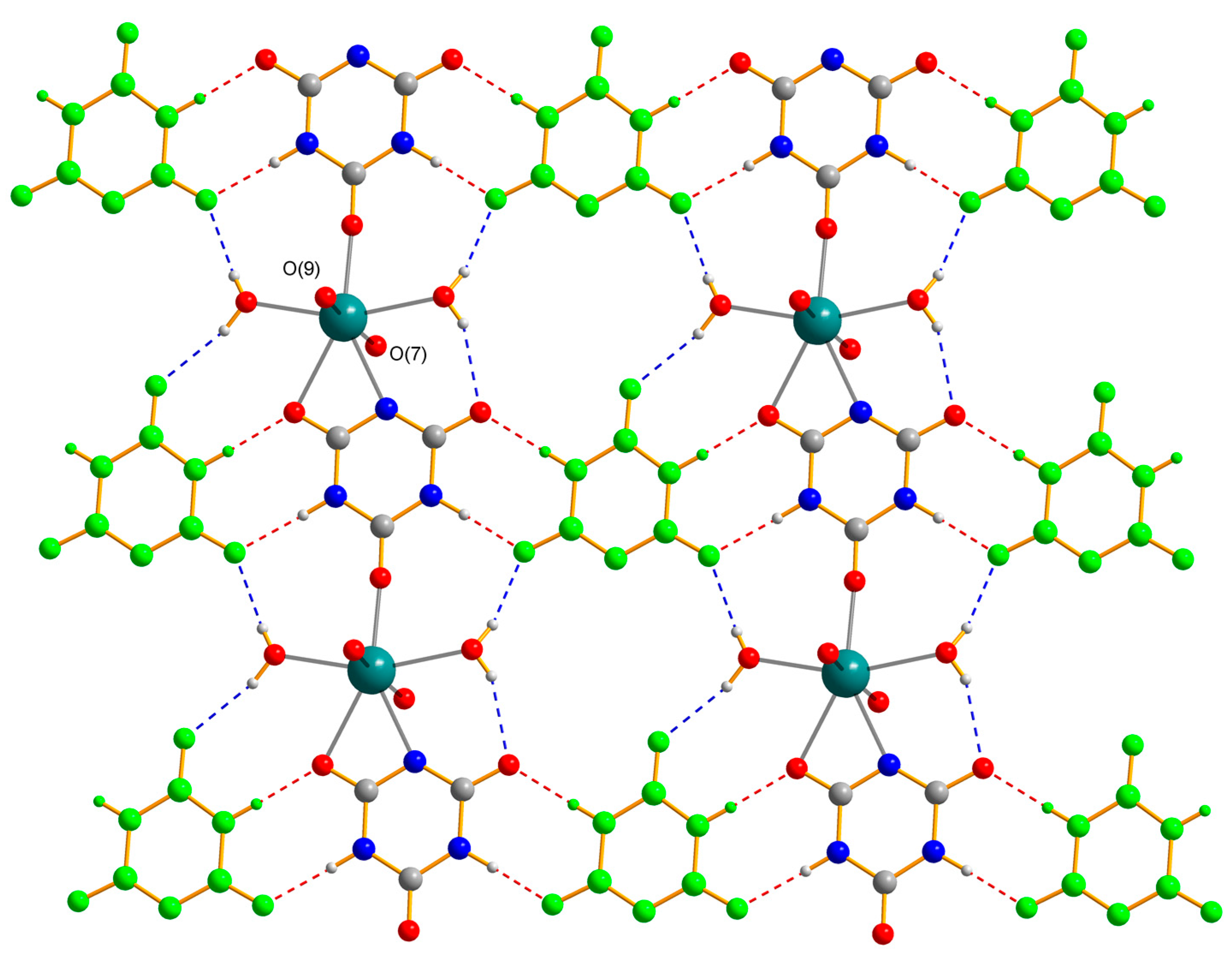
b axis. Alternate four-membered rings of this chain are further spanned by two inversion-related cyanurate anions (with atom numbers 4–6); each anion chelates one Ba centre through a carbonyl O atom and the deprotonated N atom and coordinates the other Ba centre through the carbonyl O atom adjacent to the coordinating N atom, leaving the carbonyl between the two NH groups uncoordinated. The other cyanurate anion (atom numbers 1–3) forms a chelate ring with Ba on the opposite side to that formed by the first anion, but its other carbonyl group adjacent to the deprotonated N atom bonds to a Ba atom in the next chain, so centrosymmetric pairs of these anions link chains together into a sheet structure. The overall arrangement is shown in
Figure 9.
All H atoms in this structure were refined with unconstrained individual isotropic displacement parameters. Distance restraints were applied to N–H bonds [0.88(1) Å] and O–H bonds [0.84(1) Å] and to water H...H distances [1.35(2) Å], corresponding to a restraint on the H–O–H angles of about 107°. Hydrogen bonding uses all N–H and O–H bonds. Aqua ligands donate to uncoordinated carbonyl O atoms and to those that are not part of chelate rings. N–H bonds donate to uncoordinated and chelating O atoms. Thus, each coordinated carbonyl O atom receives one hydrogen bond, and each uncoordinated carbonyl O atom receives two. The now-familiar hydrogen-bonded ribbons of
R22(8) motifs formed by cyanurate anions occur once again, in polymeric form, as in compounds
1 and
2, rather than as the short finite ribbons found in compound
3. These hydrogen-bonded ribbons are shown in
Figure 10, with geometrical details of all hydrogen bonds in
Table 8.
4. Conclusions
Our systematic survey of alkaline earth metal complexes of the singly deprotonated anion of cyanuric acid reveals distinct trends in the interplay of metal–ligand coordination and hydrogen bonding in the construction of the crystal structures. As the group is descended and ionic radii increase, higher coordination numbers are expected and, indeed, are found, but there is also a clear pattern in the balance of coordination by CY− and water. Listing the number of bonds from the metal centre to cyanurate, then to water, and the total coordination number, we find: Mg, 0 + 6 = 6; Ca, 3 + 4 = 7; Sr, 4 + 4 = 8; Ba, 6 + 4 = 10. Thus, the increase in coordination number is essentially taken up by an increasing involvement of CY− in the coordination, with the number of water molecules remaining constant after the initial drop from Mg to Ca. Within these simple numbers are hidden more subtle trends, including an overall but oscillating shift in balance from terminal to bridging aqua ligands (the number of bridging ligands along the series is 0, 2, 0, 4) and a reduction in the number of uncoordinated CY− ligands in what is a constant 1:2 stoichiometric ratio of metal to cyanurate (2, 1, 0, 0). The two complexes (Ca and Ba) that feature chelating cyanurate are also the two containing bridging aqua ligands, though this may be a coincidental observation rather than a consequential structural feature.
Hydrogen bonding is a significant feature in all four complexes. This is hardly surprising, given that CY− offers two potential donors in the N–H bonds and four potential acceptors in the deprotonated N atom and the three carbonyl groups, and water can serve as both the donor and the acceptor of up to two hydrogen bonds in each way. Acceptor sites are also the potential coordinating atoms, so the two functions are in competition, though these structures contain a few examples of O atoms (but not deprotonated N) serving simultaneously as hydrogen-bond acceptors and coordinating atoms. In the case of the magnesium complex 1, there is no coordination of Mg by CY− and the extensive hydrogen bonding forms a three-dimensional network. For the calcium complex 2, CY− generates a polymeric chain, with bridging aqua ligands connecting these into a double-chain polymer extending in one dimension, and hydrogen bonding provides the linkages in the other two dimensions. For the strontium complex 3, a one-dimensional polymer is generated exclusively by bridging CY− ligands, with all aqua ligands terminal. Once again, hydrogen bonding generates the connectivity in the other two dimensions. CY− bridging coordination in the barium complex 4 generates a sheet polymer that also incorporates the bridging aqua ligands, and the connection between adjacent sheets is provided by the hydrogen bonding. Thus, the dimensionality of coordinative bonding increases in the series: Mg 0, Ca 1, Sr 1, Ba 2.
We note that a similar structural survey has been conducted for alkaline earth metal complexes of the empirical formula M(TMT)
2(H
2O)
n containing the sulphur analogue of cyanuric acid, 2,4,6-trimercapto-1,3,5-triazine, abbreviated here as TMTH, with singly deprotonated anion TMT
− [
43,
54]. The softer sulphur donor sites have less affinity for the hard group 2 metal centres and do not coordinate at all to Mg, Ca, or Sr cations, which have 6, 8, and 8 terminal aqua cations, respectively. Ba is coordinated by two TMT
− anions (one chelating and one monodentate, giving one Ba–N and two Ba–S bonds) and seven water molecules in a total coordination number of 10, compared with the two Ba–N, four Ba–O(CY), and four Ba–OH
2 bonds (total 10) in compound
4. As in our study, hydrogen bonding is extensive in these structures.
The competition between cyanurate and water for coordination to the alkaline earth metal cations and that between the coordination and hydrogen bonding propensities of the cyanurate anion appear to be finely balanced, with the structural outcomes depending on the relative sizes and degrees of hardness of the four cations of the group. The extent of CY− coordination increases relative to the coordination of water as the cationic radius increases. There is also an increase in the dimensionality, from 0 through 1 to 2, of the coordination network of the structures. Extensive hydrogen bonding is an important feature of all the structures.
Potential extensions of this work are to complexes of cyanuric acid with two or more alkaline earth metals, to explore competitive or cooperative behaviour, and—given the relative paucity of relevant structures noted in the Introduction—to further complexes of transition metals. There are also numerous small-ring compounds with a similar chemical structure to that of cyanuric acid.


