Article

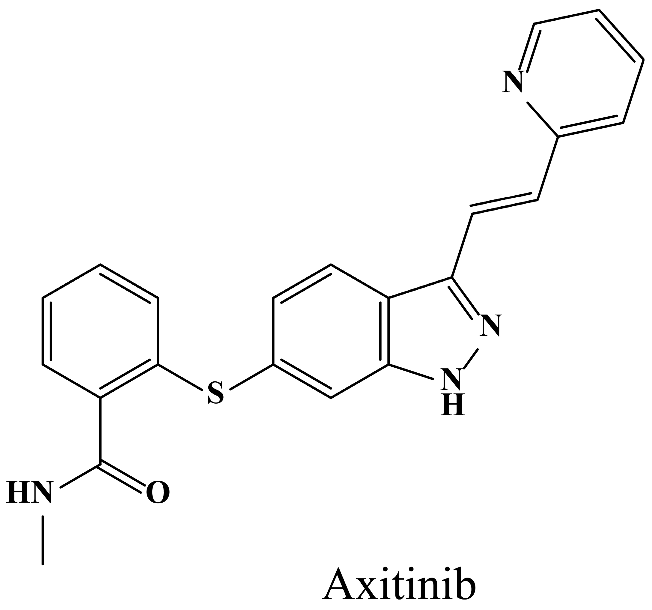
Phase Separation Investigation of Axitinib in Supersaturated Solution

1
Institute of Biopharmaceutical Research, Liaocheng University, Hunan Road, Liaocheng 252059, China
2
School of Geography and Environment, Liaocheng University, Hunan Road, Liaocheng 252059, China
*
Author to whom correspondence should be addressed.
Crystals 2024, 14(12), 1042; https://doi.org/10.3390/cryst14121042
Submission received: 19 November 2024 / Revised: 26 November 2024 / Accepted: 27 November 2024 / Published: 30 November 2024

Abstract

:
Phase separation is quite common in formulations for hydrophobic active pharmaceutical ingredients (APIs) due to their thermodynamic instability in a supersaturated state during in vitro dissolution or in vivo absorption. Phase separation possibly accompanies the formation of a disordered drug-rich phase, but this is still not thoroughly understood. In this study, the phase separation of supersaturated axitinib (Axi) in media with or without polymers was evaluated via multiple analytical methods, including UV–vis and fluorescence spectroscopy, dynamic light scattering, and microscopy. The phase separation of Axi occurred at an Axi concentration of 25–30 µg/mL in the media, while the addition of quantitative hypromellose acetate succinate (HPMCAS) MG and povidone (PVP) K30 did not alter its phase separation concentration. The second scattering dispersion phase of the system exhibited superior stability and reversibility as the formative filamentous crystalline condensates could disintegrate upon dilution. These disparate analyses consistently detected the phase separation of Axi. This manuscript could provide a better understanding of the supersaturation state of hydrophobic APIs upon pharmaceutical application.

Graphical Abstract

1. Introduction

The development of multiple new potential drug candidates (>90%) has been limited by their narrow aqueous solubility and poor oral bioavailability in recent years [1]. To assess the solubility and permeability of new drugs, the biopharmaceutical classification system (BCS) is proposed [2]. Typically, BCS class II drugs, which represent most of the previously approved active pharmaceutical ingredients (APIs), have poor solubility and high permeability [3]. As a result, numerous formulation approaches have been applied in order to enhance their solubility and bioavailability, including co-crystals [1], salt formations [4], cyclodextrin inclusion complexes [5], nano-drug delivery systems [6], emulsion/microemulsion/self-emulsifying systems [7], and amorphous solid dispersions (ASDs) [8]. The crystal form of the hydrophobic APIs in most formulations is transformed from a crystalline state to an amorphous state, at which the solubility of the APIs is enhanced significantly due to the reduced energy barrier to lattice disruption [9]. However, phase separation is quite common in these formulations as well because of the thermodynamically unstable supersaturated state of the APIs in aqueous media [10].
Hydrophobic APIs in a modest supersaturated state could incur phase or liquid–liquid phase separation (LLPS), forming a disordered drug-rich phase, while increased supersaturation ultimately results in undesirable recrystallization [11]. Thus, maintaining a stable supersaturated state for as long as possible can be achieved with the assistance of different excipients and pharmaceutical technologies [12]. Anura S. Indulka [13] investigated the effect of sodium dodecyl sulfate (SDS) on the nucleation induction time (NIT) of atazanavir (ATZ) and found that SDS at a certain minimum concentration could promote the crystallization of ATZ due to the formation of SDS hemimicelles around the crystal surface of the API. The crystallization promotion effect of SDS on ATZ could also influence the inhibitory effect of specific polymers such as polyvinylpyrrolidone/vinyl acetate (PVPVA). Bin Tian [9] analyzed the crystallization inhibitory effect of different natural polysaccharides (NPs) on curcumin and found the nucleation inhibitory effect of xanthan gum (XTG) was better than konjac glucomannan (KGM) and sodium alginate (SA).
The typical analytical methods [14] for evaluating the phase separation of hydrophobic APIs include UV–vis spectroscopy [15], fluorescence spectroscopy [16], dynamic light scattering [17], NMR spectroscopy [18], and microscopy [19]. UV–vis spectroscopy could detect both absorbance and light scattering, and the sudden change in the latter data often indicates the formation of phase separation [20]. In addition, the light scattering intensity fluctuation of particles could be measured by dynamic light scattering as well. Hydrophobic fluorescent probes preferentially interact with drug-rich phases in aqueous solutions due to similarity–intermiscibility theory, and their fluorescence spectroscopy varieties could be detected after drug crystallization and phase separation [16]. Moreover, NMR spectroscopy is utilized to assess the composition of different phases, while microscopy obtains clear internal images of them.
Axitinib (Axi, Figure 1) is a BCS class II drug with poor equilibrium solubility in water (approximately 0.4 µg/mL at pH 7) but high-level membrane permeability [21], possessing a pKa of 4.3, which promotes its solubility in acid solutions (rough 280 µg/mL at pH 1) [22]. Axi is currently approved for treating renal cell carcinoma and could be utilized as a single therapy or in combination with other agents for melanoma and lung cancer [23]. It has also been developed for topical application to treat ailments, such as anterior segment diseases or retinopathy [24]. The oral bioavailability of Axi has been elevated via multiple formulations, including cyclodextrin solubilization [25], micelles [26], hydrogels [27], nanoparticles [24], co-crystals [28], etc.
In this study, Axi was selected as the model drug in order to evaluate the application of different analytical methods to the investigation of the phase separation phenomenon of hydrophobic drugs in a supersaturated state. Dynamic light scattering (DLS) and UV-vis spectroscopy were utilized to analyze the crystal growth of Axi as a function of drug concentrations, which was further verified using an optical microscope. Pyrene was used as a fluorescence probe for detecting the phase separation character of the drug-rich solution. Typical polymers, including hypromellose acetate succinate (HPMCAS) MG and povidone (PVP) K30, were also selected to compare their influence on the phase separation of Axi. This study could provide a better understanding of the supersaturation of hydrophobic APIs in formulation processing.

2. Materials and Methods

2.1. Materials

Axitinib (Axi, ≥99%) and pyrene (≥99%) were obtained from Shanghai Aladdin Biochemical Technology Co., Ltd. (Shanghai, China). Hypromellose acetate succinate (HPMCAS) MG was provided by Ashland (Ashland, KY, USA). Povidone (PVP) K30 was supplied by BASF (Ludwigshafen, Germany). All the other chemical reagents were of analytical grade without further purification.

2.2. Determination of the Phase Separation Concentration of Axitinib

The phase separation concentration of Axi was determined when the drug-rich amorphous/crystal phase formed in the solution, which could be characterized by dynamic light scattering intensity, UV-vis extinction, or studying the microstructure. Briefly, Axi was solubilized in methanol in order to obtain a concentrated Axi methanol solution at 0.5 mg/mL. Different volumes of the Axi methanol solution were transferred into 50 mL of 10 mM sodium phosphate buffer saline solution (pH = 7.4) to formulate supersaturated Axi solutions at final concentrations of 5, 10, 15, 20, 25, 30, 35, 40, 45, and 50 µg/mL. The supersaturated Axi solutions were persistently stirred at a speed of 300 rpm and a temperature of 37 °C for 5 min. Then, 1 mL of the supersaturated Axi solution was withdrawn, and dynamic light scattering (DLS) was applied to monitor the mean count rate of the solution using a Malvern Nano ZSP instrument (Malvern, UK). The analysis was performed in the 173° backscattering mode at 25 °C with a standard 633 nm He-Ne laser (10 mV). In addition, 2.5 mL of the supersaturated Axi solution was also collected, and UV-vis spectroscopy (U-3900, Hitachi, Tokyo, Japan) was simultaneously utilized to detect the extinction of the sample at a nonabsorbing wavelength of 400 nm (different from the maximum ultraviolet absorbing wavelength of Axi at 330 nm) to evaluate the turbidity of the supersaturated Axi solutions, which is an index for verifying the presence of the second scattering dispersed phase. Each sample was measured three times, and DLS measurement was performed with 12 runs. Furthermore, 30 μL of the above supersaturated Axi solution was dropped on a slide and observed under a Nikon Eclipse Ti2-U inverted microscope (Nikon Ltd., Tokyo, Japan) as well.

2.3. Detection of the Phase Separation Property of Axitinib

UV-vis spectroscopy (U-3900, Hitachi, Tokyo, Japan) and the Malvern Nano ZSP instruments (Malvern, UK) were further applied to detect the phase separation properties of Axi. Briefly, a concentrated Axi methanol solution at 0.5 mg/mL was prepared by solubilizing quantitive Axi in methanol. Firstly, the mean count rate and UV-vis extinction of 50 mL of phosphate buffer (10 mM, pH = 7.4) were recorded, respectively, for the initial 30 min. Secondly, 0.5 mg/mL of concentrated Axi methanol solution in a quantitive volume was transferred into the phosphate buffer (10 mM, pH = 7.4) to formulate Axi solutions at a final concentration of 40 µg/mL. The detection continued for another 30 min or 50 min, after which 50 mL of phosphate buffer (10 mM, pH = 7.4) was added into the solution to dilute Axi to 20 µg/mL. The investigation lasted for another 20 min. Then, 1 mL of the supersaturated Axi solution was utilized for DLS measurement, while 2.5 mL of the supersaturated Axi solution was applied for UV-vis analysis. The assay was performed in triplicates. In the process, a Nikon Eclipse Ti2-U inverted microscope (Nikon Ltd., Tokyo, Japan) was also applied for observation.

2.4. Detection of the Phase Separation Process of Axitinib

First, 0.5 mg/mL concentrated Axi in methanol was prepared and transferred into 50 mL of phosphate buffer (10 mM, pH = 7.4) to formulate Axi solutions at a final concentration of 40 µg/mL. The solution was persistently stirred at a speed of 300 rpm and a temperature of 37 °C. Then, 1 mL of the supersaturated Axi solution was sampled at a predetermined time point. Dynamic light scattering (DLS) was applied to monitor the mean count rate of the samples using a Malvern Nano ZSP instrument (Malvern, UK). Each DLS measurement was performed three times with 12 runs. Moreover, 30 μL of the supersaturated Axi solution was dropped on a slide and observed under a Nikon Eclipse Ti2-U inverted microscope (Nikon Ltd., Tokyo, Japan).
Pyrene, which is a frequently used hydrophobic fluorescent probe, was utilized to investigate the phase separation of Axi. Briefly, 0.5 mg/mL concentrated Axi in methanol was prepared and transferred into 50 mL of phosphate buffer (10 mM, pH = 7.4) to formulate Axi solutions at a final concentration of 40 µg/mL. Then, 2 μM pyrene was added to the Axi solution. At each time point, 1 mL of the solution was withdrawn, and the fluorescent spectra of pyrene were analyzed using an F-4600 fluorescence spectrophotometer (Hitachi, Tokyo, Japan). The excitation wavelength was set at 332 nm and the emission spectra were collected from 350 to 450 nm at an interval of 0.2 nm with slits of 5 nm. The curve of the I1(373 nm)/I3(384 nm) ratio vs. time was plotted. The assay was repeated three times.

2.5. Detection of the Phase Separation Process of Axitinib in Presence of Different Polymers

Hypromellose acetate succinate (HPMCAS) MG and povidone (PVP) K30 were pre-dissolved in 50 mL of phosphate buffer (10 mM, pH = 7.4) at a final concentration of 0.5 mg/mL. A concentrated Axi methanol solution at 0.5 mg/mL was prepared and discontinuously transferred into 50 mL of phosphate buffer (10 mM, pH = 7.4) containing or not containing polymers to formulate Axi solutions at final concentrations of 5, 10, 15, 20, 25, 30, 35, 40, 45, and 50 µg/mL. The solutions were persistently stirred at a speed of 300 rpm and a temperature of 37 °C. Then, 2.5 mL of the supersaturated Axi solution was withdrawn, and UV-vis spectroscopy (U-3900, Hitachi, Tokyo, Japan) was utilized to detect the extinction of the solution at a nonabsorbing wavelength at 400 nm, which was then plotted against the concentration of Axi. The experiments were repeated three times.

3. Results and Discussion

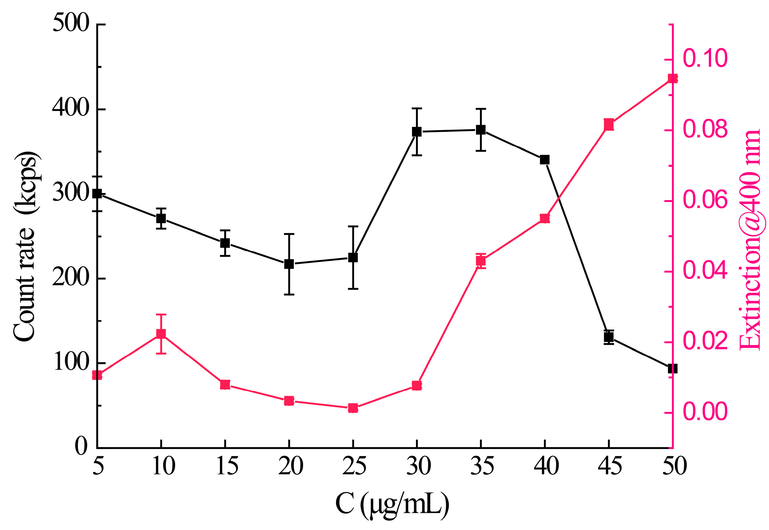
3.1. The Phase Separation Concentration of Axitinib

The mean count rate (kcps) acquired via DLS measurements and UV-vis extinction (at a wavelength of 400 nm) obtained by UV-vis spectroscopy was plotted as a function of Axi concentrations in phosphate buffer (10 mM, pH = 7.4). For homogeneous solutions without scattering centers, the UV-vis extinction at a nonabsorbing wavelength should be negligible, but the UV-vis extinction would be enhanced significantly with the formation of a second phase that scatters light [29]. Thus, the UV-vis extinction would support the formation of the second phase consisting of drug-rich particles. As shown in Figure 2, the UV-vis extinction of the system started to evolve at Axi concentrations of 25 µg/mL and increased dramatically from an Axi concentration of 30 µg/mL, followed by sustainable growth. This phenomenon indicated that a second scattering dispersed phase emerged at an Axi concentration of 25–30 µg/mL; in other words, the phase separation concentration of Axi in phosphate buffer (10 mM, pH = 7.4) was calculated as 25–30 µg/mL, which was much higher than its intrinsic solubility of around 0.37 μg/mL [30].
When light strikes particles smaller than its wavelength, the light scatters in all directions around it. Since the laser is monochromatic and phase-coherent, time-dependent scattering intensity fluctuations can be observed by using the laser as the light source. This fluctuation of scattering intensity is mainly due to the change in relative position with time caused by the Brownian motion of particles in solution. Large particles move slowly, and the intensity of scattered light fluctuates slowly, while small particles do the opposite [31]. Thus, the mean count rate (kcps) values of the system are recorded as an index of the turbidity of the solution, which is derived from the Mie scattering and is indicative of the presence of a second scattering dispersed phase. The mean count rate for the Axi system changed at an Axi concentration of 25 µg/mL (Figure 2), which indicated phase separation. However, the curve exhibited a significant decline at the Axi concentration of 40 µg/mL, which might be attributed to the crystallization and precipitation of the API.
Furthermore, a microscope was intuitively used to monitor the phase separation morphology of Axi. As shown in Figure 3, the second scattering dispersed phase emerged at an Axi concentration of 30 µg/mL (consistent with the optical photographs shown in Figure S3), with clearly visible minor crystal particles, leading to the mean count rate changing and the UV-vis extinction variation of the system. However, at an Axi concentration of 50 µg/mL, the minor crystal particles grew into major aggregates, which contributed to the decline in the mean count rate. The system displayed a satisfactorily consistent mean count rate and UV-vis extinction variation tendency.
Several hydrophobic APIs, such as ritonavir [29], could form liquid–liquid phase separation (LLPS) drug aggregates with typical circular droplets (drug–water binary aggregates) in a supersaturated state on their own. In most other cases, LLPS occurs during the dissolution of amorphous solid dispersions (ASDs) for supersaturation state maintenance, which produces drug–polymer–water ternary aggregates, and significantly elevates the bioavailability of the APIs [32]. The Axi bulk powder also underwent LLPS (Figure S3), but immediately formed minor crystal aggregates above its equilibrium solubility in the media (Figure 3).

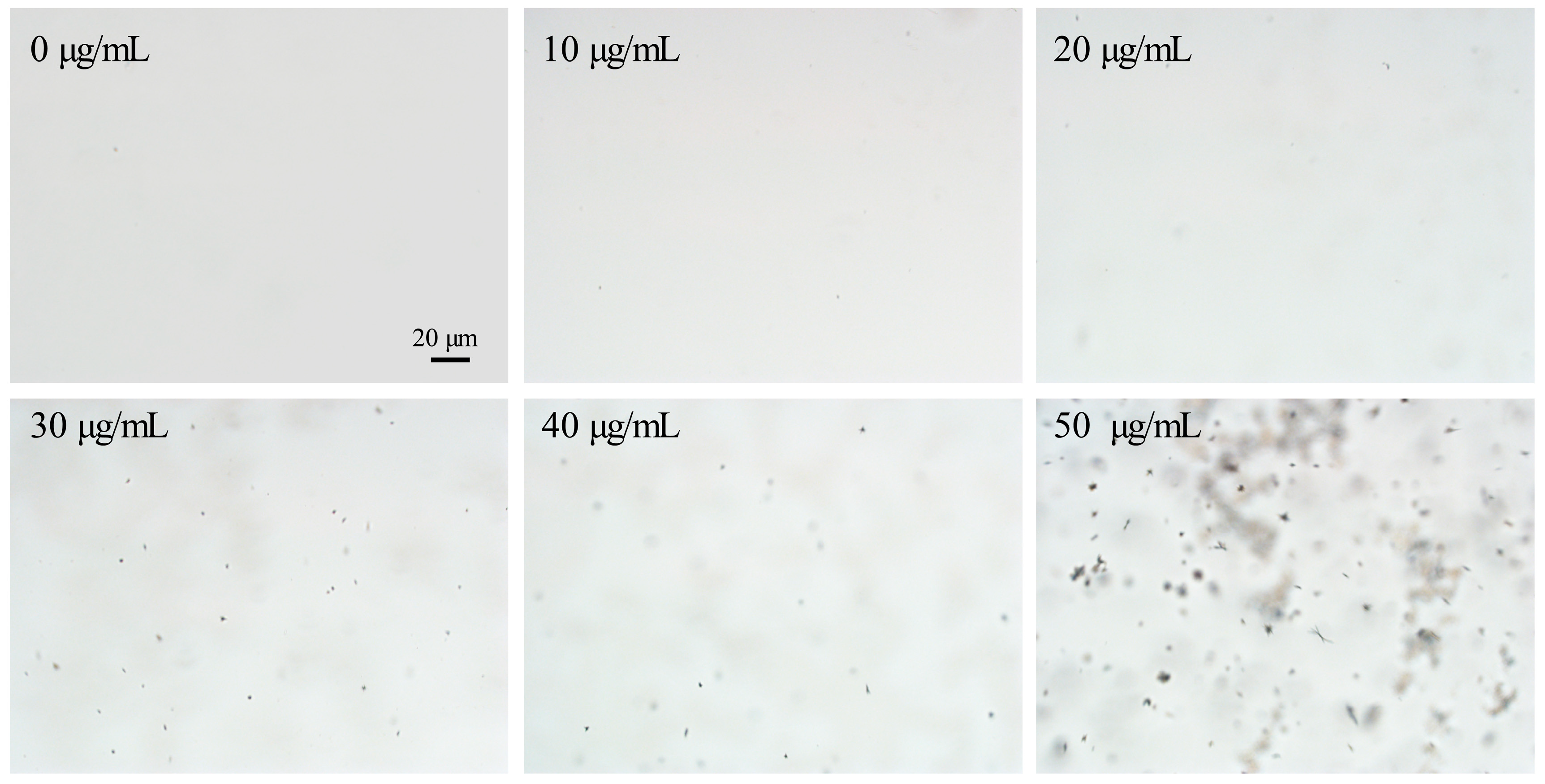
3.2. The Phase Separation Property of Axitinib

To characterize the phase separation properties of Axi, dynamic light scattering (DLS) and UV-vis spectroscopy were utilized to determine the mean count rate and UV-vis extinction of the solutions containing Axi at different concentrations, respectively. As shown in Figure 4A, the homogeneous solution without Axi (Axi concentration at 0 μg/mL) exhibited minimal UV-vis extinction in the initial 30 min, as there were no scattering centers in the media. When 40 μg/mL Axi was added to the media, the UV-vis extinction of the system was enhanced significantly. The highest point emerged at 35 min, with a 5 min lag due to minor crystal aggregate formation and growth, whose value was 3.8 times higher than the background signal. The UV-vis extinction of the system then gradually decreased over time from 40 to 60 min, indicating the formation and development of a second scattering dispersion phase. After 60 min, the system was diluted with fresh phosphate buffer (10 mM, pH = 7.4), and the concentration of Axi was reduced to 20 μg/mL. Accordingly, the UV-vis extinction value fell to ~72% of that of the system containing 40 μg/mL Axi, indicating that the second scattering dispersion phase might disappear or subsequently reduce in intensity. The mean count rate curve also showed turning points at 40 and 60 min after Axi was added or diluted, respectively. However, the stability of the mean count rate values for the Axi system was relatively poor during the whole process. Furthermore, the experiment was repeated, except the maintenance time of the second scattering dispersion phase was prolonged from 30 min to 60 min. As shown in Figure 4B, the variation in the UV-vis extinction of the system was similar to that displayed in Figure 4A, but smoother and steadier, indicating the superior stability of the second colloidal dispersion phase for Axi, which could be sustained for 60 min or longer. The second scattering dispersion phase of Axi was reversible, which could disperse faster upon dilution to below the phase separation concentration. In contrast, the variation in the mean count rate of the system still fluctuated, but the turning point occurred in accordance with the UV-vis extinction of the system at 40 and 80 min.
A microscope was utilized to observe the whole process displayed in Figure 4A. As shown in Figure 5, when Axi was added into the media, the second scattering dispersed phase started to form at 30 min, as well as minor filamentous crystalline condensates which grew into major aggregates over time. The particle size of the aggregates varied from ~5 µm to ~50 µm. At 60 min, Axi was diluted to the concentration below its phase separation concentration, so the major filamentous crystalline aggregates spread out and left stable condensates with a particle size of 30–50 µm. The microscopical observation of the process was consistent with the mean count rate and UV-vis extinction records as well.

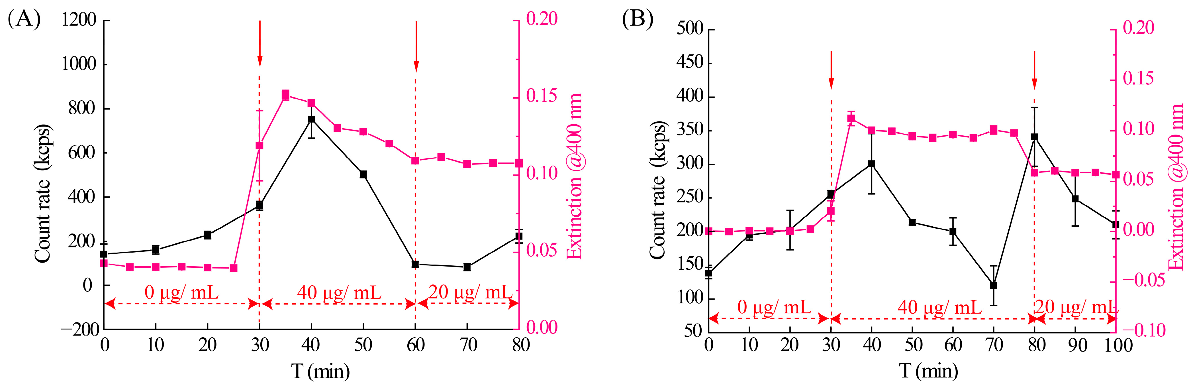
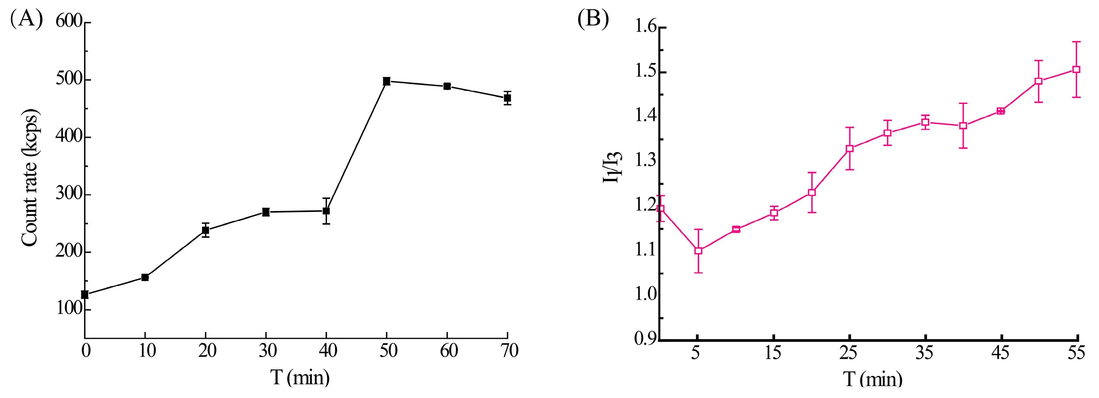
3.3. The Phase Separation Process of Axitinib

In order to further illustrate the phase separation process of Axi, the mean count rate of 10 mM phosphate buffer (pH = 7.4) containing 40 µg/mL Axi was monitored for 70 min (Figure 6A). The mean count rate of the Axi solution at this concentration was enhanced gradually over time, but the system tended to stabilize after 50 min. The observations verified this tendency as the minor crystal particles grew into major aggregates from 0 to 40 min (Figure 7). With the increase in the particle size, the amount of the crystal aggregates also increased, followed by dispersed into crystal particles of 30–50 µm. The amount of crystal aggregates also decreased after 50 min.
The hydrophobic fluorescent probe pyrene was commonly utilized as an index of the hydrophilicity variety of the local environments in aqueous media. Upon the formation of a second phase consisting of drug-rich particles, the hydrophobic probe molecules would be divided into the new phase, resulting in changed fluorescent emission spectra of the probe [33,34]. The ratio of the I1 emission peak at 373 nm and the I3 emission peak at 384 nm is relatively sensitive to the varying fluorescent emission characteristics of pyrene, which could be recorded as an index of the formation of a second phase in the supersaturated solution containing Axi (Figure 6B). The I1/I3 ratio of pyrene decreased in the first 5 min due to the formation of a hydrophobic drug-rich colloidal dispersion phase, which promoted its dissolution. However, the I1/I3 ratio of pyrene was enhanced significantly over time, indicating a more hydrophilic environment, which resulted from the crystallization of Axi that excluded pyrene [35].

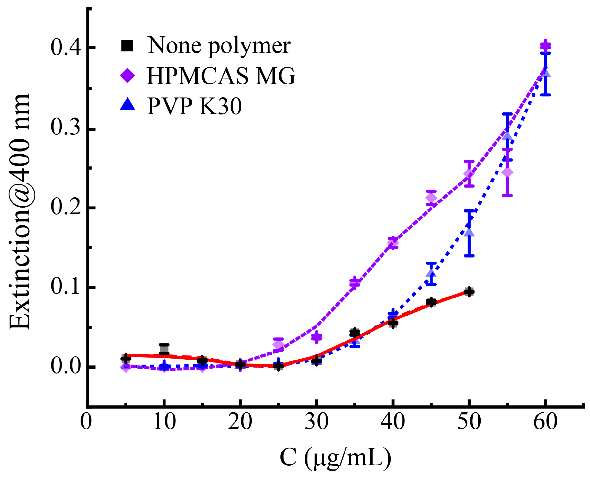
3.4. The Influence of Different Polymers on the Phase Separation Process of Axitinib

In order to evaluate the applicability of the above analytical methods in analyzing the phase separation in a binary system containing both the API and a polymer, hypromellose acetate succinate (HPMCAS) MG and povidone (PVP) K30 were utilized to explore their influence on the phase separation process of Axi. As illustrated in Figure 8, the phase separation of Axi bulk powder occurred at a drug concentration of 25–30 μg/mL when the UV-vis extinction curve changed. After the addition of PVP K30, the phase separation concentration did not undergo a transformation as the UV-vis extinction curve of the Axi-PVP K30 binary system coincided with Axi bulk powder, but a significant enhancement appeared at an Axi concentration larger than 40 μg/mL, indicating PVP K30 was ineffective for inhibiting drug recrystallization. In contrast to PVP K30, the addition of HPMCAS MG downgraded the phase separation concentration of Axi to 20–25 μg/mL, followed by a rapidly escalating trend against the Axi concentrations, which indicated the rapid crystallization of Axi.
Typically, the presence of effective recrystallization inhibitors in formulations for hydrophobic APIs is highly desirable to prolong drug supersaturation of the drugs, which could be characterized as a “spring-parachute” process due to the enhanced equilibrium solubility and maintained supersaturation state of the APIs [36]. Appropriate intermolecular interactions between polymers and drugs, including hydrogen bonding or electrostatic attraction, are vital for the effectiveness of a recrystallization inhibitor, as well as the viscosity property of a polymer which provides steric hindrance for inhibiting recrystallization [37]. Apparently, PVP K30 was an inefficient recrystallization inhibitor for Axi due to weak intermolecular interaction between them, while HPMCAS MG even induced nucleation at lower Axi concentrations. HPMCAS MG consists of hydrophobic acetyl functional groups and hydrophilic succinyl functional groups, which contributes to its amphipathy. In aqueous media containing supersaturated Axi, HPMCAS MG could form polymer hemimicelles around the crystal surface of Axi and decrease the interfacial energy between the crystal of the API and water, which finally promoted the nucleation of the drug. Similar report could be found for amphiphilic surfactant sodium dodecyl sulfate (SDS) [13]. SDS could induce the crystallization of atazanavir at concentrations below its critical micelle concentration (CMC), while at concentrations above the CMC, SDS did not influence its crystallization. At lower concentrations, monomers of SDS solubilized in the media, adsorbed on the nuclei surface of the drug, and formed hemimicelles, which promoted crystal growth due to reduced interfacial energy. However, when the CMC was exceeded, SDS micelles formed, and the crystallization of the drug did not emerge inside the constitutionally stable micelles. Thus, future studies on the suitable excipient type or excipient concentration utilized in the formulation of Axi should be conducted in order to fabricate effective preparations.

4. Conclusions

In this study, the phase separation of the hydrophobic API Axi was evaluated using different analytical methods including dynamic light scattering (DLS), UV-vis spectroscopy, fluorescence spectroscopy, and optical microscopy. The phase separation of Axi bulk powder occurred at a concentration of 25–30 µg/mL, at which clearly visible minor crystal aggregates were observed and quickly grew into major ones with an enhanced Axi concentration. At an appropriate Axi concentration, such as 40 µg/mL, the second scattering dispersion phase of the system exhibited superior stability and reversibility because the formative filamentous crystalline condensates could disintegrate upon dilution. The addition of PVP K30 did not affect the recrystallization of Axi in the media, while HPMCAS MG promoted the nucleation of Axi due to hemimicelle formation. This manuscript verified the consistency of disparate analysis strategies for detecting the phase separation of hydrophobic APIs and could promisingly provide a better understanding of the supersaturation of hydrophobic APIs in formulation processing.

Supplementary Materials

The following supporting information can be downloaded at https://www.mdpi.com/article/10.3390/cryst14121042/s1: Figure S1: The DSC curve (A) and XRD pattern (B) of axitinib; Figure S2. The UV-vis spectra of axitinib at concentrations of 5 µg/mL and 50 µg/mL; Figure S3. Pictures of 10 mM PBS (pH = 7.4) containing axitinib at different concentrations.

Author Contributions

Conceptualization, Y.Z.; data curation, J.S., H.Z. and Z.D.; formal analysis, J.S.; funding acquisition, N.Z. and Y.Z.; investigation, J.X.; methodology, J.X.; project administration, Y.Z.; supervision, N.Z.; validation, R.B.; visualization, H.Z.; writing—original draft, Y.Z. All authors have read and agreed to the published version of the manuscript.

Funding

This work was financially supported by the Natural Science Foundation of Shandong Province of China (No. ZR2022LSW018) and “Guangyue Young Scholar Innovation Team” of Liaocheng University (No. LCUGYTD2023-03).

Data Availability Statement

All data are included in this manuscript.

Acknowledgments

The above work was also technically supported by Shandong Collaborative Innovation Center for Antibody Drugs, Shandong Province Engineering Research Center for Nanomedicine and Drug Delivery Systems, and Shandong Province Engineering Laboratory of Anti-Viral Drugs.

Conflicts of Interest

The authors declare no competing financial interests.

References

  1. O’Sullivan, A.; Long, B.; Verma, V.; Ryan, K.M.; Padrela, L. Solid-state and particle size control of pharmaceutical cocrystals using atomization-based techniques. Int. J. Pharm. 2022, 621, 121798. [Google Scholar] [CrossRef] [PubMed]
  2. Amidon, G.L.; Lennernäs, H.; Shah, V.P.; Crison, J.R. A Theoretical Basis for a Biopharmaceutic Drug Classification: The Correlation of in Vitro Drug Product Dissolution and in Vivo Bioavailability. Pharm. Res. 1995, 12, 413–420. [Google Scholar] [CrossRef] [PubMed]
  3. Zhao, Y.; Xie, X.; Zhao, Y.; Gao, Y.; Cai, C.; Zhang, Q.; Ding, Z.; Fan, Z.; Zhang, H.; Liu, M.; et al. Effect of plasticizers on manufacturing ritonavir/copovidone solid dispersions via hot-melt extrusion: Preformulation, physicochemical characterization, and pharmacokinetics in rats. Eur. J. Pharm. Sci. 2019, 127, 60–70. [Google Scholar] [CrossRef] [PubMed]
  4. Hiew, T.N.; Taylor, L.S. Combining drug salt formation with amorphous solid dispersions—A double edged sword. J. Control. Release 2022, 352, 47–60. [Google Scholar] [CrossRef] [PubMed]
  5. Liu, X.; Niu, W.; Liu, J.; Cui, Z.; Li, J.; Zhang, Z.; Ju, J. Preparation of β-cyclodextrin and hydroxypropyl-β-cyclodextrin inclusion complexes of baicalein and evaluation of their effects on dextran sulfate sodium-induced acute ulcerative colitis in mice. J. Drug Deliv. Sci. Technol. 2023, 87, 104714. [Google Scholar] [CrossRef]
  6. Sultana, A.; Zare, M.; Thomas, V.; Kumar, T.S.S.; Ramakrishna, S. Nano-based drug delivery systems: Conventional drug delivery routes, recent developments and future prospects. Med. Drug Discov. 2022, 15, 100134. [Google Scholar] [CrossRef]
  7. Kim, J.S.; Din, F.u.; Cho, H.J.; Choi, Y.J.; Woo, M.R.; Cheon, S.; Ji, S.H.; Park, S.; Youn, Y.S.; Oh, K.T.; et al. Impact of carrier hydrophilicity on solid self nano-emulsifying drug delivery system and self nano-emulsifying granule system. Int. J. Pharm. 2023, 648, 123578. [Google Scholar] [CrossRef]
  8. Jiang, X.; Zhao, Y.; Guan, Q.; Xiao, S.; Dong, W.; Lian, S.; Zhang, H.; Liu, M.; Wang, Z.; Han, J. Amorphous solid dispersions of cyclosporine A with improved bioavailability prepared via hot melt extrusion: Formulation, physicochemical characterization, and in vivo evaluation. Eur. J. Pharm. Sci. 2022, 168, 106036. [Google Scholar] [CrossRef] [PubMed]
  9. Tian, B.; Li, L.; Kang, K.; Peng, D.; Shi, Y.; Wang, P. Crystallization inhibitory effects of konjac glucomannan, sodium alginate and xanthan gum on curcumin in supersaturated solution. Int. J. Biol. Macromol. 2023, 245, 125489. [Google Scholar] [CrossRef] [PubMed]
  10. Han, Y.R.; Ma, Y.; Lee, P.I. Impact of phase separation morphology on release mechanism of amorphous solid dispersions. Eur. J. Pharm. Sci. 2019, 136, 104955. [Google Scholar] [CrossRef] [PubMed]
  11. Kawakami, K.; Sato, K.; Fukushima, M.; Miyazaki, A.; Yamamura, Y.; Sakuma, S. Phase separation of supersaturated solution created from amorphous solid dispersions: Relevance to oral absorption. Eur. J. Pharm. Biopharm. 2018, 132, 146–156. [Google Scholar] [CrossRef] [PubMed]
  12. Tres, F.; Posada, M.M.; Hall, S.D.; Mohutsky, M.A.; Taylor, L.S. Mechanistic understanding of the phase behavior of supersaturated solutions of poorly water-soluble drugs. Int. J. Pharm. 2018, 543, 29–37. [Google Scholar] [CrossRef] [PubMed]
  13. Indulkar, A.S.; Gao, Y.; Raina, S.A.; Zhang, G.G.Z.; Taylor, L.S. Impact of Monomeric versus Micellar Surfactant and Surfactant–Polymer Interactions on Nucleation–Induction Times of Atazanavir from Supersaturated Solutions. Cryst. Growth Des. 2020, 20, 62–72. [Google Scholar] [CrossRef]
  14. Zhao, P.; Han, W.; Shu, Y.; Li, M.; Sun, Y.; Sui, X.; Liu, B.; Tian, B.; Liu, Y.; Fu, Q. Liquid–liquid phase separation drug aggregate: Merit for oral delivery of amorphous solid dispersions. J. Control. Release 2023, 353, 42–50. [Google Scholar] [CrossRef] [PubMed]
  15. Taylor, L.S.; Zhang, G.G.Z. Physical chemistry of supersaturated solutions and implications for oral absorption. Adv. Drug Deliv. Rev. 2016, 101, 122–142. [Google Scholar] [CrossRef] [PubMed]
  16. Almeida e Sousa, L.; Reutzel-Edens, S.M.; Stephenson, G.A.; Taylor, L.S. Assessment of the Amorphous “Solubility” of a Group of Diverse Drugs Using New Experimental and Theoretical Approaches. Mol. Pharm. 2015, 12, 484–495. [Google Scholar] [CrossRef]
  17. Alonzo, D.E.; Gao, Y.; Zhou, D.; Mo, H.; Zhang, G.G.Z.; Taylor, L.S. Dissolution and Precipitation Behavior of Amorphous Solid Dispersions. J. Pharm. Sci. 2011, 100, 3316–3331. [Google Scholar] [CrossRef] [PubMed]
  18. Liu, C.; Chen, Z.; Chen, Y.; Lu, J.; Li, Y.; Wang, S.; Wu, G.; Qian, F. Improving Oral Bioavailability of Sorafenib by Optimizing the “Spring” and “Parachute” Based on Molecular Interaction Mechanisms. Mol. Pharm. 2016, 13, 599–608. [Google Scholar] [CrossRef] [PubMed]
  19. Zhang, S.-W.; Yu, L.; Huang, J.; Hussain, M.A.; Derdour, L.; Qian, F.; de Villiers, M.M. A Method to Evaluate the Effect of Contact with Excipients on the Surface Crystallization of Amorphous Drugs. AAPS PharmSciTech 2014, 15, 1516–1526. [Google Scholar] [CrossRef] [PubMed]
  20. Li, W.; Song, J.; Li, J.; Li, M.; Tian, B.; He, Z.; Liu, X.; Fu, Q. Co-amorphization of atorvastatin by lisinopril as a co-former for solubility improvement. Int. J. Pharm. 2021, 607, 120971. [Google Scholar] [CrossRef]
  21. Benet, L.Z. Predicting drug disposition via application of a Biopharmaceutics Drug Disposition Classification System. Basic Clin. Pharmacol. Toxicol. 2010, 106, 162–167. [Google Scholar] [CrossRef]
  22. Dodd, S.; Kollipara, S.; Sanchez-Felix, M.; Kim, H.; Meng, Q.; Beato, S.; Heimbach, T. Prediction of ARA/PPI Drug-Drug Interactions at the Drug Discovery and Development Interface. J. Pharm. Sci. 2019, 108, 87–101. [Google Scholar] [CrossRef]
  23. Dilli Babu, A.; Singh, S.; Thota, A.; Duhan, S.; Sainatham, C.; Gill, H.; Raghavakurup, L.; Tantry, U.; Bliden, K.; Gurbel, P. Systemic hypertension with VEGFR inhibitor (axitinib) therapy in cancer patients: A meta-analysis and systematic review of randomized controlled trials. IJC Heart Vasc. 2024, 52, 101415. [Google Scholar] [CrossRef] [PubMed]
  24. Huang, Y.; Zhu, Y.; Cai, D.; Guo, Q.; Wang, J.; Lei, L.; Li, X.; Shi, S. Penetrating-peptide-mediated non-invasive Axitinib delivery for anti-neovascularisation. J. Control. Release 2022, 347, 449–459. [Google Scholar] [CrossRef] [PubMed]
  25. Jansook, P.; Loftsson, T.; Stefánsson, E. Drug-like properties of tyrosine kinase inhibitors in ophthalmology: Formulation and topical availability. Int. J. Pharm. 2024, 655, 124018. [Google Scholar] [CrossRef] [PubMed]
  26. Shi, S.; Peng, F.; Zheng, Q.; Zeng, L.; Chen, H.; Li, X.; Huang, J. Micelle-solubilized axitinib for ocular administration in anti-neovascularization. Int. J. Pharm. 2019, 560, 19–26. [Google Scholar] [CrossRef] [PubMed]
  27. Lessmann, T.; Jones, S.A.; Voigt, T.; Weisbrod, S.; Kracker, O.; Winter, S.; Zúñiga, L.A.; Stark, S.; Bisek, N.; Sprogøe, K. Degradable Hydrogel for Sustained Localized Delivery of Anti-Tumor Drugs. J. Pharm. Sci. 2023, 112, 2843–2852. [Google Scholar] [CrossRef]
  28. Qu, H.; Li, Z.; Zhang, G.; Zhou, Z.; Wu, S. Tuning the physicochemical properties of axitinib by crystallization: Preparation, calculation and Structure-property relationship. J. Ind. Eng. Chem. 2023, 124, 570–578. [Google Scholar] [CrossRef]
  29. Ilevbare, G.A.; Taylor, L.S. Liquid–Liquid Phase Separation in Highly Supersaturated Aqueous Solutions of Poorly Water-Soluble Drugs: Implications for Solubility Enhancing Formulations. Cryst. Growth Des. 2013, 13, 1497–1509. [Google Scholar] [CrossRef]
  30. Praphanwittaya, P.; Saokham, P.; Jansook, P.; Loftsson, T. Aqueous solubility of kinase inhibitors: I the effect of hydrophilic polymers on their γ-cyclodextrin solubilization. J. Drug Deliv. Sci. Technol. 2020, 55, 101462. [Google Scholar] [CrossRef]
  31. Fan, X.; Zheng, W.; Singh, D.J. Light scattering and surface plasmons on small spherical particles. Light Sci. Appl. 2014, 3, 179. [Google Scholar] [CrossRef]
  32. Kong, Y.; Wang, W.; Wang, C.; Li, L.; Peng, D.; Tian, B. Supersaturation and phase behavior during dissolution of amorphous solid dispersions. Int. J. Pharm. 2023, 631, 122524. [Google Scholar] [CrossRef] [PubMed]
  33. Guan, Q.; Ma, Q.; Zhao, Y.; Jiang, X.; Zhang, H.; Liu, M.; Wang, Z.; Han, J. Cellulose derivatives as effective recrystallization inhibitor for ternary ritonavir solid dispersions: In vitro-in vivo evaluation. Carbohydr. Polym. 2021, 273, 118562. [Google Scholar] [CrossRef]
  34. Li, Y.; Xu, J.; Guan, Q.; Zhang, H.; Ding, Z.; Wang, Q.; Wang, Z.; Han, J.; Liu, M.; Zhao, Y. Impact of hypromellose acetate succinate and Soluplus® on the performance of β-carotene solid dispersions with the aid of sorbitan monolaurate: In vitro-in vivo comparative assessment. Int. J. Biol. Macromol. 2023, 253, 126639. [Google Scholar] [CrossRef] [PubMed]
  35. Ilevbare, G.A.; Liu, H.; Pereira, J.; Edgar, K.J.; Taylor, L.S. Influence of Additives on the Properties of Nanodroplets Formed in Highly Supersaturated Aqueous Solutions of Ritonavir. Mol. Pharm. 2013, 10, 3392–3403. [Google Scholar] [CrossRef] [PubMed]
  36. Shi, N.-Q.; Wang, S.-R.; Zhang, Y.; Huo, J.-S.; Wang, L.-N.; Cai, J.-H.; Li, Z.-Q.; Xiang, B.; Qi, X.-R. Hot melt extrusion technology for improved dissolution, solubility and “spring-parachute” processes of amorphous self-micellizing solid dispersions containing BCS II drugs indomethacin and fenofibrate: Profiles and mechanisms. Eur. J. Pharm. Sci. 2019, 130, 78–90. [Google Scholar] [CrossRef] [PubMed]
  37. Ilevbare, G.A.; Liu, H.; Edgar, K.J.; Taylor, L.S. Impact of Polymers on Crystal Growth Rate of Structurally Diverse Compounds from Aqueous Solution. Mol. Pharm. 2013, 10, 2381–2393. [Google Scholar] [CrossRef] [PubMed]
Figure 1. The chemical structure of Axitinib.
Crystals 14 01042 g001
Figure 2. The mean count rate and UV-vis extinction of 10 mM phosphate buffer (pH = 7.4) containing Axitinib at different concentrations. n = 3.
Crystals 14 01042 g002
Figure 3. Microscope images of 10 mM phosphate buffer (pH = 7.4) containing Axitinib at different concentrations. Scale bar: 20 µm.
Crystals 14 01042 g003
Figure 4. The mean count rate and UV-vis extinction variation of 10 mM phosphate buffer (pH = 7.4) containing Axi against time during dilution. The detection of Axi solutions at 40 µg/mL continued for 30 min (A) or 50 min (B). n = 3.
Crystals 14 01042 g004
Figure 5. Microscope images of 10 mM phosphate buffer (pH = 7.4) containing Axitinib during concentration change at different time points related to Figure 4A. Scale bar: 20 µm.
Crystals 14 01042 g005
Figure 6. The mean count rate (A) and I1/I3 ratio of pyrene (B) for 10 mM phosphate buffer (pH = 7.4) containing Axitinib of 40 µg/mL against time. n = 3.
Crystals 14 01042 g006
Figure 7. Microscope images of 10 mM phosphate buffer (pH = 7.4) containing Axitinib at 40 µg/mL against time. Scale bar: 20 µm.
Crystals 14 01042 g007
Figure 8. The UV-vis extinction of 10 mM phosphate buffer (pH = 7.4) containing Axitinib at different concentrations with hypromellose acetate succinate (HPMCAS) MG, povidone (PVP) K30, or nothing. n = 3.
Crystals 14 01042 g008
Disclaimer/Publisher’s Note: The statements, opinions and data contained in all publications are solely those of the individual author(s) and contributor(s) and not of MDPI and/or the editor(s). MDPI and/or the editor(s) disclaim responsibility for any injury to people or property resulting from any ideas, methods, instructions or products referred to in the content.

