Article

Going Inside a Historical Brazilian Diamond from the Spada Collection (19th Century)

Dipartimento di Scienze della Terra e Geoambientali, Università degli Studi “Aldo Moro”, 70121 Bari, Italy
*
Author to whom correspondence should be addressed.
Crystals 2024, 14(9), 779; https://doi.org/10.3390/cryst14090779
Submission received: 31 July 2024 / Revised: 29 August 2024 / Accepted: 29 August 2024 / Published: 31 August 2024
(This article belongs to the Section Mineralogical Crystallography and Biomineralization)

Abstract

:
The characterization of objects of historical and cultural interest represents a crucial topic, specifically when it regards gemstones. Actually, the advanced investigation of precious minerals of gemological interest requires exclusively non-destructive analyses which are also suitable for determining their provenance when it is unknown. In this study, a non-destructive analytical protocol, previously tested on diamonds for petrogenetic studies, has been applied to a natural diamond of very high historical and gemological value, donated in 1852 by Monsignor Lavinio de ‘Medici Spada to the Museum of Earth Sciences of Sapienza University (Rome). The analytical protocol used includes X-ray diffraction topography, micro-computed X-ray tomography, single-crystal micro-X-ray diffraction and Fourier-transform infrared spectroscopy. The results show the presence of dislocations originating from inclusions and a very low degree of plastic deformation. The aggregation states of its N impurities show that this diamond is type IaAB, while the inclusions consist of olivine (Fo92-93), suggesting a lithospheric origin. The historical references found in the catalogs of the Museum indicate only a Brazilian origin, without any reference to the mining district. The information acquired in this study, enhanced by document research on mining in Brazil since 1700, suggests that the diamond likely comes from the district of Diamantina, Mina Gerais, Brazil.

1. Introduction

Natural diamonds represent a unique material coming directly from the unexplored depths of the Earth. Their study allows for obtaining precious information about the Earth’s Mantle [1]. The main findings are gained by studying the inclusions hosted in these minerals, which provide important information about their formation environment. As an example, the mineralogical and crystallographic analyses of these inclusions allow for defining the depth at which the studied diamond grew, distinguishing diamonds from the upper Mantle, named lithospheric diamonds, which represent about 99% of global production, from diamonds coming from the transition zone and lower Mantle, named sublithospheric and super-deep diamonds [2,3,4,5,6,7,8,9,10,11].
Some studies have also been devoted to the analyses of defects inside the diamond crystal structure, using X-ray diffraction topography (XRDT) to characterize the relationships between the inclusions and the host, with the aim of determining the protogenetic, syngenetic or epigenetic origin of the inclusions [12]. XRDT is a technique specifically employed to obtain information about crystal growth and the genetic environment of minerals by the visualization of the spatial distribution of lattice defects in the whole sample and has been extensively used to study the internal structures of natural and synthetic diamonds ([13] and references therein). The characterization of structural defects also provides information about the possible plastic deformation experienced by diamonds during their residence in the Mantle and/or during their ascent [14,15]. The occurrence of structural defects in natural diamonds is very common and characterizes the well-known anomalous birefringence of this cubic mineral. The causes of this anomalous optical anisotropy are due mainly to a local change in the refractive index, which depends on internal lattice strains associated with (i) dislocations, (ii) lattice parameter variations due to impurity absorption, (iii) inclusions, (iv) fractures, and also plastic deformation [16]. The study of structural defects can be very useful to distinguish between growth defects formed during crystallization and post-growth defects formed during plastic deformation episodes [17].
The experiences gained in the studies of diamonds of high petrogenetic value have required the development of a protocol of non-destructive analyses to preserve the integrity of samples, allowing, on the same crystals, for the acquisition and comparison of data by different techniques. To “map” information about the origin of the diamond and the “material” trapped in it while completely preserving the specimen, we have combined different non-destructive techniques: X-ray diffraction topography (XRDT), micro-computed X-ray tomography analysis (μ-CXRT), micro-X-ray diffraction (μ-XRD) on the inclusions and Fourier-transform infrared spectroscopy (FT-IR). μ-CXRT adds new perspectives to the investigation of the volumetric distribution of inclusions, also identifying those of a multiphasic composition; μ-XRD allows for obtaining data on the inclusions, focusing the X-ray beam directly on the foreign crystals trapped in the diamond to identify the mineralogical species of inclusions without the necessity of exposing them on the surface of the sample [18]. These techniques, used jointly with XRDT for extended defect analyses and FT-IR to analyze the impurity (point defects), can also be usefully applied to the characterization of a natural diamond of high gemological and historical value.
The purpose of this study is to characterize, by using the aforementioned analytical non-destructive protocol, the various growth features and defects of a diamond coming from the mineralogical section of the MUST (Museum of Earth Sciences of Sapienza University, Rome, Italy). The historical references found in the catalogs of the Museum indicate only a Brazilian origin, without any reference to the mining district, so the geologic and geographic origins of this specimen were unknown. We have investigated its geologic origin and explored its possible geographic origin by conducting document research on the mining activity in Brazil in the past.

2. Materials and Methods

The diamond crystal studied in this paper comes from the mineralogical section of the MUST (Museum of Earth Sciences of Sapienza University) in Rome. The specimen was donated by Monsignor Lavinio de‘ Medici Spada in 1852. His collection, consisting of 12,228 specimens, purchased in 1851 by Pope Pius IX (1846–1878) for 20,000 scudi, is the foundation of the present Museum.
The diamond is a beautiful transparent and colorless crystal about 1.59 carat in weight, exhibiting an octahedral morphology (Figure 1a,b). The specimen was cataloged as coming from Brazil, with the catalog number 16/16, but the mining district from which it comes is not known (Figure 1c).
The techniques used for this study were XRDT to investigate its extended defects; μ-CXRT to investigate a 3D reconstruction of the diamond with the distribution, form and dimension of its inclusions; FT-IR to investigate the impurities and their aggregation state; and, finally, µ-XRD to identify the mineralogical species corresponding to the inclusions.

2.1. X-ray Diffraction Topography (XRDT)

To obtain images of the deformation fields associated with extended defects, we used X-ray diffraction topography (XRDT) in transmission mode (Lang’s camera). This method is particularly suitable for the non-destructive study of extended defects in diamonds because this mineral’s low attenuation coefficient makes it highly transparent to X-rays, allowing for the investigation of the structural imperfections of the whole sample, contrary to cathodoluminescence, which is sensitive only of a depth of a few micrometers of the surface of the sample.
The X-ray topographs, taken with Laue geometry, were carried out using a Rigaku camera with monochromatic radiation (MoKα1) and with a micro-focus X-ray tube. Their spatial resolution is about 1–2 μm. Topographic images were recorded on high-resolution photographic films (SR Kodak, Rochester, NY, USA). Characterization of the structural defects was performed by applying the extinction criteria to their diffraction contrasts, according to kinematic and dynamic X-ray diffraction theories [19]. For an in-depth description of this method, see Agrosì et al. (2016) [12].

2.2. Micro-Computed X-ray Tomography Analysis (µ-CXRT)

This method is a non-destructive way of obtaining 3D information on the internal structures of a large variety of objects [20]. It is able to distinguish highly X-ray-absorbing mineral inclusions from X-ray-transparent diamonds, providing a 3D reconstruction of the specimen and the three-dimensional distribution of the inclusions trapped in it, as well as an image of their shapes [21].
In this study, the instrument utilized was a Bruker Skyscan 1172 high-resolution μX-CT scanner equipped with a W tube. A 49 kV X-ray source was used with a current of 200 µA. A total of 1202 absorption radiographs were acquired over a 360° rotation with an angular step of 0.3°. Random movement of the vertical axis and multiple-frame averaging were used to minimize the Poisson noise in the projection images. An Al filter of about 0.5 mm was positioned between the source and the detector to reduce the beam hardening. The nominal spatial resolution was 1.49 µm. Bruker’s NRecon software (version 2.0) was used to reconstruct raw data in two-dimensional slices. During the reconstruction process, corrections for the beam-hardening effect and ring artifacts were also made. µ-CXRT datasets were examined using Bruker’s CTAn and CTvox software.

2.3. Micro-X-ray Diffraction (µ-XRD)

X-ray diffraction analyses were performed using a Rigaku Oxford Diffraction SuperNova single-crystal diffractometer, with MoKα radiation, operating at 50 kV and 0.8 mA, equipped with a Dectris Pilatus 200 K area detector and a Mova X-ray microsource (beam spot ~0.12 mm) [22]. The sample–detector distance was 68 mm. Data reduction was performed using the CrysAlis software (Rigaku Oxford Diffraction, Tokyo, Japan). The instrument can also work in “micro-powder diffraction mode” to acquire X-ray diffractograms on inclusions trapped in diamonds of sizes down to 20–10 µm. The data were collected at 0–360 degrees around the phi angle with 120 s of exposure for each degree.

2.4. Fourier-Transform Infrared Spectroscopy (FT-IR)

FT-IR absorption spectra were acquired over a 400–4000 cm−1 range using a Thermo Nicolet 6700 spectrometer equipped with KBr beam splitters and a Diffuse Reflectance Infrared Fourier Transform (DRIFT) accessory. Spectra with a resolution of 4 cm−1 were obtained over 64 scans. During the analyses, the apparatus and sample chambers were constantly purged with dry air to reduce ambient contamination.

3. Results

The brilliant colorless diamond exhibits a rounded octahedral morphology with well-visible trigons on its {111} faces (Figure 2a). Some very small colorless inclusions are visible in the inner part of the crystal, and some little green halos can be seen on some faces (Figure 2c). The corners are slightly rounded, and a fracture healed by dark material can be seen in the core of the diamond (Figure 2d).
The specimen exhibits a slightly anomalous birefringence under crossed polars (Figure 2b).
The FT-IR spectrum shows the two-phonon intrinsic absorption of the diamond from 2666 cm−1 to 1332 cm−1, whereas the absorption in the one-phonon region reveals that the diamond has a significant content of nitrogen impurities, corresponding to A centers (neighbor nitrogen pairs), B centers (aggregates of nitrogen and vacancies with the atomic structure N4V) and platelets (extended planar defects associated with aggregated forms of nitrogen) (Figure 3 and Table 1). The occurrence of N impurities and their aggregation states allow for the classification of the historical diamond S16 as type IaAB [23].
Table 1 shows the main peaks associated with impurities and defects in the FT-IR spectrum: the peak at 3107 cm−1 corresponds to H impurities, whereas the numerosity of sharp peaks in the spectrum reveals the complexity of the N-related defects and the presence of dislocations. Also, an imperceptible peak at 948 cm−1, sensitive to very little radiation damage, likely corresponding to the small green halos, can be seen.
The X-ray topographs were carried out in a non-destructive way, irrespective of their ideal thickness, to obtain the best kinematic contributions of defect imaging. Nevertheless, the high crystalline quality of this diamond, consisting of a low density of defects, allowed for the obtaining of a good resolution of dark contrasts corresponding to the deformation fields related to the lattice imperfections. In Figure 4a,b, there are two topographs acquired according to Bragg’s diffraction conditions for the set of planes (2 2 ¯ 0) and ( 1 ¯ 11), respectively. In the first instance, it can be noted that there is a low density of dark elements. In the inner part of the crystal, there is a dark contrast corresponding to the core, labeled c, from which linear contrasts corresponding to dislocations (D) start and run towards the outer regions of the crystal. In particular, the dislocations form bundles radiating away from the core region.
Below the core, a dark contrast corresponding to a fracture (F) is clearly visible. It is worth noting that the dislocations observable in the topograph in Figure 4a are out of contrast in the topograph in Figure 4b, whereas the dislocations visible in the topograph in Figure 4b are out of contrast in the image in Figure 4a. This phenomenon depends on the extinction criterion, according to which the extinction of linear contrast arises when the Burgers vector (b) of dislocations is perpendicular to their diffraction vector (g) and their scalar product is zero. Since the direction of the diffraction vectors is known, as indicated by the arrows in the topographs, the extinction criterion allows for the determination of the directions of the Burgers vectors, which, to satisfy the equation g·b = 0, must lie on a perpendicular plane to the [2 2 ¯ 0] direction (Figure 4a) or must lie on a perpendicular plane to the [ 1 ¯ 11] direction (Figure 4b). Unfortunately, the limitations associated with the non-destructivity of the analyses did not allow for the determination of, for each dislocation bundle, the angle between the dislocation lines and the Burgers vectors. Consequently, the information that could be used to determine whether these dislocations are screw, edge or mixed is insufficient. Nevertheless, the acquired images show unequivocally that these linear defects formed during crystallization.
The three-dimensional reconstruction using µ-CXRT reveals the occurrence of very small inclusions (about of 16–54 µm in diameter), which are mainly trapped in the core region (Figure 4d). The X-ray absorption of the inclusions is the same (see the same red color in the video in Supplementary Materials), suggesting that all the inclusions consist of the same mineralogical phase. In the µ-CXRT video, the fracture is still visible even when the X-ray attenuation coefficient of the material filling the fracture is very low, suggesting that it is graphite. Actually, natural diamonds often exhibit graphite-filled cleavage fractures [4]. Comparisons between the X-ray topograph images, micro-tomographic images and video show that the region labeled c in the topographs (Figure 4a,b) contains the most inclusions. Therefore, it can be concluded that the dislocations nucleated from the small inclusions contained in the core during the crystal growth of this diamond.
Although the identification of the mineralogical phase of the inclusions was very difficult to determine in a non-destructive way because of their small sizes and their high depths (a few mm) in the diamond host with respect to its surfaces, µ-XRD data were acquired on some inclusions. The oX-ray diffraction data revealed that they consist of olivine (Fo92-93) with the cell parameters a = 4.75 (4), b = 10.4 (2) and c = 5.96 (4) and elementary cell volume V = 293 (7) Ǻ (in brackets their mean square errors). The systematic absences clearly indicate the Pbnm space group, as expected for olivine.

4. Discussion

The results obtained show that the natural diamond donated by Monsignor Lavinio d‘ Medici Spada in 1852 and belonging to the collection of the Museum of Mineralogy of the University La Sapienza (Rome, Italy) is type IaAB, as it exhibits a good crystalline quality, characterized by a low density of structural defects, no visible plastic deformation signatures and forsteritic olivine as inclusions. Nitrogen is the most abundant impurity in diamonds; its concentration and degree of aggregation depend on the temperature, total content of nitrogen impurities and duration of thermal action. The content of N impurities and their high degree of aggregation (Type IaAB) is strictly related to high oversaturation of the crystallization medium and high internal temperature gradients, which are typical of the upper mantle. . On the contrary, sublithospheric diamond crystals are commonly nitrogen-free (type IIa) [31]. Therefore, the point defects found by FT-IR suggest a lithospheric origin for the S16 diamond.
The main extended defects found by XRDT in this historical sample are dislocations nucleated by small inclusions of olivine trapped in the core region of the crystal during the first stages of the formation of the crystal. The analyses of structural defects generally allow for the visualization of the contrasts associated with deformation fields and allow us to distinguish the defects formed during crystallization from the post-growth defects formed during plastic deformation. The straight development of dislocations that start from the inclusions and end on the surface of the S16 diamond is typical of growth defects formed during crystallization. Actually, the dislocations commonly nucleate from the inclusions to minimize the lattice mismatch related to the entrapment of volume defects and propagate themselves perpendicularly to the growing lattice planes, providing the direction of the development of each growth sector (see Figure 4c) [32]. Plastic deformation occurs during the residence of diamonds in the Mantle and during their ascent to the Earth’s surface; through this process, the straight lines of dislocations suffer movement and bending that can induce a very complex network of defects [33]. Plastic deformation also causes the development of deformation twinning, disoriented microdomains, grain boundaries and many other features [34] not found in S16.
The finding of only straight dislocation bundles, running from the core to the surface of the crystal, testify that this sample is a rare case of a diamond with no or very few plastic deformation features and with a lack of the dissolution and recrystallization signs commonly found in diamonds.
In addition, the nucleation of dislocations from volume defects during crystallization testify to the protogenetic origin of these inclusions, which, consequently, allows for the determination of the geologic origin of this diamond [12]. In fact, the presence of forsteritic olivine as inclusions is typical of lithospheric diamonds of peridotitic origin, indicating a growth environment at depths of 120–200 km and at an average temperature of 1160 °C and a pressure of 5–6 GPa [35,36]. This finding confirms the lithospheric origin of the S16 diamond, as suggested by the FT-IR data.
In terms of the origin of the green halos on some faces, it is known that radiation commonly imparts a green or brown coloration to diamonds. Green coloration is generally interpreted as being due to alpha radiation emanating from radioactive substances (i.e., minerals or fluids) located in direct contact with or close to the diamond surface [37]. The green halos become orange–brown when heating to a few hundred degrees Celsius occurs after irradiation [38]. The presence of only green radiation halos suggests a history of burial at low temperatures for the S16 sample, as also indicated by the very small peak found on the FT-IR spectrum at 948 cm−1.
Therefore, it can be concluded that S16 is a lithospheric diamond, with no or very low plastic deformation signs, that suffered alpha radiation. Its rounded edges testify a transport mechanism, suggesting an origin from a mining district corresponding to sedimentary deposits.
Once the geological origin of the S16 diamond has been established, the geographical origin remains to be determined.
What is the provenance of this specimen? To determine the geographic origin of this beautiful sample, a bibliographic search was conducted on diamond mining activities in Brazil, starting from 1700.
Brazil commanded the global production of diamonds from the 1720 to the 1870. For a century, the country remained the world’s greatest producer, losing its position only after the discovery of the Kimberley field in South Africa in 1867 [39]. Nevertheless, recordkeeping was inadequate or nonexistent. Today, Brazil represents less than 1% of world’s production of diamonds.
The sources of Brazilian diamonds are mainly alluvial. The main places of their discovery are in riverbeds, in unconsolidated sediments and in compacted sedimentary conglomerates [40].
The initial finding of diamonds took place in alluvial sediments or in sedimentary rocks in the state of Minas Gerais. The production and transport of diamonds to Portugal favored, in 1772, the birth of Diamantina, a very important center of diamond mining that, along with Coromandel and Aebetè, formed the famous Triangulo Mineiro [41]. Subsequently, diamonds were also found in several other widely separated regions of the vast country of Brazil, principally in the states of Bahia, Goiás and Mato Grosso, up to the early 1900s. Brazilian diamonds coming directly from Kimberlite-type rocks were discovered only in the late 1960s, first in the Coromandel area in Minas Gerais and later in the states of Goiás, Mato Grosso, Rondônia and Piauí [42]. Since the S16 diamond was donated by Monsignor Lavinio de‘ Medici Spada in 1852, our historical research confirms that the S16 diamond comes from secondary deposits such as modern alluvial/eluvial/colluvial gravel deposits, or conglomerates, suggesting that the provenance area of this diamond could be the district of Diamantina in Minas Gerais, which belongs to the Triangulo Mineiro. This hypothesis was further confirmed by a comparison between the information acquired on the S16 diamond and the literature data acquired on other diamonds from Diamantina [43]. Many similarities were found; in fact, other diamonds from Diamantina are also predominantly of type IaAB, with a significant occurrence of peridotitic inclusions and typical green and brown radiation spots on their surfaces. Therefore, it can be concluded that the historical S16 diamond, originally of uncertain origin, comes from the Diamantina district and was formed in the upper Mantle under a peridotitic environment.

5. Conclusions

A non-destructive protocol of analyses consisting of XRDT, μ-CXRT, FT-IR and µ-XRD, previously tested on diamonds of petrogenetic interest, was successfully applied to a historical Brazilian diamond from the Spada collection (19th century), belonging to the collection of Museum of Mineralogy of the University La Sapienza (Rome, Italy), to discover its unknown geologic and geographic origin.
The analyses of its structural defects allow for the finding of features typical of the lithospheric origin of this diamond. Actually, point defects, such as nitrogen impurities and their aggregation state, allowed for the finding of A and B centers and platelets. This finding allowed for the classification of the S16 sample as type IaAB. The analyses of extended defects showed the diamond’s good crystalline quality, characterized by a low density of structural defects. The protogenetic inclusions of forsterite compositions suggest a peridotitic origin in the upper Mantle. Moreover, its rounded morphology indicates that the S16 diamond suffered a transport process, suggesting a provenance from sedimentary deposits. Finally, document research on mining in Brazil since 1700 and comparisons of our data with those in the literature suggest that the diamond comes from the district of Diamantina, Mina Gerais, Brazil.

Supplementary Materials

The following are available online at https://www.mdpi.com/article/10.3390/cryst14090779/s1, Video S1: S16 diamond’s volume rendering by µ-CXRT.

Author Contributions

Conceptualization, G.A., D.M. and G.T.; methodology, G.A., D.M. and G.T.; formal analysis, G.A., D.M. and G.T.; investigation, G.A., D.M. and G.T.; writing—original draft preparation, G.A.; writing—review and editing, G.A. All authors have read and agreed to the published version of the manuscript.

Funding

This research received no external funding.

Informed Consent Statement

Informed consent was obtained from all subjects involved in the study.

Data Availability Statement

The original contributions presented in the study are included in the article.

Acknowledgments

The authors are grateful to the MUST (Museo Universitario di Scienze della Terra dell’Università la Sapienza di Roma, Italy) and its curator, Michele Macrì, for the loan of the diamond analyzed in this study. Fabrizio Nestola from the Geosciences Department of the University of Padua, Italy, is thanked for the X-ray measurements of the inclusions. The authors also thank Marco Torelli from Masterstones Gemological Laboratory for acquiring the FT-IR spectroscopic data presented herein. The Bruker Skyscan 1172 high-resolution μX-CT scanner was purchased with funds from “PON Ricerca e Competività 2007–2013”.

Conflicts of Interest

The authors declare no conflicts of interest.

References

  1. Nestola, F.; Pamato, M.G.; Novella, D. Going Inside a Diamond. In Celebrating the International Year of Mineralogy; Bindi, L., Cruciani, G., Eds.; Springer Mineralogy; Springer: Cham, Switzerland, 2023. [Google Scholar] [CrossRef]
  2. Nestola, F.; Korolev, N.; Kopylova, M.; Rotiroti, N.; Pearson, D.G.; Pamato, M.G.; Alvaro, M.; Peruzzo, L.; Gurney, J.J.; Moore, A.E.; et al. CaSiO3 perovskite in diamond indicates the recycling of oceanic crust into the lower mantle. Nature 2018, 555, 237–241. [Google Scholar] [CrossRef]
  3. Stachel, T.; Brey, G.P.; Harris, J.W. Inclusions in Sublithospheric Diamonds; Glimpses of Deep Earth. Elements 2005, 1, 73–78. [Google Scholar] [CrossRef]
  4. Stachel, T.; Aulbach, S.; Harris, J.W. Mineral inclusions in lithospheric diamonds. Rev. Mineral. Geochem. 2022, 88, 307–392. [Google Scholar] [CrossRef]
  5. Anzolini, C.; Nestola, F.; Mazzucchelli, M.L.; Alvaro, M.; Nimis, P.; Gianese, A.; Morganti, S.; Marone, F.; Campione, M.; Hutchison, M.T.; et al. Depth of Diamond Formation Obtained from Single Periclase Inclusions. Geology 2019, 47, 219–222. [Google Scholar] [CrossRef]
  6. Shirey, S.B.; Cartigny, P.; Frost, D.J.; Keshav, S.; Nestola, F.; Nimis, P.; Pearson, D.G.; Sobolev, N.V.; Walter, M.J. Diamonds and the Geology of Mantle Carbon. Rev. Mineral. Geochem. 2013, 75, 355–421. [Google Scholar] [CrossRef]
  7. Nestola, F.; Regier, M.E.; Luth, R.W.; Pearson, D.G.; Stachel, T.; McCammon, C.; Wenz, M.D.; Jacobsen, S.D.; Anzolini, C.; Bindi, L.; et al. Extreme redox variations in a superdeep diamond from a subducted slab. Nature 2023, 613, 85–89. [Google Scholar] [CrossRef]
  8. Gu, T.; Pamato, M.G.; Novella, D.; Alvaro, M.; Fournelle, J.; Brenker, F.E.; Wang, W.; Nestola, F. Hydrous peridotitic fragments of Earth’s mantle 660 km discontinuity sampled by a diamond. Nat. Geosci. 2022, 15, 950–954. [Google Scholar] [CrossRef]
  9. Harris, J.W.; Smit, K.V.; Fedortchouk, Y.; Moore, M. Morphology of Monocrystalline Diamond and its Inclusions. Rev. Mineral. Geochem. 2022, 88, 119–166. [Google Scholar] [CrossRef]
  10. Agrosì, G.; Tempesta, G.; Mele, D.; Caggiani, M.C.; Mangone, A.; Della Ventura, G.; Cestelli-Guidi, M.; Allegretta, I.; Hutchison, M.T.; Nimis, P.; et al. Multiphase inclusions associated with residual carbonate in a transition zone diamond from Juina (Brazil). Lithos 2019, 350–351, 105279. [Google Scholar] [CrossRef]
  11. Nimis, P.; Nestola, F.; Schiazza, M.; Reali, R.; Agrosì, G.; Mele, D.; Tempesta, G.; Howell, D.; Hutchison, M.T.; Spiess, R. Fe-rich ferropericlase and magnesiowüstite inclusions reflecting diamond formation rather than ambient mantle. Geology 2019, 47, 27–30. [Google Scholar] [CrossRef]
  12. Agrosì, G.; Nestola, F.; Tempesta, G.; Bruno, M.; Scandale, E.; Harris, J. X-Ray Topographic Study of a Diamond from Udachnaya: Implications for the Genetic Nature of Inclusions. Lithos 2016, 248–251, 153–159. [Google Scholar] [CrossRef]
  13. Moore, M. Imaging diamond with x-rays. J. Physiscs Condens. Matter 2009, 21, 364217. [Google Scholar] [CrossRef] [PubMed]
  14. Agrosì, G.; Tempesta, G.; Della Ventura, G.C.; Cestelli Guidi, M.A.; Hutchison, M.T.; Nimis, P.; Nestola, F. Non-destructive in situ study of plastic deformations in diamonds: X-ray Diffraction Topography and µFT-IR mapping of two super deep diamond crystals from Sao Luiz (Juina, Brazil). Crystals 2017, 7, 233. [Google Scholar] [CrossRef]
  15. Agrosì, G.; Tempesta, G.; Mele, D.; Allegretta, I.; Terzano, R.; Shirey, S.B.; Pearson, G.; Nestola, F. Non-destructive, multi-method, internal analysis of multiple inclusions in a single diamond: First occurrence of mackinawite (Fe,Ni)1+xS. Am. Mineral. 2017, 102, 2235–2243. [Google Scholar] [CrossRef]
  16. Lang, A.R. Causes of birefringence in diamond. Nature 1967, 213, 248–251. [Google Scholar] [CrossRef]
  17. Agrosì, G.; Tempesta, G.; Scandale, E.; Harris, J.W. Growth and post-growth defects of a diamond from Finsch mine (South Africa). Eur. J. Mineral. 2013, 25, 551–559. [Google Scholar] [CrossRef]
  18. Angel, R.J.; Alvaro, M.; Nestola, F. Crystallographic methods for non-destructive characterization of mineral inclusions in diamonds. Rev. Mineral. Geochem. 2022, 88, 257–305. [Google Scholar] [CrossRef]
  19. Authier, A.; Zarka, A. X-ray topographic study of the real structure of minerals. In Composition, Structure and Properties of Mineral Matter; Marfunin, A.S., Ed.; Springer: Berlin/Heidelberg, Germany, 1994; pp. 221–233. [Google Scholar]
  20. Cnudde, V.; Boone, M.N. High-resolution X-ray computed tomography in geosciences: A review of the current technology and applications. Earth-Sci. Rev. 2013, 123, 1–17. [Google Scholar] [CrossRef]
  21. Nestola, F.; Merli, M.; Nimis, P.; Parisatto, M.; Kopylova, M.; De Stefano, A.; Longo, M.; Ziberna, L.; Manghnani, M. In situ analysis of garnet inclusion in diamond using single-crystal X-ray diffraction and X-ray micro-tomography. Eur. J. Mineral. 2012, 24, 599–606. [Google Scholar] [CrossRef]
  22. Nestola, F.; Nimis, P.; Ziberna, L.; Longo, M.; Marzoli, A.; Harris, J.W.; Manghnani, M.H.; Fedortchouk, Y. First crystal-structure determination of olivine in diamond: Composition and implications for provenance in the Earth’s mantle. Earth Planet. Sci. Lett. 2011, 305, 249–255. [Google Scholar] [CrossRef]
  23. Breeding, C.M.; Shigley, J.E. The “Type” Classification System of Diamonds and Its Importance in Gemology. Gems Gemol. 2009, 45, 96–111. [Google Scholar] [CrossRef]
  24. Woods, G.S.; Collins, A.T. Infrared absorption spectra of hydrogen complexes in type I diamonds. J. Phys. Chem. Solids 1983, 44, 471–475. [Google Scholar] [CrossRef]
  25. Woods, G.S. Platelets and the infrared Absorption of Type Ia Diamonds. Proc. R. Soc. London Ser. A 1986, 407, 219–238. [Google Scholar] [CrossRef]
  26. Zaitsev, A.M.; Prelas, M. Handbook of Industrial Diamonds and Diamond Films; Prelas, M., Popovici, G., Bigelow, L., Eds.; Dekker: New York, NY, USA, 1998; pp. 227–376. [Google Scholar]
  27. Woods, G.S.; Purser, G.C.; Mtimkulu, A.S.S.; Collins, A.T. The nitrogen content of Type Ia natural diamonds. J. Phys. Chem. Solids 1990, 51, 1191–1197. [Google Scholar] [CrossRef]
  28. Boyd, S.R.; Kiflawi, I.; Woods, G.S. Infrared absorption by the B nitrogen aggregate in diamond. Philos. Mag. B 1995, 72, 351–361. [Google Scholar] [CrossRef]
  29. Collins, A.T. Vacancy enhanced aggregation of nitrogen in diamond. J. Phys. C Solid St. Phys. 1980, 13, 2641–2650. [Google Scholar] [CrossRef]
  30. Zaitsev, A.M. Optical Properties of Diamond: A Data Handbook; Springer: Berlin/Heidelberg, Germany, 2001; p. 502. [Google Scholar]
  31. Kaminsky, F.V.; Khachatryan, G.K. Characteristics of nitrogen and other impurities in diamond, as revealed by infrared absorption data. Can. Mineral. 2001, 39, 1733–1745. [Google Scholar] [CrossRef]
  32. Klapper, H. Generation and propagation of dislocations during crystal growth. Mater. Chem. Phys. 2000, 66, 101–109. [Google Scholar] [CrossRef]
  33. Diehl, R.; Herres, N. X-ray fingerprinting routine for cut diamonds. Gems Gemol. 2004, 40, 40–57. [Google Scholar] [CrossRef]
  34. Titkov, S.V.; Krivovichev, S.V.; Organova, N.I. Plastic deformation of natural diamonds by twinning: Evidence from X-ray diffraction studies. Mineral. Mag. 2012, 76, 143–149. [Google Scholar] [CrossRef]
  35. Wang, Y.; Nestola, F.; Li, H.; Hou, Z.; Lorenzetti, A.; Antignani, P.; Cornale, P.; Nava, J.; Dong, G.; Qu, K. In Situ Single-Crystal X-ray Diffraction of olivine inclusion in Diamond from Shandong, China: Implications for the Depth of Diamond Formation. Eur. J. Mineral. 2023, 35, 361–372. [Google Scholar] [CrossRef]
  36. Day, M.C.; Pamato, M.G.; Novella, D.; Nestola, F. Imperfections in natural diamond: The key to understanding diamond genesis and the mantle. Riv. Nuovo Cim. 2023, 46, 381–471. [Google Scholar] [CrossRef]
  37. Nasdala, L.; Grambole, D.; Wildner, M.; Gigler, A.; Hainschwang, T.; Zaitsev, A.; Harris, J.W.; Milledge, J.; Schulze, D.J.; Hofmeister, W.; et al. Radio-colouration of diamond: A spectroscopic study. Contrib. Mineral. Petrol. 2013, 165, 843–861. [Google Scholar] [CrossRef]
  38. Vance, E.R.; Milledge, H.J. Natural and laboratory α-particle irradiation of diamond. Mineral. Mag. 1972, 38, 878–881. [Google Scholar] [CrossRef]
  39. Hoal, K.O. The Occurrence of Diamonds in South Africa.: M.G.C. Wilson, N. McKenna and M.D. Lynn, with contributions by T.R. Marshall and A. van der Westhuizen. 2007. Pp. 105, South Africa Council for Geoscience, Mineral Resources Series 1. ISBN 978-1-920226-00-8. Econ. Geol. 2008, 103, 1380. [Google Scholar] [CrossRef]
  40. Svisero, D.P.; Shigley, J.E.; Weldon, R. Brazilian diamonds: A historical and recent perspective. Gems Gemol. 2017, 53, 2–33. [Google Scholar] [CrossRef]
  41. Karfunkel, J.; Hoover, D.; Fonseca Fernandes, A.; Sgarbi, G.N.; Kambrock, C.K.; Oliveira, G.D. Diamonds from the Coromandel Area, West Minas Gerais State, Brazil: An update and new data on surface sources and origin. Braz. J. Geol. 2014, 44, 325–338. [Google Scholar] [CrossRef]
  42. Svisero, D.P. Distribution and origin of diamonds in Brazil: An overview. J. Geodyn. 1995, 20, 493–514. [Google Scholar] [CrossRef]
  43. de Carvalho, L.D.V.; Schnellrath, J.; Medeiros, S.R.d. Mineral inclusions in diamonds from Chapada Diamantina, Bahia, Brazil: A Raman spectroscopic characterization. REM-Int. Eng. J. 2018, 71, 27–35. [Google Scholar] [CrossRef]
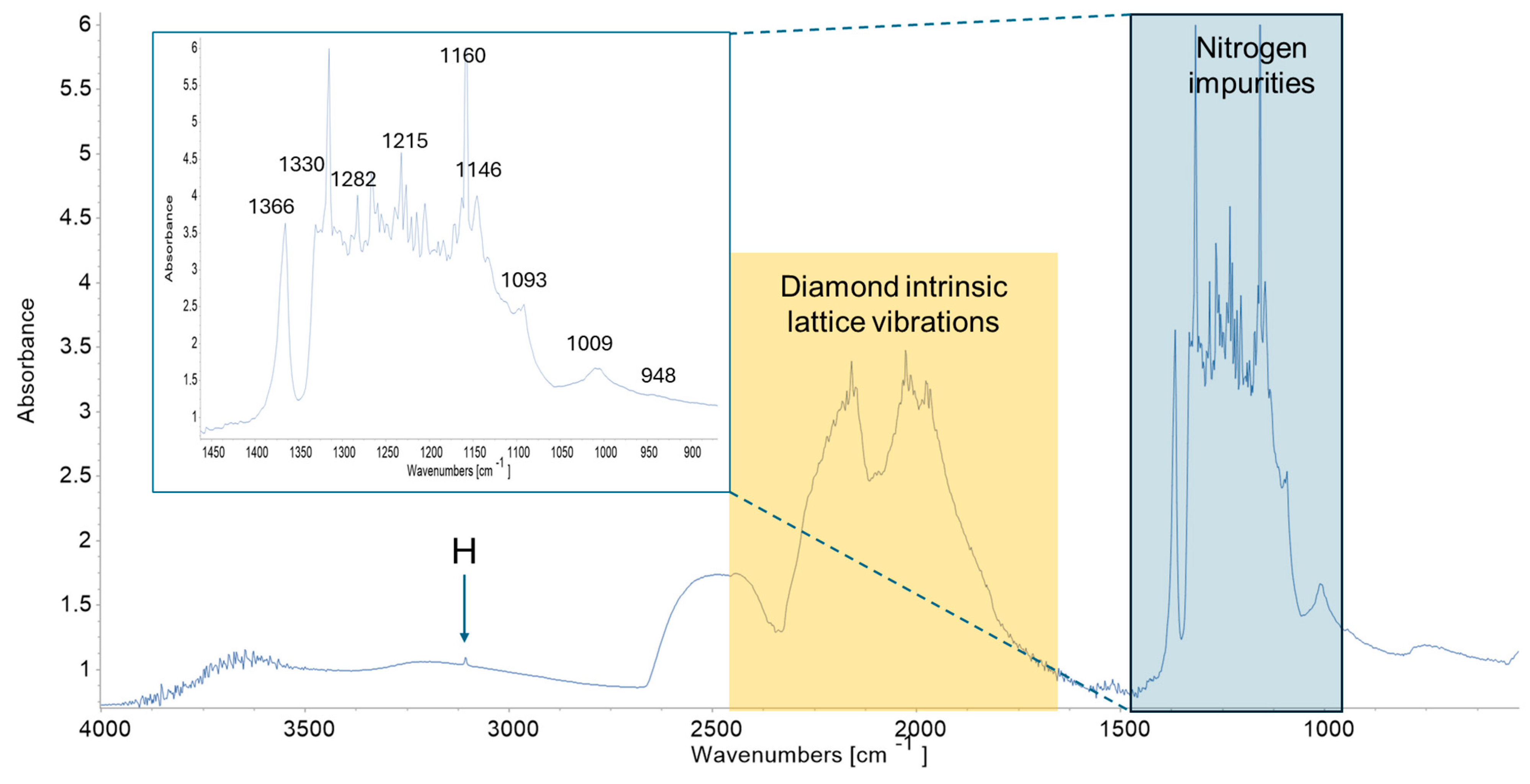
Figure 1. S16 diamond: (a) optical image of specimen belonging to Spada collection (Museum of Mineralogy of the Sapienza University, Rome); (b) its round octahedral morphology; and (c) historical document of the Museum, attesting the Brazilian provenance of specimen.
Crystals 14 00779 g001
Figure 2. S16 diamond: (a) trigons on face {111}; (b) anomalous birefringence under crossed polars; (c) green halos; and (d) healed fracture filled with dark material.
Crystals 14 00779 g002aCrystals 14 00779 g002b
Figure 3. S16 diamond’s FT-IR spectrum. The absorbance from 1000 to 1500 cm−1, corresponding to nitrogen defects, allows for classifying the diamond as type IaAB (see the enlarged detail of the spectrum).
Crystals 14 00779 g003
Figure 4. S16 diamond: (a) X-ray topograph taken with diffraction vector g = 2 2 ¯ 0 (see arrow); (b) X-ray topograph taken with diffraction vector g = 1 ¯ 11 (see arrow); (c) growth sector projection corresponding to the images of the topographs; and (d) µ-CXRT 3D reconstruction of sample oriented in agreement with the images of the topographs. D: dislocations, F: fracture, C: core. Note the white contrasts corresponding to the inclusions.
Crystals 14 00779 g004
Table 1. S16 diamond. Main peaks of FT-IR spectrum and related defects.
Peak Position cm−1Defect CenterReferences
3107HWoods and Collins (1983) [24]
1366B’ or PlateletsWoods (1986) [25]
1330BZaitsev (1998) [26]
1282AWoods et al. (1990) [27]
1215AWoods (1986) [25]
1160BWoods (1986) [25]
1146DislocationsBoyd et al. (1995) [28]
1130CCollins (1980) [29]
1093A and BZaitsev (2001) [30]
1009BWoods (1986) [25]
948Radiation damageZaitsev (2001) [30]
Disclaimer/Publisher’s Note: The statements, opinions and data contained in all publications are solely those of the individual author(s) and contributor(s) and not of MDPI and/or the editor(s). MDPI and/or the editor(s) disclaim responsibility for any injury to people or property resulting from any ideas, methods, instructions or products referred to in the content.

