Article

Structural and Optical Properties of Defected and Exotic Calcium Monochalcogenide Nanoparticles: Insights from DFT and TD-DFT Calculations

by
Panagiotis G. Moustris
1,
Alexandros G. Chronis
1,
Fotios I. Michos
1,
Nikos Aravantinos-Zafiris
2,* and
Mihail M. Sigalas
1,*
1
Department of Materials Science, University of Patras, 26504 Patras, Greece
2
Department of Environment, Ionian University, 29100 Zakynthos, Greece
*
Authors to whom correspondence should be addressed.
Crystals 2025, 15(5), 392; https://doi.org/10.3390/cryst15050392
Submission received: 9 March 2025 / Revised: 17 April 2025 / Accepted: 20 April 2025 / Published: 23 April 2025
(This article belongs to the Special Issue Celebrating the 10th Anniversary of International Crystallography)

Abstract

:
In this work, the structural and optical properties of calcium monochalcogenide nanoparticles were numerically examined by using Density Functional Theory and Time Dependent Density Functional Theory. The composition of the examined nanoparticles was obtained from an initial cubic-like building block of the form Ca4Y4, where Y could be one of the chalcogen elements sulfur, selenium, and tellurium, after its proper numerical examination to check their structural stability. The examined nanoparticles were then created from these initial cubic-like building blocks after their elongation along one, two, and three perpendicular directions. Τhe Absorption Spectrum, the Binding Energy, together with the highest occupied-lowest unoccupied molecular orbital (HOMO-LUMO) gap, were included in the calculations of the studied calcium monochalcogenides. The calculations provided numerical evidence regarding the existence of stable structures for a wide range of morphologies. In addition, the examination of the properties of such nanostructures after placing different kinds of defects was also included in the calculations, thus leading to new groups of nanoparticles with several potential uses in technological applications, such as hydrogen storage, CO2 capture, and ultraviolet-responsive devices.

1. Introduction

The growing global energy demands, coupled with the ongoing challenges of climate change, are two of the most critical issues currently faced by the international community. Energy remains an essential need for all nations, with fossil fuels still being the dominant source of power. However, the burning of carbon-based fuels results in significant CO2 emissions, which are the primary contributors to climate change. As global energy consumption continues to rise, efforts to decarbonize remain insufficient. In this context, renewable energy sources, such as wind and solar, as well as innovative technologies like photocatalysis, light-emitting diodes (LEDs), and advanced energy storage mechanisms, are increasingly viewed as solutions to mitigate the effects of climate change. Much of the current scientific research is focusing on the development and improvement of green energy technologies, with particular attention to photovoltaics, solar cells, hydrogen storage, and Li-ion batteries. Within this framework, the investigation of the structural and optical properties of calcium (Ca) monochalcogenide nanoparticles holds great promise. These materials could contribute significantly to the development of more efficient and sustainable energy systems [1,2,3].
Thin films of chalcogenides exhibit tunable optical and electrical properties, highly influenced by deposition method, composition, thickness, and annealing temperature, making precise control of these parameters essential for optimizing their performance in advanced technological applications [4]. In recent years, magnesium-based nanoparticles have shown potential in optoelectronics and energy storage, primarily due to their ability to be engineered at the nanoscale to optimize light absorption and electron transport properties [5]. These attributes make them promising candidates for solar cells and photocatalytic systems, with applications also extending to sustainable energy fields such as CO2 reduction and pollutant degradation [6]. Additionally, calcium monochalcogenide NPs are under exploration for biomedical uses due to their non-toxic nature and stability, which make them suitable for imaging and targeted drug delivery applications [7].
Calcium monochalcogenide quantum dots (QDs), composed of elements such as sulfur (S), selenium (Se), and tellurium (Te), have become a focal point in materials science, given their adaptable synthesis methods and significant potential for varied technological applications. These nanoparticles are synthesized through precise techniques, allowing their structural and optical properties to be engineered for specific applications. The tunable band gaps in CaS, CaSe, and CaTe make them especially promising for photovoltaic applications, as they offer low-cost and efficient alternatives to traditional solar cell materials [8,9]. Advances in group II–VI monochalcogenides have yielded durable nanostructures capable of enhancing solar energy capture, as well as photodetectors with increased sensitivity, efficiency, and stability. Calcium-based QDs are showing promise in environmental applications, particularly in the photocatalytic degradation of pollutants and CO2 reduction, thus contributing to sustainable solutions for environmental challenges [10]. Additability of Ca-Y QDs is beginning to open avenues in biomedical imaging and targeted drug delivery, further broadening their potential across interdisciplinary fields [11,12,13].
Recent theoretical studies have further highlighted the potential of group IV monochalcogenide nanoparticles to form exotic geometries, offering unique properties suitable for advanced applications such as photodetectors and solar cells [14]. The optical and electronic properties of rocksalt alkaline earth and transition metal monochalcogenide nanoparticles have been extensively examined using Density Functional Theory (DFT) and Time-Dependent DFT, demonstrating their versatility in optoelectronic technologies [15]. Notably, magnesium and aluminum hydride nanoparticles have been studied for their distinctive absorption spectrum, providing insights into their potential for spectrum-specific applications [16]. Moreover, calcium-based nanostructures, including nanorods and nanodisks, have revealed significant variations in their optical gaps and morphologies, underscoring their adaptability and utility in tailoring materials for specific energy-related uses [17]. These findings collectively underscore the expanding opportunities for calcium monochalcogenide quantum dots (QDs) in both fundamental research and practical applications, ranging from energy technologies to advanced biomedical solutions.
In this study, we examine the structural and optical properties of calcium monochalcogenide (Ca-Y, where Y = S, Se, Te) nanoparticles by employing Density Functional Theory (DFT) and Time-Dependent Density Functional Theory (TD-DFT) methods. This research builds on an initial cubic-like Ca4Y4 nanoparticle model, elongated along one, two, and three perpendicular directions, to investigate their potential for stability across various morphological configurations. The calculations for the examined nanoparticles include the determination of the Absorption Spectrum (AS), the Binding Energy (BE), and the Highest Occupied-Lowest Unoccupied Molecular Orbital (HOMO-LUMO) gap. Also, by analyzing the absorption spectrum and the structural stability through vibrational frequencies, we assess the suitability of these nanoparticles for potential applications in sustainable energy technologies. As will be extracted by the numerical data of this contribution, the optical properties of the examined nanostructures are strongly related to their size, their shape, and their constitutive components. The optical gap, as will be presented, varies significantly with the constitutive components of the nanoparticle, and it is strongly affected by the presence of a defect. The results of this study provide a foundation for further investigation of calcium-based nanoparticles, with promising implications for applications such as hydrogen storage, CO2 capture, and ultraviolet (UV)-responsive devices.

2. Computational Methods

In this study, Density Functional Theory and Time-Dependent Density Functional Theory were utilized to investigate the structural and optical properties of over 30 distinct nanostructures. These nanostructures were systematically designed and analyzed for their structural stability, originating from an initial configuration that was extended along one, two, and three perpendicular directions, while defected nanostructures were also studied by substituting sulfur atoms with fluorine, chlorine, nitrogen, and phosphorus. Initial assessments of these configurations confirmed the presence of structurally stable morphologies. The three cubic structures that were analyzed exhibit similar overall geometry but differ notably in their interatomic distances, reflecting variations in bonding and atomic arrangement. The optimized geometry reveals a non-ideal cubic arrangement, as evidenced by the variation in interatomic distances.
In the Ca4S4 cubic structure, the distance between neighboring Ca and S atoms is 2.66 Å, indicating relatively strong bonding interactions. However, a noticeable asymmetry is observed in the diagonal distances across the cube faces: the Ca–Ca face diagonal measures 3.53 Å, while the corresponding distance between two S atoms (face diagonal) is slightly longer, at 3.97 Å. This discrepancy suggests a distortion from perfect cubic symmetry, likely induced by differences in atomic radius and electronic distribution between the two elements. The shortest interatomic distance in the Ca4Se4 cubic structure, measured between a Ca and a Se atom, is 2.80 Å. The distance between two Ca atoms along the face diagonal is 3.62 Å. Similarly, the Se–Se distance along a face diagonal is 4.256 Å. The shortest distance in the Ca4Te4 cubic structure is 3.03 Å between Ca and Te atoms. The Ca–Ca face diagonal distance is 3.75 Å, slightly longer than the Ca–Te bond, consistent with their positioning along neighboring edges. The distance between two Te atoms along the face diagonal is 4.71 Å, representing a distorted lattice compared to the previous structures.
The structural optimizations were conducted using the Perdew, Burke, and Ernzerhof (PBE) gradient-corrected functional [18], in combination with the triple-ζ quality def2-TZVP basis set [19]. Following optimization, the lowest singlet vertical excitation energies and the ultraviolet-visible absorption spectra of the nanoparticles were calculated through TD-DFT, applied to the ground-state geometries. For all calculations, the exchange functional in TD-DFT was modeled using the hybrid CAM-B3LYP functional [20], which is well-regarded for its accuracy in predicting excitation energies. Due to the absence of experimental data for the absorption spectra of the proposed structures and limitations in computational resources, the structures were analyzed using the CAM-B3LYP functional. This functional was chosen for its balance of high accuracy and relatively lower computational demands compared to the EOM-CCSD functional, which, while offering higher precision, imposes significantly higher computational requirements. In all cases examined, the exchange functional in the TD-DFT calculations was approximated using the hybrid CAM-B3LYP functional. The computed absorption spectra were generated using a predefined scenario, representing each excitation as a Gaussian function centered at the TD-DFT calculated excitation energy. The absorption spectra were generated using a specified script that modeled each excitation as a Gaussian function centered at the excitation energy calculated by TD-DFT, with a standard deviation (broadening) set to 0.1 eV. This broadening value was selected to closely approximate the peak shapes typically observed in experimental measurements. All computations were carried out using the TURBOMOLE software package [21].

3. Results and Discussion
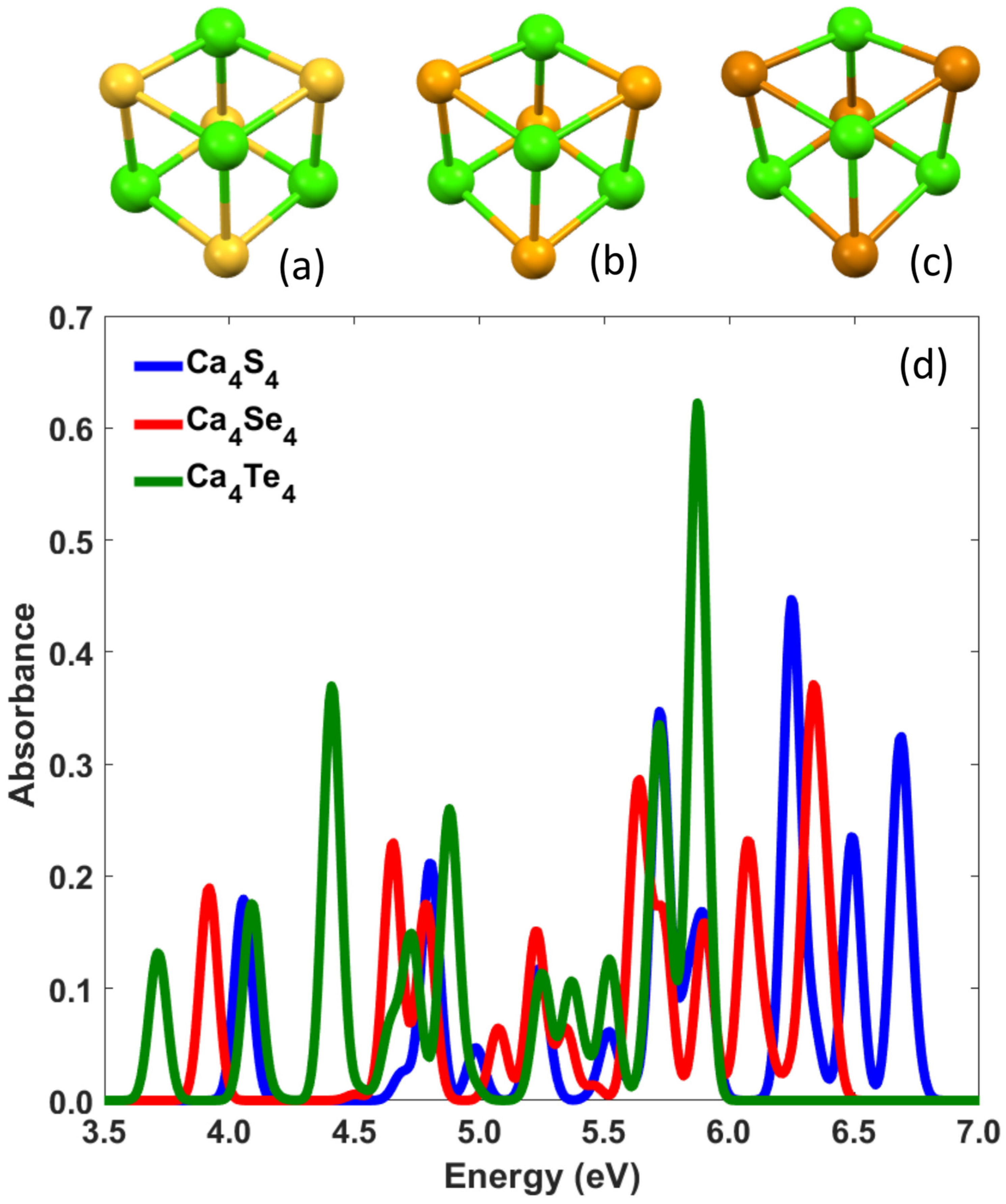
Calcium-based nanoclusters, including Ca2Mgn (n = 1–15) clusters, have been studied for their structural and electronic properties using Density Functional Theory, with focus on Binding Energy and HOMO-LUMO gaps, indicating significant stability for specific cluster sizes, similar to trends observed in magnesium nanostructures [22]. The optical absorption spectra of calcium chalcogenide nanoparticles (CaxYx, where Y = S, Se, Te), containing up to 15 atoms, exhibit a notable enhancement in the energy range of 3.50–4.50 eV, as determined using Time-Dependent Density Functional Theory [23]. Furthermore, studies of their electronic characteristics showed notable changes after doping with selenium and tellurium. Several studies have investigated the photoabsorption spectra of small calcium clusters, including the effect of their low-lying isomers. For instance, a recent study by Sahoo et al. investigated the linear and non-linear optical properties of small CaCn (n = 2–7) clusters, providing insights into their electronic structure and optical behavior using theoretical calculations [24]. Also, the vibrational properties of calcium monochalcogenide nanostructures offer an intriguing parallel to those observed in magnesium-based nanoparticles. Recent studies on MgxYx systems (Y = S, Se, Te) demonstrate how vibrational modes, analyzed via Density Functional Theory, reveal size-dependent stability and unique phononic behaviors [25]. These insights highlight the potential for analogous explorations in calcium nanoparticles to deepen our understanding of their optoelectronic and mechanical adaptability for advanced applications.
The calcium NPs analyzed in this study were categorized into distinct structural groups, all derived from a fundamental cubic-like unit of the form Ca4Y4, where Y = S, Se, Te. By systematically elongating this primary building block, which is illustrated in Figure 1a–c for the cases where Y = S, Se, Te, respectively, geometries were identified exhibiting notable stability and promising optical characteristics. These structural variations enable a deeper exploration of their potential in optoelectronic applications. Thus, the calcium nanoparticles investigated in this study can be classified into four categories. The initial building block’s extension in a single direction produced the first class of NPs. The original building block was extended along two and three perpendicular directions, respectively, to produce the other two categories. The fourth category of the examined nanoparticles consists of two groups and was derived from the previously examined CaxYx NPs, where Y = S, Se, Te. The first group consists of NPs that were created from the initial cubic unit elongated along one dimension and by substituting a sulfur atom with a fluorine, chlorine, nitrogen, or phosphorus atom. The second group consists of nanostructures that were modified by creating a hole in the NP after extracting atoms from it, and thus forming a set that could be characterized as “exotic” NPs. The optical properties of the examined Ca-based NPs were analyzed, revealing intriguing results that highlight their potential for various technological applications.

3.1. CaxYx NPs

The absorption spectra reported in this study were determined using the TD-DFT approach, as previously described. A range of exchange–correlation functionals was initially tested to select the most appropriate one for the nanoparticle under investigation. For CaxYx nanoclusters (with x = 4 and Y = S, Se, or Te), the absorption spectra were computed employing several commonly used functionals, including EOM-CCSD [26], CAM-B3LYP [20], M06–2X [27], PBE0 [28], B3LYP [29], and PBE [18]. The structure with the lowest first excitation energy and an Oscillation Strength (OS) greater than 0.01 was used to define the Optical Gap (OG) of the nanoparticle. The excitation energies obtained with EOM-CCSD served as a benchmark due to the method’s high reliability. In the case of Ca4S4, excitation energies of 4.18, 4.06, 3.97, 3.80, 3.51, and 3.16 eV were calculated using EOM-CCSD, CAM-B3LYP, M06-2X, PBE0, B3LYP, and PBE, respectively. Similarly, for the Ca4Se4 structure, values of 4.06, 3.92, 3.84, 3.67, 3.40, and 3.08 eV were obtained in the same order of functionals. Regarding Ca4Te4, the first excitation energies were determined as 3.86 eV (EOM-CCSD), 3.72 eV (CAM-B3LYP), 3.61 eV (M06-2X), 3.47 eV (PBE0), 3.25 eV (B3LYP), and 2.96 eV (PBE). These values are summarized in Table 1. It is evident that CAM-B3LYP offers results closely aligned with those from EOM-CCSD, outperforming the remaining functionals in accuracy. Considering the substantial computational cost associated with EOM-CCSD, all subsequent calculations in this work were carried out using CAM-B3LYP. Furthermore, the convergence parameter for Energy was set to 10−5. In addition, regarding the rest of the computational parameters, the Fermi setting was on, the initial and final temperatures were set to 300 K and 150 K, respectively, the annealing factor was set to 0.98, the gap criterion was 0.1 eV, and the stop criterion was 0.001 Hartree. Also, the lowest excitation energy of any nanoparticle investigated, with a significant Oscillator Strength larger than 0.01, is also known as the Optical Gap of the corresponding NP for all the diagrams shown subsequently in this article.
First, the absorption spectrum of the initial cubic-like building blocks was calculated for the nanostructures studied in this work. The results of these calculations are presented in Figure 1d. For the Ca4S4 nanoparticle, the first four excitation energies in the absorption spectrum are 4.05, 4.91, 4.97, and 5.26 eV, with corresponding absorbance values of 0.18, 0.21, 0.03, and 0.12. Similarly, for Ca4Se4, the excitation energies appear at 3.92, 4.27, 4.81, and 5.16 eV, with absorbance values of 0.19, 0.22, 0.17, and 0.05. For the case of Ca4Te4, the excitation energies are 3.71, 4.08, 4.41, and 4.71 eV, with absorbance values of 0.12, 0.18, 0.38, and 0.27, respectively. Additionally, the energy difference between the highest occupied molecular orbital (HOMO) and the lowest unoccupied molecular orbital (LUMO), referred to as the HOMO-LUMO gap, was determined for the studied NPs. Thus, the HOMO-LUMO gap is 6.35 eV for Ca4S4, 6.16 eV for Ca4Se4, and 5.82 eV for Ca4Te4. The stability of each analyzed NP of the form CaxYx is assessed by calculating the Binding Energy per formula unit (BE per f.u.) [5]. The BE, EB, is defined as follows:
EB(CaxYx) = xE(Ca) + xE(Y) − E(CaxYx),
where the index x indicates each examined formula of the structure (formula unit), E(Ca) is the energy of isolated atoms of Ca, E(Y) is the energy of isolated atoms of Y = S, Se, Te, and E(CaxYx) is the total energy of the NP. The BE per f.u., (Eb) is thus given by the following relation:
Eb(CaxYx) = EB(CaxYx)/x = E(Ca) + E(Y) − E(CaxYx)/x
For the initial cubic-like NPs, the BE per f.u. was found to be 7.00, 6.49, and 5.67 eV for Ca4S4, Ca4Se4, and Ca4Te4 NPs, respectively.

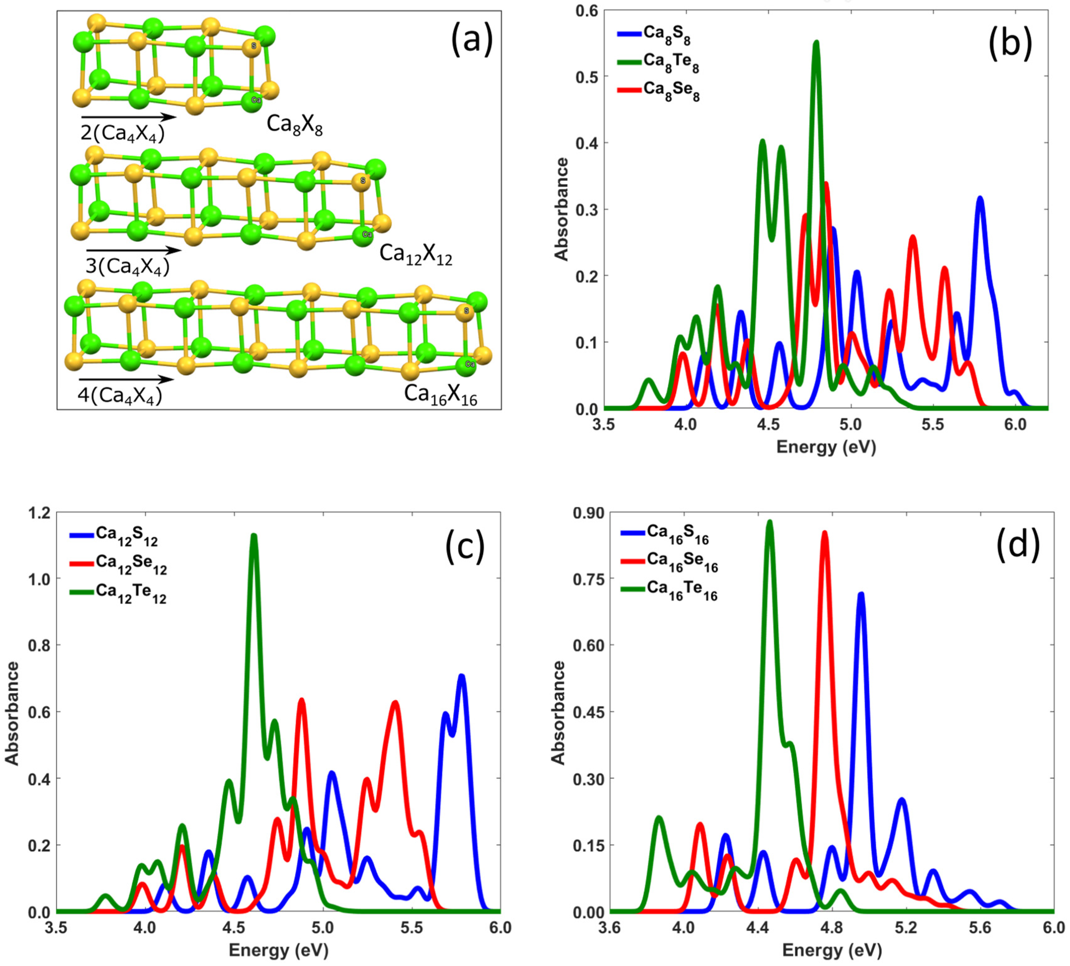
3.1.1. Elongated CaxYx NPs Along One Direction

The first category of calcium nanoparticles analyzed in this study was derived by extending the initial cubic-like structure, Ca4Y4 (Y = S, Se, Te), along one direction, as illustrated in Figure 2a. The nanoparticles produced in this group can be described by the formula CaxYx, where x takes the values of 8, 12, and 16, while the corresponding absorption spectra are presented in Figure 2b–d, respectively. For a more concise presentation of the calculated results, Table 2 shows the optical properties of all the nanostructures studied, as determined from the collected data.
Specifically, in the first case of this group, where x = 8, as shown in Figure 2b, the optical gaps are 4.33, 4.18, and 3.96 eV for Ca8S8, Ca8Te8, and Ca8Se8, respectively. Additionally, the Ca8Te8 nanostructure exhibits significantly higher absorption peaks compared to the corresponding structures with S and Se. For example, the highest absorption peak is 0.56, which is approximately 1.5 times greater than the highest peaks of the structures with S and Se, with values 3.40 at 4.80 eV, and 3.30 at 5.80 eV, respectively. Furthermore, for the Ca8S8, Ca8Se8, and Ca8Te8 examined NPs, the HOMO-LUMO gap was found to be 6.41, 6.17, and 5.75 eV, respectively. The same trend in absorption is also observed in Figure 2c, where x = 12. Specifically, the nanostructure containing Te exhibits the highest absorption peak with a value of 1.18 at 4.70 eV, while the structures with S and Se have maximum peak values of 0.62 at 4.85 eV and 0.70 at 5.8 eV, respectively. In addition, the optical gaps and the HOMO-LUMO gaps for the corresponding Ca12S12, Ca12Se12, and Ca12Te12 NPs are 4.36, 4.20, and 3.97 eV, and 6.45, 6.20, and 5.76 eV, respectively. Regarding the structures in this group with x = 16, the absorption trends are similar. As shown in Figure 2d, the maximum absorption peaks are 0.72 at 5.00 eV, 0.86 at 4.80 eV, and 0.88 at 4.50 eV for the Ca16S16, Ca16Se16, and Ca16Te16 structures, respectively, while the corresponding optical gaps are 4.36, 4.21, and 3.97 eV. Finally, comparing the three UV–Visible spectra, we can conclude that the structures in this group that contain Te exhibit higher absorbance than those that contain S and Se. The highest absorbance is observed for Ca12Te12 at 4.7 eV. Moreover, regarding the optical gap of the nanostructures, it is observed that elongation in one direction does not lead to significant changes in the optical gap. For instance, the optical gap of the CaxSx nanostructures is 4.05, 4.33, 4.36, and 4.36 eV for x = 4, 8, 12, and 16, respectively. Similarly, for the CaxSex nanostructures, the optical gap values are 3.92, 4.18, 4.20, and 4.08 eV, while for the CaxTex nanostructures, the corresponding values are 3.71, 3.96, 3.97, and 3.91 eV for x = 4, 8, 12, and 16. Thus, although the considered one-dimensional nanostructures provided good structural stability, the elongation along one direction did not significantly affect the optical gaps, as the numerical results indicated. Furthermore, as shown in Figure 2b–d, elongation in one direction does not significantly change the HOMO-LUMO gap. For example, for the structures Ca8S8, Ca12S12, and Ca16S16, the HL gap is 4.33, 4.36, and 4.36 eV, respectively. Similarly, when selenium is the second element in the nanoparticle, the HOMO-LUMO gap is 4.18, 4.20, and 4.21 eV for the Ca8Se8, Ca12Se12, and Ca16Se16 NPs, respectively. In contrast, when tellurium is the second element, the HOMO-LUMO gap is 3.96, 3.97, and 3.97 eV for the Ca8Te8, Ca12Te12, and Ca16Te16 NPs, respectively.

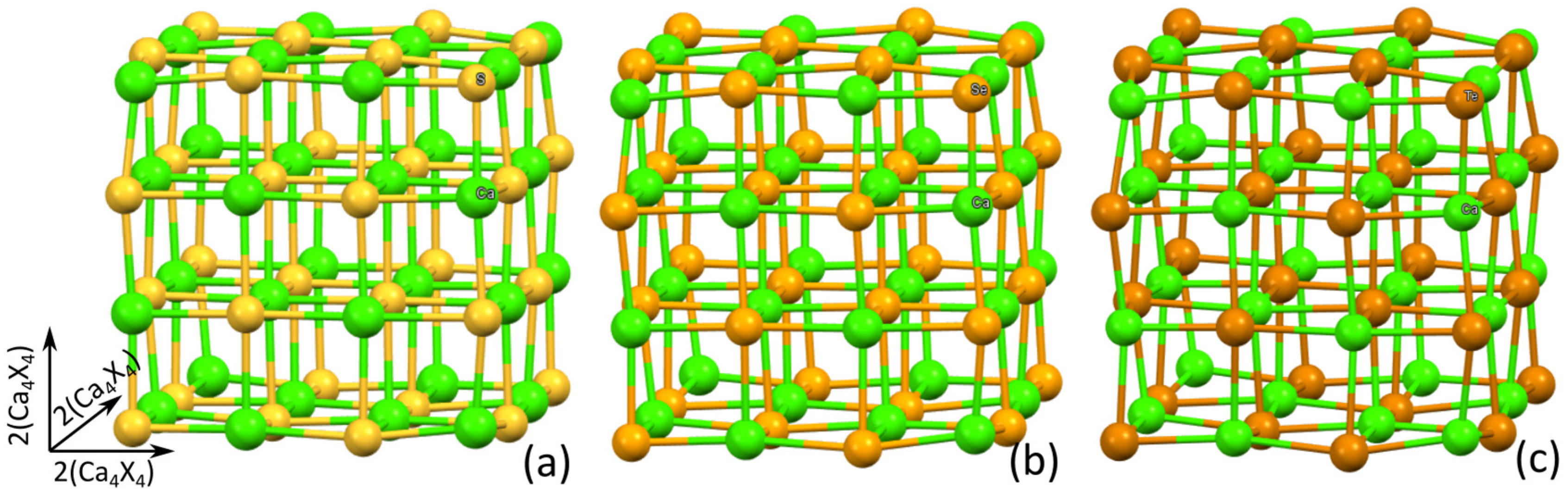
3.1.2. Elongated CaxYx NPs Along Two Perpendicular Directions

The second category of nanostructures studied in this work is formed from the initial building block, as in the previous case, with the difference that, in this case, they are extended in two perpendicular directions in a way that forms L-shaped NPs. The general form of the examined L-shaped NPs is CaxYx, where x = 12 and 16, and Y = S, Se, Te. For x = 12 and x = 16, two different L-shaped two-dimensional (2D) geometries were formed, named L1 and L2 as indicated in the relevant diagrams of Figure 3. The UV–Vis spectra from these cases are shown in Figure 3a,b. As an illustration of the examined geometries, the corresponding structures from the case where the second component is sulfur are presented as insets within each of the relevant diagrams.
In more detail, as shown in Figure 3a, the calculated optical gaps are 4.11, 3.98, and 3.76 eV for the nanostructures Ca12S12, Ca12Se12, and Ca12Te12, respectively. Additionally, the first four excitation energies are 4.11, 4.35, 4.45, and 4.65 eV when the second element of the structure is S, while the first four excitation energies are 3.98, 4.22, 4.36, and 4.52 eV for Se as the second element, and 3.76, 3.95, 4.10, and 4.28 eV for the case where Te is the second element of the NP. Regarding the absorbance, as in previous cases, presented in Section 3.1.1, the structure composed of Te exhibits the highest absorbance, which was 0.27 at 4.28 eV. Regarding the HOMO-LUMO gap, it was found to be 6.36, 5.51, and 5.72 eV for Ca12S12, Ca12Se12, and Ca12Te12 nanoparticles, respectively. It is important to notice here that by comparing the HOMO-LUMO gaps of nanoparticles elongated in one and two directions, it can be concluded that elongation in two directions slightly reduces the HOMO-LUMO gap, as also illustrated in the collected results of Table 2.
The UV–visible spectra for the cases where the structures Ca16Y16 were elongated along two perpendicular directions, thus forming the second case of 2D L-shaped NPs, named L2, are presented in Figure 3b. When the second element of the structure is sulfur, the optical gap is 4.11 eV, while the optical gaps are 3.97 and 3.77 eV for selenium and tellurium, respectively. Furthermore, compared to all the previously examined cases, only for this case, the peak absorbance value of 0.16 at 3.95 eV for the case of tellurium is not the highest, as the peak absorbance for the selenium case is 0.22 at 4.20 eV and for the sulfur case is 0.20 at 4.37 eV. In addition, from the diagrams presented in Figure 3b, the HOMO-LUMO gaps were found to be 6.41 eV, 6.16 eV, and 5.74 eV for the Ca16S16, Ca16Se16, and Ca16Te16 nanoparticles, respectively. These values are slightly reduced compared to the corresponding HOMO-LUMO gaps of the structures elongated in one direction, as presented in Table 2. Finally, the comparison of the two diagrams in Figure 3 shows that the optical gaps do not change significantly with an increase in the number of atoms in the nanostructure and remain nearly at the same values. In more detail, for the L1 2D L-shaped cases, the optical gaps were calculated at 4.11, 3.98, and 3.76 eV for Ca12S12, Ca12Se12, and Ca12Te12, respectively. Relevantly, for the L2 2D L-shaped cases, the optical gaps were calculated at 4.11, 3.97, and 3.77 eV, respectively.

3.1.3. Elongated NPs Along Three Perpendicular Directions

Within the framework of this study, three-dimensional (3D) structures were also created after elongation of the initial cubic unit along three perpendicular directions. These nanostructures have the general form CanYn, where n = 32 and Y = S, Se, Te. Their corresponding geometries are presented in Figure 4. The structural optimizations for these larger NPs validated their structural stability. It should be mentioned here, though, that due to the lack of computational resources, only the HOMO-LUMO gaps were calculated for these nanostructures. Thus, the HOMO-LUMO gap is 6.17 eV for Ca32S32, 5.98 eV for Ca32Se32, and 5.65 eV for Ca32Te32.

3.2. Defected and Exotic Ca-Based Nanostructures

The findings presented in the preceding section, particularly the exceptional structural stability demonstrated by the examined nanoparticles, provided a solid foundation for investigating further nanostructures derived from the initial cubic-like nanoparticle design. Thus, new groups of NPs were created. The first group of Ca-based NPs was considered from the one-dimensional case Ca8S8, where one sulfur atom was replaced by another atom, thus creating a defect in this one-dimensional nanostructure. The second group of Ca-based NPs was created from the two-dimensional case of Ca36S36 when a cubic unit cell of Ca4S4 was extracted from the middle of the nanostructure. These nanostructures, due to their intricate morphologies, could be classified as “exotic” NPs. The stability of these defected and exotic nanostructures was checked through the calculation of their vibrational spectrum, where no negative frequencies were calculated. In more detail, these defected NPs were constructed by substitution of a sulfur atom in calcium nanoparticles with halogen atoms, such as fluorine, chlorine, or pnictogens such as nitrogen or phosphorus in order to investigate the effect of this substitution on the structure and the relevant spectral properties of the nanoparticle. The introduction of halogens and pnictogens into calcium nanoparticles is expected to significantly affect the electronic structure of the material, thereby influencing the absorption spectrum. Halogens, due to their high electronegativity, affect electron transfer and interactions at the nanoscale, while pnictogens add additional dynamics to the formed structure. These changes are anticipated to lead to variations in light absorption and accelerated surface reactions, potentially making the nanoparticles more sensitive to environmental conditions and more effective in biological applications. Alterations in the chemical composition of the nanoparticles may modify their spectral characteristics, such as absorption, emission, and photosensitivity, which could have applications in fields such as photocatalysis and biomedical technologies. Scientific studies indicate that the incorporation of such elements may lead to improvements in the mechanistic processes of light energy conversion and the overall performance of nanomaterials [30,31,32]. It is also well-known that defects play a crucial role in determining material properties and can significantly expand their applications, especially in chalcogenide salts [33,34].

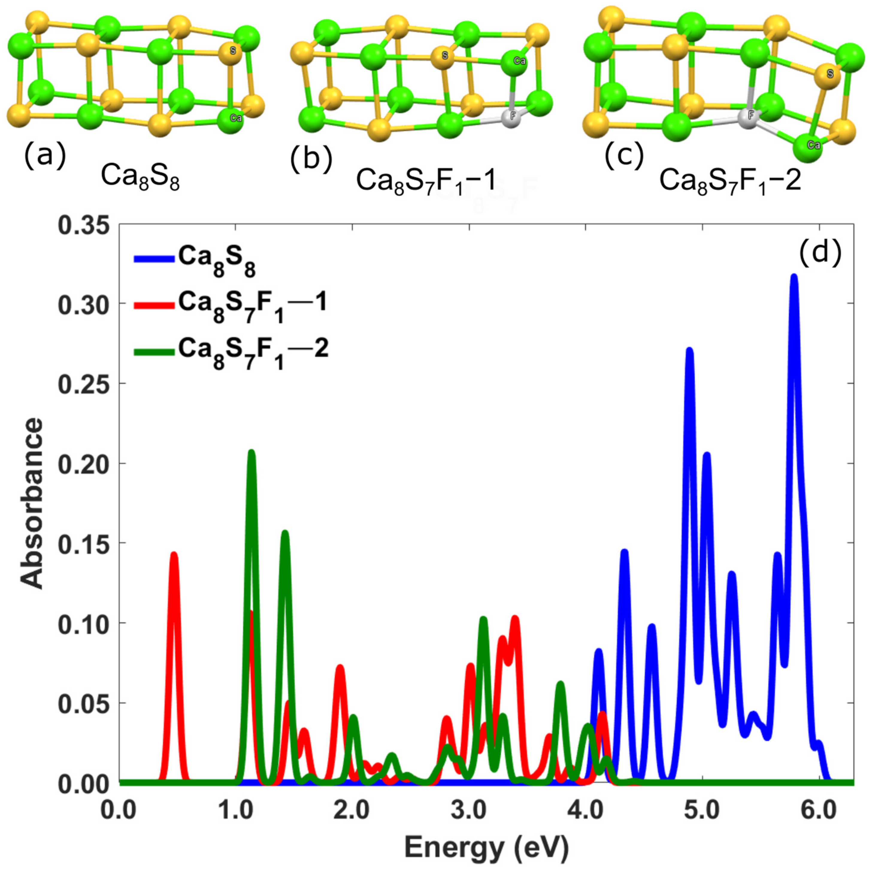
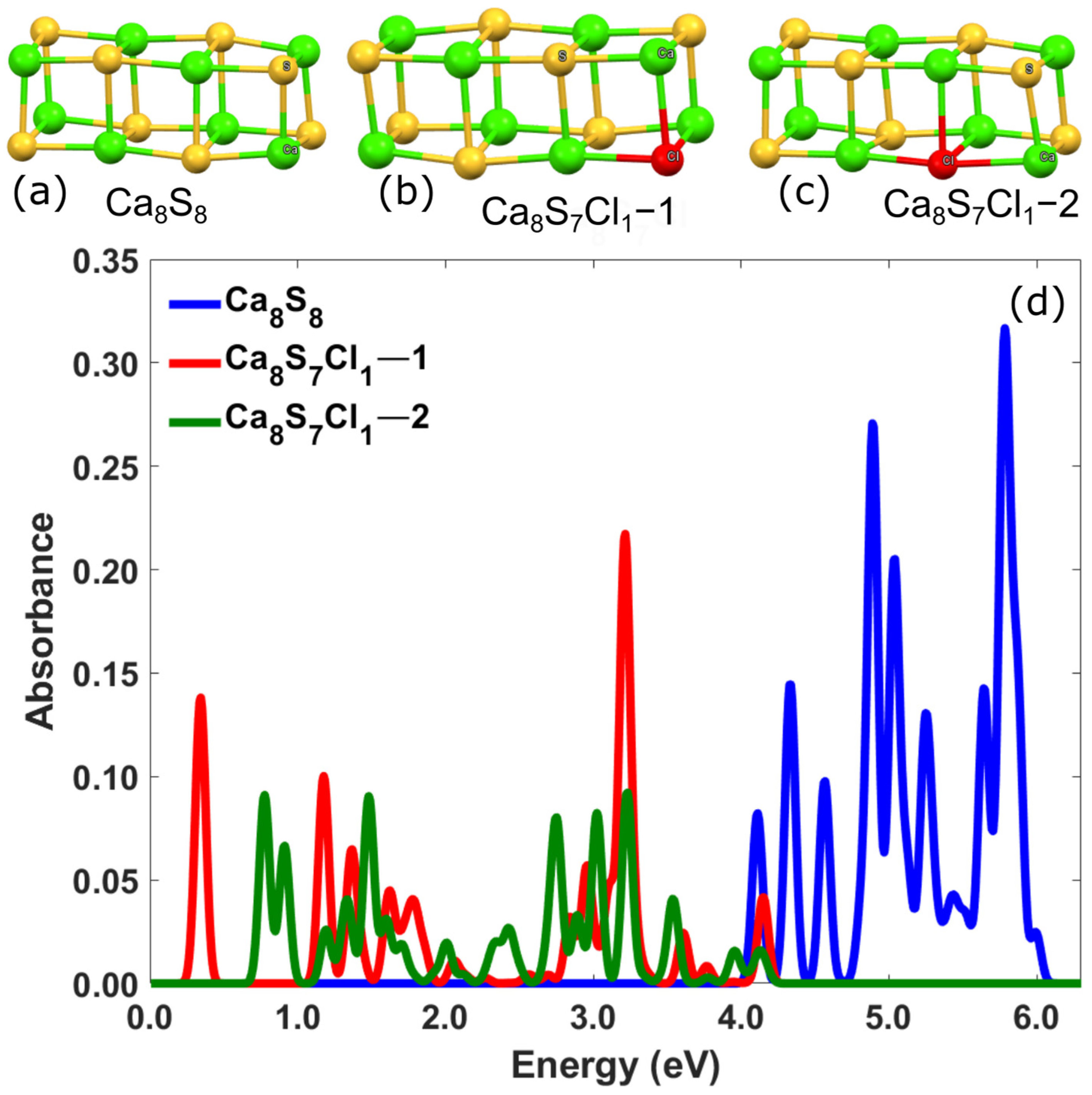
3.2.1. Ca-Based Nanostructures with Defects: Substitution of S by Halogens (F, Cl)

Two groups of defected nanostructures were studied in this category. In one group, the sulfur atom was replaced by a fluorine atom at two different positions, while in the second group, a sulfur atom was replaced by a chlorine atom, also at two different positions. In more detail, in the first group of the studied NPs, a sulfur atom, located either at the vertex of the geometry or at a subsequent position in the geometry, was replaced with a fluorine atom. The geometries of these defected nanostructures with the general form Ca8S7F1-1 and Ca8S7F1-2, respectively, together with their corresponding UV–visible spectra, are shown in the top panel of Figure 5. For a better comparison with the non-defected case, the spectrum of the Ca8S8 structure is shown again and is represented by the blue line. The defected nanostructure that was created by a substitution of a sulfur atom, located at the vertex of the geometry, with a fluorine atom, is shown with the red line. The relevant defected nanostructure that was created by the substitution of a sulfur atom, located at the second position of the geometry, with a fluorine atom, is shown in green. It is observed that by replacing the atoms, the spectrum shifts to lower energies, and frequency resonances can be found within the visible region. In particular, the first four excitation energies for the Ca8S8 nanostructure are 4.20, 4.40, 4.50, and 4.90 eV, whereas for Ca8S7F1-1 NP, the relevant excitation energies were found at 0.45, 1.10, 1.50, and 1.60 eV. Furthermore, for Ca8S7F1-2 NP, the first four excitation energies were calculated at 1.10, 1.50, 2.00, and 2.25 eV. It is also noteworthy that the highest absorption peak for Ca8S8 is approximately twice as large as the corresponding peak for Ca8S7F1-1 and about 1.5 times larger than that of the Ca8S7F1-2 NP. More specifically, the highest peak for the Ca8S8 nanostructure occurs at 5.80 eV with an absorbance of 0.33, while for the defected structure with the substitution of one S atom, located at the vertex of the geometry, with one fluorine atom, the peak occurs at 0.45 eV with an absorbance of 0.14. Finally, for the defected structure with the substitution of a sulfur atom, located at the second position of the geometry, with a fluorine atom, the peak is at 1.10 eV with an absorbance of 0.21.
The second group of the examined defected structures includes those in which, as mentioned above, one sulfur atom is substituted by one chlorine atom. The geometries of the structures studied and the corresponding absorption spectra are shown in Figure 6. As in the previous diagram, the spectrum of the Ca8S8 NP is represented in blue, the spectrum of the Ca8S7Cl1-1 NP is shown in red, and the spectrum of the Ca8S7Cl-2 NP is shown in green. As observed in the previous group of this category, the UV–Vis spectra of the defected structures shift to lower energies and fall within the visible range. The first four excitation energies for the Ca8S7Cl-1 structure are 0.35, 1.15, 1.25, and 1.55 eV, with corresponding absorbances of 0.14, 0.10, 0.06, and 0.04, respectively. For the Ca8S7Cl-2 nanostructure, the excitation energies are 0.80, 0.90, 1.15, and 1.25 eV, with corresponding absorbances of 0.09, 0.07, 0.03, and 0.04, respectively. Additionally, the absorption strength of the Ca8S8 structure is significantly higher compared to the Ca8S7Cl1-1 nanostructure and approximately three times higher than that of the Ca8S7Cl1-2 nanostructure. Specifically, the highest absorption peak for Ca8S8 occurs at 5.80 eV with an absorbance of 0.33, whereas the highest peaks for the Ca8S7Cl1-1 and Ca8S7Cl1-2 nanostructures are 0.22 (at 3.20 eV) and 0.09 (at 3.20 eV), respectively.

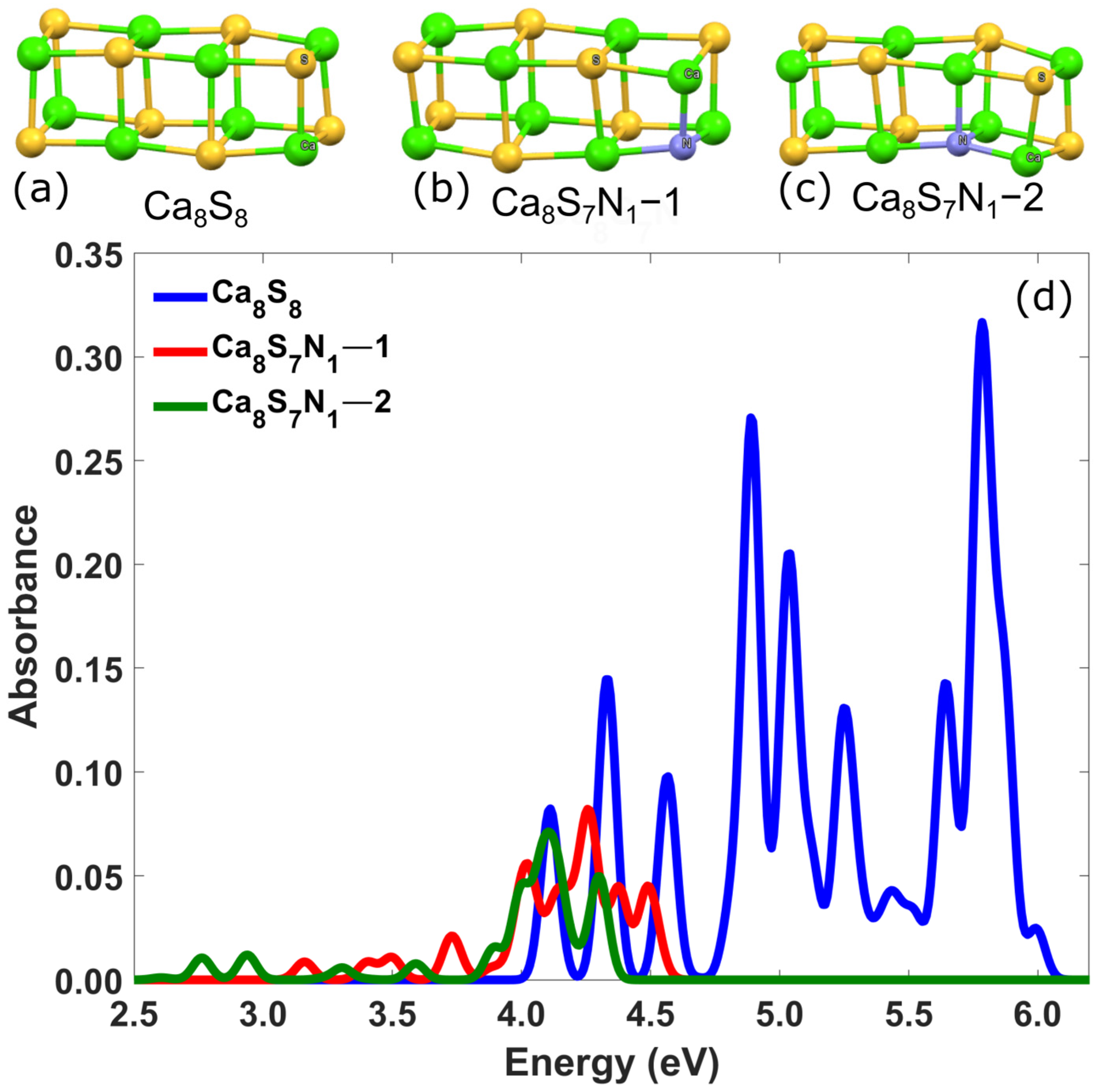
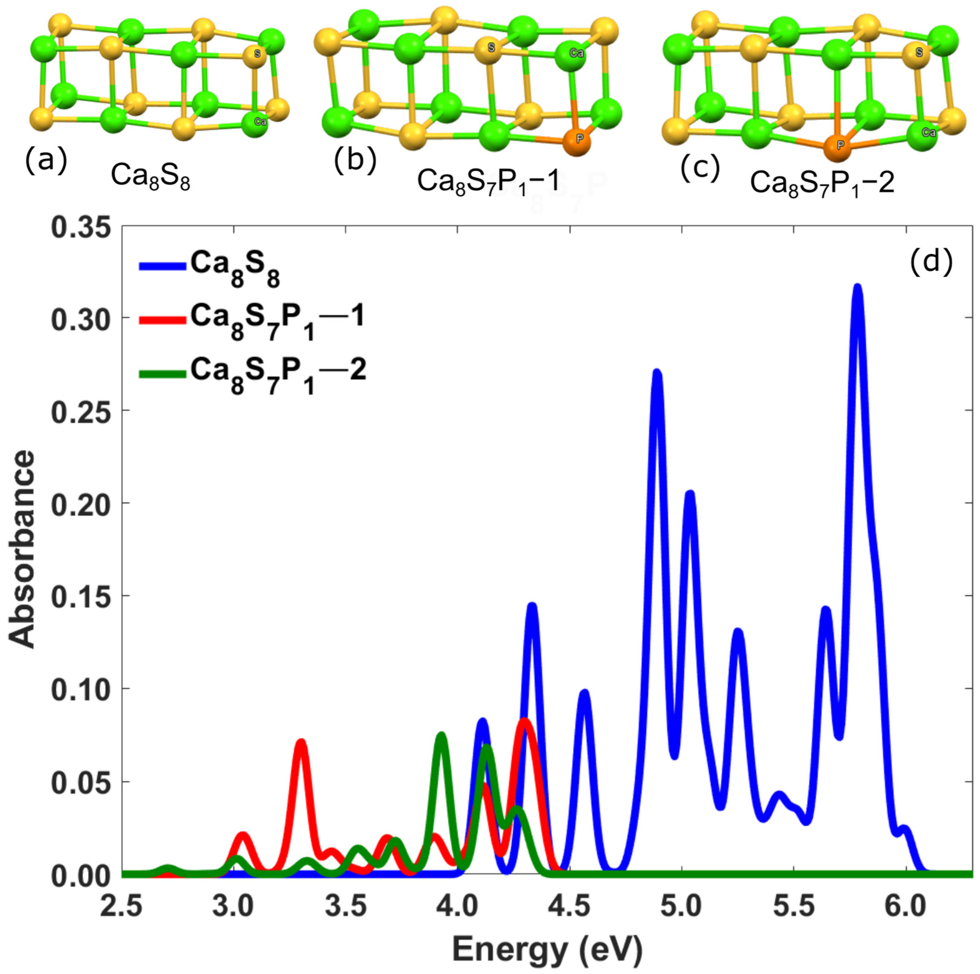
3.2.2. Ca-Based Nanostructures with Defects: Substitution of a Sulfur Atom by Pnictogens (N, P)

This category includes the investigation of two distinct groups, like the previously discussed category. The first group involves the substitution of a sulfur atom with a nitrogen atom, while the second group involves the substitution of a sulfur atom with a phosphorus atom. Specifically, in the first group, the defective NPs were created by replacing a sulfur atom with a nitrogen atom at two different positions. The corresponding geometries of these nanostructures and their absorption spectra are shown in Figure 7. As can be observed by the presented spectra of Figure 7d, the substitution of a sulfur atom by a nitrogen atom shifts the excitation energies to lower values. For instance, the first four excitation energies for the Ca8S7N1-1 nanostructure were 3.70, 4.00, 4.30, and 4.40 eV, with corresponding absorbance values of 0.02, 0.06, 0.08, and 0.05, respectively. Similarly, for the Ca8S7N1-2 nanostructure, the first four excitation energies are 3.90, 4.00, 4.10, and 4.30 eV, with absorbance values of 0.02, 0.05, 0.07, and 0.05, respectively. Furthermore, the peak absorbance is at least three times higher than that of the defected NPs, with values of 0.32, 0.08, and 0.07 for Ca8S8, Ca8S7N1-1, and Ca8S7N1-2, respectively.
In the second group of this category, similarly to the above groups, defected NPs involve the substitution of a sulfur atom with a phosphorus atom in two different positions. The resulting geometries of these NPs and their UV–visible spectra are presented in Figure 8. As in the previously mentioned cases of atom substitutions, the absorption spectra of the defected NPs in this case also shift to lower energies and exhibit lower absorbance. The first four excitation energies for Ca8S7P1-1 are 3.0, 3.3, 3.7, and 3.9 eV with corresponding absorbance values of 0.02, 0.07, 0.02, and 0.02, respectively. Similarly, for Ca8S7P1-2 NPs, the first four energies are 3.60, 3.90, 4.20, and 4.30 eV, with corresponding absorbance values of 0.02, 0.07, 0.07, and 0.04. Additionally, in this case, the peak absorbance is at least four times higher than that of the defected NPs. Specifically, the peak absorbance values are 0.32, 0.08, and 0.07 for Ca8S8, Ca8S7P1-1, and Ca8S7P1-2 NPs, respectively.

3.2.3. Exotic Ca-Based Nanostructures: Creating a Hole in the Structure

Based on some of the previously examined structures, the new group of NPs in the final section of this study may be regarded as exotic NPs because of their morphological features. The vibrational spectrum of various structures was calculated to verify their stability. For every structure examined, no negative frequencies were discovered, therefore validating their structural stability. This group includes nanoparticles created by elongating the initial cubic-like building block along two perpendicular directions and removing atoms from the center, thereby creating a void within the nanostructure. The general form of the proposed structures is Ca32Y32, where Y = S, Se, Te, and their geometries are presented in Figure 9. Relevant numerical calculations for nanostructures with defects have already been performed in several cases and have shown that such defected structures could enrich their vibrational or optical properties [35,36]. Due to limited computational resources, only the HOMO-LUMO gap was calculated for all NPs. As a representative case the together with the HOMO-LUMO gap, the Optical Gap, and the oscillator strength were calculated only for the case where the second element of the nanostructure was sulfur. The relevant values were found to be 4.30 eV for the Optical Gap with Oscillation Strength 0.05. The Binding Energy per f.u. was calculated at 7.95 eV for this case, as shown in Table 2.

4. Conclusions

Summarizing, in this work, by using Density Functional Theory and Time-Dependent Density Functional Theory, the structural and optical properties of calcium monochalcogenide nanoparticles were analyzed. The examined nanoparticles were generated by elongating an initial cubic-like nanostructure along one, two, and three perpendicular directions, and their structural stability was initially evaluated. The absorption spectra analysis demonstrated that the elongation of the cubic-like building block added new states to the spectra that were not present in the original structure. Furthermore, the absorption spectra confirmed the presence of excitation energies within the ultraviolet region for all the cubic-like building blocks analyzed in this study. These findings suggest that the proposed nanoparticles, with their unique optical properties and ultraviolet excitation energies, hold significant potential for integration into UV-based devices, such as light-emitting diodes, positioning them as promising candidates for advanced optoelectronic applications [37,38].
Furthermore, the calculated vibrational frequencies of the studied structures were all positive, providing strong evidence of their dynamic stability and the reliability of the optimized geometries. This stability, in combination with their distinct optical absorbance in the ultraviolet spectrum, highlights the potential of the proposed structures as promising candidates for various applications in advanced UV-based technologies, including optoelectronics and photonic devices. Τhe structural stability exhibited by the proposed structures motivated further exploration of new morphologies derived from the initial cubic-like building block, with a focus on how the altered shape influences their stability. Additionally, in the examined structures where one sulfur atom was substituted with one atom of nitrogen, chlorine, fluorine, or phosphorus in different positions, the excitation energies consistently shifted to lower values within the UV spectrum. These findings underscore the significant impact of substitution on the optical properties of the nanostructures, highlighting their potential for tailored applications in various UV-based optoelectronic technologies. Specifically, these modified nanostructures could be utilized in the development of ultraviolet light-emitting diodes for energy-efficient lighting, photodetectors for high-sensitivity UV detection, and solar-blind imaging devices. Furthermore, their unique optical properties make them suitable for use in photocatalysis, enabling efficient chemical reactions under UV irradiation, and in advanced coatings for UV protection. These diverse applications demonstrate the versatility of the proposed nanostructures and their promise in both scientific and industrial advancements.
The theoretical findings of this contribution, which are based on first-principles computational methods, provide the community with a new group of nanoparticles with plenty of promising potential applications. The absorption spectrum, which provides information regarding the attenuation of light when it passes through the examined nanostructure; the binding energy, which is a quantity closely related to its stability; and the HOMO-LUMO gap, which provides information regarding its absorption properties, constitute a concise and useful theoretical package that predicts the behavior of structures at the nanometer scale. Wide HOMO-LUMO gaps result in absorption in the UV region. Although this is effective for applications such as UV-screening, most of the applications come from nanoparticles that absorb in the visible spectrum. For example, photovoltaics, LEDs, biolabeling, and photocatalysis are applications based on nanoparticles that absorb in the visible spectrum. Furthermore, CO2 capture and hydrogen storage are also very interesting applications, which are planned by the authors to be part of future work, although such applications are not expected to be much affected by their HOMO-LUMO gaps. However, judging from their structural characteristics, hollow structures are expected to perform better. Thus, the findings presented here showed that there is a wide group of calcium-based nanostructures, which are generated by an initial cubic unit, and provide good structural stability. Among the key findings is the fact that their optical properties, as studied by first principles calculations, make them potential candidates for plenty of applications such as hydrogen storage, CO2 capture, and ultraviolet responsive devices. Especially in cases where defects are considered in the studied structures, the absorption frequencies were found from the visible spectrum. Thus, such nanoparticles could stand as very powerful candidates for photovoltaics, LEDs, biolabeling, and photocatalysis.
In conclusion, the findings of this study, particularly the rich variety of nanoparticle shapes, suggest that calcium monochalcogenide NPs could represent a highly promising class of materials. These materials exhibit a range of potential applications in the field of green energy technologies, such as hydrogen storage, CO2 capture, storage of other gases, and battery development. Their unique structure and physical properties open new avenues for advancing innovative technologies that could contribute to sustainable solutions for energy and environmental challenges.

Author Contributions

Conceptualization, P.G.M., A.G.C., F.I.M., N.A.-Z. and M.M.S.; methodology, P.G.M., A.G.C., F.I.M., N.A.-Z. and M.M.S.; software, P.G.M., A.G.C., F.I.M., N.A.-Z. and M.M.S.; validation, P.G.M., A.G.C., F.I.M., N.A.-Z. and M.M.S.; formal analysis, P.G.M., A.G.C., F.I.M., N.A.-Z. and M.M.S.; investigation, P.G.M., A.G.C., F.I.M., N.A.-Z. and M.M.S.; resources, P.G.M., A.G.C., F.I.M., N.A.-Z. and M.M.S.; data curation, P.G.M., A.G.C., F.I.M., N.A.-Z. and M.M.S.; writing—original draft preparation, P.G.M., A.G.C., F.I.M., N.A.-Z. and M.M.S.; writing—review and editing, P.G.M., A.G.C., F.I.M., N.A.-Z. and M.M.S.; visualization, P.G.M., A.G.C., F.I.M., N.A.-Z. and M.M.S.; supervision, N.A.-Z. and M.M.S.; project administration, N.A.-Z. and M.M.S. All authors have read and agreed to the published version of the manuscript.

Funding

This research received no external funding.

Data Availability Statement

The raw data supporting the conclusions of this article will be made available by the authors on request.

Conflicts of Interest

The authors declare no conflicts of interest.

References

  1. Ong, W.-J.; Zheng, N.; Antonietti, M. Advanced nanomaterials for energy conversion and storage: Current status and future opportunities. Nanoscale 2021, 13, 9904–9907. [Google Scholar] [CrossRef] [PubMed]
  2. Yang, Y.; Zhang, C.; Lai, C.; Zeng, G.; Huang, D.; Cheng, M.; Wang, J.; Chen, F.; Zhou, C.; Xiong, W. BiOX (X = Cl, Br, I) photocatalytic nanomaterials: Applications for fuels and environmental management. Adv. Colloid Interface Sci. 2018, 254, 76–93. [Google Scholar] [CrossRef] [PubMed]
  3. Wang, M.; Zhou, H.; Wang, F. Photocatalytic biomass conversion for hydrogen and renewable carbon-based chemicals. Joule 2024, 8, 604–621. [Google Scholar] [CrossRef]
  4. El-Ghany, W.A.A. Review on the optical and electrical properties of chalcogenide thin films: Challenges and applications. Phys. Chem. Chem. Phys. 2025, 27, 4567–4586. [Google Scholar] [CrossRef]
  5. Aravantinos-Zafiris, N.; Chronis, A.G.; Sigalas, M.M. Structural and optical properties of exotic magnesium monochalcogenide nanoparticles. Mater. Today Commun. 2021, 28, 102622. [Google Scholar] [CrossRef]
  6. Zabed, H.M.; Islam, J.; Chowdhury, F.I.; Zhao, M.; Awasthi, M.K.; Nizami, A.-S.; Uddin, J.; Thomas, S.; Qi, X. Recent insights into heterometal-doped copper oxide nanostructure-based catalysts for renewable energy conversion and generation. Renew. Sustain. Energy Rev. 2022, 168, 112887. [Google Scholar] [CrossRef]
  7. Malehmir, S.; Esmaili, M.A.; Mahabady, M.K.; Sobhani-Nasab, A.; Atapour, A.; Ganjali, M.R.; Ghasemi, A.; Hasan-Abad, A.M. A review: Hemocompatibility of magnetic nanoparticles and their regenerative medicine, cancer therapy, drug delivery, and bioimaging applications. Front. Chem. 2023, 11, 1249134. [Google Scholar] [CrossRef]
  8. Huang, H.; Feng, W.; Chen, Y. Two-dimensional biomaterials: Material science, biological effect and biomedical engineering applications. Chem. Soc. Rev. 2021, 50, 11381–11485. [Google Scholar] [CrossRef]
  9. Sharma, G.; Kumar, A.; Lichtfouse, E. Nanophotocatalysis and Environmental Applications: Materials and Technology; Asiri, A.M., Ed.; Springer: Berlin/Heidelberg, Germany, 2019; Volume 29. [Google Scholar] [CrossRef]
  10. Qi, C.; Lin, J.; Fu, L.-H.; Huang, P. Calcium-based biomaterials for diagnosis, treatment, and theranostics. Chem. Soc. Rev. 2017, 47, 357–403. [Google Scholar] [CrossRef]
  11. Sun, T.; Zhang, Y.S.; Pang, B.; Hyun, D.C.; Yang, M.; Xia, Y. Engineered nanoparticles for drug delivery in cancer therapy. In Nanomaterials and Neoplasms; Taylor & Francis Group: Abingdon, UK, 2019; pp. 31–142. [Google Scholar] [CrossRef]
  12. Lin, Y.; Zheng, Y.; Guo, Y.; Yang, Y.; Li, H.; Fang, Y.; Wang, C. Peptide-functionalized carbon dots for sensitive and selective Ca2+ detection. Sens. Actuators B Chem. 2018, 273, 1654–1659. [Google Scholar] [CrossRef]
  13. Rao, V.N.; Ravi, P.; Sathish, M.; Vijayakumar, M.; Sakar, M.; Karthik, M.; Balakumar, S.; Reddy, K.R.; Shetti, N.P.; Aminabhavi, T.M.; et al. Metal chalcogenide-based core/shell photocatalysts for solar hydrogen production: Recent advances, properties and technology challenges. J. Hazard. Mater. 2021, 415, 125588. [Google Scholar] [CrossRef]
  14. Karantagli, E.; Sigalas, M.; Garoufalis, C.S. Exotic nanoparticles of group IV monochalcogenides. Solid State Commun. 2019, 295, 38–42. [Google Scholar] [CrossRef]
  15. Wobbe, M.C.C.; Zwijnenburg, M.A. Chemical trends in the optical properties of rocksalt nanoparticles. Phys. Chem. Chem. Phys. 2015, 17, 28892–28900. [Google Scholar] [CrossRef]
  16. Chronis, A.G.; Sigalas, M.M.; Koukaras, E.N. Absorption spectrum of magnesium and aluminum hydride nanoparticles. Mater. Chem. Phys. 2019, 228, 244–253. [Google Scholar] [CrossRef]
  17. Chronis, A.G.; Michos, F.I.; Garoufalis, C.S.; Sigalas, M.M. Structural and optical properties of Be, Mg and Ca nanorods and nanodisks. Phys. Chem. Chem. Phys. 2021, 23, 1849–1858. [Google Scholar] [CrossRef]
  18. Perdew, J.P.; Burke, K.; Ernzerhof, M. Generalized gradient approximation made simple. Phys. Rev. Lett. 1996, 77, 3865–3868. [Google Scholar] [CrossRef]
  19. Weigend, F.; Ahlrichs, R. Balanced basis sets of split valence, triple zeta valence and quadruple zeta valence quality for H to Rn: Design and assessment of accuracy. Phys. Chem. Chem. Phys. 2005, 7, 3297–3305. [Google Scholar] [CrossRef]
  20. Yanai, T.; Tew, D.P.; Handy, N.C. A new hybrid exchange–correlation functional using the Coulomb-attenuating method (CAM-B3LYP). Chem. Phys. Lett. 2004, 393, 51–57. [Google Scholar] [CrossRef]
  21. TURBOMOLE; TURBOMOLE GmbH: Karlsruhe, Germany, 2019.
  22. Li, C.; Cui, Y.; Tian, H.; Ren, B.; Li, Q.; Li, Y.; Yang, H. Quantum Chemistry Study on the Structures and Electronic Properties of Bimetallic Ca2-Doped Magnesium Ca2Mgn (n = 1–15) Clusters. Nanomaterials 2022, 12, 1654. [Google Scholar] [CrossRef]
  23. Schlipf, M.; Betzinger, M.; Ležaić, M.; Friedrich, C.; Blügel, S. Structural, electronic, and magnetic properties of the europium chalcogenides: A hybrid-functional DFT study. Phys. Rev. B 2013, 88, 094433. [Google Scholar] [CrossRef]
  24. Sahoo, R.K.; Khatua, R.; Sahu, S. Theoretical study of linear and non-linear optical properties of small CaCn (n = 2–7) clusters. Mater. Today Proc. 2022, 66, 3397–3400. [Google Scholar] [CrossRef]
  25. Aravantinos-Zafiris, N.; Michos, F.I.; Sigalas, M.M. Vibrational Spectrum of Magnesium Monochalcogenide Nanoparticles. Nanomaterials 2024, 14, 1918. [Google Scholar] [CrossRef] [PubMed]
  26. Goings, J.J.; Caricato, M.; Frisch, M.J.; Li, X. Assessment of low-scaling approximations to the equation of motion coupled-cluster singles and doubles equations. J. Chem. Phys. 2014, 141, 164116. [Google Scholar] [CrossRef]
  27. Zhao, Y.; Truhlar, D.G. The M06 suite of density functionals for main group thermochemistry, thermochemical kinetics, noncovalent interactions, excited states, and transition elements: Two new functionals and systematic testing of four M06-class functionals and 12 other functionals. Theor. Chem. Acc. 2008, 120, 215–241. [Google Scholar] [CrossRef]
  28. Adamo, C.; Barone, V. Toward reliable density functional methods without adjustable parameters: The PBE0 model. J. Chem. Phys. 1999, 110, 6158–6170. [Google Scholar] [CrossRef]
  29. Becke, A.D. Density-functional thermochemistry. III. The role of exact exchange. J. Chem. Phys. 1993, 98, 5648–5652. [Google Scholar] [CrossRef]
  30. Fadlallah, M. Magnetic, electronic, optical, and photocatalytic properties of nonmetal- and halogen-doped anatase TiO2 nanotubes. Phys. E Low-Dimens. Syst. Nanostruct. 2017, 89, 50–56. [Google Scholar] [CrossRef]
  31. Carmona, F.J.; Dal Sasso, G.; Bertolotti, F.; Ramírez-Rodríguez, G.B.; Delgado-López, J.M.; Pedersen, J.S.; Masciocchi, N.; Guagliardi, A. The role of nanoparticle structure and morphology in the dissolution kinetics and nutrient release of nitrate-doped calcium phosphate nanofertilizers. Sci. Rep. 2020, 10, 12396. [Google Scholar] [CrossRef]
  32. Fatemi, A.; Jamshidi-Ghaleh, K.; Tohidi, T.; Bayat, F. CaCl2 doping effect on optical and structural properties of halide perovskites under ligand assisted reprecipitation synthesis. Phys. B Condens. Matter 2024, 691, 416360. [Google Scholar] [CrossRef]
  33. Zhang, X.; Gao, L.; Yu, H.; Liao, Q.; Kang, Z.; Zhang, Z.; Zhang, Y. Single-atom vacancy doping in two-dimensional transition metal dichalcogenides. Acc. Mater. Res. 2021, 2, 655–668. [Google Scholar] [CrossRef]
  34. Cavallini, M.; Gentili, D. Atomic Vacancies in Transition Metal Dichalcogenides: Properties, Fabrication, and Limits. Chempluschem 2022, 87, e202100562. [Google Scholar] [CrossRef] [PubMed]
  35. Aravantinos-Zafiris, N.; Sigalas, M.M. Phononic Band Gaps in Graphene-Like Materials and Nanotubes. J. Surfaces Interfaces Mater. 2013, 1, 184–188. [Google Scholar] [CrossRef]
  36. Sgouros, A.P.; Neupane, M.R.; Sigalas, M.M.; Aravantinos-Zafiris, N.; Lake, R.K. Nanoscale phononic interconnects in THz frequencies. Phys. Chem. Chem. Phys. 2014, 16, 23355–23364. [Google Scholar] [CrossRef] [PubMed]
  37. Sun, M.; Schwingenschlögl, U. Unique omnidirectional negative Poisson’s ratio in δ-phase carbon monochalcogenides. J. Phys. Chem. C 2021, 125, 4133–4138. [Google Scholar] [CrossRef]
  38. Xu, Y.; Liu, Y.; Chen, Y.; Zhang, Y.; Ma, C.; Zhang, H.; Sun, S.; Ji, Y. Two-dimensional direct semiconductor Boron Monochalcogenide γ-BTe: Room-temperature single-bound exciton and novel donor material in excitonic solar cells. ACS Appl. Mater. Interfaces 2020, 12, 58349–58359. [Google Scholar] [CrossRef] [PubMed]
Figure 1. The initial cubic-like building block of the studied Ca4Y4 NPs after the geometry optimization, where (a) Y = S, (b) Y = Se, and (c) Y = Te. (d) The UV–visible spectra of the initial cubic-like building blocks of Ca4Y4 (Y = S, Se, Te) NPs.
Crystals 15 00392 g001
Figure 2. (a) The examined CaxYx NPs where x = 8, 12, and 16, derived from the elongation of the initial cubic-like building block Cα4Y4 (Y = S, Se, and Te) along one direction two, three, and four times respectively, as indicated by the texted arrow. (bd) The corresponding UV–visible spectra of Ca8Y8 (b), Ca12Y12 (c), and Ca16Y16 (d) ΝPs.
Crystals 15 00392 g002
Figure 3. The UV–visible spectra of (a) Ca12Y12 and (b) Ca16Y16 ΝPs (Y = S, Se, and Te). The inset in each diagram provides an illustration of the relevant geometry from the case of S of the examined nanostructures, and the arrows indicate how many times the initial cubic unit block was elongated along each direction.
Crystals 15 00392 g003
Figure 4. The examined NPs with general form Ca32Y32, where Y = S (a), Se (b), and Te (c), which were created after elongation of the initial cubic-like building block Cα4Y4 along three perpendicular directions. The arrows in (a) indicate how many times the initial cubic unit block was elongated along each direction.
Crystals 15 00392 g004
Figure 5. (a) The non-defected NP Ca8S8. (b) The first case of the defected Ca8S8, named Ca8S7F1-1, which was formed by substituting one sulfur atom with one fluorine atom (shown in grey) at the vertex of the NP. (c) The second case of the defected Ca8S8, named Ca8S7F1-2, which was formed by substituting one sulfur atom with one fluorine atom (shown in grey) at a position right after the vertex of the NP. (d) The calculated UV–visible spectra of the examined NPs.
Crystals 15 00392 g005
Figure 6. (a) The non-defected NP Ca8S8. (b) The first case of the defected Ca8S8, named Ca8S7Cl1-1, which was formed by substituting one sulfur atom with one chlorine atom (shown in red) at the vertex of the NP. (c) The second case of the defected Ca8S8, named Ca8S7Cl1-2, which was formed by substituting one sulfur atom with one chlorine atom (shown in red) at a position right after the vertex of the NP. (d) The calculated UV–visible spectra of the examined NPs.
Crystals 15 00392 g006
Figure 7. (a) The non-defected NP Ca8S8. (b) The first case of the defected Ca8S8, named Ca8S7N1-1, which was formed by substituting one sulfur atom with one nitrogen atom (shown in blue) at the vertex of the NP. (c) The second case of the defected Ca8S8, named Ca8S7N1-2, which was formed by substituting one sulfur atom with one nitrogen atom (shown in blue) at a position right after the vertex of the NP. (d) The calculated UV–visible spectra of the examined NPs.
Crystals 15 00392 g007
Figure 8. (a) The non-defected NP Ca8S8. (b) The first case of the defected Ca8S8, named Ca8S7P1-1, was formed by substituting one sulfur atom with one phosphorus atom (shown in orange) at the vertex of the NP. (c) The second case of the defected Ca8S8, named Ca8S7P1-2, which was formed by substituting one sulfur atom with one phosphorus atom (shown in orange) at a position right after the vertex of the NP. (d) The calculated UV–visible spectra of the examined NPs.
Crystals 15 00392 g008
Figure 9. The defected with a hole exotic NPs Ca32Y32, where Y = S (a), Se (b), and Te (c).
Crystals 15 00392 g009
Table 1. The Ca4Y4 NPs (Y = S, Se, Te). Benchmarking the first excitation energy (eV) obtained using different density functionals.
Ca4S4Ca4Se4Ca4Te4
EOM-CCSD4.184.063.86
CAM-B3LYP4.063.923.72
M06-2X3.973.843.61
PBE03.803.673.47
B3LYP3.513.403.25
PBE3.163.082.96
Table 2. HOMO-LUMO Gap (HLG), Optical Gap (OG), Oscillator Strength (OS) and Binding Energy per f.u. for all examined CaxYx NPs (Y = S, Se, Te).
NanoparticleHOMO-LUMO Gap (eV)Optical Gap (eV)Oscillator StrengthBinding Energy per f.u. (eV)
Ca4S46.354.050.187.00
Ca4Se46.163.920.196.49
Ca4Te45.823.710.135.68
Ca8S86.414.330.147.51
Ca8Se86.174.180.156.96
Ca8Te85.753.960.106.07
Ca12S126.454.360.187.69
Ca12Se126.204.200.197.11
Ca12Te125.763.970.126.20
Ca12S12_2D_L16.364.110.067.63
Ca12Se12_2D_L15.513.980.055.03
Ca12Te12_2D_L15.723.760.036.17
Ca16S166.404.220.037.90
Ca16Se166.184.080.037.31
Ca16Te165.813.920.066.36
Ca16S16_1D6.474.360.27.77
Ca16Se16_1D6.224.210.217.20
Ca16Te16_1D5.773.970.146.27
Ca16S16_2D_L26.414.110.037.73
Ca16Se16_2D_L26.163.970.047.16
Ca16Te16_2D_L25.743.770.026.24
Ca32S326.174.470.348.22
Ca32Se325.98--7.60
Ca32Te325.65--6.59
Ca32S32–2DH6.504.300.057.95
Ca32Se32–2DH6.20--7.36
Ca32Te32–2DH5.75--6.41
Ca8S7Cl1–11.891.170.10-
Ca8S7Cl1–21.860.770.09-
Ca8S7P1–14.623.310.03-
Ca8S7P1–24.083.720.02-
Ca8S7F1–12.171.120.11-
Ca8S7F1–22.631.140.21-
Ca8S7N1–15.473.730.02-
Ca8S7N1–25.002.760.01-
Disclaimer/Publisher’s Note: The statements, opinions and data contained in all publications are solely those of the individual author(s) and contributor(s) and not of MDPI and/or the editor(s). MDPI and/or the editor(s) disclaim responsibility for any injury to people or property resulting from any ideas, methods, instructions or products referred to in the content.

