2.1. Characterization of the Phenomenon of CLC Photopatterning due to Optically Controlled Boundary Conditions
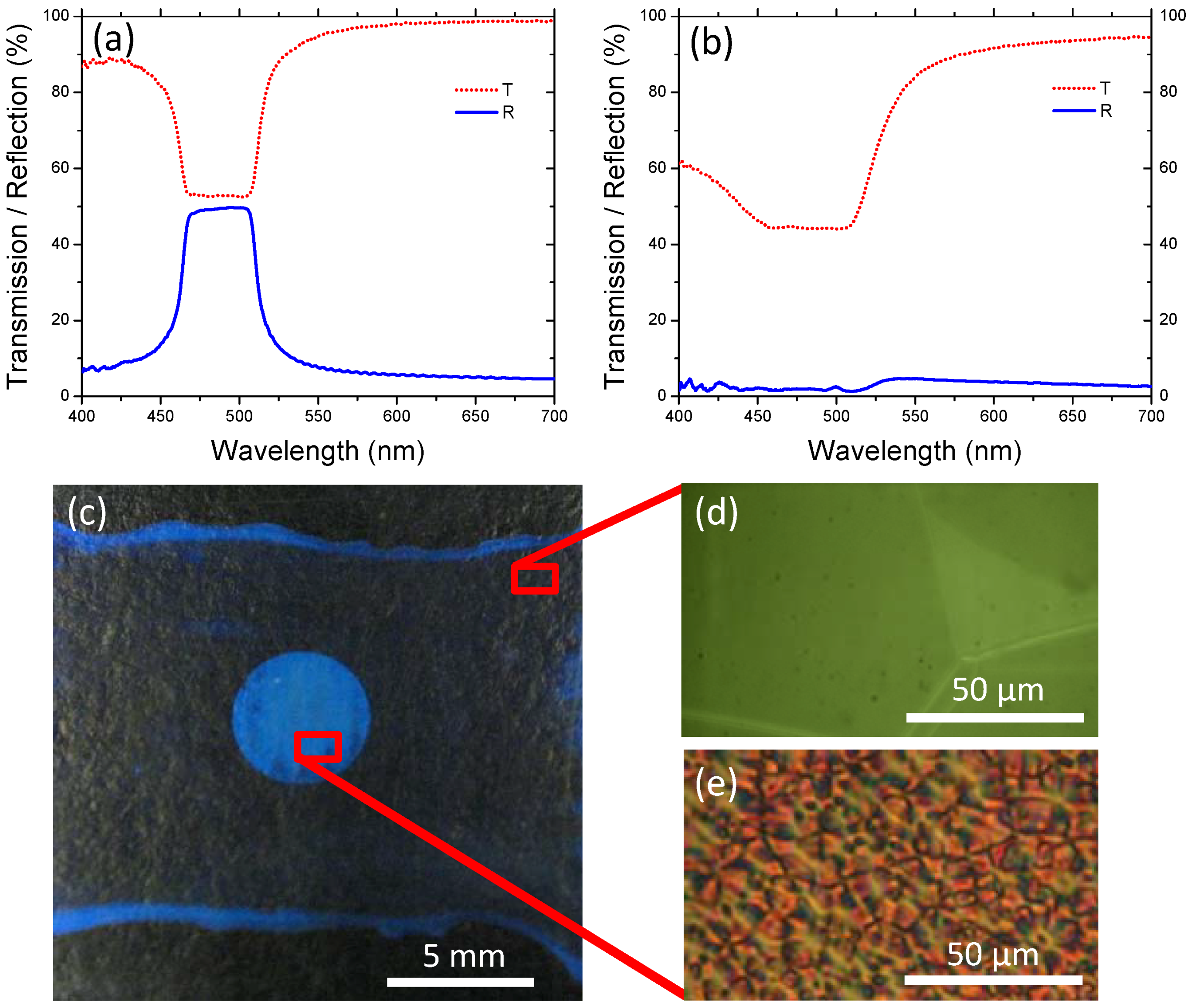
CLC cells were constructed with a commercially available photosensitive alignment material (PAAD-27) spin coated on the input substrate (substrate nearest laser) and polyvinyl alcohol (PVA) spin coated on the output substrate (substrate incident laser light exits a cell). Cell thickness was set by 20 µm glass rod spacers and the cells were filled with a mixture of 72.0 wt % NLC (1444) and 28.0 wt % left-handed (LH) chiral dopant (S811). The initial transmission and reflection spectra taken from the input side of a photoaligned (via linearly polarized light of 442 nm wavelength) LH CLC cell,
Figure 1a, indicated strong blue/green reflection centered at 485 nm. As evident in
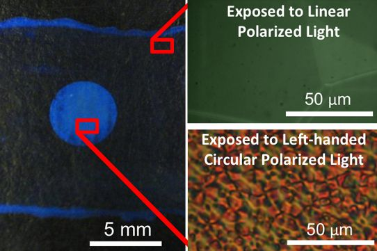
Figure 1b, exposure of this CLC cell to left-handed circularly polarized (LHCP) light of 488 nm wavelength caused a considerable decrease in the reflected intensity and a decrease in the transmitted intensity. The initially reflective cell developed a scattering spot limited to the irradiated area as exhibited by the contrast between the blue spot (scattering area) and its surroundings (off-Bragg angle reflection from well-aligned CLC) depicted in
Figure 1c. A polarized optical micrograph of the initially planar, reflective Grandjean texture is shown in
Figure 1d, and a micrograph of the defect-rich texture within the scattering region of the cell is shown in
Figure 1e. The optically-discernable, micrometer-scale disruptions to the initially Grandjean (planar) CLC texture in the scattering regions are referred to as domains. Reflectivity of the output side of the cell remains the same after domain formation indicating that the domains are localized at the input side of the cell. Although similar in appearance to domains predicted for, and observed upon, application of electric field to CLCs, the periodicities in this work are not as uniform as in previous reports and the mechanism (CP light exposure) is unique to the literature [
23,
24,
25,
26].
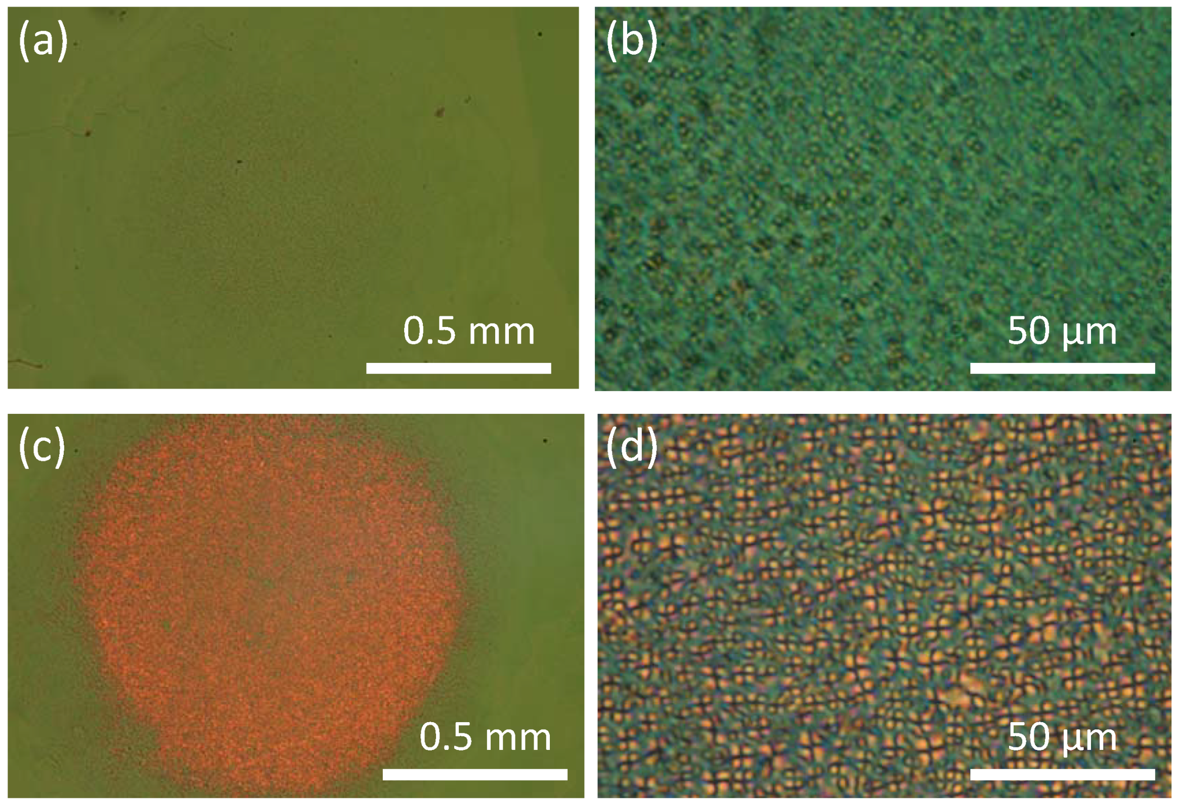
Domains start forming at an early stage of light exposure (
Figure 2a,b) and fully evolve after 5 min (
Figure 2c,d) for irradiation of 0.98 W/cm
2 power density and 488 nm. The examination of CLC cells with PAAD-27 layer irradiated with counterpropagating LHCP beams before and after filling with CLC mixture,
Figure 3, indicated
in situ interaction between PAAD-27 and CLC during irradiation. The conditions of filled cell exposure were emulated for pre-filling exposure of the PAAD layer by placing a PAAD-27 coated substrate in front of non-photosensitive LH CLC reflector with selective reflection peak centered at 485 nm. Schematics of the experimental set-ups are shown in
Figure 3a,b. POM micrographs of the corresponding microstructures,
Figure 3c,d, show that only the area irradiated after filling exhibited domains.
Figure 1.
(a) Transmission and reflection spectra from a Cholesteric liquid crystal (CLC) cell with a photosensitive layer on input substrate: (a) aligned with linearly polarized light; and (b) after 15 min exposure to 488 nm left-handed circularly polarized (LHCP) light (intensity =41 mW/cm2); (c) When imaged off of a Bragg condition the exposed spot appears blue and the rest of the aligned, reflective cell appears transparent (black background); Representative micrographs are shown of both the (d) reflective (aligned) and (e) scattering (domained) regions. Micrographs were taken in transmission mode between crossed polarizers.
Figure 2.
Temporal dependence of domain formation shown with polarized optical micrographs taken (a,b) after 1 min exposure and (c,d) after 5 min exposure.
Figure 2.
Temporal dependence of domain formation shown with polarized optical micrographs taken (a,b) after 1 min exposure and (c,d) after 5 min exposure.
Figure 3.
Schematic of counterpropagating 488 nm LHCP beam exposure via with reflected beam generated by (a) a non-photosensitive CLC reflector and (b) by a filled CLC with photosensitive alignment layer. Corresponding optical micrographs of same cell exposed (c) before and (d) after filling with CLC mixture. Note: QWP = quarter waveplate.
2.2. Similarity of Optically induced Domains to the Domains Generated in CLCs with Homeotropic Boundary Conditions
The domains evident in
Figure 1e,
Figure 2d,
Figure 3d are reminiscent of previous examinations of textures observed upon application of electric field to CLCs [
26] as well as free-standing CLC films (
i.e., no output substrate) [
27,
28,
29,
30]. The domains apparent in
Figure 4a are typical of those previously observed at CLC/air interfaces and have been attributed to focal conical domains that form as a result of the interplay between surface tension and the anchoring/distortion energies [
27,
28,
29,
30]. The molecules periodically are orthogonal to the CLC/air interface. This out-of-plane texture is similar to that observed in cells with photogenerated domains. Additional support of homeotropic boundary conditions inducing similar defects was demonstrated by the domain texture observed in a CLC cell made with weak homeotropic orienting boundary conditions (
Figure 4b). The similarities of the textures of the photogenerated domains to those observed at a CLC/air interface as well as to those in a cell with weak homeoptropic orienting conditions is evidence of the creation of out-of-plane alignment conditions in the photopatterning of scattering states.
Figure 4.
(a) Micrograph of substrate coated with photosensitive alignment material (PAAD-27) layer and CLC material S811(28%)/1444 (droplet of CLC was spread in thin layer. Output substrate is absent); (b) Observation of domain structure in a CLC cell with weak homeotropic orienting boundary conditions (i.e., 4:1 PAAD(1wt% in N,N-dimethylformamide (DMF)): lecithin(1wt% in DMF) volume ratio).
2.3. Effect of the Polarization State of Light on Photosensitive Layer
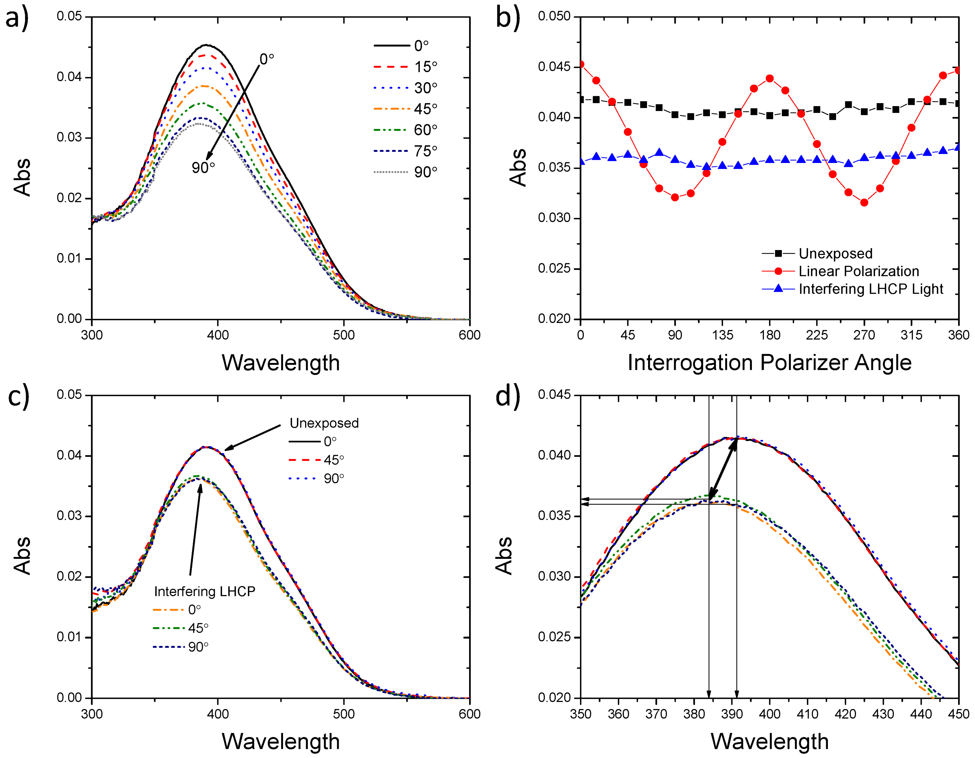
To elucidate the fundamental photochemical mechanism of domain formation, we examined the interaction of the light beam with the photosensitive orienting layer via polarized UV-Vis spectroscopy. Exposure of the photoalignment layer to linearly polarized light of a suitable wavelength (blue/green light) promoted alignment of the long axes of the chromophores in the substrate plane orthogonal to the polarization state of the light (
Figure 5a) due to repeated
trans-
cis-
trans cycling [
31]. The mechanism of photo-induced reorientation is detailed in a recent review [
9]. A hallmark of reorientation is photo-induced birefringence and dichroism [
31]. The absorption of azobenzene-based alignment layers is maximized when the electric field vector of polarized interrogation light is parallel to the alignment direction of the long molecular axis of the trans azobenzene isomer (
i.e., interrogation light polarizer angle defined at 0° in this work (see
Figure 5a)). The orientation of the PAAD-27 alignment layer was interrogated via polarized UV-Vis spectroscopy after each step in a series of exposures similar to those a CLC cell would be subjected to in writing scattering regions/domains (unexposed, exposed to linearly polarized light in the y-direction, then exposed to counterpropagating LHCP light). Representative absorbance spectra generated with linearly polarized interrogation light (0°–90° in 15° increments) are shown after exposure to linearly polarized 488 nm light. In
Figure 5b the absorbance values at 392 nm (average absorption maximum for unexposed PAAD-27 film) are plotted as a function of interrogation light polarization angle (
y = 90°, 270°;
x = 0°, 180°) after each exposure step. Polarized UV-Vis spectroscopy confirmed that the initial state of the spin coated PAAD-27 does not exhibit any preferred alignment (
Figure 5b), as the absorbance of the PAAD-27 layer did not change as a function of interrogation polarizer angle. Sinusoidal variation of the magnitude of absorbance was observed in the case of preferred alignment after exposure to linearly polarized light. Specifically, after irradiation with linearly (
y-direction) polarized 488 nm light the long molecular axis showed preferred orientation (
i.e., in the
x-direction) perpendicular to the polarization state of the laser light (
Figure 5a,b). After exposure to counterpropagating (input beam and CLC-reflected beam) LHCP 488 nm light, the PAAD-27 layer once again exhibited absorbance values independent of polarizer angle accompanied by a concurrent decrease in the magnitude of average absorbance (
Figure 5b).
Figure 5.
(a) UV-Vis absorbance spectra of spin coated PAAD-27 film on glass substrate after exposure to linearly polarized light taken with various polarization angles of interrogation light; (b) Polarized UV-Vis plots of identical spots of PAAD-27 film taken in series: as coated (■), after exposure to linearly polarized light (488 nm, 5 mW/cm2, 15 min) (●), and after exposure to counterpropagating LHCP beams(488 nm, 5 mW/cm2, 15 min) (▲); (c,d) Absorbance spectra taken with interrogation polarizer at 0°, 45° and 90° of PAAD-27 as coated versus after exposure to counterpropagating LHCP light. Note: (d) is a portion of the spectra shown in (c).
Unpolarized and circularly polarized light have been reported to randomize chromophore orientations [
31]. The average decrease in absorbance is attributed to some preferential alignment along the radiation propagation axis (
i.e., out-of-plane or along
z-axis) [
31]. Three of the unexposed absorbance spectra and three spectra obtained after counterpropagating LHCP light exposure absorbance spectra are presented in
Figure 5c, showing a decrease in absorption for the entire peak and a peak blue-shift after exposure. Each exposure state demonstrated nicely overlapping absorbance spectra for 0°, 45°, and 90° interrogation polarizer angles as expected from isotropic alignment (
Figure 5c,d). Zooming in on the peaks in
Figure 5d reveals that choosing a fixed wavelength for determining absorbance may contribute artificial loss in absorbance due to peak shift. For each exposure step the absorbance was measured at 392 nm and 384 nm. Peak to peak shifting, 392 nm (unexposed) → 384 nm (after counterpropagating LHCP) showed an only 8% decrease in the average absorbance loss when compared to the loss measured for both cases at 392 nm. Thus the bulk of the loss in absorbance after exposure is attributed to out-of-plane alignment of the chromophores along the irradiation axis. The blue-shift evident in
Figure 5c,d is potentially attributed to changes in
trans-/
cis-isomer concentration and/or the parallel alignment of chromophores referred to as H-aggregates [
31,
32,
33,
34].
At oblique incidence, even unpolarized light can induce pre-tilt of LC molecules adjacent to a photoalignment surface [
31,
35]. All polarization states of incident light have shown the propensity to promote some out-of-plane alignment but only counterpropagating CP beams of same handedness have been shown to generate domains [
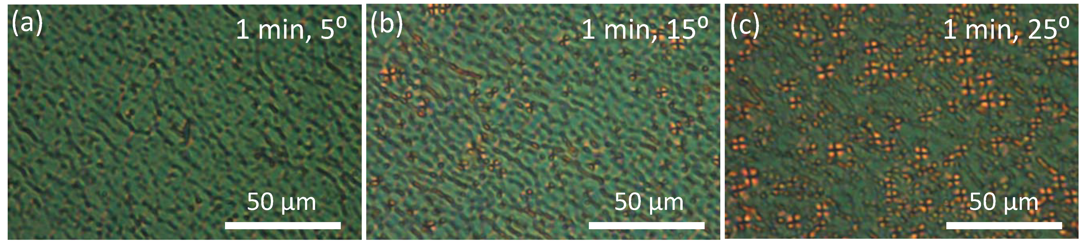
22]. By tilting the CLC cell, an increased efficiency of domain formation with increased tilt angle was observed (
Figure 6a–c).
Figure 6.
Effect of angle of incident irradiation (a) 5°; (b) 15°; and (c) 25° on domain formation after 1 min exposure.
Figure 6.
Effect of angle of incident irradiation (a) 5°; (b) 15°; and (c) 25° on domain formation after 1 min exposure.
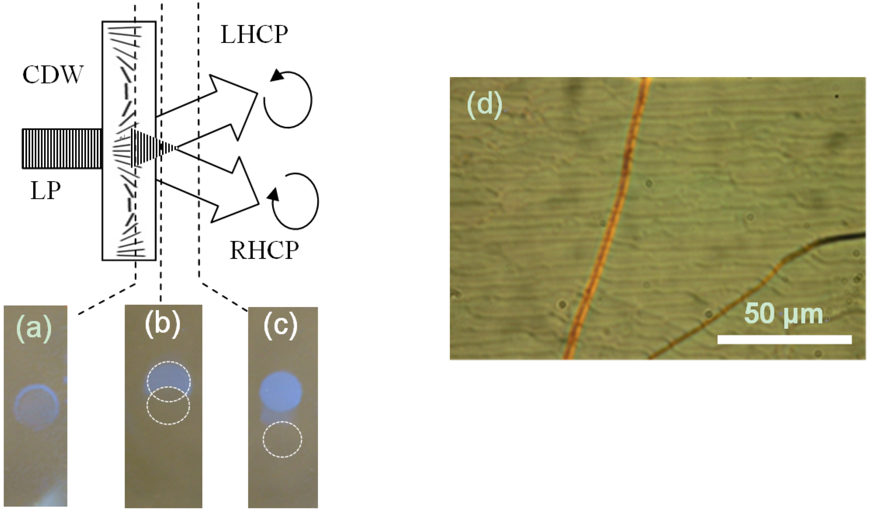
2.4. Frustrated Photoalignment
We can thus draw the following general picture for the phenomenon under discussion. Counterpropagating LHCP beams (the incident beam and the beam reflected from the CLC) overlap creating a spiral standing wave—a linear polarization state that is rotating along the irradiation propagation axis (Figure S1). It degrades the planar alignment condition near the boundary. This diminishes the bandgap reflection, thus the standing wave and the resulting linear polarization state acting at the boundary layer is lost (Figure S2). Since the incident CP light itself does not have an appreciable in-plane alignment effect on boundary conditions, the resultant textured state of the CLC is preserved.
The photosensitive alignment layer of the CLC cell, being only a few nanometers thin, is actually subject to effectively linear polarized light in the case of normal incidence or to a polarization modulation pattern if the cell is tilted with respect to the propagation direction of the beams and thus intersects the standing wave at planes corresponding to different polarization states. Experiments show that linearly polarized incident beam does not cause domain formation and can eliminate the textures [
22]. To test a hypothesis that the domain formation may have been caused by spatially modulated alignment conditions, the CLC was exposed to a laser beam propagated through a linear-to-cycloidal polarization converter [
36]. A cycloidal diffractive waveplate (CDW) was used for the purpose. The waveplate splits an input linear polarized light into ±1st diffraction orders of orthogonal circular polarization states. Spatially rotating linear polarization pattern (with 6 μm pitch in the particular case) is obtained at the overlap region of the beams, however, no domain-like textures are generated in this area or in the area subject to the RHCP beam as shown in
Figure 7.
Figure 7.
Exposing the interface of a CLC with the photosensitive boundary of the cell to spatially modulated linear polarization pattern obtained at the output of a linear-to-cycloidal polarization converter. (a) The interface is in the polarization modulation pattern present at the overlap region of right- and left-circular polarized beams outputted by the cycloidal diffractive waveplate (CDW). The latter acts as a polarization converter for a linear polarized (LP) incident beam; (b) The interface is in the partial overlap region of the orthogonal circular polarized beams; (c) The interface is out of overlap region of the orthogonal circular polarized beams. Domains are present only in area subject to LHCP beam outside of the overlap area; (d) Grating structure formed in the overlap area of the beams.
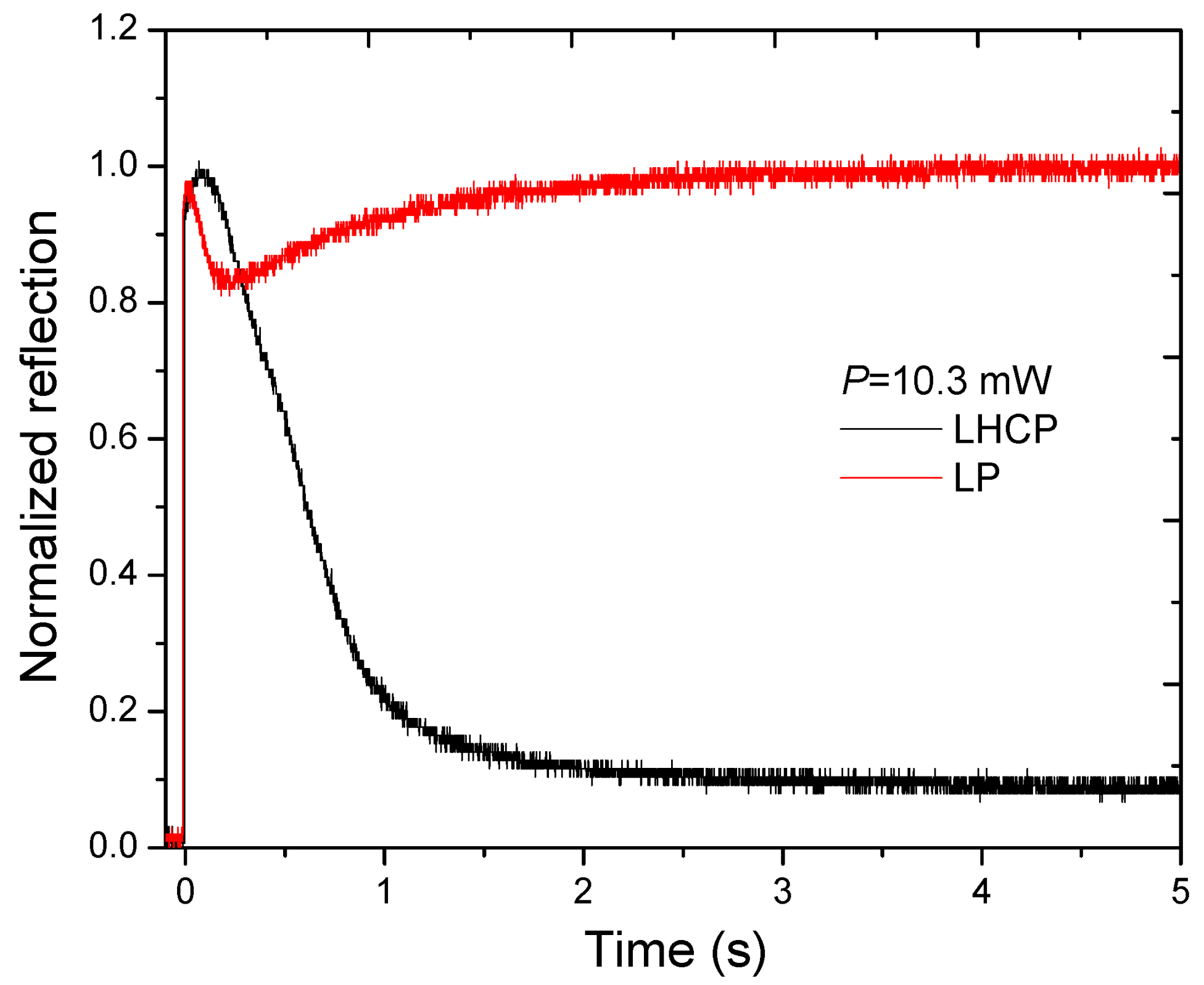
A linear polarized input, modulated or not, does not induce domain formation. To reconcile this statement with the observation that only bandgap mode CP light induces domains (Note: the initial overlap of input and reflected beams creates a linear polarization state at the photosensitive alignment layer), we need to assume that realignment of the PAAD molecules at the influence of a linear polarized light has a transient state. With no feedback (
i.e., change in polarization state), the light remains linearly polarized accomplishing the transition to the new oriented state. With frustrated feedback (
i.e., change in polarization state due to loss of reflected beam), the domained state is preserved. This was proven directly by registering the reflection dynamics for a linear polarized beam incident on the CLC,
Figure 8. The reflection of the CLC when exposed to a linearly polarized beam decreased at the initial stages of the process followed by relaxation to the highly reflective state. In contrast, the decrease in the reflection continues for the circular polarized input.
Figure 8.
Reflection dynamics of CLC upon exposure with linearly polarized (LP) and left-handed circularly polarized (LHCP) (wavelength = 488 nm, Intensity = 1.2 W/cm2).
Figure 8.
Reflection dynamics of CLC upon exposure with linearly polarized (LP) and left-handed circularly polarized (LHCP) (wavelength = 488 nm, Intensity = 1.2 W/cm2).
The origins of the transient state that results in photogenerated domain formation is apparently a pretilt induced by the PAAD due to statistical accumulation of PAAD molecules in a direction perpendicular to the polarization of light, which includes the direction normal to the substrates [
37]. The formation of domains of similar texture at a CLC/air interface or with a weak homeotropic boundary condition (PAAD/lecithin alignment layer) is evidence for this hypothesis. The UV-Vis spectroscopic data provides evidence for out-of-plane alignment which also supports this working hypothesis.


