Hydrogen-Bonding Motifs in Piperazinediium Salts


Abstract
:1. Introduction
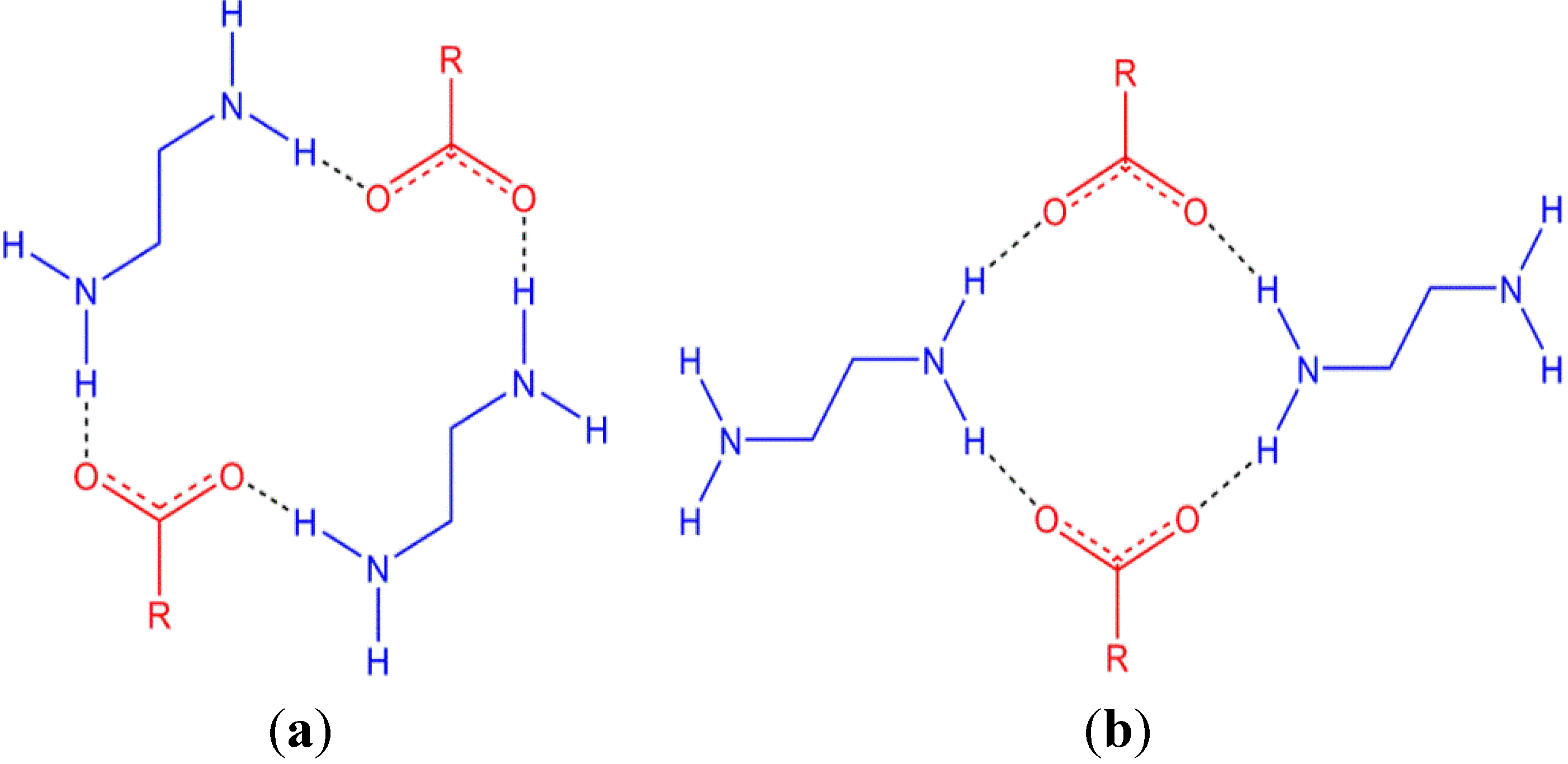
 (18) ring (using Graph-Set analysis) [20,21] formed between two cations and two anions (Figure 1a). There are two ring motifs involving piperazinediium/carboxylates that can give rise to 1D hydrogen-bonded chains which vary by the manner in which the cations bridge between anions (Figure 1). A search of the Cambridge Structural Database reveals that there are marginally more structures reported with the larger R (18) ring than with the smaller R (12) ring with 29 and 19 reported entries, respectively [22,23].(18) ring (a) and a smaller R (12) ring (b). The protonated piperazine rings are shown side-on for clarity.
(18) ring (a) and a smaller R (12) ring (b). The protonated piperazine rings are shown side-on for clarity.
(18) ring (using Graph-Set analysis) [20,21] formed between two cations and two anions (Figure 1a). There are two ring motifs involving piperazinediium/carboxylates that can give rise to 1D hydrogen-bonded chains which vary by the manner in which the cations bridge between anions (Figure 1). A search of the Cambridge Structural Database reveals that there are marginally more structures reported with the larger R (18) ring than with the smaller R (12) ring with 29 and 19 reported entries, respectively [22,23].(18) ring (a) and a smaller R (12) ring (b). The protonated piperazine rings are shown side-on for clarity.
(18) ring (a) and a smaller R (12) ring (b). The protonated piperazine rings are shown side-on for clarity.
2. Results and Discussion

2.1. p-Toluenesulfonate Structures
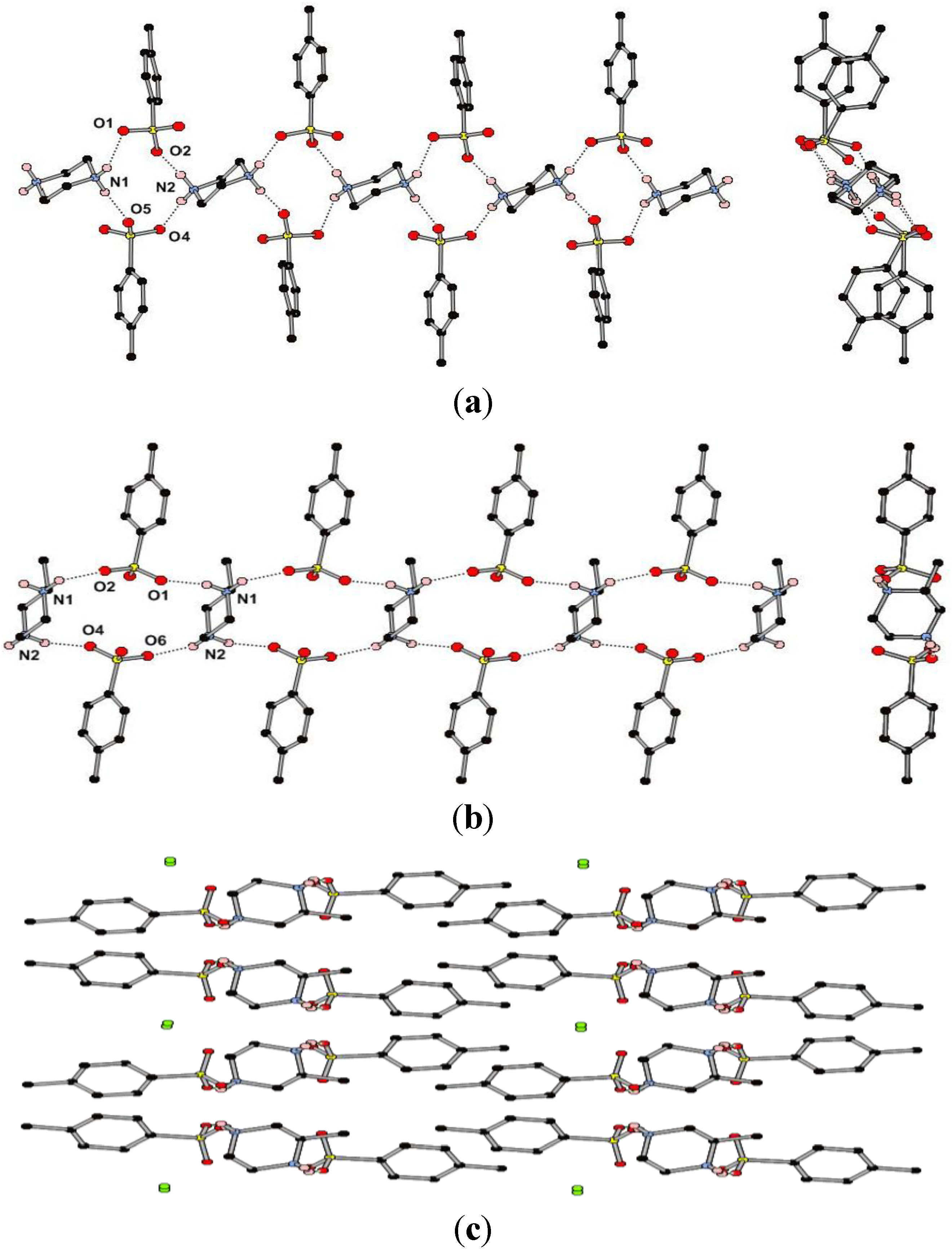
(18) ring (Figure 3b, cf. Figure 1). The hydrogen bonds in this chain have H···A distances in the range 1.91–2.00 Å and D–H···A angles in the range 143°–158° (Table 1) which are considerably smaller ranges than those observed in the structure of 1. There are longer and less linear interactions between the 1D chains with each of the NH2 groups forming an R  (6) ring with a sulfonate in a neighboring chain, giving rise to 2D sheets. These sheets interdigitate with aryl CH···π interactions between the layers. The 50% occupancy water molecules reside between sulfonate groups on the periphery of the 2D sheets with O···O distances of ca. 2.8 Å, suggesting that there may be hydrogen bonding interactions present (Figure 3c) (note: the hydrogen atoms of the partial occupancy water could not be experimentally located from the Fourier difference map). Crystallinity is lost upon heating and removal of the water molecule. Whilst both enantiomers of the cation are present in the crystal structure, with the presence of a glide plane, the non-centrosymmetry arises from the packing in which all methyl groups are orientated in one direction with respect to the a axis. The compound may potentially display piezoelectric properties as observed in the closely related (r-H2Mepip)(CCl3CO2) [28].

| 1 | 2 | ||||||
|---|---|---|---|---|---|---|---|
| Interaction | D···A (Å) | H···A (Å) | D–H···A (°) | Interaction | D···A (Å) | H···A (Å) | D–H···A (°) |
| N1···O1 | 2.9126(16) | 2.10 | 146.3 | N1···O2#3 | 2.790(5) | 2.00 | 143.0 |
| N1···O5#1 | 2.8089(17) | 1.89 | 174.1 | N1···O1#4 | 3.076(6) | 2.41 | 129.0 |
| N2···O2#2 | 2.7223(17) | 1.84 | 159.5 | N1···O1 | 2.798(5) | 1.92 | 158.0 |
| N2···O4 | 2.7542(15) | 1.86 | 163.0 | N1···O2#4 | 3.063(5) | 2.50 | 120.0 |
| N1···O2 | 3.0536(15) | 2.50 | 114.9 | N2···O6#5 | 2.772(5) | 1.98 | 142.9 |
| N1···O6 | 2.8620(16) | 2.30 | 114.9 | N2···O4 | 3.080(5) | 2.42 | 128.8 |
| – | – | – | – | N2···O4#6 | 2.784(5) | 1.91 | 157.3 |
| – | – | – | – | N2···O6 | 3.149(6) | 2.57 | 121.0 |
2.2. Chloroacetate Structures
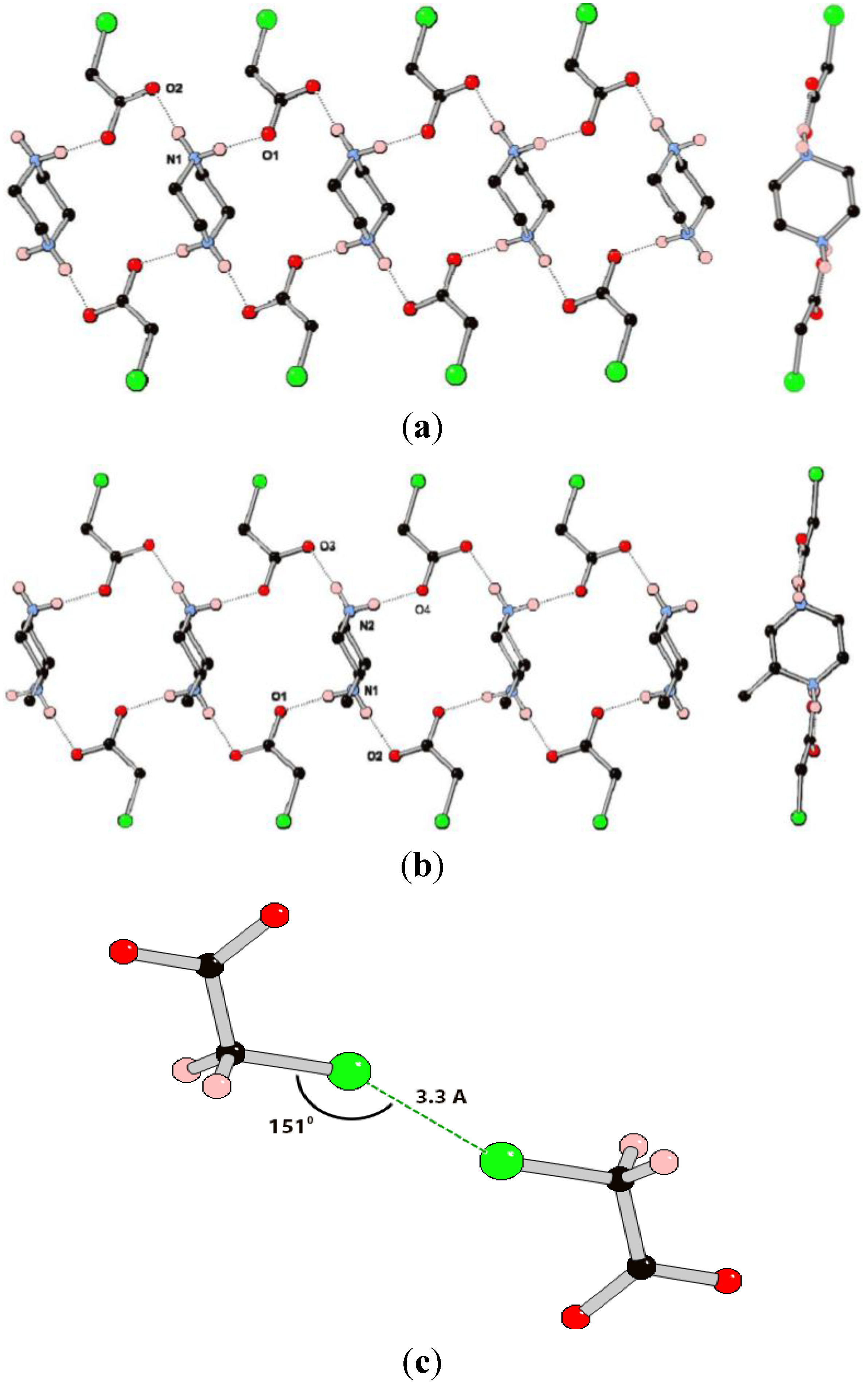
(18) ring motif (Figure 4b). Indeed, the similarity between the structures of 3 and 4 is reflected in the similar cell parameters (see experimental details). The hydrogen bonding interactions in 4 appear marginally shorter and more linear than those in 3, although there is obvious uncertainty in proton position from X-ray data. Whilst the overall structure is centrosymmetric, each chain contains only one isomer of 2-methylpiperazinediium. There are no hydrogen bonding interactions between the chains, with the only noteworthy close interaction being between Cl2 and its symmetry generated equivalent with a Cl···Cl interaction with very similar geometric parameters to those observed in 3. The second crystallographically unique anion does not have a corresponding close contact. In systems 3 and 4 it appears that the presence of a methyl substituent does not significantly alter the structure, with only very minor differences observed between the hydrogen bonding geometries.
| 3 | 4 | ||||||
|---|---|---|---|---|---|---|---|
| Interaction | D···A (Å) | H···A (Å) | D-H···A (°) | Interaction | D···A (Å) | H···A (Å) | D-H···A (°) |
| N1···O2 | 2.7517(17) | 1.85 | 165.6 | N1···O1 | 2.741(2) | 1.76 | 172.4 |
| N1···O1#1 | 2.6915(16) | 1.78 | 169.2 | N1···O2#2 | 2.741(2) | 1.76 | 171.7 |
| – | – | – | – | N2···O3 | 2.727(2) | 1.75 | 169.8 |
| – | – | – | – | N2···O4#2 | 2.713(2) | 1.73 | 174.8 |
3. Experimental Section
3.1. Synthesis

3.2. X-Ray Crystallography
| Compound | 1 AT10 | 2 CC10 | 3 AT14 | 4 CC14 |
|---|---|---|---|---|
| Formula | (H2Pip)(TsO)2 | (H2MePip)(TsO)2·½H2O | (H2Pip)(OAcCl)2 | (H2MePip)(OAcCl)2 |
| Empirical Formula | C18H26N2O6S2 | C19H29N2O6.5S2 | C8H16Cl2N2O4 | C9H18Cl2N2O4 |
| Formula Mass | 430.53 | 453.56 | 275.13 | 289.15 |
| Crystal System | Triclinic | Monoclinic | Triclinic | Monoclinic |
| Space Group | P-1 | Pc | P-1 | P21/c |
| a/Å | 5.9020(12) | 14.584(3) | 5.6260(11) | 5.7050(11) |
| b/Å | 13.059(3) | 9.863(2) | 7.2100(14) | 29.975(6) |
| c/Å | 13.581(3 | 7.6460(15) | 7.5280(15) | 7.5280(15) |
| α/° | 73.55(3) | 90 | 77.60(3) | 90 |
| β/° | 86.00(3) | 100.89(3) | 80.83(3) | 97.26(3) |
| γ/° | 84.09(3) | 90 | 85.29(3) | 90 |
| V/Å3 | 997.7(3) | 1080.0(4) | 294.07(10) | 1277.0(4) |
| μ/mm−1 | 0.305 | 0.284 | 0.553 | 0.514 |
| Refs. Collected | 36824 | 11230 | 11028 | 23550 |
| Theta Range | 1.56–31.54 | 2.71–31.50 | 2.80–31.49 | 1.36–31.51 |
| Unique Refs. (Rint) | 5606 (0.0660) | 5649 (0.0779) | 1660 (0.0706) | 3548 (0.0460) |
| Obs. Refs. (I > 2σI) | 5391 | 5038 | 1557 | 3414 |
| R1 (I > 2σI/all data) | 0.0386/0.0399 | 0.0786/0.0834 | 0.0361/0.0379 | 0.0473/0.0486 |
| wR2 (I > 2σI/all data) | 0.1027/0.1040 | 0.2118/0.2184 | 0.0940/0.0950 | 0.1130/0.1137 |
| GooF | 1.077 | 1.081 | 1.120 | 1.122 |
4. Conclusions
(12) motif in 1 and the more common R (18) motif in 2–4 (as ascertained by searches of the CSD). In the structures of 1 and 2 there is additional, weak hydrogen bonding between the chains that is absent in 3 and 4 due to the larger number of acceptor atoms present in the sulfonate versus the carboxylate. In the chloroacetate structures, the presence of a methyl group does not affect the hydrogen bonding motif that is observed, whereas in the toluenesulfonate structures it does appear that there is some influence, possibly due to the larger bulk of sulfonate compared to carboxylate. Combined with CSD searches, these results suggest that the 1D chain based on R (18) hydrogen-bonding motifs is quite a stable supramolecular synthons and future work aims to exploit this in crystal engineering applications. Such applications involve the use of rigid polycarboxylates to construct per-designed network architectures, exploring the physical properties of materials containing different synthons (with potential pharmaceutical relevance) and exploring effects such as piezoelectricity in engineered non-centrosymmetric networks of 2-methylpiperazine.Acknowledgments
Conflicts of Interest
References
- Steed, J.W.; Atwood, J.L. Supramolecular Chemistry, 2nd ed.; John Wiley & Sons: Chichester, UK, 2009. [Google Scholar]
- Steed, J.W.; Turner, D.R.; Wallace, K.J. Core Concepts in Supramolecular Chemistry and Nanochemistry; John Wiley: Chichester, UK; Hoboken, NJ, USA, 2007. [Google Scholar]
- Burrows, A.D. Crystal engineering using multiple hydrogen bonds. Struct. Bond. 2004, 108, 55–95. [Google Scholar] [CrossRef]
- Etter, M.C. Hydrogen-bonds as design elements in organic-chemistry. J. Phys. Chem. 1991, 95, 4601–4610. [Google Scholar] [CrossRef]
- Jeffrey, G.A. An Introduction to Hydrogen Bonding; OUP USA: New York, NY, USA, 1997. [Google Scholar]
- Spackman, M.A.; Jayatilaka, D. Hirshfeld surface analysis. CrystEngComm 2009, 11, 19–32. [Google Scholar] [CrossRef]
- Yates, J.R.; Pham, T.N.; Pickard, C.J.; Mauri, F.; Amado, A.M.; Gil, A.M.; Brown, S.P. An investigation of weak CH···O hydrogen bonds in maltose anomers by a combination of calculation and experimental solid-state NMR spectroscopy. J. Am. Chem. Soc. 2005, 127, 10216–10220. [Google Scholar] [CrossRef]
- Chierotti, M.R.; Gobetto, R. NMR crystallography: The use of dipolar interactions in polymorph and co-crystal investigation. CrystEngComm 2013, 15, 8599–8612. [Google Scholar] [CrossRef]
- Mafra, L.; Santos, S.M.; Siegel, R.; Alves, I.; Almeida Paz, F.A.; Dudenko, D.; Spiess, H.W. Packing interactions in hydrated and anhydrous forms of the antibiotic ciprofloxacin: A solid-state NMR, X-ray diffraction, and computer simulation study. J. Am. Chem. Soc. 2011, 134, 71–74. [Google Scholar]
- Saleh, G.; Gatti, C.; LoPresti, L.; Contreras-García, J. Revealing non-covalent interactions in molecular crystals through their experimental electron densities. Chemistry 2012, 18, 15523–15536. [Google Scholar] [CrossRef]
- Ward, M.D. Design of crystalline molecular networks with charge-assisted hydrogen bonds. Chem. Commun. 2005, 47, 5838–5842. [Google Scholar] [CrossRef]
- Brisse, F.; Denault, J.; Sangin, J.P. Study of aliphatic chain compounds 1. Synthesis and crystallographic characterization of bis (n-alkanoates) of piperazinium, 2[CxH2x−1O2−][C4H12N22+]. J. Appl. Crystallogr. 1982, 15, 279–281. [Google Scholar] [CrossRef]
- Brisse, F.; Sangin, J.P. Study of compounds with aliphatic chains 2. The structure of piperazinium bis(normal-dodecanoate). Acta Crystallogr. Sect. B 1982, 38, 215–221. [Google Scholar] [CrossRef]
- Venkatramani, L.; Craven, B.M. Disordered fatty-acid chains in piperazinium myristate and palmitate. Acta Crystallogr. Sect. B 1991, 47, 968–975. [Google Scholar] [CrossRef]
- Ponomarev, V.I.; Klimchuk, E.G.; Merzhanov, A.G.; Filipenko, O.S. Structural-chemical transformations during organic self-propagating high-temperature synthesis. Crystal structure of piperazine malonate and its crystal hydrate. Russ. Chem. B. 1997, 46, 939–943. [Google Scholar] [CrossRef]
- Vanier, M.; Belangergariepy, F.; Brisse, F. Structural studies of compounds with aliphatic chains 10. Structure of piperazinium suberate monohydrate, [C8H12O42−][C4H12N22+]H2O. Acta Crystallogr. Sect. C 1983, 39, 916–917. [Google Scholar]
- Vanier, M.; Brisse, F. Structural studies of compounds with aliphatic chains 7. The structure of piperazinium glutarate and the geometry of the piperazinium cation. Acta Crystallogr. Sect. B 1982, 38, 3060–3063. [Google Scholar] [CrossRef]
- Vanier, M.; Brisse, F. Structural studies of compounds with aliphatic chains 8. Structure of piperazinium succinate, [C4H4O42−][C4H12N22+]. Acta Crystallogr. Sect. C 1983, 39, 912–914. [Google Scholar] [CrossRef]
- Vanier, M.; Brisse, F. Structural studies of compounds with aliphatic chains 9. Structure of piperazinium adipate, [C6H8O42−][C4H12N22+]. Acta Crystallogr. Sect. C 1983, 39, 914–915. [Google Scholar] [CrossRef]
- Bernstein, J.; Davis, R.E.; Shimoni, L.; Chang, N.L. Patterns in hydrogen bonding—Functionality and graph set analysis in crystals. Angew. Chem. Int. Ed. 1995, 34, 1555–1573. [Google Scholar] [CrossRef]
- Etter, M.C.; Macdonald, J.C.; Bernstein, J. Graph-set analysis of hydrogen-bond patterns in organic-crystals. Acta Crystallogr. Sect. B 1990, 46, 256–262. [Google Scholar] [CrossRef]
- Allen, F.H. The cambridge structural database: A quarter of a million structures and rising. Acta Crystallogr. Sect. B 2002, B58, 380–388. [Google Scholar] [CrossRef]
- Cambridge Structural Database. Version 5.31 + 1 update. February 2012.
- Katagiri, H.; Morimoto, M.; Sakai, K. A pair of diastereomeric 1:1 salts of (s)- and (r)-2-methylpiperazine with (2s,3s)-tartaric acid. Acta Crystallogr. Sect. C 2009, 65, O357–O360. [Google Scholar] [CrossRef]
- Katagiri, H.; Morimoto, M.; Sakai, K. A pair of diastereomeric 1:2 salts of (r)- and (s)-2-methylpiperazine with (2s,3s)-tartaric acid. Acta Crystallogr. Sect. C 2010, 66, O20–O24. [Google Scholar] [CrossRef]
- Aakeroy, C.B.; Bahra, G.S.; Nieuwenhuyzen, M. Piperazinium l-tartrate. Acta Crystallogr. Sect. C 1996, 52, 1471–1473. [Google Scholar] [CrossRef]
- Farrell, D.M.M.; Ferguson, G.; Lough, A.J.; Glidewell, C. Chiral versus racemic building blocks in supramolecular chemistry: Tartrate salts of organic diamines. Acta Crystallogr. Sect. B 2002, 58, 272–288. [Google Scholar] [CrossRef]
- Cai, H.-L.; Zhang, T.; Chen, L.-Z.; Xiong, R.-G. The first homochiral compound with temperature-independence of piezoelectric properties. J. Mater. Chem. 2010, 20, 1868–1870. [Google Scholar] [CrossRef]
- Bondi, A. Van der waals volumes + radii. J. Phys. Chem. 1964, 68, 441–451. [Google Scholar] [CrossRef]
- Metrangolo, P.; Meyer, F.; Pilati, T.; Resnati, G.; Terraneo, G. Halogen bonding in supramolecular chemistry. Angew. Chem. Int. Ed. 2008, 47, 6114–6127. [Google Scholar] [CrossRef]
- Rissanen, K. Halogen bonded supramolecular complexes and networks. CrystEngComm 2008, 10, 1107–1113. [Google Scholar] [CrossRef]
- Awwadi, F.F.; Willett, R.D.; Peterson, K.A.; Twamley, B. The nature of halogen···halogen synthons: Crystallographic and theoretical studies. Chemistry 2006, 12, 8952–8960. [Google Scholar] [CrossRef]
- Metrangolo, P.; Resnati, G. Type II halogen···halogen contacts are halogen bonds. IUCrJ 2013, 1, 5–7. [Google Scholar] [CrossRef]
- Kabsch, W. Automatic processing of rotation diffraction data from crystals of initially unknown symmetry and cell constants. J. Appl. Cryst. 1993, 26, 795–800. [Google Scholar] [CrossRef]
- Sheldrick, G.M. A short history of SHELX. Acta Crystallogr. Sect. A 2008, 64, 112–122. [Google Scholar] [CrossRef]
- Barbour, L.J. X-Seed—A software tool for supramolecular crystallography. J. Supramol. Chem. 2001, 1, 189–191. [Google Scholar] [CrossRef]
© 2014 by the authors; licensee MDPI, Basel, Switzerland. This article is an open access article distributed under the terms and conditions of the Creative Commons Attribution license (http://creativecommons.org/licenses/by/3.0/).
Share and Cite
Hawes, C.S.; Chen, C.; Tran, A.; Turner, D.R. Hydrogen-Bonding Motifs in Piperazinediium Salts. Crystals 2014, 4, 53-63. https://doi.org/10.3390/cryst4010053
Hawes CS, Chen C, Tran A, Turner DR. Hydrogen-Bonding Motifs in Piperazinediium Salts. Crystals. 2014; 4(1):53-63. https://doi.org/10.3390/cryst4010053
Chicago/Turabian StyleHawes, Chris S., Cherry Chen, Andrew Tran, and David R. Turner. 2014. "Hydrogen-Bonding Motifs in Piperazinediium Salts" Crystals 4, no. 1: 53-63. https://doi.org/10.3390/cryst4010053
APA StyleHawes, C. S., Chen, C., Tran, A., & Turner, D. R. (2014). Hydrogen-Bonding Motifs in Piperazinediium Salts. Crystals, 4(1), 53-63. https://doi.org/10.3390/cryst4010053