Review

The Interaction of mTOR and Nrf2 in Neurogenesis and Its Implication in Neurodegenerative Diseases

1
Department of Surgery, Morsani College of Medicine, University of South Florida, Tampa, FL 33612, USA
2
Department of Medical Engineering, College of Engineering and Morsani College of Medicine, University of South Florida, Tampa, FL 33612, USA
*
Author to whom correspondence should be addressed.
Cells 2022, 11(13), 2048; https://doi.org/10.3390/cells11132048
Submission received: 23 May 2022 / Revised: 23 June 2022 / Accepted: 26 June 2022 / Published: 28 June 2022
(This article belongs to the Special Issue Nrf2 Signaling Pathway in Cardiovascular Health and Disease)

Abstract

:
Neurogenesis occurs in the brain during embryonic development and throughout adulthood. Neurogenesis occurs in the hippocampus and under normal conditions and persists in two regions of the brain—the subgranular zone (SGZ) in the dentate gyrus of the hippocampus and the subventricular zone (SVZ) of the lateral ventricles. As the critical role in neurogenesis, the neural stem cells have the capacity to differentiate into various cells and to self-renew. This process is controlled through different methods. The mammalian target of rapamycin (mTOR) controls cellular growth, cell proliferation, apoptosis, and autophagy. The transcription factor Nrf2 (nuclear factor erythroid 2-related factor 2) is a major regulator of metabolism, protein quality control, and antioxidative defense, and is linked to neurogenesis. However, dysregulation in neurogenesis, mTOR, and Nrf2 activity have all been associated with neurodegenerative diseases such as Alzheimer’s, Huntington’s, and Parkinson’s. Understanding the role of these complexes in both neurogenesis and neurodegenerative disease could be necessary to develop future therapies. Here, we review both mTOR and Nrf2 complexes, their crosstalk and role in neurogenesis, and their implication in neurodegenerative diseases.

1. Introduction

Neurogenesis is a process that gives rise to new neuronal cells from neural stem cells (NSCs) in the brain during embryonic development and throughout adulthood [1]. NSCs can differentiate into different cells and have the ability for self-renewal [1,2]. Under normal physiological conditions, neurogenesis occurs in the hippocampus [2]. Adult neurogenesis persists in two regions of the brain—the subgranular zone (SGZ) in the dentate gyrus of the hippocampus and the subventricular zone (SVZ) of the lateral ventricles [1,2,3,4]. Cells are generated in the SGZ granule and play a critical role in learning and memory. Conversely, SVZ astrocyte-like cells produce transit-amplifying cells that will then produce neuroblasts [3,5]. This then migrates to the rostral migratory stream of the olfactory bulb [3,5,6,7]. NSCs must be able to differentiate, divide, and migrate in order to function properly. In adults, the process of neurogenesis is stimulated by a variety of physiological, pathological, and pharmacological stimuli. An overproduction or alternation of these stimuli has been seen in neurodegenerative diseases. For instance, genes such as tau, presenilin (PSEN)1, and huntingtin are physiologically involved in modulating brain plasticity in an embryonic brain. Studies have shown that tau pathology is correlated to functional deficits in Alzheimer’s disease (AD). Mutations in the huntingtin gene can lead to the development of Huntington’s disease, and changes in PSEN1 expression can also cause neurodegeneration and dementia in familial Alzheimer’s disease [8,9,10,11]. Additionally, alterations in the dentate gyrus of the hippocampus, SVZ, and olfactory bulb have been observed in diseases such as Parkinson’s disease (PD) and Huntington’s disease (HD) [11]. This further enhances the question of the link between neurogenesis and the development of neurodegenerative disease.
Many cellular activities such as proliferation, cell growth, and protein synthesis are regulated by the mammalian target of the rapamycin (mTOR) pathway, a key regulator of these processes [12]. Upon activation, the mTOR complex is phosphorylated through two of its substrates—the eukaryotic initiation factor 4E (eIF4E)-binding proteins 4E-BP1 and 4E-BP2 and the ribosomal S6 kinases S6K1 and S6K 2 [13]. Several studies have shown the involvement of mTOR in neurogenesis as its activation can change NSC migration, differentiation, maturation, and dendrite development [14]. Furthermore, elevated phosphorylated mTOR and its downstream protein targets have been observed in AD pathology suggesting the implication of mTOR in neurodegenerative diseases [15,16,17]. An increase in oxidative stress is one of the key mechanisms leading to the development of AD [18]. This stress is known to interfere with neural survival proliferation and differentiation, suggesting nuclear factor erythroid 2-related factor (Nrf2) could induce NSC proliferation in the subgranular zone of the dentate gyrus during oxidative stress [18]. Neuronal cells differ from any other cells in shape, function, mode of activation, and renewal. For instance, endothelial cells such as skin cells can function independently, while neuron cells depend on the stimulation and neurotransmitters generated by other nerves to function. Nerve cells consist prominently of the nucleus, and once these cells are destroyed, new cells cannot be generated. Due to those variations, the key mechanism of activation between mTOR and Nrf2 might have differences.
After years of investigating neurodegenerative diseases such as AD, PD, and HD, researchers still have not found the best treatment methods. Studies have still not been fully able to answer the questions of how protein folding, and neuronal cell death are associated with AD, PD, and HD. Further, can neuronal replacement be used as a form of therapy? Can molecular mechanisms associated with neurodegenerative diseases and neurogenesis be used as a treatment target? All of these questions are still not answered. Moreover, even after decades of investigations in neurogenesis and the field of NSC by neurobiologists, there are questions that remain unclear, such as can the mechanism and factors that regulated this process can be used to treat neurological disease? To what extent is neurogenesis beneficial since having more new neurons is not always better [19]? Furthermore, in pathological and physiological states, what are the molecular and cellular mechanisms present within the brain that would lead to the repair or reshaping of the neurons?
As regenerative medicine, also known as stem cell therapy, opens a new direction in modern medicine, understanding key elements associated with both neurogenesis and neurodegenerative diseases could provide new insight into certain neurodegenerative treatment methods. In this review, we discuss mTOR and Nrf2 complexes, their crosstalk, and roles in neurogenesis, and the implication in neurodegenerative diseases such as AD, PD, and HD, and how a better understanding of those complexes could provide possible answers to questions mentioned previously. Our objective is to provide an overview of the implication of mTOR and Nrf2 in neurogenesis as the formation of neuronal cells and neuronal cell death play a critical role in embryonic and adult brain development. Further changes in these processes have been seen in neurodegenerative diseases, which we further evaluate in this review.

2. Nrf2 in Neurogenesis and Disease Development

2.1. Nrf2 Mechanism Pathway

One of the key regulators of cellular oxidation is nuclear factor erythroid 2-related factor 2 (Nrf2) which is a key transcriptional factor that regulates different genes in cells under both normal and stress conditions [20,21]. Nrf2 maintains cellular homeostasis by activating the expression of cytoprotective, antioxidant, and anti-inflammatory genes [21]. Nrf2 is a basic leucine zipper (bZip) transcription factor from the cap ‘n’ collar (CNC) family which includes p45 NF-E2, NRF1, NRF3, CNC homolog 1 (Bach 1) and Bach 2, broad-complex, tramtrack, and bric-a-brac (BTB) [20,22]. One difference between the members of this family of transcription factors is that Nrf1 through 3 acts as a transcriptional activator, while Bach 1 and 2 serve as transcriptional repressors, and play a role in regulating Nrf2 [20]. In the nucleus, Nrf2 can heterodimerize with small MAF or JUN proteins. When Nrf2 heterodimerizes with small MAF (sMaf), it enhances nucleotide sequences in the DNA called antioxidant response elements (ARE) or electrophile response elements (EpRE) [20,21]. Brain-derived neurotrophic factors show that Nrf2 cellular function includes metabolic regulation, protein quality control, and antioxidative defense by initiating the transcription of cytoprotective genes, including anti-inflammatory interleukin (IL)-10, peroxisome proliferator-activated receptor gamma coactivator 1-alpha (PGC-1α), and iron exporter ferroportin 1 [20,21]. The Nrf2 molecular structure consists of seven Nrf2-erythroid-derived CNC homology (ECH; Neh) domains (Neh1-7), that are critical in Nrf2 repression and activity [20]. For instance, Kelch-like ECH associated protein 1 (Keap1) is a negative regulator of Nrf2 under unstressed cellular conditions [20]. Moreover, Neh6 is important for Nrf2 degradation in stressed cells independent of Keap1 while on the other hand Neh3 interacts with other transcription factors through DNA binding and dimerization [20]. Nrf2 is regulated in both the nucleus and the cytosol [20]. Glycogen synthase kinase 3 (GSK3) mediated proteasomal degradation of Nrf2 in the nucleus while Keap1-mediated proteasomal degradation of Nrf2 occurs in the cytosol with RBX1 and Cul3 [20]. Nrf2 transcription factor is activated by Aryl hydrocarbon receptor (AhR), nuclear factor (NF)-κB (NF-κB), p53, myocyte-specific enhancer factor 2 D (MEF2D), breast cancer 1 (BRCA1), proliferator-activated receptor (PPAR)α or PPARγ, c-Jun, and c-Myc. All can activate Nrf2 transcription, while the mechanistic target of rapamycin complex 1 (mTORC1), AMP-activated protein kinase (AMPK), as well as GSK3, all play a role in Nrf2 stability and activity by directly or indirectly interacting with Nrf2 [20]. With this interconnection of Nrf2 with other genes and proteins, it is no surprise that Nrf2 might be linked to neurogenesis and the development of neurodegenerative diseases. A summary of the Nrf2 signaling pathways is presented in Figure 1. Although the NRF2 pathway plays a protective role against regulating oxygen reactive species (ROS), some studies have suggested that both the suppression of the activity of the Nrf2 transcription factor and priming are necessary for the upregulation of antioxidant molecules. However, there are contradictions among the literature regarding cell-generated memory mechanisms of activation [23,24].

2.2. Nrf2 in Neurogenesis

As previously mentioned, Nrf2 controls cellular homeostasis linked to multiple stressors by regulating other genes involved in the anti-inflammatory response, metabolic reprogramming of tumor cells, antioxidant defense, autophagy, etc. [20,21]. The expression of those genes promote self-renewal, cell survival, differentiation, cell growth, proliferation, and increased lifespan [25]; all of which are important factors to the function of stem cells [25]. Some studies have explored the Nrf2 association in neurogenesis and found that the decline in Nrf2 expression during aging was linked to the reduction of neural stem cells in the SVZ [25,26]. Other studies have looked into the role of Nrf2 in the homeostasis of the neurogenic niche in the SGZ and reported that the overexpression of Nrf2 enhances neuronal differentiation. They have also found that an upregulation of Nrf2 improves amyloid β-mediated neural stem cell death, and genetic change in Nrf2 expression can either rejuvenate or suppress the neural stem cell niche in the SGZ [18,21,25].
One of the main characteristics of Alzheimer’s disease (AD) development is the extracellular deposits of amyloid β (Aβ) and the increase in oxidative stress associated with Aβ toxicity; so understanding the mechanism of Nrf2 in this process could lead to a new therapeutic approach in AD [18,27]. Regulation of oxidative stress also has an impact on neural stem cell differentiation. Nrf2 has an impact on neural stem cell survival, differentiation, and neurogenesis by ROS [21]. ROS, which is produced in the endoplasmic reticulum (ER), membrane-bound NADPH oxidase, and cellular mitochondria, is the main oxidative stressor that is constantly produced by cells during metabolic functions [21,28]. The amounts of ROS produced has been associated with cellular survival, proliferation, and differentiation [21,28]. The overproduction of ROS in the mitochondria of neural stem cell in the presence of glucose alters endogenous antioxidant homeostasis leading to oxidative stress [21,29]. A study of neural stem cell lines (i.e., C17.2) shows that high glucose-mediated oxidative stress induces endoplasmic reticulum (ER) stress, inhibiting C17.2 cell differentiation into glia or neuron cells [21,29]. Modulating physiological ROS signaling and stimulating Nrf2-dependent developmental genes, in primarily the mitochondria as well as the ER, determines the molecular metabolism and the fate of NSC is crucial [21,30]. Studies conducted on the Nrf2 knockout rate in different ages show that Nrf2 expression regulates the glial differentiation of NSC in the dentate gyrus as well as neurogenesis-related hippocampal behaviors [21,31]. Semkova et al. presented in their study that both Nrf2 proteasomal activity and pathway response to oxidative stress was crucial to developing the nervous system, showing the importance of Nrf2 in neurogenesis [32]. However, the stress that could be encountered during neurogenesis has not been fully explored, which can present limitations on Nrf2 mechanistic pathways associated with neurogenesis. Nrf2 is an important factor in NSC proliferation, differentiation, and survival, and could provide a further understanding of the fundamental aspects of NSC biology.

2.3. Nrf2 in AD

AD is one of the most common neurodegenerative diseases and its medical management is still a challenge [33]. AD pathology is associated with an aggregate of Aβ plaques as well as intracellular aggregations of neurofibrillary tangles (NFTs), composed of hyperphosphorylated microtubule-associated τ and p-tau [34]. Aβ plaques develop and spread from the basal neocortex regions of the brain to the hippocampus, amygdala, and basal ganglia [34]. In patients with advanced AD, Aβ was found further in the cerebellar cortex and throughout the mesencephalon, while in the critical stage, it spread to the hippocampus and neocortex [34]. The abnormal concentration of Aβ causes a τ-tangle to form, which is mostly found in the locus coeruleus and transentorhinal and entorhinal are as of the brain [34].
In the aged population, Nrf2 expression decreases. This phenomenon is also observed in AD patients [35]. The correlation between low Nrf2 and AD can be explained due to Nrf2 playing a role in inflammation, oxidative stress, and influencing autophagy directly or indirectly [36,37,38]. Rojo et al. (2017) and Joshi et al. (2015) demonstrated a decline in Nrf2 in AD animal models and aggregate-like pathology enhancing Nrf2 correlation to AD [38,39]. Some of the Nrf2 target genes, Heme oxygenase-1 (HO-1), NADPH quinone oxidoreductase I (NQO1), and glutamate-cysteine ligase catalytic subunit (GCLC) have been observed in AD brains [37,40]. For instance, NQO1, which protects the plasma membrane from free radicals and lipid peroxidation, is upregulated in the AD frontal cortex region [37]. Bahn et al. (2019) in their study showed that high expression of NQO1 expression in 3xTg-AD mice leads to Aβ immunoreactivity [37,41]. Furthermore, Rojo et al. (2017) observed an increase in Aβ and p-tau expression in Nrf2-deficient mice [39]. Other studies implicate the link between Nrf2 and chaperone-mediated autophagy [37]. Pajares et al. (2018) identified Nrf2 binding sequences in lysosomal-associated membrane protein 2A (LAMP2) in both mouse models and humans. Moreover, an overexpression and under expression of Nrf2 is linked to an increase or reduction in LAMP2 [37,42]. Nrf2 deletion has been associated with an increase in intracellular Aβ42 and Aβ40 [37,38]. Bahn et al.’s study of beta-site amyloid precursor protein cleaving enzyme 1 (BACE1) revealed that Nrf2 inhibits BACE1, a rate-limiting enzyme for Aβ peptides in AD model mouse, by binding to the are promoter of BACE1 [37,41]. Finally, Nrf2 affects p-tau by inducing nuclear dot protein 52 (NDP52), an autophagy-associated protein linked in p-tau degradation and binding in its are promoter [37,43]. This suggests that Nrf2 could facilitate tau clearance [37,43]. Further study of the regulatory mechanism of Nrf2 in AD could provide a better understanding of the disease development.

2.4. Nrf2 in PD

PD is one of the most common neurodegenerative diseases that affect individual movements. The pathogenesis of PD is characterized by the degeneration of dopamine neurons in the midbrain leading to the loss of dopamine neurotransmitters, the primary motor neurons [37,44]. This results in symptoms such as ataxia, bradykinesia, and rigidity [44]. In addition to the loss of dopamine, the presence of protein inclusions such as Lewy bodies have been observed in PD [44]. Further, the upregulation of free radical-generating enzymes is another attribute that has been observed in PD [37,45]. Guo et al. observed that Nrf2 activity was reduced in the 1-methyl-4-phenyl-1,2,3,6-tetrahydropyridine (MPTP) model of PD, which enhances the PD phenotype and MPTP has been associated with iron deposit and astroglia HO-1 expression [37,46,47]. Furthermore, studies have shown that NQO1 expression in PD links Nrf2 to its neuroprotective effect [37,48]. An increase in proinflammatory cytokines released by activated microglia was observed in the cerebrospinal fluid of PD patients [37,49]. Another study by Rojo et al. has shown that Nrf2 decreases microglial activation in PD progression [50].
Moreover, studies have shown that the PI3K/AKT/GSK3β signaling axis is involved in PD neuroprotection, and a decline in AKT is associated with sporadic PD [37]. In addition, elevated expression of GSK3β expression and activity has been reported in PD, and inhibition of GSK3β is known to increase antioxidant genes as well as Nrf2 activity [37]. In summary, Nrf2 has potential neuroprotective effects in PD.

2.5. Nrf2 in HD

HD is a rare hereditary neuronal disease contrary to AD and PD, which are caused by mutations or protein misfolding. HD autosomal hereditary disease is caused by CAG trinucleotide repeat expansion in the huntingtin (HTT) gene leading to an expansion of polyglutamine repeats in the huntingtin protein (mtHtt) [51,52]. This mutation causes progressive degeneration of nerve cells in the brain and leads to a lack of movement coordination along with motor impairments [51]. Even though there is a lot to learn about HD, studies have shown that cellular antioxidants, as well as mitochondrial dysfunction, play a role in HD pathology connecting Nrf2 to HD [51]. Enhanced lipid peroxidation has been observed in HD mice. Impairment in this antioxidant mechanism can lead to the inhibition of Nrf2 activity by mtHtt [51]. mtHtt interacts with the CBP/p300 dimer, preventing its acetylation and function, which then blocks Nrf2 cellular localization and stability [51,53]. The mechanism by which Nrf2 affects the formation and aggregation of huntingtin protein is not fully understood. However, studies have found that Nrf2 activation in certain parts of the brain have a neuroprotective effect and extends the lifespan in HD animal models [51,54]. Furthermore, Saito et al. (2020) observed that Nrf2 induced the expression of p62 autophagy-related proteins and microtubule-associated protein 1A/1B-light chain 3 (LC3) [55]. Both aid in the rapid clearance of toxic of mtHtt aggregates by forming a shell around it [55].
The deposit of toxic mtHtt aggregate, astrocyte, and microglia activation all contribute to HD pathology and progression. This deposit triggers the release of cytokines and pro-inflammatory mediators [51]. The connection between NF-κB and the Nrf2 pathway is well documented and shows the activated Nrf2 upregulation of HO-1. This upregulation decreases the expression of pro-inflammatory cytokines and reduces inflammatory mediator levels [51,56,57]. The Nrf2/ARE pathway assists in the neuroinflammation caused by mtHtt accumulation [51,56,57]. The interaction between NF-κB and the Nrf2 pathway could be the target of new treatment therapies for HD.

3. mTOR in Neurogenesis and Disease Development

3.1. mTOR Mechanism Pathway

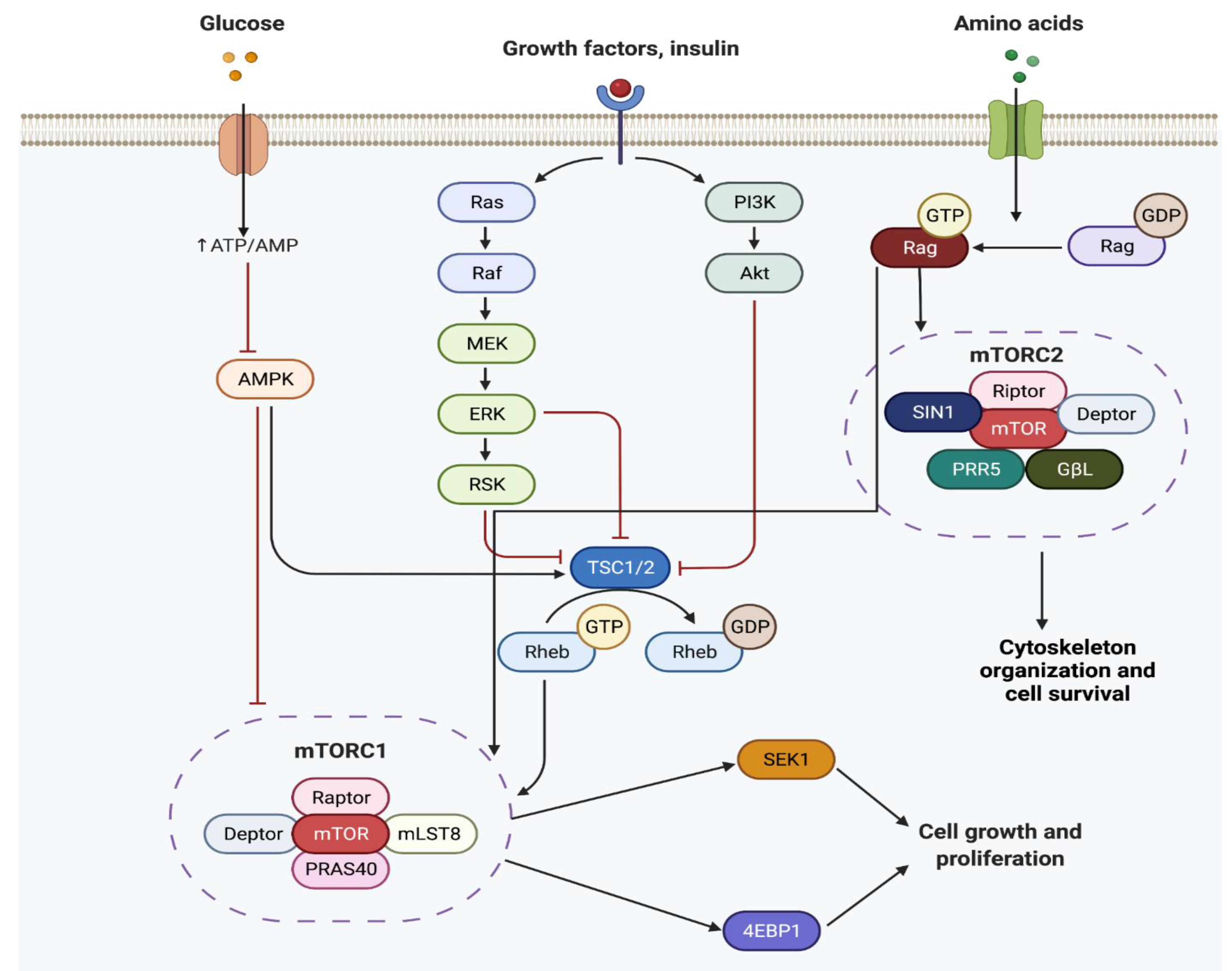
Cellular development such as cell proliferation, autophagy, apoptosis, cellular growth, and more are controlled by the mammalian target of rapamycin (mTOR) [58,59]. mTOR is a member of the phosphatidylinositol 3-kinase-related kinase family of protein kinases [58,59]. Through the phosphorylation of p70S6K and 4E-BP1 and the regulation of cap-dependent translation, mTOR activates protein synthesis [60]. mTOR plays an important role in the body and its mutation or dysregulation has been associated with diseases such as cancer, cardiovascular disease, obesity, inflammation, diabetes, and neurodegenerative disease [58,61]. mTOR has two functionally and structurally distinct complexes known as the mammalian target of rapamycin complex 1 (mTORC1) and the mammalian target of rapamycin complex 2 (mTORC2) [58] (Figure 2). mTORC1 complex consists of mTOR, GβL, regulatory-associated protein of mTOR (raptor), and DEP domain-containing mTOR interacting protein (DEPTOR) genes [58,62]. mTORC1 activity is influenced by growth factors, amino acids, energy levels, glucose availability, and stressors [58,62]. mTORC2 complex consists of mTOR, SIN1, Rictor, PRR5, GβL, and DEPTOR genes [58,61]. This complex controls cytoskeleton organization and cell survival as shown in Figure 2 [58,61]. mTOR is involved in multiple cellular signaling pathways such as adenosine 5′-monophosphate-activated protein kinase (AMPK), phosphoinositide-3-kinase (PI3K)/AKT, tuberous sclerosis complex subunit 1 and 2 (TSC1 and TSC2), Rag GTPases, Rheb, etc. [56]. Although there is still a lot to learn about mTORC2, such as the type of proteins involved in the activation mechanism or proteins that make up its complex, both downstream and upstream pathways of mTORC1 are better understood. For instance, it is known that mTORC1 activates central nervous system hormones, neuromodulators, and neurotransmitters [61]. The interaction of mTOR with its downstream substrate leads to diverse cellular responses associated with neurogenesis.

3.2. mTOR in Neurogenesis

The role of the mTORC1 and mTORC2 complex in stem cell maintenance, growth, and differentiation vary due to their downstream and upstream targets. Changes in the upstream elements in mTORC1 signaling can lead to NSC proliferation [14]. For instance, UTX, a histone demethylase, promotes PTEN expression, and the deletion of both UTX and PTEN during neural development increase AKT and mTOR phosphorylation resulting in an increase in NSC proliferation [63]. When AKT is overexpressed it can induce proliferation in cortical progenitors [14,64]. Cell proliferation is enhanced by AKT activation through mTORC1 mechanisms [14,64]. Inhibition of mTORC1 through DEPTOR plays an important role in maintaining stemness, and when the cell is about to differentiate it is downregulated [65]. mTORC1 plays an important role in NSC’s differentiation into daughter cells [14,60]. This differentiation reduces mTORC1 activity, in turn decreasing neural progenitor cell populations [14,60]. Insulin, one of the growth factors that stimulated mTORCs through the activation of AKT, PI3K, Rheb, and the TSC pathway, also promotes neuronal differentiation in NSC through mTORC1 activation [14,61]. Furthermore, activation of mTORC1 after TSC1 deletion decreases the proliferation of stem cells in the SVZ [66]. Dysregulation of mTORC1 can also alter neurite development as well as synapse formation and maintenance resulting in epilepsy [14]. Through the activation of different pathways, mTOR is involved in neurogenesis.

3.3. mTOR in AD

As mentioned earlier, AD pathology is associated with an aggregate of Aβ plaques, intracellular aggregations of neurofibrillary tangles (NFTs), and composed of hyperphosphorylated microtubule-associated τ and p-tau [34]. The implication of mTOR in AD pathogenesis is not fully understood but evidence shows that in the mouse model Aβ production was greatly reduced [67]. AMPK is activated suggesting an indirect implication of mTOR in AD [67]. Furthermore, Oddo et al. provided evidence that Aβ aggregated causes hyperactivation of mTOR which then hyperphosphorylated tau, in turn leading to PHF’s (paired helical filaments) and NFT’s formation [67,68].
The analysis conducted on the human AD brain shows a high level of p-mTOR as well as two of its downstream targets, p70S6K and eIF4E, suggesting a high mTOR activity in AD [68]. According to Jin-Jing Pei et al., an increase in p70S6K could be associated with the upregulation of tau in AD [69]. Studies have shown that mTOR activation also enhances tau pathology, leading to the dysfunction of autophagy [68]. In summary, mTOR plays an important role in both anabolic and catabolic processes that are involved in aging. However, full understanding of the mechanisms remains unclear and requires further investigations.

3.4. mTOR in PD

Recent evidence has shown that mTOR signaling is altered during PD progression. However, whether mTOR has neurotoxicity or neuroprotection remains unclear and controversial. For instance, some studies have shown that 1-methyl-4-phenyl-1,2,3,6-tetrahydropyridine (MPTP) and rotenone 6-hydroxydopamine (6-OHDA) suppress mTOR activity [70,71,72,73]. Conversely, Domanskyi et al., found that deletion of PTEN led to the activation of mTOR and protection of dopaminergic neurons against neurotoxin insult in PD mouse models [73,74].
In addition, REDD1 and mTORC1 inhibitors are upregulated in cellular PD models suggesting that while elevated REDD1 expression suppresses mTOR signaling, inhibition of REDD1 has a neuroprotective effect on PD animal models [73]. In other studies, AKT/mTOR signaling seems to have a regenerative effect in dopaminergic neurons, enhancing the neuroprotective effect of mTOR in certain PD models [73,75]. The neuroprotective effect of mTOR in PD is unclear and the findings are contradictory on whether mTOR has a neurotoxic or neuroprotective effect in PD models.

3.5. mTOR in HD

The mechanism of mHtt in HD is still not fully understood. However, since mTOR inhibits autophagy, it could be implicated in the pathology by having a protective effect in HD. Nevertheless, mTORC1 promotes many mechanisms that are impaired by mHtt [76]. In a study conducted by Lee et al. (2015), the phosphorylation of ribosomal protein S6, a marker of mTORC1 activity, was decreased in HD patients [77]. As presented in Figure 2, the mutation of mTORC1 activators, Rheb and caRheb, in mice models increased elements that promote mitochondrial function, autophagy, and cholesterol synthesis, which result in a depletion of HD [77]. Furthermore, mHTT aggregate abundance and expression of genes that promote mHtt aggregation is decreased, improving motor performance and decreased atrophy [76,77].
However, mTORC1 is predominantly regulated by Ras homolog enriched in the striatum (Rhes). A large decrease of Rhes occurs in HD patients. This suggests that the activation of mTORC1 may be beneficial in HD [76,77]. Furthermore, mTOR activity contributes to the brain atrophy and neuronal shrinkage observed in HD [78,79]. When mTOR targets S6K1, the phosphorylation and activity of mHtt is reduced [78,79]. Further investigation of the mechanism of mTOR activity in HD is required.

4. mTOR and Nrf2 Crosslink Signaling Pathways

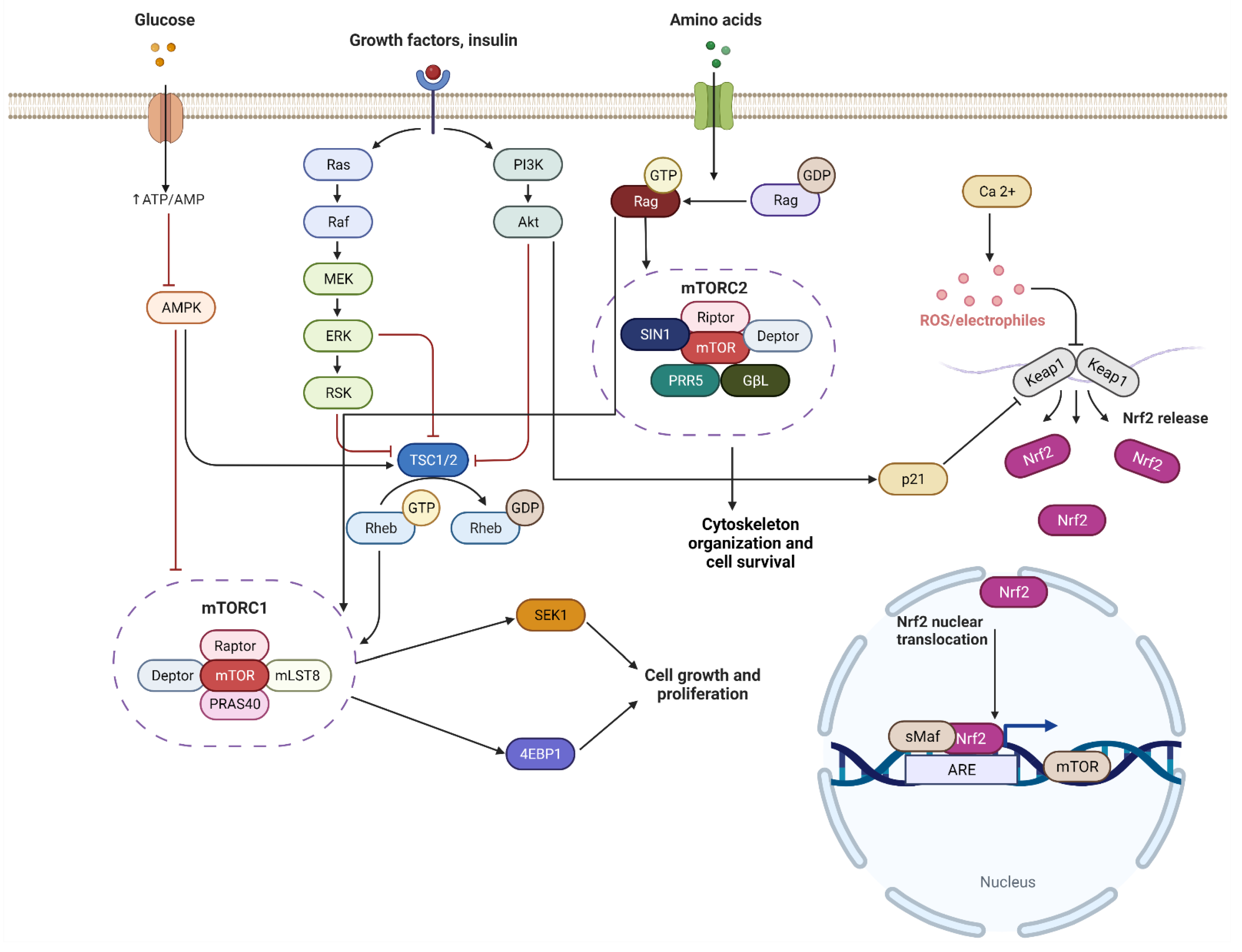
Nrf2/ARE signaling is associated with the AKT/PI3K pathway and the activation of both protects against oxidative stress [80]. Nrf2/ARE positively regulates mTOR through its interaction with RagD, small G-proteins, which activate mTORC1 [80,81,82]. In glioma cells, downregulation of the Nrf2 expression by shRNA suppresses mTOR while causing the ATP deficit to activate AMPK, leading to Nrf2 inhibition of mTOR [80,83]. One of the most obvious connections between Nrf2 and mTOR is the ARE sequence in the mTOR promoter region linking Nrf2, which has a direct role in regulating the expression of this gene. This regulation depends on the activity of PI3K/AKT [80] (Figure 3). The close involvement of the Nrf2/ARE signaling pathway in mTOR function and its association with neurogenesis and neurodegenerative diseases can open new prospective treatments for numerous neurodegenerative diseases. While the crosstalk between mTOR and Nrf2 in neurodegenerative diseases could provide novel therapeutic treatments, there are limited studies that have investigated the mechanism. In the current research available, we were able to find a possible link between memory formation and AD development. Long-term memory formation depends largely on synapse shape, formed by CA1 pyramidal neurons, which depends on N-methyl-d-aspartate glutamate receptors (NMDARs) and Schaffer collateral [84]. During long-term memory formation, an increase in postsynaptic membranes and sensitivity to neurotransmitters are required [80,85]. Through protein synthesis, mTORC1 activation plays an important role. It is involved in the regulation of proteins through cap-dependent mRNA translation of multiple proteins that participate in long-term memory formation [80,85]. To investigate the effect of Nrf2 on long-term memory, Zweig et al. (2020) studied Nrf2−/− mice that exhibited memory impairment and found a decrease in synaptic density and dendritic arborization [80,86]. In AD development, Nrf2 and mTOR function is disturbed and as presented, both play a role in synapse shape associated with memory formation, which suggested their involvement in memory depletion in AD.

5. Conclusions and Recommendations for the Future

Neurogenesis occurs in the brain during embryonic development and throughout adulthood. Even during stress conditions or disease, new neuronal cells are produced in the subgranular zone in the dentate gyrus of the hippocampus, and the subventricular zone of the lateral ventricles. However, during disease development, when certain factors occur such as an alteration in Nrf2 and mTOR function, it can lead to the increase or decrease in the activity of progenitor cells. This enhances disease development (i.e., Alzheimer’s disease). Throughout the development of the nervous system, Nrf2 and mTOR play an important role, whether it is directly or indirectly through the activation of other signaling pathways in the differentiation, proliferation, self-renewal of NSC, or in synaptic formation. They are also essential to neurogenesis and the establishment of neural circuits. Changes in their activity have been associated with a few neurodegenerative diseases. mTOR and Nrf2 complexes are associated with external signals and transduction pathways and understanding the role of these complexes are necessary to develop future therapies for current neurodevelopmental disorders.
We recommend further investigations into mTOR and Nrf2 crosstalk in neurogenesis. As we have presented in this review, a better understanding of their interaction could lead to the development of therapeutic drug treatments against neurodegenerative diseases. Although we have a general understanding of the mechanism of Nrf2 and mTOR in some cell types, there are still limitations to our knowledge of their mechanisms in neuronal cells. Further studies using mouse models and investigation into the upregulation or downregulation of various protein activity could provide a better understanding of the pathways, such as activated protein C (APC)-AKT signaling pathway and APC-AMPK signaling pathway [87,88].

Author Contributions

Writing—original draft preparation, L.I.Z.; writing—review and editing, M.K.-H., H.W., M.K.F., L.B., Z.L., P.K., D.R., J.L.; supervision, J.L.; funding acquisition, J.L. All authors have read and agreed to the published version of the manuscript.

Funding

This research was funded by NIH R01HL158515, NIH R01GM124108, VA Merit Award I01BX005625, Florida Department of Health 22K08.

Institutional Review Board Statement

Not applicable.

Informed Consent Statement

Not applicable.

Data Availability Statement

Not applicable.

Conflicts of Interest

The authors declare no conflict of interest.

References

  1. Ming, G.; Song, H. Adult Neurogenesis in the Mammalian Brain: Significant Answers and Significant Questions. Neuron 2011, 70, 687–702. [Google Scholar] [CrossRef] [PubMed] [Green Version]
  2. Vago, D.R.; Wallenstein, G.V.; Morris, L.S. Hippocampus. In Reference Module in Neuroscience and Biobehavioral Psychology; Elsevier: Amsterdam, The Netherlands, 2017; ISBN 978-0-12-809324-5. [Google Scholar]
  3. Lacar, B.; Young, S.; Platel, J.-C.; Bordey, A. Imaging and Recording Subventricular Zone Progenitor Cells in Live Tissue of Postnatal Mice. Front. Neurosci. 2010, 4, 43. [Google Scholar] [CrossRef] [PubMed] [Green Version]
  4. Hamilton, G.F.; Rhodes, J.S. Chapter Sixteen-Exercise Regulation of Cognitive Function and Neuroplasticity in the Healthy and Diseased Brain. In Progress in Molecular Biology and Translational Science; Bouchard, C., Ed.; Molecular and Cellular Regulation of Adaptation to Exercise; Academic Press: Cambridge, MA, USA, 2015; Volume 135, pp. 381–406. [Google Scholar]
  5. Doetsch, F.; Caillé, I.; Lim, D.A.; García-Verdugo, J.M.; AlvAREz-Buylla, A. Subventricular Zone Astrocytes ARE Neural Stem Cells in the Adult Mammalian Brain. Cell 1999, 97, 703–716. [Google Scholar] [CrossRef] [Green Version]
  6. Lois, C.; García-Verdugo, J.-M.; AlvAREz-Buylla, A. Chain Migration of Neuronal Precursors. Science 1996, 271, 978–981. [Google Scholar] [CrossRef]
  7. Peretto, P.; Merighi, A.; Fasolo, A.; Bonfanti, L. Glial Tubes in the Rostral Migratory Stream of the Adult Rat. Brain Res. Bull. 1997, 42, 9–21. [Google Scholar] [CrossRef]
  8. Ando, K.; Houben, S.; Homa, M.; de Fisenne, M.-A.; Potier, M.-C.; Erneux, C.; Brion, J.-P.; Leroy, K. Alzheimer’s Disease: Tau Pathology and Dysfunction of Endocytosis. Front. Mol. Neurosci. 2021, 13, 583755. [Google Scholar] [CrossRef]
  9. Sari, Y. Huntington’s Disease: From Mutant Huntingtin Protein to Neurotrophic Factor Therapy. Int. J. Biomed. Sci. IJBS 2011, 7, 89–100. [Google Scholar]
  10. Kelleher, R.J.; Shen, J. Presenilin-1 Mutations and Alzheimer’s Disease. Proc. Natl. Acad. Sci. USA 2017, 114, 629–631. [Google Scholar] [CrossRef] [Green Version]
  11. Winner, B.; Kohl, Z.; Gage, F.H. Neurodegenerative Disease and Adult Neurogenesis. Eur. J. Neurosci. 2011, 33, 1139–1151. [Google Scholar] [CrossRef]
  12. Castellanos, M.; Gubern, C.; Kadar, E. Chapter 7-MTOR: Exploring a New Potential Therapeutic Target for Stroke. In Molecules to Medicine with mTOR; Maiese, K., Ed.; Academic Press: Boston, MA, USA, 2016; pp. 105–122. ISBN 978-0-12-802733-2. [Google Scholar]
  13. Bond, P. Regulation of MTORC1 by Growth Factors, Energy Status, Amino Acids and Mechanical Stimuli at a Glance. J. Int. Soc. Sports Nutr. 2016, 13, 8. [Google Scholar] [CrossRef] [Green Version]
  14. LiCausi, F.; Hartman, N.W. Role of MTOR Complexes in Neurogenesis. Int. J. Mol. Sci. 2018, 19, 1544. [Google Scholar] [CrossRef] [PubMed] [Green Version]
  15. Griffin, R.J.; Moloney, A.; Kelliher, M.; Johnston, J.A.; Ravid, R.; Dockery, P.; O’Connor, R.; O’Neill, C. Activation of AKT/PKB, Increased Phosphorylation of AKT Substrates and Loss and Altered Distribution of AKT and PTEN ARE Features of Alzheimer’s Disease Pathology. J. Neurochem. 2005, 93, 105–117. [Google Scholar] [CrossRef] [PubMed]
  16. Norwitz, N.G.; Querfurth, H. MTOR Mysteries: Nuances and Questions About the Mechanistic Target of Rapamycin in Neurodegeneration. Front. Neurosci. 2020, 14, 775. [Google Scholar] [CrossRef] [PubMed]
  17. Wong, M. A Critical Review of MTOR Inhibitors and Epilepsy: From Basic Science to Clinical Trials. Expert Rev. Neurother. 2013, 13, 657–669. [Google Scholar] [CrossRef] [Green Version]
  18. Kärkkäinen, V.; Pomeshchik, Y.; Savchenko, E.; Dhungana, H.; Kurronen, A.; Lehtonen, S.; Naumenko, N.; Tavi, P.; Levonen, A.-L.; Yamamoto, M.; et al. Nrf2 Regulates Neurogenesis and Protects Neural Progenitor Cells Against Aβ Toxicity. Stem Cells 2014, 32, 1904–1916. [Google Scholar] [CrossRef]
  19. Tang, G.; Gudsnuk, K.; Kuo, S.-H.; Cotrina, M.L.; Rosoklija, G.; Sosunov, A.; Sonders, M.S.; Kanter, E.; Castagna, C.; Yamamoto, A.; et al. Loss of MTOR-Dependent Macroautophagy Causes Autistic-like Synaptic Pruning Deficits. Neuron 2014, 83, 1131–1143. [Google Scholar] [CrossRef] [Green Version]
  20. Zang, H.; Mathew, R.O.; Cui, T. The Dark Side of Nrf2 in the Heart. Front. Physiol. 2020, 11, 722. [Google Scholar] [CrossRef]
  21. Kahroba, H.; Ramezani, B.; Maadi, H.; Sadeghi, M.R.; Jaberie, H.; Ramezani, F. The Role of Nrf2 in Neural Stem/Progenitors Cells: From Maintaining Stemness and Self-Renewal to Promoting Differentiation Capability and Facilitating Therapeutic Application in Neurodegenerative Disease. Ageing Res. Rev. 2021, 65, 101211. [Google Scholar] [CrossRef]
  22. Sandberg, M.; Patil, J.; D’Angelo, B.; Weber, S.G.; Mallard, C. NRF2-Regulation in Brain Health and Disease: Implication of Cerebral Inflammation. Neuropharmacology 2014, 79, 298–306. [Google Scholar] [CrossRef] [Green Version]
  23. Bischoff, L.J.M.; Kuijper, I.A.; Schimming, J.P.; Wolters, L.; Braak, B.T.; Langenberg, J.P.; Noort, D.; Beltman, J.B.; van de Water, B. A Systematic Analysis of Nrf2 Pathway Activation Dynamics during Repeated Xenobiotic Exposure. Arch. Toxicol. 2019, 93, 435–451. [Google Scholar] [CrossRef] [Green Version]
  24. Shah, S.Z.A.; Zhao, D.; Hussain, T.; Sabir, N.; Mangi, M.H.; Yang, L. P62-Keap1-NRF2-ARE Pathway: A Contentious Player for Selective Targeting of Autophagy, Oxidative Stress and Mitochondrial Dysfunction in Prion Diseases. Front. Mol. Neurosci. 2018, 11, 310. [Google Scholar] [CrossRef] [Green Version]
  25. Robledinos-Antón, N.; Rojo, A.I.; Ferreiro, E.; Núñez, Á.; Krause, K.-H.; Jaquet, V.; Cuadrado, A. Transcription Factor NRF2 Controls the Fate of Neural Stem Cells in the Subgranular Zone of the Hippocampus. Redox Biol. 2017, 13, 393–401. [Google Scholar] [CrossRef]
  26. Corenblum, M.J.; Ray, S.; Remley, Q.W.; Long, M.; Harder, B.; Zhang, D.D.; Barnes, C.A.; Madhavan, L. Reduced Nrf2 Expression Mediates the Decline in Neural Stem Cell Function during a Critical Middle-Age Period. Aging Cell 2016, 15, 725–736. [Google Scholar] [CrossRef] [Green Version]
  27. Yanker, B.A. New Clues to Alzheimer’s Disease: Unraveling the Roles of Amyloid and Tau. Nat. Med. 1996, 2, 850–852. [Google Scholar] [CrossRef]
  28. Hu, Q.; Khanna, P.; Wong, B.S.E.; Heng, Z.S.L.; Subhramanyam, C.S.; Thanga, L.Z.; Tan, S.W.S.; Baeg, G.H. Oxidative Stress Promotes Exit from the Stem Cell State and Spontaneous Neuronal Differentiation. Oncotarget 2017, 9, 4223–4238. [Google Scholar] [CrossRef] [Green Version]
  29. Chen, X.; Shen, W.-B.; Yang, P.; Dong, D.; Sun, W.; Yang, P. High Glucose Inhibits Neural Stem Cell Differentiation Through Oxidative Stress and Endoplasmic Reticulum Stress. Stem Cells Dev. 2018, 27, 745–755. [Google Scholar] [CrossRef] [Green Version]
  30. Khacho, M.; Clark, A.; Svoboda, D.S.; Azzi, J.; MacLaurin, J.G.; Meghaizel, C.; Sesaki, H.; Lagace, D.C.; Germain, M.; Harper, M.-E.; et al. Mitochondrial Dynamics Impacts Stem Cell Identity and Fate Decisions by Regulating a Nuclear Transcriptional Program. Cell Stem Cell 2016, 19, 232–247. [Google Scholar] [CrossRef] [Green Version]
  31. Ray, S.; Corenblum, M.J.; Anandhan, A.; Reed, A.; Ortiz, F.O.; Zhang, D.D.; Barnes, C.A.; Madhavan, L. A Role for Nrf2 Expression in Defining the Aging of Hippocampal Neural Stem Cells. Cell Transplant. 2018, 27, 589–606. [Google Scholar] [CrossRef]
  32. Semkova, V.; Haupt, S.; Segschneider, M.; Bell, C.; Ingelman-Sundberg, M.; Hajo, M.; Weykopf, B.; Muthukottiappan, P.; Till, A.; Brüstle, O. Dynamics of Metabolic Pathways and Stress Response Patterns during Human Neural Stem Cell Proliferation and Differentiation. Cells 2022, 11, 1388. [Google Scholar] [CrossRef]
  33. Zhang, X.-X.; Tian, Y.; Wang, Z.-T.; Ma, Y.-H.; Tan, L.; Yu, J.-T. The Epidemiology of Alzheimer’s Disease Modifiable Risk Factors and Prevention. J. Prev. Alzheimers Dis. 2021, 8, 313–321. [Google Scholar] [CrossRef]
  34. Tiwari, S.; Atluri, V.; Kaushik, A.; Yndart, A.; Nair, M. Alzheimer’s Disease: Pathogenesis, Diagnostics, and Therapeutics. Int. J. Nanomed. 2019, 14, 5541–5554. [Google Scholar] [CrossRef] [Green Version]
  35. Zhang, H.; Davies, K.J.A.; Forman, H.J. Oxidative Stress Response and Nrf2 Signaling in Aging. Free Radic. Biol. Med. 2015, 88, 314–336. [Google Scholar] [CrossRef] [Green Version]
  36. Riley, B.E.; Kaiser, S.E.; Kopito, R.R. Autophagy Inhibition Engages Nrf2-P62 Ub-Associated Signaling. Autophagy 2011, 7, 338–340. [Google Scholar] [CrossRef] [Green Version]
  37. Saha, S.; Buttari, B.; Profumo, E.; Tucci, P.; Saso, L. A Perspective on Nrf2 Signaling Pathway for Neuroinflammation: A Potential Therapeutic Target in Alzheimer’s and Parkinson’s Diseases. Front. Cell. Neurosci. 2022, 15, 787258. [Google Scholar] [CrossRef]
  38. Joshi, G.; Gan, K.A.; Johnson, D.A.; Johnson, J.A. Increased Alzheimer’s Disease–like Pathology in the APP/PS1ΔE9 Mouse Model Lacking Nrf2 through Modulation of Autophagy. Neurobiol. Aging 2015, 36, 664–679. [Google Scholar] [CrossRef] [Green Version]
  39. Rojo, A.I.; PajAREs, M.; Rada, P.; Nuñez, A.; Nevado-Holgado, A.J.; Killik, R.; Van Leuven, F.; Ribe, E.; Lovestone, S.; Yamamoto, M.; et al. NRF2 Deficiency Replicates Transcriptomic Changes in Alzheimer’s Patients and Worsens APP and TAU Pathology. Redox Biol. 2017, 13, 444–451. [Google Scholar] [CrossRef]
  40. Silva-Palacios, A.; Ostolga-Chavarría, M.; Zazueta, C.; Königsberg, M. Nrf2: Molecular and Epigenetic Regulation during Aging. Ageing Res. Rev. 2018, 47, 31–40. [Google Scholar] [CrossRef]
  41. Bahn, G.; Park, J.-S.; Yun, U.J.; Lee, Y.J.; Choi, Y.; Park, J.S.; Baek, S.H.; Choi, B.Y.; Cho, Y.S.; Kim, H.K.; et al. NRF2/ARE Pathway Negatively Regulates BACE1 Expression and Ameliorates Cognitive Deficits in Mouse Alzheimer’s Models. Proc. Natl. Acad. Sci. USA 2019, 116, 12516–12523. [Google Scholar] [CrossRef] [Green Version]
  42. Pajares, M.; Rojo, A.I.; Arias, E.; Díaz-Carretero, A.; Cuervo, A.M.; Cuadrado, A. Transcription Factor NFE2L2/NRF2 Modulates Chaperone-Mediated Autophagy through the Regulation of LAMP2A. Autophagy 2018, 14, 1310–1322. [Google Scholar] [CrossRef]
  43. Jo, C.; Gundemir, S.; Pritchard, S.; Jin, Y.N.; Rahman, I.; Johnson, G.V.W. Nrf2 Reduces Levels of Phosphorylated Tau Protein by Inducing Autophagy Adaptor Protein NDP52. Nat. Commun. 2014, 5, 3496. [Google Scholar] [CrossRef]
  44. MacMahon Copas, A.N.; McComish, S.F.; Fletcher, J.M.; Caldwell, M.A. The Pathogenesis of Parkinson’s Disease: A Complex Interplay Between Astrocytes, Microglia, and T Lymphocytes? Front. Neurol. 2021, 12, 771. [Google Scholar] [CrossRef] [PubMed]
  45. Pajares, M.; Jiménez-Moreno, N.; García-Yagüe, Á.J.; Escoll, M.; de Ceballos, M.L.; Van Leuven, F.; Rábano, A.; Yamamoto, M.; Rojo, A.I.; Cuadrado, A. Transcription Factor NFE2L2/NRF2 Is a Regulator of Macroautophagy Genes. Autophagy 2016, 12, 1902–1916. [Google Scholar] [CrossRef] [PubMed] [Green Version]
  46. Guo, X.; Han, C.; Ma, K.; Xia, Y.; Wan, F.; Yin, S.; Kou, L.; Sun, Y.; Wu, J.; Hu, J.; et al. Hydralazine Protects Nigrostriatal Dopaminergic Neurons From MPP+ and MPTP Induced Neurotoxicity: Roles of Nrf2-ARE Signaling Pathway. Front. Neurol. 2019, 10, 271. [Google Scholar] [CrossRef] [PubMed]
  47. Youdim, M.B.H.; Stephenson, G.; Shachar, D.B. Ironing Iron Out in Parkinson’s Disease and Other Neurodegenerative Diseases with Iron Chelators: A Lesson from 6-Hydroxydopamine and Iron Chelators, Desferal and VK-28. Ann. N. Y. Acad. Sci. 2004, 1012, 306–325. [Google Scholar] [CrossRef]
  48. Lastres-Becker, I.; García-Yagüe, A.J.; Scannevin, R.H.; CasAREjos, M.J.; Kügler, S.; Rábano, A.; Cuadrado, A. Repurposing the NRF2 Activator Dimethyl Fumarate as Therapy Against Synucleinopathy in Parkinson’s Disease. Antioxid. Redox Signal. 2016, 25, 61–77. [Google Scholar] [CrossRef] [Green Version]
  49. Stephenson, J.; Nutma, E.; van der Valk, P.; Amor, S. Inflammation in CNS Neurodegenerative Diseases. Immunology 2018, 154, 204–219. [Google Scholar] [CrossRef] [Green Version]
  50. Rojo, A.I.; Innamorato, N.G.; Martín-Moreno, A.M.; De Ceballos, M.L.; Yamamoto, M.; Cuadrado, A. Nrf2 Regulates Microglial Dynamics and Neuroinflammation in Experimental Parkinson’s Disease. Glia 2010, 58, 588–598. [Google Scholar] [CrossRef]
  51. Zgorzynska, E.; Dziedzic, B.; Walczewska, A. An Overview of the Nrf2/ARE Pathway and Its Role in Neurodegenerative Diseases. Int. J. Mol. Sci. 2021, 22, 9592. [Google Scholar] [CrossRef]
  52. Tabrizi, S.J.; Leavitt, B.R.; Landwehrmeyer, G.B.; Wild, E.J.; Saft, C.; Barker, R.A.; Blair, N.F.; Craufurd, D.; Priller, J.; Rickards, H.; et al. Targeting Huntingtin Expression in Patients with Huntington’s Disease. N. Engl. J. Med. 2019, 380, 2307–2316. [Google Scholar] [CrossRef]
  53. Ganner, A.; Pfeiffer, Z.-C.; Wingendorf, L.; Kreis, S.; Klein, M.; Walz, G.; Neumann-Haefelin, E. The Acetyltransferase P300 Regulates NRF2 Stability and Localization. Biochem. Biophys. Res. Commun. 2020, 524, 895–902. [Google Scholar] [CrossRef]
  54. Tsvetkov, A.S.; Arrasate, M.; Barmada, S.; Ando, D.M.; Sharma, P.; Shaby, B.A.; Finkbeiner, S. Proteostasis of Polyglutamine Varies among Neurons and Predicts Neurodegeneration. Nat. Chem. Biol. 2013, 9, 586–592. [Google Scholar] [CrossRef] [Green Version]
  55. Saito, Y.; Yako, T.; Otsu, W.; Nakamura, S.; Inoue, Y.; Muramatsu, A.; Nakagami, Y.; Shimazawa, M.; Hara, H. A Triterpenoid Nrf2 Activator, RS9, Promotes LC3-Associated Phagocytosis of Photoreceptor Outer Segments in a P62-Independent Manner. Free Radic. Biol. Med. 2020, 152, 235–247. [Google Scholar] [CrossRef]
  56. Dowling, R.J.O.; Topisirovic, I.; Fonseca, B.D.; Sonenberg, N. Dissecting the Role of MTOR: Lessons from MTOR Inhibitors. Biochim. Biophys. Acta BBA Proteins Proteom. 2010, 1804, 433–439. [Google Scholar] [CrossRef]
  57. Luo, J.-F.; Shen, X.-Y.; Lio, C.K.; Dai, Y.; Cheng, C.-S.; Liu, J.-X.; Yao, Y.-D.; Yu, Y.; Xie, Y.; Luo, P.; et al. Activation of Nrf2/HO-1 Pathway by Nardochinoid C Inhibits Inflammation and Oxidative Stress in Lipopolysaccharide-Stimulated Macrophages. Front. Pharmacol. 2018, 9, 911. [Google Scholar] [CrossRef]
  58. Zou, Z.; Tao, T.; Li, H.; Zhu, X. MTOR Signaling Pathway and MTOR Inhibitors in Cancer: Progress and Challenges. Cell Biosci. 2020, 10, 31. [Google Scholar] [CrossRef]
  59. Saxton, R.A.; Sabatini, D.M. MTOR Signaling in Growth, Metabolism, and Disease. Cell 2017, 168, 960–976. [Google Scholar] [CrossRef] [Green Version]
  60. Paliouras, G.N.; Hamilton, L.K.; Aumont, A.; Joppé, S.E.; Barnabé-Heider, F.; Fernandes, K.J.L. Mammalian Target of Rapamycin Signaling Is a Key Regulator of the Transit-Amplifying Progenitor Pool in the Adult and Aging Forebrain. J. Neurosci. 2012, 32, 15012–15026. [Google Scholar] [CrossRef] [Green Version]
  61. Takei, N.; Nawa, H. MTOR Signaling and Its Roles in Normal and Abnormal Brain Development. Front. Mol. Neurosci. 2014, 7, 28. [Google Scholar] [CrossRef] [Green Version]
  62. Takahara, T.; Amemiya, Y.; Sugiyama, R.; Maki, M.; Shibata, H. Amino Acid-Dependent Control of MTORC1 Signaling: A Variety of Regulatory Modes. J. Biomed. Sci. 2020, 27, 87. [Google Scholar] [CrossRef]
  63. Lei, X.; Jiao, J. UTX Affects Neural Stem Cell Proliferation and Differentiation through PTEN Signaling. Stem Cell Rep. 2018, 10, 1193–1207. [Google Scholar] [CrossRef] [Green Version]
  64. Sinor, A.D.; Lillien, L. AKT-1 Expression Level Regulates CNS Precursors. J. Neurosci. 2004, 24, 8531–8541. [Google Scholar] [CrossRef]
  65. Agrawal, P.; Reynolds, J.; Chew, S.; Lamba, D.A.; Hughes, R.E. DEPTOR Is a Stemness Factor That Regulates Pluripotency of Embryonic Stem Cells*. J. Biol. Chem. 2014, 289, 31818–31826. [Google Scholar] [CrossRef] [Green Version]
  66. Magri, L.; Cambiaghi, M.; Cominelli, M.; Alfaro-Cervello, C.; Cursi, M.; Pala, M.; Bulfone, A.; Garcìa-Verdugo, J.M.; Leocani, L.; Minicucci, F.; et al. Sustained Activation of MTOR Pathway in Embryonic Neural Stem Cells Leads to Development of Tuberous Sclerosis Complex-Associated Lesions. Cell Stem Cell 2011, 9, 447–462. [Google Scholar] [CrossRef] [Green Version]
  67. Mueed, Z.; Tandon, P.; Maurya, S.K.; Deval, R.; Kamal, M.A.; Poddar, N.K. Tau and MTOR: The Hotspots for Multifarious Diseases in Alzheimer’s Development. Front. Neurosci. 2019, 12, 1017. [Google Scholar] [CrossRef]
  68. Oddo, S. The Role of MTOR Signaling in Alzheimer Disease. Front. Biosci. 2012, 4, 941–952. [Google Scholar] [CrossRef] [Green Version]
  69. Pei, J.-J.; Hugon, J. MTOR-Dependent Signalling in Alzheimer’s Disease. J. Cell. Mol. Med. 2008, 12, 2525–2532. [Google Scholar] [CrossRef] [Green Version]
  70. Selvaraj, S.; Sun, Y.; Watt, J.A.; Wang, S.; Lei, S.; Birnbaumer, L.; Singh, B.B. Neurotoxin-Induced ER Stress in Mouse Dopaminergic Neurons Involves Downregulation of TRPC1 and Inhibition of AKT/MTOR Signaling. J. Clin. Investig. 2012, 122, 1354–1367. [Google Scholar] [CrossRef] [Green Version]
  71. Zhou, Q.; Liu, C.; Liu, W.; Zhang, H.; Zhang, R.; Liu, J.; Zhang, J.; Xu, C.; Liu, L.; Huang, S.; et al. Rotenone Induction of Hydrogen Peroxide Inhibits MTOR-Mediated S6K1 and 4E-BP1/EIF4E Pathways, Leading to Neuronal Apoptosis. Toxicol. Sci. 2015, 143, 81–96. [Google Scholar] [CrossRef] [PubMed] [Green Version]
  72. Xu, Y.; Liu, C.; Chen, S.; Ye, Y.; Guo, M.; Ren, Q.; Liu, L.; Zhang, H.; Xu, C.; Zhou, Q.; et al. Activation of AMPK and Inactivation of AKT Result in Suppression of MTOR-Mediated S6K1 and 4E-BP1 Pathways Leading to Neuronal Cell Death in in Vitro Models of Parkinson’s Disease. Cell. Signal. 2014, 26, 1680–1689. [Google Scholar] [CrossRef] [Green Version]
  73. Lan, A.; Chen, J.; Zhao, Y.; Chai, Z.; Hu, Y. MTOR Signaling in Parkinson’s Disease. NeuroMolecular Med. 2017, 19, 1–10. [Google Scholar] [CrossRef]
  74. Domanskyi, A.; Geiβler, C.; Vinnikov, I.A.; Alter, H.; Schober, A.; Vogt, M.A.; Gass, P.; Parlato, R.; Schütz, G. PTEN Ablation in Adult Dopaminergic Neurons Is Neuroprotective in Parkinson’s Disease Models. FASEB J. 2011, 25, 2898–2910. [Google Scholar] [CrossRef] [PubMed]
  75. Kim, H.J.; Kim, J.; Kang, K.S.; Lee, K.T.; Yang, H.O. Neuroprotective Effect of Chebulagic Acid via Autophagy Induction in SH-SY5Y Cells. Biomol. Ther. (Seoul) 2014, 22, 275–281. [Google Scholar] [CrossRef] [PubMed] [Green Version]
  76. Ferrarelli, L.K. Is MTOR a Good Guy or Bad Guy in Huntington’s Disease? Sci. Signal. 2015, 8, ec26. [Google Scholar] [CrossRef]
  77. Lee, J.H.; Tecedor, L.; Chen, Y.H.; Monteys, A.M.; Sowada, M.J.; Thompson, L.M.; Davidson, B.L. Reinstating Aberrant MTORC1 Activity in Huntington’s Disease Mice Improves Disease Phenotypes. Neuron 2015, 85, 303–315. [Google Scholar] [CrossRef] [Green Version]
  78. Ravikumar, B.; Vacher, C.; Berger, Z.; Davies, J.E.; Luo, S.; Oroz, L.G.; Scaravilli, F.; Easton, D.F.; Duden, R.; O’Kane, C.J.; et al. Inhibition of MTOR Induces Autophagy and Reduces Toxicity of Polyglutamine Expansions in Fly and Mouse Models of Huntington Disease. Nat. Genet. 2004, 36, 585–595. [Google Scholar] [CrossRef] [Green Version]
  79. Rubinsztein, D.C. Lessons from Animal Models of Huntington’s Disease. Trends Genet. 2002, 18, 202–209. [Google Scholar] [CrossRef]
  80. Gureev, A.P.; Popov, V.N.; Starkov, A.A. Crosstalk between the MTOR and Nrf2/ARE Signaling Pathways as a Target in the Improvement of Long-Term Potentiation. Exp. Neurol. 2020, 328, 113285. [Google Scholar] [CrossRef]
  81. Shibata, T.; Saito, S.; Kokubu, A.; Suzuki, T.; Yamamoto, M.; Hirohashi, S. Global Downstream Pathway Analysis Reveals a Dependence of Oncogenic NF-E2–Related Factor 2 Mutation on the MTOR Growth Signaling Pathway. Cancer Res. 2010, 70, 9095–9105. [Google Scholar] [CrossRef] [Green Version]
  82. Sasaki, H.; Shitara, M.; Yokota, K.; Hikosaka, Y.; Moriyama, S.; Yano, M.; Fujii, Y. RagD Gene Expression and NRF2 Mutations in Lung Squamous Cell Carcinomas. Oncol. Lett. 2012, 4, 1167–1170. [Google Scholar] [CrossRef] [Green Version]
  83. Jia, Y.; Wang, H.; Wang, Q.; Ding, H.; Wu, H.; Pan, H. Silencing Nrf2 Impairs Glioma Cell Proliferation via AMPK-Activated MTOR Inhibition. Biochem. Biophys. Res. Commun. 2016, 469, 665–671. [Google Scholar] [CrossRef]
  84. Baltaci, S.B.; Mogulkoc, R.; Baltaci, A.K. Molecular Mechanisms of Early and Late LTP. Neurochem. Res. 2019, 44, 281–296. [Google Scholar] [CrossRef]
  85. Lenz, G.; Avruch, J. Glutamatergic Regulation of the P70S6 Kinase in Primary Mouse Neurons. J. Biol. Chem. 2005, 280, 38121–38124. [Google Scholar] [CrossRef] [Green Version]
  86. Zweig, J.A.; Caruso, M.; Brandes, M.S.; Gray, N.E. Loss of NRF2 Leads to Impaired Mitochondrial Function, Decreased Synaptic Density and Exacerbated Age-Related Cognitive Deficits. Exp. Gerontol. 2020, 131, 110767. [Google Scholar] [CrossRef]
  87. Murphy, J.; Le, T.N.V.; Fedorova, J.; Yang, Y.; Krause-Hauch, M.; Davitt, K.; Zoungrana, L.I.; Fatmi, M.K.; Lesnefsky, E.J.; Li, J.; et al. The Cardiac Dysfunction Caused by Metabolic Alterations in Alzheimer’s Disease. Front. Cardiovasc. Med. 2022, 9, 850538. [Google Scholar] [CrossRef]
  88. Ren, D.; Fedorova, J.; Davitt, K.; Le, T.N.V.; Griffin, J.; Liaw, P.C.; Esmon, C.T.; Rezaie, A.R.; Li, J. Activated Protein C Strengthens Cardiac Tolerance to Ischemic Insults in Aging by Modulating the Metabolic Response. Cir. Res. 2022, 130, 252–272. [Google Scholar] [CrossRef]
Figure 1. Nrf2 mechanism pathway. Nrf2 is released in the cytoplasm then migrates into the nucleus where it is transcripted during oxidative stress and degraded when it is no longer needed through ubiquitination (see text for more information). The figure was prepared by software provided by Biorender.com (accessed 3 March 2022).
Cells 11 02048 g001
Figure 2. mTOR mechanism pathway. Glucose and growth factors activate a series of pathways that either activate or inhibit (AMPK) mTORC1. Amino acids can either activate mTORC1 or mTORC2, which lead to cytoskeleton organization and cell survival or cell growth and proliferation (see text for more details). The figure was prepared by software provided by Biorender.com (accessed on 3 March 2022).
Cells 11 02048 g002
Figure 3. Nrf2 and mTOR mechanism pathway. AKT is one of the procedures of mTOR activity, which plays an important role in cellular mechanisms such as cell proliferation, cell cycle, and activated p21, which then inhibits Keap1, which alternates the Nrf2 mechanism. The figure was prepared by software provided by Biorender.com (accessed 3 March 2022).
Cells 11 02048 g003
Publisher’s Note: MDPI stays neutral with regard to jurisdictional claims in published maps and institutional affiliations.