Article

Regulation of APD and Force by the Na+/Ca2+ Exchanger in Human-Induced Pluripotent Stem Cell-Derived Engineered Heart Tissue

1
Institute of Experimental Pharmacology and Toxicology, University Medical Center Hamburg-Eppendorf, 20246 Hamburg, Germany
2
Department of Cardiology, University Heart & Vascular Center Hamburg, University Medical Center Hamburg-Eppendorf, 20246 Hamburg, Germany
3
DZHK (German Centre for Cardiovascular Research), Partner Site Hamburg/Kiel/Lübeck, 20246 Hamburg, Germany
4
Department of Biomedical Sciences, University of Copenhagen, 2200 Copenhagen, Denmark
5
Department of Cardiovascular Surgery, University Heart and Vascular Center, 20246 Hamburg, Germany
6
Institute of Cardiovascular Sciences, College of Medical and Dental Sciences, University of Birmingham, Birmingham B15 2TT, UK
7
BioMediTech, Faculty of Medicine and Health Technology, Tampere University, 33520 Tampere, Finland
*
Authors to whom correspondence should be addressed.
Present address: Nanion Technologies GmbH, 80339 München, Germany.
Present address: Idorsia Pharmaceuticals Ltd., 4123 Allschwil, Switzerland.
§
These authors contributed equally to this work.
Cells 2022, 11(15), 2424; https://doi.org/10.3390/cells11152424
Submission received: 24 May 2022 / Revised: 21 July 2022 / Accepted: 26 July 2022 / Published: 5 August 2022
(This article belongs to the Special Issue Calcium Signaling in Skeletal and Cardiac Health and Diseases)

Abstract

:
The physiological importance of NCX in human-induced pluripotent stem cell-derived cardiomyocytes (hiPSC-CMs) is not well characterized but may depend on the relative strength of the current, compared to adult cardiomyocytes, and on the exact spatial arrangement of proteins involved in Ca2+ extrusion. Here, we determined NCX currents and its contribution to action potential and force in hiPSC-CMs cultured in engineered heart tissue (EHT). The results were compared with data from rat and human left ventricular tissue. The NCX currents in hiPSC-CMs were larger than in ventricular cardiomyocytes isolated from human left ventricles (1.3 ± 0.2 pA/pF and 3.2 ± 0.2 pA/pF for human ventricle and EHT, respectively, p < 0.05). SEA0400 (10 µM) markedly shortened the APD90 in EHT (by 26.6 ± 5%, p < 0.05) and, to a lesser extent, in rat ventricular tissue (by 10.7 ± 1.6%, p < 0.05). Shortening in human left ventricular preparations was small and not different from time-matched controls (TMCs; p > 0.05). Force was increased by the NCX block in rat ventricle (by 31 ± 5.4%, p < 0.05) and EHT (by 20.8 ± 3.9%, p < 0.05), but not in human left ventricular preparations. In conclusion, hiPSC-CMs possess NCX currents not smaller than human left ventricular tissue. Robust NCX block-induced APD shortening and inotropy makes EHT an attractive pharmacological model.

1. Introduction

The development of human-induced pluripotent stem cell-derived cardiomyocytes (hiPSC-CMs) is expected to become a relevant step for the translational facet of cardiovascular basic research. HiPSC-CMs provide human disease models, enabling the detailed functional study of patient-specific hiPSC-CMs in a dish [1,2]. Nevertheless, there is concern that hiPSC-CMs may not fully reflect human heart physiology.
Ca2+ signaling is crucial for proper heart function and defective intracellular calcium signaling is a key process driving heart failure, arrhythmias, and vascular dysfunction, but may differ in hiPSC-CMs that lack the highly organized expression of the electromechanical coupling components. HiPSC-CMs have a less well developed t-tubular network, and their sarcomeric structures are less organized than those of adult cardiomyocytes (CMs) [3]. Despite these morphological differences, a recent report suggests that Ca2+ signaling in hiPSC-CMs appears to resemble that of rabbit ventricular CMs [4]. Authors used established and sophisticated methods to measure calcium influx via Ca2+ channels, Ca2+ cycling via sarcoplasmic reticulum, and Ca2+ extrusion via a sodium calcium exchanger (NCX). However, as the experimental conditions necessary for performing such studies often are far from the physiologically relevant situation, a comparison with in vivo calcium regulation may be questionable. Furthermore, results are difficult to interpret and have resulted in some controversy [5].
Here, we used a different approach. We directly measured NCX currents using an established patch clamp protocol [6]. To estimate the NCX contribution to the force and action potential duration (APD), we applied the NCX blocker SEA0400 in hiPSC-CMs cultured as engineered heart tissue (EHT). Importantly, the hiPSC-CM results were compared with the data from rat and human left ventricular tissue, obtained under comparable experimental conditions.

2. Methods

2.1. Rat Tissue

The study was conducted according to guidelines for laboratory animal welfare of the National Institutes of Health (publication no. 85-23, revised 1985). All experiments with rat tissue were performed in compliance with the German Law for the Protection of Animals. Moreover, 160 to 240 g male Wistar rats (Charles River Laboratories, Wilmington, MA, USA) were decapitated under light CO2 anesthesia (70% CO2 and 30% O2) and the excised hearts were placed in oxygenated and modified Tyrode’s solution at 37°C containing (in mM): NaCl—126.9, KCl—5.4, CaCl2—1.8, MgCl2—1.05, NaHCO3—22, NaH2PO4—0.45, EDTA—0.04, ascorbic acid—0.2, pyruvate—5, and glucose—5. For contractility and action potential measurements, small ventricular chunks were prepared from ventricular tissue. For patch clamp measurements, rat CMs were isolated via enzymatic dissociation using collagenase type 1 (254 U/mL; Worthington Biochemical, Lakewood, NJ, USA) on a Langendorff apparatus, as described earlier [7].

2.2. Human Tissue

The study followed the declaration of Helsinki and was approved by the ethics committee of the Medical Council of Hamburg, Germany (approval number: OB-088/04 and PV3759). Human left ventricular free wall tissue samples were obtained with informed consent from 13 patients undergoing implantation of the left ventricular assist device, heart transplantation, or aortic valve surgery at the University Heart and Vascular Center Hamburg. The patients were 53.7 ± 4.3 years old and left ventricular ejection fraction was 27.5 ± 4.7%. Further patient characteristics are outlined in Table S1. After the excision of ventricular tissue, samples were transferred in cardioplegic solution to the laboratory. The composition of cardioplegic solution in mM: NaCl—100, taurine—50, glucose—20, KCl—10, MgSO4—5, MOPS (3-(N-morpholino) propanesulfonic acid)—5, KH2PO4—1.2, and 2.3-butanedione monoxime (BDM)—30. For patch clamp measurements, human CMs were isolated via enzymatic dissociation using protease type XXIV (5 U/mL; Sigma, St. Louis, MO, USA) and collagenase type 1 (254 U/mL; Worthington Biochemical, Lakewood, NJ, USA), as described previously in detail [8].

2.3. Human-Induced Pluripotent stem cell-Derived Engineered Heart Tissue

Ventricular-like hiPSC-CMs were differentiated from an in-house control hiPSC cell line [9]. Differentiated hiPSC-CMs were used to generate three-dimensional EHT (1 × 10⁶ cells/100 μL EHT). The expansion and differentiation of hiPSC-CMs and the generation of EHT were performed according to published in-house standardized protocol [10]. EHT was cultured for 24–29 days under identical conditions at 37 °C in a 7% CO2 and 40% O2humidified cell culture incubator with a medium consisting of DMEM (Biochrom, Berlin, Germany), 10% heat-inactivated horse serum (Gibco, Paisley, Scotland), 1% penicillin–streptomycin (Gibco, Paisley, Scotland), insulin (10 μg/mL; Sigma, St. Louis, MO, USA), and aprotinin (33 μg/mL; Sigma, St. Louis, MO, USA). After culturing, hiPSC-CMs applied for patch clamp measurements were isolated with collagenase II (200 U/mL; Worthington Biochemical, Lakewood, NJ, USA) for 5 h. Isolated cells were plated on gelatine-coated (0.1%) glass coverslips (12 mm diameter; Carl Roth GmbH + Co, Karlsruhe, Germany) and kept in culture for 24–48 h to maintain adherence under superfusion in the recording chamber during patch clamp measurements.

2.4. Patch Clamp Measurements

The whole-cell configuration of the single-electrode patch clamp technique was used to record membrane current measurements with the Axopatch 200B (Axon Instruments, Foster City, CA, USA). Cells were investigated in a recording chamber (RC-26G; Warner Instruments, Hamden, CT, USA) placed on the stage of an inverted microscope (Axiovert 40 CFL; Zeiss, Oberkochen, Germany) at 37 °C. Heat-polished pipettes were pulled from borosilicate-filamented glass (Hilgenberg, Malsfeld, Germany) using the DMZ Universal Puller (Zeitz Instruments GmbH, Martinsried, Germany). Tip resistances were 2–5 MΩ and seal resistances were 3–6 GΩ. The data were acquired and analyzed using the ISO2 software (MFK, Niedernhausen, Germany). To measure the NCX current, we applied previously described electrophysiological protocols [6]. In brief, we used K+-free bath and pipette solutions. The composition of bath solutions in mM: NaCl—135, CsCl—10, CaCl2—1, MgCl2—1, BaCl2—0.2, NaH2PO4—0.33, TEACl—10, HEPES—10, glucose—10, pH adjusted to 7.4 with NaOH. The solution contained (in mM) lidocaine (0.05), nisoldipine (0.001), and ouabain (0.02) to block the Na+, Ca2+, and Na+/K+ pump currents. The composition of pipette solutions in mM: CsOH—140, aspartic acid—75, TEA-Cl—20, Mg2+-ATP—5, HEPES—10, NaCl—20, EGTA—20, CaCl2—10, pH adjusted to 7.2 with CsOH. The cell capacitance was calculated from the current response to a 5 mV voltage step and was than compensated for measuring the membrane current. The currents were measured in a constant flow (2 mL/min) low-volume chamber (300 µL) through ramp pulses (frequency 0.1 Hz), initially leading to depolarization from the holding potential of −40 mV to 60 mV, followed by hyperpolarization to −100 mV, and depolarization back to the holding potential of −40 mV (Figure 1). The NCX current was defined as the Ni2+-sensitive current measured by application of 10 mM NiCl2.

2.5. Action Potential Measurements

The action potentials (APs) in rat and human ventricular tissue and EHT were recorded with standard sharp microelectrodes, as previously described [7,11]. In brief, both ventricular tissue and EHT were pinned down in a recording chamber and field-stimulated by two platinum wires at 1 Hz (unipolar pacing, 0.5 ms stimulus duration, 50% above the stimulation threshold). Sharp microelectrodes were pulled from borosilicate glass (Hilgenberg, Malsfeld, Germany) using the DMZ Universal Puller (Zeitz-Instruments GmbH, Martinsried, Germany). The tip resistances ranged from 20 to 50 MΩ when filled with 3 M of KCl. The signals were amplified with a BA-1s npi amplifier (npi electronic GmbH, Tamm, Germany). The APs were recorded and analyzed using the Lab-Chart software (AD Instruments Pty Ltd., Castle Hill NSW, Australia). The compound effects were measured after an equilibration period of 30 min. In order to calculate the AP parameters, five consecutive APs were averaged.

2.6. Contractility Measurements

To measure the contractile force in rat and human ventricular muscle, strip preparations were mounted in organ baths filled with 50 mL of Tyrode solution, as described previously [7]. Rat preparations were stretched to 5 mN [12]. Human preparations were pre-stretched to a 50% length, giving the maximum force, and paced at a frequency of 1 Hz with 5 ms electrical pulses 10% above the threshold intensity. The tissue samples were pretreated for 90 min with the irreversible α-adrenoceptor blocker phenoxybenzamine (6 µM) [13]. The compound effects were measured after an equilibration period of 30 min. The recordings were analyzed using Chart software (AD Instruments Pty Ltd., Castle Hill NSW, Australia). Force data were not normalized to the muscle square area [14].
To measure the contractile force in EHT, a video-optical contractility test system (EHT Technologies GmbH, Hamburg, Germany) was applied, as described previously [15]. Briefly, video files generated by a software-based automated video-optical recording system were analyzed by deflecting the silicon posts during EHT contraction. Based on the EHT shortening during contractions, force and kinetic parameters were calculated using CTMV software (CTMV GmbH & Co. KG, Pforzheim, Germany).

2.7. Computational Simulations with Rat, Human and hiPSC Cardiomyocyte Models

Computational modelling and simulations were performed using recently published, yet established, rat ventricular [16] and human ventricular [17] CM models. For the hiPSC-CM model, we used the same approach as previously described [11,18]. Briefly, hiPSC-CM model version was obtained by increasing the calcium current (ICa) density 1.5-fold and increasing the density of the rapidly activating potassium current (IKr) density 4-fold, as well as decreasing the inwardly rectifying potassium current density (IK1) 0.5-fold in the human ventricular CM model [17]. For the sake of simplicity, we omitted the T-type calcium current from this implementation, as its role was found insignificant to excitation–contraction coupling in our previous study [11]. As suggested by the experiments presented in this study, we extended the hiPSC-CM model version to include 2.5-fold stronger NCX current. As the EHT experiments were carried out using an external pacing stimulus current, we excluded the pacemaking current, If, from the hiPSC-CM model implementation, similar to our earlier approach [11].
The gradual NCX block in silico experiments were simulated by multiplying the parameter that defines the maximum NCX rate with the corresponding value constant. In all cases, in silico results were obtained by running the CM models first to the steady-state control at 1 Hz pacing, switching the NCX block parameter, and finally running the model to the new drug-induced steady state. The data from one AP cycle were recorded from each of those steady states for plotting and further analysis.
By readjusting the sodium–calcium exchanger plasma membrane Ca2+ ATPase (NCX-PMCA), the Ca2+ extrusion ratio rat ventricular and human ventricular CM models were carried out via simple hand-tuning, as it was deemed sufficient for the purpose of our hypothesis testing. In the case of the rat ventricular CM model, the maximum exchange rate of NCX was decreased by 20% and maximum pump rate of plasma membrane Ca2+ ATPase (PMCA) was increased by 50%. To reset the sarcoplasmic reticulum [Ca2+] to the original range, the conductance of background Ca2+ current was decreased by 20%. In the case of the human ventricular CM model, a slightly larger set of tuning parameters was needed, because the PMCA pump was so weak in the original model version [17] that it hardly made any impact on Ca2+ extrusion. To fix this, the PMCA pump rate was increased 740-fold (from 5 × 10⁻⁴ to 0.37). The maximum exchange rate of NCX was decreased by 60%. Furthermore, we decreased the maximum permeability of the L-type Ca2+ channel by 33% to reset the sarcoplasmic reticulum [Ca2+] to the original range. As the modifications needed for readjusting the NCX-PMCA ratio were more dramatic for the human ventricular CM model, the additional tuning of K+ current conductances was needed to reset the APD to the original range. We accomplished this using the global multiplier with a value of 0.73 to reduce the maximum conductance of all K+ currents by 27%.
The EC50 values for SEA0400 blocking NCX lie between 2.2 µM in the HEK cells expressing human NCX [19] and 780 nM in the human atrial CMs [7]. From these findings, we would expect between 82 and 92% of the NCX block to use 10 µM SEA0400. To cover that range, we ran the models with 50, 70, and 90% of the NCX block.

2.8. Drugs

SEA0400 (2-[4-[(2.5-difluorophenyl)methoxy]phenoxy]-5-ethoxyaniline) was synthesized in the Institute of Pharmaceutical Chemistry, University of Szeged (Szeged, Hungary). The stock solutions were prepared in 100% DMSO and stored at −20 °C. BaCl2 and NiCl2 were obtained from Calbiochem (Merck, Darmstadt, Germany). All other drugs and chemicals were obtained from Sigma (St. Louis, MO, USA).

2.9. Statistics

Statistical analyses and curve fitting were performed using GraphPad Prism software version 7 (GraphPad Software, San Diego, CA, USA). The data were compared using Student’s unpaired or paired t test or 1-way ANOVA, where appropriate. Differences with a value of p < 0.05 were considered statistically significant. Data are presented as mean ± SEM.

3. Results

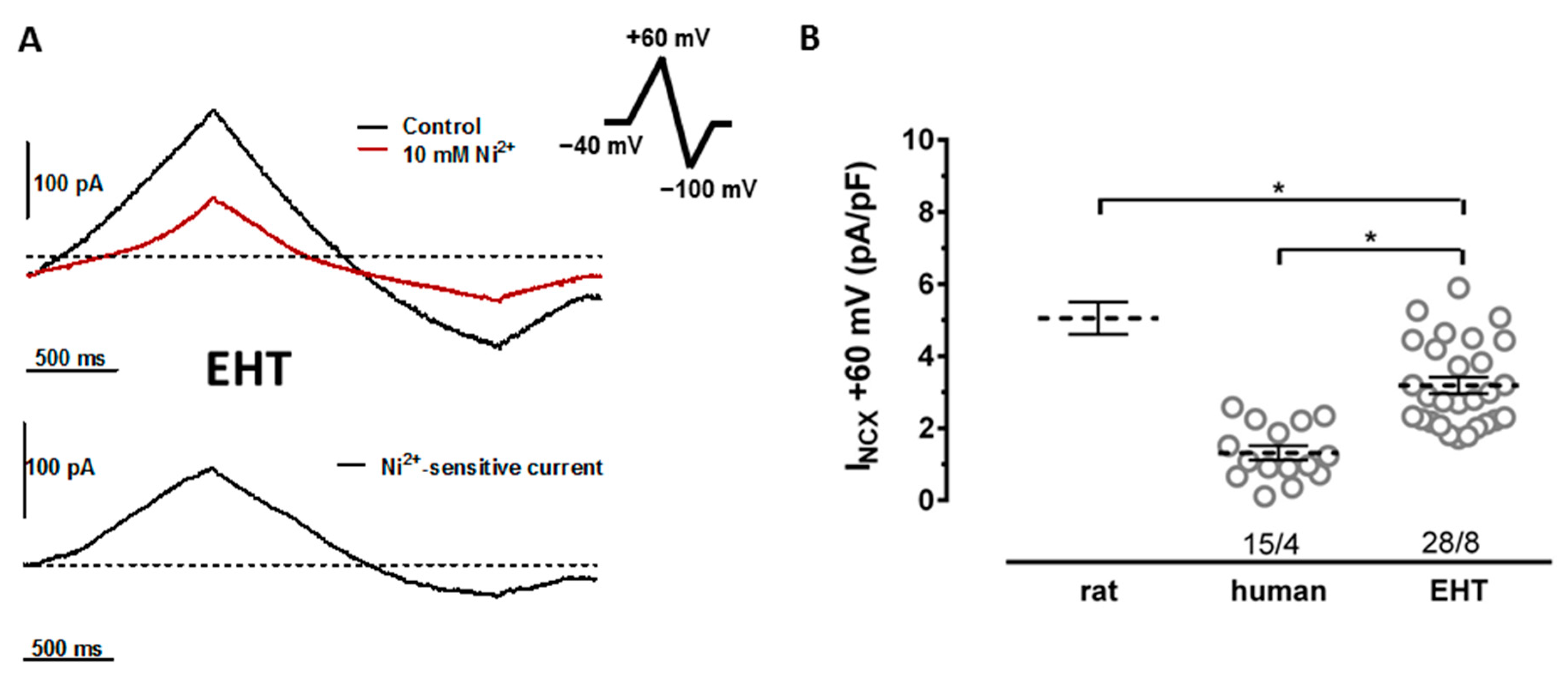
3.1. NCX Currents in hiPSC-CMs Are Not Smaller Than in Human Ventricular Cardiomyocytes

First, we measured mRNA expression for the NCX isoforms NCX1-3 (genes: SLC8A1, SLC8A2 and SLC8A3) [20]. The mRNA for all three isoforms of NCX was expressed in the human left ventricle (LV) and also in EHT. Expressions for NCX1, the dominant isoform, were slightly but significantly higher in EHT vs. human LV (Supplementary Figure S1).
The NCX currents were measured in isolated CMs. In order to facilitate comparisons to the literature, we used voltage ramps, which have been frequently used in previous studies [6]. The NCX currents were defined as Ni2+-sensitive currents evoked during voltage ramp protocols. We found NCX currents in each single cell from all four batches of hiPSC-CMs cultured from EHT (Supplementary Figure S2). In EHT, the NCX currents were larger than in the human left ventricular CMs (Figure 1). However, the NCX current densities in EHT did not reach the level recently reported by us for rat left ventricular CMs studied using the same protocol [7].

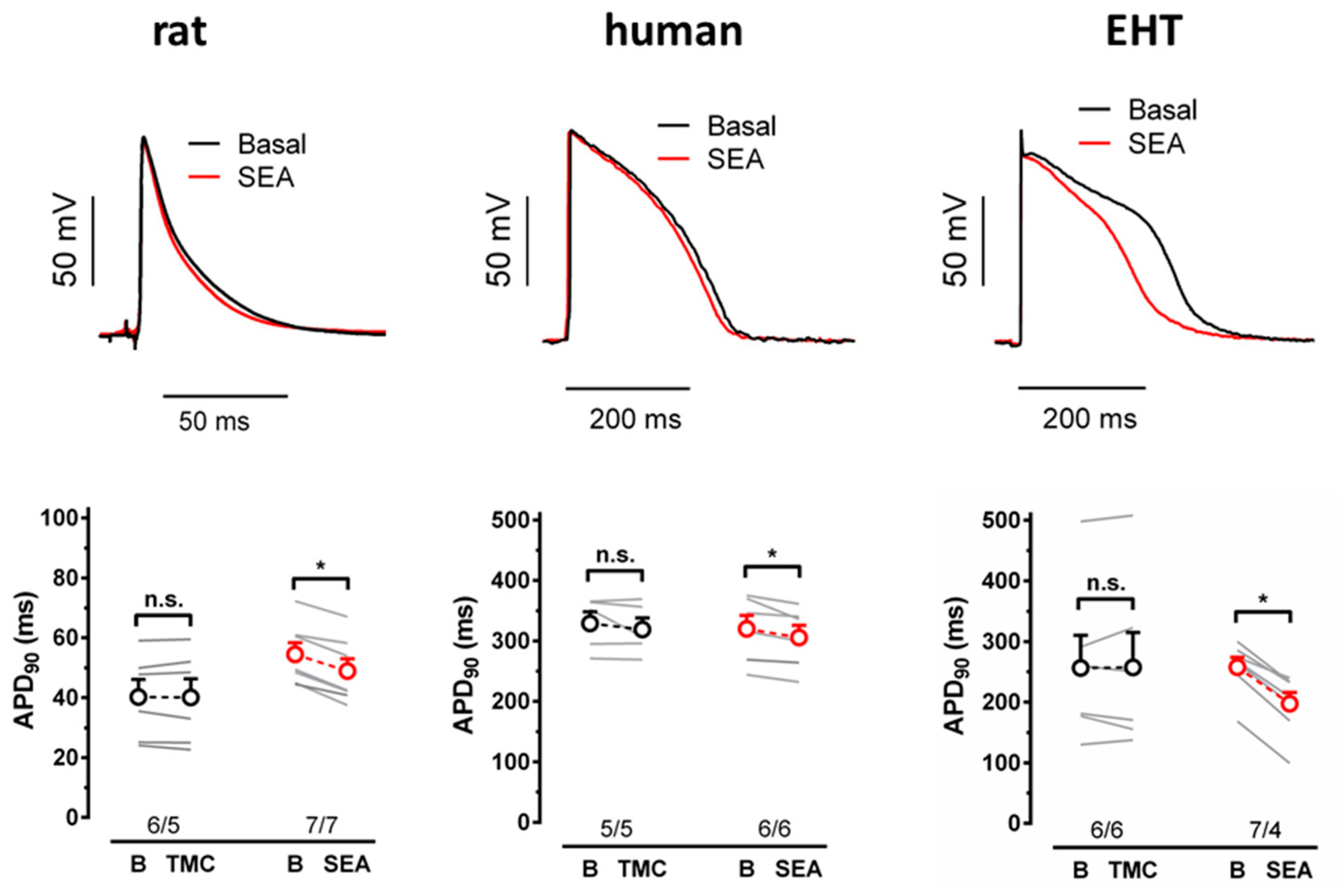
3.2. NCX Block Shortens the APD in EHT but Not in the Human Left Ventricle

During the plateau phase of the APs, NCX should mediate a depolarizing current [21]. Thus, the block of NCX should shorten the APD. Therefore, we measured the effects of the acute block of NCX by SEA0400 on the APD in EHT and in the left ventricular tissue from humans and rats. In all three tissues, we did not see significant changes in the APD in time-matched controls (TMCs) over 30 min (Figure 2). In contrast, the APD90 was markedly shortened by SEA0400 (10 µM) in EHT (by 24.6 ± 3.5%). In the left ventricular papillary muscles of rats, SEA0400-induced shortening of the APD90 was significantly smaller than in EHT, but still significant compared to TMCs (by 10.7 ± 1.6%). In left ventricular preparations from humans, SEA0400 tended to shorten the APD90, but the apparent effect size was not larger than the non-significant trend in TMCs (by 4.4 ± 1.1% vs. 2.9 ± 2.5% in TMCs).

3.3. SEA0400 Does Not Block Calcium Currents in hiPSC-CMs

SEA0400 shortened the APD90 in EHT, but not in the human LV compared to TMCs. This finding may imply that SEA0400 non-selectively blocks other depolarizing currents in hiPSC-CMs, but not in the human LV. However, the action potential amplitude (APA) or maximum upstroke velocity were not affected (in the rat and human LV or in EHT, Table S2), arguing against the relevant block of Na+ currents using SEA0400. HiPSC and human CMs differ with respect to Ca2+ channels. While human ventricular CMs express only L-type Ca2+ channels, T-type Ca2+ channels contribute substantially to the total Ca2+ currents in hiPSC-CMs [22]. Therefore, we measured the effects of SEA0400 on ICa in hiPSC-CMs. As shown before [22], there was a slight, but still significant, decrease in ICa in TMCs. More importantly, the decrease was not larger in hiPSC-CMs exposed to 10 µM of SEA0400 (Supplementary Figure S3).

3.4. NCX Block Increases Force in EHT but Not in the Human Left Ventricle

NCX is believed to represent one of the main Ca2+-extruding mechanisms; therefore, the block of NCX should increase intracellular Ca2+ and consecutively increase the force generation [21]. We measured the effects of the acute NCX block with SEA0400 on the force in EHT and in LV preparations from rats and humans. In the human LV and EHT, the force in time-matched controls declined slowly over 30 min (Figure 3). In EHT, SEA0400 (10 µM) significantly increased the force (to 22.3 ± 3.3% of pre-drug control values). In contrast to the human LV, SEA0400 did not show any consistent effect on force (−3.7 ± 4.3% vs. −6.2 ± 3.7% in TMCs). For comparison, we measured the effects of the compound in intact rat ventricular papillary muscles. The force remained stable over 30 min in TMCs, but increased significantly upon SEA0400 exposure (to 31 ± 5.4% of pre-drug control values).

3.5. Effects of NCX Block on AP and Force in Comparison to Computational Model Predictions

To better understand whether the effects of SEA0400 on the APD and force are in line with common paradigms on NCX contribution to Ca2+ handling in human ventricle, we employed established computational models for rat [16] and human [17] ventricular CMs, as well as for hiPSC-CMs. Unfortunately, in the initial simulations, in which the original model versions were used, the NCX block turned out to be an intolerable condition, leading to intracellular Ca2+ overload and consequent AP abnormalities. For the rat CM model, a 50% NCX block was already too great of a challenge (Supplementary Figure S4). In contrast, the human CM model was a bit more robust, tolerating NCX blocks up to 70% (Supplementary Figure S5).
We hypothesized that the standard models may underestimate the Ca2+ extrusion using the PMCA, likely causing a lack of robustness. It is generally assumed that, in murine CMs, sarcoplasmic reticulum Ca2+ ATPase (SERCA) pumps ~90% of Ca2+ back to the SR and NCX extrudes ~10%, while the PMCA contributes 1% or less [23]. However, there is substantial experimental evidence that the PMCA can remove Ca2+ at a rate of 30% of that of NCX [24,25,26]. When we readjusted the rat CM model accordingly, it became a bit more robust, capable of handling a 50% NCX block (Supplementary Figure S4). The simulated drug impact resulted in a 11.1% reduction in the APD90 (Figure 4A). This is in the same range as the SEA0400-induced shortening of APs in the experiments. However, the simulated block of NCX is only 50%, compared to the ~90% block to be expected in the experiments, so the conditions are not fully comparable.
To our knowledge, a thorough analysis of SERCA-NCX-PMCA Ca2+ removal fractions for human ventricular CMs has not been published. However, there are several data sets, from which these values can be integratively estimated (please see Supplementary Computations for details). When we readjusted the human CM model accordingly, it functioned a bit more robustly, being able to also handle a 90% NCX block. The simulated impact of the 90% NCX block on the APD90 (−18.3%) was much greater than the tiny non-significant change observed in the experiments. However, the effect was substantially decreased compared to the original model (by 34.9%), as shown in Supplementary Figure S5A. Similarly, the great increase in force due to the NCX block (Figure 4E) in simulation does not match the experimental observation of no change.
We also repeated the same NCX block simulations with a hiPSC-CM model that is described in detail in the Methods section. Interestingly, the model prediction for AP shortening due to the NCX block (APD90 −22.8%) fits perfectly with the experimentally observed change (−26.6 ± 5%). However, the simulated increase in force (almost nine-fold; Figure 4H) is far greater than in the experiments (+20.8 ± 3.9%). This indicates that even with a readjusted NCX-PMCA Ca2+ extrusion ratio, the computational CM model is not robust enough to resist intracellular Ca2+ accumulation, when NCX function is almost fully blocked.

4. Discussion

In our study, we compared the densities of the NCX current in hiPSC-CMs to ventricular CMs from rat and human and estimated the NCX contribution to AP and force. The main finding is that NCX currents in hiPSC-CMs from EHT are larger than in the human LV. Furthermore, the block of NCX evokes measurable effects on the APD and force in EHT and in the LV from rat but not from humans.

4.1. NCX Current Density

We found slightly higher transcript abundances for NCX1 in EHT vs. the human LV (Supplementary Figure S1), which were associated with substantially larger NCX currents in EHT, indicating relevant differences in functional channel-forming proteins. Interestingly, the ~2.4-fold larger NCX in EHT fits closely to the ~2 fold larger ICa in EHT vs. the human LV (12 pA/pF vs. 6 pA/pF) [22,27], and could thereby be interpreted as a balanced activity between transsarcolemmal Ca2+ load and Ca2+ extrusion. In rat ventricular myocytes, the NCX current was much higher than in EHT, despite the fact that ICa did not substantially differ (10.2 pA/pF) [28]. A much higher ratio of NCX/ICa is likely needed to extrude Ca2+ during shorter APs.

4.2. Contribution of NCX to the APD in Heart Muscle

As expected, SEA0400 shortened the APD both in rat ventricular tissue and in EHT. Even though basal APD90 values widely differ between the two tissues, the effect size was clearly stronger in EHT than in rats, both absolutely and relatively. This indicates that NCX is more important for the regulation of repolarisation in human EHT than in rats. As for force, we cannot provide an explanation for the lack of SEA0400 effects on the APD in the human LV. Results are in line with several papers demonstrating an absence of APD shortening by the NCX block in ventricular tissue from dogs, closely resembling the electrophysiology of the human heart [29,30,31]. Furthermore, our results are in line with findings on a new (even more selective than SEA0400) NCX blocker that did not shorten the APD in human left ventricular preparations but in hiPSC-CMs [19]. Thus, the data suggest fundamental differences in NCX contribution to Ca2+ handling, and hence APD regulation between hiPSC-CMs and the mature human LV.

4.3. Contribution of NCX to Force in Heart Muscle

The first NCX blockers such as KB-R7943 showed poor selectivity [21]. Thus, there was a huge interest in knock-out (KO) models of NCX. Unexpectedly, NCX1 KO mice showed regular Ca2+ transients [32], indicating that chronic adaptation can compensate for the lack of NCX function in the KO animals. While Ca2+ cycling via sarcoplasmic reticulum remained unchanged in NCX-KO mice, the ICa density was smaller, thereby reducing Ca2+ entry and subsequently the need to extrude Ca2+ via NCX. In addition, mice may not be the species of choice to study human NCX physiology, since NCX contributes to the regulation of [Ca2+]i predominantly during the APs, which is much shorter in mice than in humans.
Nevertheless, positive inotropy upon the acute pharmacological NCX block could be consistently demonstrated in rat ventricular tissue. Our findings in isolated rat ventricular tissue are in line with data obtained in Langendorff-perfused rat hearts [33]. With this in mind, one would expect a larger positive inotropic effect by the NCX block in species with a longer APD, but SEA0400 was devoid of positive inotropy in guinea pig hearts (APD ~150–200 ms) [34,35] and in human LV tissue (this study). Comparing the size of the positive inotropic effects in rat ventricular tissue vs. EHT is difficult because the dynamic range of force regulation differs. In rat ventricular tissue, catecholamines and/or high concentrations of calcium increase force by ~5 mN (the same experimental conditions) [28]. The increase in force using the NCX block amounts to about 20% of maximum force. In contrast, the maximum force increases as high calcium in EHT only amounts to ~0.15 mN [36]. Thus, the positive inotropy using the NCX block in EHT reached a 60% maximum force in this model. This finding could be interpreted as evidence for a larger contribution of NCX to Ca2+ handling in EHT.

4.4. Effect of NCX Block on the APD and Force Are Not Compromised by SEA0400’s Selectivity

Unexpectedly, SEA0400 did not increase force in heart tissues from several animals (Table 1). One hypothesis proposes the nonselective block of ICa by SEA0400 that may have masked positive inotropy induced by less Ca2+ extrusion [37]; in fact, some papers reported the ICa block using SEA0400 [38,39,40]. However, others did not see an effect of SEA0400 on ICa [41] and two considerations argue against the assumption that the block of ICa could explain the unexpected lack of effect of SEA0400 in guinea pig and human (Table 1). First, the block of ICa by SEA0400 should aggravate (not level off) APD shortening. However, SEA0400 does not shorten the APD90 in the human LV (this study and also recently reported) [19], human atrium [7], and dog ventricle [29,30,31]. Second, in our study, a decline in ICa in hiPSC-CMs exposed to increasing concentrations of SEA0400 was not larger than in time-matched controls (Supplementary Figure S3). These data argue against the nonselective block of ICa by SEA0400 under our experimental conditions, under which the compound induced strong APD shortening.

4.5. Effects of NCX Block: Immature EHT vs. Mature LV

The results of this study may be perplexing. Both LV and EHT express NCX and possess robust NCX currents when using established patch clamp protocols. HiPSC-CMs that are clearly immature with respect of intracellular architecture show the effects predicted by computer models which integrate our current understanding of Ca2+ handling, but native LV tissue from patients does not. The reasons for this are unclear. One could argue that LV tissue was obtained from patients in end-stage heart failure with well-known remodeling of Ca2+ handling. However, with regards to heart failure, the contribution of sarcoplasmic reticulum is reported to decrease and the contribution of NCX increased [47,48]. The PMCA represents an alternative way to extrude Ca2+ [49]. Therefore, we readjusted the PMCA contribution to Ca2+ removal to fit our experimental findings, increasing its maximum pump rate 740-fold in the computational human ventricular CM model. Under this condition, APD shortening upon the NCX block was almost halved, but still remained substantial (Supplementary Figure S5), deviating from the experimental findings. We therefore changed the modeling conditions further and tested a purely speculative human ventricular CM model version. In addition to higher pump rate, the PMCA was altered to be electroneutral. Under this condition, APD shortening upon the NCX block was decreased only very slightly (Supplementary Figure S6). In summary, our comparison of experimental and simulation results suggests that the computational human ventricular CM model is likely missing some elements of autoregulatory mechanisms related to Ca2+ handling, as the NCX block leads to substantial intracellular Ca2+ accumulation and consequent AP shortening.
Methodological issues could be relevant. Tissue penetration by SEA0400 could be an issue. However, the compound was active in intact rat left ventricular tissue strips (both APD and force in this study). No data are available about the effects of SEA0400 on the APD in whole rat hearts, but it induced a strong tendency for a concentration-dependent shortening of the QT interval in Langendorff-perfused rat hearts [50]. In addition, SEA0400 increased LV dp/dtmax in Langendorff-perfused rat hearts [50]. The data argue against the idea that a lack of APD or force effects of NCX blockers in human heart preparations can be explained by a lack of tissue penetration.

5. Limitations of the Study

We measured SEA0400 effects in rat ventricular tissue at 1 Hz, much lower than the physiological heart rate in this species. However, we used this rate for comparison with human tissue. We used only one hiPSC-CM cell line for our study. Access to the human LV is limited. The success rate of AP measurements is much smaller than for force recording. Thus, we could have overlooked the small effects of the NCX block on the APD in the human LV.
Before EHT can be recommended or discarded as an experimental tool to study human ventricular NCX, other NCX functions such as a contribution to the generation of early afterdepolarizations [6,41] or Na+-driven inotropy should be studied in adult human ventricular tissue and in EHT in parallel.

6. Conclusions

The pharmacological block of NCX shortens the APD and increases force in rat ventricle, as expected from textbook knowledge. This finding may illustrate that simple but rapidly acting compounds can provide better insight in NCX physiology than slowly developing mice KO models, prone to chronic adaptation. The NCX block shows the predicted effects on the APD in hiPSC-CMs, but not in the human LV, despite the fact that transsarcolemmal Ca2+ influx via ICa and Ca2+ extrusion via NCX, estimated from patch clamp data, do not differ widely. Obviously, the current density does not necessarily correlate to NCX contribution to the APD and force regulation. The lack of effect of the NCX block in human ventricle is in line with several other studies in the hearts of larger animals and should therefore be taken seriously. Reasons for the NCX block insensitivity of human ventricle needs to be clarified before hiPSC-CMs can be used as a human model to study NCX. However, the marked effects of NCX inhibition in EHT indicate high sensitivity in preclinical drug screening. For this purpose, EHT appears to be suitable, even better than human myocardial tissue.

Supplementary Materials

The following supporting information can be downloaded at: https://www.mdpi.com/article/10.3390/cells11152424/s1, Figure S1: RNA expression of NCX; Figure S2: NCX currents in cardiomyocytes from human ventricle and EHT culture divided by different patients and different batches; Figure S3: Effect of SEA0400 on calcium current (ICa) in EHT; Figure S4: Comparison of NCX block impact in the original and readjusted rat ventricular cardiomyocyte models; Figure S5: Comparison of NCX block impact in the original and readjusted human ventricular cardiomyocyte models; Figure S6: NCX block impact in the readjusted human ventricular cardiomyocyte model, with electroneutral PMCA; Figure S7: Comparison of readjusted human ventricular cardiomyocyte and hiPSC-CM models; Table S1: Patient characteristics; Table S2: Action potential parameters (mean ± SEM) before and after superfusion with SEA0400 (10 µM) in rat ventricle, human ventricle and EHT; Supplemental Experimental and Computational Procedures.

Author Contributions

Conceptualization, D.I., J.T.K. and T.C.; methodology, D.I., K.G., A.H. (András Horváth), L.Y., M.D.L., J.T.K. and T.C.; software, J.T.K.; validation, D.I. and T.C.; formal analysis, D.I., K.G., A.H. (András Horváth), M.D.L., J.T.K. and T.C.; investigation, D.I., K.G., A.H. (András Horváth), L.Y., M.D.L., C.S., J.S., J.T.K. and T.C.; resources, J.P., H.R., T.E. and A.H. (Arne Hansen); data curation, D.I., K.G., A.H. (András Horváth), L.Y., M.D.L., C.S., J.S., J.T.K. and T.C.; writing—original draft preparation, D.I., J.T.K. and T.C.; writing—review and editing, P.K., T.J., T.E., A.H. (Arne Hansen), J.T.K. and T.C.; visualization, D.I., K.G., A.H. (András Horváth), L.Y., M.D.L., C.S., J.T.K. and T.C.; supervision, T.J., T.E., A.H. (Arne Hansen), and T.C.; project administration, D.I., T.E. and T.C.; funding acquisition, T.E., A.H. (Arne Hansen), and T.C. All authors have read and agreed to the published version of the manuscript.

Funding

This study was supported by the German Ministry of Education and Research (BMBF, FKZ161L0236), the European Research Council (ERC-AG IndivuHeart), Deutsche Forschungsgemeinschaft (DFG Es 88/12-1, DFG HA 3423/5-1), the Centre for Cardiovascular Research (DZHK), the Freie und Hansestadt Hamburg, the Academy of Finland Centre of Excellence in Body-on-Chip Research, the Finnish Foundation for Cardiovascular Research (170047, 200101), the Paavo Nurmi Foundation (20170129) and the Pirkanmaa regional fund of the Finnish Cultural Foundation (50171514, 50201322).

Institutional Review Board Statement

The study on human tissue followed the declaration of Helsinki and was approved by the ethics committee of the Medical Council of Hamburg, Germany (approval number: OB-088/04 and PV3759). The study on rat tissue was conducted according to guidelines for laboratory animal welfare of the National Institutes of Health (Publication no. 85-23, revised 1985). All experiments with rat tissue were performed in compliance with the German Law for the Protection of Animals.

Informed Consent Statement

Informed consent was obtained from all subjects involved in the study.

Data Availability Statement

Not applicable.

Acknowledgments

The authors gratefully acknowledge the patients who donated tissue; the members of the hiPSC-CM working group at the Department of Experimental Pharmacology and Toxicology, UKE-Hamburg, for their help in providing EHTs from Marta Lemme, Ingke Braren, Mirja Schulze, Umber Saleem, Thomas Schulze, and Birgit Klampe; and the expert technical assistance of Anna Steenpaß.

Conflicts of Interest

T.E. and A.H. (Arne Hansen) were co-founders of EHT Technologies GmbH.

Abbreviations

APsaction potentials
APAaction potential amplitude
APDaction potential duration
APD90action potential duration at 90% repolarization
CaTcalcium transient
CMcardiomyocyte
Cscell shortening
EHTengineered heart tissue
hiPSC-CMhuman-induced pluripotent stem cell-derived cardiomyocyte
ICacalcium current
IK1inwardly rectifying potassium current density
IKrrapidly activating potassium current
KOknock-out
LVleft ventricle
NCXsodium calcium exchanger
PMCAplasma membrane Ca2+-ATPase
TMCstime-matched controls

References

  1. Eschenhagen, T.; Carrier, L. Cardiomyopathy phenotypes in human-induced pluripotent stem cell-derived cardiomyocytes–A systematic review. Pflugers Arch. 2019, 471, 755. [Google Scholar] [CrossRef] [Green Version]
  2. Paci, M.; Penttinen, K.; Pekkanen-Mattila, M.; Koivumäki, J.T. Arrhythmia Mechanisms in Human Induced Pluripotent Stem Cell-Derived Cardiomyocytes. J. Cardiovasc. Pharmacol. 2020, 77, 300–316. [Google Scholar] [CrossRef]
  3. Zhang, X.h.; Morad, M. Ca2+ signaling of human pluripotent stem cells-derived cardiomyocytes as compared to adult mammalian cardiomyocytes. Cell Calcium. 2020, 90, 102244. [Google Scholar] [CrossRef]
  4. Hwang, H.S.; Kryshtal, D.O.; Feaster, T.K.; Sánchez-Freire, V.; Zhang, J.; Kamp, T.J.; Hong, C.C.; Wu, J.C.; Knollmann, B.C. Human induced pluripotent stem cell (hiPSC) derived cardiomyocytes to understand and test cardiac calcium handling: A glass half full. J. Mol. Cell. Cardiol. 2015, 89, 379–380. [Google Scholar] [CrossRef]
  5. Kane, C.; Couch, L.; Terracciano, C.M.N. Excitation–contraction coupling of human induced pluripotent stem cell-derived cardiomyocytes. Front. Cell Dev. Biol. 2015, 3, 1–8. [Google Scholar] [CrossRef] [Green Version]
  6. Jost, N.; Nagy, N.; Corici, C.; Kohajda, Z.; Horváth, A.; Acsai, K.; Biliczki, P.; Levijoki, J.; Pollesello, P.; Koskelainen, T.; et al. ORM-10103, a novel specific inhibitor of the Na+/Ca2+ exchanger, decreases early and delayed afterdepolarizations in the canine heart. Br. J. Pharmacol. 2013, 170, 768–778. [Google Scholar] [CrossRef] [Green Version]
  7. Christ, T.; Kovács, P.P.; Acsai, K.; Knaut, M.; Eschenhagen, T.; Jost, N.; Varró, A.; Wettwer, E.; Ravens, U. Block of Na+/Ca2+ exchanger by SEA0400 in human right atrial preparations from patients in sinus rhythm and in atrial fibrillation. Eur. J. Pharmacol. 2016, 788, 286–293. [Google Scholar] [CrossRef]
  8. Dobrev, D.; Wettwer, E.; Himmel, H.M.; Kortner, A.; Kuhlisch, E.; Schüler, S.; Siffert, W.; Ravens, U. G-protein β3-subunit 825T allele is associated with enhanced human atrial inward rectifier potassium currents. Circulation 2000, 102, 692–697. [Google Scholar] [CrossRef] [Green Version]
  9. Lemme, M.; Ulmer, B.M.; Lemoine, M.D.; Zech, A.T.L.; Flenner, F.; Ravens, U.; Reichenspurner, H.; Rol-Garcia, M.; Smith, G.; Hansen, A.; et al. Atrial-like Engineered Heart Tissue: An In Vitro Model of the Human Atrium. Stem Cell Rep. 2018, 11, 1378–1390. [Google Scholar] [CrossRef] [Green Version]
  10. Breckwoldt, K.; Letuffe-Brenière, D.; Mannhardt, I.; Schulze, T.; Ulmer, B.; Werner, T.; Benzin, A.; Klampe, B.; Reinsch, M.C.; Laufer, S.; et al. Differentiation of cardiomyocytes and generation of human engineered heart tissue. Nat. Protoc. 2017, 12, 1177–1190. [Google Scholar] [CrossRef]
  11. Lemoine, M.D.; Krause, T.; Koivumäki, J.T.; Prondzynski, M.; Schulze, M.L.; Girdauskas, E.; Willems, S.; Hansen, A.; Eschenhagen, T.; Christ, T. Human Induced Pluripotent Stem Cell-Derived Engineered Heart Tissue as a Sensitive Test System for QT Prolongation and Arrhythmic Triggers. Circ. Arrhythmia Electrophysiol. 2018, 11, e006035. [Google Scholar] [CrossRef]
  12. Pecha, S.; Flenner, F.; Söhren, K.D.; Lorenz, K.; Eschenhagen, T.; Christ, T. β1 Adrenoceptor antagonistic effects of the supposedly selective β2 adrenoceptor antagonist ICI 118,551 on the positive inotropic effect of adrenaline in murine hearts. Pharmacol. Res. Perspect. 2015, 3, e00168. [Google Scholar] [CrossRef]
  13. Kloth, B.; Pecha, S.; Moritz, E.; Schneeberger, Y.; Söhren, K.D.; Schwedhelm, E.; Reichenspurner, H.; Eschenhagen, T.; Böger, R.H.; Christ, T.; et al. AkrinorTM, a cafedrine/theodrenaline mixture (20:1), increases force of contraction of human atrial myocardium but does not constrict internal mammary artery in vitro. Front. Pharmacol. 2017, 8, 272. [Google Scholar] [CrossRef] [Green Version]
  14. Pecha, S.; Koivumäki, J.; Geelhoed, B.; Kempe, R.; Berk, E.; Engel, A.; Reichenspurner, H.; Eschenhagen, T.; Ravens, U.; Kaumann, A.; et al. Normalization of force to muscle cross-sectional area: A helpful attempt to reduce data scattering in contractility studies? Acta Physiol. 2018, 224, e13202. [Google Scholar] [CrossRef] [Green Version]
  15. Hansen, A.; Eder, A.; Bonstrup, M.; Flato, M.; Mewe, M.; Schaaf, S.; Aksehirlioglu, B.; Schworer, A.; Uebeler, J.; Eschenhagen, T.; et al. Development of a Drug Screening Platform Based on Engineered Heart Tissue. Circ. Res. 2010, 107, 35–44. [Google Scholar] [CrossRef] [Green Version]
  16. Gattoni, S.; Røe, Å.T.; Frisk, M.; Louch, W.E.; Niederer, S.A.; Smith, N.P. The calcium–frequency response in the rat ventricular myocyte: An experimental and modelling study. J. Physiol. 2016, 594, 4193–4224. [Google Scholar] [CrossRef] [Green Version]
  17. Margara, F.; Wang, Z.J.; Levrero-Florencio, F.; Santiago, A.; Vázquez, M.; Bueno-Orovio, A.; Rodriguez, B. In-silico human electro-mechanical ventricular modelling and simulation for drug-induced pro-arrhythmia and inotropic risk assessment. Prog. Biophys. Mol. Biol. 2021, 159, 58–74. [Google Scholar] [CrossRef]
  18. Prondzynski, M.; Lemoine, M.D.; Zech, A.T.; Horváth, A.; di Mauro, V.; Koivumäki, J.T.; Kresin, N.; Busch, J.; Krause, T.; Krämer, E.; et al. Disease modeling of a mutation in α-actinin 2 guides clinical therapy in hypertrophic cardiomyopathy. EMBO Mol. Med. 2019, 11, e11115. [Google Scholar] [CrossRef]
  19. Otsomaa, L.; Levijoki, J.; Wohlfahrt, G.; Chapman, H.; Koivisto, A.-P.; Syrjanen, K.; Koskelainen, T.; Peltokorpi, S.-E.; Finckenberg, P.; Heikkila, A.; et al. Discovery and characterization of ORM-11372, a unique and positively inotropic sodium-calcium exchanger/inhibitor. Br. J. Pharmacol. 2020, 177, 5534–5554. [Google Scholar] [CrossRef]
  20. Khananshvili, D. The SLC8 gene family of sodium–Calcium exchangers (NCX)–Structure, function, and regulation in health and disease. Mol. Aspects Med. 2013, 34, 220–235. [Google Scholar] [CrossRef]
  21. Shattock, M.J.; Ottolia, M.; Bers, D.M.; Blaustein, M.P.; Boguslavskyi, A.; Bossuyt, J.; Bridge, J.H.B.; Chen-Izu, Y.; Clancy, C.E.; Edwards, A.; et al. Na+/Ca2+ exchange and Na+/K+-ATPase in the heart. J. Physiol. 2015, 593, 1361–1382. [Google Scholar] [CrossRef] [Green Version]
  22. Uzun, A.U.; Mannhardt, I.; Breckwoldt, K.; Horváth, A.; Johannsen, S.S.; Hansen, A.; Eschenhagen, T.; Christ, T. Ca2+-currents in human induced pluripotent stem cell-derived cardiomyocytes effects of two different culture conditions. Front. Pharmacol. 2016, 7, 300. [Google Scholar] [CrossRef] [Green Version]
  23. Bers, D.M. Cardiac excitation contraction coupling. Nature 2002, 415, 198–205. [Google Scholar] [CrossRef]
  24. Negretti, N.; O’Neill, S.C.; Eisner, D.A. The relative contributions of different intracellular and sarcolemmal systems to relaxation in rat ventricular myocytes. Cardiovasc. Res. 1993, 27, 1826–1830. [Google Scholar] [CrossRef]
  25. Choi, H.S.; Eisner, D.A. The effects of inhibition of the sarcolemmal Ca-ATPase on systolic calcium fluxes and intracellular calcium concentration in rat ventricular myocytes. Pflügers Arch. 1999, 437, 966–971. [Google Scholar] [CrossRef]
  26. Choi, H.S.; Eisner, D.A. The role of sarcolemmal Ca2+-ATPase in the regulation of resting calcium concentration in rat ventricular myocytes. J. Physiol. 1999, 515, 109–118. [Google Scholar] [CrossRef]
  27. Ismaili, D.; Geelhoed, B.; Christ, T. Ca2+ currents in cardiomyocytes: How to improve interpretation of patch clamp data? Prog. Biophys. Mol. Biol. 2020, 157, 33–39. [Google Scholar] [CrossRef]
  28. Christ, T.; Galindo-Tovar, A.; Thoms, M.; Ravens, U.; Kaumann, A.J. Inotropy and L-type Ca2+ current, activated by β 1-and β 2 -adrenoceptors, are differently controlled by phosphodiesterases 3 and 4 in rat heart. Br. J. Pharmacol. 2009, 156, 62–83. [Google Scholar] [CrossRef] [Green Version]
  29. Nagy, N.; Kormos, A.; Kohajda, Z.; Szebeni, Á.; Szepesi, J.; Pollesello, P.; Levijoki, J.; Acsai, K.; Virág, L.; Nánási, P.P.; et al. Selective Na+/Ca2+ exchanger inhibition prevents Ca2+ overload-induced triggered arrhythmias. Br. J. Pharmacol. 2014, 171, 5665–5681. [Google Scholar] [CrossRef] [Green Version]
  30. Kohajda, Z.; Farkas-Morvay, N.; Jost, N.; Nagy, N.; Geramipour, A.; Horváth, A.; Varga, R.S.; Hornyik, T.; Corici, C.; Acsai, K.; et al. The Effect of a Novel Highly Selective Inhibitor of the Sodium/Calcium Exchanger (NCX) on Cardiac Arrhythmias in In Vitro and In Vivo Experiments. PLoS ONE 2016, 11, e0166041. [Google Scholar] [CrossRef]
  31. Geramipour, A.; Kohajda, Z.; Corici, C.; Prorok, J.; Szakonyi, Z.; Oravecz, K.; Márton, Z.; Nagy, N.; Tóth, A.; Acsai, K.; et al. The investigation of the cellular electrophysiological and antiarrhythmic effects of a novel selective sodium–calcium exchanger inhibitor, GYKB-6635, in canine and guinea-pig hearts. Can. J. Physiol. Pharmacol. 2016, 94, 1090–1101. [Google Scholar] [CrossRef] [PubMed]
  32. Pott, C.; Philipson, K.D.; Goldhaber, J.I. Excitation-contraction coupling in Na+-Ca2+ exchanger knockout mice: Reduced transsarcolemmal Ca2+ flux. Circ. Res. 2005, 97, 1288–1295. [Google Scholar] [CrossRef] [PubMed] [Green Version]
  33. Szentandrássy, N.; Birinyi, P.; Szigeti, G.; Farkas, A.; Magyar, J.; Tóth, A.; Csernoch, L.; Varró, A.; Nánási, P.P. SEA0400 fails to alter the magnitude of intracellular Ca2+ transients and contractions in Langendorff-perfused guinea pig heart. Naunyn-Schmiedeberg’s Arch. Pharmacol. 2008, 378, 65–71. [Google Scholar] [CrossRef] [PubMed]
  34. Namekata, I.; Nakamura, H.; Shimada, H.; Tanaka, H.; Shigenobu, K. Cardioprotection without cardiosuppression by SEA0400, a novel inhibitor of Na+-Ca2+ exchanger, during ischemia and reperfusion in guinea-pig myocardium. Life Sci. 2005, 77, 312–324. [Google Scholar] [CrossRef] [PubMed]
  35. Tanaka, H.; Shimada, H.; Namekata, I.; Kawanishi, T.; Iida-Tanaka, N.; Shigenobu, K. Involvement of the Na+/Ca2+ Exchanger in Ouabain-Induced Inotropy and Arrhythmogenesis in Guinea-Pig Myocardium as Revealed by SEA0400. J. Pharmacol. Sci. 2007, 103, 241–246. [Google Scholar] [CrossRef] [PubMed] [Green Version]
  36. Saleem, U.; Ismaili, D.; Mannhardt, I.; Pinnschmidt, H.; Schulze, T.; Christ, T.; Eschenhagen, T.; Hansen, A. Regulation of ICa,L and force by PDEs in human-induced pluripotent stem cell-derived cardiomyocytes. Br. J. Pharmacol. 2020, 177, 3036–3045. [Google Scholar] [CrossRef] [PubMed] [Green Version]
  37. Birinyi, P.; Tóth, A.; Jóna, I.; Acsai, K.; Almássy, J.; Nagy, N.; Prorok, J.; Gherasim, I.; Papp, Z.; Hertelendi, Z.; et al. The Na+/Ca2+ exchange blocker SEA0400 fails to enhance cytosolic Ca2+ transient and contractility in canine ventricular cardiomyocytes. Cardiovasc. Res. 2008, 78, 476–484. [Google Scholar] [CrossRef] [Green Version]
  38. Birinyi, P.; Acsai, K.; Bányász, T.; Tóth, A.; Horváth, B.; Virág, L.; Szentandrássy, N.; Magyar, J.; Varró, A.; Fülöp, F.; et al. Effects of SEA0400 and KB-R7943 on Na+/Ca2+ exchange current and L-type Ca2+ current in canine ventricular cardiomyocytes. Naunyn-Schmiedeberg’s Arch. Pharmacol. 2005, 372, 63–70. [Google Scholar] [CrossRef]
  39. Acsai, K.; Kun, A.; Farkas, A.S.; Fülöp, F.; Nagy, N.; Balázs, M.; Szentandrássy, N.; Nánási, P.P.; Papp, J.G.; Varró, A.; et al. Effect of partial blockade of the Na+/Ca2+-exchanger on Ca2+ handling in isolated rat ventricular myocytes. Eur. J. Pharmacol. 2007, 576, 1–6. [Google Scholar] [CrossRef]
  40. Bourgonje, V.J.A.; Vos, M.A.; Ozdemir, S.; Doisne, N.; Acsai, K.; Varro, A.; Sztojkov-Ivanov, A.; Zupko, I.; Rauch, E.; Kattner, L.; et al. Combined Na+/Ca2+ Exchanger and L-Type Calcium Channel Block as a Potential Strategy to Suppress Arrhythmias and Maintain Ventricular Function. Circ. Arrhythmia Electrophysiol. 2013, 6, 371–379. [Google Scholar] [CrossRef] [Green Version]
  41. Nagy, Z.A.; Virág, L.; Tóth, A.; Biliczki, P.; Acsai, K.; Bányász, T.; Nánási, P.; Papp, J.G.; Varró, A. Selective inhibition of sodium–calcium exchanger by SEA-0400 decreases early and delayed afterdepolarization in canine heart. Br. J. Pharmacol. 2004, 143, 827–831. [Google Scholar] [CrossRef] [PubMed] [Green Version]
  42. Oravecz, K.; Kormos, A.; Gruber, A.; Márton, Z.; Kohajda, Z.; Mirzaei, L.; Jost, N.; Levijoki, J.; Pollesello, P.; Koskelainen, T.; et al. Inotropic effect of NCX inhibition depends on the relative activity of the reverse NCX assessed by a novel inhibitor ORM-10962 on canine ventricular myocytes. Eur. J. Pharmacol. 2018, 818, 278–286. [Google Scholar] [CrossRef]
  43. Ozdemir, S.; Bito, V.; Holemans, P.; Vinet, L.; Mercadier, J.-J.; Varro, A.; Sipido, K.R. Pharmacological Inhibition of Na/Ca Exchange Results in Increased Cellular Ca2+ Load Attributable to the Predominance of Forward Mode Block. Circ. Res. 2008, 102, 1398–1405. [Google Scholar] [CrossRef] [PubMed] [Green Version]
  44. Bögeholz, N.; Schulte, J.S.; Kaese, S.; Bauer, B.K.; Pauls, P.; Dechering, D.G.; Frommeyer, G.; Goldhaber, J.I.; Kirchhefer, U.; Eckardt, L.; et al. The Effects of SEA0400 on Ca2+ Transient Amplitude and Proarrhythmia Depend on the Na+/Ca2+ Exchanger Expression Level in Murine Models. Front. Pharmacol. 2017, 8, 649. [Google Scholar] [CrossRef] [Green Version]
  45. Tanaka, H.; Namekata, I.; Takeda, K.; Kazama, A.; Shimizu, Y.; Moriwaki, R.; Hirayama, W.; Sato, A.; Kawanishi, T.; Shigenobu, K. Unique excitation–contraction characteristics of mouse myocardium as revealed by SEA0400, a specific inhibitor of Na+–Ca2+ exchanger. Naunyn-Schmiedeberg’s Arch. Pharmacol. 2005, 371, 526–534. [Google Scholar] [CrossRef]
  46. Amran, M.S.; Hashimoto, K.; Homma, N. Effects of Sodium-Calcium Exchange Inhibitors, KB-R7943 and SEA0400, on Aconitine-Induced Arrhythmias in Guinea Pigs in Vivo, in Vitro, and in Computer Simulation Studies. J. Pharmacol. Exp. Ther. 2004, 310, 83–89. [Google Scholar] [CrossRef] [PubMed] [Green Version]
  47. Pogwizd, S.M.; Schlotthauer, K.; Li, L.; Yuan, W.; Bers, D.M. Arrhythmogenesis and Contractile Dysfunction in Heart Failure. Circ. Res. 2001, 88, 1159–1167. [Google Scholar] [CrossRef] [Green Version]
  48. Studer, R.; Reinecke, H.; Bilger, J.; Eschenhagen, T.; Böhm, M.; Hasenfuß, G.; Just, H.; Holtz, J.; Drexler, H. Gene expression of the cardiac Na+-Ca2+ exchanger in end-stage human heart failure. Circ. Res. 1994, 75, 443–453. [Google Scholar] [CrossRef] [Green Version]
  49. Carafoli, E. Calcium pump of the plasma membrane. Physiol. Rev. 1991, 71, 129–153. [Google Scholar] [CrossRef]
  50. Farkas, A.S.; Acsai, K.; Nagy, N.; Tóth, A.; Fülöp, F.; Seprényi, G.; Birinyi, P.; Nánási, P.P.; Forster, T.; Csanády, M.; et al. Na+/Ca2+ exchanger inhibition exerts a positive inotropic effect in the rat heart, but fails to influence the contractility of the rabbit heart. Br. J. Pharmacol. 2008, 154, 93–104. [Google Scholar] [CrossRef] [Green Version]
Figure 1. NCX currents in cardiomyocytes from rat ventricle, human ventricle, and EHT culture. (A) NCX current traces elicited by a ramp pulse from a holding potential of −40 mV to +60 mV, then to −100 mV, and back to the holding potential of −40 mV (see inset) at the pre-drug control (black trace) and after superfusion of cells with 10 mM Ni2+ (dark red trace). (B) NCX current density in the presence of 10 mM Ni2+ given as individual data points (circles) and respective mean values ± SEM measured in CMs from human ventricle and EHT culture. For comparison of mean ± SEM NCX currents measured in rat ventricular, CMs from Christ et al. [7] are given. n/n indicates the number of cells/number patients for human and the number of cells/number of isolations for EHT, * indicates significance.
Cells 11 02424 g001
Figure 2. Action potentials in rat ventricle, human ventricle, and EHT. (Up) Action potential traces at pre-drug control (black trace) and after 20 min superfusion with 10 µM SEA0400 in rat ventricle, human ventricle, and EHT. (Bottom) Individual data points (light gray) and respective mean values ± SEM for APD90 given as time-matched controls (TMCs) and in the presence of 10 µM SEA0400 in rat ventricle, human ventricle, and EHT. n/n indicates the number of cells/number rats or humans and the number of cells/number of isolations for EHT, * indicates significance, n.s. indicates non-significance.
Cells 11 02424 g002
Figure 3. Force measurements in rat ventricle, human ventricle, and EHT. (Up) Time course of force in rat and human ventricle given as time-matched controls (TMCs) and exposed to 10 µM SEA0400. Single contraction of force in EHT given as TMCs and exposed to 10 µM SEA0400. (Bottom) Individual data points (light gray) and respective mean values ± SEM for force given as time-matched controls (TMCs) and in the presence of 10 µM SEA0400 in rat ventricle, human ventricle, and EHT. n/n indicates the number of cells/number rats or humans and the number of cells/number of isolations for EHT, * indicates significance, n.s. indicates non-significance.
Cells 11 02424 g003
Figure 4. Simulated effect of NCX block in the rat ventricular, human ventricular, and hiPSC cardiomyocyte models. The principal outputs: membrane voltage (A,C,F), calcium transient (B,D,G), and active tension (E,H) with different degrees of NCX block. For details of the used models and simulation protocols, please see the Methods section.
Cells 11 02424 g004
Table 1. Literature research: the effect of NCX block on the APD90 and contractility parameters in different species.
SpeciesCell TypeSubstanceConcentration (in µM)APD90Peak CaTCsForceReference
DogVentricleSEA04001Nagy et al. [41]
SEA04001Birinyi et al. [37]
SEA04001Bourgonje et al. [40]
SEA04001Nagy et al. [29]
ORM-1010310
GYKB-66351Geramipouretal. [31]
ORM-109621Kohajda et al. [30]
ORM-109621Oravecz et al. [42]
HumanVentricleORM-1137210Otsomaa et al. [19]
AtriumSEA040010Christ et al. [7]
HiPSCORM-113720.1; 0.3Otsomaa et al. [19]
RatVentricleSEA04000.3Acsai et al. [39]
SEA04001Szentandrássy et al. [33]
MouseVentricleSEA04000.3; 1Ozdemir et al. [43]
SEA04001Bögeholz et al. [44]
SEA04001; 10Tanaka et al. [45]
Guinea pigVentricleSEA04001Szentandrássy et al. [33]
SEA04001Tanaka et al. [35]
SEA04001; 10; 100Amran et al. [46]
SEA04001Namekata et al. [34]
PigventricleSEA04000.3; 1Ozdemir et al. [43]
Legend: ↑ = increase; ↓ = reduction; ↔ = no effect; — = not measured.
Publisher’s Note: MDPI stays neutral with regard to jurisdictional claims in published maps and institutional affiliations.

