Article

The Inactivation of LPS Biosynthesis Genes in E. coli Cells Leads to Oxidative Stress

by
Tatiana A. Seregina
,
Irina Yu. Petrushanko
,
Rustem S. Shakulov
,
Pavel I. Zaripov
,
Alexander A. Makarov
,
Vladimir A. Mitkevich
* and
Alexander S. Mironov
Engelhardt Institute of Molecular Biology, Russian Academy of Science, 119991 Moscow, Russia
*
Author to whom correspondence should be addressed.
Cells 2022, 11(17), 2667; https://doi.org/10.3390/cells11172667
Submission received: 22 July 2022 / Revised: 22 August 2022 / Accepted: 25 August 2022 / Published: 27 August 2022

Abstract

:
Impaired lipopolysaccharide biosynthesis in Gram-negative bacteria results in the “deep rough” phenotype, which is characterized by increased sensitivity of cells to various hydrophobic compounds, including antibiotics novobiocin, actinomycin D, erythromycin, etc. The present study showed that E. coli mutants carrying deletions of the ADP-heptose biosynthesis genes became hypersensitive to a wide range of antibacterial drugs: DNA gyrase inhibitors, protein biosynthesis inhibitors (aminoglycosides, tetracycline), RNA polymerase inhibitors (rifampicin), and β-lactams (carbenicillin). In addition, it was found that inactivation of the gmhA, hldE, rfaD, and waaC genes led to dramatic changes in the redox status of cells: a decrease in the pool of reducing NADPH and ATP equivalents, the concentration of intracellular cysteine, a change in thiol homeostasis, and a deficiency in the formation of hydrogen sulfide. In “deep rough” mutants, intensive formation of reactive oxygen species was observed, which, along with a lack of reducing agents, such as reactive sulfur species or NADPH, leads to oxidative stress and an increase in the number of dead cells in the population. Within the framework of modern ideas about the role of oxidative stress as a universal mechanism of the bactericidal action of antibiotics, inhibition of the enzymes of ADP-heptose biosynthesis is a promising direction for increasing the effectiveness of existing antibiotics and solving the problem of multidrug resistance.

1. Introduction

At present, a large number of studies are devoted to solving the problem of antibiotic resistance to bacterial infections. The most effective direction of this work is not the creation of new antibacterial drugs, but the search for cellular targets, exposure to which would lead to a significant increase in the sensitivity of bacteria to antibiotics, widely used in modern regimens for the treatment of clinically significant infections. Previously, it was described that the disruption of biosynthesis and the assembly of lipopolysaccharide (LPS) components of the outer membrane of Gram-negative bacteria, due to mutations in the rfa-operon (“deep-rough” mutants), led to an increase in the content of lipids in LPS as well as a simultaneous decrease in proteins. As a result, there was an increase in membrane permeability for hydrophobic substances and an enhancement in cell sensitivity to various hydrophobic compounds, including antibiotics novobiocin, actinomycin D, erythromycin, etc. [1,2]. The rfaD, waaF, and waaC genes are part of the first of three rfa-operons and encode proteins involved in the biosynthesis and transfer of activated heptose residues to the LPS inner core [3]. The gmhA, gmhB, and hldE genes are not included in the rfa-operon; their protein products are involved in the isomerization of sedoheptulose-7P and its subsequent activation reactions, resulting in the formation of ADP-glycero-manno-heptose, which, with the participation of the WaaC and WaaF proteins, is attached to the inner core (Kdo) LPS (Figure 1).
The main intracellular source of sedoheptulose in most organisms is the pentose phosphate pathway (PPP) [6]. Sedoheptulose is formed in the reversible branch of PPP by transketolase from two pentose molecules or in the opposite direction by transaldolase from erythrose and fructose. In E. coli cells, sedoheptulose-7P is attached to the backbone of LPS (2-keto-3-deoxyoctanate, Kdo) after activation in the form of ADP-D-glycero-β-D-manno-heptose. This process involves the gmhA, gmhB, hldE genes, and the raf-operon genes rfaD (or gmhD), waaC (rfaF), and waaF (rfaC) (Figure 2) [7,8]. ADP-heptose biosynthetic enzymes are quite conserved among a large spectrum of Gram-negative bacteria of clinical significance [8,9].
A detailed study of mutants with a violation of different stages of LPS synthesis has not been carried out, just as the supersensitivity mechanism of these mutants to various antibiotics remains unclear.
The interest in a more detailed study of LPS synthesis mutants is primarily due to practical considerations. Increased sensitivity to antibiotics makes them promising targets for the search for small-molecule inhibitors which can be used as new antibacterial drugs that enhance the action of traditional antibiotics. In this study, the effect of deletion of the genes gmhA, hldE, rfaD, gmhB, waaC, and waaF on the redox status of cells, their production of hydrogen sulfide, cell viability, and cellular sensitivity to different antibiotics was evaluated.

2. Materials and Methods

2.1. Strains and Growth Conditions

E. coli strains used in the present study are listed in Table 1. The sequences of the studied genes were downloaded from the Ecocyc database (ecocyc.org). Construction of gmhA, hldE, gmhB, rfaD, waaC, and waaF deletion strains of E. coli was produced according to the method of Datsenko and Wanner [10]. The primers are listed in Table 2. When necessary, the kanamycin drug resistance marker was excised from strains using the FLP activity of pMW int-xis-ts, followed by loss of the plasmid at the nonpermissive temperature. All mutations were verified by PCR and gel analysis. Luria-Bertani (LB) complete medium was used for the general cultivation of E. coli. To obtain strains, antibiotics were added at 30 μg/mL (for chloramphenicol) and 100 μg/mL (for ampicillin). A solid medium was prepared using 1.5% agar. All reagents used in the work produced by Sigma-Aldrich, St. Louis, MO, USA, unless otherwise stated.

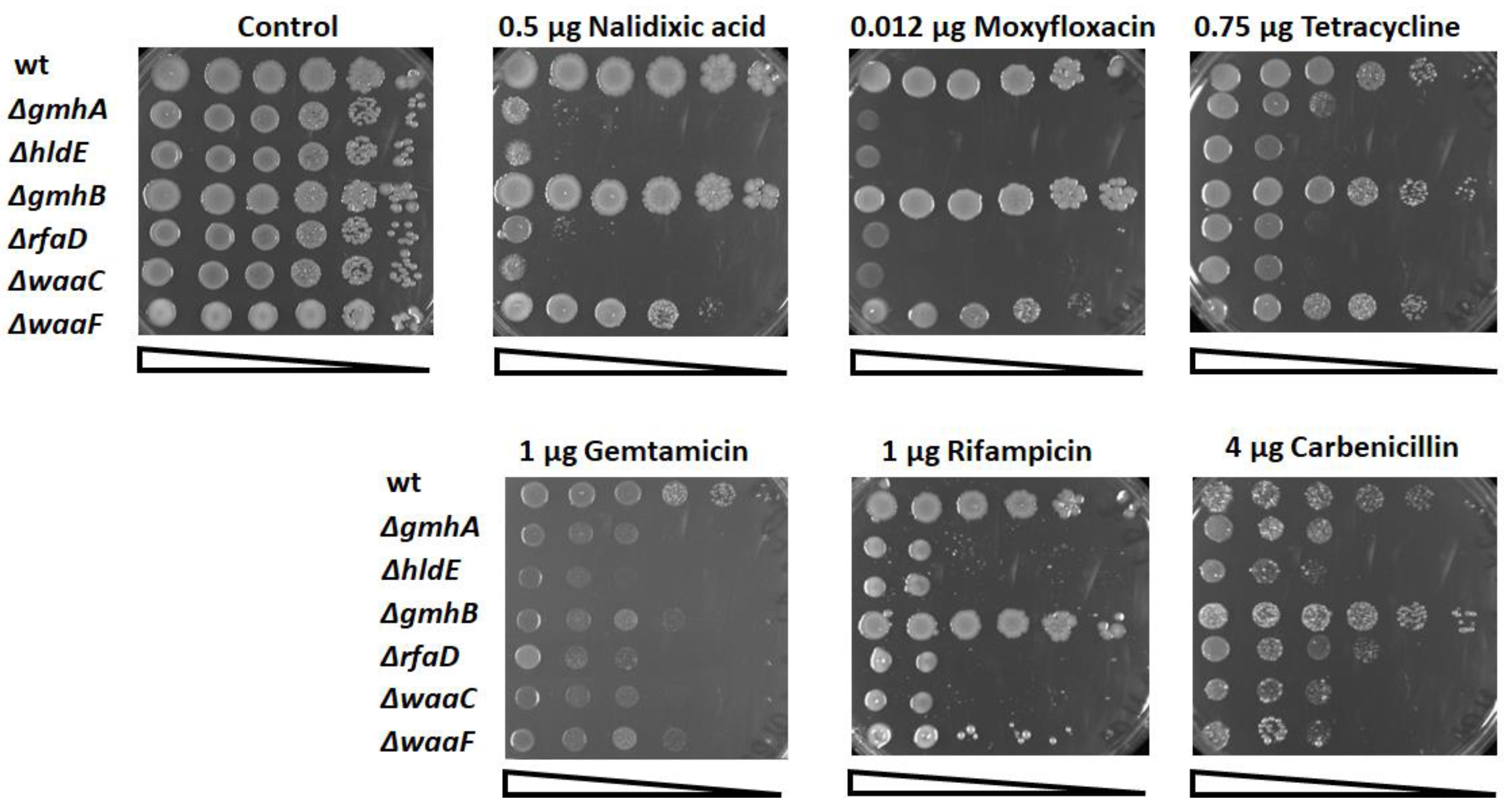
2.2. Determination of Sensitivity to Antibiotics

Overnight cultures were inoculated into a LB broth and grown at 37 °C to ~2 × 10⁷ cells per 1 mL. Cell suspensions were aligned according to optical density, and a series of 6 consecutive tenfold dilutions was prepared. Then the cells were placed on solid media containing various concentrations of antibiotics. To determine the viability of bacterial cells, suspensions were incubated in the presence of lethal doses of antibiotics and then plated on solid media for counting colony-forming units (CFU) [11].

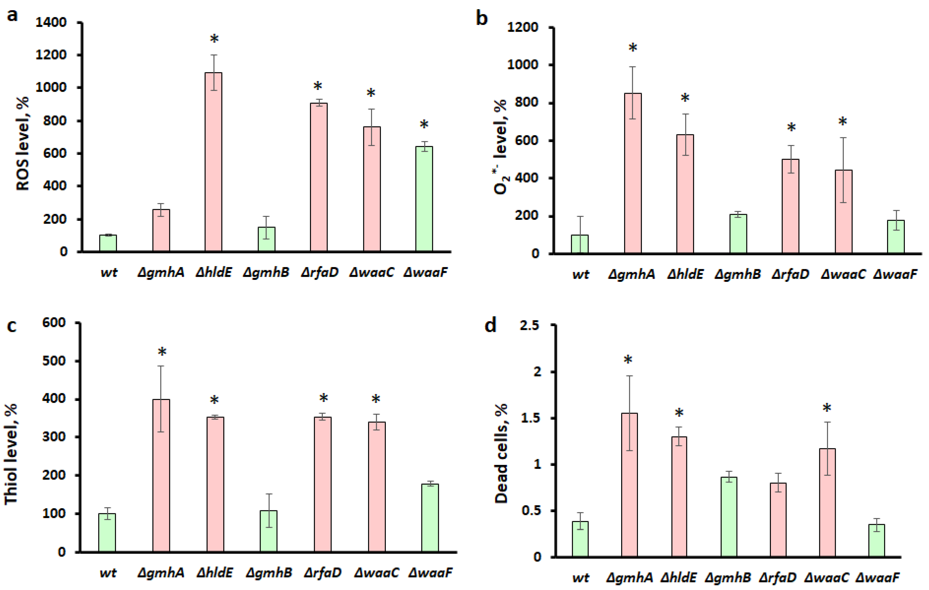
2.3. Determination of Viability and Redox Status of Cells Using Flow Cytometry

Cells were grown in a complete medium to an optical density of 0.4 and then washed twice with phosphate-buffered saline (1xPBS), centrifuged, the supernatant was removed, and cells were resuspended in 100 μL of PBS. Cell parameters were analyzed using flow cytometry on a BD LCR Fortessa flow cytometer (Becton Dickinson, Franklin Lakes, NJ, USA). The cell population for analysis was selected according to the parameters of forward (FSC) and side scattering (SSC), which characterize the size and granularity of cells.
The percentage of dead cells in the cell population was assessed using propidium iodide (Ex/Em = 535/617 nm, Sigma-Aldrich, St. Louis, MO, USA), which was added to the cells at a concentration of 10 μg/mL per minute before the start of the analysis. Propidium iodide penetrates the cells with a damaged membrane and, after binding to DNA, has a bright fluorescence in the red region of the spectrum. The redox status of E. coli cells was assessed by the level of reactive oxygen species (ROS) and intracellular thiols, a significant part of which is intracellular reduced glutathione (GSH). The ROS level was assessed using the dye Dihydrorhodamine 123 (DHR123) (Ex/Em = 507/525 nm, ThermoFisher Scientific, Waltham, MA, USA) [12], which was added to the cells to a final concentration of 7.5 µM, and the cells were incubated for an hour in the dark at 37 °C. The levels of intracellular thiols were assessed using ThiolTracker Violet dye (Ex/Em = 405/526 nm, ThermoFisher Scientific) [13], which was added at a concentration of 10 μM, after which the cells were incubated for one hour in the dark at 37 °C. Specific levels of superoxide anion were evaluated using Dihydroethidium (DHE) (Ex/Em = 518/606 nm, ThermoFisher Scientific) as described earlier [14]. Cells were incubated with 20 µM Dihydroethidium for 1 h in the dark at 37 °C. These parameters were evaluated in the cells with intact membranes, which were not propidium iodide stained. Each value is the mean of at least three independent experiments with triplicate samples ± SD.

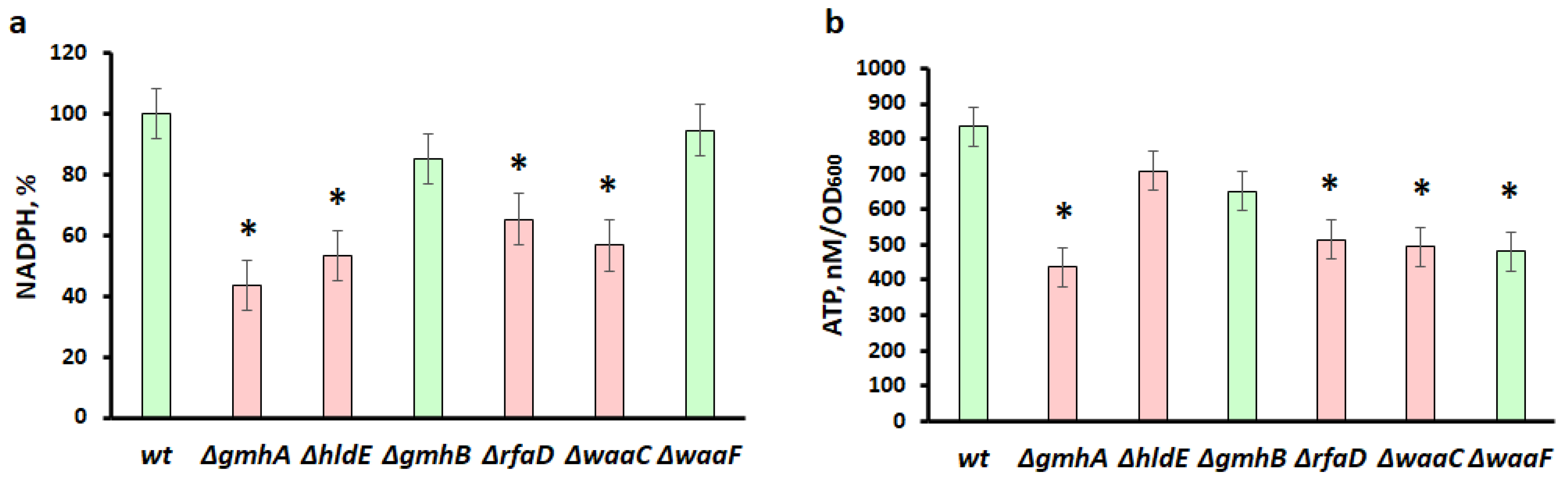
2.4. Measurement of NADPH

Cells were grown in 10 mL of LB medium at 37 °C to an optical density of 0.4 and collected by centrifugation. The concentration of reductive NADPH equivalents was determined using the High Sensitivity NADPH Quantitation Fluorometric Assay Kit (Sigma-Aldrich, St. Louis, MO, USA) on a Tecan Spark tablet reader. The obtained values were related to the optical density of the culture. Data were obtained from three independent experiments.

2.5. Measurement of ATP
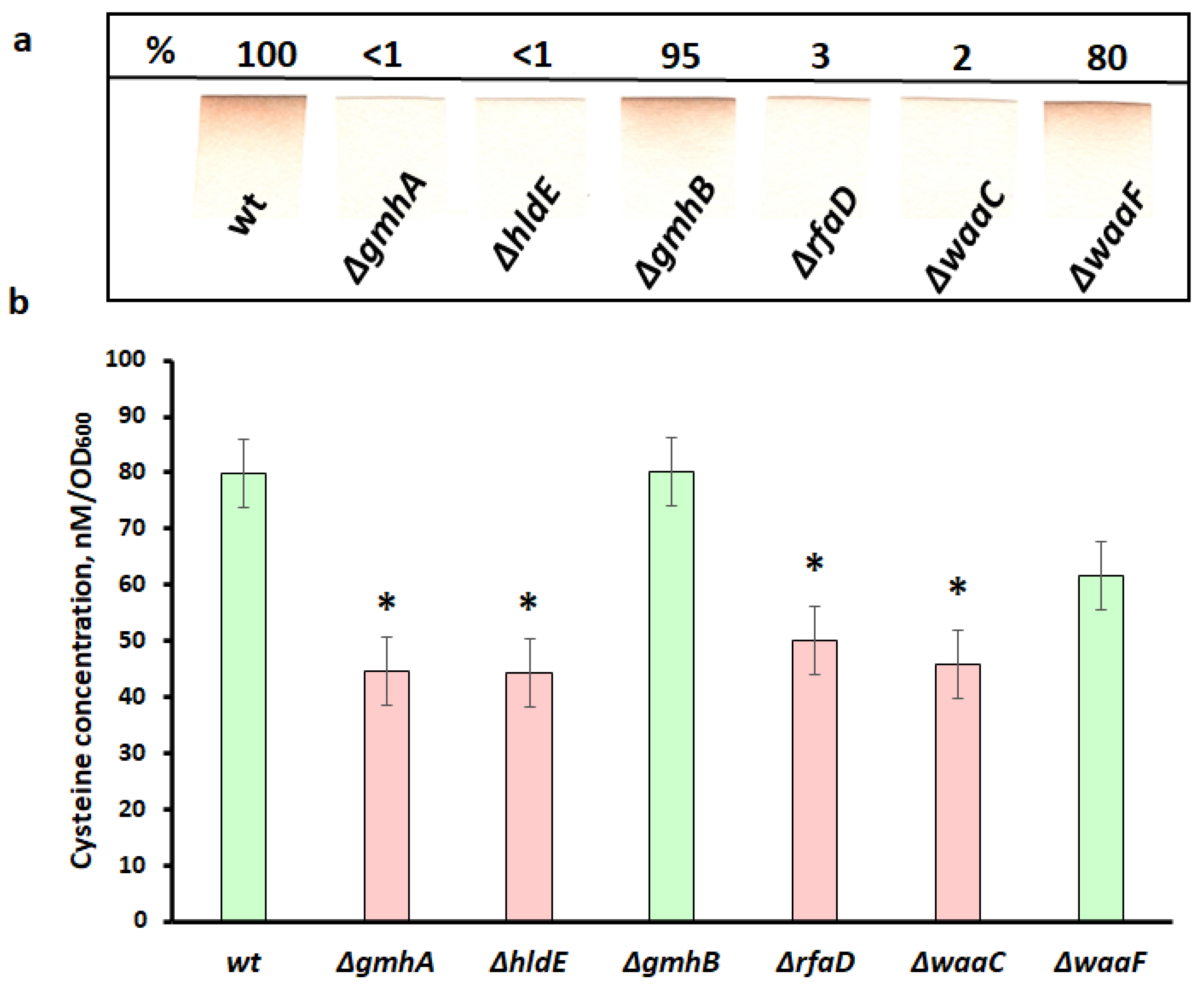
The amount of ATP in cells was determined by the luminescent method using the ATP Detection Assay Kit (Abcam Laboratories, Chicago, IL, USA) on a Tecan Spark tablet reader. One hundred microliters of overnight culture were inoculated with 10 mL of fresh LB medium and grown to an optical density of 0.4. Cells were destroyed by ultrasound. Debris was removed by centrifugation. The resulting extracts were transferred to a plate with the reaction mixture, and the luminescence was immediately measured at a wavelength of 535 nm. Three independent repeats were prepared for each sample.

2.6. H2S Detection

To detect H2S production in wild-type (wt) and mutant cells, the lead acetate detection method was used [15]. Paper strips saturated by 2% of Pb(Ac)2 were affixed to the inner wall of a cultural tube, above the level of the liquid culture of wild-type or mutant bacteria. Overnight cultures were diluted at 1:500 in LB and incubated for 16–18 h at 37 °C with aeration. Stained paper strips were scanned and quantified with an Alpha Imager (Imgen Technologies, Alexandria, WA, USA). The results were normalized per ODs.

2.7. Determination of Intracellular Cysteine

Intracellular cysteine was determined in cell extracts by the method of Gaitonde [16]. Bacterial cultures were grown in 50 mL LB medium to OD600 0.4. Cells were harvested by centrifugation and suspended in 20 mM EDTA. After that, the cells were destroyed by sonication. Proteins were precipitated by the addition of 5 M HClO4. The suspension was centrifuged, and the pH of the supernatant was adjusted to 8.5 by adding KOH. Potassium perchlorate was removed by centrifugation. Samples were treated with 50 mM DTT. The reaction mixture consisted of 500 µL of the sample and 500 µL of Gaitonde’s reagent (250 mg ninhydrin, 4 mL HCl, and 16 mL glacial acetic acid). A calibration curve was built using the known concentrations of cysteine. The assigned values were related to the OD600 of the culture. Data were obtained from three independent experiments.

2.8. Statistical Analysis

The data are shown as mean ± standard deviation measures from triplicate values obtained from 3–4 independent experiments. The statistical difference between experimental groups was analyzed by one-way ANOVA with Tukey correction for multiple comparisons. Probability values (p) less than 0.05 were considered significant. Statistical analysis was performed using the GraphPad Prism 9.1.2 software (GraphPad Software Inc, San Diego, CA, USA).

3. Results

3.1. Oxidative Stress and Antibiotic Sensitivity

Previously, it was shown that Gram-negative bacteria with impaired LPS synthesis were extremely sensitive to various hydrophobic bactericidal agents [17]. During the study, we tested antibacterial drugs with various mechanisms of action: DNA gyrase inhibitors (nalidixic acid, moxifloxacin), protein biosynthesis inhibitors (gentamicin, tetracycline), RNA polymerase inhibitors (rifampicin), and β-lactams (carbenicillin). The sensitivity of the studied mutants was determined by applying a series of dilutions on solid media containing various concentrations of antibiotics (Figure 3).
Deletions in the gmhA, hldE, rfaD, and waaC genes lead to a dramatic increase in sensitivity (Figure 3).
In addition, survival analysis was performed for each mutant on nalidixic acid, tetracycline, and carbenicillin. Bacterial cells were incubated in the presence of lethal doses (means the minimum bactericidal concentration) of antibiotics and plated on a solid medium for further CFU counting (Figure 4). Survival was expressed as a percentage of untreated control.
Deletions of gmhA, hldE, rfaD, and waaC sensitize E. coli cells to all the studied classes of antibiotics, while ∆waaF exhibits very moderate sensitivity, and the ∆gmhB mutant does not differ from wild-type bacteria.
There is ample evidence for the involvement of ROS in antibiotic toxicity by inducing oxidative damage to DNA and proteins [18]. Evaluation of reactive oxygen species (ROS) in cells showed that deletions of the hldE gene induced maximal oxidative stress (Figure 5a). The level of ROS increased by more than 10 times. Deletions of the gmhA, rfaD, waaC, and waaF genes led to a less pronounced increase in ROS by 3, 9, 8, and 6 times, correspondingly. The ROS level in the ∆gmhB mutant did not differ from the wild-type bacteria. It was found that the O2*− level for ∆hldE, ∆rfaD, and ∆waaC correlated with the ROS level (Figure 5b). Unexpectedly, the O2*− level was maximal for the gmhA mutant, which did not have such a high ROS level as other mutants. At the same time, O2*− levels in ∆waaF and ∆gmhB were similar and close to values for the wild-type cells. Deletions of gmhA, hldE, rfaD, and waaC led to a significant (more than three-fold) increase in the thiol group level in E. coli. In cells with a deletion of waaF, the increase in thiols was less pronounced (less than 2 times). At the same time, the amounts of thiols in the ∆gmhB mutant did not differ from wild-type bacteria (Figure 5c). The percentage of dead cells in the population was also maximal in the case of a deletion of gmhA. In the cell populations with deletions of hldE, and waaC, the percentages of dead cells were slightly lower than at ∆gmhA, and in the case of ∆gmhB, ∆rfaD, and ∆waaF, it statistically did not differ from the wild-type bacteria.
The data obtained demonstrate a clear correlation between the amount of ROS, dead cells, an increase in reduced thiols, and sensitivity to antibiotics in “deep rough” mutants. It is interesting, that the mutants showing increased sensitivity to antibiotics (red color in the histogram) differ sharply in the redox status of the cells from the wild-type bacteria (Figure 5).

3.2. Reducing NADPH Equivalents and the ATP Pool

The cofactors ATP, NADH, and NADPH constitute the core of an intricate biochemical network, the operation of which must be clearly regulated in changing environmental conditions. Reducing equivalents of NADPH are the main electron donors in the redox reactions and are essential for many anabolic reactions to occur. One of the main sources of NADPH formation in a bacterial cell is the oxidative branch of the pentose phosphate shunt. In addition, it is in this metabolic pathway that the precursor of ADP-heptose, sedoheptulose 7-phosphate, is formed.
Against the background of deletions of gmhA, hldE, rfaD, and waaC, there was a significant decrease in the level of NADPH compared to the wild-type strain (Figure 6a). The effect of gmhB and waaF deletions on NADPH generation was much weaker and it statistically did not differ from the wild-type bacteria. It can be assumed that NADPH deficiency occurs as a result of its consumption to extinguish oxidative stress, restore glutathione, and support other protective mechanisms.
A significant decrease in NADPH in ADP-heptose of deficient strains is possibly associated with the suppression of oxidative stress that occurred against the background of changes in cell wall permeability (Figure 6a).
ATP deficiency can be a consequence of both a malfunction of the respiratory chain and other metabolic processes associated with its consumption (Figure 6b). It was shown that the greatest decrease in the ATP pool occurs in mutants containing deletions of the gmhA, rfaD, waaC, and waaF genes (Figure 6b).

3.3. H2S Generation and Cysteine Deficiency

Hydrogen sulfide in bacterial cells has an extremely important physiological function and is a component of the most ancient redox-regulatory system involving active forms of sulfur. Previously, it was shown that H2S could protect bacteria from antibiotics and oxidative stress [19,20]. It was found that bacterial cells with a low level of hydrogen sulfide generation were more sensitive to the action of antibiotics and oxidizing agents such as hydrogen peroxide. The main source of H2S generation in E. coli cells is the pathway of cysteine degradation; therefore, hydrogen sulfide can also be considered an indicator of the intensity of cysteine metabolism.
During the experiments, it was found that mutants carrying deletions of the gmhA, hldE, rfaD, and waaC genes stopped generating hydrogen sulfide, which may indicate a change in the metabolism of cysteine, as the main precursor of H2S biosynthesis, while the gmhB and waaF mutants practically did not differ from the control in this parameter (Figure 7a).
Previously, it was shown that the level of endogenous H2S generation directly depended on the intracellular content of cysteine [20], as it is a precursor in the hydrogen sulfide biosynthetic pathway. The main role in the generation of H2S in E. coli cells, as was shown earlier, is played by the enzyme 3-mercaptopyruvate sulfo-transferase (3-MST) encoded by the mstA gene [21].
To test the assumption that the decrease in the level of H2S generation in the gmhA, hldE, rfaD, and waaC mutants is due to a low level of intracellular cysteine content, we performed a quantitative determination of this amino acid in living cells (Figure 7b). The intracellular concentration of cysteine was determined spectrophotometrically using the Gaitonde reagent [16].
As follows from the data presented in Figure 7, in all mutants, with the exception of gmhB, a marked decrease in the cytoplasmic pool of cysteine was observed. These data correlate well with the level of hydrogen sulfide generation in these mutants. Thus, in the strains where H2S generation was practically absent (gmhA, hldE, rfaD, waaC) (Figure 7a), the intracellular cysteine concentration was significantly reduced (Figure 7b).

4. Discussion

The results obtained reveal that strong oxidative stress develops in E. coli cells against the background of impaired LPS biosynthesis. The results of this work clearly indicate that the level of sensitivity of different mutants to antibiotics correlates well with a decrease in the intracellular content of NADPH, ATP, and cysteine, the generation of hydrogen sulfide, thiol levels, and an increase in the intracellular ROS. In the strains ∆gmhA, ∆hldE, ∆rfaD, and ∆waaC with high sensitivity, there is a dramatic increase in ROS, one type of which is superoxide (Figure 5). On the other hand, the mutant ∆waaF is significantly less sensitive to the tested antibiotics, which can be partly explained by the emergence of compensatory mechanisms (fitness costs) [22,23]. The mutant ∆gmhB is practically the same as the wild-type strain. In these strains, changes in redox status and other measured parameters were not as considerable. This is due to the fact that ∆gmhB does not lead to a complete disruption of ADP-heptose synthesis due to the presence of homologous enzymes. The gmhB gene product (D-glycero-β-D-manno-heptose-1,7-bisphosphate 7-phosphatase) is an enzyme belonging to the phosphatase class. Several similar enzymes are present in E. coli cells, for example, the imidazoleglycerol-phosphate dehydratase/histidinol-phosphatase HisB involved in histidine biosynthesis, according to the literature [1], can replace the gmhB deletion defect. In addition, there are phosphatases involved in the central carbon metabolism, the activity of which is critical for cell viability; therefore, it is not possible to completely block this stage of ADP-heptose biosynthesis [24,25]. However, a tendency to slightly increase in dead cells in the ∆gmhB and ∆rfaD populations was detected. Furthermore, the outer membrane integrity is monitored by the envelope stress response system, which initiates the activity of genes and enzymes involved in membrane remodulation. The loss of GmhB activates periplasmic serine endoprotease DegP production, resulting in porin OmpF degradation [26]. Changes in the number of porins in the outer membrane can affect the osmotic resistance of cells.
An interesting fact is that strains with impaired ADP-heptose synthesis actively produce colanic acid, which forms a capsule and compensates for the defect in the barrier function of the impaired cell wall [27,28,29]. In contrast with other mutants, ∆waaF secretes large amounts of colanic acid, this biosynthetic pathway can deplete ATP [29]. However, the release of colanic acid into the growth medium leads to an increase in viscosity and deterioration of gas exchange [30], which can lead to the activation of the respiratory chain, the main source of reactive oxygen species. Apparently, the increased generation of ROS in the ∆waaF strain can be explained by changes in the functioning of the respiratory chain [31]. On the other hand, keeping enough reduction equivalents in ∆waaF (Figure 6 and Figure 7) will compensate for oxidative stress [32] and prevent an increase in the number of dead cells (Figure 5d).
Furthermore, an increase in the number of reduced thiol groups also indicates significant changes in the metabolism of reactive sulfur species in LPS mutants (∆gmhA, ∆hldE, ∆rfaD, and ∆waaC). Cysteine, along with hydrogen sulfide and other low-molecular-weight thiols form an important regulatory system that maintains the redox balance of the bacterial cell and its resistance to stress. Cysteine, being in the external environment mainly in the oxidized form (cystine), is transported into the cell, where it undergoes rapid recovery. Cystine is present in bacterial cells in trace amounts that cannot be detected [33]. Currently, cysteine is considered one of the main protectors against ROS in the periplasm of gram-negative bacteria [34,35]. On the other hand, the concentration of cysteine in the cell cytoplasm is strictly controlled, since an excess of cysteine can promote the genotoxic Fenton reaction [35,36]. As a reason for the increased sensitivity of mutants to antibiotics, one can observe a decrease in the intracellular concentration of cysteine (Figure 7). It can be hypothesized that the disruption of LPS integrity leads to the uncontrolled outflow of cysteine from the cell, and also, possibly, to the difficulty in the functioning of the L-cysteine/cystine shuttle system. A decrease in the intracellular concentration of cysteine, in turn, leads to a decrease in the level of H2S generation (Figure 7), which, as was shown earlier, protects cells from oxidative stress and the action of antibiotics [19,20]. It is important to note that in the experiments on measuring the generation of H2S in mutants, a complete LB medium was used. Under these conditions, an important contribution to the intracellular pool of cysteine is made by its transport in the form of cystine, which is a component of the LB environment and is carried out with the participation of specific transporters tcyP and tcyJ [34]. This explanation is supported by experiments to determine the intracellular content of cysteine in selected LPS mutants. Moreover, the development of oxidative stress in ADP-heptose deficient cells can promote the activation of metabolic fluxes in order to compensate for the increased consumption of reducing agents. The formation and reduction/oxidation of NADP/NADPH is in strict relationship with energy metabolism. One of the nonspecific effects of antibiotics is an increase in cellular respiration, which is directly related to the synthesis of ATP [31]. This process requires reductive NAD(H) equivalents, which in turn are linked to the NADP(H) pool via the enzymes pyridine nucleotide transhydrogenases PntAB and UdhA [32]. Previous research suggests that the metabolic state of bacteria influences their susceptibility to antibiotics [37]. According to our data, a significant reduction in the pool of NADPH and ATP is observed in ∆gmhA, ∆rfaD, and ∆waaC mutants, which indicates an imbalance in central metabolism (Figure 6). It is possible that the deletion of hldE leads to the conservation of ATP compared to other deep-rough mutants, since this enzyme catalyses the ATP-dependent addition of phosphate in the first position to D-glycero-manno-heptose 7-phosphate and subsequent transformation of D-glycero-β-D-manno-heptose 1-phosphate into ADP-L-glycero-β-D-manno-heptose.
Thus, a change in the normal structure of the LPS of the cell wall of gram-negative bacteria not only leads to a violation of its barrier functions but also significantly affects the key protective mechanisms. Redox imbalance of deep-rough mutants deprives cells of the opportunity to form an adequate response to oxidative stress caused by the antibacterial drugs. The most significant changes in redox homeostasis in E. coli cells occur when the gmhA, hldE, rfaD, and waaC genes are deleted (Figure 5, Figure 6 and Figure 7) and these cells show a dramatic increase in antibiotic sensitivity (Figure 3 and Figure 4). In the future, it would be interesting to analyze the homology of these enzymes among clinically significant pathogens, as well as to determine their influence on antibiotic susceptibility.
Previously, attempts were made to create inhibitors of the LPS biosynthesis enzyme [3,7,9,38]. The idea of (should these words “The idea of” be removed?) inactivating the biosynthesis of ADP-heptose is considered a way to create antivirulent adjuvants, since this component of LPS is responsible for the formation of biofilms and, in fact, is an endotoxin [38]. After nearly a decade of inactivity, new data emerges now. Recent research has revealed that ADP-heptose biosynthesis affects antibiotic susceptibility in Helicobacter pylori and Mycobacterium tuberculosis [39,40]. Based on previous and our data we consider it promising to continue this work, since the inactivation of these enzymes leads to an unprecedented increase in cell sensitivity, and will make it possible to overcome the problems of antibiotic resistance of gram-negative pathogens.

Author Contributions

Conceptualization, A.S.M.; methodology, T.A.S. and I.Y.P.; validation, T.A.S., I.Y.P., V.A.M. and A.S.M.; investigation, T.A.S., I.Y.P., P.I.Z.; resources, V.A.M. and A.A.M.; data curation, V.A.M. and A.S.M.; writing—original draft preparation, T.A.S., I.Y.P., R.S.S., V.A.M., A.S.M.; writing—review and editing, T.A.S., I.Y.P., R.S.S., A.S.M.; supervision, V.A.M., A.A.M.; project administration, V.A.M.; funding acquisition, V.A.M. All authors have read and agreed to the published version of the manuscript.

Funding

The research was funded by the Ministry of Science and Higher Education of the Russian Federation (grant agreement No. 075-10-2021-113 unique project ID RF----193021 × 0001).

Institutional Review Board Statement

Not applicable.

Informed Consent Statement

Not applicable.

Data Availability Statement

This study did not report any data.

Acknowledgments

The authors thank Elizaveta Melnikova for help with flow cytometry for gmhA mutant. We are also grateful to Evgeny Nudler for helpful discussion.

Conflicts of Interest

The author declares no conflict of interest.

References

  1. Kneidinger, B.; Marolda, C.; Graninger, M.; Zamyatina, A.; McArthur, F.; Kosma, P.; Valvano, M.A.; Messner, P. Biosynthesis Pathway of ADP-l-Glycero-β-d-Manno-Heptose in Escherichia coli. J. Bacteriol. 2002, 184, 363–369. [Google Scholar] [CrossRef] [PubMed]
  2. Taylor, P.L.; Blakely, K.M.; de Leon, G.P.; Walker, J.R.; McArthur, F.; Evdokimova, E.; Zhang, K.; Valvano, M.A.; Wright, G.D.; Junop, M.S. Structure and Function of Sedoheptulose-7-Phosphate Isomerase, a Critical Enzyme for Lipopolysaccharide Biosynthesis and a Target for Antibiotic Adjuvants *. J. Biol. Chem. 2008, 283, 2835–2845. [Google Scholar] [CrossRef] [PubMed]
  3. Durka, M.; Tikad, A.; Périon, R.; Bosco, M.; Andaloussi, M.; Floquet, S.; Malacain, E.; Moreau, F.; Oxoby, M.; Gerusz, V.; et al. Systematic Synthesis of Inhibitors of the Two First Enzymes of the Bacterial Heptose Biosynthetic Pathway: Towards Antivirulence Molecules Targeting Lipopolysaccharide Biosynthesis. Chem. A Eur. J. 2011, 17, 11305–11313. [Google Scholar] [CrossRef]
  4. Huang, K.C.; Mukhopadhyay, R.; Wen, B.; Gitai, Z.; Wingreen, N.S. Cell Shape and Cell-Wall Organization in Gram-Negative Bacteria. Proc. Natl. Acad. Sci. USA 2008, 105, 19282–19287. [Google Scholar] [CrossRef] [PubMed]
  5. Schnaitman, C.A.; Klena, J.D. Genetics of Lipopolysaccharide Biosynthesis in Enteric Bacteria. Microbiol. Rev. 1993, 57, 655–682. [Google Scholar] [CrossRef] [PubMed]
  6. Stincone, A.; Prigione, A.; Cramer, T.; Wamelink, M.M.C.; Campbell, K.; Cheung, E.; Olin-Sandoval, V.; Grüning, N.-M.; Krüger, A.; Tauqeer Alam, M.; et al. The Return of Metabolism: Biochemistry and Physiology of the Pentose Phosphate Pathway. Biol. Rev. 2015, 90, 927–963. [Google Scholar] [CrossRef] [PubMed]
  7. De Leon, G.P.; Elowe, N.H.; Koteva, K.P.; Valvano, M.A.; Wright, G.D. An In Vitro Screen of Bacterial Lipopolysaccharide Biosynthetic Enzymes Identifies an Inhibitor of ADP-Heptose Biosynthesis. Chem. Biol. 2006, 13, 437–441. [Google Scholar] [CrossRef]
  8. Valvano, M.A. Biosynthesis and Genetics of ADP-Heptose. J. Endotoxin Res. 1999, 5, 90–95. [Google Scholar] [CrossRef]
  9. Desroy, N.; Moreau, F.; Briet, S.; Fralliec, G.L.; Floquet, S.; Durant, L.; Vongsouthi, V.; Gerusz, V.; Denis, A.; Escaich, S. Towards Gram-Negative Antivirulence Drugs: New Inhibitors of HldE Kinase. Tetrahedron Young Investig. Award. 2006 Jonathan Ellman 2009, 17, 1276–1289. [Google Scholar] [CrossRef]
  10. Datsenko, K.A.; Wanner, B.L. One-Step Inactivation of Chromosomal Genes in Escherichia Coli K-12 Using PCR Products. Proc. Natl. Acad. Sci. USA 2000, 97, 6640–6645. [Google Scholar] [CrossRef] [Green Version]
  11. Cockerill, F.R.; Clinical and Laboratory Standards Institute. Methods for Dilution Antimicrobial Susceptibility Tests for Bacteria That Grow Aerobically: Approved Standard; Clinical and Laboratory Standards Institute: Wayne, PA, USA, 2012; ISBN 1-56238-784-7. [Google Scholar]
  12. Mitkevich, V.A.; Kretova, O.V.; Petrushanko, I.Y.; Burnysheva, K.M.; Sosin, D.V.; Simonenko, O.V.; Ilinskaya, O.N.; Tchurikov, N.A.; Makarov, A.A. Ribonuclease Binase Apoptotic Signature in Leukemic Kasumi-1 Cells. Biochimie 2013, 95, 1344–1349. [Google Scholar] [CrossRef] [PubMed]
  13. Mitkevich, V.A.; Petrushanko, I.Y.; Yegorov, Y.E.; Simonenko, O.V.; Vishnyakova, K.S.; Kulikova, A.A.; Tsvetkov, P.O.; Makarov, A.A.; Kozin, S.A. Isomerization of Asp7 Leads to Increased Toxic Effect of Amyloid-Β42 on Human Neuronal Cells. Cell Death Dis. 2013, 4, e939. [Google Scholar] [CrossRef] [PubMed]
  14. Ivanov, A.V.; Smirnova, O.A.; Petrushanko, I.Y.; Ivanova, O.N.; Karpenko, I.L.; Alekseeva, E.; Sominskaya, I.; Makarov, A.A.; Bartosch, B.; Kochetkov, S.N.; et al. HCV Core Protein Uses Multiple Mechanisms to Induce Oxidative Stress in Human Hepatoma Huh7 Cells. Viruses 2015, 7, 2745–2770. [Google Scholar] [CrossRef] [PubMed]
  15. Tille, P.M. Bailey & Scott’s Diagnostic Microbiology, 13th ed.; Elsevier: St. Louis, MS, USA, 2014. [Google Scholar]
  16. Gaitonde, M. A Spectrophotometric Method for the Direct Determination of Cysteine in the Presence of Other Naturally Occurring Amino Acids. Biochem. J. 1967, 104, 627–633. [Google Scholar] [CrossRef]
  17. Coleman, W.G.; Leive, L. Two Mutations Which Affect the Barrier Function of the Escherichia Coli K-12 Outer Membrane. J. Bacteriol. 1979, 139, 899–910. [Google Scholar] [CrossRef]
  18. Dwyer Daniel, J.; Belenky Peter, A.; Yang Jason, H.; MacDonald, I.C.; Martell Jeffrey, D.; Takahashi, N.; Chan Clement, T.Y.; Lobritz Michael, A.; Braff, D.; Schwarz Eric, G.; et al. Antibiotics Induce Redox-Related Physiological Alterations as Part of Their Lethality. Proc. Natl. Acad. Sci. USA 2014, 111, E2100–E2109. [Google Scholar] [CrossRef]
  19. Shatalin, K.; Shatalina, E.; Mironov, A.; Nudler, E. H2S: A Universal Defense Against Antibiotics in Bacteria. Science 2011, 334, 986–990. [Google Scholar] [CrossRef]
  20. Mironov, A.; Seregina, T.; Nagornykh, M.; Luhachack, L.G.; Korolkova, N.; Lopes, L.E.; Kotova, V.; Zavilgelsky, G.; Shakulov, R.; Shatalin, K.; et al. Mechanism of H2S-Mediated Protection against Oxidative Stress in Escherichia coli. Proc. Natl. Acad. Sci. USA 2017, 114, 6022–6027. [Google Scholar] [CrossRef]
  21. Colnaghi, R.; Cassinelli, G.; Drummond, M.; Forlani, F.; Pagani, S. Properties of the Escherichia Coli Rhodanese-like Protein SseA: Contribution of the Active-Site Residue Ser240 to Sulfur Donor Recognition. FEBS Lett. 2001, 500, 153–156. [Google Scholar] [CrossRef]
  22. Linkevicius, M.; Sandegren, L.; Andersson, D.I. Mechanisms and Fitness Costs of Tigecycline Resistance in Escherichia coli. J. Antimicrob. Chemother. 2013, 68, 2809–2819. [Google Scholar] [CrossRef] [Green Version]
  23. Coleman, W.G., Jr. The RfaD Gene Codes for ADP-L-Glycero-D-Mannoheptose-6-Epimerase. An Enzyme Required for Lipopolysaccharide Core Biosynthesis. J. Biol. Chem. 1983, 258, 1985–1990. [Google Scholar] [CrossRef]
  24. Fraenkel, D.G.; Horecker, B.L. Fructose-1,6-Diphosphatase and Acid Hexose Phosphatase of Escherichia coli. J. Bacteriol. 1965, 90, 837–842. [Google Scholar] [CrossRef] [PubMed]
  25. Fraenkel, D.G.; Pontremoli, S.; Horecker, B.L. The Specific Fructose Diphosphatase of Escherichia coli: Properties and Partial Purification. Arch. Biochem. Biophys. 1966, 114, 4–12. [Google Scholar] [CrossRef]
  26. Alzayn, M.; Dulyayangkul, P.; Satapoomin, N.; Heesom Kate, J.; Avison Matthew, B. OmpF Downregulation Mediated by Sigma E or OmpR Activation Confers Cefalexin Resistance in Escherichia Coli in the Absence of Acquired β-Lactamases. Antimicrob. Agents Chemother. 2021, 65, e01004-21. [Google Scholar] [CrossRef] [PubMed]
  27. Meredith, T.C.; Mamat, U.; Kaczynski, Z.; Lindner, B.; Holst, O.; Woodard, R.W. Modification of Lipopolysaccharide with Colanic Acid (M-Antigen) Repeats in Escherichia coli*. J. Biol. Chem. 2007, 282, 7790–7798. [Google Scholar] [CrossRef] [PubMed]
  28. Joloba Moses, L.; Clemmer Katy, M.; Sledjeski Darren, D.; Rather Philip, N. Activation of the Gab Operon in an RpoS-Dependent Manner by Mutations That Truncate the Inner Core of Lipopolysaccharide in Escherichia coli. J. Bacteriol. 2004, 186, 8542–8546. [Google Scholar] [CrossRef]
  29. Ren, G.; Wang, Z.; Li, Y.; Hu, X.; Wang, X. Effects of Lipopolysaccharide Core Sugar Deficiency on Colanic Acid Biosynthesis in Escherichia coli. J. Bacteriol. 2016, 198, 1576–1584. [Google Scholar] [CrossRef]
  30. Samain, E. 1.23—Production of Oligosaccharides in Microbes. In Comprehensive Glycoscience; Kamerling, H., Ed.; Elsevier: Oxford, UK, 2007; pp. 923–947. ISBN 978-0-444-51967-2. [Google Scholar]
  31. Lobritz Michael, A.; Belenky, P.; Porter Caroline, B.M.; Gutierrez, A.; Yang Jason, H.; Schwarz Eric, G.; Dwyer Daniel, J.; Khalil Ahmad, S.; Collins James, J. Antibiotic Efficacy Is Linked to Bacterial Cellular Respiration. Proc. Natl. Acad. Sci. USA 2015, 112, 8173–8180. [Google Scholar] [CrossRef]
  32. Canonaco, F.; Hess, T.A.; Heri, S.; Wang, T.; Szyperski, T.; Sauer, U. Metabolic Flux Response to Phosphoglucose Isomerase Knock-out in Escherichia coli and Impact of Overexpression of the Soluble Transhydrogenase UdhA. FEMS Microbiol. Lett. 2001, 204, 247–252. [Google Scholar] [CrossRef]
  33. Reitzer, L.; DiRita, V.J. Death by Cystine: An Adverse Emergent Property from a Beneficial Series of Reactions. J. Bacteriol. 2015, 197, 3626–3628. [Google Scholar] [CrossRef] [Green Version]
  34. Ohtsu, I.; Wiriyathanawudhiwong, N.; Morigasaki, S.; Nakatani, T.; Kadokura, H.; Takagi, H. The L-Cysteine/l-Cystine Shuttle System Provides Reducing Equivalents to the Periplasm in Escherichia coli*. J. Biol. Chem. 2010, 285, 17479–17487. [Google Scholar] [CrossRef] [PubMed]
  35. Mironov, A.; Seregina, T.; Shatalin, K.; Nagornykh, M.; Shakulov, R. Nudler, E. CydDC Functions as a Cytoplasmic Cystine Reductase to Sensitize Escherichia Coli to Oxidative Stress and Aminoglycosides. Proc. Natl. Acad. Sci. USA 2020, 117, 23565–23570. [Google Scholar] [CrossRef]
  36. Park, S.; Imlay, J. High Levels of Intracellular Cysteine Promote Oxidative DNA Damage by Driving the Fenton Reaction. J. Bacteriol. 2003, 185, 1942–1950. [Google Scholar] [CrossRef] [PubMed]
  37. Stokes, J.M.; Lopatkin, A.J.; Lobritz, M.A.; Collins, J.J. Bacterial Metabolism and Antibiotic Efficacy. Cell Metab. 2019, 30, 251–259. [Google Scholar] [CrossRef] [PubMed]
  38. Moreau, F.; Desroy, N.; Genevard, J.M.; Vongsouthi, V.; Gerusz, V.; Le Fralliec, G.; Oliveira, C.; Floquet, S.; Denis, A.; Escaich, S.; et al. Discovery of New Gram-Negative Antivirulence Drugs: Structure and Properties of Novel, E. coli WaaC Inhibitors. Bioorg. Med. Chem. Lett. 2008, 18, 4022–4026. [Google Scholar] [CrossRef]
  39. Chiu, S.-F.; Teng, K.-W.; Wang, P.-C.; Chung, H.-Y.; Wang, C.-J.; Cheng, H.-C.; Kao, M.-C. Helicobacter Pylori GmhB Enzyme Involved in ADP-Heptose Biosynthesis Pathway Is Essential for Lipopolysaccharide Biosynthesis and Bacterial Virulence. Virulence 2021, 12, 1610–1628. [Google Scholar] [CrossRef] [PubMed]
  40. Karan, S.; Pratap, B.; Yadav, S.P.; Ashish, F.; Saxena, A.K. Structural and Functional Characterization of M. tuberculosis Sedoheptulose- 7-Phosphate Isomerase, a Critical Enzyme Involved in Lipopolysaccharide Biosynthetic Pathway. Sci. Rep. 2020, 10, 20813. [Google Scholar] [CrossRef]
Figure 1. Cell wall structure of gram-negative bacteria. ADP-heptose attaches O-antigen polysaccharide chains to the inner core (Kdo) [4]. The “deep-rough” mutant LPS structure is unstable due to the lack of negatively charged phosphate groups on heptose residues, which normally stabilize the structure through the interaction with positive ions. Furthermore, the “deep-rough” phenotype leads to increased susceptibility to hydrophobic compounds [5].
Cells 11 02667 g001
Figure 2. Biosynthesis of ADP-heptose from sedoheptulose 7-phosphate and its translocation to LPS. Disruption of any step in this pathway disturbs the incorporation of the heptose residues in the LPS structure.
Cells 11 02667 g002
Figure 3. Determination of the sensitivity of strains containing deletions in the gmhA, hldE, gmhB, rfaD, waaC, and waaF genes to various groups of antibiotics. Cells were spotted on LB agar plates in serial 10-fold dilutions and incubated at 37 °C for 24 h.
Cells 11 02667 g003
Figure 4. Survival of E. coli MG1655 strains containing knockouts for ADP-heptose activation genes (gmhA, hldE, gmhB, rfaD, waaC, and waaF). Representative curves show the effect of deficiency ADP-heptose biosynthesis on antibiotic-mediated killing. Mean values ± SD from at least three independent experiments are shown.
Cells 11 02667 g004
Figure 5. Changes in the level of intracellular ROS, O2*− the level of thiols, and the percentage of dead cells in the population of E. coli cells with deletions of the gmhA, hldE, gmhB, rfaD, waaC, waaF genes. The level of ROS (a) was assessed using the dye DHR123; the O2*− level (b) with DHE dye; the thiols level (c) with ThiolTracker Violet; the percentage of dead cells (d) was estimated using propidium iodide. Parameters for cells with gmhA, hldE, rfaD, and waaC deletions that result in a dramatic increase in antibiotic susceptibility in E. coli cells are shown in red, while parameters for wt, gmhB, and waaF are shown in green. Mean values ± SD from at least three independent experiments are shown. *—p < 0.05, compared to the wild-type cells.
Cells 11 02667 g005
Figure 6. Changes in the intracellular content of NADPH and ATP in the population of E. coli cells with deletions of the gmhA, hldE, gmhB, rfaD, waaC, waaF genes. (a) Changes in the intracellular NADPH level. (b) Changes in the intracellular ATP level. Parameters for cells with gmhA, hldE, rfaD, and waaC deletions that result in a dramatic increase in antibiotic susceptibility in E. coli cells are shown in red, while parameters for wt, gmhB, and waaF are shown in green. Mean values ± SD from at least three independent experiments are shown. *—p < 0.05, compared to the wild-type cells.
Cells 11 02667 g006
Figure 7. Formation of hydrogen sulfide and intracellular concentration of cysteine in the population of E. coli cells with deletions of the gmhA, hldE, gmhB, rfaD, waaC, waaF genes and wild-type cells. (a) Formation of H2S by E. coli cells. Pb-acetate-soaked paper strips show a brown colour of PbS as a result of the reaction with gaseous H2S exiting liquid bacterial cultures (b) Intracellular concentration of cysteine. Parameters for cells with gmhA, hldE, rfaD, and waaC deletions that result in a dramatic increase in antibiotic susceptibility in E. coli cells are shown in red, while parameters for wt, gmhB, and waaF are shown in green. Mean values ± SD from at least three independent experiments are shown. *—p < 0.05, compared to the wild-type cells.
Cells 11 02667 g007
Table 1. Genotype of obtained mutants with deletions of LPS biosynthesis genes.
GenotypeNature of MutationReference
wtF- wild typeLaboratory collection
gmhADeletion of the gmhA D-sedoheptulose 7-phosphate isomeraseThis work
hldEDeletion of the hldE gene encoding heptose-7-phosphate kinase
gmhBDeletion of the gmhB gene encoding
D,D-heptose 1,7-bisphosphate phosphatase
rfaDDeletion of the rfaD gene encoding
ADP-L-glycero-D-mannoheptose-6-epimerase
waaCDeletion of the waaC gene encoding
ADP-heptose:LPS heptosyltransferase I
waaFDeletion of the waaF gene encoding
ADP-heptose:LPS heptosyltransferase II
Table 2. Primers for obtaining deletions of LPS biosynthesis genes.
NameSequence
GmhA15′–atg-tac-cag-gat-ctt-att-cgt-acc-gaa-ctg-aac-gac-gct-caa-gtt-agt-ata-aaa-aag-ct–3′
GmhA25′–tta-ctt-aac-cat-ctc-ttt-ttc-aat-taa-ctg-gat-cag-tga-agc-ctg-ctt-ttt-tat-act-aag–3′
GmhA35′–ccg-tac-ttc-tcg-ctt-ttg-gc–3′
GmhA45′–taa-gac-gcg-tca-gca-tcg-ca–3′
HldE15′–atg-aaa-gta-acg-ctg-cca-gag-ttt-gaa-cgt-gca-ggc-gct-caa-gtt-agt-ata-aaa-aag-ct–3′
HldE25′–tta-gcc-ttt-ttt-atc-ctg-ttg-gat-ctt-ctt-gat-gat-tga-agc-ctg-ctt-ttt-tat-act-aag–3′
HldE35′–ggt-gga-aga-atg-aag-tat-gg–3′
HldE45′–gtt-gaa-aaa-aca-aca-gcg-tca–3′
GmhB15′–gtg-gcg-aag-agc-gta-ccc-gca-att-ttt-ctt-gac-cgc-gct-caa-gtt-agt-ata-aaa-aag-ct–3′
GmhB25′–tca-ttg-tgc-cgg-ttt-ttg-ctg-ctt-ttt-tat-cgc-ttg-tga-agc-ctg-ctt-ttt-tat-act-aag–3′
GmhB35′–ttc-ttg-cag-gtc-gaa-aca-tg–3′
GmhB45′–c-tca-gga-aga-caa-gcg-gaa–3′
RfaD15′–atg-atc-atc-gtt-acc-ggc-ggc-gcg-ggc-ttt-atc-ggc-gct-caa-gtt-agt-ata-aaa-aag-ct–3′
RfaD25′–tta-tgc-gtc-gcg-att-cag-cca-ggc-cat-gta-ttc-cgt-tga-agc-ctg-ctt-ttt-tat-act-aag–3′
RfaD35′–atg-att-aca-gac-att-cgt-gtc–3′
RfaD45′–ga-ctt-tgc-gac-atc-atc-atg–3′
WaaC15′–atg-cgg-gtt-ttg-atc-gtt-aaa-aca-tcg-tcg-atg-ggc-gct-caa-gtt-agt-ata-aaa-aag-ct–3′
WaaC25′–tta-taa-tga-tga-taa-ctt-ttc-caa-aac-tgc-ttg-act-tga-agc-ctg-ctt-ttt-tat-act-aag–3′
WaaC35′–gcg-tac-tgg-aag-aac-tca-ac–3′
WaaC45′–gat-ttc-aga-gtg-taa-ggt-ttc–3′
WaaF15′–atg-aaa-ata-ctg-gtg-atc-ggc-ccg-tct-tgg-gtt-ggc-gct-caa-gtt-agt-ata-aaa-aag-ct–3′
WaaF25′–tca-ggc-ttc-ctc-ttg-taa-caa-tag-cgc-gtt-gag-ttc-tga-agc-ctg-ctt-ttt-tat-act-aag–3′
WaaF35′–gtt-gct-gaa-ggt-gta-acg-ga–3′
WaaF45′–ggc-aac-gta-tgg-aga-aca-tc–3′
Publisher’s Note: MDPI stays neutral with regard to jurisdictional claims in published maps and institutional affiliations.

