Article

Characterization of Gfat1 (zeppelin) and Gfat2, Essential Paralogous Genes Which Encode the Enzymes That Catalyze the Rate-Limiting Step in the Hexosamine Biosynthetic Pathway in Drosophila melanogaster

by
Shawn Cotsworth
1,
Catherine J. Jackson
1,2,3,4,
Graham Hallson
1,
Kathleen A. Fitzpatrick
1,
Monika Syrzycka
1,5,
Alistair B. Coulthard
6,
Amy Bejsovec
7,
Marcella Marchetti
8,
Sergio Pimpinelli
8,
Simon J. H. Wang
1,
Robert G. Camfield
9,
Esther M. Verheyen
1,
Donald A. Sinclair
1,
Barry M. Honda
1 and
Arthur J. Hilliker
6,*
1
Department of Molecular Biology and Biochemistry (MBB), Simon Fraser University, 8888 University Dr., Burnaby, BC V5A 1S6, Canada
2
Department of Plastic and Reconstructive Surgery, Institute for Surgical Research, University of Oslo, N-0424 Oslo, Norway
3
The Department of Medical Biochemistry, Oslo University Hospital, N-0424 Oslo, Norway
4
Institute of Oral Biology, Faculty of Dentistry, University of Oslo, N-0424 Oslo, Norway
5
Allergan Canada, 500-85 Enterprise Blvd, Markham, ON L6G 0B5, Canada
6
Department of Biology, York University, Toronto, ON M3J 1P3, Canada
7
Department of Biology, Duke University, Durham, NC 27708, USA
8
Department of Biology and Biotechnology “C. Darwin”, “Sapienza” University of Rome, 00185 Rome, Italy
9
BC Genome Science Centre, 675 West 10th Avenue, Vancouver, BC V5Z 1L3, Canada
*
Author to whom correspondence should be addressed.
Cells 2022, 11(3), 448; https://doi.org/10.3390/cells11030448
Submission received: 30 December 2021 / Revised: 21 January 2022 / Accepted: 22 January 2022 / Published: 27 January 2022

Abstract

:
The zeppelin (zep) locus is known for its essential role in the development of the embryonic cuticle of Drosophila melanogaster. We show here that zep encodes Gfat1 (Glutamine: Fructose-6-Phosphate Aminotransferase 1; CG12449), the enzyme that catalyzes the rate-limiting step in the hexosamine biosynthesis pathway (HBP). This conserved pathway diverts 2%–5% of cellular glucose from glycolysis and is a nexus of sugar (fructose-6-phosphate), amino acid (glutamine), fatty acid [acetyl-coenzymeA (CoA)], and nucleotide/energy (UDP) metabolism. We also describe the isolation and characterization of lethal mutants in the euchromatic paralog, Gfat2 (CG1345), and demonstrate that ubiquitous expression of Gfat1+ or Gfat2+ transgenes can rescue lethal mutations in either gene. Gfat1 and Gfat2 show differences in mRNA and protein expression during embryogenesis and in essential tissue-specific requirements for Gfat1 and Gfat2, suggesting a degree of functional evolutionary divergence. An evolutionary, cytogenetic analysis of the two genes in six Drosophila species revealed Gfat2 to be located within euchromatin in all six species. Gfat1 localizes to heterochromatin in three melanogaster-group species, and to euchromatin in the more distantly related species. We have also found that the pattern of flanking-gene microsynteny is highly conserved for Gfat1 and somewhat less conserved for Gfat2.

1. Introduction

The HBP diverts fructose-6-phosphate from glycolysis to generate UDP-N-acetylglucosamine, an important precursor used in the formation of glycoproteins (e.g., GlcNAcylation by OGT, O-GlcNAc transferase), proteoglycans, and other important biomolecules (Figure 1) [1,2,3,4]. The rate-limiting step in the pathway is catalyzed by Glutamine-fructose-6-phosphate transaminase 1 or GFPT1 in humans (hereafter called hGfat1) and Gfat1 in Drosophila, and Glutamine-fructose-6-phosphate transaminase 2 or GFPT2 in humans (hereafter called hGfat2) and Gfat2 in Drosophila. In Drosophila, C. elegans, mice, and humans, Gfat1 and Gfat2 are encoded by two separate genes, and this may also be the case for Aedes aegypti [5,6,7,8]. In contrast, it appears that there is only a single Gfat gene/enzyme in fungi [9,10,11]. The distinction between hGfat1 and hGfat2 is the presence of two putative Protein Kinase A (PKA) sites in hGfat1, but only a single site in hGfat2 [5,12,13]. In Drosophila melanogaster, one of the PKA sites is conserved in both Gfat1 and Gfat2. Although the other site is partially conserved (albeit presumably non-functional) in Gfat1, it is absent from Gfat2 [5].
Gfat enzymes convert fructose-6-phosphate and glutamine to glucosamine-6-phosphate (GlcN-6P) and glutamate (Figure 1). The GlcN-6P is then rapidly processed by a series of other enzymes in the HBP, ultimately generating the final product UDP-GlcNAc, an essential substrate for a variety of highly conserved cellular and organismal functions [4,6]. These include (1) N-glycosylation; (2) O-linked glycosylation (e.g., mucin-type O-glycosylation); (3) O-linked GlcNAcylation, an extremely important post-translational modification of serine and threonine residues of target proteins catalyzed by O-GlcNAc transferase (OGT); and (4) the formation of structural biomolecules such as chitin, a long chain GlcNAc polymer that is one of the most abundant macromolecules found in nature and is the primary component of the arthropod cuticle/peritrophic matrix [6,14].
Given the multifaceted cellular requirements for the HBP, it is not surprising that suboptimal functioning of the pathway has been linked to a wide variety of diseases, including heart disease, diabetes, neurodegenerative diseases, cellular stress diseases, premature aging, and cancer [1,2,3,6,15,16,17,18,19]. Many of these diseases stem from aberrant O-linked GlcNAcylation [20,21].
Since chitin is a major component of the insect cuticle [22] and because the end product of the HBP serves as the substrate for chitin synthesis, a reasonable prediction is that genetic defects in HBP enzymes will block chitin synthesis and, thus, cuticle production in flies. Indeed, this is true for mummy (mmy), which encodes UDP-N-acetylglucosamine diphosphorylase (UAP1), the enzyme that catalyzes the final step in the HBP (Figure 1). Hypomorphic mmy alleles exhibit a bloated embryonic cuticle, while null alleles fail to secrete an embryonic cuticle [23]. In an earlier study, workers determined that devitellinized embryos homozygous for a mutant allele of a previously unknown gene called zeppelin (zep) also displayed an expanded embryonic cuticle, which was dubbed the “blimp” phenotype [24]. The zep locus was mapped to 3R pericentric heterochromatin (3R het). Interestingly, Gfat1 has been previously mapped to 81F in 3R het [5].
The notorious silencing properties of centromeric heterochromatin with respect to relocated euchromatic genes, coupled with the paradoxical finding that heterochromatic genes require a heterochromatic environment for optimal expression (reviewed in [25]), have long intrigued Drosophila researchers. Hence, Hilliker and Honda have participated in a collaboration aimed at contributing to the characterization of heterochromatin in Drosophila, with the view to obtaining more insight into the relationship between chromatin structure and gene expression, as well as the evolution of heterochromatin. Much of our work has focused on correlating genetically identified loci with existing gene models in autosomal heterochromatin in Drosophila melanogaster e.g., see [25,26]. In the current work, we show that the essential zep locus corresponds to the Gfat1 gene. Furthermore, we have expanded our analysis by exploring the relationship between the Gfat1 and Gfat2 genes and their products. Thus, we describe the isolation and characterization of lethal mutants in the euchromatic paralog, Gfat2, and show that ubiquitously expressed Gfat1+ and Gfat2+ transgenes can rescue lethal alleles in either gene, indicating that the two enzymes are functionally equivalent in D. melanogaster. In addition, our RT-qPCR analysis and the available RNAseq and proteomic data reveal distinct embryonic expression patterns for the two genes. We also provide evidence that while there are essential requirements for Gfat2 in the nervous system and muscles, this does not appear to be the case for Gfat1. This essential tissue-specificity of Gfat2 is likely due, at least in part, to the need to provide UDP-GlcNAc for essential functions of OGT. In contrast, our data show that both Gfat1 and Gfat2 have essential functions in the trachea.
We have also carried out an evolutionary cytogenetic analysis to determine where the two paralogues are located in five other Drosophila species. The Gfat2 gene is located in euchromatin in D. melanogaster and all five other species examined. On the other hand, the Gfat1 gene is located in heterochromatin in Drosophila melanogaster and the two other species in the melanogaster group but it is located in euchromatin in the three more distantly related species. Finally, we report microsynteny data for genes that immediately flank Gfat1 and Gfat2 in the six species. These data show that the gene arrangement in D. melanogaster is completely conserved for Gfat1 and mostly conserved for Gfat2. The Gfat1 data suggest that the evolutionarily recent acquisition of a hetereochromatic environment by this gene occurred by the relocation of a genomic block containing several genes, a finding similar to that reported for the light gene and its neighbours [27].

2. Materials and Methods

2.1. Pre-Existing Drosophila Stocks, Routine Culture Conditions, Identification of New Zep/Gfat1 Alleles and Routine Genetic Crosses

Stock centre transgenic RNAi lines used in the present study are described in Table S1. It is noteworthy that both v24539 and B42892 target all nine identified Gfat1 transcripts that are listed at Flybase ([28] and data not shown). Most other stocks used were either acquired from the Bloomington Drosophila stock centre (BDSC) or in some cases, directly from research groups. The generation/origin of Gfat1 RNAi and Gfat1+ transgenes is described below. Standard Drosophila medium was used throughout this study. Fly stocks were maintained at 25 °C or 18 °C and most experiments were conducted in vials at 25 °C or 29 °C. New EMS-induced Gfat1/zep mutant alleles were identified from two sources via non-complementation with the original zepLP13 allele. Five alleles, each designated with a z superscript and identifying number, were identified among a collection of third chromosome recessive lethal lines (each was marked with st and ry) kindly provided by the Zuker lab [29]. Two additional alleles, I400#1 and I400# were among several third chromosome recessive lethal lines (each marked with th st cp in ri pp) kindly provided by Dr. M. Leptin [30]. Df(3R)10-65, kniri−1 pp/TM3, Ser (BDSC #2597) and Df(3R)4-75, kniri−1 pp/TM3, Ser (BDSC #2598) were generated previously by Marchant and Holm [31,32]. More recently, it has been determined that Df(3R)4-75 is a multi-breakpoint pericentric inversion [33,34]. w1118; Df(3R)BSC460/TM6C, Sb cu (BDSC stock#24964) and w1118; Df(3R)BSC567/TM6C, Sb (BDSC stock#25390) are two deletions that uncover Gfat2. Four additional deletions that uncover zep lethality and delete Gfat1 were also used in the current study: Df(3R)8740#20 (also called zep8740#20), Df(3R)8740#22 (also called zep8740#22), and Df(3R)EP-167 (also called zep167), were recovered in a P-element-induced male recombination study, and Df(3R)7B-90, e was X-ray-induced [35,36].
Inter se complementation analysis involving all pairwise combinations of the eight zep alleles was carried out at 25 °C, scoring a minimum of 100 emerging adult progeny per cross. The RNAi crosses were performed at 29 °C. Six of ten different UAS-RNAi transgenes for Gfat1 generated in this study (see below) were tested for viability using either of the strong ubiquitously-expressing drivers, Tub-GAL4/TM3, Sb, or Act5C-GAL4/CyO, and all were lethal (Table S2). A heat-shock GAL4 driver was also used to express specific Gfat1 RNAi transgenes with a view to identifying adult phenotypes. The btl-GAL4 experiments were completed later than the others using an incubator set at 30 °C.

2.2. Cuticle Analysis of Newly-Isolated Zep Alleles

Embryonic cuticle preparations to test the new zep alleles for the blimp phenotype were carried out as described in [24].

2.3. Sequence Analysis of Zep Alleles

Genomic DNA corresponding to 500 bp segments spanning the entire coding sequence plus the exon/intron splice junctions of the Gfat1 gene was isolated from embryos homozygous for each of the eight zep alleles by PCR using the primers listed in Table S3. The resulting DNA segments were then subjected to standard DNA sequence analysis by Macrogen Inc. (Seoul, Korea; http://dna.macrogen.com/eng/ (accessed on 29 December 2021)). The sequences were then compared to the Flybase [28] genomic sequence using BLASTN to identify point mutations and BLASTP to identify changes at the amino acid level.

2.4. Isolation and Genetic and Molecular Characterization of Putative Gfat2 Excision Mutations

The Gfat2 gene is located in euchromatin on the right arm of chromosome 3 at cytological position 98C4 [28]. We generated lethal excisions by using Sb Δ 2.3 to mobilize a P-element located in the 5′ UTR of the gene, w67c23 P{EPgy2}Gfat2EY21762 (or EY21762: BDSC #22502) [28], selecting for w excisions and then testing each for recessive lethality. w/TM6, Sb males from each of the lethal lines were crossed separately to either Df(3R)BSC460/TM6, Tb, or Df(3R)BSC567/TM6, Tb in order to confirm that the lethality mapped to the segment containing the Gfat2 gene. Putative excision mutations that were lethal in combination with either deletion were re-balanced with TM3, Sb Ser es twiGAL4-EGFP, and each was tested for complementation with w*; PBac{GAL4D,EYFP}larp43/TM3, Sb (BDSC #78330), a lethal allele of the La-related protein (larp) gene, which lies immediately proximal to Gfat2 [28]. Since there were no available lethal mutations in the immediately distal gene, Moca-cyp, DNA from single embryos homozygous for each of putative excision lesions that complemented the larp allele, was separately subjected to PCR analysis in order to identify molecular excisions that did not extend into Moca-cyp. The Moca-cyp primers used for this purpose were: forward primer 5′-AGTTCTGAGTAGAGCTGGCAACGCC-3′ and reverse primer 5′-ACAGCAGCACACACACACAAGCG-3′.

2.5. Sequence Analysis of Gfat2 Mutants

DNA from homozygotes for each of the two Gfat2-specific excision mutants was subjected to sequence analysis as follows: The entire gene was PCR amplified from single homozygous embryos using primers that annealed to segments flanking the coding region. Each PCR product was blunt-end ligated into the Fermentas pJET 1.2 vector and sent to UBC-NAPS for DNA sequencing using the forward primer 5′- GCGCCGTTCACTTGTCTTGTCAAT-3′ and the reverse primer 5′-TCACACCCTTGTACTGCAGCTTCT-3′, and the sequence data obtained were compared to the wild type Gfat2 sequence [28].

2.6. Lethal Phase Analysis of a Gfat2 Deletion Mutant

Gfat210A−2/TM3 Sb Ser twi 2x-eGFP virgin females were crossed to Df(3R) BSC460/TM3 Sb Ser twi 2X-eGFP males and incubated at 25 °C for 5 days. The cross was then caged over an embryo lay plate for 6 h at 25 °C after which adult flies were removed and the plate incubated overnight. Gfat210A−2/Df(3R) BSC460 embryos that did not fluoresce under examination by standard GFP fluorescence microscopy were placed on a new plate and examined the following day. The number of unhatched embryos and first instar larvae (L1) remaining on the plate was used to determine the number of organisms that died during embryogenesis. The surviving larvae were counted again the following day.

2.7. Generation of a Gfat1+ Rescue cDNA Construct

We obtained the RE72989 EST with cDNA corresponding to Gfat1-RA, inserted it into the pBluescript shuttle vector and ultimately cloned it into pUAST [36,37]. This construct was sequenced by Macrogen Inc. and then sent to BestGene Inc. (2140 Grand Ave, Chino Hills, CA, USA) for the generation of transgenic lines.

2.8. Gfat1 and Gfat2 Rescue Crosses

The test for genetic rescue of zep mutants by a ubiquitously expressed UAS-Gfat1+ cDNA was performed by crossing UAS-Gfat1+/CyRoi; Gfat1LP13/TM3, Sb males to Actin5C-GAL4/CyO; Df(3R)7B-90, e/TM3, Sb females. The test for rescue analysis of Gfat2 excision mutants by a UAS-Gfat1+ cDNA was performed by crossing UAS-Gfat1+/CyRoi; Gfat210A−2/TM3, Ser males to Actin5C-GAL4/CyO; Df(3R)567/TM3, Ser females. Finally, a UAS-Gfat2+ cDNA transgenic line (generously provided by Dr. L. Partridge) was tested for the rescue of Gfat1 and Gfat2 mutants in the following crosses: (i) UAS-Gfat2+/Cy, Roi; Df(3R)BSC460/TM6B, Tb females and Actin5C-GAL4/CyO; Gfat210A−2/TM3, Ser males; (ii) UAS-Gfat2+/CyRoi; Gfat1z−1904/TM6 B, Tb females, and Actin5C-GAL4/CyO; Gfat1I400#8/TM3, Ser males [38]. In all rescue tests, all surviving progeny of the diagnostic crosses were classified and counted.

2.9. Gfat1 RNAi Stocks and Crosses

The Gfat1 RNAi stocks were generated by amplifying a 290 bp fragment from the RE72989 cDNA clone using the forward primer 5′-GACTCCTTCCTCGAGCTGT-3′ and the reverse primer 5′-TCAGAATTCCTTTCCGAACGC-3′. Underlined base pairs were altered from the known Gfat1 sequence ([28] in order to create artificial XhoI and EcoRI restriction sites in the forward and reverse primers, respectively [28]. The 290 bp amplicon was ligated into the pTZ57R shuttle vector then digested with XhoI and EcoRI and inserted into the pSympUast vector. Ten transgenic strains containing the Gfat1 RNAi construct were generated in the w1118 background by BestGene Inc. (2140 Grand Ave, Chino Hills, CA, USA).

2.10. Real-Time qPCR

RNA was isolated from wild type Oregon-R embryos (synchronized at specific time intervals after egg-lay) using TRIZOL. One μg of each RNA sample was treated with DNAseI for 1 h at 37 °C and 364 ng of DNA free RNA from each sample was used to create cDNAs using the BioRad iScript select cDNA synthesis kit using its random primers. qPCR primers specific for amplifying RpL32, Gfat1, and Gfat2 were obtained from IDT along with a probe possessing a 5′ 6-FAM fluorophore, a 3′ Iowa Black FQ quencher, and an intermediate ZEN quencher located 9bp from the 5′end of the probe that would anneal to each individual amplicon (see Table S3 for qPCR primer sequences). Each qPCR reaction was prepared using 2 µL of the cDNA sample, 1 µL of a cocktail consisting of the primers and the fluorophore probe, 10 µL of BioRad iTaq Supermix, and 7 µL of ddH2O and cycled using an Applied Biosystems Step-One Real-Time PCR System Machine. Standard curves were generated using each primer set in order to determine PCR efficiency. A relative quantification of Gfat1 and Gfat2 expression at each of these stages was determined relative to the 0–90 min sample according to the method described in [39].

2.11. Tests for Essential Tissue-Specific Requirements for Gfat1 and Gfat2

In order to test for possible essential requirements for Gfat1 and Gfat2 in specific tissues, the effects of knockdown of their respective genes in the nervous system and muscles using various RNAi transgenes for Gfat1 and Gfat2 were examined as follows: Males bearing individual Gfat1 or Gfat2 RNAi transgenes were mated to females bearing the larval pan-neural driver Appl-GAL4 (generously provided by Dr. U. Pandey) or the muscle driver Mef2-GAL4, and the offspring were raised at 29 °C. Since the product of the HBP is used as the substrate for GlcNAcylation by Ogt/sxc (CG10392), analogous crosses were carried out using a TRiP Ogt RNAi line. In a subsequent experiment, to test for essential requirements of Gfat1 and Gfat2 in tracheae, males bearing specific Gfat1 or Gfat2 RNAi transgenes were mated to females bearing the trachea driver breathless (btl)-GAL4, and the offspring were raised in an incubator set at 30 °C. In each case, adult survival associated with specific RNAi-induced knockdown was assessed and knockdown was deemed to be lethal if no diagnostic adult offspring were observed in comparison to the survival of a minimum of 60 internal control adults from the same cross. Where relevant, the designations of male semi-lethality or weak semi-lethality are explained in the table footings.

2.12. Fluorescence In Situ Hybridization (FISH) Localization of Gfat1 and Gfat2 in Different Species of Drosophila

Polytene and mitotic in situ analyses were carried out as described previously [40]. Probes were differentially labeled by nick translation with digoxigenin- or biotin-coupled dUTP, and, after hybridization at 37 °C overnight, the signal was detected with a fluorescein avidin or antidigoxigenin–rhodamine antibody. DNA was counterstained with DAPI before image capture. Microsynteny analysis was performed using species data available at FB2017_05 (Dmel Release 6.18) from http://www.flybase.org and/or https://blast.ncbi.nlm.nih.gov/Blast.cgi (accessed on 29 December 2021).

3. Results

3.1. The Zep Locus Corresponds to Gfat1

The zep gene was originally mapped to 3R het based on non-complementation between zepLP13 and Df(3R)4-75 [24]. Since both mmy and zepLP13 mutants exhibit expanded embryo phenotypes and because mmy encodes an HBP enzyme, a reasonable hypothesis is that the 3R het gene Gfat1 [5] corresponds to the zep locus. This hypothesis is strongly supported by the finding that zep is the only essential gene deleted by Df(3R)8740#20 and Df(3R)8740#22, plus the fact that both deficiencies also remove Gfat1 (Figure S1) [35,36,41]. The fact that the Gfat1 insertion allele, y w*; Mi{MIC}Gfat1MI11277/TM3, Sb Ser (BDSC 56582), failed to complement zepLP13 (data not shown) is further evidence that the zep locus corresponds to the Gfat1 gene.
We extended our analysis by isolating and characterizing seven new zep alleles based on the failure to complement zepLP13 from a large collection of EMS-induced recessive lethal mutations provided by the Zuker and Leptin groups [29,30]. The inter se complementation data for the eight zep alleles are shown in Figure 2. Most pairwise combinations were lethal. However, several combinations, involving transheterozyotes between zepz1904, zepz1914, or zep3−52 and various other alleles, were either semi-lethal (less than 50% of expected progeny) or viable, suggesting that these may be hypomorphic zep alleles. Indeed, the weakest alleles, zepz1904 and zepz1914, exhibited the same complementation pattern, and they are semi-lethal in combination with Gfat1 deletions (data not shown). Interestingly, many surviving transheterozygotes had extended legs with melanin deposits at the joints. This phenotype, which was also observed for some survivors when a Gfat1-RNAi transgene was expressed continuously at 29 °C using a heat-shock GAL4 transgene (Figure S2), resembles the effects of reduced expression of the Splayed locus, a putatively haplo-abnormal gene positioned in 81F-82A [42,43]. Thus, it is possible that the Gfat1/zep is allelic to Spl.
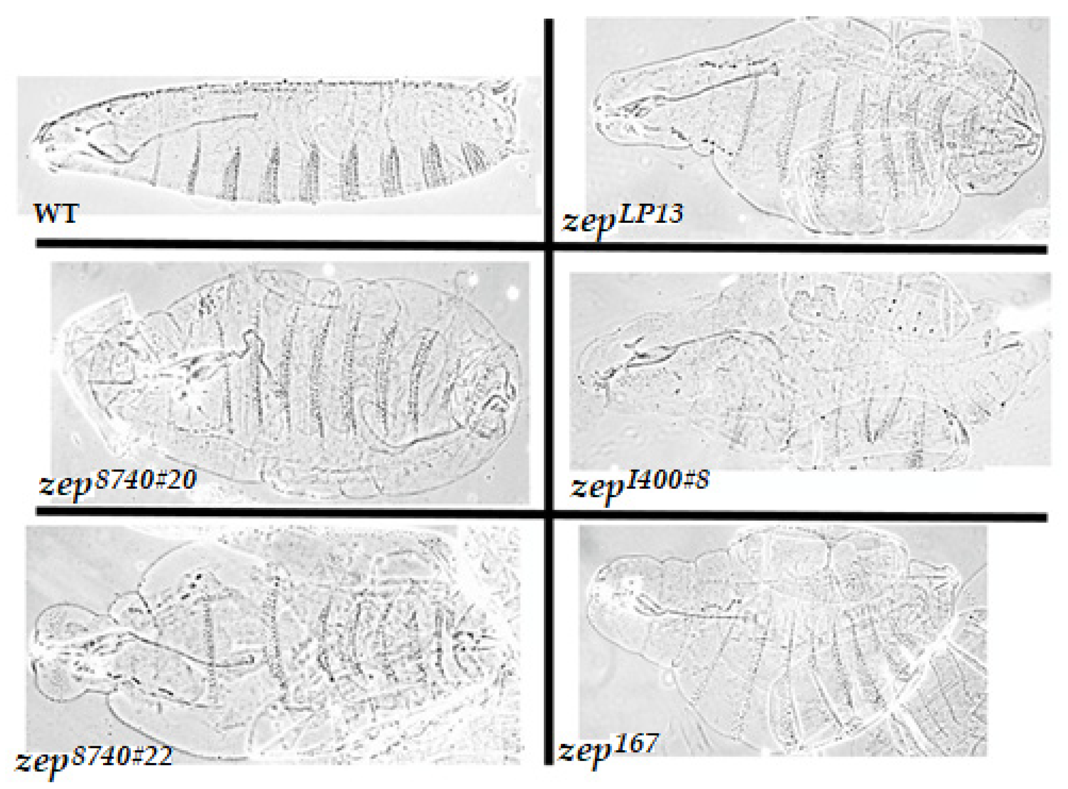
In order to test whether the newly isolated zep alleles also exhibited the blimp phenotype, cuticle preparations of mechanically devitellinized homozygous mutant embryos were performed as previously described [24]. Indeed, embryos homozygous for each of the seven new EMS zep alleles and three Gfat1 deletions exhibit the diagnostic expanded cuticle phenotype to varying degrees that correlate with allele severity (Figure 3 and data not shown), consistent with the hypothesis that the zep locus is Gfat1.
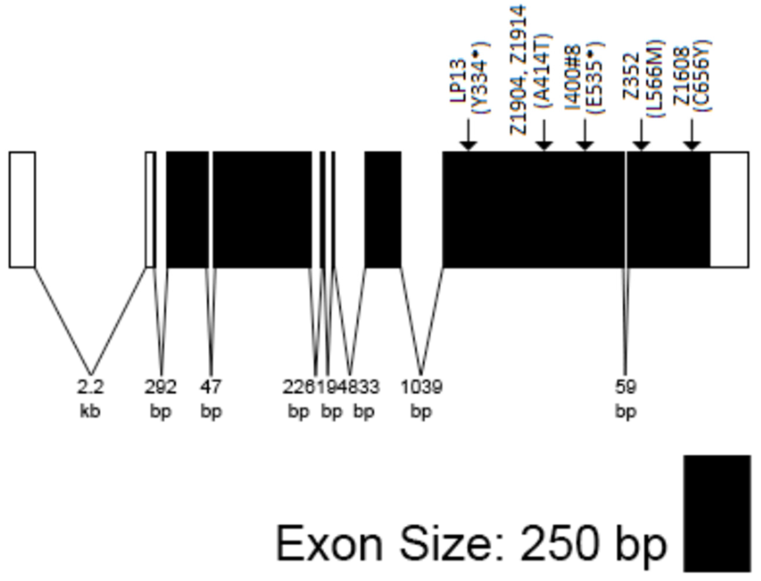
Sequence data confirmed that Gfat1 and zep are the same genes (summarized in Figure 4). A total of Six of the zep alleles possess either a nonsense or missense mutation in their coding sequence. Thus, zepLP13 and zep1400#8 contain premature stop codons: zepLP13: Tyr334*, and zepI400#8: Gln535* presumably results in truncated proteins, each possessing the Glutaminase domain, but lacking one or both Isomerase domains. In contrast, each of the other four alleles contains a missense mutation: zep3−52: L588M; zepz1608: C656Y; zepz1904, and zepz1914 contain the identical lesion: A414T. Each of these represents a substitution of a conserved residue in one of the Isomerase domains. However, based on the complementation data (see above), the zepz1904, zepz1914, and zep3−52 lesions do not appear to block Gfat1 activity completely. The remaining two alleles, zepz1014 and zepI400#1, have no non-polymorphic changes in their protein-coding or exon/intron junction sequences.

3.2. Isolation, Sequence and Lethal Phase Analysis of Gfat2 Mutant Alleles

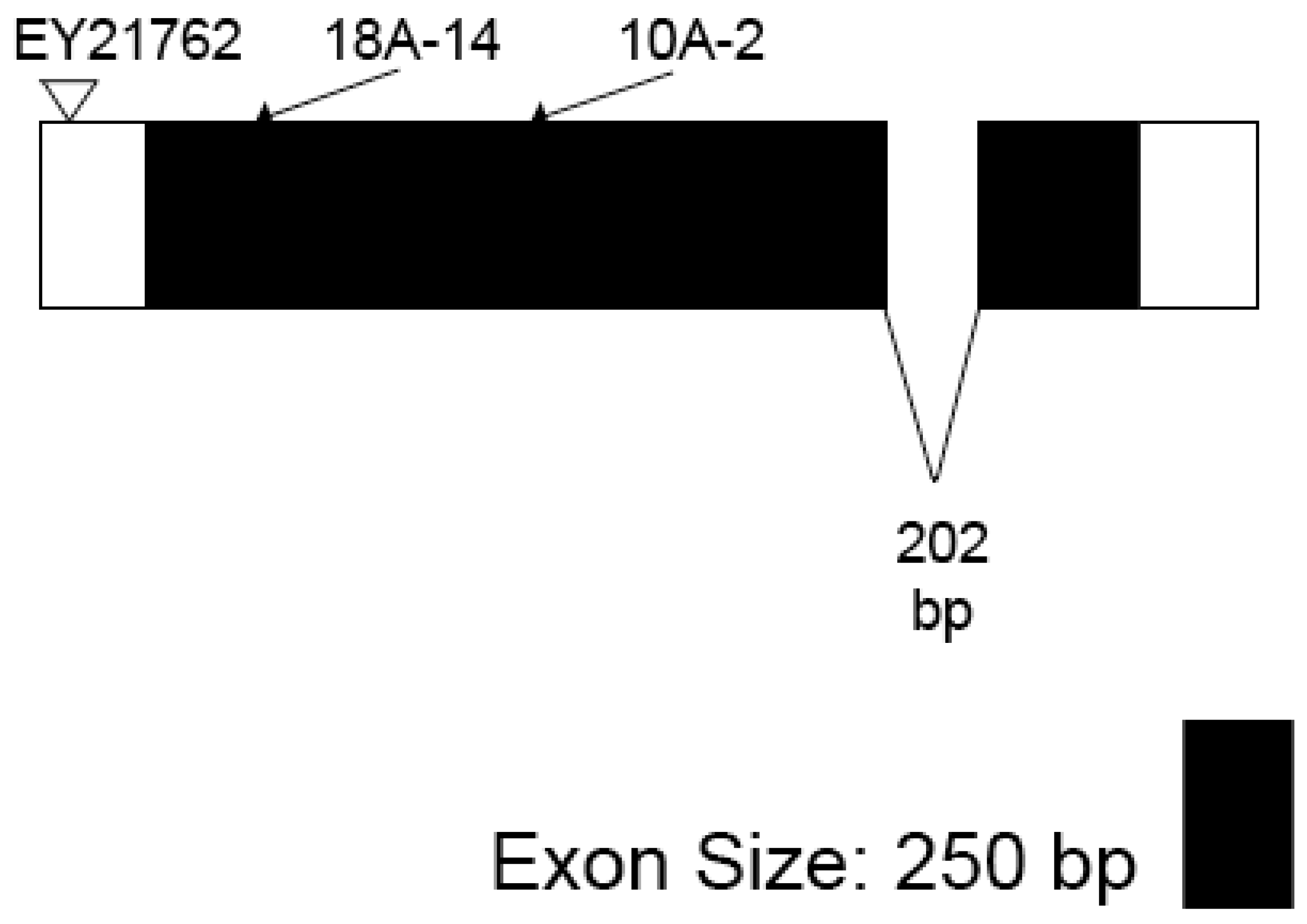
We identified several putative Gfat2 lesions from the P-element-excision study described in the Materials and Methods. A total of thirty-eight w excisions were recessive lethal and nine of these that were lethal with one of the Gfat2 deletions, Df(3R)BSC460/TM6, Tb or Df(3R)BSC567/TM6, Tb were viable in combination with larp43. All nine were subjected to PCR analysis to check for genomic integrity of Moca-cyp as described. This analysis identified three lesions for which Moca-cyp was intact: 1C-25, 10A-2, and 18A-14 [44]. Sequence analysis of two of these, designated as Gfat210A−2 and Gfat218A−14, revealed that each involved a Gfat2-specific deletion (Figure 5), thereby confirming that the gene is essential. The Gfat210A-2 mutant is a deletion beginning 50 bp from the 5′ end in the 5′UTR, extending 1038 bp towards the 3′ end, deleting part of exon 1 and all of the glutaminase domain. The Gfat218A-14 mutant is a deletion beginning 50 bp from the 5′ end of the 5′UTR and extending 498 bp towards the 3′ end, deleting part of exon 1 and just under a third of the glutaminase domain. Both deletions retained the 5′-CATGATGAAATAA-3′ sequence that was originally part of the terminal repeat of the P-element in the EY21762 line. Lethal phase analysis of 100 Gfat210-A2/Df(3R)BSC460 hemizygotes revealed a biphasic pattern (Table 1): 59% died during embryogenesis and 41% survived to the L1 stage. The Gfat2- L1 larvae failed to grow and died shortly after hatching. Interestingly, the dead Gfat2-embryos displayed no obvious cuticle phenotype (data not shown). This contrasts with the embryonic lethal phase and blimp phenotypes of zep/Gfat1 alleles.

3.3. Gfat1/Zep and Gfat2 Mutants Can Be Rescued by Ubiquitous Expression of Gfat1+ and Gfat2+ cDNA Transgenes

Rescue analysis also confirmed that Gfat1 and zep are the same genes. We were able to rescue the lethality of a zep/Gfat1 allele in combination with Df(3R)7B-90e by using a UAS-Gfat1+ cDNA under the control of Act5C-GAL4 (top panel, Table 2). Furthermore, constitutive expression of the UAS-Gfat1+ allowed the rescue of flies hemizygous for either Gfat210A−2 or Gfat218A−14 (second panel in Table 2). Finally, both Gfat1 and Gfat2 mutant alleles were rescued by constitutive expression of the UAS-Gfat2+ transgene (bottom two panels, Table 2). Together, these data suggest that the two Gfat enzymes are functionally equivalent.

3.4. Gfat1 and Gfat2 Genes Exhibit Different Expression Patterns during Development

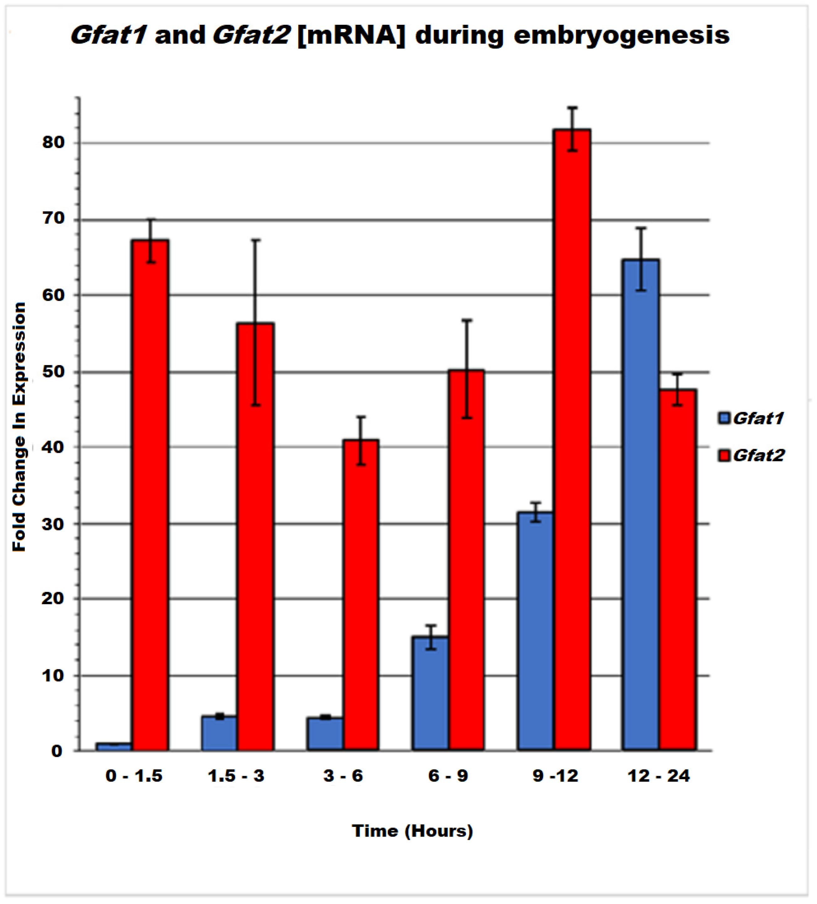
The different lethal phases and embryonic phenotypes of Gfat1 and Gfat2 mutants raised the possibility that Gfat1 and Gfat2 may have somewhat different physiological roles in the fly. We investigated this possibility initially by profiling the mRNA pattern of each gene at specific intervals during embryogenesis using RT-qPCR analysis (Figure 6). Our data show that Gfat2 mRNA is relatively abundant throughout embryogenesis, with the highest level observed at 9–12 h after egg-lay (AEL). Furthermore, perceptible expression of the gene within 0–1.5 h AEL indicates the significant maternal contribution of the Gfat2 mRNA. In contrast, Gfat1 mRNA levels were relatively low until 6–9 h AEL, reaching a peak near the end of embryogenesis, which coincides with the time of embryonic cuticle deposition [24]. These mRNA patterns are similar but not identical to those revealed by the more refined modENCODE Refseq data [28]. These show moderately high expression of Gfat2 throughout most embryogenesis, with high expression in the 8–12 h (~stages 12–15) and 14–16 h AEL (~stage 16) intervals. Similar to our data, they found that Gfat1 expression is either absent or very low until 10–14 h AEL (~stages 14–15) and, thereafter, expression is high or very high, with peak expression during the 16–18 h AEL interval (~stage 16).
Our results and the RNAseq data are consistent with the current in situ data for embryonic stages 1–3 indicating maternal deposition of Gfat2 mRNA, but not of Gfat1 mRNA [28,45]. Moreover, there is little if any Gfat1 expression until embryonic stages 13–14 and then very gradually it transitions to a high level throughout the embryo by stages 16–17, during which mRNA appears to be concentrated in dorsal, ventral, and head epidermis and the salivary glands. In distinct contrast, Gfat2 mRNA is reasonably abundant and ubiquitously distributed in blastoderm embryos (stages 4–6). By embryonic stages 7–8, expression is ubiquitous, with some concentration in various germ layers. The expression then transitions through stages 9–10, during which the mRNA is faintly ubiquitous, but by stages 11–16 it becomes highly abundant and ubiquitous and noticeably concentrated in various tissues and structures.
Protein levels during embryogenesis indicate very low expression from 0–80 min AEL, followed by a very long period of no or extremely low expression from 2 h to ~12 h AEL (embryonic stages 4–16) and then a transition through low expression from 14 to 18 h AEL (stages 16–17), eventually culminating in moderate expression by 20 h AEL [28,43]. The near absence of Gfat1 protein during a long segment of embryogenesis is consistent with the paucity of Gfat1 mRNA during most of this same period (our data and the RNAseq results). Since there is no clear maternal deposition of Gfat1 mRNA, it is possible that a small amount of Gfat1 protein is contributed maternally. Proteomic analysis of Gfat2 levels during embryogenesis indicates very low expression from 0–140 min AEL, but thereafter its expression increases transiently to low levels, followed by an increase to moderate expression from 4–14 h AEL, and then ultimately to high expression by 14–20 h AEL. The higher levels of Gfat2 versus Gfat1 protein expression throughout embryogenesis are consistent with the moderately high to high levels of Gfat2 mRNA throughout embryogenesis described above.
RNAseq analysis during post-embryonic development reveals moderately high or high levels of Gfat1 expression during most stages, with peak expression during larval stages L2 and 12 h L3, and especially during the 12 h prepupal stage; however, Gfat1 expression is much lower in adult males and females [28]. The corresponding analysis of Gfat2 also indicates high expression in L1 and L2, and thereafter predominantly moderately high expression, except for low expression in mid and late pupae. In contrast with Gfat1, the expression of Gfat2 is also moderately high in adult males and females. Interestingly, a very recent study of links between nutrient availability, protein O-GlcNAcylation, and diurnal rhythm in adult flies also found much higher levels of Gfat2 mRNA versus very low levels of Gfat1 mRNA [46]. Indeed, based on these and other data, these authors contend that Gfat2 is the primary functional paralogue in adults. Proteomic data indicate high to extremely high Gfat2 protein expression during all three larval stages and during the white prepupal and days 1 and 3 of pupal development. High levels of the protein were also observed in adult males [28,47]. Corresponding proteomic data for Gfat1 in post-embryonic stages of development are not currently available.

3.5. Evidence for Differences in Essential Tissue-Specific Requirements for Gfat1 and Gfat2

Owing to the phenotypic and expression pattern differences observed for Gfat1 and Gfat2, we decided to investigate the question of whether Gfat1 and Gfat2 might have different essential roles in the fly. Thus, we used Gfat1 and Gfat2 RNAi transgenes to explore essential tissue-specific requirements for the two versions of the Gfat enzyme (for a description of the RNAi lines used, see Table S1). For the first experiment, we used the Appl-GAL4 pan-neural driver and the Mef2-GAL4 muscle driver. We chose Appl-GAL4 because of the importance of the HBP in CNS development [48,49,50]. Furthermore, since pilot tests using a previously-generated RNAi transgene showed that RNAi-induced pan-neural and muscle knockdown of Ogt/sxc was lethal (data not shown), we reasoned that one or both of the Gfat enzymes might be required for HBP generation of the UDP-GlcNAc substrate for essential Ogt catalytic functions (see Figure 1) in these tissues. Therefore, we included RNAi knockdown of Ogt/sxc in this experiment.
The results of the first experiment are shown in Table 3 and Table 4. As expected, the TRiP RNAi transgene for Ogt/sxc is lethal when expressed in either the nervous system or muscles. Furthermore, the data also show that one of the Gfat2 RNAi transgenes is lethal when expressed in both tissues, while the other Gfat2 RNAi transgene is lethal when expressed in muscles and semi-lethal when expressed in the nervous system. In striking contrast, none of three Gfat1 RNAi transgenes is lethal when expressed in either the nervous system or muscles. These data support the contention that Gfat2, but not Gfat1, has an essential role in the structure/function of these tissues.
The requirement for proper chitin synthesis in tracheal development in Drosophila melanogaster is well documented [49,51,52]. Since mmy mutants are defective in chitin production and trachea morphogenesis, we decided to test for possible lethal effects of specific downregulation of Gfat1 and Gfat2 in the trachea using btl-GAL4-driven RNAi transgenes [48,53]. The results of this second experiment are presented in Table 5 and they show that RNAi knockdown of Gfat1 or Gfat2 in the trachea is either completely lethal or weakly semi-lethal. These data suggest that both versions of the enzyme have important roles in tracheal development/function.

3.6. FISH Localization of the Gfat1 and Gfat2 Genes in Six Different Drosophila Species

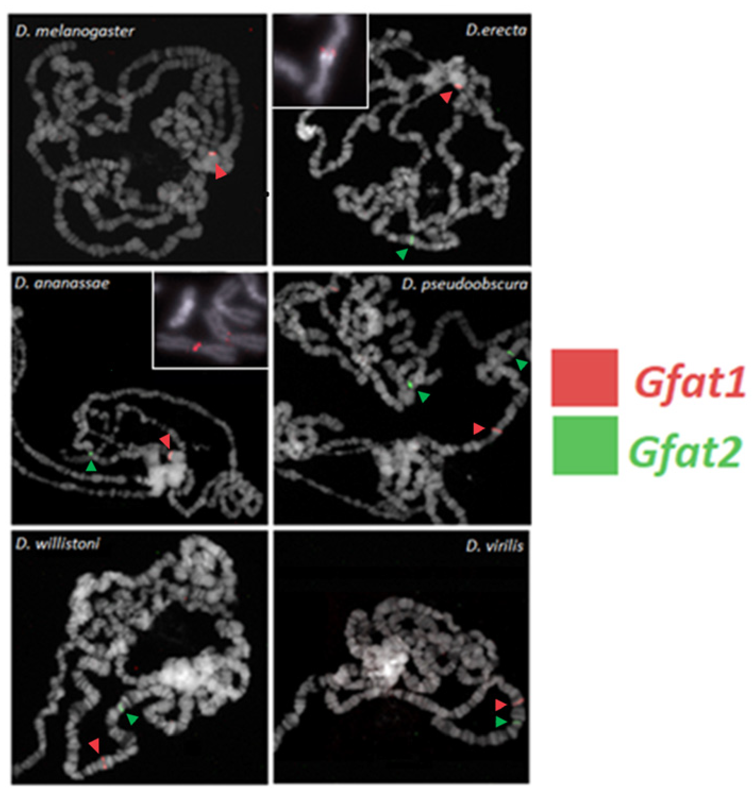
It was of interest to investigate the degree to which the heterochromatic location of Gfat1 was conserved during Drosophila evolution. Thus, we determined the chromosomal locations of the Gfat1 and Gfat2 genes in six Drosophila species, two of which are closely related to D. melanogaster. The data show that Gfat2 is located in euchromatin in all six species, whereas Gfat1 is located in heterochromatin in D. melanogaster and the two most closely related species, D. erecta and D. annanasae, but it is located in euchromatin in the three distantly related species, D. pseudoobscura, D. virilis, and D. willistoni (Figure 7). The most straightforward explanation is that Gfat1 acquired a position in heterochromatin at or just after the divergence of the melanogaster and obscura groups approximately 25 million years ago (see Figure S3).

3.7. Micro-Synteny of Gfat1 and Gfat2 and Flanking Genes

We examined the degree of conservation of the arrangement of genes flanking Gfat1 and Gfat2 in the various Drosophila species and the results of this analysis are shown in Table 6 and Table 7. It is noteworthy that the amino acid sequences of Gfat1 and Gfat2 are highly conserved across all six species. This is especially the case for Gfat1. Furthermore, the same two genes, CG42402 and CG40198 (or their orthologs), flank Gfat1 in all six species; although the 5′ to 3′ orientation of CG42402 relative to Gfat1 is highly conserved, that of CG40198 is variable (Table 6). In addition, for the most part, the distances between the two flanking genes and Gfat1 are variable, and this is particularly true for CG40198. The fact that Gfat1 and at least two other genes are found together in all six Drosophila species indicates that an entire genomic segment containing Gfat1 was relocated to heterochromatin during the evolutionary divergence of the melanogaster and obscura groups. On the other hand, the identities and relative positions of the genes/orthologs flanking Gfat2, larp (CG42551), and Moca-cyp (CG1866), are somewhat less conserved than those flanking Gfat1 (Table 7). Thus, while Moca-cyp is present adjacent to Gfat2 in all species, its relative position 5′ to Gfat2 is maintained from D. melanogaster to D. pseudoobscura but it is located 3′ to Gfat2 in D. willistoni and D. virilis. Similarly, the position of larp 3′ relative to Gfat2 is conserved to D. pseudoobscura. However, larp is located 5′ to Gfat2 in D. virilis and there is no larp gene in the immediate vicinity of Gfat2 in D. willistoni. Finally, the distances between the flanking genes and Gfat2 in the six species are variable; however, for the most part, they are shorter than those observed for the genes flanking Gfat1.

4. Discussion

In this study, we confirm that in Drosophila melanogaster, the genes that encode Gfat1 and Gfat2, the rate-limiting enzymes in the HBP are essential, and further, we show that the Gfat1 gene corresponds to the zep locus. The vital nature of the two genes in Drosophila melanogaster was reported previously [52,54]. Mattila et al. defined an L1 lethal phase for CRISPR-induced Gfat2 mutants and Chen et al. reported L1 lethal phases for their CRISPR-induced alleles of both Gfat1 and Gfat2, with some embryonic death [55]. We have found that all zep alleles (including the original zepLP13 and newly identified alleles) die as embryos and display the diagnostic blimp embryonic phenotype when homozygous (Figure 3 and data not shown) [47]. In addition, transheterozygotes for some mutant alleles and escapers from RNAi crosses, survive to adulthood at low frequencies and exhibit Splayed-like phenotypes. These phenotypes are consistent with the presumptive role of Gfat1 and the HBP in chitin synthesis and thus cuticle formation, as proposed previously [5,24]. Interestingly, peak expression of Gfat1 mRNA occurs at embryonic stages 16–17, which overlap with the time of synthesis and deposition of the chitinous embryonic cuticle [5,24]. In contrast, our analysis of one of the Gfat2 deletion mutants revealed a biphasic lethal phase encompassing embryogenesis and early L1, but the mutant embryos showed no cuticle defects. Although there is also a considerable expression of the Gfat2 mRNA and protein at stages 16–17, it appears that endogenous expression of the Gfat2 gene is unable to compensate for the effects of Gfat1 mutants on cuticle deposition. This may be due in part to differences in expression of Gfat1 versus Gfat2 in cells that synthesize chitin. However, the data do not rule out the possibility that Gfat2 also contributes to chitin synthesis and cuticle production in the fly.
Our rescue data suggest that the paralogous Gfat1 and Gfat2 genes encode functionally equivalent enzymes. However, the different embryonic expression profiles for Gfat1 and Gfat2, coupled with the blimp phenotype exhibited by Gfat1/zep alleles, suggest that the two genes and their products may have undergone some degree of functional divergence in Drosophila. Our finding of essential requirements for Gfat2, but not for Gfat1, in the nervous system and muscles, provide in vivo support for our contention of functional divergence. We cannot completely rule out the possibility that all three Gfat1 RNAi lines tested are not sufficiently potent to cause lethality when expressed in these tissues. However, we have observed that the VDRC Gfat1 RNAi line is lethal in embryogenesis, even when driven by the weak ubiquitous driver, Armadillo-GAL4 (data not shown), suggesting that this RNAi line is quite potent.
The different essential tissue-specific requirements for Gfat1 and Gfat2 reported here could reflect differences in tissue-specific expression. Although there is considerable overlap with respect to tissue mRNA expression for the two genes, it is noteworthy that modENCODE RNAseq anatomical data show moderately high and low expression, respectively, of Gfat2 and Gfat1 mRNA in the central nervous system (CNS) of third instar larvae (http://www.flybase.org (accessed on 29 December 2021)), although comparable anatomical data for Gfat1 and Gfat2 protein expression are as yet unavailable [28]. Interestingly, a study of expression in human tissues has reported complementary, though not entirely mutually exclusive, tissue mRNA expression patterns for hGfat1 and hGfat2. Thus, there is a much higher expression of hGfat2 in elements of the central nervous system, whereas hGfat1 shows prominent and abundant expression in a wide variety of tissues, including the pancreas, heart, skeletal muscle, placenta, prostate, testis, etc. [7,56] Very recently it has been determined that protein O-GlcNAcylation in mammalian cardiomyocytes is specifically dependent on Gfat1 but not Gfat2 activity. Indeed, it appears that only Gfat1 is expressed in these cells [57].
Another potential contributory factor to differential tissue-specific activity of Gfat1 versus Gfat2 proteins could involve post-translational modification. For example, it has been shown that the phosphorylation of the PKA site common to both enzymes causes the inhibition of hGfat1 but the activation of hGfat2 [13,58]. In contrast, other studies have reported that this modification causes the activation of both hGfat1 and Drosophila melanogaster Gfat1 [5,12]. In addition, there is evidence for the inhibition of hGfat1 by AMP-activated protein kinase (AMPK) phosphorylation at yet another site in the enzyme [59]. Interestingly, the AMPK consensus recognition sequence containing the target serine is conserved in hGfat2 and Drosophila Gfat1 but appears to be absent from Drosophila Gfat2 [5,59]. Thus, in principle, the differential regulation of Gfat1 and Gfat2 by AMPK could contribute to differences in tissue-specific requirements for the activity of these enzymes in Drosophila.
In contrast to our nervous system and muscle results, it appears that both Gfat1 and Gfat2 are essential for tracheal development/function. It has been established that proper tracheal development requires optimal chitin synthesis and deposition [48,51,52]. The fact that there are essential requirements for both enzymes in the trachea suggests that both may contribute to critical chitin synthesis in these cells. However, because of the severity of tracheal phenotypes exhibited by mmy mutants, it has been proposed that there are chitin-independent requirements for HBP functions in tracheal development [23,24]. Therefore, it is possible that one of the Gfat enzymes is predominantly or even exclusively responsible for contributing to chitin production in the trachea, whereas the other contributes to other critical functions in these cells.
We have determined that in D. melanogaster and other members of the melanogaster group, the Gfat1 and Gfat2 genes are located in heterochromatin and euchromatin, respectively, whereas, in several other more distantly related species, both genes have euchromatic locations. The most straightforward hypothesis is that, in the ancestral Drosophila configuration, both genes were euchromatic and that, during the evolutionary divergence of the melanogaster group, Gfat1 acquired a heterochromatic location (see Figure S2). It is noteworthy that both genes are located on a single chromosomal element in at least four of the six species (Figure 7). Since Gfat1 is located in the chromocenter in D. erecta and D. annanasae, it is not possible to determine in which chromosome arm they are located, at least from the polytene FISH analysis. Interestingly, both genes are located on chromosome 2 or Muller element E in D. pseudoobscura, and this element corresponds to chromosome 3R in D. melanogaster (gene link at https://blast.ncbi.nlm.nih.gov/Blast.cgi (accessed on 29 December 2021)), suggesting some degree of evolutionary conservation of the element-specific location of the two genes [60,61]. We have also examined the microsynteny of genes that immediately flank Gfat1 and Gfat2 in the six species. The same genes/orthologs in the same 5′ and 3′ positions flank Gfat1 in all six species. By and large, this is also the case for Gfat2, although there are some differences for D. willistoni and D. virilis. These examples of flanking gene conservation contrast with the pattern for genes that flank another highly conserved gene, RpL15, which is located in 3L het in D. melanogaster (Table S5). In this case, there is a considerable deviation for most species. The significance of conservation of gene arrangements in the vicinities of Gfat1 and Gfat2 is unclear but may warrant future investigation.
It is instructive to compare our evolutionary data to those of an earlier seminal analysis of the structure and evolution of light and neighbouring heterochromatic genes and their euchromatic orthologues [30]. These workers determined that a large chromosomal segment containing the light gene and adjacent genes was juxtaposed to heterochromatin during the evolutionary divergence of the melanogaster subgroup. In contrast, although our results also indicate that a multigene segment that contains the Gfat1 gene was relocated to heterochromatin, this event occurred during the evolutionary divergence of the melanogaster and obscura groups.
The relationship between Gfat1 and Gfat2 and other duplicate gene-pairs in Drosophila melanogaster is, as yet, undefined [62]. However, it is noteworthy that there are at least two other Drosophila paralogous gene-pairs for which one gene is heterochromatic and the other is euchromatic: SNAP25 (3L het)/SNAP24 (euchromatic) and spok (3R het)/spo (euchromatic) [63,64,65]. As is true for Gfat1/Gfat2, these studies show that the two protein products of each of these gene-pairs are functionally equivalent. Interestingly, in all three cases, the two genes exhibit different patterns of mRNA expression [28]. As mentioned, Gfat2 is expressed at earlier stages and, for the most part, at consistently higher levels than Gfat1 during embryogenesis. Similarly, spo expression occurs during the first half of embryogenesis, whereas spok expression begins later in embryogenesis and continues at higher levels in the larval and pupal stages. Finally, SNAP-24 expression extends throughout embryogenesis at moderately-high/high levels and then decreases somewhat during the larval and pupal stages, whereas SNAP-25 expression commences rather late in embryogenesis and extends through the larval and pupal stages.
An interesting correlation exists between gene structure and differential expression of the aforementioned gene pairs. Thus, while in each case the heterochromatic paralog either contains multiple introns or one very large intron, the euchromatic paralog either contains a single small intron or is intronless, and this pattern is conserved in the five other Drosophila species included in the current study (Gfat1/Gfat2: Figures S4 and S5; SNAP25/SNAP24 and spok/spo data are available in Flybase release FB2017_05 [28]). One possible explanation is that, in each case, the euchromatic paralog has been subject to selective pressure for maintenance of a compact structure that could facilitate rapid and abundant transcription and mRNA processing for earlier developmental requirements. Consistent with this explanation, at least two Drosophila studies have reported enriched identification of intronless and intron-poor genes among genes exhibiting early zygotic expression, particularly those expressed during syncytial stages [66,67]. Moreover, the prevalence of early-expressed zygotic genes with short transcription units is observed for several different species that span the Drosophila phylogeny, suggesting that this pattern is evolutionarily conserved [67].
Future investigations of specific mechanisms of Gfat1 regulation should provide important insight into the regulation of heterochromatic gene expression. In addition, the question of how Gfat2 expression is regulated is of considerable interest. Moreover, given the potential links between the HBP and a wide variety of human diseases, further studies of the functional interplay between the Gfat1 and Gfat2 enzymes in flies should prove worthwhile.

Supplementary Materials

The following are available online at https://www.mdpi.com/article/10.3390/cells11030448/s1. Figure S1: Map of proximal 3R correlating the molecular location of the Gfat1 gene with the genetic position of zep; Figure S2. Splayed phenotype displayed by hs-Gal4/2664-1-5M-CH2 fly raised at 29 °C; Figure S3. Schematic representation of Drosophila evolution over the last 50 million years; Figure S4. Gene architecture of the longest versions of Gfat1 in six species of Drosophila; Figure S5. Gene architecture of Gfat2 in six species of Drosophila; Table S1. RNAi strains from Drosophila stock centres used in the present study; Table S2. Tests for viability effects of Gfat1 RNAi transgenes when driven ubiquitously; Table S3. Primers used for the sequencing analysis of zep alleles; Table S4. qPCR primer sequences; Table S5. Microsynteny of two genes flanking RpL15 or its orthologue in six Drosophila species.

Author Contributions

Conceptualization: C.J.J., S.C., D.A.S., K.A.F., M.S., G.H. and R.G.C.; methodology: C.J.J., S.C., D.A.S., K.A.F., M.S. and S.J.H.W.; formal analysis: C.J.J., S.C., K.A.F., M.M., M.S., S.J.H.W. and S.P.; investigation: C.J.J., S.C., K.A.F., M.M. and S.J.H.W.; writing—original draft preparation: S.C., A.J.H. and D.A.S.; writing—review and editing: C.J.J., S.C., K.A.F., A.B., A.B.C., A.J.H., B.M.H., D.A.S., G.H., E.M.V., M.M., R.G.C. and S.P.; supervision: A.J.H., B.M.H., D.A.S., E.M.V. and S.P.; funding acquisition: A.J.H., B.M.H., D.A.S., E.M.V. and S.P. All authors have read and agreed to the published version of the manuscript.

Funding

This work was supported by the following: an operating grant of the Canadian Institutes of Health Research to BMH, DARS and AJH; operating grants from the Natural Sciences and Engineering Research Council to BMH and to AJH; and the National Science Foundation Grant No. IOS-1555749 to AB.

Conflicts of Interest

The authors declare no conflict of interest.

References

  1. Darley-Usmar, M.V.; Ball, L.E.; Chatham, J.C. Protein O-linked β-N-acetylglucosamine: A novel effector of cardiomyocyte metabolism and function. J. Mol. Cell. Cardiol. 2012, 52, 538–554. [Google Scholar] [CrossRef] [PubMed] [Green Version]
  2. Mattila, J.; Hietakangas, V. Regulation of carbohydrate energy metabolism in Drosophila melanogaster. Genetics 2017, 207, 1231–1253. [Google Scholar] [CrossRef] [PubMed]
  3. Chiaradonna, F.; Ricciardiello, F.; Palorini, R. The nutrient-sensing hexosamine biosynthetic pathway as the hub of cancer metabolic rewiring. Cells 2018, 7, 53. [Google Scholar] [CrossRef] [Green Version]
  4. Akella, N.M.; Ciraku, L.; Reginato, M.J. Fueling the fire: Emerging role of the hexosamine biosynthetic pathway in cancer. BMC Biol. 2019, 17, 52–65. [Google Scholar] [CrossRef] [PubMed] [Green Version]
  5. Graack, H.-R.; Cinque, U.; Kress, H. Functional regulation of glutamine:fructose-6-phosphate aminotransferase 1 (Gfat1) of Drosophila melanogaster in a UDP-N-acetylglucosamine and cAMP manner. Biochem. J. 2001, 360, 401–412. [Google Scholar] [CrossRef] [PubMed]
  6. Denzel, M.S.; Antebi, A. Hexosamine pathway and (ER) protein quality control. Curr. Opin. Cell Biol. 2015, 33, 14–18. [Google Scholar] [CrossRef] [PubMed]
  7. Oki, T.; Yamazaki, K.; Kuromitsu, J.; Okada, M.; Tanaka, I. cDNA cloning and mapping of a novel subtype of Glutamine:fructose-6-phosphate amidotransferase (Gfat2) in human and mouse. Genomics 1999, 57, 227–234. [Google Scholar] [CrossRef]
  8. Kato, N.; Dasgupta, R.; Smartt, C.T.; Christensen, B.M. Glucosamine:fructose-6-phosphate aminotransferase: Gene characterization, chitin biosynthesis and peritrophic matrix formation in Aedes aegypti. Insect Mol. Biol. 2002, 11, 207–216. [Google Scholar] [CrossRef]
  9. Smith, R.J.; Milewski, S.; Brown, A.J.P.; Gooday, G.W. Isolation and characterization of the GFA1 gene encoding glutamine:fructose-6-phosphate amidotransferase of Candida albicans. J. Bacteriol. 1996, 178, 2320–2327. [Google Scholar] [CrossRef] [Green Version]
  10. Ram, A.F.J.; Arentshorst, M.; Damveld, R.A.; vanKuyk, P.A.; Klis, F.M.; van den Hondel, C.A. The cell wall stress response in Aspergillus niger involves increased expression of the glutamine: Fructose-6-phosphate amidotransferase-encoding gene (gfaA) and increased deposition of chitin in the cell wall. Microbiology 2004, 150, 3315–3326. [Google Scholar] [CrossRef] [Green Version]
  11. Luo, C.; Shao, W.; Li, X.; Chen, Z.; Liu, Y. Molecular cloning, sequencing and expression of a l-glutamine d-fructose 6-phosphate amidotransferase gene from Volvariella volvacea. Protein J. 2009, 28, 34–43. [Google Scholar] [CrossRef] [PubMed]
  12. Zhou, J.; Huynh, Q.K.; Hoffman, R.T.; Crook, E.D.; Daniels, M.C.; Gulve, E.A.; McClain, D.A. Regulation of glutamine:fructose-6-phosphate amidotransferase by cAMP-dependent protein kinase. Diabetes 1998, 47, 1836–1840. [Google Scholar] [CrossRef] [PubMed]
  13. Hu, Y.; Riesland, L.; Paterson, A.J.; Kudlow, J.E. Phosphorylation of mouse glutamine:fructose-6-phosphate amidotransferase 2 (GFAT2) by cAMP-dependent protein kinase increases the enzyme activity. J. Biol. Chem. 2004, 279, 29988–29993. [Google Scholar] [CrossRef] [PubMed] [Green Version]
  14. Huang, X.; Tsuji, N.; Miyoshi, T.; Motobu, M.; Islam, M.K.; Alim, M.A.; Fujisaki, K. Characterization of glutamine:Fructose-6-phosphate amino transferase from the ixodid tick, Haemaphysalis longicornis, and its critical role in host blood feeding. Int. J. Parasitol. 2007, 37, 383–392. [Google Scholar] [CrossRef]
  15. Denzel, M.S.; Storm, N.J.; Gutschmidt, A.; Baddi, R.; Hinze, Y.; Jarosch, E.; Sommer, T.; Hoppe, T.; Antebi, A. Hexosamine pathway metabolites enhance protein quality and prolong life. Cell 2014, 156, 1167–1178. [Google Scholar] [CrossRef] [Green Version]
  16. Wang, Z.V.; Deng, Y.; Gao, N.; Pedrozo, Z.; Li, D.L.; Morales, C.R.; Criollo, A.; Luo, X.; Tan, W.; Jiang, N. Spliced-box protein 1 couples the unfolded protein response to hexosamine biosynthetic pathway. Cell 2014, 156, 1179–1192. [Google Scholar] [CrossRef] [Green Version]
  17. Yang, X.; Qian, K. Protein O-GlcNAcylation: Emerging mechanisms and functions. Nat. Rev. Mol. Cell. Biol. 2017, 18, 452–465. [Google Scholar] [CrossRef]
  18. Hanover, J.A.; Chen, W.; Bond, M.R. O-GlcNAc in cancer: An Oncometabolism-fueled vicious cycle. J. Bioenerg. Biomembr. 2018, 50, 155–173. [Google Scholar] [CrossRef]
  19. Hart, G.W. Nutrient regulation of signaling and transcription. J. Biol. Chem. 2019, 294, 2211–2231. [Google Scholar] [CrossRef] [Green Version]
  20. Parker, M.P.; Peterson, K.R.; Slawson, C. O-GlcNAcylation and O-GlcNAc cycling regulate gene transcription: Emerging roles in cancer. Cancers 2021, 13, 1666. [Google Scholar] [CrossRef]
  21. Sun, L.; Lv, S.; Song, T. 2021 O-GlcNAcylation links oncogenic signals and cancer epigenetics. Discov. Oncol. 2021, 12, 54. [Google Scholar] [CrossRef]
  22. Moussian, B.; Schwarz, H.; Bartoszewski, S.; Nusslein-Volhard, C. Involvement of chitin in exoskeleton morphogenesis in Drosophila melanogaster. J. Morphol. 2005, 264, 117–130. [Google Scholar] [CrossRef] [PubMed]
  23. Tonning, A.; Helms, S.; Schwarz, H.; Uv, A.E.; Moussian, B. Hormonal regulation of mummy is needed for extracellular formation and epithelial morphogenesis in Drosophila. Development 2006, 133, 331–341. [Google Scholar] [CrossRef] [PubMed] [Green Version]
  24. Ostrowski, S.; Dierick, H.A.; Bejsovec, A. Genetic control of cuticle formation during embryonic development of Drosophila melanogaster. Genetics 2002, 161, 171–182. [Google Scholar] [CrossRef] [PubMed]
  25. Syrzycka, M.; Hallson, G.; Fitzpatrick, K.A.; Kim, I.; Cotsworth, S.; Hollebakken, R.E.; Simonetto, K.; Yang, L.; Luongo, S.; Beja, K.; et al. Genetic and molecular analysis of essential genes in centromeric heterochromatin of the left arm of chromosome 3 in Drosophila melanogaster. Genes Genomics Genet. 2019, 7, 1581–1595. [Google Scholar] [CrossRef] [Green Version]
  26. Coulthard, A.B.; Alm, C.; Cealiac, I.; Sinclair, D.A.; Honda, B.M.; Rossi, F.; Dimitri, P.; Hilliker, A.J. Essential loci in centromeric heterochromatin of Drosophila melanogaster. I: The right arm of chromosome 2. Genetics 2010, 185, 479–495. [Google Scholar] [CrossRef] [Green Version]
  27. Yasuhara, J.C.; DeCrease, C.H.; Wakimoto, B.T. Evolution of heterochromatic genes of Drosophila. Proc. Natl. Acad. Sci. USA 2005, 102, 10959–10963. [Google Scholar] [CrossRef] [Green Version]
  28. Thurmond, J.; Goodman, J.L.; Strelets, V.B.; Attrill, H.; Gramates, L.S.; Marygold, S.J.; Matthews, B.B.; Millburn, G.; Antonazzo, G.; Trovisco, V.; et al. FlyBase 2.0: The next generation. Nucleic Acids Res. 2019, 47, D759–D765. [Google Scholar] [CrossRef] [PubMed] [Green Version]
  29. Koundakjian, E.J.; Cowana, D.M.; Hardy, R.W.; Beckera, A.H. The Zuker Collection: A Resource for the Analysis of Autosomal Gene Function in Drosophila melanogaster. Genetics 2004, 167, 203–206. [Google Scholar] [CrossRef] [Green Version]
  30. Irion, U.; Leptin, M. Developmental and cell biological functions of the Drosophila DEAD-box protein abstrakt. Curr. Biol. 1999, 9, 1373–1381. [Google Scholar] [CrossRef] [Green Version]
  31. Marchant, G.E.; Holm, D.G. Genetic analysis of the heterochromatin of chromosome 3 in Drosophila melanogaster. I. Products of compound autosome detachment. Genetics 1988, 120, 503–517. [Google Scholar] [CrossRef]
  32. Marchant, G.E.; Holm, D.G. Genetic analysis of the heterochromatin of chromosome 3 in Drosophila melanogaster. II. Vital loci identified through EMS mutagenesis. Genetics 1988, 120, 519–532. [Google Scholar] [CrossRef]
  33. Koryakov, D.E.; Zhimulev, I.F.; Dimitri, P. Cytogenetic analysis of the third chromosome heterochromatin of Drosophila melanogaster. Genetics 2002, 160, 509–517. [Google Scholar] [CrossRef]
  34. Koryakov, D.E.; Domanitskaya, E.V.; Belyakin, S.N.; Zhimulev, E.F. Abnormal tissue-dependent polytenization of chromosome 3 pericentric heterochromatin in Drosophila melanogaster. J. Cell Sci. 2003, 116, 1035–1044. [Google Scholar] [CrossRef] [PubMed] [Green Version]
  35. Fitzpatrick, K.A. Genetic and Molecular Characterization of Chromosome 3 Heterochromatin in Drosophila melanogaster. Ph.D.Thesis, W.A.C. Bennett Library QH 599 F584 2005, Simon Fraser University, Burnaby, BC, Canada, 2005. [Google Scholar]
  36. Jackson, C. Gfat1/Zeppelin is an Essential Gene Involved in Cuticle Formation in D. melanogaster. Master’s Thesis, W.A.C. Bennett Library QH 470 D7 J32, Simon Fraser University, Burnaby, BC, Canada, 2007. [Google Scholar]
  37. Stapleton, M.; Carlson, J.; Brokstein, P.; Yu, C.; Champe, M.; George, R.; Guarin, H.; Kronmiller, B.; Pacleb, J.; Park, S.; et al. A Drosophila full-length cDNA resource. Genome Biol. 2002, 3, 1–8. [Google Scholar] [CrossRef]
  38. Niccoli, T.; Cabecinha, M.; Tillmann, A.; Kerr, F.; Wong, C.T.; Cardenes, A.; Vincent, A.J.; Bettedi, L.; Li, L.; Grönke, S.; et al. Increased glucose transport into neurons rescues Abeta toxicity in Drosophila. Curr. Biol. 2016, 26, 2550. [Google Scholar] [CrossRef] [PubMed] [Green Version]
  39. Pfaffl, M.W. A new mathematical model for relative quantification in real-time RT-PCR. Nucleic Acids Res. 2001, 1, e45. [Google Scholar] [CrossRef] [PubMed]
  40. Pimpinelli, S.; Bonaccorsi, S.; Fanti, L.; Gatti, M. Preparation and Analysis of Drosophila mitotic Chromosomes, in Drosophila Protocols; Sullivan, W., Ashburner, M., Hawley, R.S., Eds.; Cold Spring Harbor Laboratory Press: Cold Spring Harbor, NY, USA, 2000; pp. 3–23. [Google Scholar]
  41. Fitzpatrick, K.A.; Sinclair, D.A.; Schulze, S.R.; Syrzycka, M.; Honda, B.M. A genetic and molecular profile of chromosome 3 centric heterochromatin in Drosophila melanogaster. Genome 2005, 48, 571–584. [Google Scholar] [CrossRef]
  42. Lindsley, D.L.; Sandler, L.; Baker, B.S.; Carpenter, A.T.C.; Denell, R.E.; Hall, J.C.; Jacobs, P.A.; Miklos, G.L.; Davis, B.K.; Gethmann, R.C.; et al. Segmental aneuploidy and the genetic gross structure of the Drosophila genome. Genetics 1972, 71, 157–184. [Google Scholar] [CrossRef]
  43. Tasaka, S.E.; Suzuki, D.T. Temperature-sensitive mutations in Drosophila melanogaster. XVII. Heat- and cold-sensitive lethals on chromosome 3. Genetics 1973, 74, 509–520. [Google Scholar] [CrossRef]
  44. Cotsworth, S.E.P. Gfat1 and Gfat2 Encode Functionally Equivalent Enzymes in Drosophila melanogaster: A Molecular, Genetic and Evolutionary Analysis. Master’s Thesis, W.A.C. Bennett Library Simon Fraser University, Burnaby, BC, Canada, 2018. [Google Scholar]
  45. Fisher, B.; Weiszmann, R.; Frise, E.; Hammonds, A.; Tomancak, P.; Beaton, A.; Berman, B.; Quan, E.; Shu, S.; Lewis, S.; et al. Patterns of Gene Expression in Drosophila embryogenesis. BDGP in Situ Homepage. 2012. Available online: https://insitu.fruitfly.org/cgi-bin/ex/insitu.pl (accessed on 20 January 2022).
  46. Liu, X.; Blaženović, I.; Contreras, A.J.; Pham, T.M.; Tabuloc, C.A.; Li, Y.H.; Ji, J.; Fiehn, O.; Chiu, J.C. Hexosamine biosynthetic pathway and O-GlcNAc-processing enzymes regulate daily rhythms in protein O-GlcNAcylation. Nat. Commun. 2021, 12, 4173. [Google Scholar] [CrossRef]
  47. Casas-Vila, N.; Bluhm, A.; Sayols, S.; Dinges, N.; Dejung, M.; Altenhein, T.; Kappei, D.; Altenhein, B.; Roignant, J.-Y.; Butter, F. The developmental proteome of Drosophila melanogaster. Genome Res. 2017, 27, 1273–1285. [Google Scholar] [CrossRef] [PubMed] [Green Version]
  48. Araujo, S.J.; Aslam, H.; Tear, G.; Casanova, J. Mummy/cystic encodes an enzyme required for chitin and glycan synthesis, involved in trachea, embryonic cuticle and CNS development—Analysis of its role in Drosophila morphogenesis. Dev. Biol. 2005, 288, 179–193. [Google Scholar] [CrossRef] [PubMed] [Green Version]
  49. Schimmelpfeng, K.; Strunk, M.; Stork, T.; Klambt, C. mummy encodes an UDP-N-acetylglucosamine-diphosphorylase and is required during dorsal closure and nervous system development. Mech. Dev. 2006, 123, 487–499. [Google Scholar] [CrossRef] [PubMed]
  50. Manavalan, M.A.; Jayasinghe, V.R.; Grewal, R.; Bhat, K.M. The glycosylation pathway is required for the secretion of Slit receptor Robo on axons. Sci. Signal. 2017. [CrossRef] [Green Version]
  51. Sinclair, D.A.R.; Syrzycka, M.; Macauley, M.S.; Rastgardani, T.; Komljenovic, I.; Vocadlo, D.J.; Brock, H.W.; Honda, B.M. Drosophila O-GlcNAc transferase (OGT) is encoded by the Polycomb group (PcG) gene, super sex combs (sxc). Proc. Natl. Acad. Sci. USA 2009, 106, 13427–13432. [Google Scholar] [CrossRef] [Green Version]
  52. Ozturk-Colak, A.; Moussian, B.; Araujo, S. Drosophila chitinous aECM and its cellular interactions during embryogenesis. Dev. Dyn. 2016, 245, 259–267. [Google Scholar] [CrossRef] [Green Version]
  53. Devine, W.P.; Lubarsky, B.; Shaw, K.; Luschnig, S.; Messina, L.; Krasnow, M.A. Requirement for chitin biosynthesis in epithelial tube morphogenesis. Proc. Nat. Acad. Sci. USA 2005, 102, 17014–17019. [Google Scholar] [CrossRef] [Green Version]
  54. Chen, P.; Visokay, S.; Abrams, J.M. Drosophila GFAT1 and GFAT2 enzymes encode obligate developmental functions. Fly 2020, 14, 9. [Google Scholar] [CrossRef]
  55. Mattila, J.; Kokki, K.; Hietakangas, V.; Boutros, M. Stem cell intrinsic hexosamine metabolism regulates intestinal adaptation to nutrient content. Dev. Cell 2018, 47, 112–121. [Google Scholar] [CrossRef] [Green Version]
  56. Oliveira, I.A.; Allonso, D.; Fernandes, T.V.A.; Lucena, D.M.S.; Ventura, G.T.; Dias, W.B.; Mohana-Borges, R.S.; Pascutti, P.G.; Todeschini, A.R. Enzymatic and structural properties of human glutamine:fructose-6-phosphate amidotransferase 2 (hGFAT2). J. Biol. Chem. 2020, 2020, 17. [Google Scholar] [CrossRef] [PubMed]
  57. Nabeebaccus, A.A.; Verma, S.; Zoccarato, A.; Emanuelli, G.; Santos, C.X.C.; Streckfuss-Bömeke, K.; Shah, A.M. Cardiomyocyte protein O-GlcNAcylation is regulated by GFAT1 not GFAT2. Biochem. Biophys. Res. Commun. 2021, 583, 121–127. [Google Scholar] [CrossRef]
  58. Chang, Q.; Su, K.; Baker, R.J.; Yang, X.; Paterson, A.J.; Kudlow, J.E. Phosphorylation of human glutamine:fructose-6-phosphate amidotransferase by cAMP-dependent protein kinase at Serine 205 blocks the enzyme activity. J. Biol. Chem. 2000, 275, 21981–21987. [Google Scholar] [CrossRef] [PubMed] [Green Version]
  59. Eguchi, S.; Oshiro, N.; Miyamoto, T.; Yoshino, K.; Okamoto, S.; Ono, T.; Kikkawa, U.; Yonezawa, K. AMP-activated protein kinase phosphorylates glutamine: Fructose-6-phosphate amidotransferase 1 at Ser243 to modulate enzymatic activity. Genes Cells 2009, 14, 179–189. [Google Scholar] [CrossRef] [PubMed]
  60. Schulze, S.R.; McAllister, B.F.; Sinclair, D.A.R.; Fitzpatrick, K.A.; Marchetti, M.; Pimpinelli, S.; Honda, B.M. Heterochromatic genes in Drosophila: A comparative analysis of two genes. Genetics 2006, 173, 1433–1445. [Google Scholar] [CrossRef] [Green Version]
  61. Kulathinal, R.J.; Hartl, D.L. The latest buzz in comparative genomics. Genome Biol. 2005, 6, 201–203. [Google Scholar] [CrossRef] [Green Version]
  62. Assis, R. Drosophila duplicate genes evolve new functions on the fly. Fly 2014, 8, 91–94. [Google Scholar] [CrossRef] [Green Version]
  63. Vilinsky, I.; Stewart, B.A.; Drummond, J.; Robinson, I.; Deitcher, D.L. A Drosophila SNAP-25 null mutant reveals context-dependent redundancy with SNAP-24 in neurotransmission. Genetics 2002, 162, 259–271. [Google Scholar] [CrossRef]
  64. Ono, H.; Rewitz, K.F.; Shinoda, T.; Itoyama, K.; Petryk, A.; Rybczynski, R.; Jarcho, M.; Warren, J.T.; Marqués, G.; Shimell, M.J.; et al. Spook and Spookier code for stage-specific components of the ecdysone biosynthetic pathway in Diptera. Dev. Biol. 2006, 298, 555–570. [Google Scholar] [CrossRef] [Green Version]
  65. Syrzycka, M. Genetic and Molecular Characterization of Heterochromatic Genes in Drosophila melanogaster. Ph.D. Thesis, W.A.C. Bennett Library QH 5, Simon Fraser University, Burnaby, BC, Canada, 2009. [Google Scholar]
  66. De Renzis, S.; Elemento, O.; Tavazoie, S.; Wieschaus, E.F. Unmasking activation of the zygotic genome using chromosomal deletions in the Drosophila embryo. PLoS Biol. 2007, 5, e117. [Google Scholar] [CrossRef] [Green Version]
  67. Artieri, C.G.; Fraser, H.B. Transcript length mediates developmental timing expression across Drosophila. Mol. Biol. Evol. 2014, 31, 2879–2889. [Google Scholar] [CrossRef] [PubMed] [Green Version]
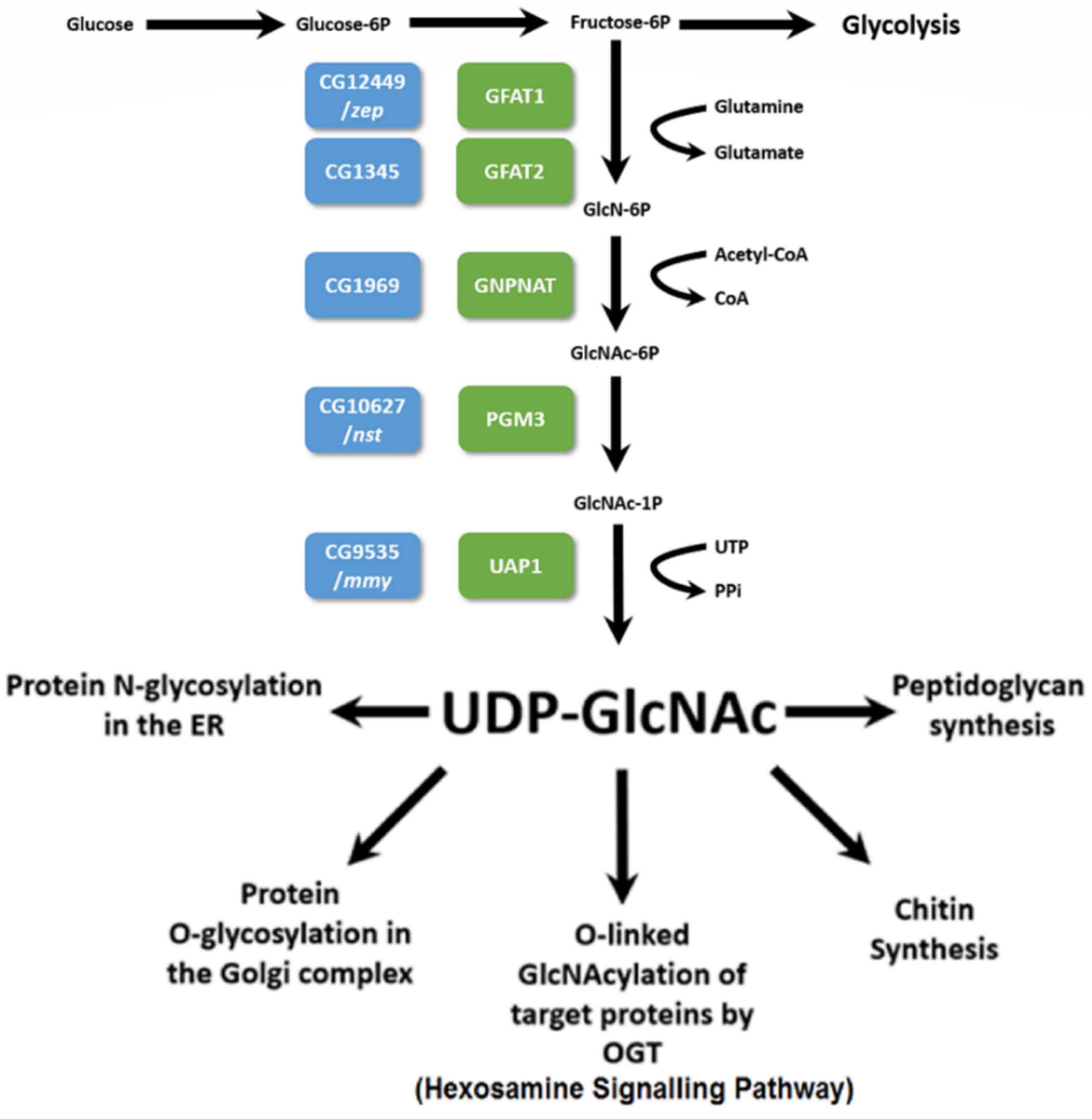
Figure 1. Schematic depiction of the Hexosamine Biosynthetic Pathway (HBP) showing the key metabolic inputs, the reaction steps, along with the enzymes/genes responsible for catalysis and the important outputs. Blue boxes correspond to the gene names in Drosophila melanogaster and green boxes correspond to the enzymes that they encode (see the Introduction). Modified from [3,4].
Cells 11 00448 g001
Figure 2. Results of inter se complementation analysis of zep alleles. The mutant third chromosomes were balanced with either TM3, Sb Ser, or TM3 Ser. A minimum of 100 progeny per cross was examined. L = lethal; SL = semi-lethal (less than 50% of expected transheterozygous or homozygous progeny relative to balancer progeny); V = viable.
Cells 11 00448 g002
Figure 3. Expanded cuticle phenotypes of the strongest zep alleles. zepLP13 and zepI400#8 contain stop codons (see Figure 4) whereas zep8740#20, zep8740#22, and zep167 delete Gfat1 plus two or more flanking genes (see Figure S1). A wildtype (Oregon-R) embryo is shown in the top left panel. Homozygous mutant and wildtype embryos were prepared as described in Ostrowski et al. [24] The embryos are oriented with anterior to the left and posterior to the right. Note the pronounced bloating of the mutant embryos relative to wildtype.
Cells 11 00448 g003
Figure 4. Transcript model of Gfat1-RN. Locations of mutations are given above and are marked by arrows along the gene model. zepLP13 and zepI400#8 contain stop codons (indicated by asterisks) after Tyr334 and Glu535, respectively; zepz1904 and zepz1914 contain the identical missense mutation, A414T; zep3−52 contains the missense mutation, L566M; and zepz1608 contains the missense mutation, C656Y.
Cells 11 00448 g004
Figure 5. Transcript model of Gfat2-RA. The approximate location of the P-element insert used for the excision experiment is shown above by an inverted triangle. The approximate end points for the excision deletions Gfat218A−14 (498 bp in length) and Gfat210A−2 (1038 bp in length) are shown above by arrows.
Cells 11 00448 g005
Figure 6. RT-qPCR analysis of Gfat1 and Gfat2 mRNA using RpL32 as a reference gene and the Gfat1 0–1.5 h sample as the control group, at different stages of embryogenesis.
Cells 11 00448 g006
Figure 7. FISH localization of Gfat1 (red) and Gfat2 (green) in polytene chromosomes in six and five Drosophila species, respectively. For a description of the methods used, see Pimpinelli et al. [40] Note that Gfat1 is located within heterochromatin in D. melanogaster, D. erecta, and D. ananassae. This is confirmed for FISH using mitotic chromosomes for D. erecta and D. ananassae (see the insets). Gfat1 is euchromatic in D. pseudoobscura, D. willistoni, and D. virilis. Gfat2 is euchromatic in all species.
Cells 11 00448 g007
Table 1. Lethal phase analysis of a Gfat2 mutation.
CrossGfat210A2/Df(3R)BSC460 Offspring
Unhatched EmbryosFirst Instar LarvaeSecond Instar Larvae
Gfat210A2/TM3, Sb, Ser, Twi-GFP x Df(3R)BSC460/ TM3, Sb, Ser, Twi-GFP 59410
Table 2. Mutant rescue with Gfat1+ and Gfat2+ transgenes.
Number of Relevant Progeny
Mutant/TM3 or TM6BMutant/Deficiency or Mutant/Mutant
CrossTotalCyO/Gfat1+ or Gfat2+Act5C-GAL4/Gfat1+ or Gfat2+Cy/Gfat1+ or Gfat2+Act5C-GAL4/Gfat1+ or Gfat2+
UAS-Gfat1+/CyRoi; Gfat1LP13/TM3, Sb
X310139120051
Actin5C-GAL4/CyO; Df(3R)7B-90, e/TM3, Sb
UAS-Gfat1+/CyRoi; Gfat210A−2/TM3 Ser
X17911549015
Actin5C-GAL4/CyO; Df(3R)BSC567/TM3, Ser
UAS-Gfat2+/CyRoi; Df(3R)BSC460/TM6B
X1779550032
Actin5C-GAL4/CyO; Gfat210A−2/TM3, Ser
UAS-Gfat2+/CyRoi; Gfat1z−1904/TM6B
X1568454018
Actin5C-GAL4/CyO; Gfat1I400#8/TM3, Ser
Table 3. Tests for viability effects of Gfat2, Gfat1, and Ogt RNAi knockdown in the larval nervous system at 29 °C using Appl-GAL4 *.
RNAiNumber of ProgenyTotalComments on RNAi
Gfat RNAiControl (Balancer)
Gfat2/CyO (v105129)123159282male semi-lethality **
frequent unfurled wings
Gfat2/TM3, Sb (B34740)0218218lethal
Gfat1/TM3, Sb (HL)175111286viable
Gfat1/CyO (B42892)258260518viable
Gfat1 (v24539)539-539viable
Ogt/CyO (B50909)0275275lethal
* Males heterozygous or homozygous for RNAi transgenes mated to Appl-GAL4 females. See Table S1 for information about the RNAi lines from stock centres. HL: 2664-1-6M-CH3 line from the Honda lab (see Table S2 for viability data of ubiquitously-expressed RNAi). ** The designation of male semi-lethality (less than 30% of expected) is based on relative viability = the number of observed RNAi males/the number of CyO males = 11/44 = 0.25.
Table 4. Tests for viability effects of Gfat2, Gfat1, and Ogt knockdown in muscle cells at 29 °C using Mef2-GAL4 *.
RNAiNumber of ProgenyTotalComments on RNAi
Gfat RNAiControl (Balancer)
Gfat2/CyO (v105129)0148148lethal
Gfat2/TM3, Sb (B34740)06060lethal
Gfat1/TM3, Sb (HL)227165392viable
Gfat1/CyO (B42892)250252502viable
Gfat1
(v24539)
60-60viable
Ogt/CyO
(B50909)
0240240lethal
* Males heterozygous or homozygous for RNAi transgenes mated to Mef2-GAL4 females; genotype of driver: y w; P{GAL4-Mef2.R}3 (BDSC #27390). See the legend to Table 3 and Table S1 for information about the RNAi lines used.
Table 5. Tests for viability effects of Gfat2 and Gfat1 RNAi knockdown in the trachea at 30 °C using breathless-GAL4 *.
Control or RNAiNumber of ProgenyTotalComments on RNAi
Gfat RNAiControl (Balancer)
w1118 control92116208NA
Gfat2/CyO (v105129)40132172weak semi-lethality **
Gfat2/TM3, Sb (B34740)0269269lethal
Gfat1/CyO (B42892)2789116weak semi-lethality **
Gfat1
(v24539)
0202202lethal
* Males heterozygous or homozygous for RNAi transgenes were mated to btl-GAL4/CyO females; genotype of driver: w; P{w+mC = GAL4-btl.S}2, P{w+mC = UASp-Act5C.T:GFP}2/CyO, P{w+m = lacZ. W+}276 (BDSC #8807); see the legend to Table 3 and Table S1 for information about the RNAi lines used. ** The designation of weak semi-lethality (less than 62% of expected) is based on relative viability = number of observed RNAi adults/half the number of CyO adults.
Table 6. Microsynteny of two genes flanking Gfat1 or its ortholog in six Drosophila species.
Species and Designation of Gfat1 Gene Sequence Homology of Orthologous Gfat1 Proteins Versus D. m. Gfat1 * CG42402 or Ortholog 5′ to Gfat1 **5′ to 3′ Orientation Relative to That of Gfat1CG40198 or Ortholog 3′ to Gfat15′ to 3′ Orientation Relative to That of Gfat1
melanogaster CG12449-yes (22)sameyes (23)same
erecta
GG12143
98/98yes (31)sameyes (3)opposite
ananassae
GF23135
95/97yes (35)sameyes (12)same
pseudoobscura
GA26267
97/98yes (35)sameyes (6)opposite
willistoni
GK12920
96/98yes (38)sameyes (8)opposite
virilis
GJ24380
96/98yes (28)sameyes (28)opposite
* percent identity/percent similarity; ** distance from Gfat1 or ortholog in kbp given in parentheses; data from FB2017_05 release and confirmed via NCBI BLAST.
Table 7. Microsynteny of two genes flanking Gfat2 or its ortholog in six Drosophila species.
Species and Designation of Gfat2 Gene Sequence Homology of Orthologous Gfat2 Protein Versus D. m. Gfat2 * Moca-cyp or Ortholog 5′ to Gfat2 **5′ to 3′ Orientation Relative to That of Gfat2larp or Ortholog 3′ to Gfat25′ to 3′ Orientation Relative to That of Gfat2
melanogaster CG1345-yes (0.5)oppositeyes (0.6)same
erecta
GG12070
98/99yes (0.4)oppositeyes (1.3)same
ananassae
GF16128
95/97yes (0.9)oppositeyes (0.7)same
pseudoobscura
GA12297
92/95yes (2)oppositeyes (6)same
willistoni
GK12142
92/95located 3′ (0.3)same5′ gene: GK12141 (3)same
virilis
GJ22773
91/94located 3′ (0.4)samelocated 5′ (0.3)opposite
* percent identity/percent similarity; ** distance from Gfat2 or ortholog in kbp given in parentheses; data from FB2017_05 release and confirmed via NCBI BLAST.
Publisher’s Note: MDPI stays neutral with regard to jurisdictional claims in published maps and institutional affiliations.