Article

Genome- and Transcriptome-Wide Association Studies Identify Susceptibility Genes and Pathways for Periodontitis

1
Department of Orthodontics, The Affiliated Stomatology Hospital of Nanjing Medical University, Nanjing 210029, China
2
Jiangsu Province Key Laboratory of Oral Diseases, Nanjing Medical University, Nanjing 210029, China
3
Jiangsu Province Engineering Research Center of Stomatological Translational Medicine, Nanjing Medical University, Nanjing 210029, China
4
State Key Laboratory of Reproductive Medicine, Nanjing Medical University, Nanjing 210029, China
*
Authors to whom correspondence should be addressed.
These authors contributed equally to this work.
Cells 2023, 12(1), 70; https://doi.org/10.3390/cells12010070
Submission received: 22 October 2022 / Revised: 17 December 2022 / Accepted: 21 December 2022 / Published: 23 December 2022

Abstract

:
Several genes associated with periodontitis have been identified through genome-wide association studies (GWAS); however, known genes only explain a minority of the estimated heritability. We aimed to explore more susceptibility genes and the underlying mechanisms of periodontitis. Firstly, a genome-wide meta-analysis of 38,532 patients and 316,185 healthy controls was performed. Then, cross- and single-tissue transcriptome-wide association studies (TWAS) were conducted based on GWAS summary statistics and the Genotype-Tissue Expression (GTEx) project. Risk genes were evaluated to determine if they were differentially expressed in periodontitis sites compared with unaffected sites using public datasets. Finally, gene co-expression network analysis was conducted to identify the functional biology of the susceptible genes. A total of eight single nucleotide polymorphisms (SNPs) within the introns of lncRNA LINC02141 approached genome-wide significance after meta-analysis. EZH1 was identified as a novel susceptibility gene for periodontitis by TWAS and was significantly upregulated in periodontitis-affected gingival tissues. EZH1 co-expression genes were greatly enriched in the cell-substrate junction, focal adhesion and other important pathways. Our findings may offer a fundamental clue for comprehending the genetic mechanisms of periodontitis.

1. Introduction

Periodontitis is a multifactorial disease with bacteria, genetic and environmental factors playing etiologic roles [1]. It is characterized by the loss of periodontal tissue support manifested by clinical attachment loss (CAL) and radiographically assessed alveolar bone loss, presence of periodontal pocketing and gingival bleeding [2,3]. Periodontitis is initiated and sustained by microbial infection in subgingival dental biofilm by periodontal pathogens including Porphyromonas gingivalis, Tannerella forsythia, and so on [4]. The innate and adaptive immune responses against these periodontal bacteria account for most of the periodontitis-related tissue damage [5].
Genetic factors considerably contributed to the development of periodontitis. Periodontitis had approximately 50% heritability after adjusting for behavioral variables in a twin study, suggesting a substantial genetic contribution to the etiology of periodontitis [6]. A systematic review revealed that twenty-nine per cent of the variance of periodontitis traits is related to genetic factors based on twin and family studies [7]. Variants in at least 65 genes, especially genes involved in inflammation, have been implicated to be associated with periodontitis [8,9]. Besides the genetic factors, the impacts of environmental risk have been acknowledged for decades. Tobacco use is one of the most significant avoidable risk factors for the occurrence and development of periodontal disease [10]. Moreover, medicines, poverty, obesity, uncontrolled diabetes and mental stress are also strongly associated with periodontitis [11]. Thus, the severity of periodontitis is determined by genetic and environmental factors [12].
In recent years, genome-wide association studies (GWAS) have identified several loci in the genome linked to periodontitis. According to the GWAS catalog, nineteen GWAS studies on periodontitis have been conducted [13]. A total of six variants (rs1537415, GLT6D1; rs242016, EFCAB4B; rs729876, SHISA9; rs11084095, SIGLEC5; rs4284742, SIGLEC5; and rs2738058, DEFA1A3) meet the significant threshold (p < 5 ×10⁻⁸) [14,15,16,17].
Although GWASs have achieved success in identifying associated loci, the statistical power and biological interpretation of GWAS results remain to be addressed [18]. Meta-analysis, gene-gene and gene-environment interactions analysis, larger samples, wider computing and some other methods are used to improve statistical power [18]. Many GWAS hits are non-coding variants residing in intergenic or intronic regions, and little is known about their biological significance. Furthermore, strong linkage disequilibrium (LD) within gene-content haplotypes makes it difficult to identify candidate variants and genes [19,20]. To overcome this issue, it is essential to translate GWAS findings into a better understanding of the biology underlying periodontitis risk by integrating other methods.
The Genotype-Tissue Expression (GTEx) database of expression quantitative trait loci (eQTL), a tissue bank to investigate the correlation between single nucleotide polymorphisms (SNPs) and gene expression in human tissues, serves to fill the gap in variants, genes, and complex traits [21]. Transcriptome-wide association study (TWAS), an integrated analysis of eQTL mapping with GWAS, can be applied to explore the most likely target genes and reveal the regulatory mechanisms underlying complex traits [22]. Single-tissue TWAS methods such as MetaXcan [23], PrediXcan [24], and FUSION [25] are widely used for diseases or traits with relevant tissues. Considering a certain number of relevant tissues were scarce and the similarity in transcription regulation across multiple tissues, researchers created a cross-tissue analysis method called UTMOST (Unified Test for MOlecular SignaTures) [20,26]. Cristina et al. used UTMOST to analyze the largest autism spectrum disorder (ASD) GWAS summary statistics and found that NKX2-2 and BLK were associated with ASD but not restricted to the brain tissue [27]. Using UTMOST and FUSION in combination, Zhu et al. identified two novel susceptibility genes (DCAF16 and CBL) for lung cancer [28].
In this study, we aimed to explore new genes associated with periodontitis and discover the biological mechanism underlying the association. A meta-analysis was performed to identify variants associated with periodontitis in Europeans using two large-scale GWAS datasets. We then conducted a TWAS analysis using both the UTMOST and FUSION methods to explore more susceptibility genes. Potential genes were validated using two gene expression datasets. Finally, downstream pathway enrichment analysis was conducted to confirm the function of susceptible genes in the etiology of periodontitis.

2. Materials and Methods

2.1. Periodontitis GWAS Data

Summary statistics for periodontitis from the Gene-Lifestyle Interactions in Dental Endpoints (GLIDE) consortium (https://data.bris.ac.uk/data/dataset/2j2rqgzedxlq02oqbb4vmycnc2, accessed on 16 June 2022) and FinnGen (https://www.finngen.fi/en/access_results, accessed on 16 June 2022) were downloaded (Table S1) [29,30]. GLIDE consists of systematic meta-analysis statistics based on nine studies with 17,353 periodontitis cases and 28,210 controls from Europe ranging from 17 to 93 years, the specifics of which have been described before [29]. FinnGen comprises 21,179 periodontitis cases and 287,975 controls with a median age of 63 years of European descent. Additional information on recruitment, genotyping, and quality control was previously provided [30]. Before analysis, LiftOver altered the FinnGen location (hg38) to match the location of GLIDE (hg19) (https://genome.sph.umich.edu/wiki/LiftOver, accessed on 21 October 2022). Then, results from GLIDE and FinnGen were incorporated into a fixed effect inverse-variance weighted meta-analysis using Metal software, with a classical approach using effect size estimates and standard errors [31]. The Manhattan and quantile-quantile plots were generated using the R package “qqman”. LocusZoom was used to visualize the genomic regions of significance (http://locuszoom.sph.umich.edu/, accessed on 21 October 2022) [32].

2.2. Cross-Tissue TWAS Analysis Using UTMOST

UTMOST integrated GWAS summary statistics and gene expression data of 44 tissues from 450 individuals in GTEx V7 to execute a cross-tissue association test [20]. First, we downloaded UTMOST pre-calculated covariance matrices (https://github.com/Joker-Jerome/UTMOST, accessed on 21 October 2022). Single-tissue association tests for 44 tissues were then performed by integrating GWAS periodontitis summary statistics and genetic expression weights. Finally, the relationships between 17,290 genes and periodontitis in 44 tissues were calculated by a joint GBJ test in combination with single-tissue gene-trait association results. The transcriptome-wide significance for the joint test was set as p-value < 1 × 10⁻⁴.

2.3. Single-Tissue TWAS Analysis Using FUSION

In our study, a single-tissue TWAS analysis was performed using FUSION [25] to minimize false positive errors and train independent imputation models for various tissues. The precomputed predictive models for tissues in GTEx V7 were downloaded from the official FUSION website (http://gusevlab.org/projects/fusion/, accessed on 21 October 2022). We then estimated the association of each gene with periodontitis, taking precomputed gene expression weights together with GWAS summary statistics. LD references were derived for Europeans from the 1000 Genome Project. We selected significant UTMOST genes with PFUSION < 0.05 in at least five tissues for further analysis.

2.4. Expression Analysis of Candidate Genes in the Gingival Tissues of Periodontitis Patients

GSE10334 [33] and GSE16134 [34] (https://www.ncbi.nlm.nih.gov/geo/, accessed on 21 October 2022) provided gene expression signatures in healthy and diseased gingival tissues. We queried these two datasets to verify whether candidate genes were differentially expressed in periodontitis sites compared with unaffected sites. In GSE10334, 90 non-smokers with periodontitis provided a total of 247 tissue samples (64 from healthy sites and 183 from periodontitis). RNA was extracted, amplified, reverse-transcribed, labeled, and hybridized using Affymetrix Human Genome U133 Plus 2.0 arrays. GSE16134 contained 120 systemically healthy non-smokers with periodontitis, including 310 gingival tissue samples (241 samples from affected and 69 from unaffected sites). Differentially expressed genes (DEGs) between periodontitis and healthy sites were screened using the interactive web tool GEO2R (http://www.ncbi.nlm.nih.gov/geo/geo2r/, accessed on 21 October 2022) with Padj < 0.05. The plots were plotted using GraphPad Prism software (GraphPad Software, San Diego, CA, USA).

2.5. Gene Annotation and Enrichment Analyses

We queried the Comparative Toxicogenomics Database (CTD; http://ctdbase.org/, accessed on 21 October 2022), which seeks to clarify links between chemicals, genes, and diseases as well as enhance the understanding of how environmental exposures affect human health [35]. Furthermore, considering that genes with comparable expression patterns across a set of samples may have an intrinsic and functional relationship, we quantified genes co-expressed with the causal genes to determine and understand their biological functions and mechanisms. The correlations were determined using 310 tissues from 120 samples in GSE16134 by Pearson correlation analysis. Statistically significant co-expressed genes with p-values satisfying the Bonferroni correction threshold of 9.14 × 10⁻⁷ (0.05/54,676) were included in the Gene Ontology (GO) and Kyoto Encyclopedia of Genes and Genomes (KEGG) enrichment analyses using “clusterProfiler” from the R package [36].

3. Results

3.1. Novel Risk Locus Discovered by Meta-Analysis

A flowchart of the study is displayed in Figure S1. GLIDE and FinnGen included 13,284,112 and 16,962,023 SNPs, respectively. A total of 4,467,917 SNPs were matched on the chromosome, nucleotide position, alleles, and direction of the log odds ratios (OR) in 38,532 periodontitis cases and 316,185 controls. Quantile-quantile plots did not show any evidence of a substantial inflation rate, ruling out a substantial cryptic population substructure and differing genotypic variants between the periodontitis cases and controls (λGLIDE = 1.00, λFINNGEN = 1.08, λMeta = 1.05, Figure S2).
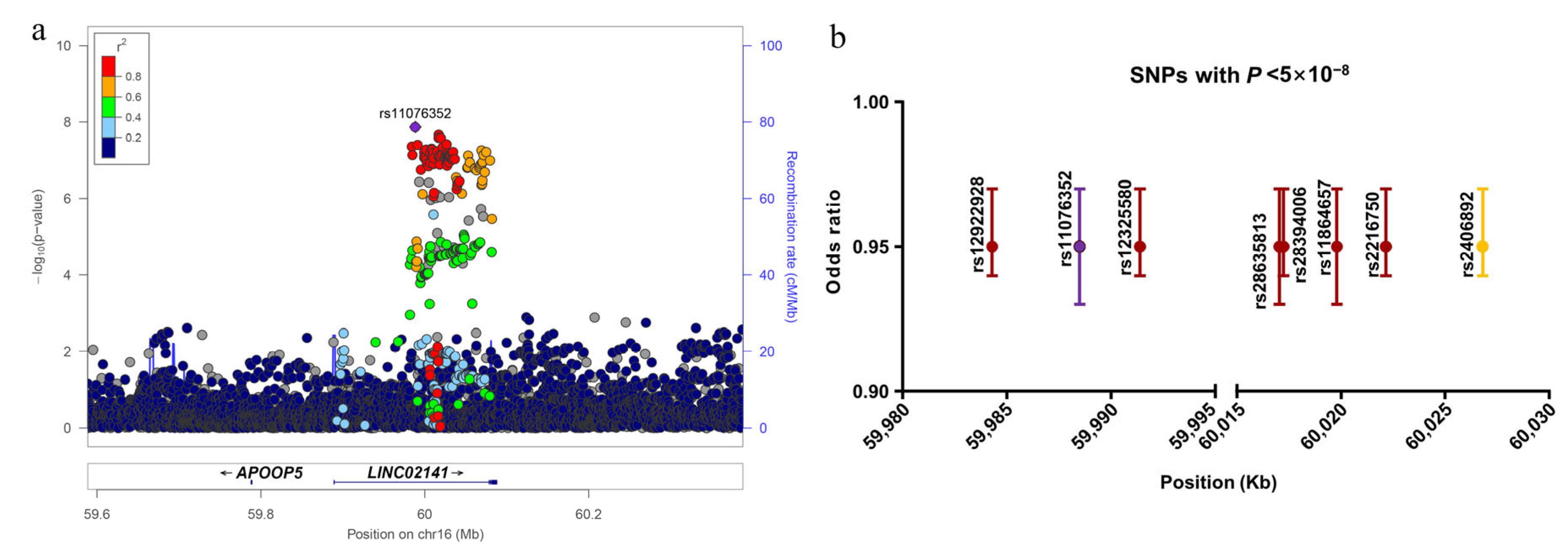
Neither GLIDE nor FinnGen SNPs exhibited any significant evidence of an association with periodontitis (p < 5 × 10⁻⁸, Figure 1a,b). After meta-analysis, eight SNPs mapped to 16q21 physically (rs11076352, rs28635813, rs28394006, rs11864657, rs2406892, rs12325580, rs12922928, and rs2216750) passed the genome-wide significance threshold after adjusting for multiple testing (p < 5 × 10⁻⁸, Figure 1c, Table S2). All of these SNPs were non-coding variants situated in introns of lncRNA LINC02141 and were in high LD (Figure 2a). Figure 2b presents their estimated OR, 95% confidence interval (CI), and physical positions.

3.2. Cross-Tissue Transcriptome-Wide Significant Genes

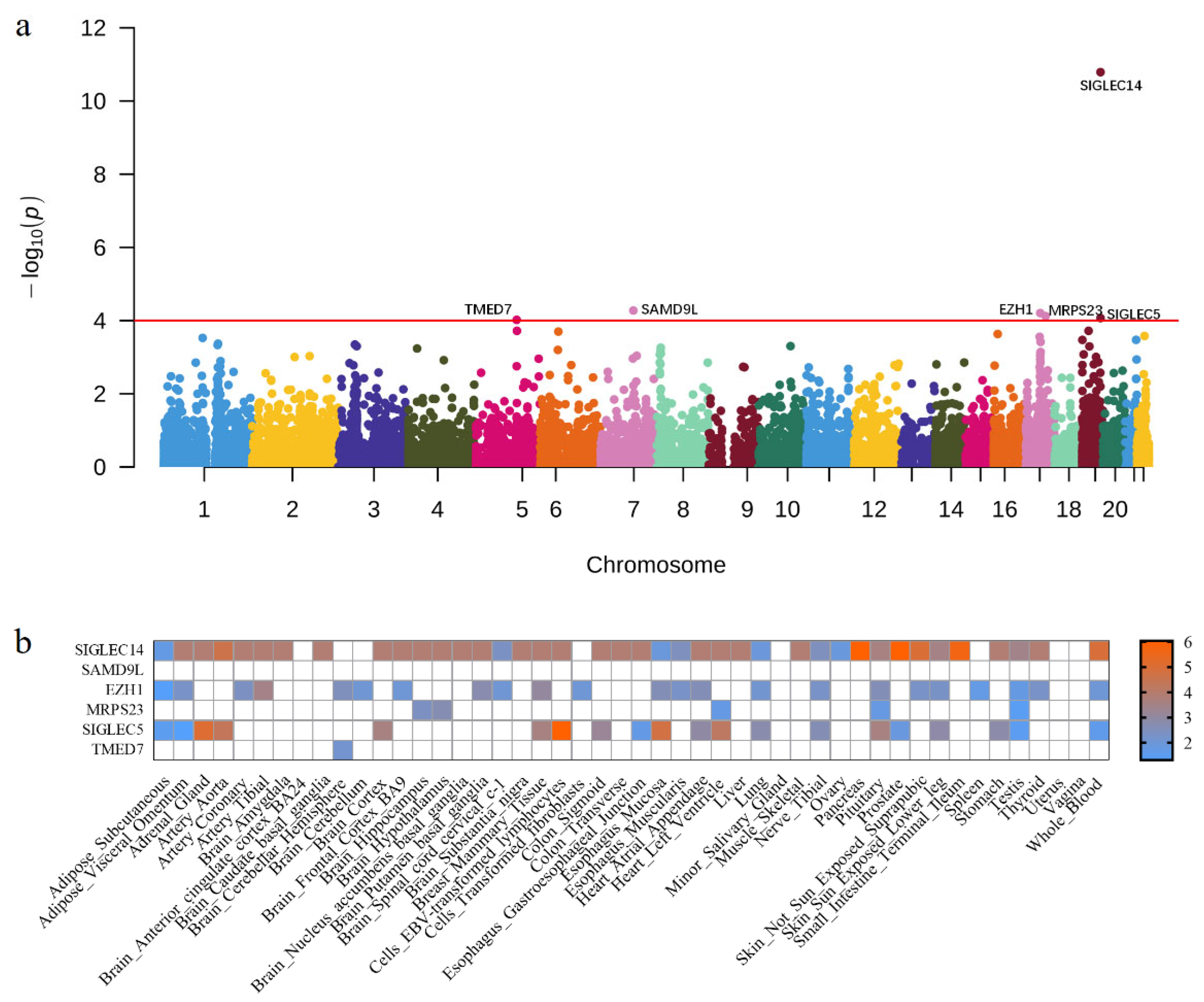
To further explore the associations between gene expression levels and periodontitis, we integrated our large-scale GWAS summary statistics with the gene expression matrix of 44 different tissues from 450 participants from the GTEx project using UTMOST. Since other genes except SIGLEC14 did not pass the Bonferroni correction, we set the threshold as 1 × 10⁻⁴ in the study to minimize false negative errors. Six genes (SIGLEC14, SAMD9L, EZH1, MRPS23, SIGLEC5, and TMED7) reached statistical significance (Figure 3a, Table 1 and Table S4).

3.3. Single-Tissue Transcriptome-Wide Significant Genes

To better characterize the six candidate genes, we separately evaluated their associations in every tissue using FUSION. Among them, four genes (SIGLEC14, EZH1, MRPS23, and SIGLEC5) were significant in at least five tissues, with PFUSION < 0.05 (Figure 3b, Table 1, Tables S3 and S5). Interestingly, EZH1 and MRPS23 were newly identified, whereas previous studies have verified SIGLEC14 and SIGLEC5 as genetic risk factors for periodontitis [37]. However, SAMD9L was not significant in any tissue and TMED7 was only significant in brain cerebellar hemisphere tissue.

3.4. Significant Upregulation of EZH1 in Periodontitis

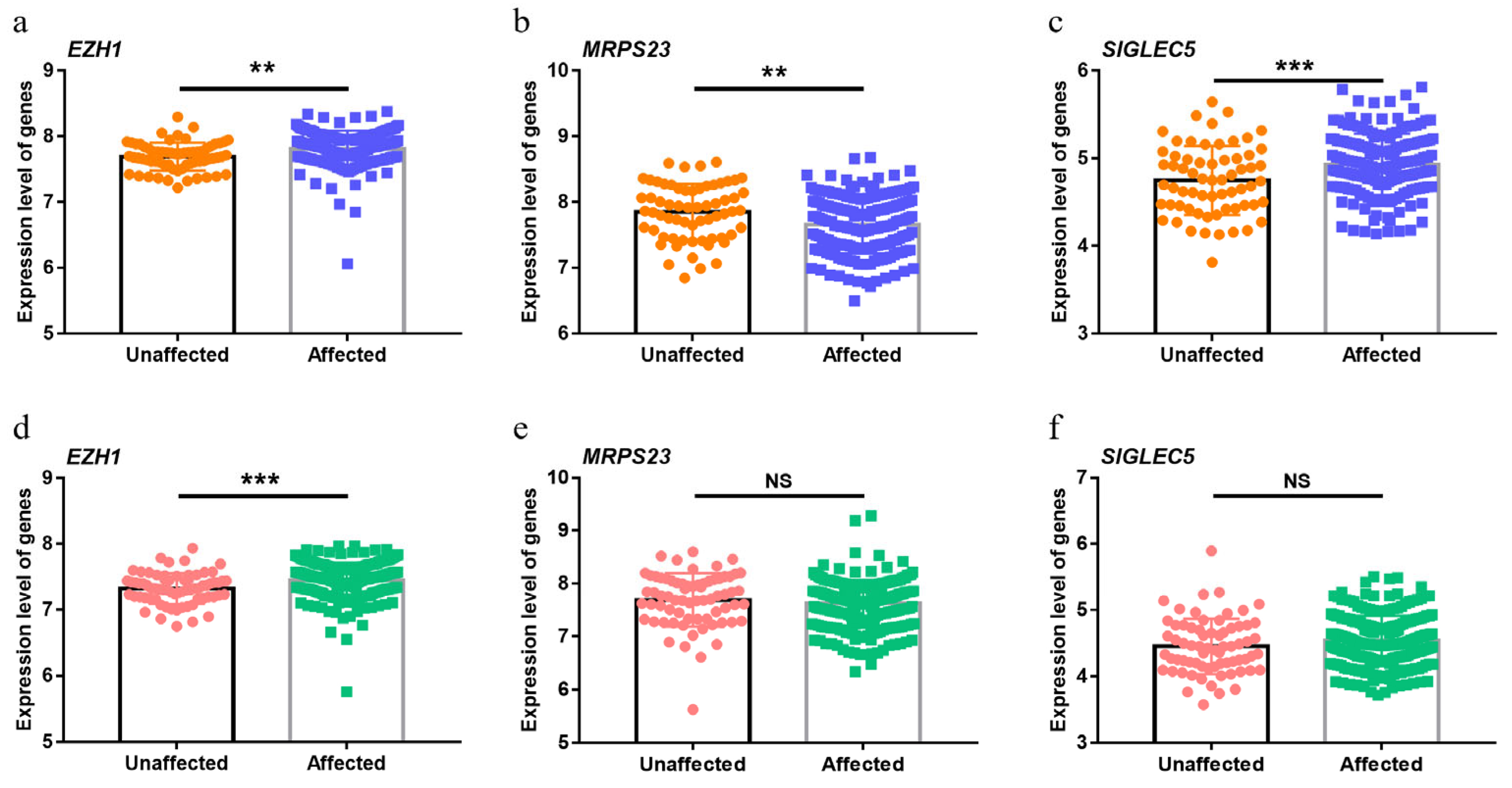
TWAS is widely used to infer disease-related genes based on SNP genotypes. Therefore, if the genes identified by TWAS are relevant, their expression should be dysregulated in periodontitis. Because the whole-genome microarrays did not include SIGLEC14, its differential expression was impossible to evaluate according to GSE10334 and GSE16134. MRPS23 and SIGLEC5 were upregulated in periodontitis-affected sites of gingival tissue in GSE10334 compared with unaffected sites, but there were no differences in GSE16134 (Figure 4b,c,e,f). Notably, the expression of the newly identified gene EZH1 increased in periodontitis sites in both GEO datasets (Figure 4a,d).

3.5. Functional Annotation of EZH1

As the only differentially expressed gene in both GEO datasets, we performed a functional annotation on EZH1. According to the CTD, some diseases such as inflammation and necrosis were associated with EZH1 (Figure 5a). Among them, inflammation was inferred to have the top score of 140.01 through interaction with 41 chemicals including smoke, soot and so on (Figure 5a,b, Table S6). To address the pathogenesis of EZH1 in periodontitis, we also explored the genes co-expressed with EZH1. The expression levels of 4724 genes were significantly correlated with EZH1 after Bonferroni correction using the expression data in GSE16134 owing to its larger sample size. Robust statistical evidence for GO enrichment was observed for cell-substrate junctions (GO: 0030055, Padjust =1.04 × 10⁻¹¹) and focal adhesion (GO: 0005925, Padjust =1.04 × 10⁻¹¹) (Figure 5c, Table S7). The KEGG analysis revealed that genes co-expressed with EZH1 were closely associated with Epstein-Barr virus (EBV) infection (KEGG: hsa05169, Padjust = 9.93 × 10⁻⁷), lysosome (KEGG: hsa04142, Padjust = 9.93 × 10⁻⁷), and protein processing in the endoplasmic reticulum (KEGG: hsa04141, Padjust = 9.93 × 10⁻⁷), all of which are associated with periodontitis (Figure 5d, Table S7).

4. Discussion

In this study, we first meta-analyzed two large GWASs to detect periodontitis-associated SNPs. Eight SNPs within the introns of lncRNA LINC02141 were associated with periodontitis. UTMOST then served as a cross-tissue TWAS method and identified six genes associated with periodontitis, four of which were validated by FUSION. Furthermore, EZH1 was significantly upregulated at periodontitis sites. Genes co-expressed with EZH1 were considerably enriched in biological processes such as cell-substrate junctions and focal adhesion. In summary, we identified eight SNPs associated with periodontitis and highlighted the possible mechanisms of EZH1.
In 16q21, there were eight SNPs within the introns of lncRNA LINC02141 with p < 5 × 10⁻⁸. LncRNAs (e.g., AWPPH and FGD5-AS1) were reported to offer a strong foundation for periodontitis diagnosis and prognosis [38,39,40]. Unfortunately, LINC02141 was not detected by GSE10334 and GSE16134, making it difficult to study its expression level. Motif analyses using HaploReg v4.1 [41] (http://pubs.broadinstitute.org/mammals/haploreg/haploreg.php, accessed on 21 October 2022) revealed that the A allele of rs11076352, the most significant SNP, altered the affinity of GATA (score: −6.9 to 5). GATA has repeatedly been reported as a transcription factor associated with smoking and periodontitis [42,43]. Although associations between the SNPs within LINC02141 and periodontitis were observed, their specific molecular mechanisms need to be elucidated in further studies.
The integration of GWAS and eQTL data has been successfully used to explore gene-trait associations. Li et al. used a Bayesian statistical method called Sherlock to systematically incorporate GWAS and eQTL data for periodontitis from whole blood. They identified ten significant genes, including SIGLEC5 and SIGLEC14 [44]. In our study, UTMOST was applied to develop imputation models for 44 tissues, considering the substantial shared gene regulation across tissues and compensating for the lack of adequate gingival tissue data. To decrease false-positive results, FUSION assisted in the evaluation of the associations in different tissues. Finally, we identified two previously reported genes (SIGLEC5, SIGLEC14) and two novel genes (EZH1, MRPS23), proving that TWAS is a supplemental strategy for studying periodontitis’ etiology.
Further differential expression analysis revealed that EZH1 was consistently upregulated in periodontitis sites among patients with periodontitis. EZH1, also known as an enhancer of the zeste one polycomb repressive complex 2 subunit, is a protein-coding gene. According to the CTD database, inflammation was associated with EZH1 with the highest inference score. Smoke, soot and another 39 chemicals belonged to the inference network. It is well known that periodontitis is an inflammatory disease characterized by the destruction of the supporting tissues around the teeth. Meanwhile, Ezh1 is the most upregulated histone methyltransferase during dendritic cell maturation, silencing of which significantly suppressed Toll-like receptors and triggered production of cytokines, including IL-6, TNF-α, and IFN-β, in dendritic cells (DCs) and macrophages [45]. These cytokines are known as pro-inflammatory cytokines and have pleiotropic effects on lymphocyte promotion as well as tissue damage [46]. For example, IL-6 can participate in alveolar bone resorption and attachment loss supported by the modulation of the Th17 periodontal response [47]. Therefore, EZH1 may participate in the development of periodontitis through inflammation.
Moreover, GO enrichment analysis indicated that genes co-expressed with EZH1 were mainly involved in cell-substrate junctions and focal adhesion. Focal adhesion is also a kind of cell-substrate junction that anchors the cell to the extracellular matrix. When infected with Porphyromonas gingivalis, a major pathogen of periodontitis, both cell-matrix and cell-cell adhesion interfered in the oral keratinocyte cell line, which could compromise the integrity of the epithelia and result in the generation of gingival pockets between the cementum and periodontal epithelium [48]. Thus, genes correlated with EZH1 may play a role in cell and extracellular matrix interactions and participate in the development of periodontitis. The KEGG pathway showed a strong correlation with the EBV infection, lysosomes, and protein processing in the endoplasmic reticulum (ER). EBV plays a crucial role in inflammatory cytokine production and osteoclast differentiation by interacting with oral cells or the macrophage lineage, which initiate and advance periodontitis [49]. Kenichi et al. provided evidence that EBV interacts with some periodontopathic bacteria and is intimately related to the beginning and progressive stages of chronic periodontitis [50]. Lysosomes can fuse with autophagosomes to form autolysosomes during autophagy [51]. Porphyromonas gingivalis in the cytosol is usually degraded by lysosomes, but Porphyromonas gingivalis co-localized with double-membrane vacuoles survives by impairing the formation of autolysosomes [52]. In addition, if cells are infected with pathogenic bacteria, expression of autophagy-related proteins and autophagosomes increases [53]. When periodontal disease develops, autophagy has an impact on immunological function, inflammation, and the homeostasis of alveolar bone [52]. Therefore, lysosomes, as an integral part of autophagy, could play an important role in periodontitis. When protein processing fails in the endoplasmic reticulum, the accumulation of misfolded proteins in the ER causes ER stress. ER stress could affect the development of periodontal diseases [54]. Altogether, the GO and KEGG enrichment analysis results provide additional evidence for the potential role of genes co-expressed with EZH1 in the pathological process of periodontitis.
Although this study identified exciting genes and pathways, there are limitations worth mentioning. Even though a large sample size was included in the GWAS and two TWAS methods were used to reduce false positives, functional research is necessary to better reveal the biological mechanisms of periodontitis. Additionally, a lack of gingival-related cells or tissue is another limitation in our study, despite genetic correlation studies demonstrating similar expression regulation between different tissues [20]. In the future, available eQTL data from the gingiva will be of great benefit to validate our findings.

5. Conclusions

In the present study, several SNPs reached genome-wide significance for periodontitis. Furthermore, TWAS and in silico analyses illustrated that EZH1, genes co-expressed with which were significantly enriched in cell-substrate junction and other important pathways, may contribute to the risk of periodontitis. Further research is required to corroborate our findings and to dissect the genetic architecture of periodontitis.

Supplementary Materials

The following supporting information can be downloaded at: https://www.mdpi.com/article/10.3390/cells12010070/s1, Table S1: Characteristics of the periodontitis cases and controls; Table S2: Significant SNPs of periodontitis in the GWAS; Table S3: The significant results of FUSION for candidate susceptibility genes; Table S4: The results of UTMOST for periodontitis; Table S5: The results of FUSION for candidate susceptibility genes; Table S6: Top 10 diseases associated with the EZH1 gene according to CTD; Table S7: Top 10 GO and KEGG terms identified by enrichment analysis for genes co-expressed with EZH1; Figure S1: The flow chart of this study; Figure S2: Quantile-quantile plot of −log10 (p-values) from all autosomal SNPs.

Author Contributions

Conceptualization, L.W. and Y.P.; Methodology, G.Z. and L.F.; Software, G.Z.; Validation, X.C.; Formal analysis, G.Z.; Investigation, G.Z.; Resources, X.C.; Data curation, X.C.; Writing—original draft preparation, G.Z.; Writing—review and editing, L.W., Y.P., L.F.; Visualization, X.C.; Supervision, L.W.; Project administration, L.W.; Funding acquisition, L.W. and Y.P. All authors have read and agreed to the published version of the manuscript.

Funding

This research was funded by the National Natural Science Foundation of China, grant numbers 82270496, 81830031, 81970969, 82001088, and 82101054; and the Priority Academic Program Development of Jiangsu Higher Education Institutions, grant number PAPD-2018-87.

Institutional Review Board Statement

Not applicable.

Informed Consent Statement

Not applicable.

Data Availability Statement

Genome-wide summary statistics of the GLIDE consortium are available at: https://data.bris.ac.uk/data/dataset/2j2rqgzedxlq02oqbb4vmycnc2. Genome-wide summary statistics of FINNGEN are available at: https://storage.googleapis.com/finngen-public-data-r7/summary_stats/finngen_R7_K11_PERIODON_CHRON_INCLAVO.gz.

Acknowledgments

The authors acknowledge and thank the GWAS consortia of the original GWAS studies for sharing the summary data used in this study. In addition, we especially wish to thank Catherine Huihan Chu for providing English writing assistance.

Conflicts of Interest

The authors declare no conflict of interest.

References

  1. Shimizu, S.; Momozawa, Y.; Takahashi, A.; Nagasawa, T.; Ashikawa, K.; Terada, Y.; Izumi, Y.; Kobayashi, H.; Tsuji, M.; Kubo, M.; et al. A genome-wide association study of periodontitis in a Japanese population. J. Dent. Res. 2015, 94, 555–561. [Google Scholar] [CrossRef]
  2. Tonetti, M.S.; Greenwell, H.; Kornman, K.S. Staging and grading of periodontitis: Framework and proposal of a new classification and case definition. J. Clin. Periodontol. 2018, 45 (Suppl. 20), S149–S161. [Google Scholar] [CrossRef] [PubMed] [Green Version]
  3. Papapanou, P.N.; Sanz, M.; Buduneli, N.; Dietrich, T.; Feres, M.; Fine, D.H.; Flemmig, T.F.; Garcia, R.; Giannobile, W.V.; Graziani, F.; et al. Periodontitis: Consensus report of workgroup 2 of the 2017 World Workshop on the Classification of Periodontal and Peri-Implant Diseases and Conditions. J. Periodontol. 2018, 89 (Suppl. 1), S173–S182. [Google Scholar] [CrossRef] [PubMed] [Green Version]
  4. Kwon, T.; Lamster, I.B.; Levin, L. Current Concepts in the Management of Periodontitis. Int. Dent. J. 2021, 71, 462–476. [Google Scholar] [CrossRef] [PubMed]
  5. Gera, I.; Vari, M. Genetic background of periodontitis. Part II. Genetic polymorphism in periodontal disease. A review of literature. Fogorv. Szle. 2009, 102, 131–140. [Google Scholar]
  6. Michalowicz, B.S.; Diehl, S.R.; Gunsolley, J.C.; Sparks, B.S.; Brooks, C.N.; Koertge, T.E.; Califano, J.V.; Burmeister, J.A.; Schenkein, H.A. Evidence of a substantial genetic basis for risk of adult periodontitis. J. Periodontol. 2000, 71, 1699–1707. [Google Scholar] [CrossRef]
  7. Nibali, L.; Bayliss-Chapman, J.; Almofareh, S.A.; Zhou, Y.; Divaris, K.; Vieira, A.R. What Is the Heritability of Periodontitis? A Systematic Review. J. Dent. Res. 2019, 98, 632–641. [Google Scholar] [CrossRef]
  8. Loos, B.G.; Van Dyke, T.E. The role of inflammation and genetics in periodontal disease. Periodontol. 2000 2020, 83, 26–39. [Google Scholar] [CrossRef]
  9. Brodzikowska, A.; Gorski, B. Polymorphisms in Genes Involved in Inflammation and Periodontitis: A Narrative Review. Biomolecules 2022, 12, 552. [Google Scholar] [CrossRef]
  10. Ryder, M.I.; Couch, E.T.; Chaffee, B.W. Personalized periodontal treatment for the tobacco- and alcohol-using patient. Periodontol. 2000 2018, 78, 30–46. [Google Scholar] [CrossRef]
  11. Bartold, P.M. Lifestyle and periodontitis: The emergence of personalized periodontics. Periodontol. 2000 2018, 78, 7–11. [Google Scholar] [CrossRef] [PubMed]
  12. Kinane, D.F.; Stathopoulou, P.G.; Papapanou, P.N. Periodontal diseases. Nat. Rev. Dis. Primers 2017, 3, 17038. [Google Scholar] [CrossRef] [PubMed]
  13. Macarthur, J.; Bowler, E.; Cerezo, M.; Gil, L.; Hall, P.; Hastings, E.; Junkins, H.; Mcmahon, A.; Milano, A.; Morales, J.; et al. The new NHGRI-EBI Catalog of published genome-wide association studies (GWAS Catalog). Nucleic Acids Res. 2017, 45, D896–D901. [Google Scholar] [CrossRef] [PubMed]
  14. Schaefer, A.S.; Richter, G.M.; Nothnagel, M.; Manke, T.; Dommisch, H.; Jacobs, G.; Arlt, A.; Rosenstiel, P.; Noack, B.; Groessner-Schreiber, B.; et al. A genome-wide association study identifies GLT6D1 as a susceptibility locus for periodontitis. Hum. Mol. Genet. 2010, 19, 553–562. [Google Scholar] [CrossRef] [PubMed]
  15. Bevilacqua, L.; Navarra, C.O.; Pirastu, N.; Lenarda, R.D.; Gasparini, P.; Robino, A. A genome-wide association study identifies an association between variants in EFCAB4B gene and periodontal disease in an Italian isolated population. J. Periodontal Res. 2018, 53, 992–998. [Google Scholar] [CrossRef] [PubMed] [Green Version]
  16. Munz, M.; Richter, G.M.; Loos, B.G.; Jepsen, S.; Divaris, K.; Offenbacher, S.; Teumer, A.; Holtfreter, B.; Kocher, T.; Bruckmann, C.; et al. Meta-analysis of genome-wide association studies of aggressive and chronic periodontitis identifies two novel risk loci. Eur. J. Hum. Genet. 2019, 27, 102–113. [Google Scholar] [CrossRef] [Green Version]
  17. Munz, M.; Willenborg, C.; Richter, G.M.; Jockel-Schneider, Y.; Graetz, C.; Staufenbiel, I.; Wellmann, J.; Berger, K.; Krone, B.; Hoffmann, P.; et al. A genome-wide association study identifies nucleotide variants at SIGLEC5 and DEFA1A3 as risk loci for periodontitis. Hum. Mol. Genet. 2017, 26, 2577–2588. [Google Scholar] [CrossRef] [Green Version]
  18. Tam, V.; Patel, N.; Turcotte, M.; Bosse, Y.; Pare, G.; Meyre, D. Benefits and limitations of genome-wide association studies. Nat. Rev. Genet. 2019, 20, 467–484. [Google Scholar] [CrossRef]
  19. Gallagher, M.D.; Chen-Plotkin, A.S. The Post-GWAS Era: From Association to Function. Am. J. Hum. Genet. 2018, 102, 717–730. [Google Scholar] [CrossRef] [Green Version]
  20. Hu, Y.; Li, M.; Lu, Q.; Weng, H.; Wang, J.; Zekavat, S.M.; Yu, Z.; Li, B.; Gu, J.; Muchnik, S.; et al. A statistical framework for cross-tissue transcriptome-wide association analysis. Nat. Genet. 2019, 51, 568–576. [Google Scholar] [CrossRef]
  21. Carithers, L.J.; Moore, H.M. The Genotype-Tissue Expression (GTEx) Project. Biopreserv. Biobank. 2015, 13, 307–308. [Google Scholar] [CrossRef] [PubMed]
  22. Zhu, Z.; Zhang, F.; Hu, H.; Bakshi, A.; Robinson, M.R.; Powell, J.E.; Montgomery, G.W.; Goddard, M.E.; Wray, N.R.; Visscher, P.M.; et al. Integration of summary data from GWAS and eQTL studies predicts complex trait gene targets. Nat. Genet. 2016, 48, 481–487. [Google Scholar] [CrossRef] [PubMed]
  23. Barbeira, A.N.; Dickinson, S.P.; Bonazzola, R.; Zheng, J.; Wheeler, H.E.; Torres, J.M.; Torstenson, E.S.; Shah, K.P.; Garcia, T.; Edwards, T.L.; et al. Exploring the phenotypic consequences of tissue specific gene expression variation inferred from GWAS summary statistics. Nat. Commun. 2018, 9, 1825. [Google Scholar] [CrossRef] [PubMed]
  24. Gamazon, E.R.; Wheeler, H.E.; Shah, K.P.; Mozaffari, S.V.; Aquino-Michaels, K.; Carroll, R.J.; Eyler, A.E.; Denny, J.C.; Nicolae, D.L.; Cox, N.J.; et al. A gene-based association method for mapping traits using reference transcriptome data. Nat. Genet. 2015, 47, 1091–1098. [Google Scholar] [CrossRef] [Green Version]
  25. Gusev, A.; Ko, A.; Shi, H.; Bhatia, G.; Chung, W.; Penninx, B.W.; Jansen, R.; de Geus, E.J.; Boomsma, D.I.; Wright, F.A.; et al. Integrative approaches for large-scale transcriptome-wide association studies. Nat. Genet. 2016, 48, 245–252. [Google Scholar] [CrossRef] [Green Version]
  26. Battle, A.; Brown, C.D.; Engelhardt, B.E.; Montgomery, S.B. Genetic effects on gene expression across human tissues. Nature 2017, 550, 204–213. [Google Scholar] [CrossRef] [Green Version]
  27. Rodriguez-Fontenla, C.; Carracedo, A. UTMOST, a single and cross-tissue TWAS (Transcriptome Wide Association Study), reveals new ASD (Autism Spectrum Disorder) associated genes. Transl. Psychiatry 2021, 11, 256. [Google Scholar] [CrossRef]
  28. Zhu, M.; Fan, J.; Zhang, C.; Xu, J.; Yin, R.; Zhang, E.; Wang, Y.; Ji, M.; Sun, Q.; Dai, J.; et al. A cross-tissue transcriptome-wide association study identifies novel susceptibility genes for lung cancer in Chinese populations. Hum. Mol. Genet. 2021, 30, 1666–1676. [Google Scholar] [CrossRef]
  29. Shungin, D.; Haworth, S.; Divaris, K.; Agler, C.S.; Kamatani, Y.; Keun, L.M.; Grinde, K.; Hindy, G.; Alaraudanjoki, V.; Pesonen, P.; et al. Genome-wide analysis of dental caries and periodontitis combining clinical and self-reported data. Nat. Commun. 2019, 10, 2773. [Google Scholar] [CrossRef] [Green Version]
  30. Kurki, M.I.; Karjalainen, J.; Palta, P.; Donner, K. FinnGen: Unique genetic insights from combining isolated population and national health register data. medRxiv 2022. [Google Scholar] [CrossRef]
  31. Willer, C.J.; Li, Y.; Abecasis, G.R. METAL: Fast and efficient meta-analysis of genomewide association scans. Bioinformatics 2010, 26, 2190–2191. [Google Scholar] [CrossRef] [PubMed] [Green Version]
  32. Pruim, R.J.; Welch, R.P.; Sanna, S.; Teslovich, T.M.; Chines, P.S.; Gliedt, T.P.; Boehnke, M.; Abecasis, G.R.; Willer, C.J. LocusZoom: Regional visualization of genome-wide association scan results. Bioinformatics 2010, 26, 2336–2337. [Google Scholar] [CrossRef] [PubMed] [Green Version]
  33. Demmer, R.T.; Behle, J.H.; Wolf, D.L.; Handfield, M.; Kebschull, M.; Celenti, R.; Pavlidis, P.; Papapanou, P.N. Transcriptomes in healthy and diseased gingival tissues. J. Periodontol. 2008, 79, 2112–2124. [Google Scholar] [CrossRef] [PubMed]
  34. Kebschull, M.; Demmer, R.T.; Grun, B.; Guarnieri, P.; Pavlidis, P.; Papapanou, P.N. Gingival tissue transcriptomes identify distinct periodontitis phenotypes. J. Dent. Res. 2014, 93, 459–468. [Google Scholar] [CrossRef] [PubMed] [Green Version]
  35. Davis, A.P.; King, B.L.; Mockus, S.; Murphy, C.G.; Saraceni-Richards, C.; Rosenstein, M.; Wiegers, T.; Mattingly, C.J. The Comparative Toxicogenomics Database: Update 2011. Nucleic Acids Res. 2011, 39, D1067–D1072. [Google Scholar] [CrossRef] [PubMed] [Green Version]
  36. Wu, T.; Hu, E.; Xu, S.; Chen, M.; Guo, P.; Dai, Z.; Feng, T.; Zhou, L.; Tang, W.; Zhan, L.; et al. clusterProfiler 4.0: A universal enrichment tool for interpreting omics data. Innovation (Camb) 2021, 2, 100141. [Google Scholar] [CrossRef]
  37. Masumoto, R.; Kitagaki, J.; Fujihara, C.; Matsumoto, M.; Miyauchi, S.; Asano, Y.; Imai, A.; Kobayashi, K.; Nakaya, A.; Yamashita, M.; et al. Identification of genetic risk factors of aggressive periodontitis using genomewide association studies in association with those of chronic periodontitis. J. Periodontal Res. 2019, 54, 199–206. [Google Scholar] [CrossRef]
  38. Wang, X.; Ma, F.; Jia, P. LncRNA AWPPH overexpression predicts the recurrence of periodontitis. Biosci. Rep. 2019, 39, BSR20190636. [Google Scholar] [CrossRef] [Green Version]
  39. Yu, M.; Chi, C. lncRNA FGD5-AS1 and miR-130a Can Be Used for Prognosis Analysis of Patients with Chronic Periodontitis. Biomed Res. Int. 2021, 2021, 8544914. [Google Scholar] [CrossRef]
  40. Zhang, X.; Ren, L.; Yan, X.; Shan, Y.; Liu, L.; Zhou, J.; Kuang, Q.; Li, M.; Long, H.; Lai, W. Identification of immune-related lncRNAs in periodontitis reveals regulation network of gene-lncRNA-pathway-immunocyte. Int. Immunopharmacol. 2020, 84, 106600. [Google Scholar] [CrossRef]
  41. Ward, L.D.; Kellis, M. HaploReg v4: Systematic mining of putative causal variants, cell types, regulators and target genes for human complex traits and disease. Nucleic Acids Res. 2016, 44, D877–D881. [Google Scholar] [CrossRef] [PubMed]
  42. Goncalves, R.B.; Coletta, R.D.; Silverio, K.G.; Benevides, L.; Casati, M.Z.; Da, S.J.; Nociti, F.J. Impact of smoking on inflammation: Overview of molecular mechanisms. Inflamm. Res. 2011, 60, 409–424. [Google Scholar] [CrossRef]
  43. Walther, K.A.; Gonzales, J.R.; Groger, S.; Ehmke, B.; Kaner, D.; Lorenz, K.; Eickholz, P.; Kocher, T.; Kim, T.S.; Schlagenhauf, U.; et al. The Role of Polymorphisms at the Interleukin-1, Interleukin-4, GATA-3 and Cyclooxygenase-2 Genes in Non-Surgical Periodontal Therapy. Int. J. Mol. Sci. 2022, 23, 7266. [Google Scholar] [CrossRef]
  44. Li, W.; Zheng, Q.; Meng, H.; Chen, D. Integration of genome-wide association study and expression quantitative trait loci data identifies AIM2 as a risk gene of periodontitis. J. Clin. Periodontol. 2020, 47, 583–593. [Google Scholar] [CrossRef]
  45. Liu, Y.; Zhang, Q.; Ding, Y.; Li, X.; Zhao, D.; Zhao, K.; Guo, Z.; Cao, X. Histone lysine methyltransferase Ezh1 promotes TLR-triggered inflammatory cytokine production by suppressing Tollip. J. Immunol. 2015, 194, 2838–2846. [Google Scholar] [CrossRef] [PubMed] [Green Version]
  46. Pan, W.; Wang, Q.; Chen, Q. The cytokine network involved in the host immune response to periodontitis. Int. J. Oral Sci. 2019, 11, 30. [Google Scholar] [CrossRef] [Green Version]
  47. Apolinario, V.G.; Aparecida, R.A.; Figueiredo, C.K.; Ferreira, O.L.; Tanaka, S.K.; Reis, M.M.; Sprone, R.M.; Goncalves, D.A.A.; Taba, M.J. Specific inhibition of IL-6 receptor attenuates inflammatory bone loss in experimental periodontitis. J. Periodontol. 2021, 92, 1460–1469. [Google Scholar] [CrossRef]
  48. Hintermann, E.; Haake, S.K.; Christen, U.; Sharabi, A.; Quaranta, V. Discrete proteolysis of focal contact and adherens junction components in Porphyromonas gingivalis-infected oral keratinocytes: A strategy for cell adhesion and migration disabling. Infect. Immun. 2002, 70, 5846–5856. [Google Scholar] [CrossRef] [Green Version]
  49. Yokoe, S.; Hasuike, A.; Watanabe, N.; Tanaka, H.; Karahashi, H.; Wakuda, S.; Takeichi, O.; Kawato, T.; Takai, H.; Ogata, Y.; et al. Epstein-Barr Virus Promotes the Production of Inflammatory Cytokines in Gingival Fibroblasts and RANKL-Induced Osteoclast Differentiation in RAW264.7 Cells. Int. J. Mol. Sci. 2022, 23, 809. [Google Scholar] [CrossRef]
  50. Imai, K.; Ogata, Y. How Does Epstein-Barr Virus Contribute to Chronic Periodontitis? Int. J. Mol. Sci. 2020, 21, 1940. [Google Scholar] [CrossRef] [PubMed] [Green Version]
  51. Mcphee, C.K.; Baehrecke, E.H. Autophagy shows its animal side. Cell 2010, 141, 922–924. [Google Scholar] [CrossRef]
  52. Yang, Y.; Huang, Y.; Li, W. Autophagy and its significance in periodontal disease. J. Periodontal Res. 2021, 56, 18–26. [Google Scholar] [CrossRef]
  53. Jiang, M.; Li, Z.; Zhu, G. The role of autophagy in the pathogenesis of periodontal disease. Oral Dis. 2020, 26, 259–269. [Google Scholar] [CrossRef]
  54. Jiang, M.; Li, Z.; Zhu, G. The role of endoplasmic reticulum stress in the pathophysiology of periodontal disease. J. Periodontal Res. 2022, 57, 915–932. [Google Scholar] [CrossRef]
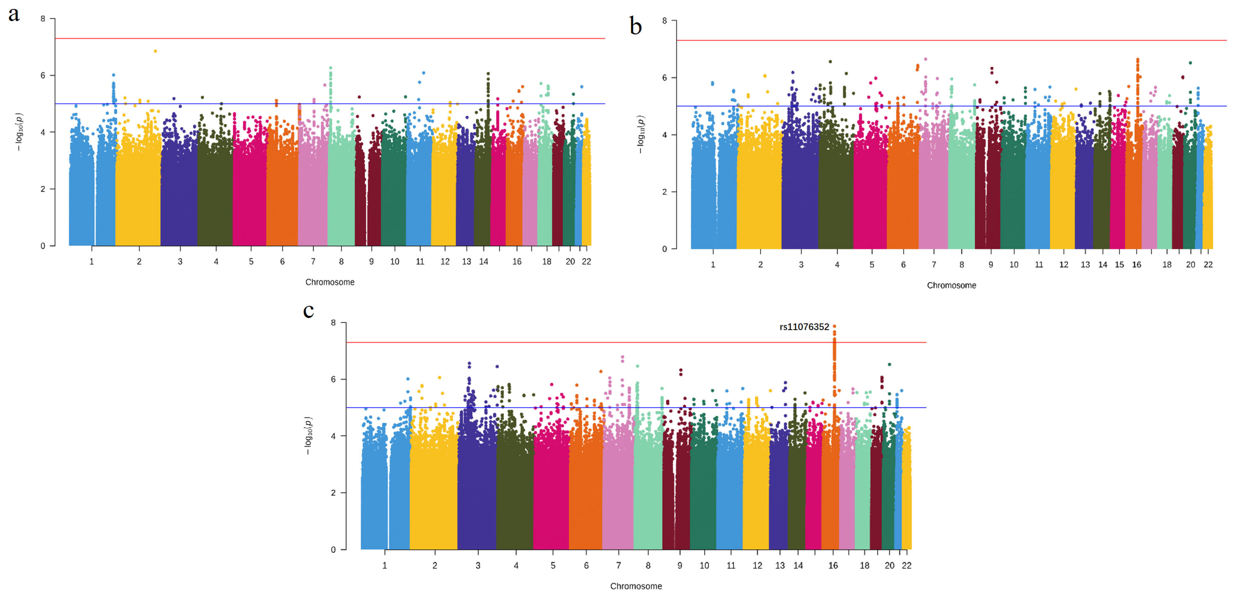
Figure 1. Manhattan plot of −log10 (p-values) of the GWAS results. (a) Summary statistics from the GLIDE consortium. (b) Summary statistics from FinnGen. (c) Meta-analysis results. The red and blue horizontal lines indicate the level of genome-wide significance (p = 5.0 × 10⁻⁸) and nominal significance (p = 1.0 × 10⁻⁵), respectively. GWAS, genome-wide association study.
Cells 12 00070 g001
Figure 2. Regional plots and effective size of significant SNPs. (a) Regional plots of association results and recombination rates for the genome-wide significant SNP rs11076352. (b) OR and 95% CI under an additive model for SNPs with p < 5 × 10 −8. SNPs, single nucleotide polymorphisms; OR, odds ratio; 95% CI, 95% confidence interval.
Cells 12 00070 g002
Figure 3. Transcriptome-wide association study results for periodontitis. (a) Manhattan plot of cross-tissue TWAS using UTMOST. The y-axis represents the p-value in −log10 scale. Genes with p-value < 1 × 10⁻⁴ are labeled. (b) The heatmaps of the six genes are segregated by tissues. The statistics represent the p-value of FUSION on a −log10 scale. The white lattice represents PFUSION > 0.05 or unavailable statistics. TWAS, transcriptome-wide association study.
Cells 12 00070 g003
Figure 4. Dysregulation of top causal genes in periodontitis. Differentially expressed genes in the affected and unaffected gingival tissue sites among periodontitis patients. Data from GSE10334 (ac) and GSE16134 (df). EZH1 was significantly upregulated in the periodontal ligament in periodontitis sites compared with unaffected sites. NS, p > 0.05; ** p < 0.01; *** p < 0.001.
Cells 12 00070 g004
Figure 5. Functional annotation of EZH1. (a) Disease categories associated with EZH1 gene in the CTD database with inference scores. (b) A total of 41 chemicals interacted with EZH1 and correlated with inflammation. Circle diagrams of GO (c) and KEGG (d) enriched pathways for genes co-expressed with EZH1 from GSE16134. CTD, the Comparative Toxicogenomics Database; GO, Gene Ontology; KEGG, Kyoto Encyclopedia of Genes and Genomes.
Cells 12 00070 g005
Table 1. Significant genes in the TWAS analysis of periodontitis.
GeneChrBPPUTMOSTPFUSIONnReported
SIGLEC1419521458061.63 × 10−119.34 × 10−740Yes
SAMD9L7927593685.32 × 10−5NA0No
EZH117408522936.24 × 10−53.83 × 10−323No
MRPS2317559162877.55 × 10−52.14 × 10⁻³⁵No
SIGLEC519521147568.54 × 10−59.34 × 10−720Yes
TMED751149489059.56 × 10−57.53 × 10⁻³¹No
TWAS, transcriptome-wide association study; Chr, chromosome; BP, start base gene position; PFUSION, the most significant p-value in 44 tissues; n, the number of tissues with PFUSION < 0.05; NA, p > 0.05 in every tissue; Reported, whether reported to be associated with periodontitis before; Bold, significant genes after UTMOST and FUSION analysis.
Disclaimer/Publisher’s Note: The statements, opinions and data contained in all publications are solely those of the individual author(s) and contributor(s) and not of MDPI and/or the editor(s). MDPI and/or the editor(s) disclaim responsibility for any injury to people or property resulting from any ideas, methods, instructions or products referred to in the content.