Article

DNA-Binding Activity of CAMTA3 Is Essential for Its Function: Identification of Critical Amino Acids for Its Transcriptional Activity

by
Kasavajhala V. S. K. Prasad
1,*,†,
Amira A. E. Abdel-Hameed
1,2,†,
Qiyan Jiang
1,3 and
Anireddy S. N. Reddy
1,*
1
Department of Biology and Program in Cell and Molecular Biology, Colorado State University, Fort Collins, CO 80523, USA
2
Department of Botany and Microbiology, Faculty of Science, Zagazig University, Zagazig 44519, Egypt
3
National Key Facility for Crop Gene Resources and Genetic Improvement, Institute of Crop Sciences, Chinese Academy of Agricultural Sciences, Beijing 100081, China
*
Authors to whom correspondence should be addressed.
These authors contributed equally to this work.
Cells 2023, 12(15), 1986; https://doi.org/10.3390/cells12151986
Submission received: 27 June 2023 / Revised: 22 July 2023 / Accepted: 26 July 2023 / Published: 2 August 2023
(This article belongs to the Section Plant, Algae and Fungi Cell Biology)

Abstract

:
Calmodulin-binding transcription activators (CAMTAs), a small family of highly conserved transcription factors, function in calcium-mediated signaling pathways. Of the six CAMTAs in Arabidopsis, CAMTA3 regulates diverse biotic and abiotic stress responses. A recent study has shown that CAMTA3 is a guardee of NLRs (Nucleotide-binding, Leucine-rich repeat Receptors) in modulating plant immunity, raising the possibility that CAMTA3 transcriptional activity is dispensable for its function. Here, we show that the DNA-binding activity of CAMTA3 is essential for its role in mediating plant immune responses. Analysis of the DNA-binding (CG-1) domain of CAMTAs in plants and animals showed strong conservation of several amino acids. We mutated six conserved amino acids in the CG-1 domain to investigate their role in CAMTA3 function. Electrophoretic mobility shift assays using these mutants with a promoter of its target gene identified critical amino acid residues necessary for DNA-binding activity. In addition, transient assays showed that these residues are essential for the CAMTA3 function in activating the Rapid Stress Response Element (RSRE)-driven reporter gene expression. In line with this, transgenic lines expressing the CG-1 mutants of CAMTA3 in the camta3 mutant failed to rescue the mutant phenotype and restore the expression of CAMTA3 downstream target genes. Collectively, our results provide biochemical and genetic evidence that the transcriptional activity of CAMTA3 is indispensable for its function.

1. Introduction

Plants in their environment are continuously subjected to biotic and abiotic stresses, which limit plant growth and crop yield [1,2]. Therefore, plants have evolved various physiological and biochemical mechanisms for stress adaptation that rely mainly on changes in gene expression patterns [3,4]. In this process, transcription factors (TFs) represent the master switches that target stress-responsive genes and regulate their expression [5]. The calmodulin-binding transcription activators (CAMTAs) family is one of the well-characterized calmodulin (CaM)-binding TFs that work downstream of the calcium signaling pathway to elicit plant responses to various biotic and abiotic stresses [6,7]. CAMTAs have been identified in plants, humans, Drosophila as well as worms and are evolutionarily conserved from plants to humans [8,9,10,11,12,13]. A typical CAMTA protein has various ordered functional domains with amino acid sequence stretches that are evolutionarily conserved [8,14,15]. These domains include a DNA-binding domain (CG-1), a non-specific DNA-binding domain, a Transcription-associated Immuno Globulin domain (TIG), ankyrin repeats (ANK) for protein–protein interactions, followed by five tandem repeats of a Ca2+-independent CaM-binding domain (IQ), and a Ca2+-dependent CaM-binding domain (CaMBD).
In Arabidopsis thaliana (referred to as Arabidopsis henceforth), there are six CAMTAs (AtCAMTA1 to AtCAMTA6, referred to as CAMTA1 to CAMTA6 henceforth), which are differentially expressed in response to multiple stresses [6,10,16]. Among these, CAMTA3 (also known as signal-responsive1, SR1), is the most studied member and regulates diverse biotic and abiotic stress responses, as well as thigmomorphogenesis [8,17,18,19,20,21,22]. Several transcriptomic studies comparing WT and camta3 mutants revealed a dual regulatory role for CAMTA3 in multiple cellular pathways [23,24,25]. Mutants of camta3 exhibit up-regulation of plant immune system, salicylic acid (SA) biosynthesis, and salt stress-responsive genes [17,23,25], while genes involved in insect tolerance and low temperature stress response showed down-regulation [25,26]. In line with this, camta3 mutants exhibited autoimmune phenotypes including stunted growth, chlorosis and leaf lesions, SA accumulation, constitutive expression of defense genes and enhanced resistance against bacterial and fungal pathogens [14,17,23,27]. Our studies have shown that CAMTA3 functions as a negative regulator for salt tolerance in Arabidopsis [25]. On the other hand, CAMTA3 acts as a positive regulator for cold and drought stress response as well as defense against insect herbivory and wound-induced response [20,28,29]. CAMTA3 works in concert with CAMTA1, CAMTA2 and CAMTA5 to increase plant freezing tolerance by inducing the expression of the CRT/DRE-Binding Factor (CBF) genes and other cold-induced genes [24,26,30]. The camta3 mutants were also more susceptible to herbivore attack and showed decreased levels of glucosinolates, as compared to the wild-type (WT) plants (Laluk et al., 2012). Moreover, CAMTA3 has been recently reported to be a positive regulator for the suppression of plant viral infection by activating the RNAi defense machinery [22]. Therefore, CAMTA3 functions both as a transcriptional repressor for defense- and salt stress-related genes as well as a transcriptional activator for genes involved in cold, drought, glucosinolate metabolism, herbivory, wound-induced and general stress-mediated responses [25,26,28,29,31].
The CAMTA proteins have a CG-1 domain, which binds to CGCG or CGTG core motifs with the CGCG-core consensus motif (A/C)CGCG(C/G/T), whereas the CGTG-core consensus motif is (A/C)CGTGT [10,17,23,26,32]. Furthermore, CAMTA3 has been shown to act as a transcriptional activator of early general stress response genes by binding to the Rapid Stress Response Element (RSRE) [33], which is CGCGTT motif [31,34]. Previous studies have shown that CAMTA3 regulates plant immunity by modulating the expression of the defense genes EDS1, NDR1, EIN3 and NPRI by binding to the CGCG elements in their promoters [17,19,27]. It is thought that CAMTA3 regulates plant immunity by regulating the expression of defense genes. However, Lolle et al. [35] have shown that the enhanced defense phenotype observed in camta3 is primarily because of the activation of the NLRs (Nucleotide-binding, Leucine-rich Repeat Receptors) - DOMINANT SUPPRESSOR OF camta3 NUMBER 1 (DSC1) and DOMINANT SUPPRESSOR OF camta3 NUMBER (2 DSC2), rather than the loss of the transcriptional activity of CAMTA3 as a negative regulator of gene expression. In a genetic screen, they identified two dominant negative NLRs, DSC1-DN and DSC2-DN, that suppressed the autoimmune phenotype of the camta3 mutant [35]. The expression of DSC1-DN or DSC2-DN in camta3 mutants suppressed the autoimmune phenotype, restored the expression of the defense genes (PR1 and EDS1) to WT levels and repressed plant resistance to fungal and bacterial pathogens [35]. Furthermore, the expression of DSC1 or DSC2 in Nicotiana benthamiana triggered the HR cell death response, but co-expression of CAMTA3 prevented this [35]. Furthermore, BiFC and FRET assays showed CAMTA3 and DSCs to be a part of a nuclear-localized complex [35]. Together these results led to the suggestion that DSC1/DSC2 and CAMTA3 form a guard–guardee complex, similar to that observed with RESISTANCE TO P. SYRINGAE PV MACULICOLA 1 (RPM1) and RESISTANT TO P. SYRINGAE 2 (RPS2) NLRs and RPM1-INTERACTING PROTEIN 4 (RIN4) [36]. These results, further suggest that modification of CAMTA3 by effectors or loss of CAMTA3 triggers the corresponding NLR guards to activate plant defense genes [35]. Here, we tested if the DNA-binding and transcriptional activity of CAMTA3 is necessary for its function in plant immunity. We generated point mutations in the conserved amino acids of the CAMTA3 CG-1 domain, evaluated the DNA-binding and transcriptional activity of the mutated versions, and tested their ability to complement the camta3 mutant phenotype. Our results show that the CAMTA3 DNA-binding activity of the CG-1 and its modulation of transcription is essential for its function in plant immunity. Furthermore, our study identified six amino acids in the CG-1 domain that are critical for its DNA-binding activity.

2. Materials and Methods

2.1. Identification and Selection of the Conserved Amino Acids in the CG-1 Domain for Mutational Studies

The genomic and full-length protein sequences of 1343 CAMTA transcription factors (including the splice variants) belonging to 165 plant species obtained from the “Plant Transcription Factor Database” (http://planttfdb.gao-lab.org, accessed on 16 January 2020) were used for analysis (File S1). A full-length protein sequence alignment across the various plant species was generated using the “T-Coffee” software, as described at http://planttfdb.gao-lab.org, accessed on 16 January 2020. The protein sequences from multiple species were truncated to include only the CG-1 domain, which were then aligned using the Hidden Markov Model-guided method (http://planttfdb.gao-lab.org, accessed on 16 January 2020) to generate a sequence logo showing the highly conserved amino acids [37]. Furthermore, the Human HsCAMTA (HsCAMTA1, Q9Y6Y1) and the Drosophila dmCAMTA (ABI94369) amino acid sequences were obtained from the NCBI database, and their CG-1 domains were compared to that of the AtCAMTA3 sequence. Based on the results of these alignments, some of the amino acids are highly conserved across all plants, human, and insects, and these were selected as candidates for mutational studies.

2.2. Generation of CAMTA3 CG-1 Domain Mutants

Total RNA was isolated from Arabidopsis using the Plant RNAeasy Kit (Qiagen, USA). About 2 µg of DNAse-treated total RNA was used for the first strand cDNA synthesis using Superscript III (Invitrogen, Waltham, MA, USA) as per manufacturer’s instructions. The full-length CAMTA3 cDNA was PCR amplified using the HS Primestar DNA polymerase (Takara, San Jose, CA, USA) with forward and reverse primers bearing BamHI and XhoI, respectively. The amplified product was gel extracted, digested with BamHI and XhoI enzymes and cloned in pET28a(+) digested with the same enzymes. After sequence verification, the full-length coding sequence clone of CAMTA3 in pET28a(+) was used as a template for the generation of the mutants.
Mutants in the CG-1 domain were created using the Q5 site-directed mutagenesis kit (New England Biolabs, Ipswich, MA, USA) as per manufacturer instructions. The primers used for creating the mutations were designed using the online design tool, NEB Base changer web portal (https://nebasechanger.neb.com, accessed on 16 January 2020). The sequence of primers used for mutagenesis are shown in Table S1. The generated plasmids carrying the mutations in the CG-1 domain were subsequently transformed into high-efficiency E. coli competent cells. The mutants were then sequenced using different primers to cover the entire length of the CAMTA3 coding sequence.

2.3. Expression and Purification of the CG-1 Domain in E. coli and DNA-Binding Assays

2.3.1. Protein Expression and Purification

To prepare CG-1 domain (153 aa) expression constructs, the CAMTA3 CG-1 domain (1–459 bps) was PCR amplified from the pET-28a(+) expression vector bearing WT or individual six-point mutations (M1 to M6) using HS Primestar DNA polymerase (Takara, San Jose, CA, USA) with forward and reverse primers bearing BamHI and XhoI, respectively. The amplified product was gel extracted, digested with BamHI and XhoI enzymes and cloned in pET28a(+) digested with the same enzymes. After verifying the sequence, the pET-28a(+) plasmid bearing WT CG-1 domain or its six-point mutants (M1 to M6) were transformed into BL21-Codon Plus (DE3)-RIL (Stratagene, San Diego, CA, USA) cells. A single transformed colony was inoculated into 5 mL of LB medium with kanamycin (50 mg/L). The culture was then incubated at 37 °C in a shaker maintained at 200 rpm for 15 h. About 500 µL of overnight culture was added to 200 mL of liquid LB medium supplemented with 50 mg/L of kanamycin and incubated at 37 °C with vigorous shaking until the O.D. reached 0.5 to 0.6 and was then induced with 0.1 mM IPTG for 4 h at 37 °C. The cells were harvested by centrifugation at 8000 rpm for 15 min at 4 °C. The pelleted cells were quickly washed with lysis buffer prior to the preparation of the extracts. The pelleted cells were resuspended in 20 mL of lysis buffer (20 mM HEPES-KOH pH 8.0, 1 M NaCl, 2 mM ß-ME, one tablet of EDTA-free complete Protease Inhibitor Cocktail, 100 mM PMSF, 1 mM imidazole). The resuspended cells were sonicated four times with a pulse interval of 10 s followed by 2 min incubation on ice. The cell extract was clarified by centrifugation (11,000 rpm, at 4 °C for 60 min). This clarified extract was used for the purification of the His-tagged proteins using Ni-NTA agarose resin (Qiagen, Germantown, MD, USA). The agarose resin was washed twice by resuspending in lysis buffer followed by centrifugation at 3000 rpm and removal of the supernatant. The clarified cell lysate was incubated with washed Ni-NTA agarose resin on a rotatory shaker for 1 h at 4 °C. After incubation, proteins bound to the resin were collected via centrifugation at 4 °C, 500× g for 5 min. The resin was repeatedly washed by suspending it in wash buffer 1 (lysis buffer +10 mM imidazole) followed by incubation on a rotatory shaker for 5 min. This step was repeated till the Abs280 of the supernatant reached zero. This was followed by a final wash with wash buffer II (lysis buffer + 20 mM imidazole). For elution of the His-Tagged proteins, the washed resin was resuspended in wash buffer II, transferred to the column and allowed to settle down. His-tagged proteins were eluted using elution buffer (lysis buffer + 250 mM imidazole). Depending upon the bead volume, multiple fractions of fixed volume were collected. His-tagged proteins in the fractions were detected using Bradford assay. The fractions resolved on polyacrylamide gel and were transblotted on to the PVDF membrane. The membrane was then probed with an anti-6X His Tag monoclonal antibody (Abcam; Waltham, MA, USA; ab49746) conjugated to alkaline phosphatase at 1:2000 dilution. The fractions that exhibited single bands of the expected size were pooled and dialyzed overnight at 4 °C against dialysis buffer (20 mM HEPES-KOH pH 8.0, 200 mM NaCl, 20% glycerol, 1 mM DTT) in a dialysis membrane bag (3500 kDa cut-off) (Spectrum, New Brunswick, NJ, USA). The dialyzed His-tagged proteins were aliquoted and then flash-frozen in liquid nitrogen and stored at −80 °C.
The flash-frozen purified proteins were thawed on ice, and the protein concentration was estimated using Bradford reagent (Bio-Rad, Hercules, CA, USA). Then, 10 µg of the purified protein was boiled in 1X sample buffer, resolved on 12% polyacrylamide gel with 10% SDS and electro-blotted onto a PVDF (Bio-Rad, Hercules, CA, USA) membrane. The blot was blocked overnight with 5% nonfat milk in TBST buffer (50 mM Tris-HCl, pH 7.5, 150 mM NaCl, 0.05% Tween-20). The membrane was then probed with an anti-6X His Tag monoclonal antibody (Abcam; ab49746) conjugated to alkaline phosphatase at 1:2000 dilution and detected with an alkaline phosphatase detection system.

2.3.2. EMSA Assays

Electrophoretic mobility shift assay (EMSA) for studying DNA protein interaction was performed with purified CG-1 (DNA-binding domain) variants using a non-isotopic method. For this purpose, the LightShift Chemiluminescent EMSA kit (Thermo Fischer Scientific, Waltham, MA, USA) was used as per the manufacturer’s instructions. The DNA probe was made by combining two oligos with one being 3′-end biotin labeled containing the binding site of CAMTA3. The binding studies were conducted by incubating the 3′-end-labeled DNA probe (20 fmol) with the purified protein in a final reaction volume of 20 µL consisting of 1X binding buffer, 50 ng/µl Poly (dI. dC) with or without 4 pmol of unlabeled DNA at room temperature for 30 min. The reaction was terminated using 1X loading dye, resolved on 5% native polyacrylamide gel (in 0.5X TBE) electrophoresis and electro-blotted onto a nylon membrane. Subsequently, the membrane was cross-linked at 120 mJ/cm2 using a UV cross-linker with 254 nm bulbs for 1 min. The membrane was developed using a chemiluminescent detection module (Thermo Fischer Scientific, Waltham, MA, USA) according to the manufacturer’s instructions. Briefly, the membrane was blocked with blocking buffer for 15 min, transferred to a blocking solution supplemented with stabilized Streptavidin-Horseradish Peroxidase Conjugate and incubated on a shaker for an additional 15 min. Subsequently, the membrane was washed four times with 1X wash buffer and incubated in substrate buffer for 5 min. The chemiluminescent substrate solution was poured onto the membrane and incubated for 5 min in dark. The membrane was wrapped in saran wrap and exposed to a gel documentation system equipped with a CCD camera (BioRad, Hercules, CA, USA) to capture the signal.
To determine the minimum amount of protein required for binding to the probe, EMSA assays were performed using different protein concentrations in the presence of a constant amount of the labeled DNA. The chemiluminescence signal for each band was quantified using gel documentation software (Image Lab, BioRad, Hercules, CA, USA), and the fraction bound was plotted as a function of protein concentration.

2.4. Generation of Effector and Reporter Constructs

2.4.1. Effector Constructs

The WT as well as the mutated versions of CAMTA3 CG-1 mutants (M1M6) were PCR amplified using the primers indicated in Table S1. The fragments were then cloned into pFGC5941 binary vector (downstream to CaMV35S promoter) between the Asc1 and BamH1 sites using the restriction sites for AscI and BamHI that were added to the forward and reverse primers, respectively. All the generated constructs were sequence verified, and then each construct was transformed into Agrobacterium tumefaciens strain GV3101 and subsequently used for the generation of transgenic lines and transient assay experiment.

2.4.2. Reporter Constructs

For making the reporter constructs for the transient assay experiments, a previously reported 4X RSRE sequence together with the upstream sequence of a minimal NOS promoter (−101 to +4) [34] (4XRSRE NOS) was synthesized. A mutated version (4XmRSRE NOS) was also synthesized by changing the core binding motifs (Supplementary Figure S4). The 4XRSRE NOS sequence was amplified with PstI and StuI restriction sites at the 5′ end and SalI and NcoI at the 3′ end to facilitate cloning upstream of the luciferase reporter gene. The synthesized DNA fragments were amplified using high-fidelity PrimeStar HS DNA polymerase with the end primers (Table S1) that contained the above-mentioned restriction sites. The amplicons were gel purified and digested with PstI and SalI, and then used for replacing the CaMV35S promoter upstream of the luciferase gene in the pUC18 vector, resulting in pUC18 4XRSRE NOS::LUC or 4XmRSRE NOS::LUC expression constructs (Supplementary Figure S5a). The resulting constructs were then digested with the SacI, and the ends were blunted with the Fast End Repair Kit (Thermofisher, Waltham, MA, USA) followed by another digestion with StuI to obtain blunt-end fragments consisting of 4XRSRE NOS::LUC or 4XmRSRE NOS::LUC. These fragments were then inserted into binary vector pFGC5941, which was previously digested with StuI and SmaI. The resulting plasmids were pFGC5941 4XRSRE NOS::LUC-OCS and pFGC5941 4XmRSRE NOS::LUC-OCS (Supplementary Figure S5b).

2.5. Generation of the Transgenic Lines

Each of the generated pFGC5941 constructs with WT or CG-1 mutants of CAMTA3 was transformed into Agrobacterium tumefaciens and subsequently used to transform camta3 mutant plants using the floral dipping method. The transgenic plants were then selected on MS plates containing Glufosinate ammonium (Crescent Chemical Co, Islandia, NY, USA) (10 µg ml−1) and genotyped using RT-PCR using the primers listed in Table S1. The selected plants were then selfed to obtain homozygous lines, and three independent lines were chosen for each construct.

2.6. RNA Extraction and Gene Expression Analysis

For RNA isolation, leaf tissue was collected from 4-week-old plants grown in soil and flash-frozen in liquid nitrogen. Total RNA was isolated using TRIzol reagent (Invitrogen, Waltham, MA, USA) and treated with an RNase-free DNase (Promega, Madison, WI, USA) to remove any genomic DNA contamination. Then, 2 µg of the DNAse-treated RNA was reverse transcribed into cDNA using oligo dT primer and Superscript II reverse transcriptase (Invitrogen, Waltham, MA, USA) according to the manufacturer’s instructions. The cDNA was diluted with 80 µL sterile nuclease-free water. Expression analysis was performed using RT-qPCR in a Roche LC480 machine (Roche Diagnostics, Indianapolis, IN, USA) using the preprogrammed “SYBR green-I 96 well program”. For every qPCR reaction, 10 µL of 2X LightCycler 480 SYBR Green I Master mix (Roche Diagnostics, Indianapolis, IN, USA) was used along with 1 µL of 5 µM of each primer and 2.5 µL cDNA template in a final reaction volume of 20 µL. ACTIN2 was used as a reference gene as this gene does not exhibit any difference in its expression level among the various genotypes under different conditions [25]. Fold change in expression was calculated and plotted with respect to control treatments. Three biological replicates were used for each experiment. Primers (see Table S1) for Real-time qPCR (RT-qPCR) were designed using the Primer Quest web tool (http://www.idtdna.com/Primerquest/Home/Index, accessed on 10 November 2017) from Integrated DNA Technologies (IDT, Boulder, CO, USA).

2.7. Transient Expression Assay in Nicotiana Benthamiana

For the transient expression assay, we used Agrobacterium tumefaciens cells containing CaMV35S::CAMTA3-OCS or CaMV 35S::CAMTA3-OCS mutants (M1M6) as effector constructs and pFGC5941 4XRSRE NOS::LUC:OCS or pFGC5941 4XmRSRE NOS::LUC-OCS as reporter constructs. The Agrobacterium cultures were grown for 36 h to OD600 ~1.0–1.2. Individual cultures were then centrifuged at 3000 rpm for 20 min. Supernatants were then removed, and the pellets were re-suspended in 10 mL infiltration medium (2 mM Na3PO4, 50 mM MES, 0.5% glucose, 100 μM Acetosyringone) followed by incubation at room temperature for 3 h. Each culture was then adjusted to OD600 = 1.0, and the transgenes to be co-expressed were mixed in a 1:1 ratio. Each combination was then mixed with p19 culture in a 1:1 ratio before infiltration to suppress post-transcriptional gene silencing (PTGS) and enhance transient expression [38]. Specifically, the RSRE NOS::LUC construct or its dysfunctional mutant versions, mRSRE NOS::LUC, were infiltrated independently in Nicotiana benthamiana leaves, alone or together with CaMV35S::CAMTA3 or one of its mutant versions (M1M6). The combined cultures were spot-infiltrated into six-week-old Nicotiana benthamiana leaves. Three plants (with three leaves per plant) were infiltrated with each combination. After 3 days, infiltrated leaves were collected and then flash-frozen in liquid nitrogen for protein extraction and quantification of LUC activity.

2.8. Protein Extraction and Quantification of LUC Activity

Infiltrated leaves were flash-frozen, ground to a fine powder in TissueLyser and suspended at room temperature in 1X CCLR (25 mM trisphosphate (pH 7.8), 2 mM DTT, 2 mM 1,2-diaminocyclohexane-N,N,N’,N´-tetraacetic acid, 10% glycerol, 1% Triton® X-100) (Promega, Madison, WI, USA) with further homogenization on a rocker. After cell lysis, the extract was clarified by centrifugation at 4°C for 10 min at 13,500 rpm using standard assay conditions. The LUC activity was assayed in the supernatant using the Luciferase Assay System (Promega, Madison, WI, USA). Briefly, 20 μL of cell lysate was added to 100 μL of the Luciferase Assay Reagent into a tube and vortexed for 10 s prior to placing it in a cuvette holder. The generated luminescence was measured using a luminometer (Turner Designs, San Jose, CA, USA) programmed to perform a 2 s measurement delay followed by a 10 s measurement read. Protein concentration was determined using the Bradford reagent (Bio-Rad, Hercules, CA, USA).

2.9. Protein Extraction and Immunoblot Analysis

For extraction of total protein, the leaves of the 30-day-old plants were flash-frozen, ground to fine powder in TissueLyser and dissolved in 500 µL protein extraction buffer (40 mM K2HPO4, 10 mM KH2PO4, 1 mg mL−1 ascorbate, 0.05% β-mercaptoethanol (v/v) 0.1% TritonX-100, 1 mM PMSF) containing 1% protease inhibitor cocktail (Sigma-Aldrich, St. Louis, MO, USA). The extract was centrifuged for 10 min at 13,500 rpm at 4 °C. Protein concentration was determined using the Bradford reagent (Bio-Rad Hercules, CA, USA). An amount of 40 µg of total protein from each sample was resolved in 12% SDS gel and blotted onto a PVDF membrane (Millipore, Burlington, MA, USA). The blot was blocked with 5% non-fat milk in TBST buffer (50 mM Tris–HCl, pH 7.5, 150 mM NaCl, 0.05% Tween-20). Then, the membrane was probed with an anti-AtCAMTA3 polyclonal antibody at a 1:500 dilution and detected with an antirabbit secondary antibody conjugated with horseradish peroxidase using the chemiluminescence detection system (Pierce, ThermoFischer Scientific, Waltham, MA, USA).

3. Results

3.1. Identification of Highly Conserved Amino Acids in the DNA Binding

AtCAMTA3, like other typical CAMTAs, also has five distinct domains, which include a DNA-binding domain (CG-1), a TIG domain involved in non-specific DNA interaction as well as protein dimerization, ankyrin (ANK) repeats implicated in protein–protein interactions, a Ca2+-independent CaM-binding domain (IQ domain), and a Ca2+-dependent CaM-binding domain (CaMB) (Figure 1a). Previous studies have shown that the CAMTA3 CG-1 domain directly interacts with the promoters of some CAMTA3-regulated genes. To identify conserved amino acids in the CG-1 domain that might be critical for their DNA-binding activity, we aligned the amino acid sequence of this domain in 1343 CAMTAs belonging to 165 species representing different taxonomic plant groups (File S1) [37]. The analysis revealed very high conservation of several amino acids with a bit score of over 4 (Figure 1b and Figure S1). We also compared the amino acid sequences of the CG-1 domain in CAMTAs from human and fruit fly with that of AtCAMTA3 and identified the highly conserved amino acid stretches (indicated as boxes) across the entire domain between plants and animals (Figure 1c). Since these amino acids are evolutionarily conserved among phylogenetically diverse organisms, we postulated that they could be critical for CAMTA3 function and would be good candidates for point mutation to understand their role in the DNA-binding activity. Therefore, to generate point mutations, we have selected six amino acids across the entire stretch of the CG-1 domain that exhibit a high bit score of >4 and are also conserved among plants, human and Drosophila (Figure 1d). These mutations are represented as M1 to M6 and the conserved amino acids replaced with alanine are shown in parenthesis next to each mutant: M1 (W26A), M2 (W75A), M3 (H88A), M4 (YY102/103/AA), M5 (W118A) and M6 (Y132A) (Figure 1d).

3.2. Identification of Critical Amino Acids Necessary for DNA-Binding Activity of the CG-1 Domain

To test the DNA-binding activity of the CG-1 mutants, we performed EMSA assays using the CG-1 mutant proteins expressed in E. coli and a well-known CAMTA3 target DNA probe (shown in Figure 2b). The wild-type and mutant proteins expressed in E. coli were purified using a His-tag column, and their purity and size were confirmed through immunoblot analysis using an anti-His-tag antibody (Figure 2a). The immunoblot analysis showed a single band of an expected size (~24 kDa) for all mutants (Supplementary Figure S2). As CAMTA3 is known to play an important role in plant defense response, we chose the promoter of PDF1.4 (a defense-related gene) as a candidate for designing the probe as it contains a well-characterized CAMTA3 CG-box binding site with “CGCG” core element [39]. Using electrophoretic mobility shift assay (EMSA), we initially performed binding studies with WT CG-1 recombinant protein in a concentration-dependent manner to confirm that the purified protein has DNA-binding activity and determine the minimal protein concentration required for binding. As expected, EMSA showed a significant binding of the purified WT CG-1 protein to the PDF1.4 probe in a concentration-dependent manner as indicated by a shift in the probe due to a DNA–protein complex formation (Figure 2b). Notably, a signal of the DNA–protein complex was detected even at a 25 ng protein concentration. Our study indicated that a protein concentration of 50 ng or more was required for good binding to the PDF1.4 probe in vitro. Based on these results, we chose a protein concentration of 500 ng for the rest of the EMSA experiments so that we could detect even the weak binding of CG-1 mutants to the probe.
To further evaluate the binding and specificity of WT and CG-1 mutants (M1 to M6) to the PDF1.4 probe, 10X unlabeled oligo of PDF1.4 was used as a competitor in a separate reaction. Apart from the WT and M5 mutant, none of the other CG-1 mutant proteins exhibited DNA binding (Figure 3a), indicating that these mutated amino acids in the CG-1 domain are required for its binding to the target DNA. The inclusion of competitor oligo in the reaction significantly reduced the binding of the WT and M5 mutant to the target DNA, indicating a highly specific interaction (Figure 3a). However, despite the ‘W118′ amino acid also being highly conserved across the kingdoms, its mutation to ‘A’ in the M5 mutant showed DNA-binding activity. Therefore, to rule out the possibility that the binding of the M5 mutant to the probe was attributed to a high protein concentration, and to compare the M5 activity with that of WT CG-1, we performed an EMSA using serial concentrations of the M5 protein. As shown in Figure 3b and Figure S3, the DNA–protein complex signal for the M5 mutant was detectable beginning with a 100 ng protein concentration, which is four times more than the WT CG-1 protein concentration (25 ng). Furthermore, a significantly lower signal intensity level was observed with the M5 protein as compared to that observed with the WT protein, indicating a relatively weak binding for the M5 mutant with the target DNA.

3.3. DNA-Binding Activity Is Required for Transcriptional Activation of Stress-Responsive Reporter Gene Expression

Previous genetic screens have shown that AtCAMTA3 is an important regulator of rapid stress-responsive genes [31,34]. It has been shown that AtCAMTA3 can activate the reporter gene (Luciferase; LUC) driven by the Rapid Stress Response Element (RSRE::LUC reporter) [31,33]. Consistently, the camta3 mutant exhibited reduced RSRE::LUC activity [40]. Therefore, to further validate our in vitro binding results of CG-1 mutant, we transiently co-expressed the RSRE::LUC construct or its mutated dysfunctional RSRE (mRSRE::LUC) (Figure 4a) together with constitutively expressed CAMTA3 (CaMV35S::CAMTA3) or individual mCAMTA3 variants (M1, M2, M3, M4, M4, M5 or M6) effector constructs (Figure 4b) in N. benthamiana leaves. Consistently with our EMSA results, among all the CG-1 mutants, only the M5 mutant behaved like WT CAMTA3 and clearly displayed strong reporter gene activation. However, all other mutants (M1, M2, M3, M4 and M6) exhibited highly reduced RSRE-driven LUC activity (Figure 4c). Interestingly, in agreement with previous reports, reporter gene activation was only detected in leaves co-infiltrated with the RSRE::LUC, and no activation was detected in the assays with the mutated dysfunctional mRSRE::LUC construct. Taken together, these data imply that except for the M5 mutant, the amino acid residues mutated in all the mutant lines (M1, M2, M3, M4 and M6) are essential for the function of CAMTA3 in activating the RSRE-driven reporter.

3.4. DNA-Binding Activity Is Essential for CAMTA3 Function in Plant Immunity

At 19–21 °C, Atcamta3 loss-of-function mutants exhibit autoimmune phenotype that includes reduced growth, chlorosis associated with leaf lesions and constitutive expression of the defense genes [17]. To investigate the impact of the CAMTA3 CG-1 domain mutations on the phenotype as well as the expression of CAMTA3 downstream target genes, we complemented the camta3 mutant with WT or individual CAMTA3 CG-1 mutants (M1, M2, M3, M4, M5 or M6) (Figure 5a). Immunoblot analysis of the protein from WT, CAMT3-YFP (CAMTA3-O.E.), camta3 and lines complemented with the CAMTA3 CG-1 mutants (M1 to M6) with an antibody specific to CaMBD of CAMTA3 indicated the expression of the CAMTA3 in all lines except for camta3 mutant (Figure 5b). However, except for the mutant line M5, all mutant lines (M1, M2, M3, M4 and M6) showed autoimmune phenotype similar to the camta3 mutant phenotype (Figure 5c), indicating that these mutant proteins are non-functional in vivo, whereas the mutant line M5 showed a similar phenotype to that of the CAMTA3 overexpression line.
Furthermore, in these lines, we analyzed the expression of some of the CAMTA3 downstream target genes that are differentially regulated by CAMAT3 [25]. We selected a few genes that are implicated in plant immunity (CBP60, EDS1, ICS1 and ALPHA-DOX1) and cold tolerance (COR15 and KIN1) and analyzed their expression in CAMTA3 CG-1 mutant lines. It was shown previously that these defense response genes are upregulated, whereas the cold response genes are downregulated in the camta3 knockout mutant [25] (Figure 6). Similar to the camta3 knockout mutant, the lines expressing M1, M2, M3, M4 and M6 showed enhanced expression of CBP60, EDS1, ICS1 and ALPHA-DOX1 genes and suppressed expression of KIN1 and COR15 as compared to the WT. In contrast, the mutant line M5 showed expression patterns for these genes like those of the WT and CAMTA3 overexpression line.Taken together, these data imply that, except for the M5 mutant, the amino acid residues mutated in all the other mutant lines (M1, M2, M3, M4 and M6), and the DNA-binding activity is essential for the function of CAMTA3.

4. Discussion

Several genetic and biochemical studies have shown that the transcriptional regulatory activity of CAMTAs is associated with their binding to the (A/C/G) CGCG(T/C/G), (A/C)CGTGT or CG(C/T)G cis-elements in the promoter regions of their target genes, primarily through the CG-1 domain [8,10,11,17,41]. However, more recently, Lolle et al. [35] suggested that CAMTA3 acts as a guardee for NLR immune receptors in modulating plant immunity rather than having a direct transcriptional activity role in regulating plant immunity. They have identified two TIR-type NLR proteins, DSC1 and DSC2, to be guards for the host effector target (guardee) CAMTA3. They suggested that the autoimmunity in camta3 plants results from the ectopic activation of the two NLRs, DSC1 and DSC2, rather than the loss of the CAMTA3 transcriptional repressor function per se [35]. The camta1 or camta2 null alleles in camta3 mutants enhance the latter’s autoimmune phenotype, whereas neither camta1 nor camta2 shows an autoimmune phenotype [24]. Interestingly, the expression of DSC1-DN or DSC2-DN versions in the camta1 camta3 double mutant did not rescue the autoimmune phenotype [35]. Therefore, if CAMTA3 works only as a guardee for DSC1, the expression of DSC1-DN or DSC2-DN into the camta1 camta3 background should be able to repress the autoimmune phenotype. In another study, the expression of a truncated version of CAMTA31–344 that includes only the CAMTA3 CG-1 domain suppressed the autoimmune phenotype of camta2 camta3 [18]. Moreover, the expression of the DSC1 orthologue of Gossypium hirsutum (GhDSC1) as well as CAMTA3 was found to be induced in response to the Verticillium wilt and JA treatment in Arabidopsis, suggesting a coordinated DSC1 and CAMTA3 response in Arabidopsis to Verticillium wilt [42]. CAMTA3 has also been reported as a negative regulator for plant immunity by direct binding to the promoter regions of the SA positive regulator CBP60g as well as the TNL- and CNL-mediated defense pathways regulators, EDS1 and NDR1, respectively, and suppressing their expression [14,17,27]. Additionally, Doherty et al. [26] demonstrated that the CG-1 domains of CAMTA1, 2, 3 and 5 bind to the CM2 motif in the promoter region of CBF2. They also provided evidence that CAMTA3 acts as a positive regulator for cold-induced CBFs gene expression in Arabidopsis [30].
One way to address whether DNA binding and transcriptional activities of CAMTA3 are necessary for regulating plant immunity is to generate CAMTA3 mutants that are unable to exhibit DNA binding and thus abolish their transcriptional activity. Irrespective of the plant species, virtually all CAMTAs have a CG-1 domain in the N-terminal region of the protein, indicating that the position of this domain is evolutionarily conserved [8,9,14]. The CG-1 domain is also present in human and animal CAMTAs [8] (Figure 1). Our analysis of the CG-1 domain revealed a high conservation of aromatic amino acids such as W, H, Y and F in the CG-1 domain indicating their likely role in DNA binding [43]. Similarly, a high occurrence of these W and Y amino acids was also noted in the DBD of WRKY, ERF, MYB and MYB-related TFs that are also involved in plant immune response [44]. Since camta3 mutants exhibit temperature-dependent constitutive autoimmune response, we hypothesized that the necessity of CAMTA3 transcriptional activity can be effectively scored by expressing WT and CAMTA3 CG-1 mutant variants in the camta3 background and see if it will rescue the mutant phenotype. Remarkably, the majority of the CG-1 mutant variants (M1–M4 and M6) that exhibited no DNA-binding activity also failed to rescue the mutant phenotype, while WT and M5 with DNA-binding activity did complement the phenotype (Figure 5). Together, these results suggest that the transcriptional activity of CAMTA3 is required for its function in plant immunity. We further validated this by analyzing the expression of several CAMTA3-regulated genes that were previously reported to be involved in plant immunity and cold response [17,18,25,26,27]. The expression levels of all the immune and cold response genes in the M1 to M4 and M6 variants were similar to that in the camta3 mutant, whereas their expression in the M5 mutant was like WT plants (Figure 6). Furthermore, to exclude the possibility that some of these results were due to the in vivo destabilization of CAMTA3 proteins as a result of the mutation, we analyzed the proteins levels and compared them with that of WT. Our results revealed similar levels of protein in all the CG-1 mutants and WT plants, implying that these CG-1 mutations did not alter the stability of the CAMTA3 protein. Collectively, our results provide strong biochemical and genetic evidence that the transcriptional activity of CAMTA3 is essential for its function. The requirement of transcriptional activity of CAMTA3 for its repressor and activator function was further supported by the function exhibited by the NRM module (CAMTA1–334), a truncated version of CAMTA3 protein that includes the N terminal CG-1 DNA-binding domain and lacks other domains [18,45]. The expression of NRM in the camta2 camta3 double mutant not only suppressed the expression of ICS1, CBP60g and PRI but also rescued the autoimmune phenotype of this mutant [18]. In another study, NRM (CAMTA3334) was also reported to be sufficient to rapidly induce the expression of CBF2 and two other cold-inducible genes, EXPansin-Like A1 (EXPL1) and Nine-Cis-Epoxycarotenoid Dioxygenase 3 (NCED3), under cold stress [18,45]. However, these studies did not identify the amino acids involved in the DNA-binding activity of the NRM. In this context, our study provides evidence for the role of the amino acids involved in the transcriptional activity of CAMTA3.
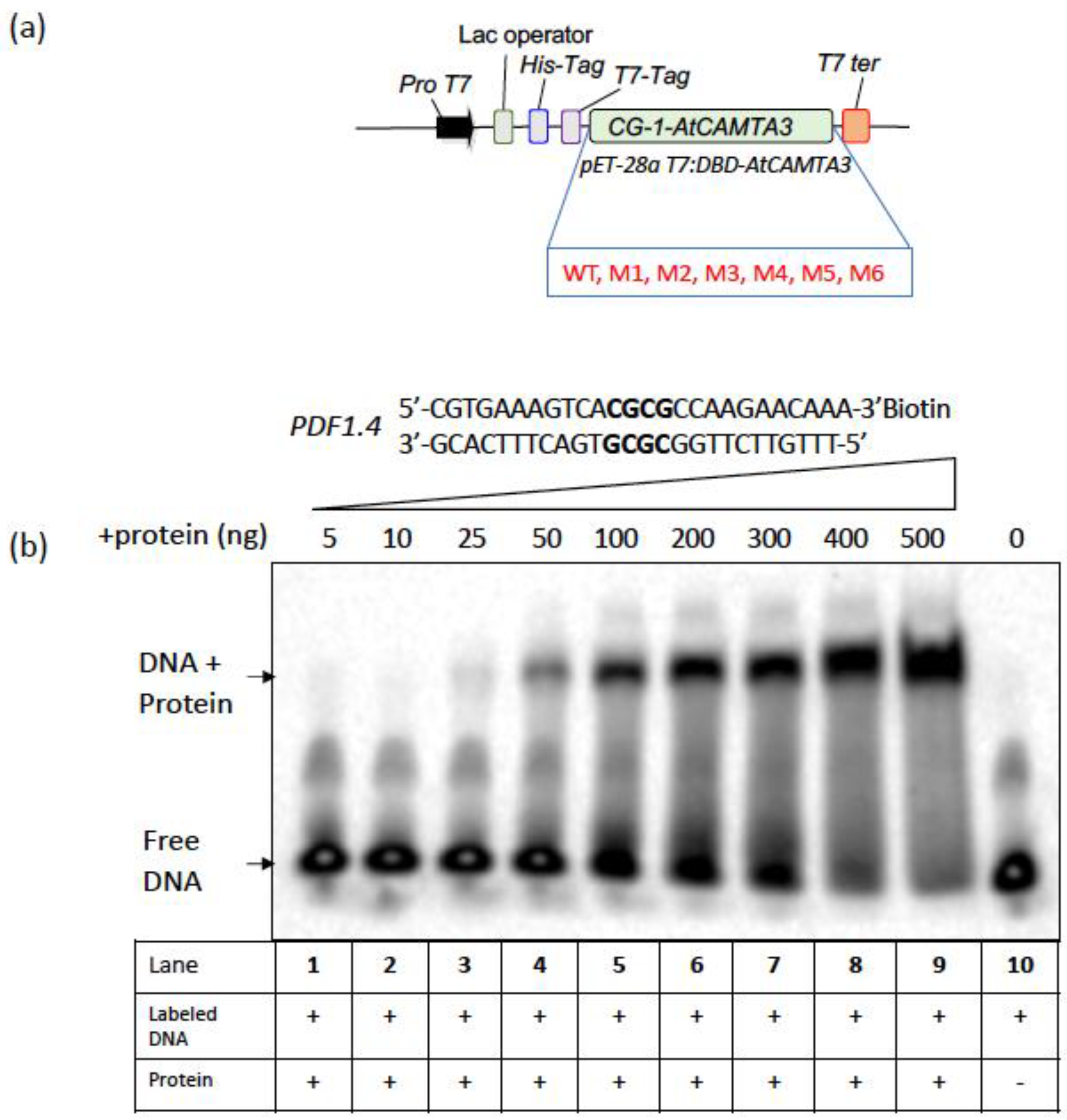
As CAMTA3 stability and turnover are known to be critical for its function as a repressor for the immune response [46,47], we show that the minimum concentration of 25 ng of WT CAMTA3 CG-1 domain to be required for binding to PDF1.4 (Figure 2). However, most of the CAMTA3 CG-1 mutants (M1, M2, M3, M4 and M6) failed to bind to the same probe even at a higher protein concentration of 500 ng (Figure 3), indicating that these amino acids are critical for DNA-binding and CAMTA3 transcriptional activity. Interestingly, the M5 mutant exhibited binding to the probe, albeit at higher concentrations (100 ng), indicating weak binding (Figure 3). Previously, it has been shown that the expression of the truncated variant of CAMTA3, comprising the CG-1 domain (334 amino acids, NRM), exhibited transcriptional activation and repressor activities in plants [18]. Interestingly, mutations of K108A and K141E in the CG-1 domain of the human CAMTA2 and CAMTA1, respectively (Figure 1), completely abolished their DNA-binding activity, indicating a critical role for these amino acids in the DNA-binding activity of these CAMTAs [48]. Indeed, this amino acid (K) is also conserved across the plant kingdom; however, the degree of conservation is slightly lower than that observed with other aromatic amino acids (Figure 1). However, the role of K in DNA-binding cannot be ruled out in plant CAMTAs, and additional mutational studies are needed.
Previously, CAMTA3 has been shown to bind to the RSRE elements and activate downstream genes [31]. In that study, the expression of full-length WT and CAMTA3 mutant proteins transiently in tobacco, and their ability to activate the expression of luciferase gene driven by RSRE (vCGCGb) was tested. In agreement with earlier reports, the binding of WT CAMTA3 is highly specific, and it transcriptionally activates the luciferase gene expression. However, except for the M5 mutant, all other mutants failed to activate the luciferase reporter gene expression (Figure 4), which concurred with our results in Figure 2 and Figure 3, with truncated CAMTA3 proteins.

5. Concluding Remarks

Our studies with multiple mutants in the CG-1 domain of CAMTA3 indicate that the DNA-binding and transcriptional activities of this protein are essential for its function in plant immunity. The current evidence suggests that CAMTA3 performs a dual role as a guardee of NLRs (DSC1/2) and as a transcriptional regulator. It is possible that elicitor-mediated modification of CAMTA3 may not only activate the NLR-mediated pathway but also regulate the transcriptional activity of CAMTA3. Furthermore, the autoimmunity exhibited by lines expressing CG-1 domain mutants could also be due to the monitoring of the transcriptional activity of CAMTA3 either by DSC1/DSC2 or other NLRs, or the CAMTA3-NLRs complex may modulate the CAMTA3 transcriptional activity. These could be addressed by testing (a) if DSC1/2-induced HR is suppressed by CG-1 mutants, (b) the effect of CAMTA3 DNA-binding mutations on its interaction with DSC1/DSC2, and (c) how this interaction modulates CAMTA3 transcriptional activity. These studies are likely to elucidate further mechanistic details about CAMTA3 and DSC1/DSC2 functions in autoimmunity.

Supplementary Materials

The following supporting information can be downloaded at: https://www.mdpi.com/article/10.3390/cells12151986/s1, Figure S1: Identification of evolutionarily conserved amino acid residues in the CG-1 domain of six CAMTAs in Arabidopsis thaliana; Figure S2: Expression and purification of WT and CG-1 mutants in E. coli; Figure S3: Significant differences in the binding activity of WT and M5 CG-1 mutant to the DNA probe; Figure S4: Sequence of synthesized RSRE::NOS and mRSRE::NOS promoter for the reporter construct; Figure S5: Schematic diagram illustrating the strategy used to generate RSRE::NOS:LUC and mRSRE::NOS: LUC constructs for Agrobacterium-mediated transient expression analysis. File S1. Fasta file of all CAMTA sequence from plants. Table S1: Primers used for cloning, genotyping, RT-PCR and generation of mutants.

Author Contributions

A.S.N.R. directed the project. K.V.S.K.P., A.A.E.A. and A.S.N.R. designed the experiments. K.V.S.K.P. and Q.J. performed EMSA experiments; K.V.S.K.P. generated the CG-1 mutants and reporter and effector constructs; A.A.E.A.-H. cloned the CG-1 mutants into a bacterial expression vector, cloned all mutants into a binary vector, generated transgenic lines and performed transient assays and gene expression analyses. K.V.S.K.P., A.A.E.A.-H. and A.S.N.R. wrote the manuscript. Q.J. read and provided comments on the manuscript. All authors have read and agreed to the published version of the manuscript.

Funding

This work was supported by a grant from the National Science Foundation (MCB #5333470) and the Agriculture and Food Research Initiative competitive grant (2019-67013-29239) of the USDA National Institute of Food and Agriculture to A.S.N.R.

Institutional Review Board Statement

Not applicable.

Informed Consent Statement

Not applicable.

Data Availability Statement

Data sharing is not applicable.

Acknowledgments

A.A.E.A. was supported by the Egyptian Cultural and Educational Bureau and Q.J. was supported by the China Scholarship Council.

Conflicts of Interest

The authors declare no conflict of interest. The funders had no role in the design of the study; in the collection, analyses, or interpretation of data; in the writing of the manuscript; or in the decision to publish the results.

References

  1. Miller, G.; Suzuki, N.; Ciftci-Yilmaz, S.; Mittler, R. Reactive oxygen species homeostasis and signalling during drought and salinity stresses. Plant Cell Environ. 2010, 33, 453–467. [Google Scholar] [CrossRef]
  2. Dhlamini, Z.; Spillane, C.; Moss, J.P.; Ruane, J.; Urquia, N.; Sonnino, A. Status of Research and Applications of Crop Biotechnologies in Developing Countries: Preiminary Assessment; Food and Agriculture Organization of the United Nations: Rome, Italy, 2005. [Google Scholar]
  3. Hadiarto, T.; Tran, L.S. Progress studies of drought-responsive genes in rice. Plant Cell Rep. 2011, 30, 297–310. [Google Scholar] [CrossRef]
  4. Fujita, M.; Fujita, Y.; Noutoshi, Y.; Takahashi, F.; Narusaka, Y.; Yamaguchi-Shinozaki, K.; Shinozaki, K. Crosstalk between abiotic and biotic stress responses: A current view from the points of convergence in the stress signaling networks. Curr. Opin. Plant Biol. 2006, 9, 436–442. [Google Scholar] [CrossRef] [PubMed]
  5. Brivanlou, A.H.; Darnell, J.E., Jr. Signal transduction and the control of gene expression. Science 2002, 295, 813–818. [Google Scholar] [CrossRef] [PubMed] [Green Version]
  6. Reddy, A.S.; Reddy, V.S.; Golovkin, M. A calmodulin binding protein from Arabidopsis is induced by ethylene and contains a DNA-binding motif. Biochem. Biophys. Res. Commun. 2000, 279, 762–769. [Google Scholar] [CrossRef] [PubMed] [Green Version]
  7. Yang, T.; Poovaiah, B.W. An early ethylene up-regulated gene encoding a calmodulin-binding protein involved in plant senescence and death. J. Biol. Chem. 2000, 275, 38467–38473. [Google Scholar] [CrossRef] [PubMed] [Green Version]
  8. Finkler, A.; Ashery-Padan, R.; Fromm, H. CAMTAs: Calmodulin-binding transcription activators from plants to human. FEBS Lett. 2007, 581, 3893–3898. [Google Scholar] [CrossRef] [Green Version]
  9. Bouche, N.; Scharlat, A.; Snedden, W.; Bouchez, D.; Fromm, H. A novel family of calmodulin-binding transcription activators in multicellular organisms. J. Biol. Chem. 2002, 277, 21851–21861. [Google Scholar] [CrossRef] [PubMed] [Green Version]
  10. Yang, T.; Poovaiah, B.W. A calmodulin-binding/CGCG box DNA-binding protein family involved in multiple signaling pathways in plants. J. Biol. Chem. 2002, 277, 45049–45058. [Google Scholar] [CrossRef] [Green Version]
  11. Choi, M.S.; Kim, M.C.; Yoo, J.H.; Moon, B.C.; Koo, S.C.; Park, B.O.; Lee, J.H.; Koo, Y.D.; Han, H.J.; Lee, S.Y. Isolation of a calmodulin-binding transcription factor from rice (Oryza sativa L.). J. Biol. Chem. 2005, 280, 40820–40831. [Google Scholar] [CrossRef] [Green Version]
  12. Han, J.H.; Gong, P.; Reddig, K.; Mitra, M.Y.; Guo, P.; Li, H.S. The fly CAMTA transcription factor potentiates deactivation of rhodopsin, a G protein-coupled light receptor. Cell 2006, 127, 847–858. [Google Scholar] [CrossRef] [Green Version]
  13. Song, K.; Backs, J.; McAnally, J.; Qi, X.; Gerard, R.D.; Richardson, J.A.; Hill, J.A.; Bassel-Duby, R.; Olson, E.N. The transcriptional coactivator CAMTA2 stimulates cardiac growth by opposing class II histone deacetylases. Cell 2006, 125, 453–466. [Google Scholar] [CrossRef] [Green Version]
  14. Rahman, H.; Yang, J.; Xu, Y.P.; Munyampundu, J.P.; Cai, X.Z. Phylogeny of Plant CAMTAs and Role of AtCAMTAs in Nonhost Resistance to Xanthomonas oryzae pv. oryzae. Front. Plant Sci. 2016, 7, 177. [Google Scholar] [CrossRef]
  15. Iqbal, Z.; Shariq Iqbal, M.; Singh, S.P.; Buaboocha, T. Ca(2+)/Calmodulin Complex Triggers CAMTA Transcriptional Machinery Under Stress in Plants: Signaling Cascade and Molecular Regulation. Front. Plant Sci. 2020, 11, 598327. [Google Scholar] [CrossRef]
  16. Xiao, P.; Feng, J.W.; Zhu, X.T.; Gao, J. Evolution Analyses of CAMTA Transcription Factor in Plants and Its Enhancing Effect on Cold-tolerance. Front. Plant Sci. 2021, 12, 758187. [Google Scholar] [CrossRef] [PubMed]
  17. Du, L.; Ali, G.S.; Simons, K.A.; Hou, J.; Yang, T.; Reddy, A.S.; Poovaiah, B.W. Ca(2+)/calmodulin regulates salicylic-acid-mediated plant immunity. Nature 2009, 457, 1154–1158. [Google Scholar] [CrossRef] [PubMed]
  18. Kim, Y.S.; An, C.; Park, S.; Gilmour, S.J.; Wang, L.; Renna, L.; Brandizzi, F.; Grumet, R.; Thomashow, M.F. CAMTA-Mediated Regulation of Salicylic Acid Immunity Pathway Genes in Arabidopsis Exposed to Low Temperature and Pathogen Infection. Plant Cell 2017, 29, 2465–2477. [Google Scholar] [CrossRef] [PubMed] [Green Version]
  19. Yuan, P.; Tanaka, K.; Poovaiah, B.W. Calmodulin-binding transcription activator AtSR1/CAMTA3 fine-tunes plant immune response by transcriptional regulation of the salicylate receptor NPR1. Plant Cell Environ. 2021, 44, 3140–3154. [Google Scholar] [CrossRef] [PubMed]
  20. Zeng, H.; Wu, H.; Wang, G.; Dai, S.; Zhu, Q.; Chen, H.; Yi, K.; Du, L. Arabidopsis CAMTA3/SR1 is involved in drought stress tolerance and ABA signaling. Plant Sci. 2022, 319, 111250. [Google Scholar] [CrossRef]
  21. Darwish, E.; Ghosh, R.; Ontiveros-Cisneros, A.; Tran, H.C.; Petersson, M.; De Milde, L.; Broda, M.; Goossens, A.; Van Moerkercke, A.; Khan, K.; et al. Touch signaling and thigmomorphogenesis are regulated by complementary CAMTA3- and JA-dependent pathways. Sci. Adv. 2022, 8, eabm2091. [Google Scholar] [CrossRef]
  22. Wang, Y.; Gong, Q.; Wu, Y.; Huang, F.; Ismayil, A.; Zhang, D.; Li, H.; Gu, H.; Ludman, M.; Fatyol, K.; et al. A calmodulin-binding transcription factor links calcium signaling to antiviral RNAi defense in plants. Cell Host Microbe 2021, 29, 1393–1406.e1397. [Google Scholar] [CrossRef] [PubMed]
  23. Galon, Y.; Nave, R.; Boyce, J.M.; Nachmias, D.; Knight, M.R.; Fromm, H. Calmodulin-binding transcription activator (CAMTA) 3 mediates biotic defense responses in Arabidopsis. FEBS Lett. 2008, 582, 943–948. [Google Scholar] [CrossRef] [PubMed] [Green Version]
  24. Kim, Y.; Park, S.; Gilmour, S.J.; Thomashow, M.F. Roles of CAMTA transcription factors and salicylic acid in configuring the low-temperature transcriptome and freezing tolerance of Arabidopsis. Plant J. 2013, 75, 364–376. [Google Scholar] [CrossRef] [PubMed]
  25. Prasad, K.V.S.K.; Abdel-Hameed, A.A.E.; Xing, D.; Reddy, A.S.N. Global gene expression analysis using RNA-seq uncovered a new role for SR1/CAMTA3 transcription factor in salt stress. Sci. Rep. 2016, in press. [Google Scholar]
  26. Doherty, C.J.; Van Buskirk, H.A.; Myers, S.J.; Thomashow, M.F. Roles for Arabidopsis CAMTA transcription factors in cold-regulated gene expression and freezing tolerance. Plant Cell 2009, 21, 972–984. [Google Scholar] [CrossRef] [PubMed] [Green Version]
  27. Nie, H.; Zhao, C.; Wu, G.; Wu, Y.; Chen, Y.; Tang, D. SR1, a calmodulin-binding transcription factor, modulates plant defense and ethylene-induced senescence by directly regulating NDR1 and EIN3. Plant Physiol. 2012, 158, 1847–1859. [Google Scholar] [CrossRef] [Green Version]
  28. Qiu, Y.J.; Xi, J.; Du, L.Q.; Suttle, J.C.; Poovaiah, B.W. Coupling calcium/calmodulin-mediated signaling and herbivore-induced plant response through calmodulin-binding transcription factor AtSR1/CAMTA3. Plant Mol. Biol. 2012, 79, 89–99. [Google Scholar] [CrossRef]
  29. Laluk, K.; Prasad, K.V.; Savchenko, T.; Celesnik, H.; Dehesh, K.; Levy, M.; Mitchell-Olds, T.; Reddy, A.S. The calmodulin-binding transcription factor SIGNAL RESPONSIVE1 is a novel regulator of glucosinolate metabolism and herbivory tolerance in Arabidopsis. Plant Cell Physiol. 2012, 53, 2008–2015. [Google Scholar] [CrossRef]
  30. Kidokoro, S.; Yoneda, K.; Takasaki, H.; Takahashi, F.; Shinozaki, K.; Yamaguchi-Shinozaki, K. Different Cold-Signaling Pathways Function in the Responses to Rapid and Gradual Decreases in Temperature. Plant Cell 2017, 29, 760–774. [Google Scholar] [CrossRef] [Green Version]
  31. Benn, G.; Wang, C.-Q.; Hicks, D.R.; Stein, J.; Guthrie, C.; Dehesh, K. A key general stress response motif is regulated non-uniformly by CAMTA transcription factors. Plant J. Cell Mol. Biol. 2014, 80, 82–92. [Google Scholar] [CrossRef]
  32. Galon, Y.; Aloni, R.; Nachmias, D.; Snir, O.; Feldmesser, E.; Scrase-Field, S.; Boyce, J.M.; Bouche, N.; Knight, M.R.; Fromm, H. Calmodulin-binding transcription activator 1 mediates auxin signaling and responds to stresses in Arabidopsis. Planta 2010, 232, 165–178. [Google Scholar] [CrossRef]
  33. Copin, J.C.; da Silva, R.F.; Fraga-Silva, R.A.; Capettini, L.; Quintao, S.; Lenglet, S.; Pelli, G.; Galan, K.; Burger, F.; Braunersreuther, V.; et al. Treatment with Evasin-3 reduces atherosclerotic vulnerability for ischemic stroke, but not brain injury in mice. J. Cereb. Blood Flow Metab. 2013, 33, 490–498. [Google Scholar] [CrossRef] [PubMed]
  34. Walley, J.W.; Coughlan, S.; Hudson, M.E.; Covington, M.F.; Kaspi, R.; Banu, G.; Harmer, S.L.; Dehesh, K. Mechanical stress induces biotic and abiotic stress responses via a novel cis-element. PLoS Genet. 2007, 3, 1800–1812. [Google Scholar] [CrossRef] [PubMed]
  35. Lolle, S.; Greeff, C.; Petersen, K.; Roux, M.; Jensen, M.K.; Bressendorff, S.; Rodriguez, E.; Sømark, K.; Mundy, J.; Petersen, M. Matching NLR Immune Receptors to Autoimmunity in camta3 Mutants Using Antimorphic NLR Alleles. Cell Host Microbe 2017, 21, 518–529.e514. [Google Scholar] [CrossRef] [PubMed] [Green Version]
  36. Jones, J.D.G.; Vance, R.E.; Dangl, J.L. Intracellular innate immune surveillance devices in plants and animals. Science 2016, 354, aaf6395. [Google Scholar] [CrossRef] [Green Version]
  37. Jin, J.; Tian, F.; Yang, D.C.; Meng, Y.Q.; Kong, L.; Luo, J.; Gao, G. PlantTFDB 4.0: Toward a central hub for transcription factors and regulatory interactions in plants. Nucleic Acids Res. 2017, 45, D1040–D1045. [Google Scholar] [CrossRef] [PubMed] [Green Version]
  38. Voinnet, O.; Rivas, S.; Mestre, P.; Baulcombe, D. An enhanced transient expression system in plants based on suppression of gene silencing by the p19 protein of tomato bushy stunt virus. Plant J. 2003, 33, 949–956. [Google Scholar] [CrossRef] [Green Version]
  39. Yuan, P.; Du, L.; Poovaiah, B.W. Ca(2+)/calmodulin-dependent AtSR1/CAMTA3 plays critical roles in balancing plant growth and immunity. Int. J. Mol. Sci. 2018, 19, 1764. [Google Scholar] [CrossRef] [Green Version]
  40. Bjornson, M.; Benn, G.; Song, X.; Comai, L.; Franz, A.K.; Dandekar, A.M.; Drakakaki, G.; Dehesh, K. Distinct roles for mitogen-activated protein kinase signaling and CALMODULIN-BINDING TRANSCRIPTIONAL ACTIVATOR3 in regulating the peak time and amplitude of the plant general stress response. Plant Physiol. 2014, 166, 988–996. [Google Scholar] [CrossRef] [Green Version]
  41. Mitsuda, N.; Isono, T.; Sato, M.H. Arabidopsis CAMTA family proteins enhance V-PPase expression in pollen. Plant Cell Physiol. 2003, 44, 975–981. [Google Scholar] [CrossRef] [Green Version]
  42. Li, T.G.; Wang, B.L.; Yin, C.M.; Zhang, D.D.; Wang, D.; Song, J.; Zhou, L.; Kong, Z.Q.; Klosterman, S.J.; Li, J.J.; et al. The Gossypium hirsutum TIR-NBS-LRR gene GhDSC1 mediates resistance against Verticillium wilt. Mol. Plant Pathol. 2019, 20, 857–876. [Google Scholar] [CrossRef] [PubMed] [Green Version]
  43. Baker, C.M.; Grant, G.H. Role of aromatic amino acids in protein-nucleic acid recognition. Biopolymers 2007, 85, 456–470. [Google Scholar] [CrossRef] [PubMed]
  44. Shoji, T.; Mishima, M.; Hashimoto, T. Divergent DNA-binding specificities of a group of ETHYLENE RESPONSE FACTOR transcription factors involved in plant defense. Plant Physiol. 2013, 162, 977–990. [Google Scholar] [CrossRef] [PubMed] [Green Version]
  45. Chao, L.; Kim, Y.; Gilmour, S.J.; Thomashow, M.F. Temperature modulation of CAMTA3 gene induction activity is mediated through the DNA binding domain. Plant J. 2022, 112, 235–248. [Google Scholar] [CrossRef] [PubMed]
  46. Zhang, L.; Du, L.Q.; Shen, C.J.; Yang, Y.J.; Poovaiah, B.W. Regulation of plant immunity through ubiquitin-mediated modulation of Ca2+-calmodulin-AtSR1/CAMTA3 signaling. Plant J. 2014, 78, 269–281. [Google Scholar] [CrossRef]
  47. Jiang, X.; Hoehenwarter, W.; Scheel, D.; Lee, J. Phosphorylation of the CAMTA3 Transcription Factor Triggers Its Destabilization and Nuclear Export. Plant Physiol. 2020, 184, 1056–1071. [Google Scholar] [CrossRef]
  48. Long, C.; Grueter, C.E.; Song, K.; Qin, S.; Qi, X.; Kong, Y.M.; Shelton, J.M.; Richardson, J.A.; Zhang, C.L.; Bassel-Duby, R.; et al. Ataxia and Purkinje cell degeneration in mice lacking the CAMTA1 transcription factor. Proc. Natl. Acad. Sci. USA 2014, 111, 11521–11526. [Google Scholar] [CrossRef]
Figure 1. Identification of the evolutionarily conserved amino acid residues in the CG-1 domain of CAMTAs. (a) Schematic representation of full-length AtCAMTA3 protein indicating various domains. (b) Sequence logo showing conserved amino acids in the CG-1 (DNA-binding domain) of 1343 CAMTAs belonging to 165 plant species. The web logo was generated using the Hidden Markov Model-guided method [37]. (c) Alignment of amino acid sequence in the CG-1 domain of AtCAMTA3, HsCAMTA1 and DmCAMTA. Highly conserved amino acid residues are marked by blue boxes. (d) The amino acid sequence of the AtCAMTA3 CG-1 domain with the highly conserved amino acids (>4 bit scored) is underlined. The letters M1–M6 indicated in (bd) represent the conserved amino acids that are mutated to alanine (A).
Cells 12 01986 g001
Figure 2. Electrophoretic mobility shift assay (EMSA) to determine the binding activity of WT CG-1 domain. (a) Schematic representation of the construct that was used for expression and purification of the WT and mutated CG-1 (DNA-binding) domain variants in E. coli. The protein was purified using a Ni-NTA agarose column. (b) Increasing concentrations of the purified recombinant WT CAMTA3 CG-1 protein (Lanes 1 to 9) were used for EMSA assays along with the PDF1.4 probe to determine the minimal protein concentration required for binding. Probe alone is shown in lane 10. (+) and (−) indicate the presence or absence of protein/DNA in the reaction mixture.
Cells 12 01986 g002
Figure 3. The majority of CG-1 point mutations abolished binding to the PDF1.4 probe. (a) EMSA assays with purified WT and CG-1 mutant proteins using the PDF1.4 probe. The majority of the CG-1 mutants (M1, M2, M3, M4 and M6) failed to bind to the PDF1.4 probe, as indicated by the absence of mobility shift in the labeled probe. Only the WT and the CG-1 domain variant (M5) with W118A mutation exhibited binding to the PDF1.4 probe. The inclusion of competitor significantly reduced WT and M5 binding to the probe. (b) A protein concentration-dependent binding of M5 protein to PDF1.4 probe. Note: the binding of M5 protein to the probe occurs only at concentrations of at or above 100 ng. (+) and (−) indicate the presence or absence of protein/DNA in the reaction mixture.
Cells 12 01986 g003
Figure 4. Full-length AtCAMTA3 CG-1 mutants curtailed the induction of RSRE::LUC reporter expression. Schematic representation of the reporter (a) and effector (b) constructs used in transient expression assays with N. benthamiana leaves for quantifying the induction of RSRE::LUC reporter expression by full-length WT and mutated AtCAMTA3. Reporter constructs include 4X RSRE elements (upper panel) and 4X mRSRE (lower panel). The effector constructs for expression of either full-length WT or CG-1 mutants (M1 to M6) of CAMTA3 driven by CaMV35S promoter are shown. (c) Transient expression of RSRE::LUC construct or its mutated dysfunctional RSRE (mRSRE::LUC) alone (control) or together with CaMV35S::AtCAMTA3 (CAMTA3-O.E) or mCAMTA3 variant (M1, M2, M3, M4, M4, M5 or M6) in N. benthamiana leaves. Only the CAMTA3 WT and M5 mutant displayed strong RSRE::LUC activation, whereas the AtCAMTA3 CG-1 mutant variants (M1, M2, M3, M4 and M6) exhibit curtailed induction of RSRE::LUC activity. Lines on top of the bars represent the standard error of the mean and asterisks above the line represent statistically significant differences (p < 0.05) in reporter gene expression. The infiltration experiments were carried out three times for each construct with three independent plants.
Cells 12 01986 g004
Figure 5. Expression and phenotypic analysis of CAMTA3 CG-1 mutants in camta3 background. (a) Schematic representation of the constructs used for transformation of camta3 (T-DNA insertion mutant) for expression of the WT and CG-1 mutant proteins of CAMTA3. (b) CAMTA3 protein levels in four-week-old WT and transgenic lines complemented with WT or CG-1 mutants (M1 to M6) of CAMTA3. The camta3-2 plants transformed with CaMV35S::CAMTA3 (CAMTA3-O.E) and CG-1 mutants (M1 to M6) were grown under day neutral conditions at 19 °C, and protein expression was detected using an antibody against CAMTA3. (c) Photographs of four-week-old plants depicting phenotypes of the WT, camta3, CAMTA3-O.E. and camta3-2 plants expressing the mutants (M1 to M6).
Cells 12 01986 g005
Figure 6. Repression of defense-related genes and activation of cold-responsive genes in camta3 plants expressing the CAMTA3 CG-1 mutants. Comparison of transcript levels of the genes associated with plant defense (CBP60g, ICS1, EDS1 ALPHA-DOX1) and cold (COR15 and KIN1) responses in WT plants and camta3-2 mutants as well as camta3-2 mutants expressing CaMV35S::CAMTA3 (CAMTA3-OE) and CG-1 mutants (M1 to M6). The plants were grown in a growth chamber at 20 °C under 12:12 h light/dark cycles, with a light intensity of 200 μmol m−2 s−1 and relative humidity of 50%. Leaves of six-week-old plants were harvested for RNA isolation. Statistically significant differences (p < 0.05) in reporter gene expression in all lines compared to WT are shown with asterisks.
Cells 12 01986 g006
Disclaimer/Publisher’s Note: The statements, opinions and data contained in all publications are solely those of the individual author(s) and contributor(s) and not of MDPI and/or the editor(s). MDPI and/or the editor(s) disclaim responsibility for any injury to people or property resulting from any ideas, methods, instructions or products referred to in the content.

