1. Introduction
Neuroendocrine Syncopes (NES) are frequent in the general population. NES is characterized by a partial or total loss of consciousness due to a drop in systolic blood pressure [
1]. The fall in blood pressure is secondary to vasoplegia and/or severe bradycardia while sometimes an atrioventricular block (AVB) may occur [
2]. In a large number of cases, loss of consciousness is preceded by prodromes like dizziness, nausea, abdominal pain or cephalalgia. Less frequently, the loss of consciousness occurs without prodromes (sudden syncope) [
3,
4]. NHS account for 3 to 5% of emergency entrance and 1% of hospitalization with a mortality of 0.28% [
5,
6,
7].
The recurrence occurs in 35% of cases while discomfort is followed by injuries in 29% of cases [
8]. Two tests are commonly used for the exploration of these syncopes: the head-up tilt test (HUT) and the intravenous bolus injection of ATP or adenosine [
1]. Both tests reproduce the symptomatology that occurs in the daily life of patients. Although it is a very common condition, physiological and molecular mechanisms of NES remained unclear, which makes the delivery of appropriate treatment a challenge. However, it is important to know the cause(s) of clinical manifestations at both physiological and molecular levels, in order to be able to deliver an appropriate treatment. In recent years, it has become clear that the cause of symptoms may be related to a dysfunction of the adenosinergic system. This is supported by the fact that (1) administration of adenosine reproduces the symptomatology of NES [
9,
10]; (2) during the head up-tilt test, a maneuver that reproduces the symptoms, an increase in adenosine plasma level (APL) was observed and was associated with the drop blood pressure [
11] and (3) antagonists of adenosine receptors are efficient in the treatment of some kind of NES [
12,
13]. In fact, adenosine, an ATP derivative, exerts a powerful cardio-inhibition and vasodilation via activation of its receptors, two phenomena which are found during the clinical manifestations of NES. The aim of this review is to highlight the latest data on the possible role of the adenosinergic system in NES.
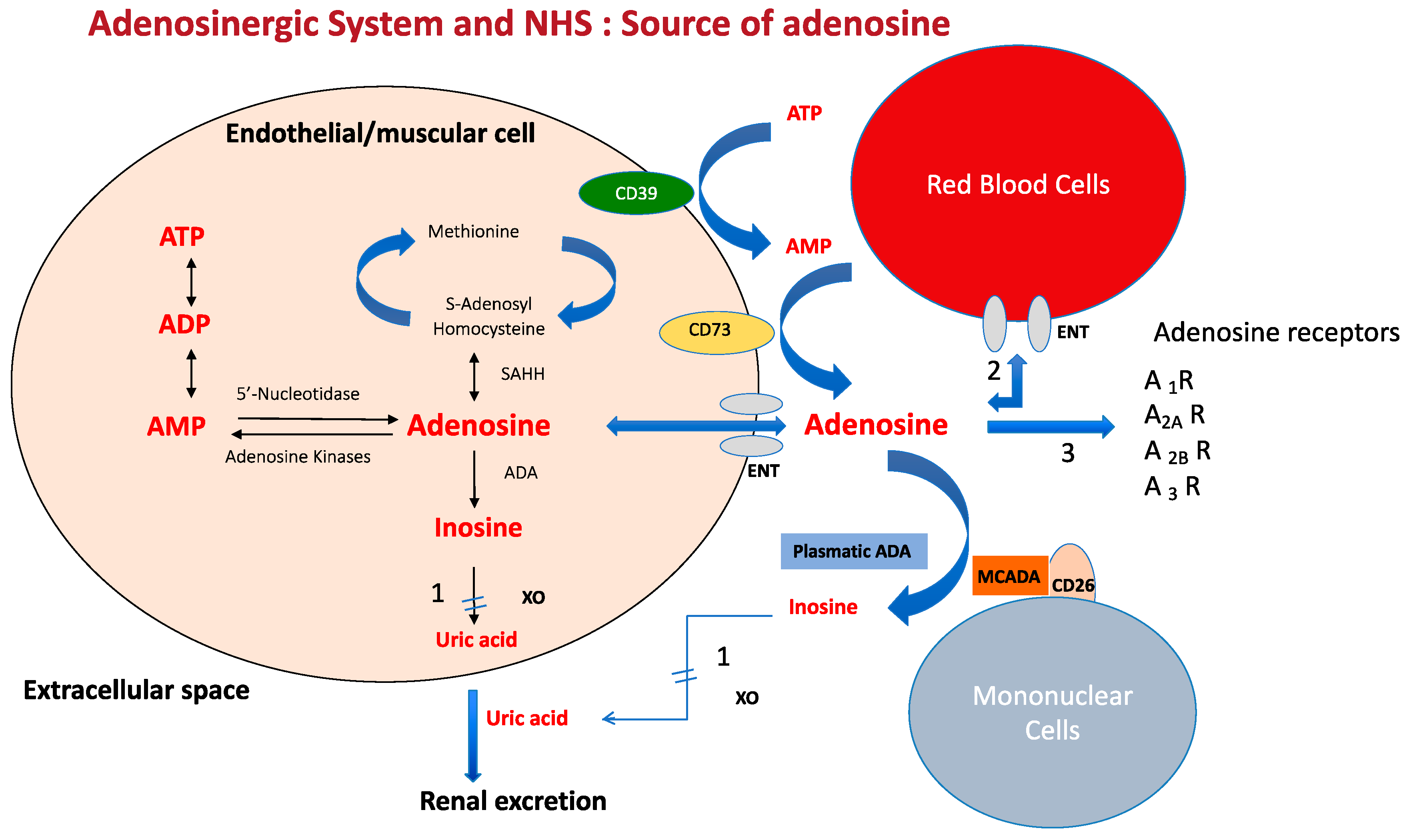
2. Source and Metabolism of Adenosine
Adenosine is a ubiquitous nucleoside, present in most cells but particularly in endothelial and muscle cells, which mainly comes from the dephosphorylation of ATP via intra-cellular nucleotidases or via ecto-nucleotidase CD39 and CD73 (
Figure 1). The methionine cycle represents only a small part of the adenosine source and does not allow rapid adenosine release, unlike ATP dephosphorylation. The ATP pathway is particularly active when there is cellular or tissue stress, such as during inflammation, hypoxia or ischemia. At the intra-cellular level, adenosine can be rephosphorylated in adenine nucleotides by kinases, but this rephosphorylation process is inhibited by the hypoxia-inducible factor (HIFa) in conditions of low oxygen tension, leading to a strong release of adenosine in the extra-cellular spaces via the equilibrative nucleoside transporter (ENT1).
Adenosine is also deaminated into inosine by intra-cellular or plasmatic ADA or by ADA linked to mononuclear cells (MCADA (
Figure 1), the final product being uric acid.
Adenosine mainly comes from the dephosphorylation of adenyl nucleotides both at the intra and extracellular levels. The dephosphorylation occurs via nucleotidases in intracellular spaces and via CD39 and CD73 at the extracellular level. Part of intra-cellular adenosine is rephosphorylate via adenosine kinases. A short part of adenosine comes from the methionine cycle.
The behavior of adenosine is trice. (1) adenosine is quickly deaminated into inosine by an intra and extracellular adenosine deaminase (ADA). Adenosine is also deaminated via MCADA (mononuclear cell adenosine deaminase) which is a plasmatic ADA linked to the CD26 transmembrane protein of the mononuclear cells via non-covalent binding. The final product is uric acid after the action of xanthine oxydase (XO). (2) Extracellular adenosine is quickly uptaken by red blood cells via the equilibrative nucleoside transporter (mainly ENT1). (3) Part of adenosine that has not been deaminated or uptaken by red blood cells acts on three subtypes of receptors named A1 R, A2 AR, A2B R and A3 R with a strong impact on the cardiovascular system.
ATP: adenosine triphosphate; ADP: adenosine diphosphate; AMP: adenosine monophosphate. SAHH: S-adenosyl-homocysteine hydrolase.
Despite its short half-life of adenosine in the extracellular space (mainly due to uptake by red blood cells and deamination by ADA), adenosine strongly impacts the cardiovascular system via its receptors.
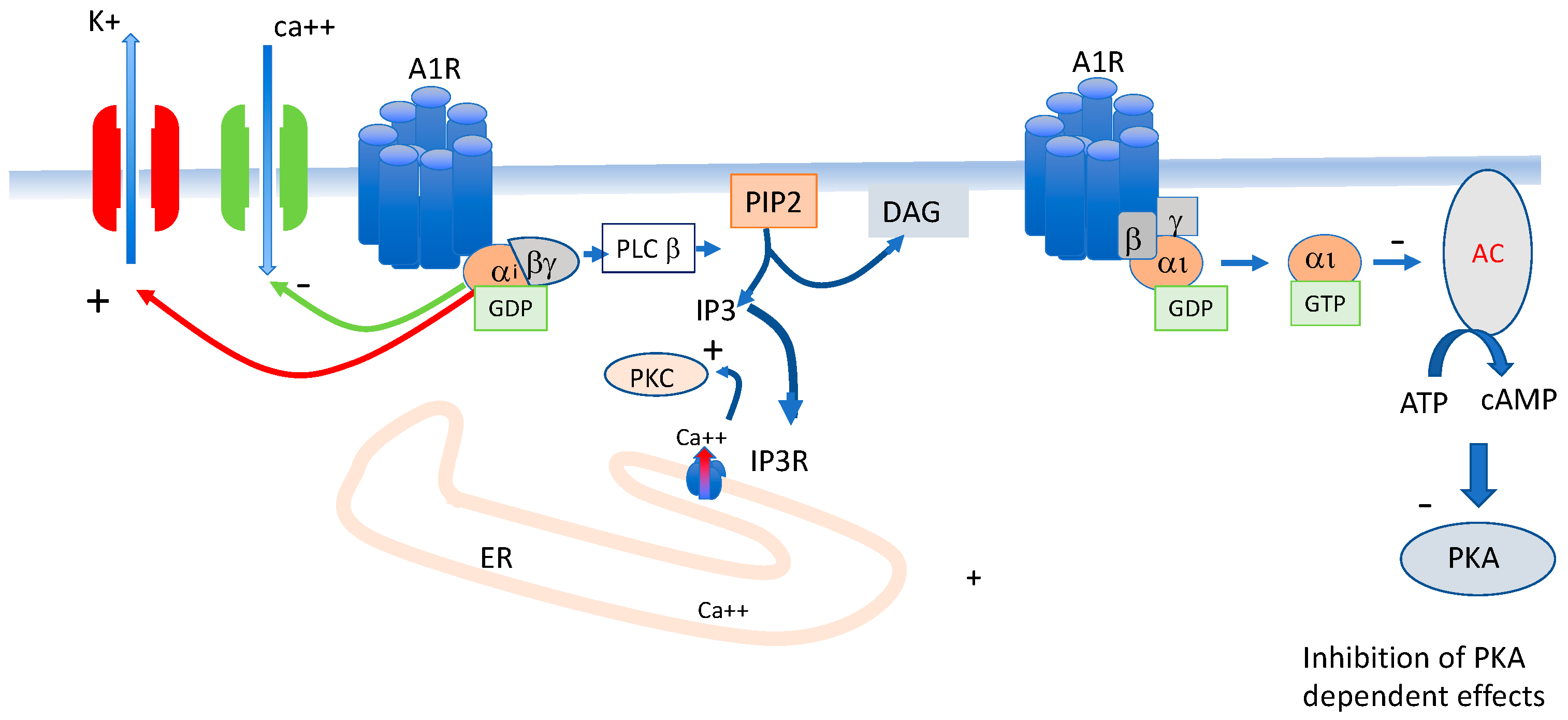
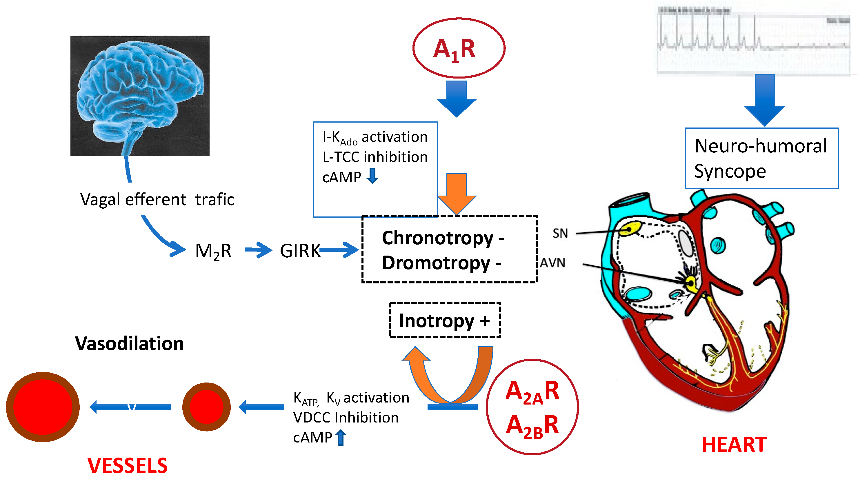
3. Adenosine Receptors Implicated in Blood Pressure and Heart Rate
Adenosine acts on the cardiovascular system via its membrane receptors namely A
1 R, A
2A R, A
2B R and A
3 R, pending on their primary sequence and their action on adenylcyclase pathway [
14,
15]. The activation of A
1 R or A
3 R leads to the inhibition of adenylcyclase and thus to the decrease in cAMP production, while conversely, the activation of A
2 R subgroups leads to cAMP production.
A
1 R is expressed in the sinus node and atrio-ventricular node where they control heart rate and AV conduction. The main effects of A
1 R activation are the “direct” (cAMP-independent) activation of the inwardly rectifying K+ current IKAdo, Ach [
16]. These channels are also activated by Ach. The direct effect occurs via the protein–protein interaction of the G-protein (probably the beta/gamma dimere complex [
17]). The activation of these inward rectifying potassium channels leads to hyperpolarization of the synaptic membrane resulting in a decreased excitability and slowing of the heart rate with sinus arrest or atrio-ventricular block. The activation of A
1 R also leads to phospholipase C stimulation (PLC) and thus activates the IP3 cascade, modulating the calcium release from its reticulum storage via the IP3 receptor (
Figure 2). Finally, A
1 R activation leads to the inhibition of adenylcylase activity and thus inhibiting the cAMP production and beyond the cAMP-dependent neurotransmission [
18].
On target cells, activation of A1 R leads to the activation of the alpha i subunit of the G protein, leading to the inhibition of the production of cAMP (indirect effects). Activation of A1 R also leads to the activation of potassium channels, mainly the inward rectifier (IK ado/Ach) and the inhibition of L-type voltage gate calcium channels (Belardinelli 1995).
A
2A R is strongly expressed in vessels where they are implicated in the blood flow via smooth cell relaxation. The activation of A
2A R leads to the stimulation of adenylcyclase activity and thus the cAMP production and the activation of a protein kinase cAMP-dependent (PKA) (
Figure 3). The three consequences of the PKA activation are: (i) activation of K
ATP and K
V channels; (ii) the inhibition of L-type voltage-gated calcium channels; and (iii) the activation of NO synthase leading to NO release. These effects are all consistent with a vasodilatory effect. Note that the activation of A
2B R leads to the same vasodilatory effects but the implication of these receptors in NHS while likely probable is not yet demonstrated especially since their activation needs a high concentration of adenosine [
19].
On target cells, activation of A2A R leads to the activation of the alpha S subunit of the G protein, leading to the production of cAMP through the activation of adenylcyclase (AC).
cAMP increase leads to the activation of a protein kinase cAMP-dependent (PKA) with phosphorylates potassium channels (mainly KATP and KV) and also to the inhibition of voltage-sensitive calcium channels (mainly L Type channels). Finally, the activation of A2A R leads to the activation of the enzyme NO synthase (eNOS) via the PKA pathway and thus to NO release. The main effects due to A2A R activation through these signal transduction pathways is vasodilation.
Figure 4 summarizes the main consequences of activation of adenosine receptors on the cardiovascular system in an integrative point of view.
4. Role of the Adenosinergic System in NES
4.1. Adenosinergic Profile in NHS Patients
The role of adenosine in NES has long been discussed [
4,
20]. Thus, there is evidence that an adenosinergic disturbance is implicated in NES. In vasovagal syncope: high basal APL was reported with a significant increase during the HUT associated with fainting [
11], while an increase in APL was also reported during HUT [
11,
21,
22]. This increase may explain the lack of prodromes in this patient population. The APL increase was much higher in patients with predominant vasodilation [
21]. This is in agreement with the fact that the K
D value for A
2A R is much higher (1.8 µM, [
23]) than for A
1 R (0.8 µM, [
24]). Indeed, in the case of high basal APL, A
1 R is mostly desensitized and only A
2A R could be activated [
25]. The increase in basal APL may be explained by the decrease in ADA activity [
21]. Furthermore, high A
2A R expression was reported in this patient population [
26,
27] while a specific SNP (c 1083 C > T) in the second exon of the gene encoding A
2A R was found [
26] but not confirmed by others [
27]. In some kinds of syncope, low APL has been described. These forms are most often associated with the absence of prodromes. In these forms, the drop in blood pressure is often associated with severe bradycardia or AVB rather than vasodilation, suggesting that activation of A
1 R is predominantly at play [
3,
28,
29].
The cause of high APL in VVS patients could be explained by a vicious circle (see
Figure 5). At the beginning, a possible genetic predisposition with the presence of the specific CC (SNP) although a silent polymorphism, is associated with overproduction of A
2A R [
26]. The activation of A
2A R, via adenosine leads to the overexpression of ENT at the membrane of the PBMCs, triggering the release of adenosine in blood, and thus creating a vicious circle.
CC variant from the second exon of the gene encoding A2A R is associated with an increase in mRNA production followed by an increase in A2A R production. Adenosine acts on A2A R leading to an increase in the number of equilibrative nucleoside transporters (ENT) that control the outflow of adenosine in the extracellular spaces. PKA: protein kinas A; AC: adenylcyclase; GTP: guanosine triphosphates; ATP: adenosine triphosphates; cAMP: cyclic adenosine monophosphate; ENT1: equilibrative nucleoside transporter 1.
Furthermore, a decrease in ADA activity was found in VVS patients with positive HUT [
30] which could also contribute to the high APL. The cause of low adenosine in some kinds of NHS remains unknown. Possible ENT1 abnormalities cannot be ruled out as well as abnormal plasma or MCADA activity. However, these hypotheses need further investigation.
4.2. Effects of Exogenous Adenosine
ATP injection was used to explore unexplained syncope for a quarter of a century [
9,
10,
31]. Exogenous ATP or adenosine reproduces symptomatology in the subgroup of patients suffering from NES. Positive HUT is associated with an increase in endogenous APL while ATP/adenosine injection reproduces the symptoms in a subgroup of NES patients [
1,
28]. However, while a small portion of patients exhibit both positive tests, it seems that HUT and ATP tests identify two distinct populations [
28,
32]. In positive HUT the basal APL is high which increases further during HUT, this increase activates mainly A
2A R. In patients with very low basal APL, the adenosine release that occurs during HUT is not sufficient to recruit even high-affinity adenosine receptors, whereas a rapid bolus injection is likely to produce sufficient adenosine concentration to activate at least the high-affinity receptors (mainly A
1 R) inducing sinus bradycardia and AVB.
4.3. The Question of Adenosine Measurement
Numerous methods have been tried to measure adenosine in blood with varying degrees of success, including high-performance liquid chromatography [
33], amperometry [
34], and mass spectrometry LC-MS/MS [
34,
35,
36]. The issue is not so much the assay method as the sampling conditions. The half-life of adenosine is relatively short in blood but longer in a sample tube. Amperometry, which measures adenosine in real time and thus permits kinetic studies, has shown that the half-life of adenosine at room temperature is of the order of 45 s, longer than in circulating blood (unpublished data). A comparative study a showed good correlation between high-performance liquid chromatography, amperometry and mass spectrometry (LC-MS/MS), the latter being the gold standard [
34]. Adenosine determination after blood collection on blotting paper is convenient because it does not require the use of a stop solution, unlike tube sampling. It also allows easy fingertip capillary sampling and samples can be mailed. Finally, the use of blot paper followed by LC-MS/MS appears to be reliable and useful to the dosage of adenosine in blood.
4.4. Effects of Dioxygene on the Adenosinergic System
Hypoxia is associated with an increase in intracellular adenosine, (via the HIF pathway see 2.1) with subsequent release in the extra-cellular spaces. Hypoxia also induces an up-regulation of A
2A R [
36]. Conversely hyperoxia, in animal studies, leads to a drop in APL and a down-regulation of A
2A R [
37]. This drop is associated with vasoconstriction leading to an increase in blood pressure.
Interestingly in healthy subjects, hyperoxia was shown to increase blood pressure during tilt testing, rendering tilt test negative in subjects with previously positive HUT. These hemodynamic changes were associated with a decrease in APL [
38]. This highlights another impact of adenosine in the regulation of blood pressure during HUT via the dioxygene pathway.
5. Peripheral Blood Mononuclear Cells (PBMCs) Are a Useful Tool for the Exploration of the Adenosinergic System
PBMCs express on their surface in particular the A
1 R and A
2A R. It has been shown that their level of expression, as well as their function (i.e., cAMP production), mirrored the expression and the function of these same receptors present in the tissues and organs of the cardiovascular system, in particular the myocardium, coronary arteries, aorta and iliac vessels [
39,
40,
41]. PBMCs are permanently in contact with the circulating blood and are therefore subject, like cardiovascular tissues, to variations in the concentration of extracellular adenosine and thus their expression regulation is the same for cardiovascular tissues.
A monoclonal antibody, against the second extra-cellular loop of the human A
2A R, which exhibits agonist properties was developed [
42]. This unique tool permits us to evaluate A
2A R expression on a lot of cardio-vascular tissues, but also PBMCs. This antibody permits also to evaluate in the same time affinity (K
D) and cAMP production (EC
50). Using this tool, it was shown that a small fraction of NHS patients exhibits a specific profile of A
2A R named receptor reserve or spare receptors [
43,
44].
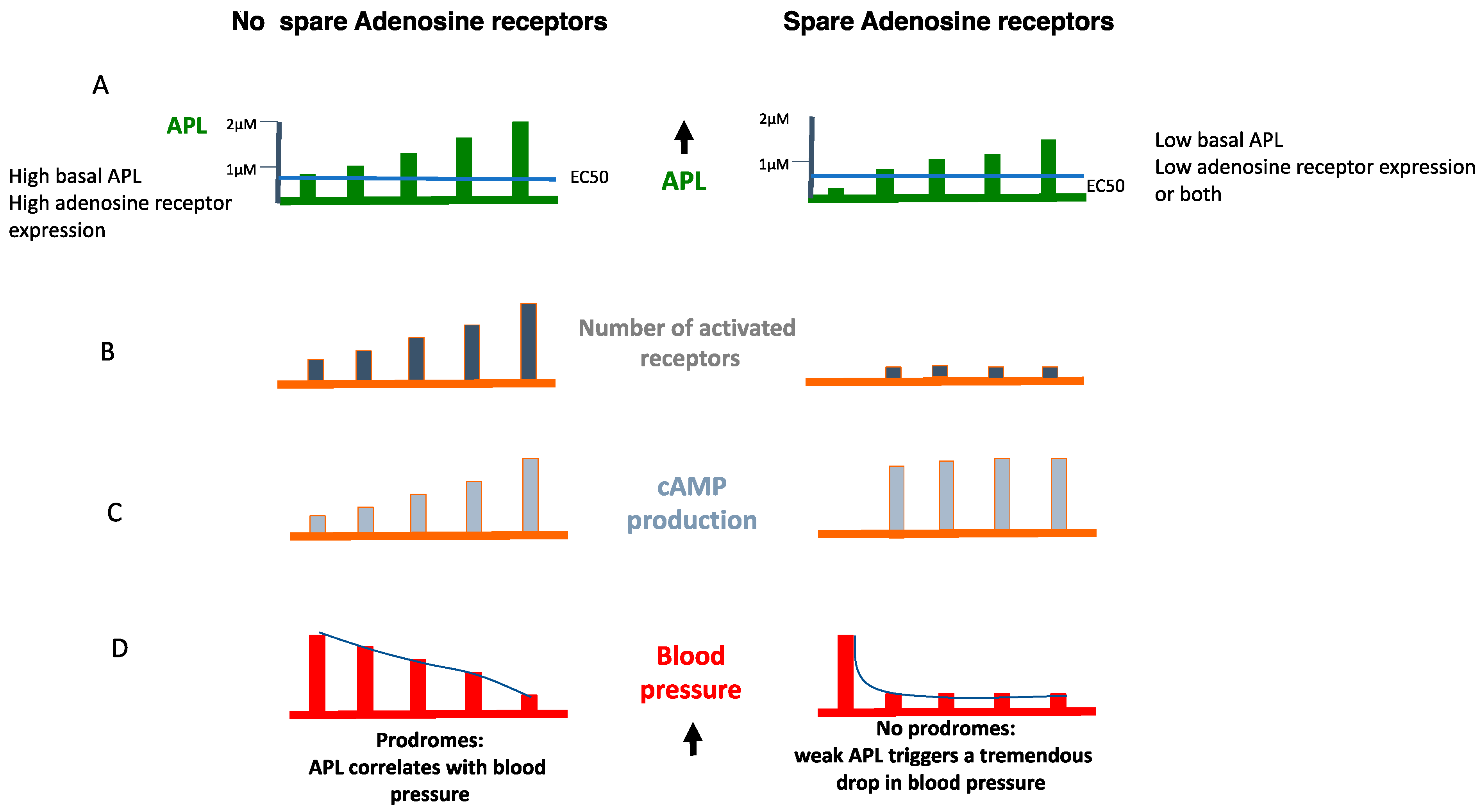
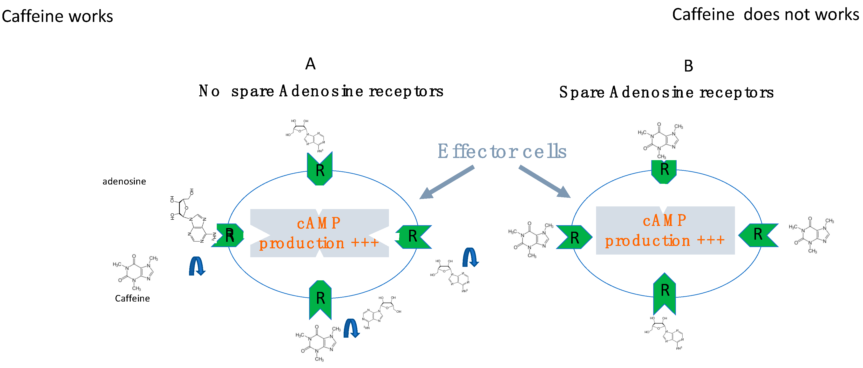
6. The Role of Receptor Reserve (Spare Receptors)
The biological response to the activation of adenosine receptors depends on tissue nature, pathophysiological situations and the nature of the ligand, agonist or antagonist.
In a general way, the intensity of the biological effects (i.e., cAMP production) correlates with the number of receptors which are activated by the ligand (here adenosine). Furthermore, the cAMP production in target cells correlates with the intensity of physiological effects (heart frequency and/or vasodilation). The concept developed by Clark in the 1930s then Stephenson [
45,
46] is that in some cases a small fraction of activated receptors is sufficient to obtain maximal biological effects (
Figure 6). From a pharmacological point of view, this concept means that the presence of spare receptors can be advocated when the half-maximal biological effects evaluated as an example by the half-maximal cAMP production (EC
50) is lower than the K
D value [
23,
43,
44,
47]. Both A
1 R and A
2A R spare receptors have been described in the heart [
23,
48,
49]. While the presence of spare adenosine receptors is usual in healthy small mammalians [
23], in humans, their presence seems to be an adaptive mechanism to compensate for low adenosine levels, low adenosine receptor expression or both [
47]. Thus, the presence of spare could provide the cell with a survival mechanism in order to be able to maintain a signal transduction process in the event of a shortage of the ligand, shortage of the receptor or both.
The presence of spare receptors is also a way for tissues to favor one biological effect over another. For example, the presence of A
2A R spare in the heart, can promote coronary vasodilation rather than heart rate, the EC
50 value for vasodilation being lower than the EC
50 value for the modulation of heart rhythm [
23]. Thus, in the event of a shortage of the ligand or receptors, the presence of spare makes it possible to favor a “protective” reaction rather than another to slow down the heart rate to save the myocardium or to dilate vessels to lower blood pressure.
The presence of spare also explains that the response to agonist or antagonist vary depending on pathophysiological situations and tissue nature and finally may differ from one patient to another. The presence of spare A
2A R in some kinds of NES has been reported [
43,
44]. Interestingly, a spare A
2A R was found in a group of patients with very low APL [
44], and sometimes in the case of low A
2A R expression or both (unpublished data).
In some patients with NES, a very low APL was measured which is 5 to 10 folds under the EC50 value, and thus in these conditions, no adenosine receptors could be activated.
For unknown reasons (infra-clinical hypoxia or inflammation), the APL increase over the EC
50 value leads to sudden maximum biological effects, objectified by a maximum production of cAMP in the target cells (
Figure 6 and
Figure 7).
The rapidity of the biological response does not leave room for prodromes. If the presence of spare receptors concerns A
1 R, the activation of the receptors during APL increase results in severe bradycardia, sinus arrest or AVB. If it concerns A
2A R, the clinical manifestations will be sudden vasoplegia (
Figure 8).
7. Effects of Adenosine Receptor Antagonists
Xanthine derivatives like caffeine or theophylline are nonspecific adenosine receptor antagonists. Caffeine binds to adenosine receptors with an affinity close to a 1 to 10 micromolar range [
50]. Furthermore, caffeine allows the inflow of calcium from the external environment via the opening of TRP channels [
51]. In addition, caffeine activates Ryanodine receptors and promotes the outflow of calcium from the storage site in the reticulum. Thus, caffeine or theophylline favors the calcium induce calcium release phenomenon leading to positive inotropy in the myocardium. In vessels, caffeine stimulates sympathetic activity leading to norepinephrine release and vasoconstriction. The administration of caffeine stimulates the juxtaglomerular apparatus and causes the secretion of renin and therefore the release of aldosterone, the reabsorption of sodium by the kidney leading to an increase in robbery and beyond the pressure arterial [
52,
53]. Finally, caffeine at high concentrations also inhibits phosphodiesterase.
Theophylline’s efficacy in preventing syncope was first demonstrated in a very small number of patients, with an efficacy that seemed superior to cardiac pacing [
54]. More recently, due to its effects, theophylline has been successfully used in the prevention of syncope recurrence [
12,
13]. While theophylline’s initial indication seemed to be reserved for syncope with low adenosine, it now appears to be effective in syncope with high or normal adenosine concentration [
12]. Thus, theophylline administration decreases the number of syncope episodes [
12], more particularly in patients affected by low adenosine syncope [
13]. Apart from the fact that they are quite often poorly supported, adenosine receptor antagonists can be ineffective in the presence of spare receptors. Indeed, in this case, the concentration of the antagonist must be very high in order to displace the adenosine from all the receptors that it occupies (see
Figure 9).
8. Effects of Pacing
In a few observational case studies performed in patients with “Low adenosine syncope”, cardiac pacing showed to be successful in preventing syncopal recurrences in patients in whom who had ECG documentation of a systolic pause due to sinus arrest or AV block [
3]. In a small multicenter trial [
55], performed in 80 highly selected elderly patients with unexplained unpredictable syncope who had a positive response to an intravenous injection of a bolus of 20 mg of ATP dual-chamber cardiac pacing, the 2-year syncope recurrence rate was significantly reduced from 69% in the control group to 23% in the active group. For the above reasons, cardiac pacing may be considered to reduce syncope recurrences in patients with the clinical features of adenosine-sensitive syncope according to the most recent ESC guidelines on syncope [
56] and on Cardiac pacing [
57] (class IIb recommendation).
9. Conclusions
While the adenosinergic system appears to be an indispensable control system for the proper functioning of the cardiovascular system, its dysfunction can be the cause of pathologies that can be disabling and spectacular. A better understanding of the molecular mechanisms that lead to clinical manifestations should enable better management of NES.
Author Contributions
Conceptualization, R.G., P.M., B.M., M.H. and J.C.D.; writing—original draft preparation, R.G., M.H., M.B., J.C.D., J.F. and A.G.; Figures and Bibliography, S.C., M.M. and Y.B.; writing—review and editing, N.L. and G.M.; visualization; supervision, M.H., M.B. and R.G. All authors have read and agreed to the published version of the manuscript.
Funding
This work was supported by Aix Marseille University.
Conflicts of Interest
The authors declare no conflict of interest.
References
- Goldberger, Z.D.; Petek, B.J.; Brignole, M.; Shen, W.-K.; Sheldon, R.S.; Solbiati, M.; Deharo, J.-C.; Moya, A.; Hamdan, M.H. ACC/AHA/HRS Versus ESC Guidelines for the Diagnosis and Management of Syncope. J. Am. Coll. Cardiol. 2019, 74, 2410–2423. [Google Scholar] [CrossRef]
- Brignole, M.; Deharo, J.-C.; Guieu, R. Syncope and Idiopathic (Paroxysmal) AV Block. Cardiol. Clin. 2015, 33, 441–447. [Google Scholar] [CrossRef]
- Brignole, M.; Guieu, R.; Tomaino, M.; Iori, M.; Ungar, A.; Bertolone, C.; Unterhuber, M.; Bottoni, N.; Tesi, F.; Deharo, J.C. Mechanism of syncope without prodromes with normal heart and normal electrocardiogram. Heart Rhythm. 2017, 14, 234–239. [Google Scholar] [CrossRef]
- Deharo, J.-C.; Brignole, M.; Guieu, R. Adenosine and neurohumoral syncope. Minerva. Med. 2022, 113, 243–250. [Google Scholar] [CrossRef]
- Sun, B.C. Risk prediction for patients with syncope. Ann. Emerg. Med. 2004, 44, 422–423. [Google Scholar] [CrossRef]
- Silverstein, M.D.; Singer, D.; Mulley, A.G.; Thibault, G.; Barnett, G. Patients with syncope admitted to medical intensive care units. JAMA 1982, 248, 1185–1189. [Google Scholar] [CrossRef]
- Day, S.C.; Cook, E.; Funkenstein, H.; Goldman, L. Evaluation and outcome of emergency room patients with transient loss of consciousness. Am. J. Med. 1982, 73, 15–23. [Google Scholar] [CrossRef]
- Da Silva, R.M.F.L. Syncope: Epidemiology, etiology, and prognosis. Front. Physiol. 2014, 5, 471. [Google Scholar] [CrossRef] [Green Version]
- Flammang, D.; Pelleg, A.; Benditt, D.G. The Adenosine Triphospate (ATP) Test for Evaluation of Syncope of Unknown Origin. J. Cardiovasc. Electrophysiol. 2005, 16, 1388–1389. [Google Scholar] [CrossRef]
- Flammang, D.; Church, T.; Waynberger, M.; Chassing, A.; Antiel, M. Can Adenosine 5′-Triphosphate Be Used to Select Treatment in Severe Vasovagal Syndrome? Circulation 1997, 96, 1201–1208. [Google Scholar] [CrossRef]
- Saadjian, A.Y.; Lévy, S.; Franceschi, F.D.; Zouher, I.; Paganelli, F.; Guieu, R.G.P. Role of Endogenous Adenosine as a Modulator of Syncope Induced During Tilt Testing. Circulation 2002, 106, 569–574. [Google Scholar] [CrossRef] [Green Version]
- Brignole, M.; Iori, M.; Solari, D.; Bottoni, N.; Rivasi, G.; Ungar, A.; Deharo, J.C.; Guieu, R. Efficacy of theophylline in patients with syncope without prodromes with normal heart and normal ECG. Int. J. Cardiol. 2019, 289, 70–73. [Google Scholar] [CrossRef]
- Brignole, M.; Solari, D.; Iori, M.; Bottoni, N.; Guieu, R.; Deharo, J.C. Efficacy of theophylline in patients affected by low adenosine syncope. Heart Rhythm. 2016, 13, 1151–1154. [Google Scholar] [CrossRef] [PubMed]
- Burnstock, G. Purinergic Signaling in the Cardiovascular System. Circ. Res. 2017, 120, 207–228. [Google Scholar] [CrossRef] [PubMed]
- Borea, P.A.; Gessi, S.; Merighi, S.; Vincenzi, F.; Varani, K. Pharmacology of Adenosine Receptors: The State of the Art. Physiol. Rev. 2018, 98, 1591–1625. [Google Scholar] [CrossRef] [Green Version]
- Belardinelli, L.; Shryock, J.C.; Song, Y.; Wang, D.; Srinivas, M.; Mozzicato, S.; Joshi, B.V.; Jacobson, K.A.; Liang, B.T.; Dougherty, C.; et al. Ionic basis of the electrophysiological actions of adenosine on cardiomyocytes. FASEB J. 1995, 9, 359–365. [Google Scholar] [CrossRef]
- De Waard, M.; Hering, J.; Weiss, N.; Feltz, A. How do G proteins directly control neuronal Ca2+ channel function? Trends Pharmacol. Sci. 2005, 26, 427–436. [Google Scholar] [CrossRef] [Green Version]
- Guieu, R.; Degioanni, C.; Fromonot, J.; De Maria, L.; Ruf, J.; Deharo, J.C.; Brignole, M. Adenosine, Adenosine Receptors and Neurohumoral Syncope: From Molecular Basis to Personalized Treatment. Biomedicines 2022, 10, 1127. [Google Scholar] [CrossRef]
- Beukers, M.W.; Dulk, H.D.; van Tilburg, E.W.; Brouwer, J.; Ijzerman, A.P. Why Are A2B Receptors Low-Affinity Adenosine Receptors? Mutation of Asn273 to Tyr Increases Affinity of Human A2B Receptor for 2-(1-Hexynyl)adenosine. Mol. Pharmacol. 2000, 58, 1349–1356. [Google Scholar] [CrossRef] [Green Version]
- Sutton, R. The role of adenosine in syncope. Int. J. Cardiol. 2022, 365, 47–48. [Google Scholar] [CrossRef]
- Lazurova, Z.; Mitro, P.; Popovnakova, M. The Role of Adenosine and Its Degradation Enzymes—Adenosinedeaminase and Adenosinekinase in Pathogenesis of Vasovagal Syncope. Eur. J. Intern. Med. 2022, 105, 77–81. [Google Scholar] [CrossRef] [PubMed]
- Fragakis, N.; Antoniadis, A.P.; Sotiriadou, M.; Virgiliou, C.; Ballauri, I.; Gika, H.G.; Bougiouklis, D.; Gerou, S.; Lazaridis, C.; Vergopoulos, S.; et al. Syncope without prodromes is associated with excessive plasma release of adenosine at the time of syncope during head-up tilt table test. Int. J. Cardiol. 2022, 363, 43–48. [Google Scholar] [CrossRef]
- Shryock, J.C.; Snowdy, S.; Baraldi, P.G.; Cacciari, B.; Spalluto, G.; Monopoli, A.; Ongini, E.; Baker, S.P.; Belardinelli, L. A2A—Adenosine Receptor Reserve for Coronary Vasodilation. Circulation 1998, 98, 711–718. [Google Scholar] [CrossRef] [Green Version]
- Cohen, F.R.; Lazareno, S.; Birdsall, N.J. The affinity of adenosine for the high- and low-affinity states of the human adenosine A1 receptor. Eur. J. Pharmacol. 1996, 309, 111–114. [Google Scholar] [CrossRef] [PubMed]
- Brignole, M.; Groppelli, A.; Brambilla, R.; Caldara, G.L.; Torresani, E.; Parati, G.; Solari, D.; Ungar, A.; Rafanelli, M.; Deharo, J.C.; et al. Plasma adenosine and neurally mediated syncope: Ready for clinical use. Eur. 2020, 22, 847–853. [Google Scholar] [CrossRef]
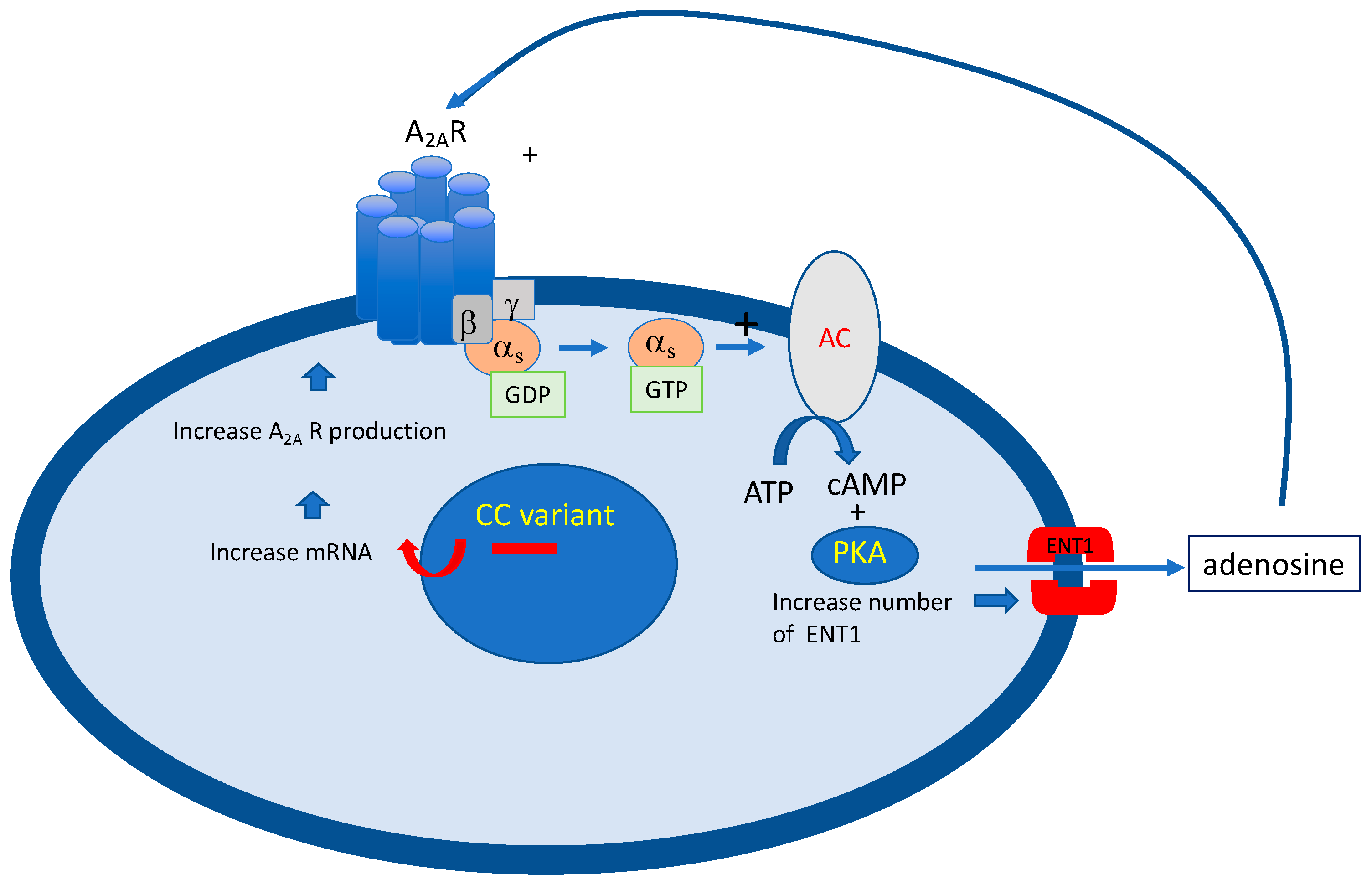
- Saadjian, A.Y.; Gerolami, V.; Giorgi, R.; Mercier, L.; Berge-Lefranc, J.-L.; Paganelli, F.; Ibrahim, Z.; By, Y.; Guéant, J.L.; Lévy, S.; et al. Head-up tilt induced syncope and adenosine A2A receptor gene polymorphism. Eur. Heart J. 2009, 30, 1510–1515. [Google Scholar] [CrossRef]
- Mitro, P.; Habalova, V.; Evin, L.; Muller, E.; Simurda, M.; Slaba, E.; Murin, P.; Valocik, G. Gene Polymorphism of the Adenosine A2a Receptor in Patients with Vasovagal Syncope. Pacing Clin. Electrophysiol. 2016, 39, 330–337. [Google Scholar] [CrossRef] [PubMed]
- Deharo, J.-C.; Mechulan, A.; Giorgi, R.; Franceschi, F.; Prevot, S.; Peyrouse, E.; Condo, J.; By, Y.; Ruf, J.; Brignole, M.; et al. Adenosine plasma level and A2A adenosine receptor expression: Correlation with laboratory tests in patients with neurally mediated syncope. Heart 2012, 98, 855–859. [Google Scholar] [CrossRef] [PubMed]
- Guieu, R.; Deharo, J.-C.; Ruf, J.; Mottola, G.; Kipson, N.; Bruzzese, L.; Gerolami, V.; Franceschi, F.; Ungar, A.; Tomaino, M.; et al. Adenosine and Clinical Forms of Neurally-Mediated Syncope. J. Am. Coll. Cardiol. 2015, 66, 204–205. [Google Scholar] [CrossRef] [Green Version]
- Mitro, P.; Lazúrová, Z.; Kerekanič, M.; Farkaš, J.; Popovňáková, M. Adenosine, adenosine-deaminase and implantable recorder outcome in syncopal patients. Pacing Clin. Electrophysiol. 2022, 45, 768–772. [Google Scholar] [CrossRef]
- Fragakis, N.; Antoniadis, A.P.; Saviano, M.; Vassilikos, V.; Pappone, C. The use of adenosine and adenosine triphosphate testing in the diagnosis, risk stratification and management of patients with syncope: Current evidence and future perspectives. Int. J. Cardiol. 2015, 183, 267–273. [Google Scholar] [CrossRef] [PubMed]
- Brignole, M.; Sutton, R.; Menozzi, C.; Garcia-Civera, R.; Moya, A.; Wieling, W.; Andresen, D.; Benditt, D.G.; Grovale, N.; De Santo, T.; et al. Lack of correlation between the responses to tilt testing and adenosine triphosphate test and the mechanism of spontaneous neurally mediated syncope. Eur. Heart J. 2006, 27, 2232–2239. [Google Scholar] [CrossRef] [PubMed] [Green Version]
- Guieu, R.; Sampiéri, F.; Bechis, G.; Rochat, H. Use of HPLC to measure circulating adenosine levels in migrainous patients. Clin. Chim. Acta 1994, 227, 185–194. [Google Scholar] [CrossRef] [PubMed]
- Marlinge, M.; Vairo, D.; Marolda, V.; Bruzzese, L.; Adjriou, N.; Guiol, C.; Kipson, N.; Bonnardel, A.; Gastaldi, M.; Kerbaul, F.; et al. Rapid measurment of adenosine concentration in human blood using fixed potential amperometry: Comparison with mass spectrometry and high performance liquid chroma-tography. J. Anal. Bioanal. Tech. 2017, 4, 1–4. [Google Scholar]
- Groppelli, A.; Brignole, M.; Chefrour, M.; Gastaldi, M.; El Oufir, F.; Deharo, J.C.; Parati, G.; Guieu, R. Adenosine Concentration in Patients with Neurally Mediated Syncope. Front. Cardiovasc. Med. 2022, 9, 900023. [Google Scholar] [CrossRef]
- Bruzzese, L.; Fromonot, J.; By, Y.; Durand-Gorde, J.-M.; Condo, J.; Kipson, N.; Guieu, R.; Fenouillet, E.; Ruf, J. NF-κB enhances hypoxia-driven T-cell immunosuppression via upregulation of adenosine A2A receptors. Cell Signal. 2014, 26, 1060–1067. [Google Scholar] [CrossRef]
- Bruzzese, L.; Rostain, J.-C.; Née, L.; Condo, J.; Mottola, G.; Adjriou, N.; Mercier, L.; Berge-Lefranc, J.-L.; Fromonot, J.; Kipson, N.; et al. Effect of hyperoxic and hyperbaric conditions on the adenosinergic pathway and CD26 expression in rat. J. Appl. Physiol. 2015, 119, 140–147. [Google Scholar] [CrossRef] [Green Version]
- Fromonot, J.; Chaumet, G.; Gavarry, O.; Rostain, J.-C.; Lucciano, M.; Joulia, F.; Brignole, M.; Deharo, J.-C.; Guieu, R.; Boussuges, A. Hyperoxia Improves Hemodynamic Status During Head-up Tilt Testing in Healthy Volunteers: A Randomized Study. Medicine 2016, 95, e2876. [Google Scholar] [CrossRef]
- Varani, K.; Laghi-Pasini, F.; Camurri, A.; Capecchi, P.L.; Maccherini, M.; Diciolla, F.; Ceccatelli, L.; Lazzerini, P.E.; Ulouglu, C.; Cattabeni, F.; et al. Changes of peripheral A2A adenosine receptors in chronic heart failure and cardiac transplantation. FASEB J. 2003, 17, 280–282. [Google Scholar] [CrossRef] [Green Version]
- Gariboldi, V.; Vairo, D.; Guieu, R.; Marlingue, M.; Ravis, E.; Lagier, D.; Mari, A.; Thery, E.; Collart, F.; Gaudry, M.; et al. Expressions of adenosine A2A receptors in coronary arteries and peripheral blood mononuclear cells are correlated in coronary artery disease patients. Int. J. Cardiol. 2017, 230, 427–431. [Google Scholar] [CrossRef]
- Gaudry, M.; Marlinge, M.; Deharo, P.; Vairo, D.; Bottone, S.; Mottola, G.; Kipson, N.; Criado, C.; Mace, P.; Chefrour, M.; et al. Pharmacological profile of adenosine A2A receptors in patients with lower extremity peripheral artery disease and associated coronary artery disease: A pilot study. Int. J. Cardiol. 2019, 285, 121–127. [Google Scholar] [CrossRef]
- By, Y.; Durand-Gorde, J.-M.; Condo, J.; Lejeune, P.-J.; Mallet, B.; Carayon, P.; Guieu, R.; Ruf, J. Production of an agonist-like monoclonal antibody to the human A2A receptor of adenosine for clinical use. Mol. Immunol. 2009, 46, 400–405. [Google Scholar] [CrossRef] [PubMed]
- Franceschi, F.; By, Y.; Peyrouse, E.; Fromonot, J.; Gerolami, V.; Kipson, N.; Boussuges, A.; Brignole, M.; Fenouillet, E.; Deharo, J.C.; et al. A2A adenosine receptor function in patients with vasovagal syncope. Europace 2013, 15, 1328–1332. [Google Scholar] [CrossRef] [PubMed]
- Jacquin, L.; Franceschi, F.; By, Y.; Durand-Gorde, J.-M.; Condo, J.; Deharo, J.-C.; Michelet, P.; Fenouillet, E.; Guieu, R.; Ruf, J. Search for adenosine A2A spare receptors on peripheral human lymphocytes. FEBS Open Bio 2012, 3, 1–5. [Google Scholar] [CrossRef] [Green Version]
- Clark, A.J. Mode of Action of Drugs on Cells; Edward Arnold and Co.: London, UK, 1933. [Google Scholar]
- Stephenson, R.P. A modification of receptor theory. Br. J. Pharmacol. Chemother. 1956, 11, 379–393. [Google Scholar] [CrossRef] [PubMed]
- Fenouillet, E.; Mottola, G.; Kipson, N.; Paganelli, F.; Guieu, R.; Ruf, J. Adenosine Receptor Profiling Reveals an Association between the Presence of Spare Receptors and Cardiovascular Disorders. Int. J. Mol. Sci. 2019, 20, 5964. [Google Scholar] [CrossRef] [PubMed] [Green Version]
- Srinivas, M.; Shryock, J.C.; Dennis, D.M.; Baker, S.P.; Belardinelli, L. Differential A1 Adenosine Receptor Reserve for Two Actions of Adenosine on Guinea Pig Atrial Myocytes. Mol. Pharmacol. 1997, 52, 683–691. [Google Scholar] [CrossRef] [PubMed] [Green Version]
- Kiss, Z.; Pak, K.; Zsuga, J.; Juhasz, B.; Varga, B.; Szentmiklosi, A.J.; Haines, D.D.; Tosaki, A.; Gesztelyi, R. The guinea pig atrial A1 adenosine receptor reserve for the direct negative inotropic effect of adenosine. Gen. Physiol. Biophys. 2013, 32, 325–335. [Google Scholar] [CrossRef] [Green Version]
- Daly, J.W.; Shi, D.; Nikodijevic, O.; Jacobson, K.A. The role of adenosine receptors in the central action of caffeine. Pharmacopsychoecologia 2015, 7, 201–213. [Google Scholar]
- Nagatomo, K.; Kubo, Y. Caffeine activates mouse TRPA1 channels but suppresses human TRPA1 channels. Proc. Natl. Acad. Sci. USA 2008, 105, 17373–17378. [Google Scholar] [CrossRef]
- Robertson, D.; Frölich, J.C.; Carr, R.K.; Watson, J.T.; Hollifield, J.W.; Shand, D.G.; Oates, J.A. Effects of Caffeine on Plasma Renin Activity, Catecholamines and Blood Pressure. N. Engl. J. Med. 1978, 298, 181–186. [Google Scholar] [CrossRef]
- Tofovic, S.P.; Kusaka, H.; Rominski, B.; Jackson, E.K. Caffeine Increases Renal Renin Secretion in a Rat Model of Genetic Heart Failure. J. Cardiovasc. Pharmacol. 1999, 33, 440–450. [Google Scholar] [CrossRef] [PubMed]
- Sra, J.S.; Jazayeri, M.R.; Avitall, B.; Dhala, A.; Deshpande, S.; Blanck, Z.; Akhtar, M. Comparison of Cardiac Pacing with Drug Therapy in the Treatment of Neurocardiogenic (Vasovagal) Syncope with Bradycardia or Asystole. N. Engl. J. Med. 1993, 328, 1085–1090. [Google Scholar] [CrossRef]
- Brignole, M.; Moya, A.; de Lange, F.J.; Deharo, J.-C.; Elliott, P.M.; Fanciulli, A.; Fedorowski, A.; Furlan, R.; Kenny, R.A.; Martín, A.; et al. 2018 ESC Guidelines for the diagnosis and management of syncope. Eur. Heart J. 2018, 39, 1883–1948. [Google Scholar] [CrossRef] [PubMed]
- Glikson, M.; Nielsen, J.C.; Kronborg, M.B.; Michowitz, Y.; Auricchio, A.; Barbash, I.M.; Barrabés, J.A.; Boriani, G.; Braunschweig, F.; Brignole, M.; et al. 2021 ESC Guidelines on cardiac pacing and cardiac resynchronization therapy. Europace 2022, 24, 71–164. [Google Scholar] [CrossRef] [PubMed]
- Flammang, D.; Church, T.R.; De Roy, L.; Blanc, J.-J.; Leroy, J.; Mairesse, G.H.; Otmani, A.; Graux, P.J.; Frank, R.; Purnode, P.; et al. Treatment of Unexplained Syncope: A multicenter, randomized trial of cardiac pacing guided by adenosin 5′-triphosphate testing. Circulation 2012, 125, 31–36. [Google Scholar] [CrossRef] [Green Version]
| Disclaimer/Publisher’s Note: The statements, opinions and data contained in all publications are solely those of the individual author(s) and contributor(s) and not of MDPI and/or the editor(s). MDPI and/or the editor(s) disclaim responsibility for any injury to people or property resulting from any ideas, methods, instructions or products referred to in the content. |
© 2023 by the authors. Licensee MDPI, Basel, Switzerland. This article is an open access article distributed under the terms and conditions of the Creative Commons Attribution (CC BY) license (https://creativecommons.org/licenses/by/4.0/).