Article

Lymphatic Defects in Zebrafish sox18 Mutants Are Exacerbated by Perturbed VEGFC Signaling, While Masked by Elevated sox7 Expression

1
Dipartimento di Bioscienze, Università degli Studi di Milano, Via Celoria 26, 20133 Milan, Italy
2
Dipartimento di Biotecnologie e Bioscienze, Università degli Studi di Milano-Bicocca, Piazza della Scienza 2, 20126 Milan, Italy
*
Author to whom correspondence should be addressed.
These authors contributed equally to this work.
Current address: Department of Endocrine and Metabolic Diseases, Lab of Endocrine and Metabolic Research, Istituto di Ricovero e Cura a Carattere Scientifico (IRCCS) Istituto Auxologico Italiano, 20149 Milan, Italy.
§
Current address: Dipartimento di Biotecnologie Mediche e Medicina Traslazionale, Università degli Studi di Milano, LITA, Via Fratelli Cervi 93, 20054 Segrate, Italy.
Cells 2023, 12(18), 2309; https://doi.org/10.3390/cells12182309
Submission received: 23 June 2023 / Revised: 5 September 2023 / Accepted: 7 September 2023 / Published: 19 September 2023
(This article belongs to the Special Issue Modeling Developmental Processes and Disorders in Zebrafish)

Abstract

:
Mutations in the transcription factor-coding gene SOX18, the growth factor-coding gene VEGFC and its receptor-coding gene VEGFR3/FLT4 cause primary lymphedema in humans. In mammals, SOX18, together with COUP-TFII/NR2F2, activates the expression of Prox1, a master regulator in lymphatic identity and development. Knockdown studies have also suggested an involvement of Sox18, Coup-tfII/Nr2f2, and Prox1 in zebrafish lymphatic development. Mutants in the corresponding genes initially failed to recapitulate the lymphatic defects observed in morphants. In this paper, we describe a novel zebrafish sox18 mutant allele, sa12315, which behaves as a null. The formation of the lymphatic thoracic duct is affected in sox18 homozygous mutants, but defects are milder in both zygotic and maternal-zygotic sox18 mutants than in sox18 morphants. Remarkably, in sox18 mutants, the expression of the closely related sox7 gene is elevated where lymphatic precursors arise. Sox7 could thus mask the absence of a functional Sox18 protein and account for the mild lymphatic phenotype in sox18 mutants, as shown in mice. Partial knockdown of vegfc exacerbates lymphatic defects in sox18 mutants, making them visible in heterozygotes. Our data thus reinforce the genetic interaction between Sox18 and Vegfc in lymphatic development, previously suggested by knockdown studies, and highlight the ability of Sox7 to compensate for Sox18 lymphatic dysfunction.

1. Introduction

SOX18, and the closely related SOX7 and SOX17 proteins, belong to the SOXF group of Sry-related HMG-box transcription factors [1], which play various roles in cardiovascular and lymphatic development [2]. Mutations in SOX18 underlie dominant and recessive forms of hypotrichosis–lymphedema–telangiectasia syndrome (HLTS) [3], combining defects in hair/eyelashes/eyebrows with lymphatic dysfunction, cutaneous red stains and, in some cases, renal failure (HLTRS) [4]. Recessive mutations correspond to missense mutations causing substitutions in key amino acids of the HMG-box DNA binding domain, while de novo dominant mutations correspond to nonsense mutations or frameshifts, leading to premature termination codons downstream of the DNA-binding domain coding sequence. The reported SOX18 pathogenic variants are very few, in a dozen of families, and yet the phenotypic spectrum is quite varied in the reported patients. Of note, two patients did not present with lymphatic dysfunction [5,6], whereas others already presented with lymphedema at birth and one died in utero with non-immune hydrops fetalis ([3,7] and references therein).
The spontaneous ragged mutants, named in the 1950s and 1960s for their characteristic sparse fur, represent the murine counterpart of the disease, with different alleles of increasing severity all associated with single-base deletions generating truncated, dominant-negative SOX18 proteins lacking transactivation ability [8,9]. The most severe allele, Sox18-RaOp, was found to cause edema, chylous ascites and superficial hemorrhage in neonates in the heterozygous state, while being embryonic lethal in the homozygous state with signs of severe lymphatic and vascular dysfunction [10,11]. Sox18-null mutant mice, produced by gene targeting, were viable and displayed only mild coat defects in the originally described mixed genetic background [12], raising the hypothesis of functional redundancy with the other SoxF group genes. In a C57BL/6 pure background, Sox18−/− mice were instead embryonic lethal by 14.5 days post-coitum (dpc) and showed gross subcutaneous edema like Sox18-RaOp homozygous embryos [11].
SOX18 is expressed in the endothelial cells (ECs) of all forming vessels in mice, including a subset of cells of the cardinal vein (CV), which also express the transcription factor NR2F2/COUP-TFII, prior to the expression of PROX1, a key transcription factor in lymphatic development [11,13,14]. SOX18 and COUP-TFII are able to directly activate the expression of Prox1, thus turning blood ECs (BECs) into lymphatic EC (LEC) progenitors. The other SOXF transcription factors, SOX7 and SOX17, were shown to share the ability to activate Prox1 transcription in vitro. However, they are not normally expressed in the CV during lymphatic development. When Sox18 is mutated, their expression is upregulated in the CV in the mixed genetic background and not in the C57BL/6 pure background, thus explaining the dramatic difference in lymphatic phenotype [15].
Vascular Endothelial Growth Factor C (VEGFC)/Vascular Endothelial Growth Factor Receptor 3 (VEGFR3) signaling plays a crucial role in lymphatic development (reviewed in [16]). In particular, the egress of LEC precursors from the CV and their subsequent migration are strictly dependent on a VEGFC gradient and on VEGFR3 in mice. Mutations in VEGFR3/FLT4, VEGFC, and genes involved in the processing of VEGFC are associated with lymphatic anomalies in humans (e.g., Milroy’s disease, Congenital Primary Lymphedema of Gordon, Hennekam syndrome; reviewed in [17]).
In zebrafish, the onset of lymphatic development can be visualized with transgenic reporter lines from around 1.5 days post-fertilization (dpf), when venous and lymphatic sprouts emerge from the posterior CV (PCV) [18,19,20]. Lymphatic precursors migrate dorsally to the horizontal myoseptum, where they constitute a transient population of parachordal lymphangioblasts (PLs), later migrating ventrally and dorsally along arterial intersegmental vessels (aISVs) to give rise to the thoracic duct (TD), the intersegmental lymphatic vessels, and the dorsal longitudinal lymphatic vessels ([21]; reviewed in [22]). The TD forms in the trunk just below the dorsal aorta (DA), between 3 and 5 dpf, from separate segments migrating rostrally and caudally to then interconnect in a single vessel [18,23].
Initial characterization of early phases of zebrafish lymphatic development, through knockdown studies, revealed a high degree of conservation of key molecular players and signaling pathways between fish and mammals. In particular, vegfc morphants showed an absence of TD formation [18,23], as vegfc is required for venous and lymphatic sprouting from the PCV [20]. The key relevance of Vegfc-Vegfr3 signaling for lymphatic development in zebrafish was later confirmed by the characterization of mutants in vegfr3/flt4 and vegfc identified through forward genetic screens [24,25,26].
Knockdown studies have also suggested conserved roles in lymphatic development for zebrafish homologues of Prox1, Coup-TFII, and Sox18 [18,27,28]. As for sox18, our and other groups have shown that the soxF genes sox7 and sox18 are largely coexpressed in the forming axial vessels and intersegmental vessels during vasculogenesis and primary angiogenesis, and that they play redundant roles in arteriovenous differentiation of ECs [29,30,31]. We noticed that sox18 expression persists in the PCV, while sox7 expression fades away in the PCV at developmental stages prior to secondary venous and lymphatic sprouting [28]. Knockdown of sox18 with morpholinos, either interfering with splicing or blocking translation, affects early trunk lymphatic development: lymphatic precursors at the horizontal myoseptum are reduced and TD formation is defective. Moreover, the synergistic effects of the simultaneous partial knockdown of sox18 and vegfc brought us to hypothesize a genetic interaction between the two factors in zebrafish lymphatic development [28].
The initial characterization of reverse genetic mutants in prox1a, prox1b, coup-TFII/nr2f2, and sox18 revealed no or marginal defects in TD formation, leading to the conclusion that the Prox1-Sox18-Coup-TFII transcription factor axis is dispensable for lymphatic development is zebrafish [32,33]. However, further analysis of a maternal-zygotic prox1a mutant pointed to a conserved crucial function of Prox1 in early phases of lymphatic differentiation [34]. Moreover, Vegfc was shown to be a key factor in enhancing prox1a expression in a subset of cells of the PCV, which later give rise to lymphatic precursors migrating towards the horizontal myoseptum [34].
This prompted us to reevaluate the role of Sox18 in zebrafish lymphatic development through the characterization of a novel mutant allele, sox18sa12315, from the Zebrafish Mutation Project [35]. Here, we combined this new mutant allele with partial vegfc knockdown to show the role of Sox18 in driving lymphatic patterning, and we unveiled the ability of Sox7 to compensate for Sox18 loss.

2. Materials and Methods

2.1. Zebrafish Lines and Maintenance

Zebrafish were raised and maintained according to established techniques [36] and to the European recommendations [37] and Italian regulations. The following lines were used: sox18sa1231 (from the Wellcome Sanger Institute, through the European Zebrafish Resource Center; [35]), Tg(fli1:EGFP)y1 [18,38], Tg(lyve1b:DsRed)nz101 [39], Tg(mrc1a:EGFP)y251;Tg(kdrl:mCherry)y171 [40].

2.2. Genotyping

The sox18sa12315 zebrafish mutant line is characterized by a G/A transition, in the sequence encoding the HMG-box domain of the sox18 gene, which causes the disruption of a restriction site for the BstNI/MvaI enzymes. Genomic DNA (gDNA) extraction was performed using incubation in lysis buffer (10 mM Tris-HCl pH 8, 1 mM EDTA pH 8, 0.3% Tween20, 0.3% glycerol) for 10 min at 98 °C, followed by 4 h of overnight digestion at 55 °C with Proteinase K (PK, 1 µg/µL final concentration) and PK inactivation for 10 min at 98 °C. gDNA was used for PCR amplification with the following primers: sox18-BstNI-F1: 5′-GATTGCATTTAGATGATGTTGTCCTG-3′ and sox18-BstNI-R1: 5′-CATCTTCTTGGGTTGTTTCTTCCTC-3′. In case of low yields, a second PCR was performed with the following internal primers: sox18-BstNI-F2: 5′-CAGTGCTCTGGCACTAGATTG-3′ and sox18-BstNI-R2: 5′-AAGCCTTGGAGAAGGAGACC-3′. PCR products were digested with MvaI, or its isoschizomer, BstNI, and analyzed on 3% agarose gels to discriminate the different genotypes, based on their digestion patterns.

2.3. MO Microinjections

Antisense morpholinos (MOs; Gene Tools, Philomath, OR, USA) were injected as described elsewhere [29]. As controls, we used uninjected embryos or injected a standard control oligo (std-MO), with no target in zebrafish embryos, to assess unspecific effects. We injected 0.125 or 0.25 pmol/embryo of sox7-MO1 [29], 0.045 pmol/embryo of vegfc-MO and 1 pmol/embryo of sox18-MO2 [28]. The sequences (5′-3′) of the morpholinos are as follows: std-MO: CCTCTTACCTCAGTTACAATTTATA; sox7-MO1: ACGCACTTATCAGAGCCGCCATGTG; vegfc-MO: AGACAGAAAATCCAAATAAGTGCAT; and sox18-MO2: gtgagtgtcttacCCAGCATTTTAC (intron-targeting sequence in lowercase).

2.4. In Situ Hybridizations

Whole-mount in situ hybridizations (ISHs) were carried out essentially as previously described [28,29,41]. The following primers were used in PCR reactions to generate DNA templates for RNA probes: sox18, fw: 5′-GGAGCCAGGAGTTACAAAACAC-3′, and rev: 5′-CTAATACGACTCACTATAGGGCTCCATATGTGCACCAGACTTC-3; sox7, fw: 5′-CCCGCTTGATAAAGATGACG-3′, and rev: 5′-CTAATACGACTCACTATAGGGTTGGAAGAGACCAGCCTCAC-3′; sox17, fw: 5′- ACGAAACAAGCGATTGGAGC-3′, and rev: 5′- CTAATACGACTCACTATAGGGTGCCATTTAAGCTGCTGACA-3′; plvapb/vsg1, fw: 5′-CTACCCACAAGTGTGACAGTGC-3′, and rev: 5′- CTAATACGACTCACTATAGGGGATCAGATTCCTTCTCCACACC-3′. Additionally, antisense DIG-labeled RNA probes for sox7 and cdh5 were transcribed in vitro using linearized plasmids as DNA templates [29,42].
Images were taken with a Leica MZFLIII epifluorescence stereomicroscope equipped with a DFC 480-R2 digital camera and LAS imaging software version 4.13.0 (Leica, Wetzlar, Germany).
The PCV/DA ratio in sox18 and sox7 ISHs was evaluated using ImageJ software version 1.47v, on images converted to 8-bit images. We set the threshold color with constant parameters and measured the number of positive pixels in 8 rectangular ROIs (regions of interest) selected in the axial vessels of the trunk. In each image, we set the ROIs in the DA (dorsal aorta) and PCV (posterior cardinal vein) under intersomitic vessels 1, 4, 7, and 10 (counting from the anus). We then calculated the ratio between the sum of all values obtained in PCV ROIs and the sum of all values of DA ROIs. Data were plotted using GraphPad Prism version 9.0.0.

2.5. Phenotypic Analyses

We evaluated embryo circulation in the trunk/tail region as reported in [29]. All larvae analyzed to study lymphatic development were circulating, and we scored them for TD formation essentially as described in [28], along 10 consecutive trunk segments, counted rostrally from the anus, in larvae at 5 dpf. Confocal microscopy was performed on a Nikon Eclipse-Ti inverted microscope (Nikon, Tokyo, Japan), and images were processed using Adobe Photoshop version CS6 or NIS-Viewer version 4.11.0.

2.6. Statistical Analyses

Statistical analyses were performed with a t-test or one-way ANOVA followed byDunnett’s Multiple Comparison post-test, when needed, using GraphPad Prism version 9.0.0 (GraphPad, San Diego, CA, USA). In the graphs, * and ** mark statistically significant data with a p-value < 0.05 and <0.01, respectively. Statistically highly significant data, with a p-value < 0.001 and <0.0001, are marked by *** and ****, respectively.

3. Results

3.1. The sa12315 Mutation Is a Loss-of-Function Allele of sox18

To better address the role of Sox18 in zebrafish lymphatic development, we decided to characterize the sa12315 mutation in the sox18 locus, generated at the Wellcome Sanger Institute within the Zebrafish Mutation Project [35]. This mutant allele corresponds to a G > A transition in the second of the two exons of the sox18 gene, introducing a premature termination codon (W132X) within the sequence coding for the DNA-binding domain of the Sox18 transcription factor (Figure 1A). This mutation is expected to truncate the protein in the second of the three alpha helices of the HMG-box domain, thus eliminating the transactivation domain, which is more C-terminal in the wild-type form, but also disrupting the DNA-binding ability of Sox18.
To confirm that the sa12315 mutant allele corresponds to a loss-of-function allele, we exploited the known functional redundancy between Sox18 and Sox7 in vascular development [29]. The progeny of fish heterozygous for the sa12315 mutation were injected with subcritical doses of a sox7 morpholino, which was already shown to cause little effects on its own but dramatic alterations in trunk–tail blood circulation when combined with the partial knockdown of sox18 [29]. At 3 days post-fertilization (3 dpf), half of the progeny showed a circulatory phenotype in the trunk–tail region when injected with as little as 0.125 pmoles of sox7-MO (n = 42, 21 noncirculating embryos). Doubling the dose of sox7-MO (0.25 pmoles) caused over three-quarters of the injected embryos to show a circulatory phenotype (n = 103, 88 noncirculating embryos). Circulatory defects were far less pronounced in controls, i.e., uninjected or injected with a standard control MO (only around 10%, n = 152 and 99, respectively) (Figure 1B). These data strongly suggest that circulatory defects are associated with the partial knockdown of sox7 combined with a reduction in or loss of functional Sox18 in heterozygous or homozygous sa12315 mutants, respectively (see also Table S1, showing genotype–phenotype analysis of a small clutch of embryos).
Moreover, we characterized, at the molecular level, the effect of partial knockdown of sox7 in the progeny of fish heterozygous for the sa12315 mutation. We previously highlighted that plvapb (also known as vsg1) is one of the genes downregulated in sox7 and sox18 double partial morphants [29]. In situ hybridizations (ISHs) revealed that the endothelial plvapb signal is reduced in a genotype-dependent way in embryos carrying one or two sa12315 alleles injected with a subcritical amount of sox7-MO (Figure 1C), whereas the expression of the endothelial marker cdh5 is largely unaffected.

3.2. sa12315 Mutants Show Mild Lymphatic Defects, Which Are Exacerbated by Perturbed VEGFC Signaling

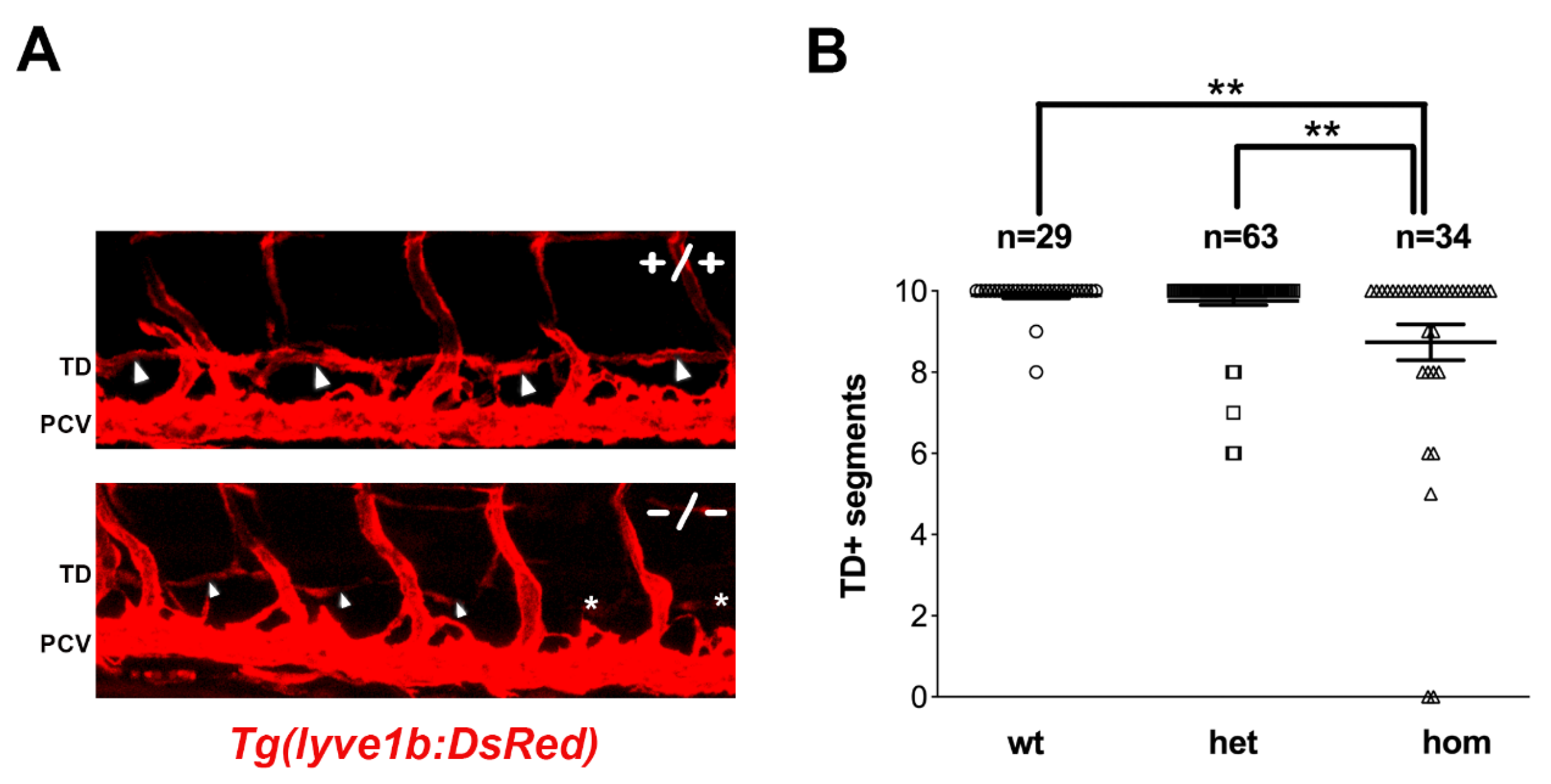
To address the impact of the sox18sa12315 mutation on lymphatic development, we scored the formation of the thoracic duct (TD) at 5 dpf in the larvae derived from matings of heterozygous mutant fish. The sox18sa12315 mutation was introduced in several transgenic reporter lines, i.e., Tg(fli1:EGFP)y1, Tg(lyve1b:DsRed)nz101, and Tg(mrc1a:EGFP)y251;Tg(kdrl:mCherry)y171, which allowed the visualization of blood and lymphatic vessels [18,39,40]. Lymphatic thoracic duct formation was scored in ten consecutive trunk segments of all larvae, which were subsequently genotyped, thus revealing a mild reduction in thoracic duct segments in homozygous sox18sa12315 mutants compared to siblings (Figure 2 and Figure 3 uninjected, Figure S2A). Though modest, this reduction was sizeable and statistically significant in all reporter lines.
We previously reported that slight perturbations in VEGFC/VEGFR3 signaling, via partial knockdown of vegfc or flt4 with morpholinos, synergize with the morpholino-mediated partial knockdown of sox18 in inducing lymphatic defects in zebrafish [28] (Figure S1B). We therefore tested the impact of the microinjection of low doses of vegfc-MO in embryos derived from matings of heterozygous sox18sa12315 mutant fish. Defects in the formation of the thoracic duct were exacerbated and became evident not only in homozygous but also in heterozygous sox18sa12315 mutant larvae compared to wild-type larvae, using both the fli1:EGFP and the lyve1b:DsRed reporters (Figure 3B and Figure S3).

3.3. sox18sa12315 Mutants Have Milder Lymphatic Phenotypes Than sox18 Morphants

To help in the comparison of the effect of the sa12315 mutation (this paper) with that of the previously reported sox18-MOs [28], we decided to plot the sox18 morphant data in the Tg(fli1:EGFP) reporter line as they are now shown here for sox18sa12315 mutants (compare Figure S1A with Figure 3). Moreover, we also directly compared thoracic duct formation in sox18sa12315 mutants and in sox18 morphants in the Tg(mrc1a:EGFP);Tg(kdrl:mCherry) double reporter line, in which both sox18 mutation and knockdown had not been previously analyzed (Figure S2). Although statistically significant, the difference in the mean number of TD+ segments in sox18 wild-type larvae versus homozygous mutant larvae is in the order of 10–20% in the two transgenic lines. On the other hand, there is an almost twofold to over threefold reduction in the mean number of TD+ segments in sox18 morphants versus control larvae injected with a standard morpholino, depending on the transgenic reporter line (Tg(fli1:EGFP) and Tg(mrc1a:EGFP);Tg(kdrl:mCherry), respectively).
Finally, given the great contribution of the maternal component in the phenotype of prox1a mutants [34], we decided to generate maternal-zygotic sox18sa12315 mutants (hereafter MZsox18 mutants). Homozygous zygotic sox18sa12315 mutants (Zsox18) are present at expected Mendelian ratios as young adults, in the progeny of sox18sa12315 heterozygotes, and are fertile. MZsox18 mutants were therefore generated by crossing Zsox18 mutant females and males and analyzed up to 5 dpf, to score for lymphatic thoracic duct formation. As shown in Figure S4A, there is a statistically significant reduction in the number of TD+ segments in MZsox18 mutants versus wild-type larvae. Nevertheless, TD formation defects appear comparable in MZsox18 mutants and in Zsox18 mutants (compare Figure S4A and Figure 3).
Taken together, these data confirm that Sox18 plays a role in lymphatic development in zebrafish but highlight that sox18sa12315 mutants have milder lymphatic phenotypes than sox18 morphants.

3.4. Ectopic Expression of sox7 in the PCV of sox18sa12315 Mutants, but Not sox18 Morphants

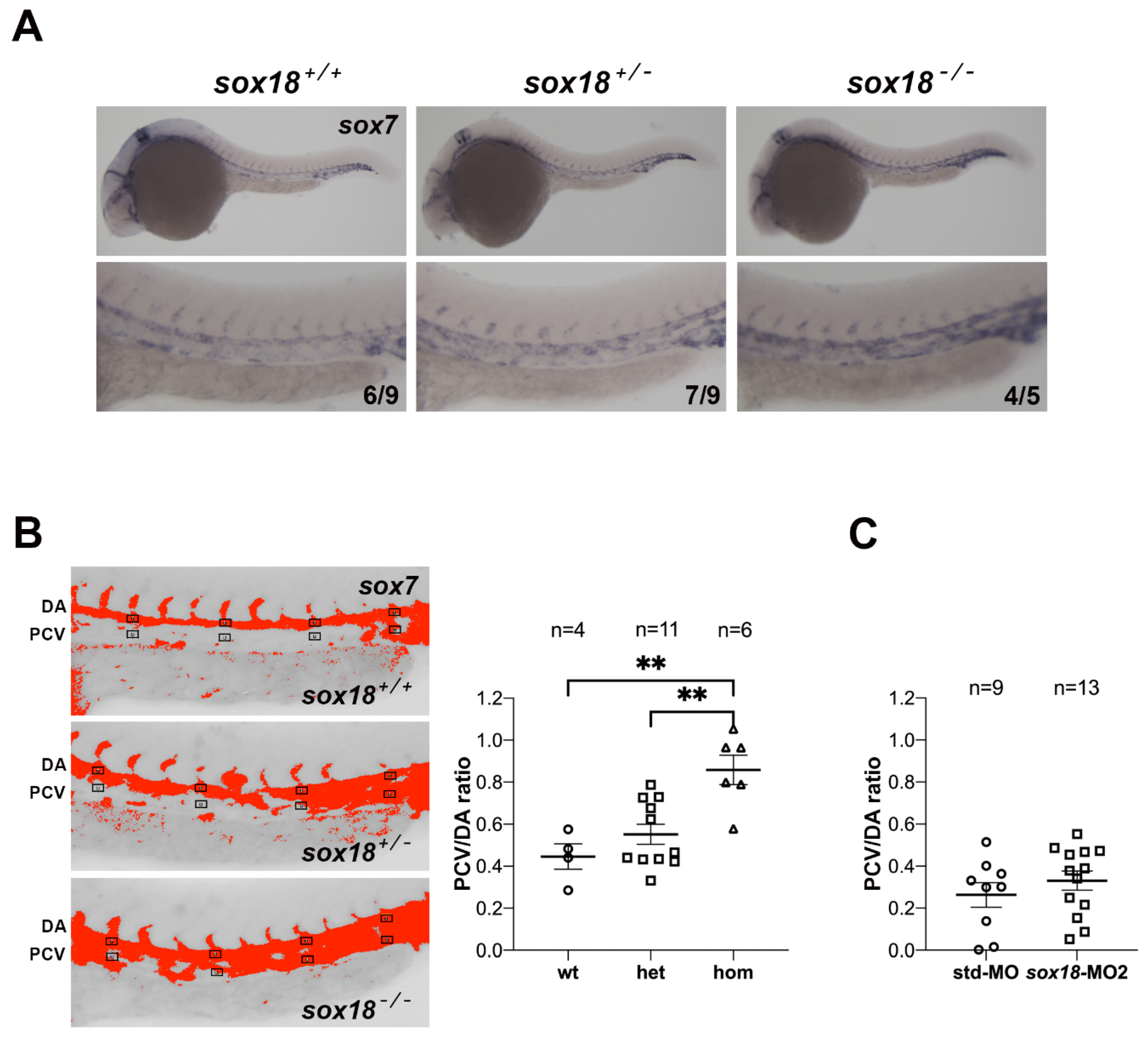
When coexpressed, sox18 and sox7 play redundant roles in vascular development in zebrafish [29,30,31]. Moreover, in mice, Sox7 and Sox17, which are not normally expressed during lymphatic development, can be activated in the absence of SOX18 function and act as modifiers of the lymphangiogenic defects caused by Sox18 dysfunction in certain strains [15]. These data prompted us to analyze the expression of sox7 in sox18sa12315 mutants at developmental stages preceding lymphatic sprouting from the posterior cardinal vein (PCV). At these stages, in wild-type conditions, sox7 ISH staining persists in the dorsal aorta (DA) and largely ends in the PCV (Figure 4A), as previously reported [28]. In homozygous zygotic sox18sa12315 mutants, we noticed clearly elevated sox7 staining in the PCV; moreover, most heterozygotes presented an intermediate sox7 ISH signal in the PCV with respect to wild-type embryos and homozygous mutants (Figure 4A, Table S2). Quantification of sox7 ISH signals in several small areas within the DA and the PCV to calculate PCV/DA ratios (Figure 4B) further highlighted the increased sox7 expression in the PCV of zygotic sox18sa12315 mutants. Remarkably, when the same analysis was performed on sox7 ISHs of sox18 morphants, no significant change was found with respect to controls injected with std-MO (Figure 4C and Figure S6).
We then checked for sox7 expression in MZsox18 mutant embryos around 30 hpf. Here, again, the sox7 ISH signal in the PCV is elevated in MZsox18 mutant embryos with respect to wild-type controls (Figure S4B).
We did not notice changes in sox18 expression by ISHs between zygotic sox18sa12315 mutants and wild-type siblings (Figure S5A): strong expression of sox18 both in the DA and in the PCV is confirmed by PCV/DA ratio analysis. Similarly, endothelial sox17 expression, which is confined to areas of the DA, is not elevated in the PCV of sox18sa12315 mutants (Figure S5B).
We therefore conclude that the elevated sox7 expression in the PCV of zygotic and maternal zygotic sox18sa12315 mutants, but not sox18 morphants, could partially compensate for the lack of functional Sox18 and explain the mild lymphatic phenotype of sox18sa12315 mutants. We currently have no molecular explanation for the specific upregulation of sox7 expression in the PCV of zebrafish sox18 mutants. Since our ISH data do not reveal reduced sox18 expression in zygotic sox18 mutants, we tend to exclude the transcriptional compensation mechanism elicited by nonsense-mediated RNA decay [43,44]. Remarkably, Sox7 expression is also upregulated in Sox18 mutants in mice, in a strain-specific way [15], possibly pointing to an evolutionarily conserved mechanism to be further addressed.

4. Discussion

Knockdown studies and reverse genetic analyses have so far stimulated controversial conclusions on the role of Sox18 in early phases of lymphatic development in zebrafish [28,33]. Here, we address this issue using an independent loss-of-function allele, sox18sa12315. Our current data support the notion that Sox18 is indeed involved in lymphatic development in zebrafish, with a close interplay with the key Vegfc signaling, as already pointed out in knockdown studies and using a dominant-negative Sox18-RaOp mutant protein [28]. Thoracic duct formation defects, though statistically significant, are much milder in both zygotic and maternal-zygotic sox18sa12315 mutants than in sox18 morphants. Remarkably, in zygotic and maternal-zygotic sox18sa12315 mutants, we found elevated expression of the closely related sox7 gene in the PCV, with respect to wild-type embryos, at developmental stages preceding lymphatic sprouting. On the contrary, sox7 expression in the PCV was unchanged in sox18 morphants with respect to control embryos, and much lower in the PCV than in the DA. Apparently, sox18 is the main soxF gene expressed in the PCV at developmental stages preceding lymphatic sprouting in wild-type conditions, but elevated expression of sox7 can partially mask the lack of a functional Sox18 protein in sox18 mutants, thus resulting in only mild lymphatic defects.
van Impel and colleagues [33] previously analyzed another sox18 mutant allele, sox18hu10320, and reported no vascular or lymphatic defects at 5 dpf when scoring for thoracic duct formation. The hu10320 mutation is a frameshift mutation upstream of the DNA-binding HMG-box domain coding sequence and is thus an LOF mutation, similarly to the sa12315 mutation we characterize here, which introduces a stop codon within the HMG-box domain coding sequence and is thus predicted to give rise to a non-functional Sox18 protein. Since the authors did not report data on the expression of the other soxF genes in the sox18hu10320 mutants, it is intriguing to speculate that sox7 expression might also be elevated in that context, but this remains to be analyzed.
Even the presence of a single mutant sox18hu10320 allele dramatically enhances the penetrance of the vascular circulatory phenotype in sox7hu5626 homozygous mutants [45], and only sox7/sox18 double homozygous mutants fully recapitulate the circulatory phenotype originally described in sox7/sox18 double knocked-down embryos, where arterio-venous shunts result from incomplete acquisition of arterio-venous identity in endothelial cells [29,30,31]. This strengthens the notion that soxF genes can play redundant roles when coexpressed.
While our current work confirms a role for Sox18 in early trunk lymphatic development, a recent paper by Arnold and colleagues revealed that SoxF transcription factors are also involved in facial lymphatic development in zebrafish [46]. The authors show reduced facial lymphatic sprouting in sox7/sox18 double partial morphants as well as in sox7/sox18 double homozygous mutants (sox7hu5626/hu5626; sox18hu10320/hu10320) or even in sox7 homozygous mutants lacking just one functional allele of sox18 (sox7hu5626/hu5626; sox18 +/hu10320).
The fine relative expression of soxF genes in different vascular beds, as well as their differential expression in mutant backgrounds, could therefore account for the relevance of each member of the F group of transcription factors in lymphatic development, as already shown in mice. Mice lacking SOX18 have an overt lymphatic phenotype in a pure C57BL/6 genetic background but are compensated by the strain-specific upregulation of Sox7 and Sox17 in the cardinal vein in a mixed 129-CD1 genetic background [11,15] Here, we report the elevated expression of sox7 in the PCV in zebrafish sox18sa12315 mutants, which show a milder trunk lymphatic phenotype than sox18 morphants, while the expression of sox17 appears to be unaffected. Zebrafish Sox17 is unique among SoxF proteins as it lacks the β-catenin interaction motif, which is otherwise present in all SOX group-F proteins [2,47]. Moreover, the temporal expression profile of sox17 in embryonic and early larval stages diverges from that of sox7 and sox18 (Figure S2 in [29]), and its spatial expression in the vascular system appears to be much more marginal and restricted to the DA, while excluded from the PCV, at embryonic stages. Recently, a new role emerged for sox17 in a novel vascularization process wherein blood vascular vessels arise from existing lymphatics in the anal fin, an adult-specific structure that is established at metamorphosis [48]. Das and colleagues report that mosaic overexpression of sox17 in ECs negatively affected TD formation in injected embryos, pointing to a new role of Sox17 in the suppression of LEC fate.
The emerging picture is that SoxF proteins are collectively at play in processes involving EC plasticity (e.g., acquisition of arterial–venous identity, BEC to LEC transition, LEC to BEC transdifferentiation) and EC migration, but their relative contribution to these processes might be different in different organisms.
Nicenboim and colleagues [49] described a niche of specialized angioblasts in the PCV of zebrafish embryos, which gives rise to cells with lymphatic fates when induced by endodermal Wnt5b. When wnt5b was knocked down, the expression of sox18, lyve1b and coup-tfII/nr2f2 was reduced in the PCV, together with the number of prox1a+ cells, whereas the expression of vegfc and ccbe1 remained unchanged [49]. Interestingly, wnt5b morphants showed reduced PL+ and TD+ segments, but no alteration in venous ISVs [49]. This highlights a specific defect in lymphatic sprouting in wnt5b morphants, resembling what we described in sox18 morphants [28].
Our previous knockdown studies suggested a genetic interaction between Sox18 and Vegfc to regulate lymphangiogenesis in zebrafish [28]. Combined partial knockdown of sox18 and vegfc, using subcritical amounts of morpholinos, synergistically impaired venous and lymphatic sprouting from the PCV and parachordal lymphatic precursors at the horizontal myoseptum and interfered with TD formation. Although sox18sa12315 mutants only show mild TD formation defects, the partial knockdown of vegfc greatly enhances the trunk lymphatic phenotype in sox18sa12315 homozygous mutants but also reveals a statistically significant reduction in TD formation in sox18sa12315 heterozygotes compared to wild-type siblings. This genotype-dependent effect of the partial knockdown of vegfc further strengthens the sox18vegfc genetic interaction while confirming a role for Sox18 in zebrafish lymphatic development. The molecular mechanisms underlying this genetic interaction remain to be elucidated and warrant further studies.
SOX18 in mammals activates the expression of Prox1 in a subset of cells of the CV, and when ectopically expressed in the CV, SOX7 and SOX17 are also able to induce Prox1 [11,15]. Not only is VEGFC signaling crucial for the budding and migration of Prox1+ lymphatic cells but VEGFC-mediated activation of VEGFR3 signaling is also necessary to maintain Prox1 expression in LEC progenitors, and the Prox1-Vegfr3 feedback loop controls the number of LEC progenitors and budding LECs [50]. In zebrafish, Vegfc plays a prominent role in regulating the division of bipotential Prox1+ precursor cells within the CV and upregulating Prox1 expression in daughter cells fated to become LECs [34]. When overexpressed, Vegfc is sufficient to induce prox1a expression in venous ECs, even if sox18 and sox7 are partially knocked down [34].
Our current data point to a more relevant role of Sox18 in zebrafish lymphatic development under limiting Vegfc conditions. It would be interesting to address whether Sox18 acts on Prox1 expression in the CV in zebrafish under slightly perturbed Vegfc conditions.
We previously reported that the heat-shock inducible expression of the dominant-negative SOX18–RaOp mutant protein in zebrafish causes impairment of lymphatic precursor sprouting from the vein and venous intersomitic vessel (vISV) formation, when induced at 29 hpf, just prior to secondary angiogenesis [28]. In heat-shocked Sox18-RaOp transgenic animals, we previously noticed a reduced level of vegfc RNA using qRT-PCR. ISH analysis of vegfc expression in sox18 morphants at one specific developmental stage did not reveal variations with respect to control embryos in the DA region, but vegfc RNA injection could partially rescue the lymphatic phenotype of sox18 morphants, thus pointing to a SoxF-VegfC axis [28].
Recently, SOX7 was shown to impact dermal lymphatic patterning in mice through direct and indirect regulation of Vegfc expression in blood ECs [51]. The authors point to fine-tuning of VEGFC levels produced in arteries, where SOX7 is expressed and can promote the expression of Hey1, a Notch effector that can repress the expression of Vegfc, or directly repress the expression of Vegfc through protein–protein interaction with HEY1 [51]. In their model, the loss of SOX7 function in arterial ECs was shown to cause an increase of endothelial VEGFC, which leads to more abundant and less polarized LEC progenitors in the CV, in the trunk, and to hyperproliferation of LEC progenitors and migration defects in dermal lymphatics.
The roles played by SoxF transcription factors in the regulation of Vegfc in zebrafish warrant future research to better elucidate evolutionarily conserved and non-conserved regulatory mechanisms.

Supplementary Materials

The following supporting information can be downloaded at: https://www.mdpi.com/article/10.3390/cells12182309/s1.

Author Contributions

Conceptualization, M.B.; validation, M.B., S.M. (Silvia Moleri) and S.M. (Sara Mercurio); formal analysis, S.M. (Silvia Moleri) and S.M. (Sara Mercurio); investigation, S.M. (Silvia Moleri), S.M. (Sara Mercurio), A.P. (Alex Pezzotta), D.D., A.B., A.P. (Alice Plebani), G.L. and M.D.F.; writing—original draft preparation, M.B. and S.M. (Silvia Moleri); writing—review and editing, M.B. and S.M. (Sara Mercurio); visualization, S.M. (Sara Mercurio), S.M. (Silvia Moleri) and M.B.; supervision, M.B., S.M. (Silvia Moleri) and S.M. (Sara Mercurio); funding acquisition, M.B. All authors have read and agreed to the published version of the manuscript.

Funding

We acknowledge financial support from Regione Lombardia (grant SAL-01 to M.B.) and Fondazione Cariplo (grant 2011-0555 to M.B.). We also acknowledge institutional support from the University of Milan (Linea 2).

Institutional Review Board Statement

Animal research was approved by the Italian Ministry of Health (208/2019-PR).

Data Availability Statement

The authors declare that the data supporting the findings of this study are available within the article and its supplementary information file.

Acknowledgments

We thank Brant M. Weinstein for the Tg(mrc1a:EGFP)y251;Tg(kdrl:mCherry)y171 line, the EZRC at KIT (Germany) for distributing the sa12315 mutant line provided by Derek Stemple (Wellcome Trust Sanger Institute, UK), Germano Gaudenzi for his advice on ImageJ analysis, and Alessandro Fantin for critical reading of the manuscript. D.D. was a fellow of the PhD Course in Molecular and Cellular Biology, University of Milan; S.M. (Silvia Moleri) was the recipient of a post-doctoral fellowship from the University of Milan. The authors acknowledge support from the University of Milan through the APC initiative.

Conflicts of Interest

The authors declare no conflict of interest.

References

  1. Bowles, J.; Schepers, G.; Koopman, P. Phylogeny of the SOX Family of Developmental Transcription Factors Based on Sequence and Structural Indicators. Dev. Biol. 2000, 227, 239–255. [Google Scholar] [CrossRef]
  2. François, M.; Koopman, P.; Beltrame, M. SoxF genes: Key players in the development of the cardio-vascular system. Int. J. Biochem. Cell Biol. 2010, 42, 445–448. [Google Scholar] [CrossRef] [PubMed]
  3. Irrthum, A.; Devriendt, K.; Chitayat, D.; Matthijs, G.; Glade, C.; Steijlen, P.M.; Fryns, J.-P.; Van Steensel, M.A.M.; Vikkula, M. Mutations in the Transcription Factor Gene SOX18 Underlie Recessive and Dominant Forms of Hypotrichosis-Lymphedema Telangiectasia. Am. J. Hum. Genet. 2003, 72, 1470–1478. [Google Scholar] [CrossRef]
  4. Moalem, S.; Brouillard, P.; Kuypers, D.; Legius, E.; Harvey, E.; Taylor, G.; François, M.; Vikkula, M.; Chitayat, D. Hypotrichosis-lymphedema-telangiectasia-renal defect associated with a truncating mutation in the SOX18 gene. Clin. Genet. 2015, 87, 378–382. [Google Scholar] [CrossRef]
  5. Wünnemann, F.; Kokta, V.; Leclerc, S.; Thibeault, M.; McCuaig, C.; Hatami, A.; Stheneur, C.; Grenier, J.-C.; Awadalla, P.; Mitchell, G.A.; et al. Aortic Dilatation Associated with a De Novo Mutationin the SOX18 Gene: Expanding the Clinical Spectrum of Hypotrichosis-Lymphedema-Telangiectasia Syndrome. Can. J. Cardiol. 2016, 32, 135.e1–135.e7. [Google Scholar] [CrossRef]
  6. Atiş, G.; Sam Sari, A.; Soylu, E.; Akgün Dogan, O. A Case with Hypotrichosis-LymphedemaTelangiectasia Syndrome with Hair Shaft Fragility. Skin Appendage Disord. 2022, 8, 511–514. [Google Scholar] [CrossRef]
  7. Coulie, R.; Niyazov, D.M.; Gambello, M.J.; Fastré, E.; Brouillard, P.; Vikkula, M. Hypotrichosis-lymphedema-telangiectasia syndrome: Report of ileal atresia associated with a SOX18 de novo pathogenic variant and review of the phenotypic spectrum. Am. J. Med. Genet. 2021, 185A, 2153–2159. [Google Scholar] [CrossRef]
  8. Pennisi, D.; Gardner, J.; Chambers, D.; Hosking, B.; Peters, J.; Muscat, G.; Abbott, C.; Koopman, P. Mutations in Sox18 underlie cardiovascular and hair follicle defects in ragged mice. Nat. Genet. 2000, 24, 434–437. [Google Scholar] [CrossRef]
  9. Downes, M.; Koopman, P. SOX18 and the Transcriptional Regulation of Blood Vessel Development. Trends Cardiovasc. Med. 2001, 11, 318–324. [Google Scholar] [CrossRef]
  10. Downes, M.; François, M.; Ferguson, C.; Parton, R.G.; Koopman, P. Vascular defects in a mouse model of hypotrichosis-lymphedema-telangiectasia syndrome indicate a role for SOX18 in blood vessel maturation. Hum. Mol. Genet. 2009, 18, 2839–2850. [Google Scholar] [CrossRef]
  11. François, M.; Caprini, A.; Hosking, B.; Orsenigo, F.; Wilhelm, D.; Browne, C.; Paavonen, K.; Karnezis, T.; Shayan, R.; Downes, M.; et al. Sox18 induces development of the lymphatic vasculature in mice. Nature 2008, 456, 643–647. [Google Scholar] [CrossRef]
  12. Pennisi, D.; Bowles, J.; Nagy, A.; Muscat, G.; Koopman, P. Mice Null for Sox18 Are Viable and Display a Mild Coat Defect. Mol. Cell. Biol. 2000, 20, 9331–9336. [Google Scholar] [CrossRef]
  13. Wigle, J.T.; Oliver, G. Prox1 Function Is Required for the Development of the Murine Lymphatic System. Cell 1999, 98, 769–778. [Google Scholar] [CrossRef]
  14. Srinivasan, R.S.; Geng, X.; Yang, Y.; Wang, Y.; Mukatira, S.; Studer, M.; Porto, M.P.R.; Lagutin, O.; Oliver, G. The nuclear hormone receptor Coup-TFII is required for the initiation and early maintenance of Prox1 expression in lymphatic endothelial cells. Genes Dev. 2010, 24, 696–707. [Google Scholar] [CrossRef]
  15. Hosking, B.; François, M.; Wilhelm, D.; Orsenigo, F.; Caprini, A.; Svingen, T.; Tutt, D.; Davidson, T.; Browne, C.; Dejana, E.; et al. Sox7 and Sox17 are strain specific modifiers of the lymphangiogenic defects caused by Sox18 dysfunction in mice. Development 2009, 136, 2385–2391. [Google Scholar] [CrossRef] [PubMed]
  16. Secker, G.A.; Harvey, N.L. VEGFR signaling during lymphatic vascular development: From progenitor cells to functional vessels. Dev. Dyn. 2015, 2443, 323–331. [Google Scholar] [CrossRef]
  17. Martin-Almedina, S.; Mortimer, P.S.; Ostergaard, P. Development and physiological functions of the lymphatic system: Insights from human genetic studies of primary lymphedema. Physiol. Rev. 2021, 101, 1809–1871. [Google Scholar] [CrossRef]
  18. Yaniv, K.; Isogai, S.; Castranova, D.; Dye, L.; Hitomi, J.; Weinstein, B.M. Live imaging of lymphatic development in the zebrafish. Nat. Med. 2006, 12, 711–716. [Google Scholar] [CrossRef]
  19. Isogai, S.; Lawson, N.D.; Torrealday, S.; Horiguchi, M.; Weinstein, B.M. Angiogenic network formation in the developing vertebrate trunk. Development 2003, 130, 5281–5290. [Google Scholar] [CrossRef]
  20. Hogan, B.M.; Bos, F.L.; Bussmann, J.; Witte, M.; Chi, N.C.; Duckers, H.J.; Schulte-Merker, S. ccbe1 is required for embryonic lymphangiogenesis and venous sprouting. Nat. Genet. 2009, 41, 396–398. [Google Scholar] [CrossRef]
  21. Bussmann, J.; Bos, F.L.; Urasaki, A.; Kawakami, K.; Duckers, H.J.; Schulte-Merker, S. Arteries provide essential guidance cues for lymphatic endothelial cells in the zebrafish trunk. Development 2010, 137, 2653–2657. [Google Scholar] [CrossRef] [PubMed]
  22. Hogan, B.M.; Schulte-Merker, S. How to Plumb a Pisces: Understanding Vascular Development and Disease Using Zebrafish Embryos. Dev. Cell 2017, 42, 576–583. [Google Scholar] [CrossRef]
  23. Küchler, A.M.; Gjini, E.; Peterson-Maduro, J.; Cancilla, B.; Wolburg, H.; Schulte-Merker, S. Development of the Zebrafish Lymphatic System Requires Vegfc Signaling. Curr. Biol. 2006, 16, 1244–1248. [Google Scholar] [CrossRef]
  24. Hogan, B.M.; Herpers, R.; Witte, M.; Heloterä, H.; Alitalo, K.; Duckers, H.J.; Schulte-Merker, S. Vegfc/Flt4 signalling is suppressed by Dll4 in developing zebrafish intersegmental arteries. Development 2009, 136, 4001–4009. [Google Scholar] [CrossRef]
  25. Villefranc, J.A.; Nicoli, S.; Bentley, K.; Jeltsch, M.; Zarkada, G.; Moore, J.C.; Gerhardt, H.; Alitalo, K.; Lawson, N.D. A truncation allele in vascular endothelial growth factor c reveals distinct modes of signaling during lymphatic and vascular development. Development 2013, 140, 1497–1506. [Google Scholar] [CrossRef] [PubMed]
  26. Le Guen, L.; Karpanen, T.; Schulte, D.; Harris, N.C.; Koltowska, K.; Roukens, G.; Bower, N.I.; van Impel, A.; Stacker, S.A.; Achen, M.G.; et al. Ccbe1 regulates Vegfc-mediated induction of Vegfr3 signaling during embryonic lymphangiogenesis. Development 2014, 141, 1239–1249. [Google Scholar] [CrossRef]
  27. Aranguren, X.L.; Beerens, M.; Vandevelde, W.; Dewerchin, M.; Carmeliet, P.; Luttun, A. Transcription factor COUP-TFII is indispensable for venous and lymphatic development in zebrafish and Xenopus laevis. Biochem. Biophys. Res. Commun. 2011, 410, 121–126. [Google Scholar] [CrossRef]
  28. Cermenati, S.; Moleri, S.; Neyt, C.; Bresciani, E.; Carra, S.; Grassini, D.R.; Omini, A.; Goi, M.; Cotelli, F.; François, M.; et al. Sox18 genetically interacts with VegfC to regulate lymphangiogenesis in zebrafish. Arterioscler. Thromb. Vasc. Biol. 2013, 33, 1238–1247. [Google Scholar] [CrossRef]
  29. Cermenati, S.; Moleri, S.; Cimbro, S.; Corti, P.; Del Giacco, L.; Amodeo, R.; Dejana, E.; Koopman, P.; Cotelli, F.; Beltrame, M. Sox18 and Sox7 play redundant roles in vascular development. Blood 2008, 111, 2657–2666. [Google Scholar] [CrossRef]
  30. Herpers, R.; van de Kamp, E.; Duckers, H.J.; Schulte-Merker, S. Redundant Roles for Sox7 and Sox18 in Arteriovenous Specification in Zebrafish. Circ. Res. 2008, 102, 12–15. [Google Scholar] [CrossRef]
  31. Pendeville, H.; Winandy, M.; Manfroid, I.; Nivelles, O.; Motte, P.; Pasque, V.; Peers, B.; Struman, I.; Martial, J.A.; Voz, M.L. Zebrafish Sox7 and Sox18 function together to control arterial–venous identity. Dev. Biol. 2008, 317, 405–416. [Google Scholar] [CrossRef]
  32. Tao, S.; Witte, M.; Bryson-Richardson, R.J.; Currie, P.D.; Hogan, B.M.; Schulte-Merker, S. Zebrafish prox1b mutants develop a lymphatic vasculature, and prox1b does not specifically mark lymphatic endothelial cells. PLoS ONE. 2011, 6, e28934. [Google Scholar] [CrossRef]
  33. van Impel, A.; Zhao, Z.; Hermkens, D.M.A.; Roukens, M.G.; Fischer, J.C.; Peterson-Maduro, J.; Duckers, H.; Ober, E.A.; Ingham, P.W.; Schulte-Merker, S. Divergence of zebrafish and mouse lymphatic cell fate specification pathways. Development 2014, 141, 1228–1238. [Google Scholar] [CrossRef]
  34. Koltowska, K.; Lagendijk, A.K.; Pichol-Thievend, C.; Fischer, J.C.; François, M.; Ober, E.A.; Yap, A.S.; Hogan, B.M. Vegfc Regulates Bipotential Precursor Division and Prox1 Expression to Promote Lymphatic Identity in Zebrafish. Cell Rep. 2015, 13, 1828–1841. [Google Scholar] [CrossRef]
  35. Kettleborough, R.N.; Busch-Nentwich, E.M.; Harvey, S.A.; Dooley, C.M.; de Bruijn, E.; van Eeden, F.; Sealy, I.; White, R.J.; Herd, C.; Nijman, I.J.; et al. A systematic genome-wide analysis of zebrafish protein-coding gene function. Nature 2013, 496, 494–497. [Google Scholar] [CrossRef]
  36. Westerfield, M. The Zebrafish Book; University of Oregon Press: Eugene, OR, USA, 1993. [Google Scholar]
  37. Aleström, P.; D’Angelo, L.; Midtlyng, P.J.; Schorderet, D.F.; Schulte-Merker, S.; Sohm, F.; Warner, S. Zebrafish: Housing and husbandry recommendations. Lab. Anim. 2020, 54, 213–224. [Google Scholar] [CrossRef]
  38. Lawson, N.D.; Weinstein, B.M. In vivo imaging of embryonic vascular development using transgenic zebrafish. Dev. Biol. 2002, 248, 307–318. [Google Scholar] [CrossRef]
  39. Okuda, K.S.; Astin, J.W.; Misa, J.P.; Flores, M.V.; Crosier, K.E.; Crosier, P.S. lyve1 expression reveals novel lymphatic vessels and new mechanisms for lymphatic vessel development in zebrafish. Development 2012, 139, 2381–2391. [Google Scholar] [CrossRef]
  40. Jung, H.M.; Castranova, D.; Swift, M.R.; Pham, V.N.; Venero Galanternik, M.; Isogai, S.; Butler, M.G.; Mulligan, T.S.; Weinstein, B.M. Development of the larval lymphatic system. Development 2017, 144, 2070–2081. [Google Scholar] [CrossRef]
  41. Thisse, C.; Thisse, B. High-resolution in situ hybridization to whole-mount zebrafish embryos. Nat. Protoc. 2008, 3, 59–69. [Google Scholar] [CrossRef]
  42. Larson, J.D.; Wadman, S.A.; Chen, E.; Kerley, L.; Clark, K.J.; Eide, M.; Lippert, S.; Nasevicius, A.; Ekker, S.C.; Hackett, P.B.; et al. Expression of VE-cadherin in zebrafish embryos: A new tool to evaluate vascular development. Dev. Dyn. 2004, 231, 204–213. [Google Scholar] [CrossRef]
  43. Wilkinson, M.F. Genetic paradox explained by nonsense. Nature 2019, 568, 179–180. [Google Scholar] [CrossRef]
  44. El-Brolosy, M.A.; Kontarakis, Z.; Rossi, A.; Kuenne, C.; Günther, S.; Fukuda, N.; Kikhi, K.; Boezio, G.L.M.; Takacs, C.M.; Lai, S.L.; et al. Genetic compensation triggered by mutant mRNA degradation. Nature 2019, 568, 193–197. [Google Scholar] [CrossRef]
  45. Hermkens, D.M.A.; van Impel, A.; Urasaki, A.; Bussmann, J.; Duckers, H.J.; Schulte-Merker, S. Sox7 controls arterial specification in conjunction with hey2 and efnb2 function. Development 2015, 142, 1695–1704. [Google Scholar] [CrossRef]
  46. Arnold, H.; Panara, V.; Hubmann, M.; Filipek-Gorniok, B.; Skoczylas, R.; Ranefall, P.; Gloger, M.; Allalou, A.; Hogan, B.; Schulte-Merker, S.; et al. mafba and mafbb differentially regulate lymphatic endothelial cell migration in topographically distinct manners. Cell Rep. 2022, 39, 1–12. [Google Scholar] [CrossRef]
  47. Sinner, D.; Rankin, S.; Lee, M.; Zorn, A.M. Sox17 and β-catenin cooperate to regulate the transcription of endodermal genes. Development 2004, 131, 3069–3080. [Google Scholar] [CrossRef]
  48. Das, R.N.; Tevet, Y.; Safriel, S.; Han, Y.; Moshe, N.; Lambiase, G.; Bassi, I.; Nicenboim, J.; Brückner, M.; Hirsch, D.; et al. Generation of specialized blood vessels via lymphatic transdifferentiation. Nature 2022, 606, 570–575. [Google Scholar] [CrossRef] [PubMed]
  49. Nicenboim, J.; Malkinson, G.; Lupo, T.; Asaf, L.; Sela, Y.; Mayseless, O.; Gibbs-Bar, L.; Senderovich, N.; Hashimshony, T.; Shin, M.; et al. Lymphatic vessels arise from specialized angioblasts within a venous niche. Nature 2015, 522, 56–61. [Google Scholar] [CrossRef] [PubMed]
  50. Srinivasan, R.S.; Escobedo, N.; Yang, Y.; Interiano, A.; Dillard, M.E.; Finkelstein, D.; Mukatira, S.; Gil, H.J.; Nurmi, H.; Alitalo, K.; et al. The Prox1–Vegfr3 feedback loop maintains the identity and the number of lymphatic endothelial cell progenitors. Genes Dev. 2014, 28, 2175–2187. [Google Scholar] [CrossRef] [PubMed]
  51. Chiang, I.K.N.; Graus, M.S.; Kirschnick, N.; Davidson, T.; Luu, W.; Harwood, R.; Jiang, K.; Li, B.; Wong, Y.Y.; Moustaqil, M.; et al. The blood vasculature instructs lymphatic patterning in a SOX7-dependent manner. EMBO J. 2023, 42, e109032. [Google Scholar] [CrossRef]
Figure 1. The sox18sa12315 mutant behaves as expected for a null allele. (A) On the left is a schematic representation of the Sox18 protein, with the HMG-box domain in blue. The G > A transition and the premature stop codon introduced in the sa12315 mutant are indicated. Fragments of the electropherograms derived using Sanger sequencing of the region surrounding the mutation in wt (sox18+/+), heterozygous (sox18+/−) or homozygous mutants (sox18−/−) are reported on the right. The restriction site for the BstNI/MvaI enzymes (boxed sequence) is disrupted by the mutation. (B) Embryos derived from sa12315 heterozygote matings and injected with subcritical doses of sox7-MO were collected in several independent experiments and analyzed in vivo at 2 dpf and 3 dpf or fixed at around 30 hpf for ISH, as shown in (C). The histogram on the right shows the trunk–tail circulatory phenotypes observed at 3 dpf. In control embryos, i.e., uninjected or injected with a standard control MO (first and second bars, respectively), trunk–tail circulatory defects are present in a small percentage of embryos. On the contrary, the partial knockdown of sox7 causes a blockage in trunk–tail circulation in a dose-dependent manner (third and fourth bars). Circulatory defects are genotype-dependent (see Table S1). (C) ISHs were performed on embryos derived from sa12315 heterozygote matings and injected with subcritical doses of sox7-MO, as shown in (B), fixed at around 30 hpf. Upper panels show control ISH performed with the endothelial marker cdh5, showing no gross alteration in embryos of the three different genotypes. Lower panels show ISHs performed with a probe for vsg1/plvapb, whose expression was particularly downregulated in double partial sox7/sox18 morphants [29]. Higher magnification images of the trunk–tail regions of the embryos are also shown. Experiments were repeated twice; all plavpb stained embryos and a subset of cdh5 stained embryos were genotyped; numbers in each image refer to a single experiment. Lateral views, anterior to the left. Pictures were taken at 40× and 63× magnification, for lower and higher magnification images respectively.
Cells 12 02309 g001
Figure 2. Homozygous sox18 mutants show subtle but statistically significant defects in thoracic duct (TD) formation. (A) Confocal trunk images representing wt (+/+) and sox18sa12315 homozygous mutant larvae (−/−) in the Tg(lyve1b:DsRed) line at 5dpf. Large and small white arrowheads point to TD+ segments (of typical or thinner aspect, respectively) while asterisks indicate the absence of TD. (B) The graph reports the mean number of TD+ segments, counted along 10 consecutive trunk segments, together with the Standard Error of the Mean (SEM), in all analyzed embryos of the three genotypes (wt: sox18+/+, het: sox18+/−, hom: sox18−/−). Data were gathered in three independent experiments, and each symbol represents the number of TD+ segments of a single analyzed larva. n = number of larvae, ** = p < 0.01. TD = thoracic duct; DA = dorsal aorta; PCV = posterior cardinal vein. Lateral view, anterior to the left.
Cells 12 02309 g002
Figure 3. TD formation defects are exacerbated upon slight perturbation of Vegfc signaling. The progeny of sox18sa12315 heterozygote matings in the Tg(fli1a:EGFP)y1 line were injected with a subcritical dose of vegfc-MO or left uninjected; TD formation was analyzed at 5dpf. (A) Confocal trunk images of uninjected wt (+/+) and sox18 homozygous mutant (−/−) larvae. Arrowheads point to TD+ segments, while asterisks indicate the absence of TD; a smaller arrowhead marks a thinner TD+ segment. (B) The graph reports the mean number of TD+ segments, counted along 10 consecutive trunk segments, together with the SEM, for all analyzed larvae of each genotype (wt: sox18+/+, het: sox18+/−, hom: sox18−/−). Each symbol represents the number of TD+ segments of a single larva; data were gathered in several independent experiments. Uninjected larvae on the left are compared to larvae with partially reduced Vegfc on the right. n = number of larvae, * = p < 0.05; ** = p < 0.01; *** = p < 0.001. TD = thoracic duct; DA = dorsal aorta; PCV = posterior cardinal vein. Lateral view, anterior to the left.
Cells 12 02309 g003
Figure 4. The expression of sox7 in the PCV is upregulated in sox18 mutants, but not in sox18 morphants. (A) Representative images of sox7 ISH on embryos at around 26 hpf derived from matings of sox18sa12315 heterozygotes in the Tg(lyve1b:DsRed) line. Higher magnifications of the trunk region are shown below the full size images. Compared to wt embryos, the sox7 ISH signal in the PCV is elevated in the great majority of sox18−/− homozygotes and, to a lesser extent, in sox18+/− heterozygotes. Numbers in each image state the number of embryos with the reported phenotype over the total analyzed embryos in one representative experiment. Lateral views, anterior to the left. Pictures were taken at 40× and 63× magnification, for lower and higher magnification images respectively. (B) Left, representative ImageJ-modified images, used to perform the quantification of the sox7 ISH signal in the PCV and the DA (as described in Materials and Methods) on 26 hpf wt (+/+), sox18sa12315 heterozygotes (+/−) or homozygous mutants (−/−). The graph on the right shows the calculated PCV/DA ratio in each embryo; embryos are grouped based on their genotypes: mean values and SEM are indicated. (C) The same analysis was performed on sox7 ISH of sox18 morphants and control embryos, as shown in Figure S6. The calculated PCV/DA ratio of each embryo is shown in the graph; mean values and SEM for std-MO injected embryos and sox18 morphants are indicated. n = number of embryos, ** = p < 0.01; DA = dorsal aorta; PCV = posterior cardinal vein. The analysis was also repeated on ISHs of sox18sa12315 mutants in the Tg(fli1a:EGFP)y1 reporter line with similar results. ISH experiments on sox18sa12315 mutants were repeated at least three times. Data shown in A and B were generated on different clutches of embryos.
Cells 12 02309 g004
Disclaimer/Publisher’s Note: The statements, opinions and data contained in all publications are solely those of the individual author(s) and contributor(s) and not of MDPI and/or the editor(s). MDPI and/or the editor(s) disclaim responsibility for any injury to people or property resulting from any ideas, methods, instructions or products referred to in the content.