Article

DAZL Knockout Pigs as Recipients for Spermatogonial Stem Cell Transplantation

1
Faculty of Veterinary Medicine, University of Calgary, Calgary, AB T2N4N1, Canada
2
Recombinetics, Inc., St. Paul, MN 55121, USA
*
Author to whom correspondence should be addressed.
Cells 2023, 12(21), 2582; https://doi.org/10.3390/cells12212582
Submission received: 21 September 2023 / Revised: 28 October 2023 / Accepted: 31 October 2023 / Published: 6 November 2023

Abstract

:
Spermatogonial stem cell (SSC) transplantation into the testis of a germ cell (GC)-depleted surrogate allows transmission of donor genotype via donor-derived sperm produced by the recipient. Transplantation of gene-edited SSCs provides an approach to propagate gene-edited large animal models. DAZL is a conserved RNA-binding protein important for GC development, and DAZL knockout (KO) causes defects in GC commitment and differentiation. We characterized DAZL-KO pigs as SSC transplantation recipients. While there were GCs in 1-week-old (wko) KO, complete GC depletion was observed by 10 wko. Donor GCs were transplanted into 18 DAZL-KO recipients at 10–13 wko. At sexual maturity, semen and testes were evaluated for transplantation efficiency and spermatogenesis. Approximately 22% of recipient seminiferous tubules contained GCs, including elongated spermatids and proliferating spermatogonia. The ejaculate of 89% of recipients contained sperm, exclusively from donor origin. However, sperm concentration was lower than the wild-type range. Testicular protein expression and serum hormonal levels were comparable between DAZL-KO and wild-type. Intratesticular testosterone and Leydig cell volume were increased, and Leydig cell number decreased in transplanted DAZL-KO testis compared to wild-type. In summary, DAZL-KO pigs support donor-derived spermatogenesis following SSC transplantation, but low spermatogenic efficiency currently limits their use for the production of offspring.

1. Introduction

Spermatogonial stem cell (SSC) transplantation is a technique that holds great potential for addressing male infertility, as well as for endangered species conservation, genetic preservation, and generation of genetically modified animal models [1,2,3,4,5,6,7,8]. SSCs are germline stem cells capable of self-renewal and differentiation into mature sperm, providing the basis of continuous spermatogenesis and male fertility [9,10,11,12,13,14]. When SSCs are transplanted into the testes of infertile males, they can colonize the recipient’s seminiferous tubules and re-establish spermatogenesis, enabling the production of functional sperm and offspring with the donor genetics [15,16,17,18,19,20].
The transplantation of gene-edited germ cells to recipient males is an effective strategy for engineering targeted mutagenesis and offers advantages over traditional methods of generating transgenic models, especially in large animals, such as shortened time and cost efficiency in comparison to cloning or embryo-based technologies, or even by replacing techniques that may be inefficient or unavailable in some species [18,21,22,23,24]. If the genetic change is introduced into the germline before the onset of spermatogenesis, this approach can be advantageous for disease modeling, where gene dosage and epigenetics play a role, or different strains of pigs are required. Therefore, SSC transplantation could serve to accelerate biomedical research, enable efficient propagation of valuable models, and rescue difficult-to-breed genotypes, besides contributing to advancements in human health and the development of novel treatments and therapies [5,25,26]. However, optimization of the transplantation techniques is still ongoing, especially in large animals, where improvement of SSC expansion and survival in vitro, colonization of the recipient testes by transplanted cells, and long-term functionality of the transplanted SSCs are research goals [2,5,7,14,27,28].
To optimize SSC transplantation efficiency, the recipient testis is ideally devoid of germ cells, as the lack of endogenous spermatogenesis creates available niches for transplanted cells to colonize and differentiate [4,29,30]. This can be achieved by using gene-edited animals or by gonadotoxic treatments such as irradiation, heat shock or chemotherapy drugs like busulfan, for example [31,32,33,34]. However, especially in large animals, there are drawbacks of using gonadotoxic treatments, their effects are variable and do not last for the long term, and they carry the risk of systemic toxicity and damage to testicular somatic cells [22,29,31,35,36,37,38,39]. Additionally, the cost and effort involved in treating each individual can be prohibitive [4,18,34]. On the other hand, genetic sterility through gene editing for germline ablation results in a phenotype that is specific to germ cell depletion, and there should be no recovery of endogenous spermatogenesis with time, so that any resulting offspring will only carry the donor genotype [15,16,29,40,41,42]. While well established in rodent models, some of these strategies cannot be easily reproduced in large animals and livestock or have important drawbacks, and genetic sterility of recipients is often considered the most effective alternative for SSC transplantation in large animals.
Gene-edited pigs with germline ablation via NANOS2 knockout were previously generated and used as recipients for SSC transplantation with limited success [41,42]. Although donor-derived sperm could be produced, these studies showed low colonization of the porcine testis by the transplanted germ cells, and sperm production was too low to allow transmission of donor genotype through natural breeding.
DAZL, also known as “Deleted in Azoospermia-like”, is a highly conserved RNA-binding protein [43,44,45] expressed in germ cells within the testes and is required for fertility [46,47,48,49]. DAZL is a key regulator in the spermatogenic process by modulating the expression of genes involved in germ cell determination, survival, and meiosis. DAZL is therefore primarily involved in the maturation of germ cells, promoting the proliferation of spermatogonia and the differentiation of spermatocytes, ultimately leading to the production of functional spermatozoa [48,49,50,51,52,53,54]. Mutations or deficiencies in the human DAZL gene have been associated with male infertility and conditions such as azoospermia, where no sperm are present in the ejaculate [55,56,57,58]. As expected, DAZL-deficient animals present a germ-cell-depleted phenotype, with defects in germ cell lineage commitment, spermatogonial differentiation, and meiosis, impairing their ability to progress through spermatogenesis and produce sperm [47,48,50,53,54,59,60,61].
Therefore, herein we took advantage of previously generated DAZL-KO male pigs [21,54] and hypothesized that these could be used as SSC transplantation recipients and enable cost-effective production of biomedical swine models.

2. Materials and Methods

2.1. Genetically Modified Pigs

DAZL knockout (DAZL-KO) males were obtained using transcription activator-like effector nuclease (TALEN)-mediated gene editing in pig fibroblasts as described by Tan and colleagues [21]. Briefly, the founder DAZL-KO pigs were developed using TALEN-stimulated homology-dependent repair followed by cloning (somatic cell nuclear transfer). Then, male and female cellular pools consisting of sequence-validated DAZL+/− clones with confirmed mutation were used to generate a breeding herd of DAZL+/− pigs, which was then used when colony expansion and sample collection were necessary. The experimental pigs obtained were a Landrace/Yorkshire crossbreed.

2.2. Spermatogonial Stem Cell Transplantation

We performed SSC transplantation into 18 DAZL-KO male recipients when they were 10–13 weeks old, with one testis being injected with donor GCs while the contralateral testis was kept intact. Transplantation was performed as previously described, with aseptic surgical conditions and under general anesthesia [18,62]. Germ cells from 9-week-old donors with a variety of genetics and breeds (Table 1) were obtained via a sequential enzymatic digestion procedure [18,63]. The resulting cell suspension was enriched for germ cells via differential plating, as germ cells remain suspended in the culture medium while somatic cells adhere to the culture plates. After three rounds of incubation when supernatants were pooled [63], germ cells were collected, centrifuged, and resuspended in DMEM and injected into the center of the rete testis by gravity flow using ultrasound-guided cannulation and a 20-gauge catheter. After transplantation, recipient boars were raised to sexual maturity and trained for semen collection.

2.3. Tissue, Sperm, and Blood Collection

Ejaculates were evaluated according to sperm presence, concentration, and motility by light microscopy. Sperm genomic DNA was differentially extracted to reduce the recipient’s non-sperm cells within the seminal plasma and concentrate the sperm heads. Single-nucleotide polymorphisms (SNP) identified for the recipient tail and donor genomic DNA were PCR-amplified and Sanger-sequenced to confirm the origin (donor/recipient) of the sperm collected (Supplementary Table S1).
Non-transplanted control, heterozygotes, and knockout pigs were castrated at 1 week and 10 weeks of age and at sexual maturity for evaluation of the testicular parenchyma, while transplanted DAZL-KO boars were castrated only at sexual maturity, when blood was also collected for hormonal analyses. Blood was allowed to clot for approximately 30 min at room temperature before being centrifuged at 1000× g for 10 min at 4 °C. Blood serum was collected, aliquoted, and frozen immediately. For intratesticular testosterone measurements, fragments of the transplanted and non-transplanted testes were homogenized in PBS containing protease inhibitors and detergent according to the service provider guidelines, centrifuged at 10,000× g for 10 min at 4 °C, and supernatant was frozen immediately. Samples were submitted to the service provider (Eve Technologies, Calgary, AB, Canada) and analyzed using the “Steroid-Thyroid Hormone 6-Plex Discovery Assay”.
Collected testes were fixed in 2% paraformaldehyde or Bouin’s solution and routinely processed for histomorphometry and immunohistochemistry analyses through dehydration via graded alcohol series and embedding in paraffin. Slides were prepared with sections of 5 μm thickness, stained with hematoxylin and eosin or periodic acid-Schiff, and imaged under light microscopy for histological evaluation and testicular morphometry.

2.4. Testis Morphometry

The volume densities of tubular and intertubular components of the testicular parenchyma were determined at 400× magnification using Image J software (version 1.53n) with a 713-point grid over digital images. Ten randomly chosen images (7130 points in total) were counted for each animal, and points were classified as one of the following: seminiferous tubules, comprising tunica propria, seminiferous epithelium, germ cells, and tubular lumen; intertubular compartment, including Leydig cells, connective tissue, and blood vessels. The volume of each component was calculated as the product of its volume density and testis volume, considering 1.0 for the specific density of the testis and excluding the testis capsule from the testis weight [64].
The mean Sertoli and Leydig cell nuclear volumes were determined by measuring the nuclear diameter of 30 nuclei showing an evident nucleolus per animal and the calculation of nuclear volume (µm3) according to the formula v = 4/3πr3, where r = nuclear diameter/2. The total number of Sertoli cells per testis was determined as follows: total number of Sertoli cells per testis = total volume of Sertoli cells in the testicular parenchyma (mL)/Sertoli cell nuclear volume (µm3). The volume of individual Leydig cells was obtained from the measurement of their nuclear and cytoplasmic volume. To calculate the proportion between nucleus and cytoplasm and relative nuclear and cytoplasmic volumes, a 713-point square lattice was projected over digital images at 630× magnification, and 1000 points over Leydig cells were counted for each animal. The total number of Leydig cells per testis was calculated from their individual volume and the volume density occupied by Leydig cells in the testis parenchyma.
Slides from the transplanted testes were prepared with sections from different regions across the sample, aiming to obtain a representation of the whole organ. These sections were digitalized using a Zeiss Axio Scan Z.1 slide scanner at 10× magnification. From the full-section images obtained, all seminiferous tubule cross sections (average of 600 tubules cross sections per slide) were evaluated and categorized into having or not having germ cells in the seminiferous epithelium.

2.5. Immunohistochemistry

Testis sections were deparaffinized with xylene and rehydrated. Antigen retrieval was performed by boiling for 10 min in citrate buffer (antigen unmasking solution, Vector, #H-3300-250). Slides were allowed to cool, washed in PBS, and permeabilized in cold methanol for 10 min, followed by 10 min in 0.2% Triton-X in PBS. Slides were then washed in PBS twice and unspecific binding was blocked by incubating with CAS-Block for 30 min (Thermo Fisher Scientific, Waltham, MA, USA, #008120). Sections were then incubated with the primary antibodies (Supplementary Table S2) diluted in PBS + 10% CAS-Block overnight at 4 °C. On the next day, slides were washed with 0.05% Tween 20 in PBS three times, incubated with the appropriate secondary antibodies (Supplementary Table S2) for 1 h at room temperature, washed with PBS, and mounted with Vectashield with DAPI (Vector, #H-1200-10) for visualization and imaging with a fluorescence microscope.

2.6. Statistical Analysis

Statistical analysis was performed using GraphPad Prism v6.0 (GraphPad Software Inc., San Diego, CA, USA). Data were tested for normality (D’Agostino and Pearson) and analyzed accordingly using either one-way ANOVA with Tukey’s multiple comparison tests or Student’s t-test. The limit of statistical significance considered was p < 0.05 (* p  <  0.05, ** p  <  0.01, and *** p  <  0.001).

3. Results

3.1. Composition of the Testicular Parenchyma in DAZL-KO Pigs

We first characterized the testes of heterozygote (Het) and homozygote DAZL-KO (KO) pigs at 1 week old (1 wko) and in adulthood. Figure 1 shows that the testicular parenchyma’s general structure in Het and KO pigs was similar to that of wild-type (WT) pigs, with both tubular and intertubular compartments clearly present. Interestingly, gonocytes were present in testes of all genotypes at 1 wko, including in KO testes (arrowheads, Figure 1). However, these germ cells were no longer observed in adulthood in the KO testes, while Het testes showed a mix of Sertoli-cell-only tubules and seminiferous tubules with full spermatogenesis (asterisks, Figure 1).
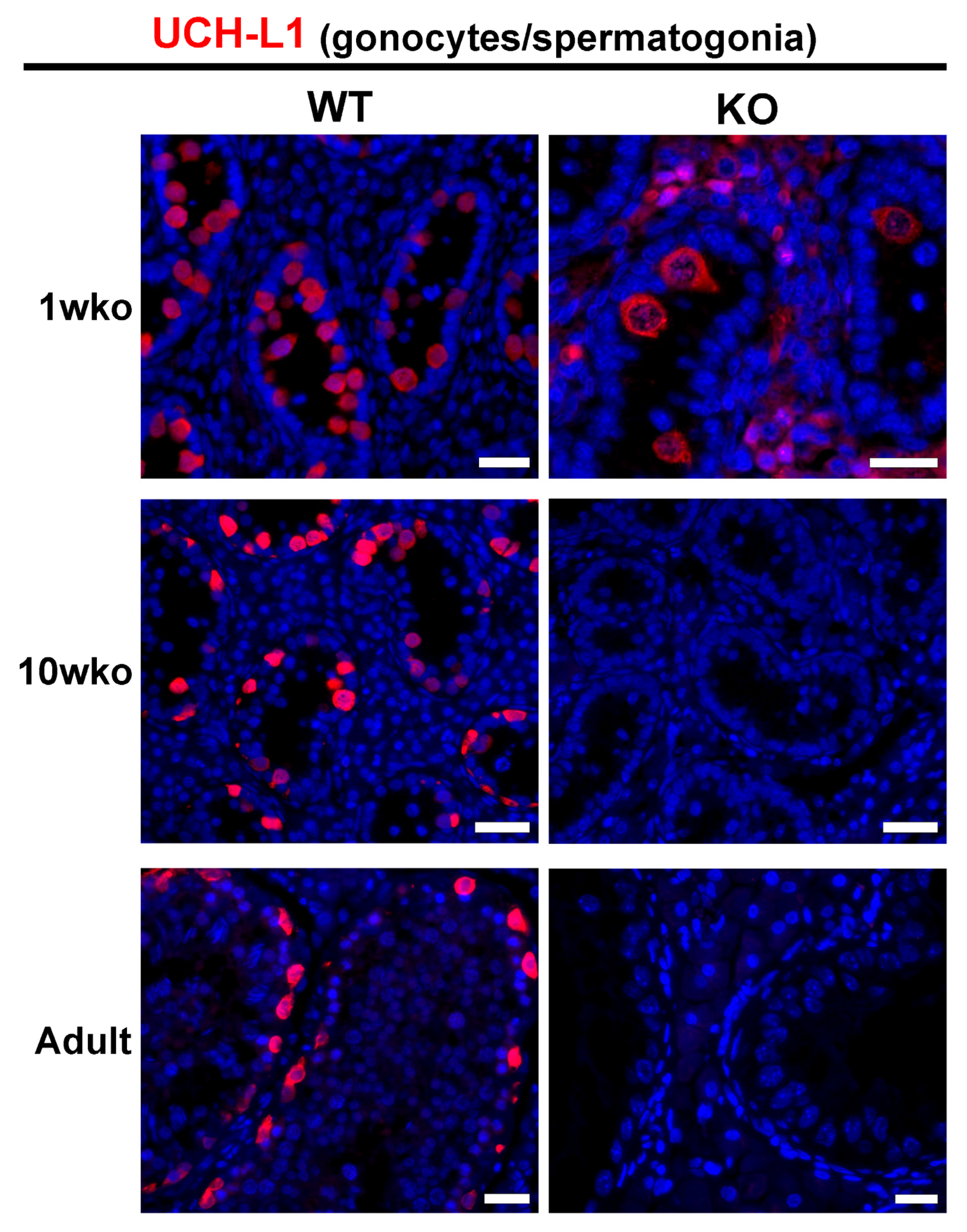
To determine an appropriate time point for SSC transplantation, we monitored the presence of germ cells based on the expression of UCH-L1, a marker for gonocytes/early spermatogonia [22,28,34]. While some germ cells were present in 1 wko DAZL-KO pig testes, complete germ cell depletion was observed at 10 weeks of age (10 wko) as well as in adult testes (Figure 2). Therefore, we performed transplantation when KO recipients were 10 to 13 weeks old.

3.2. Transplantation Efficiency

Transplantation was performed using germ cells from a variety of donor breeds and genetics (Table 1), as the intended application of the project was to use the SSC transplantation approach to efficiently propagate swine models by transplanting gene-edited germ cells. As there was no effect of donor cell origin and transplantation outcomes, all samples were combined for analysis. An overview of the transplantation parameters, the different donors, the number of germ cells injected, and sperm production efficiency is shown in Table 1.
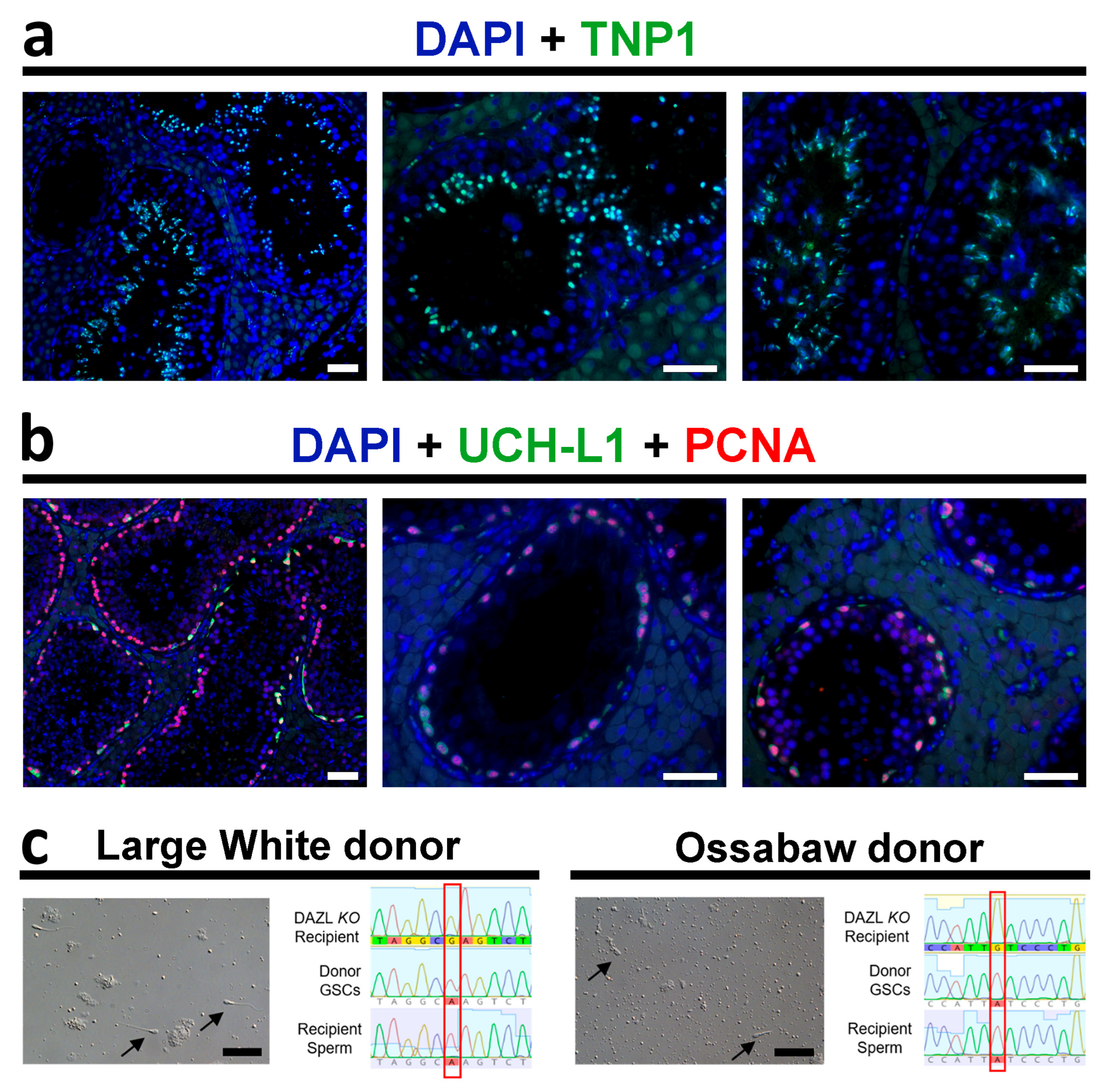
The number of germ cells transplanted into each recipient’s testis varied from 26 million to 137 million, but there was no apparent correlation between a higher number of cells injected and increased sperm production. Sperm were observed in the ejaculates of 89% (16 out of 18) of transplanted males. Peak sperm concentration ranged from 5 × 10⁴ to 7.3 × 10⁶ per mL. For comparison, wild-type sperm concentration typically ranges from 1 × 10⁹ to 5 × 10⁹ per mL. We were able to extract sperm genomic DNA from 13 transplanted animals, and SNP analysis (Figure 4c) confirmed that all sperm genotyped were exclusively from donor origin. Therefore, DAZL-KO pigs can produce donor-derived sperm after germ cell transplantation. Sperm motility was low, however, as motile sperm were observed in the ejaculates of only 4 of 18 recipients, with peak sperm motility being 0–10% for most recipients, while one recipient showed more consistent motility at 80%.
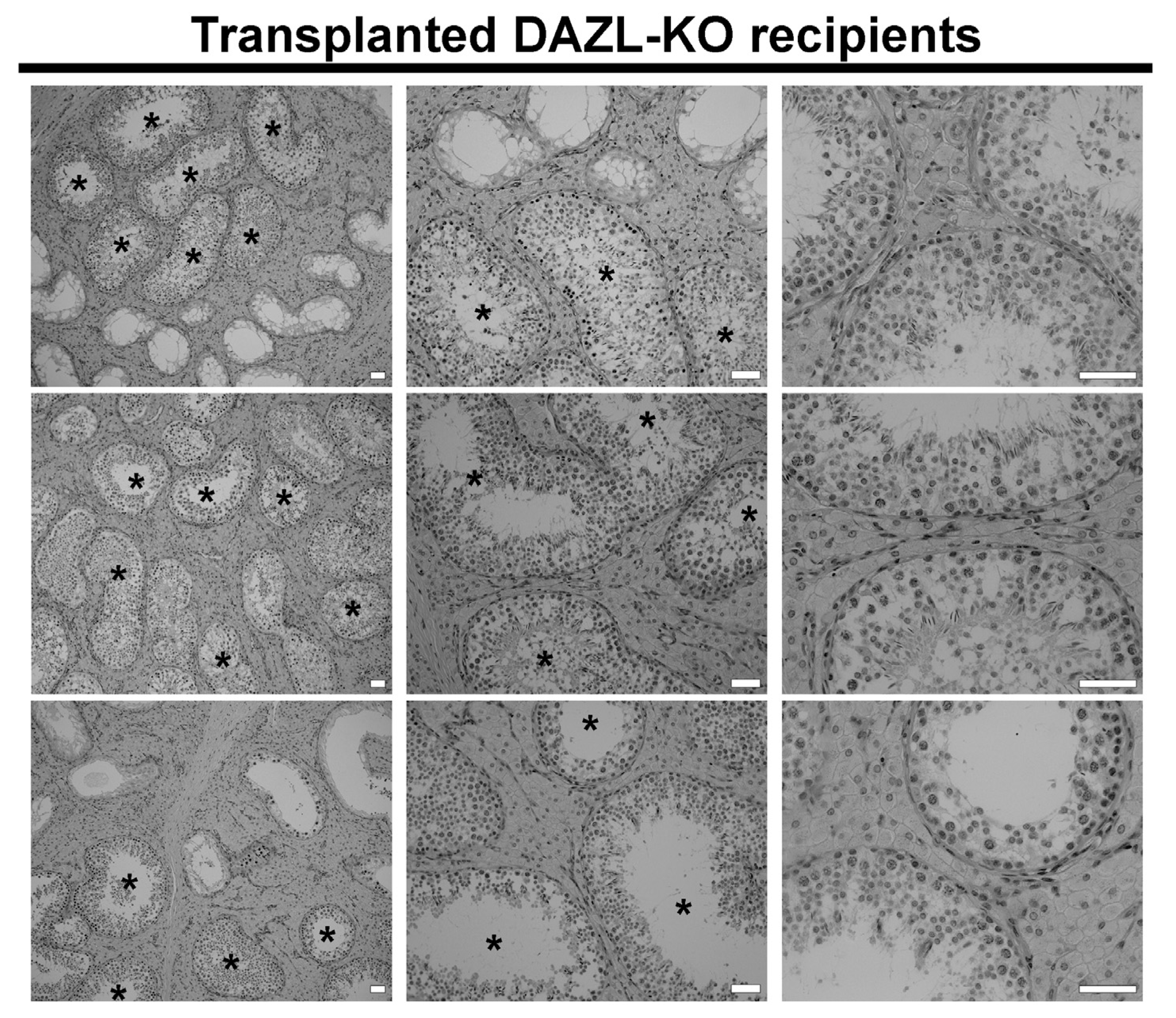
Figure 3 shows the histology of the DAZL-KO transplanted testis at the time of castration. Morphometric quantification revealed that 22.2% (±15.2, n = 4) of the seminiferous tubule cross sections contained germ cells at different stages of development, and most germ-cell-containing tubules were observed at the region close to the transplantation site (mediastinum/rete testis). Immunostaining of the recipient testes that received donor germ cells confirmed the presence of elongating spermatids expressing TNP1 (nuclear transition protein 1) (Figure 4a) as well as proliferating spermatogonia expressing UCH-L1 and the proliferating cell nuclear antigen (PCNA) (Figure 4b).
Figure 3. Histology of DAZL-KO recipient testes after germ cell transplantation. Histology of the germ cell transplantation recipient testes at the time of castration, at different magnifications. While some seminiferous tubule cross sections are still devoid of germ cells, other tubules contained germ cells at different stages of development (black asterisks). Samples represented are 2906, 2615, and 2626. Scale bars = 50 µm.
Cells 12 02582 g003
Figure 4. Presence of elongating spermatids and proliferating spermatogonia in transplanted DAZL-KO testes and sperm genotyping results. TNP1+ elongating spermatids ((a); green) and proliferating spermatogonia ((b); UCHL1+ in green and PCNA+ in red) are present in seminiferous tubules cross sections of DAZL-KO recipients after germ cell transplantation. (c) SNP analysis on sperm genomic DNA extracted from recipient pigs confirmed that all sperm (black arrows) were exclusively from donor origin (red box). Scale bars = 50 µm.
Cells 12 02582 g004

3.3. Functionality of the Recipient Testis

Given the low sperm production efficiency and sperm quality observed, we further evaluated the recipient testes for markers of somatic cell function at 1 wko and in adulthood, as well as the hormonal profile in adult DAZL-KO pigs. Table 2 and Figure 5 summarize the histomorphometry results for testis component occupancy and the Sertoli and Leydig cell volumes and numbers.
At 1 wko, the only significant difference observed was the proportion of the testis occupied by connective tissue that was increased in both Het and KO pigs when compared to control (p < 0.05, Table 2), as well as the reduction in germ cell occupancy from 6.8% in WT pigs to 0.8% in Het and 0.2% in KO (p < 0.05). In the adult pigs, the differences in testis component occupancies were more pronounced. The absence (or reduced number, in the Het) of germ cells led to a smaller proportion of the testis being occupied by seminiferous epithelium and the tubular compartment itself in both Het and KO groups when compared to WT (p < 0.05), while the tunica propria occupancy was higher in KO in comparison to WT (p < 0.05). A consequence of reduced tubular compartment occupancy is the observed increase in the intertubular compartment proportion in Het and KO pigs (p < 0.05), which was accompanied by higher occupancy of both the Leydig cells and connective tissue present in this compartment (p < 0.05).
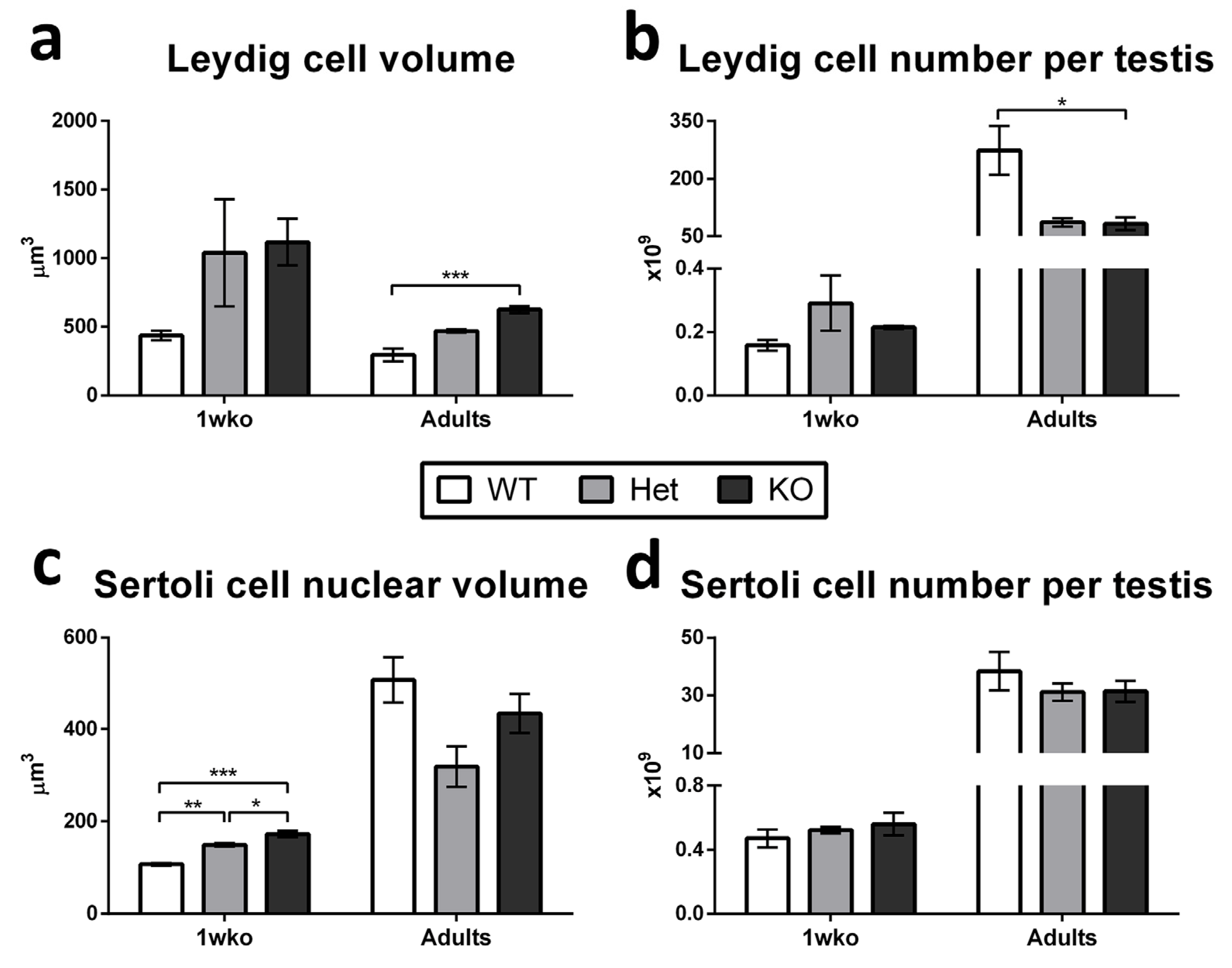
When analyzing Leydig and Sertoli cells specifically (Figure 5), we observed that Leydig cell individual volume was significantly increased (p < 0.05; Figure 5a) while the number of these cells per testis was reduced (p < 0.05; Figure 5b) in adult KO pigs when compared to WT, while no difference was observed among 1 wko pigs. Sertoli cell nuclear volume was significantly increased (p < 0.05) in Het when compared to WT, and in KO when compared to Het and WT at 1 wko (p < 0.05), but no difference was observed in mature Sertoli cell nuclear volume in adulthood (Figure 5c). Additionally, no significant changes were observed in Sertoli cell number per testis in any of the ages evaluated (Figure 5d).
We then evaluated the expression of functional markers: vimentin (VIM), an intermediate filament protein present in the cytoskeleton of testicular somatic cells; the Anti-Müllerian Hormone (AMH), produced by immature Sertoli cells; occludin (OCLN), a protein present in tight junctions among adjacent mature Sertoli cells and some interstitial cells; alpha-smooth muscle actin (SMA), present in peritubular myoid cells and endothelial cells; and aromatase (CYP19), a steroidogenic enzyme present in Leydig cells. The expression of these markers in 1 wko (Supplementary Figure S1) and adult testes (Supplementary Figure S2) was similar between the WT, Het, and KO pigs and followed the expected localization and age-appropriate patterns.
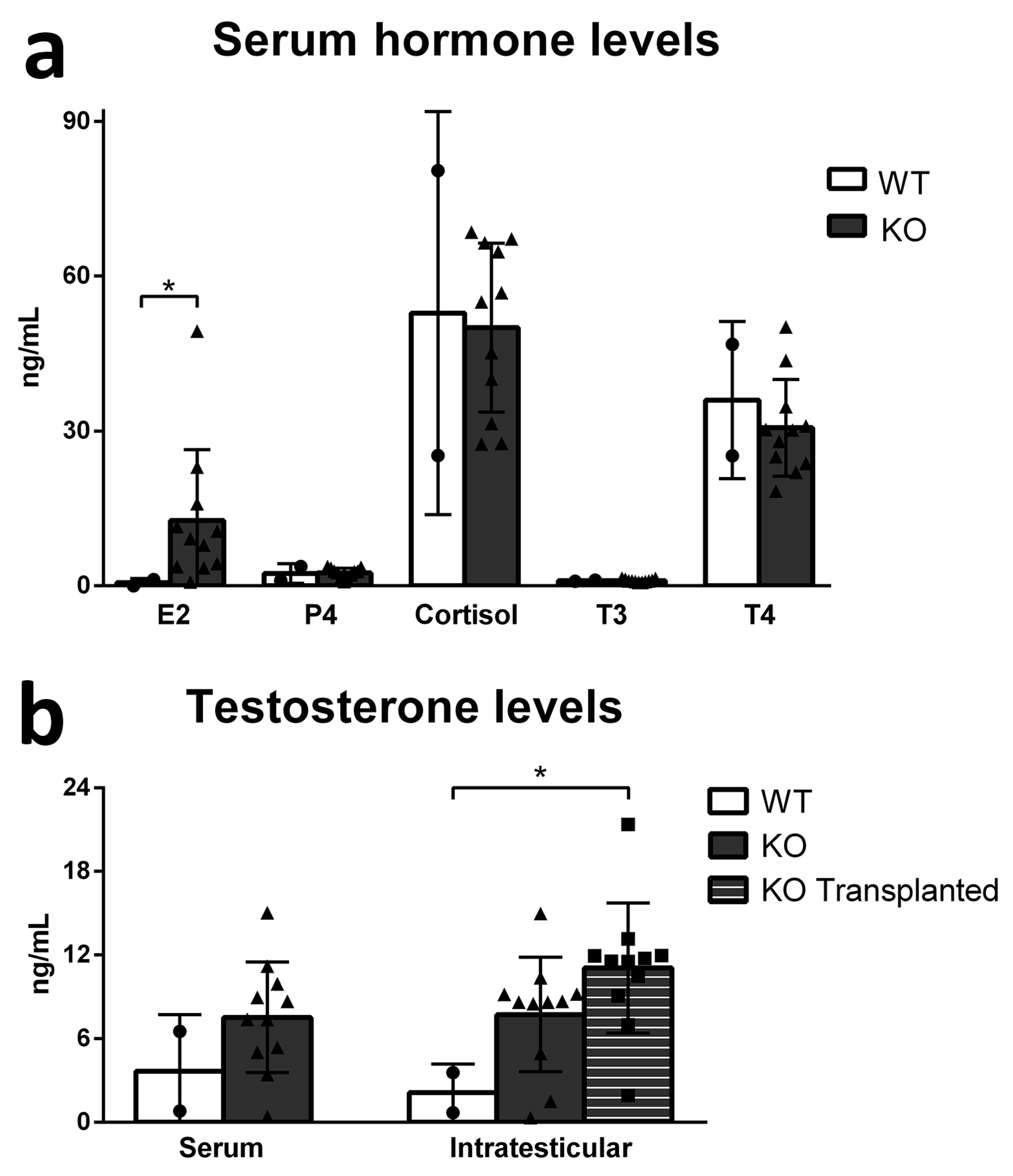
Lastly, we measured the serum levels of some hormones from blood collected during castration, as well as serum and intratesticular testosterone levels on both transplanted and non-transplanted testes (Figure 6). The serum levels of progesterone (P4), cortisol, T3, and T4 did not change between WT and KO pigs, while estradiol (E2) levels were increased in KO pigs (p < 0.05) (Figure 6a). No difference was observed in serum testosterone levels, but intratesticular levels of testosterone were increased in the KO-transplanted testis in comparison to WT (p < 0.05) (Figure 6b).

4. Discussion

The successful application of the SSC transplantation technique will allow the development of novel strategies in assisted reproduction, preservation of genetic diversity in livestock species, rescue and propagation of difficult-to-breed genotypes, and generation of genetically modified animal models. We previously reported targeted gene editing in porcine germ cells that can serve as donor cells for SSC transplantation and enable the successful generation of gene-edited pig models [65]. In the present study, we focused on investigating the potential use of DAZL-KO pigs as hosts for SSC transplantation.
We confirmed the impaired spermatogenesis previously observed in DAZL-KO pigs [54], which is consistent with the phenotype described in other species [46,47,50,55,56,57,59,60,61]. Moreover, we showed that neonatal DAZL-KO seminiferous cords still harbor gonocytes, but these GCs are no longer present in the testis of knockout pigs at 10 weeks of age and onwards. The exact age and the mechanism by which the postnatal GC development is disrupted in this model were not within the scope of this study, but these aspects certainly deserve further investigation [47,48,59]. The fact that GCs were completely absent in the KO testis at 10 wko was, however, an essential observation for choosing the age to perform SSC transplantation. At this time point, endogenous GCs are absent, leaving open niches, and testicles are still immature and relatively small to enable better distribution and colonization by the injected cells as previously established with WT recipient pigs [18,31]. Therefore, we performed SSC transplantation when recipient pigs were around 10–13 weeks of age, a time span that falls within the proposed “prime window”, which has been described to be prior to puberty [18,42,66].
The majority (~89%) of DAZL-KO recipient pigs were able to produce sperm, and all sperm evaluated were exclusively from donor origin. This confirmed that testes of DAZL-KO pigs can be hosts for transplanted SSCs and can regenerate donor-cell-derived spermatogenesis. However, the peak sperm concentration was low when compared to controls, and motile sperm were rarely observed except for one recipient which showed better sperm motility. This probably means that this individual had better colonization of the testis parenchyma by donor cells, therefore showing improved sperm parameters. However, at this point, there is no definite explanation for this observation. Based on histological analyses, the degree of colonization obtained was not very high, which could be one reason why fewer sperm were produced. Similar findings have been reported, including when NANOS2-KO pigs were used as recipients for SSC transplantation [41,42]. The degree of colonization of the recipient testis is critical to achieving SSC transplantation success and producing sufficient numbers of motile sperm, specifically if natural breeding is targeted [8,17,18,27,66,67,68]. The necessary level of colonization varies depending on species and is especially difficult to achieve in large animals, such as pigs, due to the size of the recipient testis.
Other factors of the microenvironment within DAZL-KO testes may not be optimally supportive for GC colonization, maintenance, and differentiation. To investigate this, we evaluated somatic cell function and hormonal homeostasis in these pigs. Most changes observed in the occupancy of the testicular parenchyma components were related to the lack of germ cells inside the seminiferous tubules, which led to a proportional reduction of tubular compartment occupancy and a higher proportion of intertubular compartment. No major change was observed in Sertoli cell number in either age group, while Leydig cells in the adult DAZL-KO testes were larger in size but reduced in numbers. We suspect that the changes observed in these steroidogenic cells may be related to the increase in intratesticular testosterone levels in the KO-transplanted testis in comparison to WT, and the increased serum E2 levels in DAZL-KO pigs, considering that high testosterone levels can lead to increased aromatization into E2. These results show that the steroidogenic potential of Leydig cells is maintained in DAZL-KO pigs and can be activated in the presence of spermatogenic cells, as testosterone is required for the regulation of spermatogenesis at several steps, such as meiosis and spermiogenesis [69,70,71]. Testosterone is also important for the regulation of the blood–testis barrier, which protects haploid cells within the seminiferous tubules. It can be argued that the adequate performance of this barrier after transplantation and SSC colonization is critical and may be even more important in the context of allogeneic or xenogeneic transplantation [7,69,72]. The serum levels of most hormones investigated did not show any significant changes, and the same is true for the expression of somatic cell functional markers, which did not reveal any differences between WT, Het, and KO pig testes and followed the expected patterns. This indicates that, overall, the functionality of the testicular somatic compartment is maintained in DAZL-KO pigs.
In summary, in the present study, we showed that DAZL-KO pig testes support donor-derived spermatogenesis and produce sperm following SSC transplantation, but low spermatogenic efficiency limits its use for the production of donor-derived offspring. Therefore, the technique needs to be further improved, possibly focusing on increasing the level of GC colonization of the testis parenchyma, by investigating the potential mechanisms involved or stimulating this process via hormonal modulation for example [73]. Currently, low colonization efficiency is possibly the biggest limitation to the success of routinely applying SSC transplantation in livestock breeding. Nevertheless, our findings open new avenues for SSC transplantation research in livestock that may ultimately enable the efficient production of genetically modified animals, preservation of valuable genetic traits, or other applications where assisted reproductive technologies could be used to counterbalance the low spermatogenic efficiency.

Supplementary Materials

The following supporting information can be downloaded at: https://www.mdpi.com/article/10.3390/cells12212582/s1, Figure S1: The expression of markers of somatic cell function is similar between WT, Het, and DAZL-KO pig testis at 1 week of age. Vimentin (VIM) is an intermediate filament present in the cytoskeleton of testicular somatic cells, anti-Müllerian Hormone (AMH) is produced by immature Sertoli cells, occludin (OCLN) is an intercellular tight junction protein, alpha-smooth muscle actin (SMA) is expressed by peritubular myoid and endothelial cells, and aromatase (CYP19) is a steroidogenic enzyme present on Leydig cells. Scale bar = 25 µm; Figure S2: The expression of markers of somatic cell function is similar between WT, Het, and DAZL-KO pig testis in adulthood. Vimentin (VIM) is an intermediate filament present in the cytoskeleton of testicular somatic cells, anti-Müllerian Hormone (AMH) is produced by immature Sertoli cells, occludin (OCLN) is an intercellular tight junction protein, alpha-smooth muscle actin (SMA) is expressed by peritubular myoid and endothelial cells, and aromatase (CYP19) is a steroidogenic enzyme present on Leydig cells. Scale bar = 25 µm; Table S1: List of primers used for single-nucleotide polymorphisms (SNP) analysis; Table S2: List of antibodies used.

Author Contributions

Conceptualization, D.F.C. and I.D.; methodology, investigation, formal analysis, N.L.M.L., T.G., P.R.-V., F.O., U.G., D.W., S.S., S.M.A.S.C., A.B. and S.H.; visualization, writing—original draft preparation, N.L.M.L.; writing—review and editing, T.G., A.B., D.F.C. and I.D.; project administration, funding acquisition, D.F.C. and I.D. All authors have read and agreed to the published version of the manuscript.

Funding

The research was supported by 2R44GM108150 and R01 OD016575.

Institutional Review Board Statement

Experiments conformed to the procedures and guidelines approved by the Recombinetics Institutional Animal Care and Use Committee, protocol number RCI-1805-05A.

Data Availability Statement

The datasets generated and/or analyzed during the current study are available from the corresponding author upon reasonable request.

Conflicts of Interest

T.G., P.R.-V., F.O., U.G., D.W., S.S., S.H., S.M.A.S.C., D.F.C. and I.D. are/were employees and/or shareholders in Recombinetics, Inc., a company that commercializes gene editing in livestock. The remaining authors declare that no commercial or financial relationships exist that could be construed as potential conflict of interest.

References

  1. Brinster, R.L. Germline stem cell transplantation and transgenesis. Science 2002, 296, 2174–2176. [Google Scholar] [CrossRef] [PubMed]
  2. Hill, J.R.; Dobrinski, I. Male germ cell transplantation in livestock. Reprod. Fertil. Dev. 2006, 18, 13. [Google Scholar] [CrossRef] [PubMed]
  3. Dobrinski, I.; Travis, A.J. Germ cell transplantation for the propagation of companion animals, non-domestic and endangered species. Reprod. Fertil. Dev. 2007, 19, 732. [Google Scholar] [CrossRef]
  4. Oatley, J.M. Recent advances for spermatogonial stem cell transplantation in livestock. Reprod. Fertil. Dev. 2018, 30, 44. [Google Scholar] [CrossRef] [PubMed]
  5. Goossens, E.; Jahnukainen, K.; Mitchell, R.; van Pelt, A.; Pennings, G.; Rives, N.; Poels, J.; Wyns, C.; Lane, S.; Rodriguez-Wallberg, K.; et al. Fertility preservation in boys: Recent developments and new insights. Hum. Reprod. Open 2020, 2020, hoaa016. [Google Scholar] [CrossRef] [PubMed]
  6. Doungkamchan, C.; Orwig, K.E. Recent advances: Fertility preservation and fertility restoration options for males and females. Fac. Rev. 2021, 10, 55. [Google Scholar] [CrossRef]
  7. Zhao, X.; Wan, W.; Zhang, X.; Wu, Z.; Yang, H. Spermatogonial stem cell transplantation in large animals. Animals 2021, 11, 918. [Google Scholar] [CrossRef]
  8. Zhang, W.; Nie, R.; Cai, Y.; Xie, W.; Zou, K. Progress in germline stem cell transplantation in mammals and the potential usage. Reprod. Biol. Endocrinol. 2022, 20, 59. [Google Scholar] [CrossRef]
  9. de Rooij, D.G.; Russell, L.D. All you wanted to know about spermatogonia but were afraid to ask. J. Androl. 2000, 21, 776–798. [Google Scholar] [CrossRef]
  10. de Rooij, D. Proliferation and differentiation of spermatogonial stem cells. Reproduction 2001, 121, 347–354. [Google Scholar] [CrossRef]
  11. Ehmcke, J.; Wistuba, J.; Schlatt, S. Spermatogonial stem cells: Questions, models and perspectives. Hum. Reprod. Update 2006, 12, 275–282. [Google Scholar] [CrossRef] [PubMed]
  12. Garcia, T.X.; Hofmann, M.C. Regulation of germ line stem cell homeostasis. Anim. Reprod. 2015, 12, 35–45. [Google Scholar] [PubMed]
  13. Kubota, H.; Brinster, R.L. Spermatogonial stem cells. Biol. Reprod. 2018, 99, 52–74. [Google Scholar] [CrossRef]
  14. Sharma, S.; Wistuba, J.; Pock, T.; Schlatt, S.; Neuhaus, N. Spermatogonial stem cells: Updates from specification to clinical relevance. Hum. Reprod. Update 2019, 25, 275–297. [Google Scholar] [CrossRef]
  15. Brinster, R.L.; Avarbock, M.R. Germline transmission of donor haplotype following spermatogonial transplantation. Proc. Natl. Acad. Sci. USA 1994, 91, 11303–11307. [Google Scholar] [CrossRef]
  16. Brinster, R.L.; Zimmermann, J.W. Spermatogenesis following male germ-cell transplantation. Proc. Natl. Acad. Sci. USA 1994, 91, 11298–11302. [Google Scholar] [CrossRef] [PubMed]
  17. Nagano, M.; Avarbock, M.R.; Brinster, R.L. Pattern and kinetics of mouse donor spermatogonial stem cell colonization in recipient testes. Biol. Reprod. 1999, 60, 1429–1436. [Google Scholar] [CrossRef]
  18. Honaramooz, A.; Megee, S.O.; Dobrinski, I. Germ cell transplantation in pigs. Biol. Reprod. 2002, 66, 21–28. [Google Scholar] [CrossRef]
  19. Hermann, B.P.; Sukhwani, M.; Winkler, F.; Pascarella, J.N.; Peters, K.A.; Sheng, Y.; Valli, H.; Rodriguez, M.; Ezzelarab, M.; Dargo, G.; et al. Spermatogonial stem cell transplantation into rhesus testes regenerates spermatogenesis producing functional sperm. Cell Stem Cell 2012, 11, 715–726. [Google Scholar] [CrossRef]
  20. Nakamura, Y.; Jörg, D.J.; Kon, Y.; Simons, B.D.; Yoshida, S. Transient suppression of transplanted spermatogonial stem cell differentiation restores fertility in mice. Cell Stem Cell 2021, 28, 1443–1456.e7. [Google Scholar] [CrossRef]
  21. Tan, W.; Carlson, D.F.; Lancto, C.A.; Garbe, J.R.; Webster, D.A.; Hackett, P.B.; Fahrenkrug, S.C. Efficient nonmeiotic allele introgression in livestock using custom endonucleases. Proc. Natl. Acad. Sci. USA 2013, 110, 16526–16531. [Google Scholar] [CrossRef] [PubMed]
  22. Gonzalez, R.; Dobrinski, I. Beyond the mouse monopoly: Studying the male germ line in domestic animal models. ILAR J. 2015, 56, 83–98. [Google Scholar] [CrossRef]
  23. Tang, L.; González, R.; Dobrinski, I. Germline modification of domestic animals. Anim. Reprod. 2015, 12, 93–104. [Google Scholar]
  24. Whitworth, K.M.; Green, J.A.; Redel, B.K.; Geisert, R.D.; Lee, K.; Telugu, B.P.; Wells, K.D.; Prather, R.S. Improvements in pig agriculture through gene editing. CABI Agric. Biosci. 2022, 3, 41. [Google Scholar] [CrossRef] [PubMed]
  25. Kilcoyne, K.R.; Mitchell, R.T. Testicular transplantation for fertility preservation: Clinical potential and current challenges. Reproduction 2019, 158, F1–F14. [Google Scholar] [CrossRef] [PubMed]
  26. Tran, K.T.D.; Valli-Pulaski, H.; Colvin, A.; Orwig, K.E. Male fertility preservation and restoration strategies for patients undergoing gonadotoxic therapies. Biol. Reprod. 2022, 107, 382–405. [Google Scholar] [CrossRef]
  27. Takashima, S.; Shinohara, T. Culture and transplantation of spermatogonial stem cells. Stem Cell Res. 2018, 29, 46–55. [Google Scholar] [CrossRef]
  28. Binsila, B.; Selvaraju, S.; Ranjithkumaran, R.; Archana, S.S.; Krishnappa, B.; Ghosh, S.K.; Kumar, H.; Subbarao, R.B.; Arangasamy, A.; Bhatta, R. Current scenario and challenges ahead in application of spermatogonial stem cell technology in livestock. J. Assist. Reprod. Genet. 2021, 38, 3155–3173. [Google Scholar] [CrossRef]
  29. Ogawa, T.; Dobrinski, I.; Avarbock, M.R.; Brinster, R.L. Transplantation of male germ line stem cells restores fertility in infertile mice. Nat. Med. 2000, 6, 29–34. [Google Scholar] [CrossRef]
  30. Brinster, C.J.; Ryu, B.-Y.; Avarbock, M.R.; Karagenc, L.; Brinster, R.L.; Orwig, K.E. Restoration of fertility by germ cell transplantation requires effective recipient preparation. Biol. Reprod. 2003, 69, 412–420. [Google Scholar] [CrossRef]
  31. Honaramooz, A.; Behboodi, E.; Hausler, C.L.; Blash, S.; Ayres, S.; Azuma, C.; Echelard, Y.; Dobrinski, I. Depletion of endogenous germ cells in male pigs and goats in preparation for germ cell transplantation. J. Androl. 2005, 26, 698–705. [Google Scholar] [CrossRef] [PubMed]
  32. Kanatsu-Shinohara, M.; Morimoto, H.; Shinohara, T. Fertility of male germline stem cells following spermatogonial transplantation in infertile mouse models. Biol. Reprod. 2016, 94, 112–113. [Google Scholar] [CrossRef] [PubMed]
  33. Shetty, G.; Mitchell, J.M.; Meyer, J.M.; Wu, Z.; Lam, T.N.A.; Phan, T.T.; Zhang, J.; Hill, L.; Tailor, R.C.; Peters, K.A.; et al. Restoration of functional sperm production in irradiated pubertal rhesus monkeys by spermatogonial stem cell transplantation. Andrology 2020, 8, 1428–1441. [Google Scholar] [CrossRef] [PubMed]
  34. Zhang, M.; Zhao, X.; Li, H.; Cai, G.; Yang, H.; Wu, Z. Intratesticular injection of busulfan for producing recipient male pigs for spermatogonial stem cell transplantation. Livest. Sci. 2021, 245, 104448. [Google Scholar] [CrossRef]
  35. Schlatt, S.; Foppiani, L.; Rolf, C.; Weinbauer, G.F.; Nieschlag, E. Germ cell transplantation into X-irradiated monkey testes. Hum. Reprod. 2002, 17, 55–62. [Google Scholar] [CrossRef]
  36. Zhang, Z.; Shao, S.; Meistrich, M.L. The radiation-induced block in spermatogonial differentiation is due to damage to the somatic environment, not the germ cells. J. Cell. Physiol. 2007, 211, 149–158. [Google Scholar] [CrossRef]
  37. Herrid, M.; Olejnik, J.; Jackson, M.; Suchowerska, N.; Stockwell, S.; Davey, R.; Hutton, K.; Hope, S.; Hill, J.R. Irradiation enhances the efficiency of testicular germ cell transplantation in sheep. Biol. Reprod. 2009, 81, 898–905. [Google Scholar] [CrossRef]
  38. Ma, W.; An, L.; Wu, Z.; Wang, X.; Guo, M.; Miao, K.; Ma, W.; Tian, J. Efficient and safe recipient preparation for transplantation of mouse spermatogonial stem cells: Pretreating testes with heat shock. Biol. Reprod. 2011, 85, 670–677. [Google Scholar] [CrossRef]
  39. Rasouli, M.H.; Zandi, M.; Sadeghi, A.A.; Emamjomeh-Kashan, N. Spermatogonial stem cell survival in ram lambs following busulfan treatment. Anim. Reprod. 2020, 17, e20200001. [Google Scholar] [CrossRef]
  40. Rilianawati, R.; Speed, R.; Taggart, M.; Cooke, H. Spermatogenesis in testes of Dazl null mice after transplantation of wild-type germ cells. Reproduction 2003, 126, 599–604. [Google Scholar] [CrossRef]
  41. Park, K.-E.; Kaucher, A.V.; Powell, A.; Waqas, M.S.; Sandmaier, S.E.S.; Oatley, M.J.; Park, C.-H.; Tibary, A.; Donovan, D.M.; Blomberg, L.A.; et al. Generation of germline ablated male pigs by CRISPR/Cas9 editing of the NANOS2 gene. Sci. Rep. 2017, 7, 40176. [Google Scholar] [CrossRef] [PubMed]
  42. Ciccarelli, M.; Giassetti, M.I.; Miao, D.; Oatley, M.J.; Robbins, C.; Lopez-Biladeau, B.; Waqas, M.S.; Tibary, A.; Whitelaw, B.; Lillico, S.; et al. Donor-derived spermatogenesis following stem cell transplantation in sterile NANOS2 knockout males. Proc. Natl. Acad. Sci. USA 2020, 117, 24195–24204. [Google Scholar] [CrossRef] [PubMed]
  43. Shah, C.; VanGompel, M.J.W.; Naeem, V.; Chen, Y.; Lee, T.; Angeloni, N.; Wang, Y.; Xu, E.Y. Widespread presence of human BOULE homologs among animals and conservation of their ancient reproductive function. PLoS Genet. 2010, 6, e1001022. [Google Scholar] [CrossRef] [PubMed]
  44. VanGompel, M.J.W.; Xu, E.Y. The roles of the DAZ family in spermatogenesis. Spermatogenesis 2011, 1, 36–46. [Google Scholar] [CrossRef] [PubMed]
  45. Fu, X.-F.; Cheng, S.-F.; Wang, L.-Q.; Yin, S.; De Felici, M.; Shen, W. DAZ family proteins, key players for germ cell development. Int. J. Biol. Sci. 2015, 11, 1226–1235. [Google Scholar] [CrossRef] [PubMed]
  46. Ruggiu, M.; Speed, R.; Taggart, M.; McKay, S.J.; Kilanowski, F.; Saunders, P.; Dorin, J.; Cooke, H.J. The mouse Dazla gene encodes a cytoplasmic protein essential for gametogenesis. Nature 1997, 389, 73–77. [Google Scholar] [CrossRef]
  47. Lin, Y.; Page, D.C. Dazl deficiency leads to embryonic arrest of germ cell development in XY C57BL/6 mice. Dev. Biol. 2005, 288, 309–316. [Google Scholar] [CrossRef]
  48. Li, H.; Liang, Z.; Yang, J.; Wang, D.; Wang, H.; Zhu, M.; Geng, B.; Xu, E.Y. DAZL is a master translational regulator of murine spermatogenesis. Natl. Sci. Rev. 2019, 6, 455–468. [Google Scholar] [CrossRef]
  49. Mikedis, M.M.; Fan, Y.; Nicholls, P.K.; Endo, T.; Jackson, E.K.; Cobb, S.A.; de Rooij, D.G.; Page, D.C. DAZL mediates a broad translational program regulating expansion and differentiation of spermatogonial progenitors. Elife 2020, 9, e56523. [Google Scholar] [CrossRef]
  50. Saunders, P.; Turner, J.; Ruggiu, M.; Taggart, M.; Burgoyne, P.; Elliott, D.; Cooke, H. Absence of mDazl produces a final block on germ cell development at meiosis. Reproduction 2003, 126, 589–597. [Google Scholar] [CrossRef]
  51. Reynolds, N.; Collier, B.; Maratou, K.; Bingham, V.; Speed, R.M.; Taggart, M.; Semple, C.A.; Gray, N.K.; Cooke, H.J. Dazl binds in vivo to specific transcripts and can regulate the pre-meiotic translation of Mvh in germ cells. Hum. Mol. Genet. 2005, 14, 3899–3909. [Google Scholar] [CrossRef] [PubMed]
  52. Kee, K.; Angeles, V.T.; Flores, M.; Nguyen, H.N.; Reijo Pera, R.A. Human DAZL, DAZ and BOULE genes modulate primordial germ-cell and haploid gamete formation. Nature 2009, 462, 222–225. [Google Scholar] [CrossRef] [PubMed]
  53. Gill, M.E.; Hu, Y.-C.; Lin, Y.; Page, D.C. Licensing of gametogenesis, dependent on RNA binding protein DAZL, as a gateway to sexual differentiation of fetal germ cells. Proc. Natl. Acad. Sci. USA 2011, 108, 7443–7448. [Google Scholar] [CrossRef]
  54. Nicholls, P.K.; Schorle, H.; Naqvi, S.; Hu, Y.-C.; Fan, Y.; Carmell, M.A.; Dobrinski, I.; Watson, A.L.; Carlson, D.F.; Fahrenkrug, S.C.; et al. Mammalian germ cells are determined after PGC colonization of the nascent gonad. Proc. Natl. Acad. Sci. USA 2019, 116, 25677–25687. [Google Scholar] [CrossRef]
  55. Reijo, R.; Lee, T.-Y.; Salo, P.; Alagappan, R.; Brown, L.G.; Rosenberg, M.; Rozen, S.; Jaffe, T.; Straus, D.; Hovatta, O.; et al. Diverse spermatogenic defects in humans caused by Y chromosome deletions encompassing a novel RNA–binding protein gene. Nat. Genet. 1995, 10, 383–393. [Google Scholar] [CrossRef]
  56. Lin, Y.M.; Chen, C.W.; Sun, H.S.; Tsai, S.J.; Hsu, C.C.; Teng, Y.N.; Lin, J.S.; Kuo, P.L. Expression patterns and transcript concentrations of the autosomal DAZL gene in testes of azoospermic men. Mol. Hum. Reprod. 2001, 7, 1015–1022. [Google Scholar] [CrossRef]
  57. Tung, J.Y.; Rosen, M.P.; Nelson, L.M.; Turek, P.J.; Witte, J.S.; Cramer, D.W.; Cedars, M.I.; Reijo-Pera, R.A. Novel missense mutations of the Deleted-in-AZoospermia-Like (DAZL) gene in infertile women and men. Reprod. Biol. Endocrinol. 2006, 4, 40. [Google Scholar] [CrossRef]
  58. Nailwal, M.; Chauhan, J.B. In silico analysis of non-synonymous single nucleotide polymorphisms in human DAZL gene associated with male infertility. Syst. Biol. Reprod. Med. 2017, 63, 248–258. [Google Scholar] [CrossRef]
  59. Schrans-Stassen, B.H.G.J.; Saunders, P.T.K.; Cooke, H.J.; de Rooij, D.G. Nature of the spermatogenic arrest in Dazl−/− mice. Biol. Reprod. 2001, 65, 771–776. [Google Scholar] [CrossRef]
  60. Maratou, K.; Forster, T.; Costa, Y.; Taggart, M.; Speed, R.M.; Ireland, J.; Teague, P.; Roy, D.; Cooke, H.J. Expression profiling of the developing testis in wild-type and Dazl knockout mice. Mol. Reprod. Dev. 2004, 67, 26–54. [Google Scholar] [CrossRef]
  61. McLean, Z.L.; Appleby, S.J.; Wei, J.; Snell, R.G.; Oback, B. Testes of DAZL null neonatal sheep lack prospermatogonia but maintain normal somatic cell morphology and marker expression. Mol. Reprod. Dev. 2021, 88, 3–14. [Google Scholar] [CrossRef] [PubMed]
  62. Zeng, W.; Tang, L.; Bondareva, A.; Honaramooz, A.; Tanco, V.; Dores, C.; Megee, S.; Modelski, M.; Rodriguez-Sosa, J.R.; Paczkowski, M.; et al. Viral transduction of male germline stem cells results in transgene transmission after germ cell transplantation in pigs. Biol. Reprod. 2013, 88, 27–28. [Google Scholar] [CrossRef] [PubMed]
  63. Tang, L.; Bondareva, A.; González, R.; Rodriguez-Sosa, J.R.; Carlson, D.F.; Webster, D.; Fahrenkrug, S.; Dobrinski, I. TALEN-mediated gene targeting in porcine spermatogonia. Mol. Reprod. Dev. 2018, 85, 250–261. [Google Scholar] [CrossRef] [PubMed]
  64. Avelar, G.F.; Oliveira, C.F.A.; Soares, J.M.; Silva, I.J.; Dobrinski, I.; Hess, R.A.; França, L.R. Postnatal somatic cell proliferation and seminiferous tubule maturation in pigs: A non-random event. Theriogenology 2010, 74, 11–23. [Google Scholar] [CrossRef]
  65. Webster, D.; Bondareva, A.; Solin, S.; Goldsmith, T.; Su, L.; Lara, N.L.M.; Carlson, D.F.; Dobrinski, I. Targeted gene editing in porcine spermatogonia. Front. Genet. 2021, 11, 1861. [Google Scholar] [CrossRef]
  66. Shinohara, T.; Orwig, K.E.; Avarbock, M.R.; Brinster, R.L. Remodeling of the postnatal mouse testis is accompanied by dramatic changes in stem cell number and niche accessibility. Proc. Natl. Acad. Sci. USA 2001, 98, 6186–6191. [Google Scholar] [CrossRef]
  67. Mikkola, M.; Sironen, A.; Kopp, C.; Taponen, J.; Sukura, A.; Vilkki, J.; Katila, T.; Andersson, M. Transplantation of normal boar testicular cells resulted in complete focal spermatogenesis in a boar affected by the immotile short-tail sperm defect. Reprod. Domest. Anim. 2006, 41, 124–128. [Google Scholar] [CrossRef]
  68. Mutoji, K.N.; Hermann, B.P. Defining the phenotype and function of mammalian spermatogonial stem cells. In The Biology of Mammalian Spermatogonia; Oatley, J., Griswold, M., Eds.; Springer: New York, NY, USA, 2017; pp. 67–90. [Google Scholar]
  69. Smith, L.B.; Walker, W.H. The regulation of spermatogenesis by androgens. Semin. Cell Dev. Biol. 2014, 30, 2–13. [Google Scholar] [CrossRef]
  70. Potter, S.J.; DeFalco, T. Role of the testis interstitial compartment in spermatogonial stem cell function. Reproduction 2017, 153, R151–R162. [Google Scholar] [CrossRef]
  71. Van Saen, D.; Goossens, E. Testicular Tissue Transplantation. In Female and Male Fertility Preservation; Grynberg, M., Patrizio, P., Eds.; Springer International Publishing: Berlin/Heidelberg, Germany, 2022; pp. 529–554. [Google Scholar]
  72. França, L.R.; Auharek, S.A.; Hess, R.A.; Dufour, J.M.; Hinton, B.T. Blood-tissue barriers. In Biology and Regulation of Blood Tissue Barriers; Cheng, C.Y., Ed.; Landes Bioscience and Springer Science: Berlin/Heidelberg, Germany, 2012; pp. 237–259. [Google Scholar]
  73. Shetty, G.; Wu, Z.; Lam, T.N.A.; Phan, T.T.; Orwig, K.E.; Meistrich, M.L. Effect of hormone modulations on donor-derived spermatogenesis or colonization after syngeneic and xenotransplantation in mice. Andrology 2019, 7, 257–265. [Google Scholar] [CrossRef]
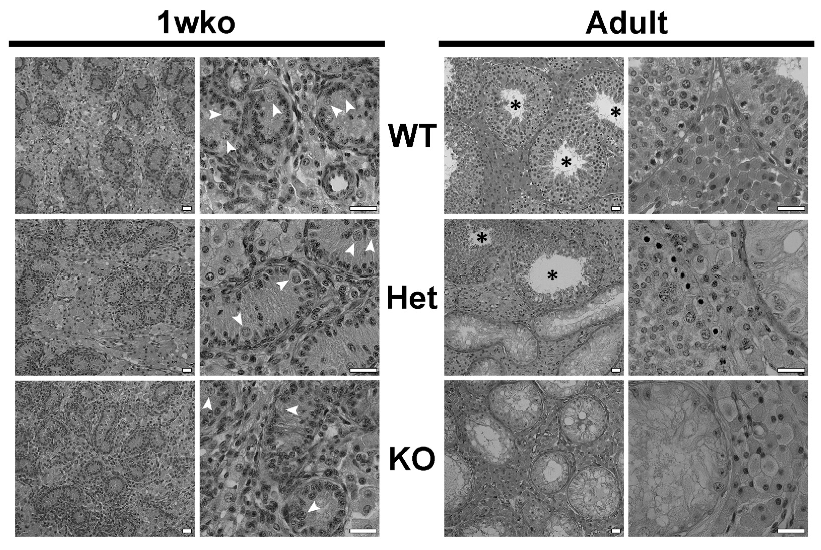
Figure 1. Testicular histology of wild-type (WT), heterozygote (Het), and DAZL-KO (KO) pigs at 1 week of age (1 wko) and in adulthood. The histology and organization of the testicular parenchyma in Het and KO testes are similar to the WT. White arrowheads point to gonocytes, present at 1 wko in the testes of all groups, including KO. In adulthood, Het testes show a mix of seminiferous tubules with no germ cells and seminiferous tubules with full spermatogenesis (black asterisks), while no germ cells are observed in the seminiferous tubules of DAZL-KO pig testes. Scale bars = 25 µm.
Cells 12 02582 g001
Figure 2. Germ cells (UCHL1+, red) are present in the testis of DAZL-KO pigs at 1 wko but no longer present at 10 weeks of age onwards. Some gonocytes were present in 1 wko DAZL-KO pig seminiferous cords, but at the time chosen for germ cell transplantation (10 wko), no UCHL1+ germ cells were observed in the KO testis, similar to what is observed in the adult KO testis. Scale bars = 25 µm.
Cells 12 02582 g002
Figure 5. Leydig and Sertoli cell parameters in WT, Het, and DAZL-KO pigs testis at 1 wko and in adulthood. (a) Leydig cell individual volume is significantly increased (p < 0.05) in adult KO pigs in comparison to WT but unchanged among the other groups. (b) The number of Leydig cells per testis is decreased (p < 0.05) in adult KO pigs in comparison to WT but unchanged among the other groups. (c) Sertoli cell nuclear volume was increased in Het when compared to WT (p < 0.05) and increased further in KO when compared to Het and KO (p < 0.05) at 1 wko, but no difference was observed in mature Sertoli cell nuclear volume in adulthood. (d) Sertoli cell number per testis did not change among the groups and ages. Data are shown as mean ± SD and * = p  <  0.05, ** = p  <  0.01, and *** = p  <  0.001.
Cells 12 02582 g005
Figure 6. Serum hormone levels and serum and intratesticular testosterone levels in WT (circle) and KO (triangle) pig testis. (a) The serum level of estradiol (E2) was higher (p < 0.05) in KO pigs, but no change was observed in the levels of progesterone (P4), cortisol, triiodothyronine (T3), and thyroxine (T4). (b) The serum testosterone level showed no significant difference between WT and KO, but the intratesticular testosterone level was increased (p < 0.05) in the KO testis that received germ cell transplantation (square) in comparison to WT. Data are shown as mean ± SD and * = p  <  0.05.
Cells 12 02582 g006
Table 1. Germ cell transplantation parameters and efficiency.
Recipient IDDonor BreedDonor GeneticsGCs Injected (million)Peak Sperm Concentration/mLSperm Genotype
2908YorkshireWT506 × 106Donor
2906YorkshireEdited504.2 × 106Donor
3412YorkshirePooled edited137.75LowDonor
2615CrossWT122LowDonor
3327CrossWT70LowDonor
3346CrossWT502.5 × 105Donor
3348CrossWT705 × 104Donor
3661CrossPooled WT685.4 × 106ND
2616CrossEdited93LowDonor
3234CrossEdited505 × 104ND
3238CrossEdited504 × 105Donor
3249OssabawWT26.460ND
2626OssabawPooled WT34.2LowDonor
2988OssabawPooled WT507.6 × 106Donor
3719OssabawEdited39.257.3 × 106Donor
3310OssabawPooled edited700ND
5043OssabawPooled edited46.41.7 × 105ND
5047OssabawPooled edited46.43 × 106Donor
Cross = Large White composite. ND = not determined.
Table 2. Testis component occupancy (volumetric density; %) in wild-type (WT), DAZL Heterozygote (Het), and DAZL Knockout (KO) 1-week-old (1 wko) and adult pigs (mean ± SEM).
1 wkoAdults
WTHetKOWTHetKO
Seminiferous tubules51.6 ± 4.736.2 ± 0.939.3 ± 2.583.4 ± 3.5 a66.1 ± 4.6 b58.4 ± 2.6 b
Seminiferous epithelium27.6 ± 2.425.5 ± 1.126.2 ± 1.959.9 ± 2.9 a46.4 ± 1.3 b33.1 ± 2.3 c
Germ cells6.8 ± 0.7 a0.8 ± 0.1 b0.2 ± 0.1 b---
Tubular lumen---17.5 ± 1.211.3 ± 4.216.1 ± 1.5
Tunica propria17.2 ± 1.99.9 ± 0.112.9 ± 0.76.0 ± 0.5 a8.4 ± 0.8 ab9.2 ± 0.9 b
Intertubular compartment48.3 ± 4.763.8 ± 0.960.7 ± 2.516.6 ± 3.5 a33.9 ± 4.6 b41.6 ± 2.6 b
Leydig cells19.6 ± 2.930.7 ± 0.924.3 ± 1.09.2 ± 1.6 a16.9 ± 2.8 ab22.8 ± 1.4 b
Connective tissue14.4 ± 0.4 a21.7 ± 0.3 b24.2 ± 1.5 b4.0 ± 0.9 a10.2 ± 1.4 b11.6 ± 1.4 b
Blood vessels14.3 ± 1.711.4 ± 0.312.2 ± 2.93.4 ± 1.26.8 ± 0.47.2 ± 0.8
Different letters for the same parameter within the same age indicate significant differences (p < 0.05).
Disclaimer/Publisher’s Note: The statements, opinions and data contained in all publications are solely those of the individual author(s) and contributor(s) and not of MDPI and/or the editor(s). MDPI and/or the editor(s) disclaim responsibility for any injury to people or property resulting from any ideas, methods, instructions or products referred to in the content.