Article

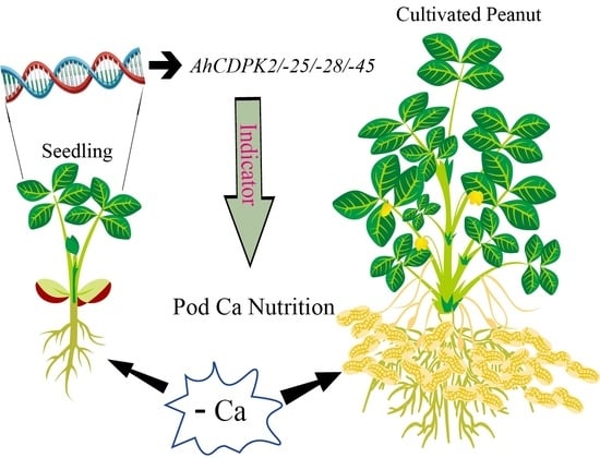
Genome-Wide Identification and Characterization of CDPK Gene Family in Cultivated Peanut (Arachis hypogaea L.) Reveal Their Potential Roles in Response to Ca Deficiency

Institute of Crop Germplasm Resources, Shandong Academy of Agricultural Sciences, Ji’nan 250100, China
*
Authors to whom correspondence should be addressed.
Cells 2023, 12(23), 2676; https://doi.org/10.3390/cells12232676
Submission received: 28 August 2023 / Revised: 13 November 2023 / Accepted: 17 November 2023 / Published: 21 November 2023

Abstract

:
This study identified 45 calcium-dependent protein kinase (CDPK) genes in cultivated peanut (Arachis hypogaea L.), which are integral in plant growth, development, and stress responses. These genes, classified into four subgroups based on phylogenetic relationships, are unevenly distributed across all twenty peanut chromosomes. The analysis of the genetic structure of AhCDPKs revealed significant similarity within subgroups, with their expansion primarily driven by whole-genome duplications. The upstream promoter sequences of AhCDPK genes contained 46 cis-acting regulatory elements, associated with various plant responses. Additionally, 13 microRNAs were identified that target 21 AhCDPK genes, suggesting potential post-transcriptional regulation. AhCDPK proteins interacted with respiratory burst oxidase homologs, suggesting their involvement in redox signaling. Gene ontology and KEGG enrichment analyses affirmed AhCDPK genes’ roles in calcium ion binding, protein kinase activity, and environmental adaptation. RNA-seq data revealed diverse expression patterns under different stress conditions. Importantly, 26 AhCDPK genes were significantly induced when exposed to Ca deficiency during the pod stage. During the seedling stage, four AhCDPKs (AhCDPK2/-25/-28/-45) in roots peaked after three hours, suggesting early signaling roles in pod Ca nutrition. These findings provide insights into the roles of CDPK genes in plant development and stress responses, offering potential candidates for predicting calcium levels in peanut seeds.

Graphical Abstract

1. Introduction

Plants have evolved complex mechanisms to adapt to their constantly changing environment. One such mechanism involves the use of calcium ions (Ca2+) as important cellular messengers, which play crucial roles in plant growth, development, and response to biotic and abiotic stress [1,2]. When external changes occur, the concentration of Ca2+ in the cytoplasm changes, triggering a series of physiological and biochemical reactions that improve plant tolerance to environmental stress [3].
To sense changes in Ca2+ concentration, plants have evolved various calcium-sensing proteins, including calmodulins (CaMs), calmodulin-like proteins (CMLs), calcineurin B-like proteins (CBLs), and Ca2+-dependent protein kinases (CDPKs) [4,5]. Among these, CDPKs are unique as they are Ser/Thr protein kinases capable of directly converting upstream Ca2+ signals into downstream protein phosphorylation events [5,6]. CDPKs are single peptide chains with four characteristic domains: a variable N-terminal domain (VNTD), a Ser/Thr protein kinase domain (PKD), an autoinhibitory junction region (JD), and a carboxyl-terminal calmodulin-like domain (CaMLD) [4,7]. The VNTD domain is poorly conserved and is involved in substrate recognition. Most CDPKs contain N-myristylation and/or S-palmitoylation sites, which enable the proteins to be localized to the membrane and contribute to the specificity of substrate recognition [8]. The PKD, which is relatively conserved in green plants, is responsible for binding ATP and phosphorylating downstream substrates [5,9,10]. The JD domain has kinase-inhibitory properties and can bind to the CaMLD domain. The CaMLD, located at the C-terminus, contains between one and four EF-hand motifs that bind to Ca2+ [9,10]. The differences in the number and amino acid sequences of these motifs enable CDPKs to decode different forms of Ca2+ signatures in the cytoplasm [7].
CDPKs play a crucial role in plant signaling, acting as key mediators of Ca2+ signals that are essential for various physiological responses [11]. CDPKs phosphorylate a range of substrates, including ion channels, transcription factors, and metabolic enzymes, which allow them to regulate diverse cellular processes [12]. As a result, CDPKs have been shown to play pivotal roles in a wide range of plant functions [7,11,12]. For example, CDPK genes are involved in plant growth and development [12]. In Arabidopsis, loss of AtCPK2,6,20,17,34 reduced pollen tube growth, while AtCPK11,24 increased [7,12]. The MaCDPK7 gene is involved in regulating banana fruit ripening [13]. TaCPK40 negatively regulates seed dormancy and positively regulates seed germination [14]. In addition, CDPKs have been shown to be involved in the response to abiotic stresses [7,11]. CPK4 and CPK11 overexpressing lines were more tolerant to drought stress through signal transduction in Arabidopsis [15]. Overexpression of OsCDPK7 enhanced the induction of some stress-responsive genes in response to salinity/drought in rice [16]. Overexpression of SiCDPK24, TaCDPK25-U-AS1, or TaCDPK25-U-AS2 enhanced drought resistance in Arabidopsis [17,18]. Overexpression of GmCDPK3 improved drought and salt resistance in Arabidopsis [19]. AtCPK28 phosphorylates and promotes the nuclear translocation of NIN-LIKE PROTEIN 7 (NLP7), thereby positively regulating plant response to cold stress in Arabidopsis [20]. The interaction between PpCDPK7 and PpRBOH may be the intersectional point of Ca2+ and ROS signal transmission during cold storage of peach fruits [21]. Genome-wide analyses have identified CDPK genes in various plant species, including Arabidopsis thaliana (34), rice (29), wheat (85), maize (40), soybean (50), cotton (84), chickpea (22), Medicago truncatula (24), peach (17), and pineapple (17) [6,17,21,22,23,24,25,26,27]. However, little is known about CDPKs in cultivated peanut (Arachis hypogaea L.).
Cultivated peanuts (Arachis hypogaea, allotetraploid, AABB, 2n = 4 × = 40) arose from the interspecific hybridization and subsequent chromosome doubling of two wild diploid ancestors, Arachis duranensis (AA genome, 2n = 2 × = 20) and Arachis ipaensis (BB genome, 2n = 2 × = 20) [28,29,30]. As an important economic and oil crop grown worldwide, peanuts often suffer from abiotic stress, such as drought, salt, and low temperatures [31]. In addition, it is important to note that peanut is a calcium-addicted crop, and calcium (Ca) deficiency can significantly impact the growth and development of the plant, resulting in various physiological changes [32,33]. Previous transcriptomic studies have indicated that Ca2+ deficiency in soil can lead to the abortion of peanut embryos or prevent kernel expansion, ultimately resulting in reduced peanut yields [34,35,36]. Certain CDPKs have been implicated in this process, although it is not yet clear if all CDPKs play a role in response to Ca deficiency. Therefore, it is necessary to comprehensively elucidate the role of the peanut CDPK gene family in response to Ca deficiency.
In our current study, we conducted a comprehensive analysis of the CDPK gene family in cultivated peanut (AhCDPK). These in silico analyses included genome-wide identification, characterization, genomic evolution, gene structure, conserved motifs, cis-regulatory elements, putative miRNA, protein-protein interactions, and functional annotations. Moreover, we also conducted expression profiling of AhCDPK genes in diverse tissues/organs, under abiotic stress, and Ca deficiency conditions using transcriptome and RT-qPCR techniques. By integrating these various bioinformatic and experimental approaches, we gained valuable insights into the evolutionary and functional roles of AhCDPK genes. Additionally, a comprehensive analysis of the response of peanut CDPK gene family members to Ca deficiency provided relevant information for elucidating the response mechanism of the CDPK gene family to the external Ca environment. These findings could have significant implications for further functional studies on the novel roles of AhCDPK genes in peanut breeding programs under challenging environmental conditions.

2. Materials and Methods

2.1. Identification of Peanut CDPK Genes

The genome file, protein file, coding sequences (CDS), and annotation files of Arachis hypogaea, Arachis duranensis, and Arachis ipaensis were obtained from Peanutbase (https://www.peanutbase.org/, accessed on 1 March 2023). The sequences of Arabidopsis isoforms were also obtained from TAIR (http://www.arabidopsis.org/, accessed on 1 March 2023). The sequences of rice isoforms were also obtained from RGAP (http://rice.uga.edu/, accessed on 1 March 2023). The Hidden Markov Model (HMM) was downloaded from the Pfam database in InterPro (https://www.ebi.ac.uk/interpro/, accessed on 1 March 2023).
To identify homologs of CDPK proteins in peanut, a series of steps were taken. Firstly, 34 Arabidopsis CDPK proteins were used as query sequences in local BLASTP searches against the peanut protein genome. Then, the HMMER v3.3.2 program was employed to search for protein kinase domains (PF00069) and EF-hand domains (PF13499). To further verify the reliability of these candidate sequences, motif scanning of the CDPKs was performed using three databases, Pfam, PROSITE profiles, and SMART in the InterPro (https://www.ebi.ac.uk/interpro/, accessed on 6 March 2023), to confirm each candidate AhCDPK protein as a member of the AhCDPK family [37]. The EF-hand motif was analyzed using the InterPro database. In addition to the identification of CDPK homologs in wild diploid peanut, similar approaches were applied to diploid parents. In A. duranensis, AdCDPK1–AdCDPK22 was discovered, while in A. ipaensis, AiCDPK1–AiCDPK23 were identified.
GPS-Lipid 1.0 was employed to predict N-terminal myristoylation sites, while GPS-Palm was used to predict palmitoylation sites. Default settings and a high threshold were utilized in these predictions [38]. An analysis of physicochemical properties such as molecular weight (MW), isoelectric point (pI), and amino acid length of the finalized set of AhCDPK genes was performed using a local Perl script. Subcellular localization prediction was conducted using the BUSCA online program (https://busca.biocomp.unibo.it, accessed on 10 March 2023) [39]. The positions of AhCDPK genes on chromosomes were mapped using TBtools [40].

2.2. Phylogenetic, Conserved Motifs, Gene Structure, and Protein Tertiary Structure Analysis of AhCDPKs

Multiple alignments of CDPK protein sequences from peanut, Arabidopsis, and rice were performed using the MUSCLE program, with default parameters implemented in MEGA11 (https://www.megasoftware.net/, accessed on 27 August 2023) [41]. The phylogenetic tree was generated using MEGA11 software, employing the neighbor-joining method. The tree was constructed with 1000 bootstrap replicates using the Poisson correction method. Default parameters were utilized for all other settings during the tree construction process. In addition, the phylogenetic tree was displayed and modified using EvolView(https://evolgenius.info//evolview-v2/, accessed on 5 April 2023) [42]. The conserved motifs of each protein were analyzed using the MEME program (version 5.5.1) (http://meme-suite.org/tools/meme, accessed on 10 April 2023) [43]. The maximum motif number was set as 10, and the maximum and minimum motif length were set as 15 and 300, respectively, the other parameters were set as default. Phylogenetic tree and motifs protein structures were visualized using TBtools [40]. The gene structures were visualized with TBtools using the CDS and genomic sequences of AhCDPK genes [40]. The protein tertiary structure of AhCDPKs was predicted by the SWISS-MODEL server (https://swissmodel.expasy.org/, accessed on 28 July 2023).

2.3. Gene Duplication Events and Synteny Analysis

The MCScanX toolkit was used to investigate gene duplication events, which were visualized using TBtools [40,44]. The synteny relationship of orthologous CDPK genes among peanut, rice, and Arabidopsis was shown using TBtools v1.098747 [40]. The synonymous and non-synonymous substitution rates (Ka = number of nonsynonymous substitutions/nonsynonymous sites; Ks = number of synonymous substitutions/synonymous sites) were computed using TBtools software v1.098747 [40]. The Ks value was used to calculate the divergence times of the duplication event (T = Ks/2λ), and the neutral substitution rate (λ) is estimated to be 8.12 × 10⁻⁹ for peanut [30].

2.4. Cis-Acting Regulatory Elements in Promoters and miRNA Target Predictions

The 2000 bp 5′-upstream sequences from the translation start site of all AhCDPKs were extracted by local Perl script and were used for the prediction of cis-acting regulatory elements according to PlantCARE (http://bioinformatics.psb.ugent.be/webtools/plantcare/html, accessed on 20 April 2023) [45], and then the results were visualized with ggplot2 (v3.4.4) in R language (v4.1.2).
MicroRNA target sites were analyzed using the psRNATarget database (https://www.zhaolab.org/psRNATarget/, accessed on 20 March 2023) [46]. The interaction network among miRNAs and AhCDPKs used Cytoscape (v3.9) [47].

2.5. Analysis of Protein Interaction Network and Functional Annotation Evaluation

To analyze the protein interaction network, the protein sequences of AhCDPKs were mapped to UniProtKB IDs using the protein interaction network prediction website (https://cn.string-db.org/cgi/input?sessionId=bzbREExWQRsE&input_page_show_search=on, accessed on 30 March 2023). Arachis hypogaea was selected as the organism to retrieve the protein network interactions map. The minimum required interaction score was set to 0.700, while default settings were used for the other parameters [48].
To perform Gene Ontology and KEGG annotation evaluations, AhCDPK protein sequences were submitted to eggNOG-mapper (http://eggnog-mapper.embl.de/, accessed on 5 May 2023) [49]. GO and KEGG enrichment evaluations were performed using TBtools [40]. The GO_Level was configured to range from three to eight, and both the p-value and corrected p-value (using the Benjamini-Hochberg method) were set to be less than 0.01.

2.6. RNA-Seq Data Analysis

The published transcriptome data (NCBI BioProject no. PRJNA291488 (one, two, or three replicates), PRJNA470988 (three replicates), PRJNA657965 (three replicates), PRJNA751249 (three replicates), PRJNA560660 (three replicates)) of peanut were used to analyze AhCDPKs expression patterns (Table S1). The cultivated peanut Tifrunner was used as a reference genome (gnm2. J5K5, https://www.peanutbase.org/genome/, accessed on 1 March 2023), and the RNA-seq data were analyzed using fastp, Hisat2, samtools, and edgeR to obtain TPM (Transcripts Per Kilobase Million) values [50,51,52,53]. The average of all replicates for each treatment was calculated, and the resulting data were visualized using heatmaps generated using TBtools [40]. For the stress treatment, the transcript levels after treatments were rescaled relative to that untreated time (at 0 h) when calculating the relative expression levels. For Ca deficiency treatment, the transcript levels after treatments were rescaled relative to control (Ca sufficiency) when calculating the relative expression levels. For all treatments, the heatmap portrayed the relative expressions after the log2 transformation. Colors from blue to red represent relative expression levels from low to high in every heatmap.

2.7. Plant Growth and Treatments

The peanut cultivar Fenghua 1 was used in this study. Seedlings of peanut were grown in 1/2 Hoagland solution in a light incubator under a 16 h photoperiod (32 °C) and 8 h dark (25 °C) period. Samples were collected from different tissues of roots, leaves, and stems at the seedling stage after two weeks of growth. For Ca deficiency treatment, two-week-old seedlings were subjected to a Ca-free nutrient solution (without Ca(NO3)2), and nitrogen (N) was balanced using NH4NO3. Root and leaf samples were collected at 3, 6, 12, and 24 h after treatment, with non-treated samples at 0 h serving as the control. For the stress treatment, two-week-old seedlings were exposed to drought (20% (m/V) PEG6000), salt (150 mM NaCl), and cold (4 °C). Leaf samples were collected at 3, 6, 12, and 24 h after treatment, with non-treated samples at 0 h serving as the control. All treatments were replicated three times. The samples were immediately frozen in liquid nitrogen and then stored at −80 °C until RNA extraction.

2.8. RNA Extraction and qRT-PCR Analysis

Total RNA was extracted using a FastPure Cell/Tissue Total RNA Isolation Kit V2 (Vazyme, Nanjing, China), and cDNA synthesis was performed using PrimeScript™ RT reagent Kit (Takara, Dalian, China) according to the manufacturer’s instructions. The Taq Pro Universal SYBR qPCR Master Mix (Vazyme, Nanjing, China) was used to conduct qRT-PCR amplification in the 7500 Fast Real-time PCR System (Applied Biosystems, Foster City, CA, USA). All primers were designed using the DNAMAN 9 software (Lynnon Corporation, Quebec, QC, Canada). The sequence information of all primers is listed in Table S2. All the genes tested resulted in a single melt peak, confirming that the primers used were appropriate and specific for amplifying the desired target gene (Figure S1). Two technical replicates were performed for each treatment. The relative expression level was calculated using the 2−ΔΔCt method [54]. For Ca deficiency and stress treatments, the expression of 0 h in each treatment was considered “1”. All expression data were means ± SD (n = 6). Means were compared using Duncan’s multiple range test at p < 0.05 in all cases in R version 4.1.2 and different letters represent significantly different values. Gene expressions were visualized using GraphPad Prism v9.5.0 (GraphPad Software, Boston, MA, USA).

3. Results

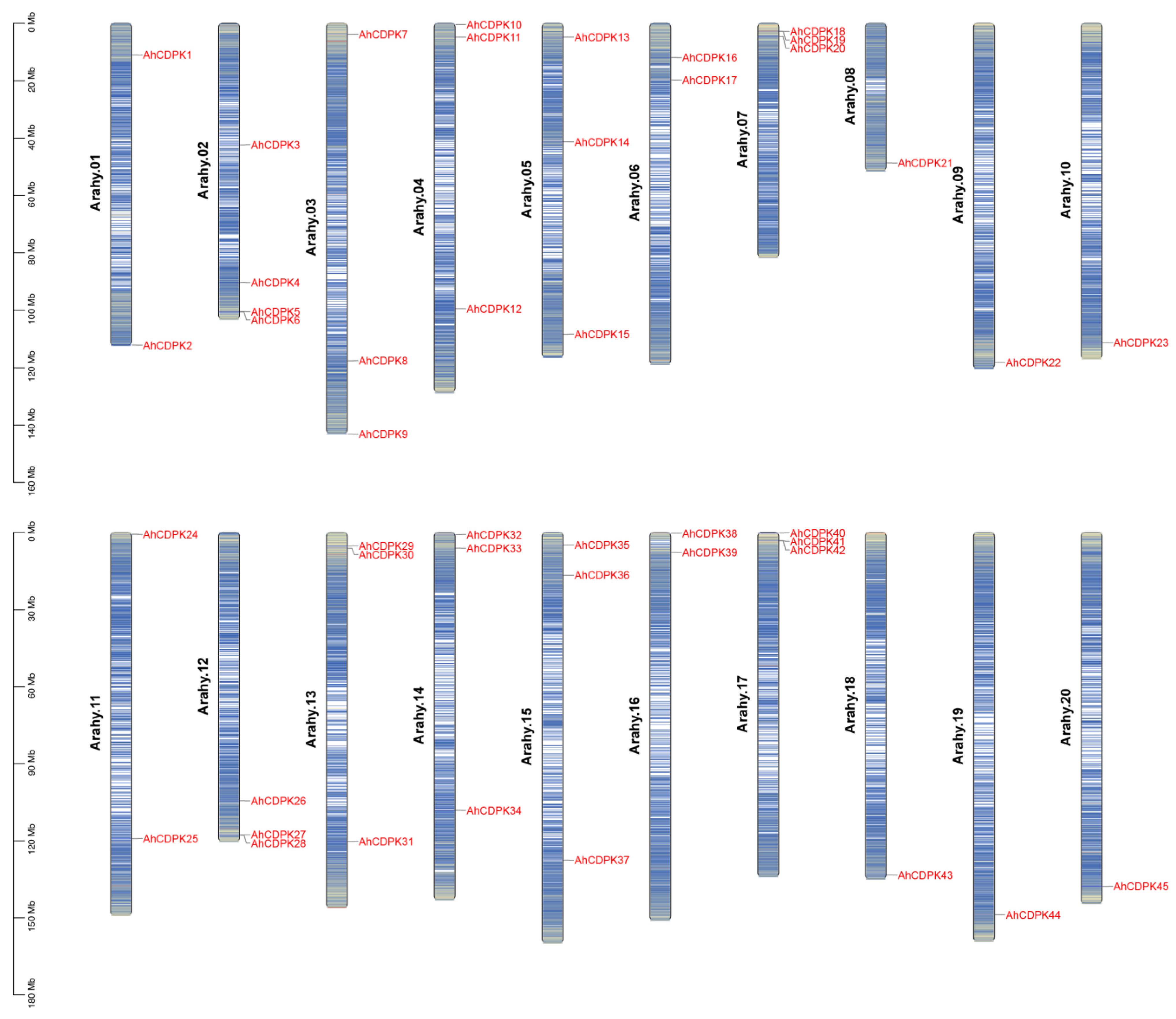
3.1. Genome-Wide Identification of 45 CDPK Genes in Peanut Genome

In this study, genome-wide analysis of the CDPK gene family has been performed on A. hypogaea genome sequence using profile HMM searches and BLASTP. A total of 45 CDPK family genes were identified in peanut after confirming each candidate using Pfam, ScanProsite, SMART, and InterProScan to verify the EF-hand and Pkinase-conserved domain. We renamed these genes AhCDPK1 to AhCDPK45 according to their chromosome positions (Table S3). The chromosomes of the A. hypogaea genome are numbered Arahy.01–Arahy.20 [29]. By chromosomal localization analysis, 45 AhCDPK genes were determined to be unevenly distributed across the 20 chromosomes and mostly concentrated at both ends of the chromosomes (Figure 1). Additionally, 23 and 22 of them belonged to the A genome (Arahy.01–10) and B genome (Arahy.11–20), respectively. The number of AhCDPKs distributed on Arahy.02 is the largest, with a total of four. Arahy.03, 04, 05, 07, 12, 13, 14, 15, and 17 all have three AhCDPKs. Arahy.01, 06, 11, and 16 all have two AhCDPKs. The number of AhCDPKs distributed on Arahy.08, 09, 10, 18, 19, and 20 is the least, with only one for each. As shown in Table S2, the gene length of AhCDPKs varies from 3335 bp (AhCDPK7) to 12,525 bp (AhCDPK33), with CDS length varying from 1425 bp (AhCDPK41) to 2325 bp (AhCDPK38). The protein lengths also vary considerably, ranging from 475 (AhCDPK41) to 774 (AhCDPK38) amino acids, and the predicted MW ranged from 53.59 (AhCDPK41) to 87.95 (AhCDPK38) kDa, while the isoelectric point (pI) ranged from 5.03 (AhCDPK3) to 9.53 (AhCDPK35). Most AhCDPKs contain both predicted N-terminal myristoylation and palmitoylation sites, while AhCDPK8, 17, 31, and 38 contain only a predicted myristoylation site. AhCDPK21, 22, 43, and 44 were predicted to have neither myristoylation nor palmitoylation sites. Next, the subcellular localizations of AhCDPKs were predicted, and the results indicate that 28 members were localized in the nucleus, 15 in the chloroplast, and 2 in the cytoplasmic membrane. In addition, 22 genes (AdCDPK1AdCDPK22) from A. duranensis and 23 genes (AiCDPK1AiCDPK23) from A. ipaensis genomes were also recognized to study the evolution of CDPK genes between tetraploid and diploid parents (Figures S2 and S3, Table S4).

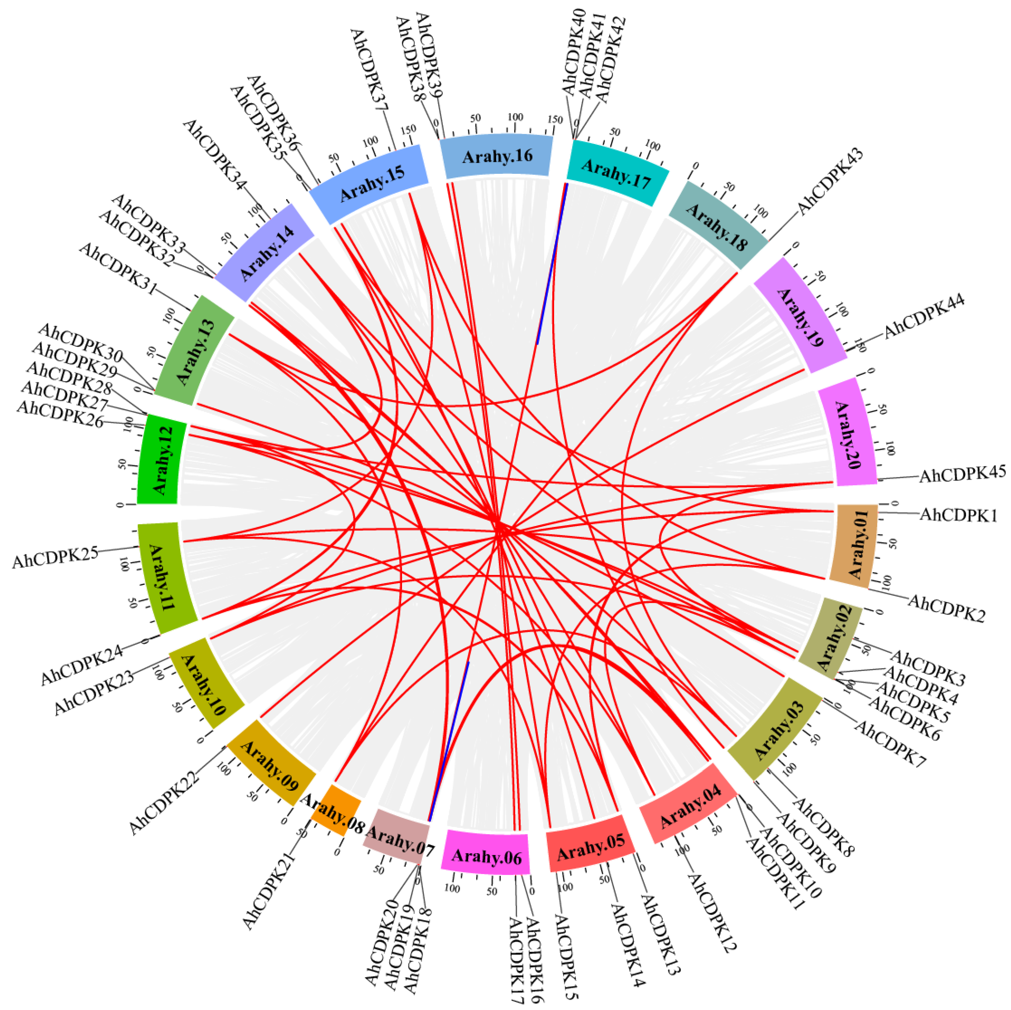
3.2. Synteny Analysis of CDPK Genes in Peanuts

Gene duplication is indeed a crucial mechanism for the amplification of large gene families in plants, including peanut. The presence of 45 AhCDPK genes in peanut suggests that the CDPK gene family has undergone significant expansion during evolution. We next examined the segmental or tandem duplication events of the CDPK gene family in the peanut genome. Almost all the AhCDPK genes experienced gene duplication events except AhCDPK3 and AhCDPK29, resulting in 47 replication events, including 33 pairs of whole-genome duplications (WGDs), 12 pairs of segmental duplications, and 2 pairs of tandem duplications (Figure 2), indicating that WGDs were the main way for expanding AhCDPK genes in peanut. We also calculated the synonymous (Ks), nonsynonymous (Ka), and Ka/Ks ratio values for each duplication event to study the evolutionary relationship among AhCDPK genes (Table S5). The results showed that the Ka values of WGDs, segmental duplications, and tandem duplications ranged from 0 to 0.2980, from 0.0476 to 0.2791, and from 0.2312 to 0.2377, respectively, while the Ks values ranged from 0 to 2.0362, from 0.6378 to 1.3353, and from 0.5499 to 0.5647, respectively. The Ka/Ks ratio (0–0.4322) was always less than 1 for every AhCDPK gene pair (Figure 2, Table S6), indicating that the AhCDPK gene family experienced strong negative selection. The divergence time for the 33 WGD pairs ranged from 0 to 125.3836 million years ago (Mya). The segmentally duplicated gene pairs had a divergence time ranging from 39.2710 to 82.2226 Mya. As for the two tandem duplicated genes, their divergence times were 33.8620 and 34.7708 Mya.
To further study the potential evolution of the AhCDPK gene family, two comparative syntenic maps of peanut with four species, including its two diploid ancestors (A. duranensis and A. ipaensis) and two other dicot species (Arabidopsis and rice), were built to clarify the syntenic relationships of CDPK genes in peanut (Figure 3). Syntenic relationships were observed between 37 AhCDPK genes and 21 AdCDPK genes in A. duranensis, 38 AhCDPK genes and 21 AiCDPK genes in A. ipaensis, 22 AhCDPK genes and 16 AtCDPK genes in Arabidopsis, and 10 AhCDPK genes and 7 OsCDPK genes in rice. The number of orthologous pairs between peanut and these four species (A. duranensis, A. ipaensis, A. thaliana, and rice) was 55, 56, 33, and 11, respectively (Tables S6 and S7).

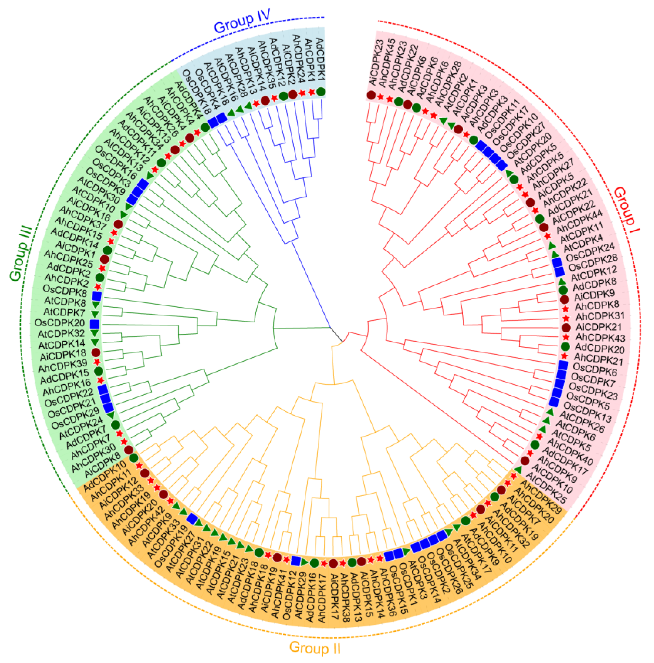
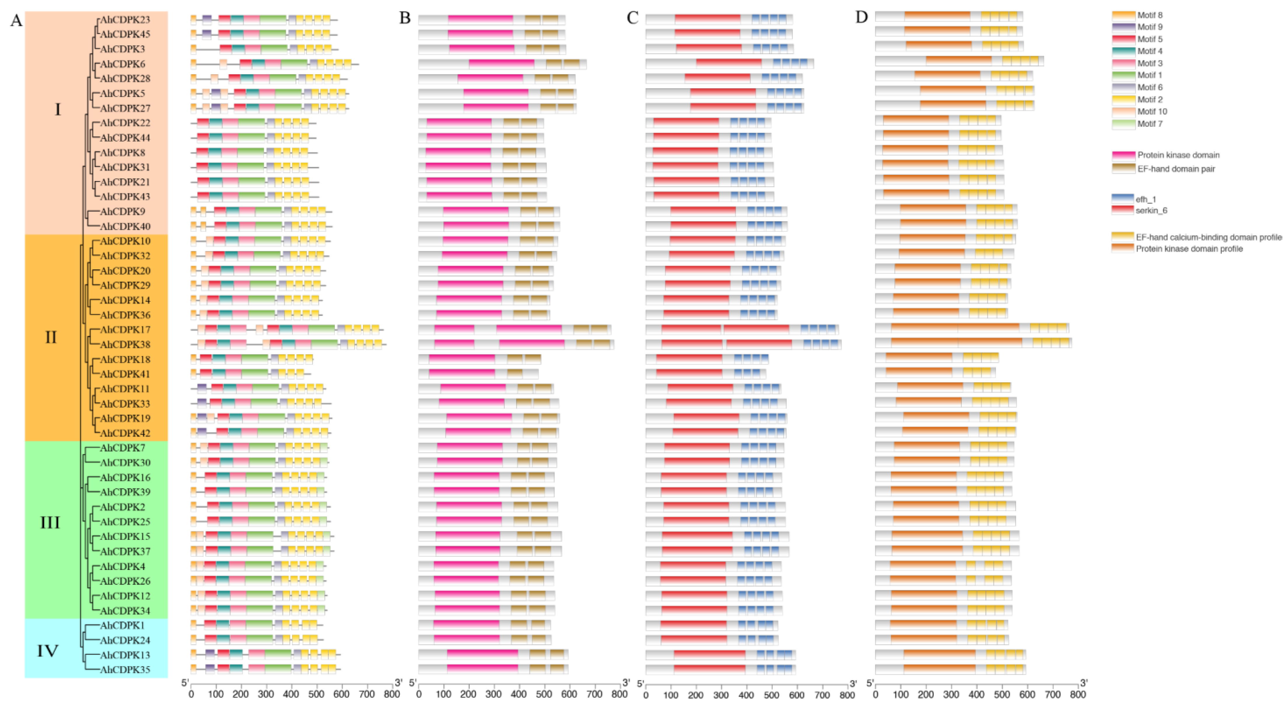
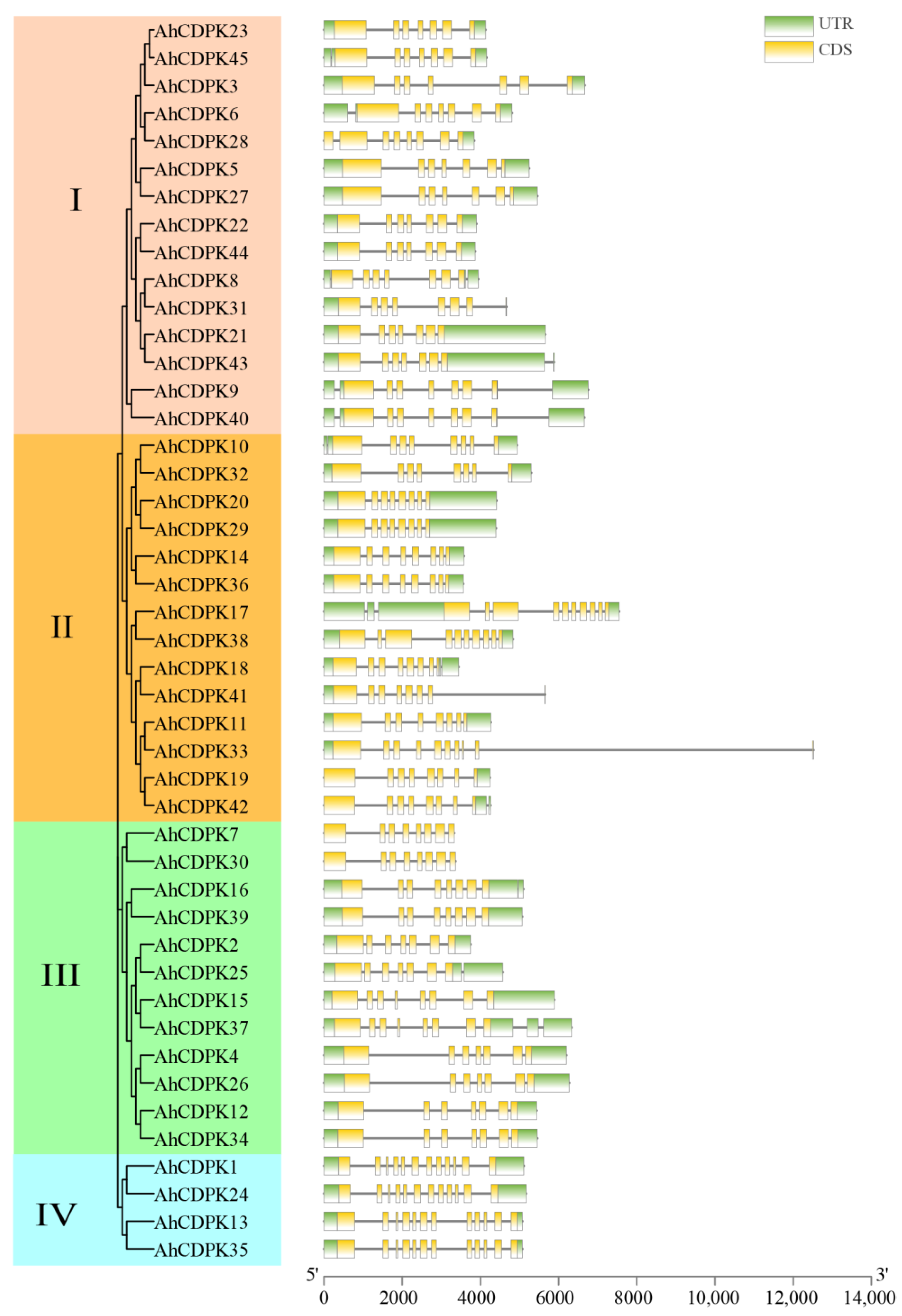
3.3. Phylogenetic Tree, Conserved Motifs, Gene Structure, and Tertiary Structures of AhCDPKs

To better elucidate the evolutionary relationship of AhCDPKs, a neighbor-joining (NJ) phylogenetic tree was constructed based on 153 CDPK protein sequences from peanut (red star), Arabidopsis (green triangle), rice (blue square), A. duranensis (dark green circle), and A. ipaensis (dark red circle) (Figure 4). CDPKs of peanut and its two diploid ancestors (A. duranensis and A. ipaensis) are more clustered together within the same branch than Arabidopsis and rice, showing a closer genetic relationship. According to the recognized classifications of the Arabidopsis and rice CDPK families [10,22], the AhCDPK/AdCDPK/AiCDPK proteins were divided into four subgroups, including 15/8/8, 14/6/7, 12/6/6, and 4/2/2 members in Subgroup I, II, III, and IV, respectively.
Phylogenetic subgroups were often related to genes and protein structures. The presence of four subgroups within the 45 AhCDPK proteins indicates that the gene family has undergone conservation within these subgroups. To illustrate the structural characteristics of the 45 CDPKs from peanut, firstly, we performed a motif and domain analysis combining the MEME program and Pfam, SMART databases, and PROSITE profiles in the InterPro (Figure 5). All AhCDPK proteins contained the four typical domains, VNTD, PKD, JD, and CaMLD. A total of 10 conserved motifs with lengths ranging from 21 to 105 amino acids were identified using MEME. (Figure 5A, Table S8). All proteins were found to contain motifs 1, 2, 3, 4, 5, and 6, while some motifs were detected only in specific subgroups. For example, motif 7 was found only in Subgroup III, motif 10 was not found in Subgroup IV, and motif 18 was found only in Subgroup I. Motif 9 was found only in AhCDPK5/-11/-13/-19/-23/-27/-33/-35/-42/-45. PKD showed conserved motifs 1, 3, 4, 5 in MEME, and the protein kinase domain in Pfam, serkin_6 in SMART, and the protein kinase domain profile in PROSITE were observed in the domain (Figure 5B–D). CaMLD were observed in motif 2 in MEME, EF-hand domain pair in Pfam, efh_1 in SMART, and EF-hand calcium-binding domain profile in PROSITE. The JD existed in motif 6, and motif 8 was found in VNTD, although nine proteins had none. All the AhCDPKs contained four EF-hands except AhCDPK41, which possessed three. Overall, the protein structure of this family was relatively conserved.
Then, the gene structure was analyzed. All peanut CDPK genes contain introns, and the number of introns ranges from 6 to 11 (Figure 6). There were 12, 18, 7, 3, and 5 genes containing 6, 7, 8, 9, and 11 introns, accounting for 26.67%, 40%, 15.56%, 6.67%, and 11.11% of the total genes, respectively. Notably, all the genes in Subgroup IV had the most at 11 introns, which showed highly similar genetic structures. In addition, there were 6 to 8 introns in Subgroup I, 7 to 11 introns in Subgroup II, and 6 to 9 introns in Subgroup III, respectively. By combining the gene structure of AhCDPKs with the phylogenetic tree, this result revealed that the exon-intron distribution of AhCDPKs was related to its classification.
Protein tertiary structures provide valuable insights into the structural characteristics of proteins. To further illustrate the structural characteristics, the tertiary structures of the CDPK proteins in peanut were investigated using AlphaFold2 as it is a novel machine learning approach and can predict protein structures with experimental accuracy based solely on their primary amino acid sequences [55]. The tertiary structures of the AhCDPK proteins were modeled using 19 template proteins identified through AFDB searches (Figure 7). The resulting models showed high quality, with GMQE values ranging from 0.666 to 0.830, sequence identities between 72.66% and 100%, and coverage from 88.1% to 100% for nearly the full length of each protein (Table S9). In addition, there were 6/7/5/1 templates in Subgroup I/II/III/IV, respectively. These templates were found to match 1, 2, or 4 proteins in each subgroup, with similar structures indicating a conserved function within each subgroup. Furthermore, transmembrane structure analysis showed that none of the AhCDPK proteins contained a transmembrane domain (Figure S4). In summary, by examining conserved motif structures, gene structures, protein tertiary structures, and phylogenetic relationships, it is evident that the gene organizations within subgroups of CDPK proteins remain remarkably consistent. This indicates that CDPK proteins possess highly conserved amino acid residues, and CDPK members belonging to the same subgroup likely perform similar functions.

3.4. Cis-Regulatory Elements Prediction in the Promotors of AhCDPKs

Considering the relatively conserved structures present within the CDPK gene family, we initially focus on exploring the potential regulatory factors affecting AhCDPK gene expression to better understand the functions of this gene family. Thus, the recognized putative cis-acting regulatory elements were analyzed from 2 kb upstream of the coding sequence of 45 AhCDPK genes using PlantCARE (Figure 8, Table S10). As shown in Figure 8, a total of 46 unique cis-acting regulatory elements with numbers equal to or greater than 10 were identified in the promoters of AhCDPKs, which were divided into the following five main categories: (1) phytohormone responsive, including ABA-responsive element (ABRE, ABRE3a, and ABRE4), auxin-responsive element (TGA-element), ethylene-responsive element (ERE), MeJA-responsive element (TGACG-motif, CGTCA-motif), GA-responsive element (P-box), and salicylic acid-responsive element (as-1, TCA, and TCA-element) (Figure S5); (2) abiotic stress, including anaerobic induction element (ARE), low-temperature-responsive element (LTR), drought-inducibility element (MBS), dehydration-responsive elements (Myb, MYB, Myb-binding site, MYB-like sequence, Myc, and MYC), stress-responsive element (STRE), and defense and stress-responsive element (TC-rich repeats) (Figure S6); (3) biotic stress, including wound-responsive element (WRE3, WUN-motif) and W box (Figure S7); (4) light responsive, including AE-box, AT1-motif, ATCT-motif, Box 4, GATA-motif, G-box, G-Box, GT1-motif, I-box, MRE, TCCC-motif, and TCT-motif (Figure S8); (5) plant growth and development, including secondary xylem development element (AAGAA-motif), A-box, maximal elicitor-mediated activation element (AT-rich sequence), meristem-expression element (CAT-box, CCGTCC-box, CCGTCC-motif), endosperm-expression element (GCN4-motif), zein metabolism regulation element (O2-site), and seed-specific regulation element (RY-element) (Figure S9). Out of the five main categories, abiotic stress had the highest number of cis-acting regulatory elements, with a total count of 603. The remaining categories, in descending order, were light responsive (546), phytohormone responsive (505), plant growth and development (155), and biotic stress (79). Among the analyzed genes, AhCDPK43 had the highest number of cis-acting regulatory elements within its promoter region (87), as it contained two prominent elements, ABRE and G-box, with a remarkable count of nineteen each (Figure 8A,B). Box 4 (197), ERE (127), MYC (121), MYB (115), and ABRE (114) were identified as the top five most prevalent cis-acting regulatory elements detected in the promoter regions of most genes (Figure 8C), indicating that AhCDPKs play an essential role in response to phytohormone, light, and abiotic stresses.

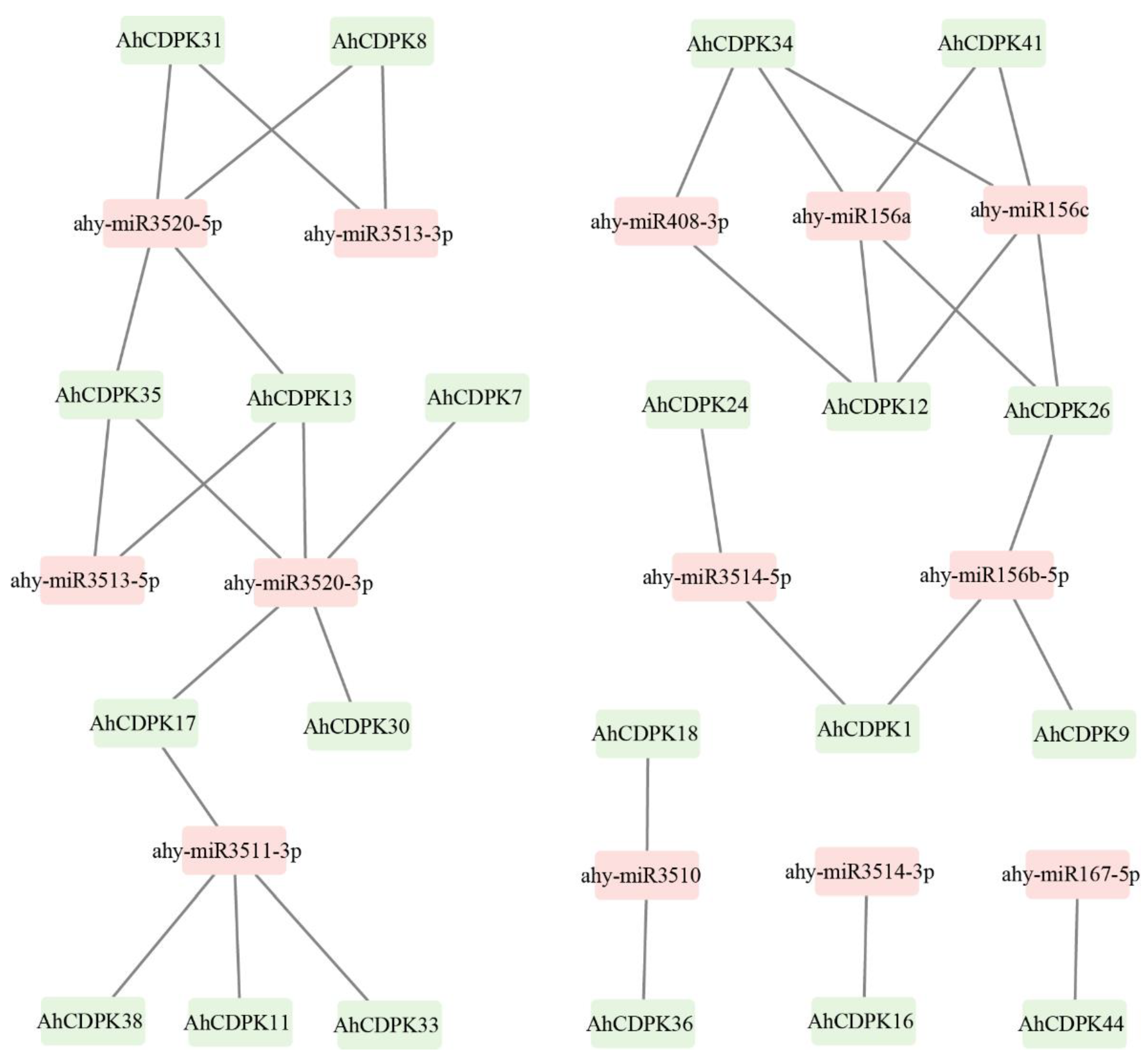
3.5. Prediction of miRNAs Targeting AhCDPK Genes

MicroRNAs (miRNAs) are crucial players in post-transcriptional regulation. To further investigate the potential regulatory mechanism in the regulation of AhCDPK genes, we identified a total of 13 miRNAs that target 21 genes. These miRNAs belonged to eight different families. The results revealed that ahy-miR3520-3p targeted the highest number (5) of genes (Figure 9, Table S11). Four miRNAs, including ahy-miR156a, ahy-miR156c, ahy-miR3511-3p, and ahy-miR3520-5p, targeted four genes, followed by ahy-miR156b-5p targeting three genes. Moreover, five miRNAs, including ahy-miR3510, ahy-miR3513-3p, ahy-miR3513-5p, ahy-miR3514-5p, and ahy-miR408-3p, targeted two genes each. Additionally, ahy-miR167-5p and ahy-miR3514-3p targeted one gene each. Interestingly, all four genes in Subgroup IV were targeted by miRNAs, while half of the genes in Subgroup II were targeted by miRNAs. One-third of the genes in both Subgroup I and Subgroup III were also targeted by miRNAs. These results suggest that miRNAs may play a crucial role in the posttranscriptional regulation of AhCDPK genes and their expression levels.

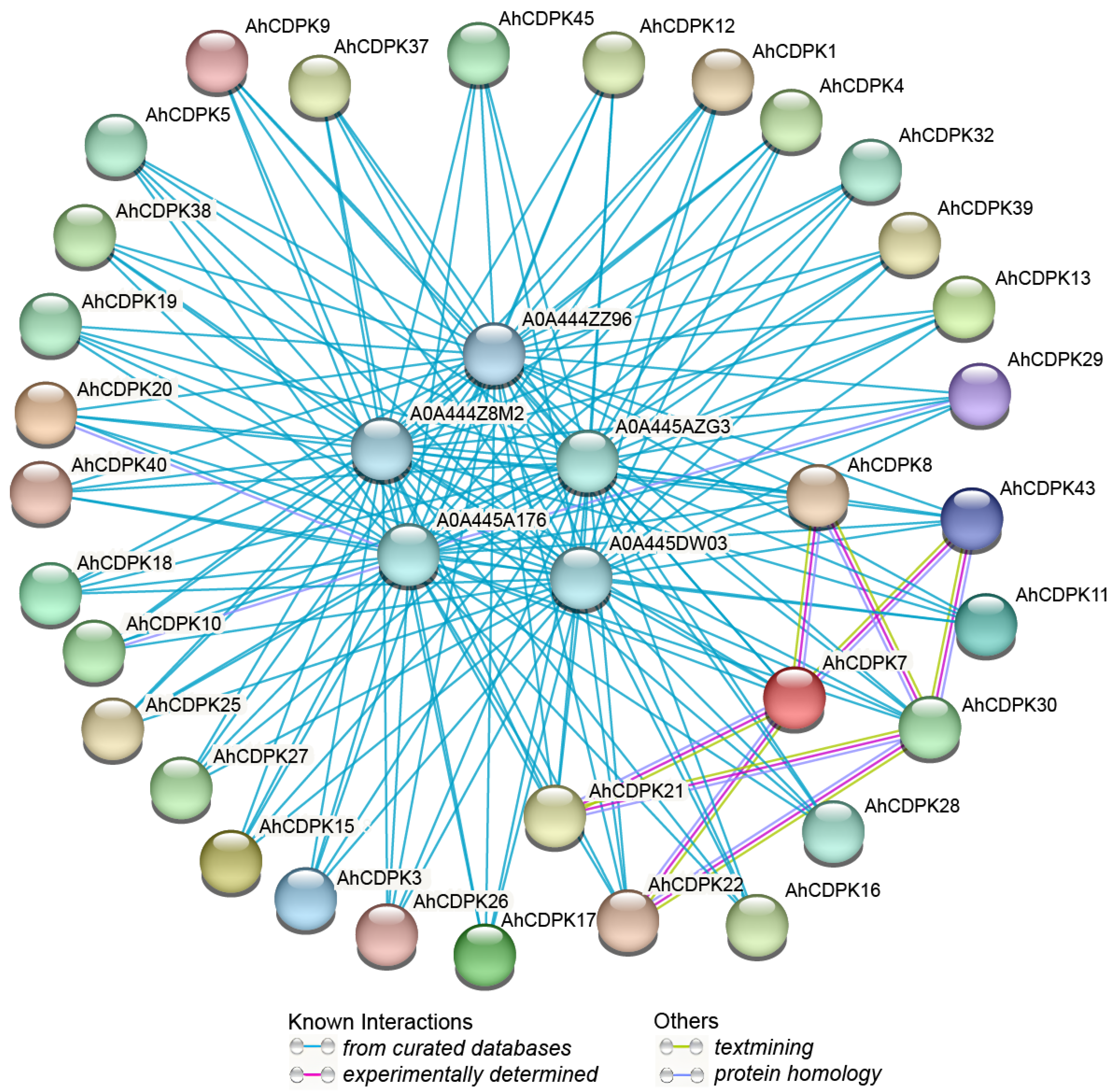
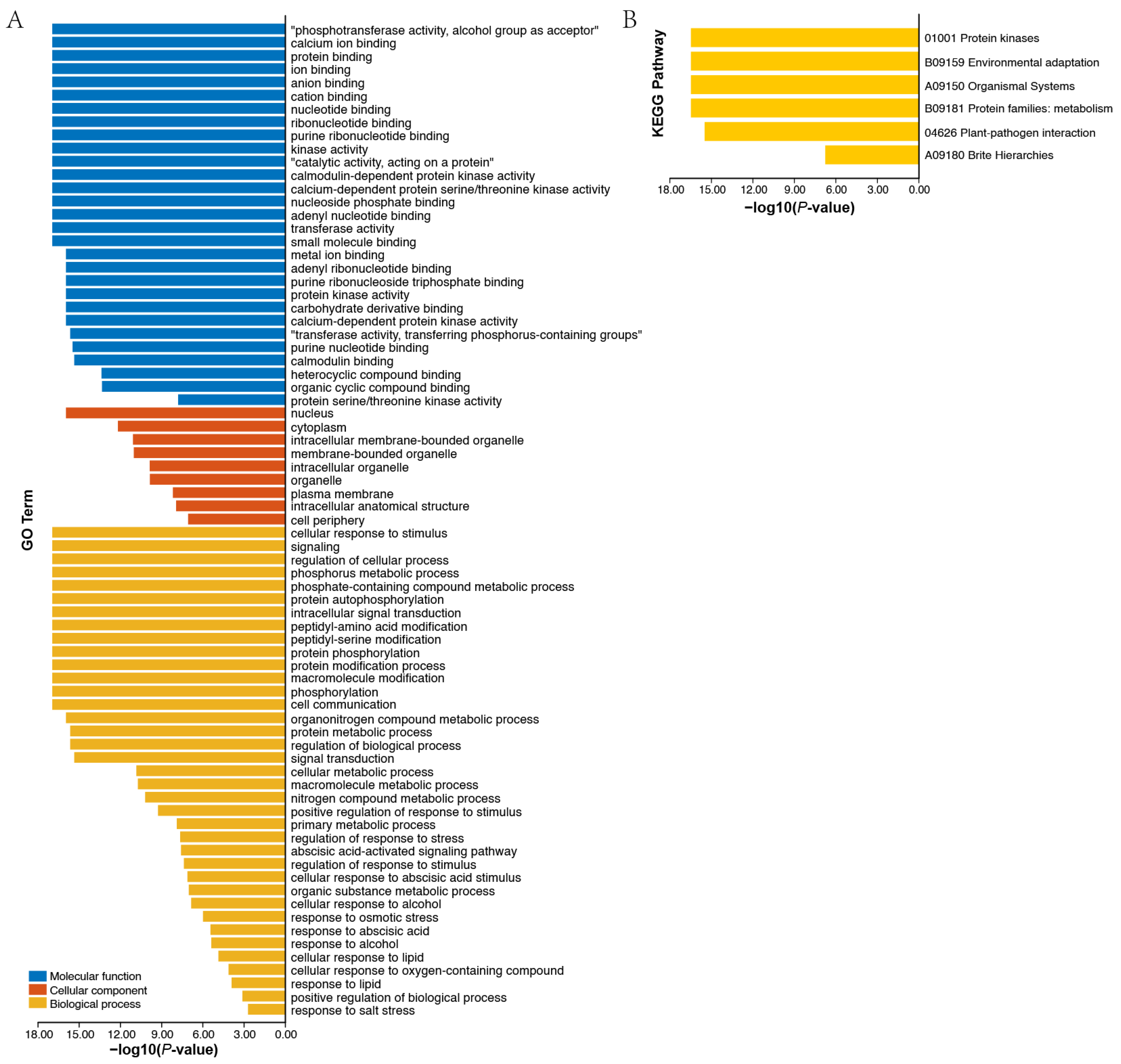
3.6. Protein Interaction Network and Functional Annotation Analysis of AhCDPKs

After analyzing the regulatory factors at the gene level, we next investigated potential regulation at the protein level. Firstly, an AhCDPKs protein interaction network was constructed between AhCDPKs and other peanut proteins using the STRING database [48]. According to the predicted results, we identified 32 AhCDPKs that interacted with 5 different peanut proteins (Figure 10, Table S12). Among these AhCDPKs, both AhCDPK22 and AhCDPK43 were found to interact with AhCDPK7 and AhCDPK30, respectively. Interestingly, AhCDPK30 was found to interact with AhCDPK21 and AhCDPK8, while AhCDPK21 was found to interact with AhCDPK7, and AhCDPK8 was found to interact with AhCDPK7. It is noteworthy that all 32 AhCDPK proteins were found to interact with five other peanut proteins, namely A0A444Z8M2, A0A444ZZ96, A0A445A176, A0A445AZG3, and A0A445DW03. These peanut proteins were identified as AhRBOHF (respiratory burst oxidase homolog F), AhRBOHI, AhRBOHE, AhRBOHB, AhRBOHA, which are homolog genes of Arabidopsis thaliana, owing to the incomplete annotation of the peanut genome. The above five peanut proteins identified in our investigation all belong to the RBOH family. RBOHs, also known as NADPH oxidases, are integral plasma membrane proteins that play a critical role in the generation of reactive oxygen species (ROS) [56]. These results provided valuable information for the further functional characterization of AhCDPK proteins. Next, we investigated the role of AhCDPK proteins at the molecular level through gene ontology (GO) and KEGG enrichment analysis (Figure 11, Table S13). To ensure the accuracy and reliability of GO and KEGG enrichment evaluations, the GO level was set to 3–8, and both the p-value and corrected p-value (BH method) were set to less than 0.01. The GO enrichment analysis can be divided into three main categories: biological processes (BP), molecular functions (MF), and cellular components (CC). For instance, in MF class, 29 enriched terms were detected, namely, calcium ion binding (GO:0005509), protein binding (GO:0005515), ion binding (GO:0043167), anion binding (GO:0043168), cation binding (GO:0043169), kinase activity (GO:0016301), calmodulin-dependent protein kinase activity (GO:0004683), calcium-dependent protein serine/threonine kinase activity (GO:0009931), protein kinase activity (GO:0004672), calcium-dependent protein kinase activity (GO:0010857), calmodulin binding (GO:0005516), protein serine/threonine kinase activity (GO:0004674), etc. In CC class, 9 enriched terms were detected, namely, nucleus (GO:0005634), cytoplasm (GO:0005737), intracellular membrane-bounded organelle (GO:0043231), membrane-bounded organelle (GO:0043227), intracellular organelle (GO:0043229), organelle (GO:0043226), plasma membrane (GO:0005886), intracellular anatomical structure (GO:0005622), cell periphery (GO:0071944). Whereas in BP class, 29 enriched terms were detected, including cellular response to stimulus (GO:0051716), signaling (GO:0023052), regulation of cellular process (GO:0050794), phosphorus metabolic process (GO:0006793), protein autophosphorylation (GO:0046777), intracellular signal transduction (GO:0035556), protein phosphorylation (GO:0006468), phosphorylation (GO:0016310), signal transduction (GO:0007165), regulation of response to stress (GO:0080134), regulation of response to stimulus (GO:0048583), response to osmotic stress (GO:0006970), response to abscisic acid (GO:0009737), response to salt stress (GO:0009651), etc. In addition, the KEGG pathway enrichment study identified six pathways involved in the different functions of the AhCDPK genes. The highly enriched pathways include protein kinases (01001), environmental adaptation (B09159), organismal systems (A09150), protein families: metabolism (B09181), plant-pathogen interaction (04626), and brite hierarchies (A09180). Overall, the GO and KEGG enrichment analyses validate the functional contribution of AhCDPK proteins in several important processes, including calcium ion binding, calmodulin-dependent protein kinase activity, regulation of cellular process, response to stresses, and environmental adaptation.

3.7. Spatial Expression Analysis of AhCDPKs

The evidence from cis-regulatory elements, miRNA, protein interaction networks, and functional annotations underscores the importance of AhCDPKs in plant development (Figure 8, Figure 9, Figure 10 and Figure 11). To gain insights into the potential biological functions of AhCDPKs in growth and development, expression pattern analysis is a useful tool. In this study, we analyzed the expression levels of AhCDPKs in 22 representative tissues using public transcriptome data [57]. A heatmap was created through the hierarchical clustering of the gene expression profiles of 45 AhCDPK genes, and these could be divided into three clusters: Cluster A, B, and C, including 17, 12, and 16 members, respectively (Figure 12). Cluster A’s CDPK genes were highly expressed in most of the 22 different tissues, implying that these genes may play important roles in peanut development. However, most of Cluster B’s CDPK genes were lowly expressed in the 22 tissues except in the lateral stem leaf, in which AhCDPK7/-30/-32 were highly expressed, suggesting that they may be involved in the growth and development of the organ. Notably, AhCDPK10/-32 had the highest expression level in the lateral stem leaf, while lightly even not expressed in other tissues, indicating their role in specific development processes. A considerable number of Cluster C’s CDPK genes exhibited moderate expression levels across the 22 tissues. Among Cluster C’s CDPK genes, AhCDPK45 was not expressed in the vegetative shoot tip or reproductive shoot tip but was highly expressed in the pattee 1 pod and pattee 10 seed. AhCDPK23 was not expressed in the vegetative shoot tip or the lateral stem leaf. The above results indicated that AhCDPK genes showed diverse expression patterns in peanut.
In order to further validate the role of AhCDPK genes in development, we conducted expression profiling of 12 genes, with 4 genes selected from each cluster, in the roots, leaves, and stems of peanut seedlings (Figure 12B). The results of RT-qPCR analysis confirmed a similar trend to that observed in the RNA-seq data. In Cluster A, the expression levels of AhCDPK8/-31/-37-40 were found to be the highest among all three tissues (roots, leaves, and stems). This indicates that these genes may play a significant role in the development of peanut seedlings. In Cluster B, the expression levels of AhCDPK12/-13/-35/-42 were relatively high, suggesting their involvement in developmental processes as well. However, we observed that the expression levels of AhCDPK7/-10/-30/-32 were hardly detected or had very low values in all three tissues. This suggests that these genes may have limited or specific functions in the development of peanut seedlings. This example demonstrates the consistency between the expression profiles obtained from RT-qPCR analysis and the RNA-seq data for all three clusters. The expression patterns of the selected AhCDPK genes in different tissues provide further evidence of their involvement in peanut development.

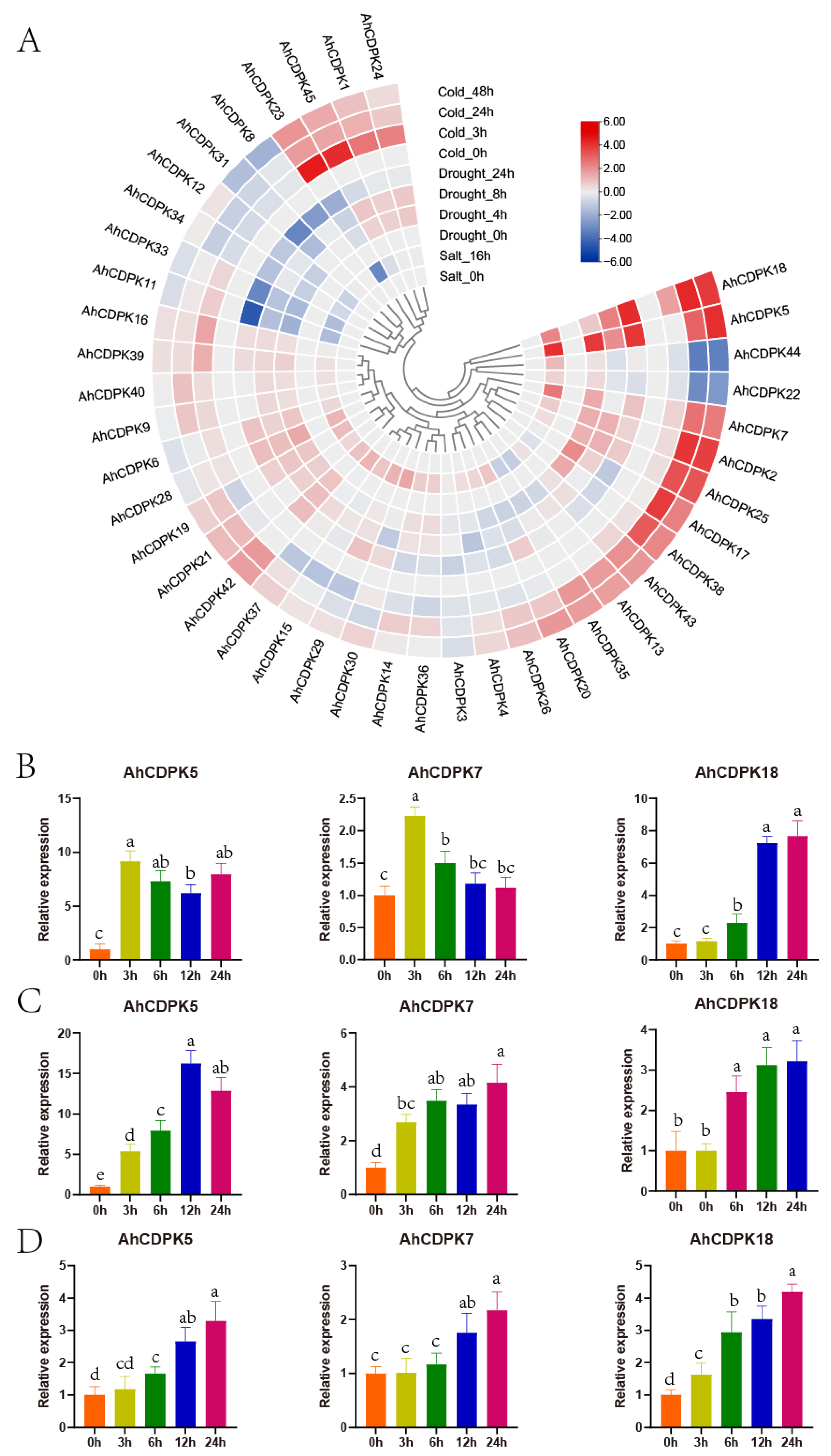
3.8. Expression Analysis of AhCDPKs under Abiotic Stresses

During growth and development, plants are significantly influenced by various environmental conditions, including low temperatures, high salinity, and drought. To investigate the potential biological functions of AhCDPKs in abiotic stress, we also analyzed the expression profiles of peanut CDPK genes in seeding responding to three abiotic stresses (salt, drought, and cold) using three public transcriptomes. As there were very low even zero TPM values in AhCDPK10/-27/-32/-41 under control conditions, the other 41 genes were considered in the following analysis. Following a 16 h salt treatment, AhCDPK5/-7/-18 were significantly induced (>2-fold increase from 0 h), while AhCDPK11/-13/-23/-33 were repressed (>2-fold decrease from 0 h) (Figure 13A). Under drought treatment, AhCDPK5 transcripts were upregulated after 4, 8, and 12 h, peaking at 4 h, whereas AhCDPK11/-33 transcripts were downregulated at all three time points. AhCDPK18 transcripts were upregulated after 8 and 12 h, whereas AhCDPK31 transcripts were downregulated at both time points. AhCDPK7/-17/-28/-38 transcripts were increased only after 4 h, whereas AhCDPK12/-34 transcripts were decreased only at this time point. AhCDPK2/-25 transcripts were increased only after 8 h, whereas AhCDPK4/-23/-30 transcripts were decreased only at this time point. Only AhCDPK8 transcripts were decreased after 12 h (Figure 13A). Under cold treatment, AhCDPK2/-5/-7/-17/-18/-25 transcripts were upregulated after 24 and 48 h, while AhCDPK22/-44 transcripts were downregulated at both time points. AhCDPK1/-23/-24/-45 transcripts were increased only after 3 h. AhCDPK38 transcripts were increased only after 24 h, while AhCDPK12/-34 transcripts were decreased only at this time point. AhCDPK8/-31 transcripts were decreased only after 48 h (Figure 13A). It is noteworthy that AhCDPK5/-7/-18 were upregulated by cold, drought, and salt stresses, while AhCDPK2/-17/-25/-38 were upregulated by cold and drought stress. On the other hand, AhCDPK11/-33 were downregulated by drought and salt stresses, while AhCDPK8/-12/-31/-34 were downregulated by cold and drought stresses. These results indicate that AhCDPKs may play an important role in response to various abiotic stresses, and different genes exhibit specific responses to stress.
To further assess the role of AhCDPKs in abiotic stresses, AhCDPK5/-7/-18 were selected to investigate their detailed expression patterns using an RT-qPCR analysis. Leaves from peanut seedlings were collected, and the expression levels of these genes were measured at different time points: 3 h, 6 h, 12 h, and 24 h (Figure 13B–D). Under drought treatment, the transcripts of AhCDPK5 were increased at all four time points, with the highest expression level observed at 3 h. AhCDPK7 transcripts were increased at 3 h and 6 h. AhCDPK18 transcripts showed a gradual increase over time, with more significant inductions at 12 h and 24 h. When subjected to salt treatment, AhCDPK5 transcripts were significantly induced at all four time points, with the peak expression observed at 12 h. AhCDPK7 transcripts were increased at all four time points. AhCDPK18 transcripts were also increased at three time points, except for the 3 h time point. Under cold treatment, AhCDPK5 transcripts were increased at three time points and showed a gradual increase over time. AhCDPK7 transcripts were increased at 12 h and 24 h. AhCDPK18 transcripts showed an increasing trend over time and peaked at 24 h. These RT-qPCR expression results were consistent with the RNA-seq results and highlighted the importance of AhCDPK5/-7/-18 in response to abiotic stress.

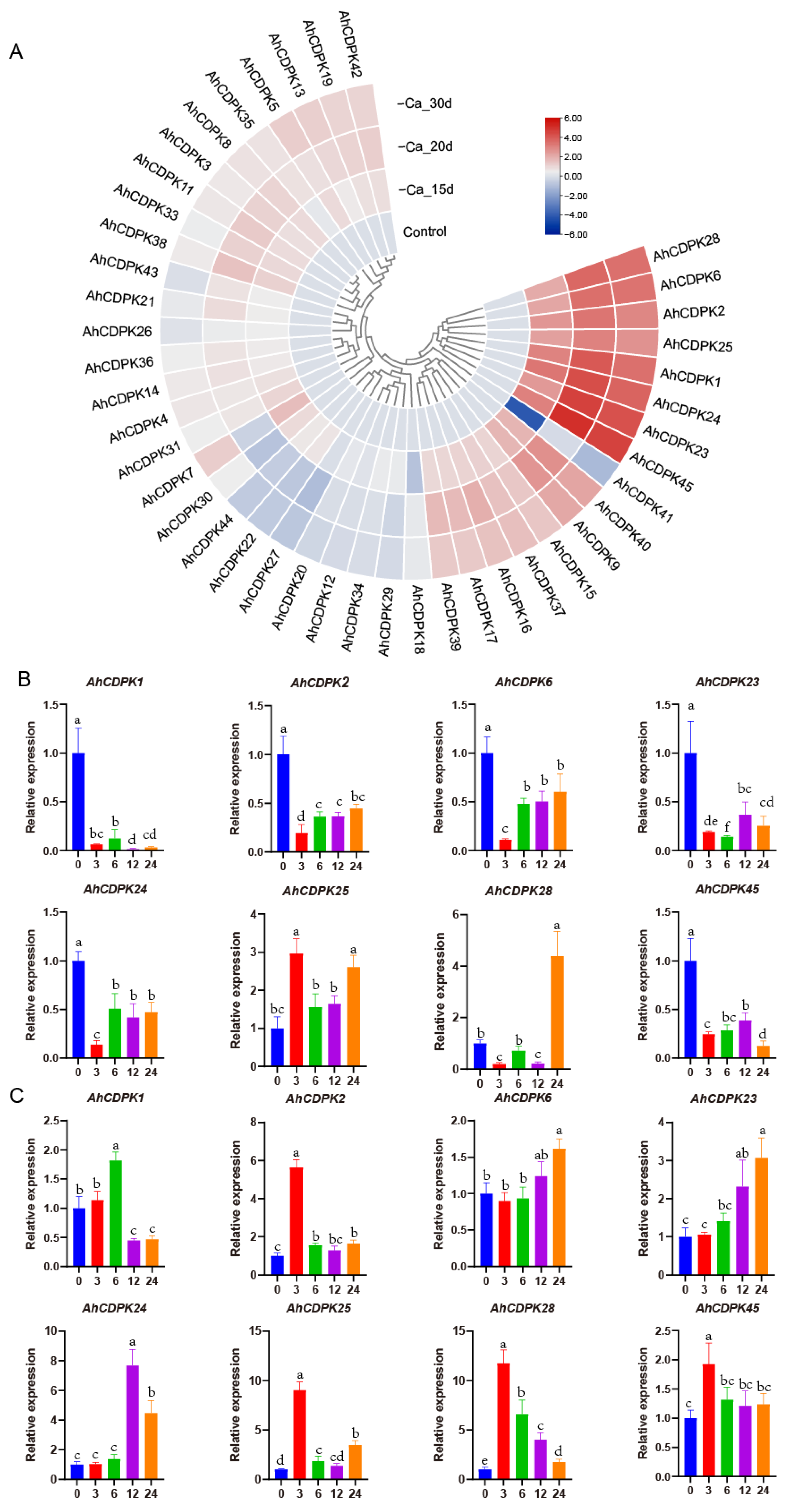
3.9. Expression Analysis of AhCDPKs in Response to Ca-Deficiency

Nutrient deficiency can greatly affect plant growth and development. For peanut, Ca is considered the third essential nutrient, following nitrogen and phosphorus. It has been reported that certain CDPKs are involved in seed and pod development when there is a deficiency of free Ca2+ in soil [33,58]. In order to explore the potential regulatory mechanisms of all AhCDPKs in the development process, the expression patterns of 43 AhCDPKs were analyzed. This was conducted using published RNA-seq data of Ca-deficient and Ca-sufficient peanut pods at 15, 20, and 30 days after pegging [36]. AhCDPK10/-32 were excluded from the analysis due to their limited expression in most pod processes. As shown in Figure 14A, 26 AhCDPK genes were raised to a significant level (>2-fold) by at least one Ca deficiency treatment, accounting for 60% of all analyzed genes. Out of these 26 genes, AhCDPK1/-2/-6/-23/-24/-25/-28/-45 exhibited >4-fold increase in all three Ca deficiency treatments, in comparison to the control. AhCDPK9/-40 soared to an elevated level (>2-fold) after 15 d and >4-fold after both 20 d and 30 d Ca deficiency treatments. In addition, AhCDPK15/-16/-17/37 were raised to a significant level (>2-fold) across all three Ca deficiency treatments. Similarly, AhCDPK7/-19/-38/-39/-42 genes were upregulated (>2-fold) in two Ca deficiency treatments, while AhCDPK3/-5/-8/-11/-13/-31/-33 were upregulated (>2-fold) in only one Ca deficiency treatment. On the contrary, AhCDPK41 experienced a significant down-regulation (>4-fold) via 15d Ca deficiency treatment and >2-fold under 30 d Ca deficiency treatments. Furthermore, AhCDPK27 was solely downregulated (>2-fold) by the 20 d Ca deficiency treatment. The above results suggest that AhCDPK genes may have specific roles in the adaptive response of peanut plants to Ca deficiency.
To comprehensively explore the involvement of the CDPK gene family in response to Ca deficiency, we undertook a detailed investigation of the expression patterns of eight AhCDPKs that were significantly induced by Ca deficiency. Specifically, we employed qRT-PCR to analyze the expression levels of these AhCDPKs at different time points (3 h, 6 h, 12 h, and 24 h) in both the leaves and roots of peanut seedlings (Figure 14B,C). The results from our analysis of leaves indicated that the transcript levels of AhCDPK1/-2/-6/-23/-24/-45 were downregulated at all four time points, while AhCDPK28 was downregulated at 3 h and 12 h, but upregulated at 24 h. Interestingly, AhCDPK25 was upregulated at 3h, 12h, and 24 h, providing us with a glimpse into the complexity of the molecular response mechanisms of plants. However, in the case of roots, we observed at least one upregulated time point of these eight AhCDPKs. Specifically, AhCDPK1 was upregulated at 6 h, AhCDPK2/-45 were upregulated at 3 h, AhCDPK6 was upregulated at 24 h, AhCDPK23/-24 were upregulated at 12 h and 24 h, and AhCDPK25/-28 were upregulated at all four time points. Notably, we discovered that four genes, AhCDPK2/-25/-28/-45, exhibited a peak expression level at 3 h, which may suggest that these genes play a crucial role in mediating the plants’ early response to low Ca conditions.

4. Discussion

As the critical sensors and decoders of calcium signals, CDPKs are essential in the regulation of plant growth, development, and stress tolerance [4,5,9,12]. In the previous study, CDPKs have been identified in Arabidopsis thaliana, rice, maize, wheat, soybean, cotton, chickpea, Medicago truncatula, peach, and pineapple [6,17,21,22,23,24,25,26,27]. However, the functions of CDPKs are still poorly understood in cultivated peanut. In this study, a total of 45 putative CDPK genes in cultivated peanut were identified using a combination of BLASTP analysis, HMM analysis, and conserved domain verification (Figure 1, Table S3). We conducted evolution analysis to reveal the gene expansion and the genetic relationship of the gene family (Figure 2 and Figure 3), which was supported by phylogenetic analysis. Phylogenetic analysis also classified 45 AhCDPKs into 4 subgroups based on their similarity (Figure 4). We further examined the structural features of the gene family, such as protein-conserved motifs, gene structure, and protein tertiary structures (Figure 5, Figure 6 and Figure 7), and found that they were consistent within each subgroup. To explore the potential function of the gene family, we predicted the regulatory mechanisms involving AhCDPKs genes and proteins. We analyzed the cis-regulatory elements and miRNA targets of AhCDPKs genes (Figure 8 and Figure 9), and the protein interaction network and functional annotations (GO and KEGG) of AhCDPKs proteins (Figure 10 and Figure 11). We discovered that the gene family had important roles in growth and development, stress response, and nutrient balance. Through expression analysis, we observed that AhCDPK genes had diverse expression patterns in different developmental stages (Figure 12). Moreover, different AhCDPK genes showed specific responses to various stresses, such as AhCDPK5/-7/-18, which were involved in cold, drought, and salt stress (Figure 13). Finally, we suggested that AhCDPK genes might have adaptive functions in peanut plants under Ca deficiency and that AhCDPK2/-25/-28/-45 could serve as indicators for pod Ca nutrition (Figure 14).
The peanut is an intriguing allotetraploid species with two subgenomes, labeled A and B. These subgenomes are genetic inheritances from two ancestral species: A. duranensis (AA) and A. ipaensis (BB) [28,29,30]. Our research has revealed that 22 CDPK genes are present in A. duranensis, while 23 CDPK genes are found in A. ipaensis (Table S4). Notably, previous research identified 35 CDPK genes in each of these wild peanuts, but only through the BLASTP method [59]. This implies that our approach is more effective. In contrast to the distribution pattern seen in the diploid parents, the cultivated peanut presents a different scenario: 23 CDPK genes are in the A-subgenome and 22 CDPK genes are in the B-subgenome (Figure 1 and Figure 2). This is largely due to the fact that the peanut subgenomes have evolved asymmetrically over time [29]. The total number of AhCDPK genes is equal to the sum of the genes in the two diploid parents, which indicates a translocation occurrence during allopolyploidization. When compared to several other plant species, the cultivated peanut stands out with a higher number of AhCDPK genes, such as Arabidopsis thaliana (34), rice (29), chickpea (22), Medicago truncatula (24), peach (17), and pineapple (17) [6,21,22,23,27,60]. However, the peanut’s total still falls short when compared to tetraploid sea-island cotton (84) and hexaploid wheat (85) [17,25]. One significant factor contributing to the large number of AhCDPK genes in peanuts is the occurrence of WGD events, which were the main duplication events (Figure 2). It is also worth noting that transposable elements make up a significant portion of the assembled genome sequence—a staggering 74% [29]. Our study further revealed evidence for two tandem duplication events within the AhCDPK genes. Specifically, AhCDPK18 and AhCDPK19 on the Arahy.07 chromosome are tandem duplicates, with their respective paralogs AhCDPK41 and AhCDPK42 on the Arahy.17 chromosome representing another tandem duplication event. We found evidence of 20 WGD events between two homologous genes, while the remaining 13 WGD events were not. The expansion of AhCDPK genes in the peanut could have occurred primarily through WGD between two homologous genes, followed by tandem duplication, segmental duplication, and allopolyploidization from the two ancient species. However, newly duplicated genes often exhibit functional redundancy, which can lead to gene loss over time [61]. To counteract this, mechanisms such as expression reduction come into play to facilitate the retention of duplicate genes and preserve their ancestral functions [61]. In our study, we observed that 12 AhCDPK genes in Cluster B displayed low expression levels in most of the 22 peanut tissues under normal conditions (Figure 12). This finding aligns with similar results reported in other gene families within the peanut genome [62,63,64]. These observations support the speculation that reduced expression may be advantageous in maintaining duplicate genes and their functional redundancy. The observation of more orthologs between peanut and Arabidopsis than between peanut and rice suggests that the expansion of the CDPK gene family in peanut is dependent on evolutionary processes (Figure 3). Orthologs are genes that have evolved from a common ancestor in different species and have retained similar functions [65]. In this case, the presence of more orthologs between peanut and Arabidopsis indicates a closer evolutionary relationship and a higher degree of conservation in the CDPK gene family.
Throughout the process of evolution, the CDPK gene family has exhibited a significant level of structural preservation across different plant species, ranging from mosses to angiosperms [4,10,12]. The representative function of CDPK is Ca2+-sensing functions and kinase activity. After the analysis of the MEME motif, Pfam, SMART, and PROSITE profiles, all 45 AhCDPK proteins included PKD and CaMLD (Figure 5, Figure S10). GO and KEGG analysis further confirmed the function of AhCDPKs (Figure 11). The VNTD and JD were also recognized by MEME. Like those in Arabidopsis, rice, maize, cotton, and peanut have CDPKs with less than four EF-hands. Only AhCDPK41 has three EF-hands. There was a tandem duplication between AhCDPK41 and AhCDPK42. Variable deletions were frequent in proximal chromosome regions [29]. AhCDPK41 and AhCDPK42 were in proximal Arahy.17. Thus, a variable deletion may come up during duplication.
In our study, we classified the CDPK gene family in peanut into four subgroups based on their general classification in Arabidopsis and rice [6,22]. However, in some species, such as sweet potato, there may be five subfamilies [66]. Interestingly, we observed that not every protein in the peanut CDPK gene family had homologous genes distributed on subchromosomes A and B. For example, AhCDPK3 did not have any paralogs. Additionally, the two pairs of homologous genes, AhCDPK20 and AhCDPK29, AhCDPK9 and AhCDPK40, were not located on the same subchromosomes, which was unexpected. To further support our findings, we analyzed the exon–intron structure and tertiary structures of the AhCDPK proteins (Figure 6 and Figure 7). These analyses provided additional evidence for the phylogenetic relationship among the AhCDPKs. We observed that most paralogs shared the same template protein, except for AhCDPK11 and AhCDPK33, as well as AhCDPK16 and AhCDPK39. The differences in template proteins between AhCDPK11 and AhCDPK33 may be attributed to the number of introns, resulting in different gene structures. AhCDPK33 had two additional introns compared to AhCDPK11, including an intron with a length of 8542, which was the longest among all AhCDPKs. While the exon-intron structure of AhCDPK16 and AhCDPK39 appeared similar, the reasons for the differences in their template proteins remain unclear and require further investigation.
Most of the homologous paralogs exhibited similar biochemical properties, which were dependent on the subgroup classification [4]. In terms of pI, AhCDPK proteins in Subgroup I were found to be acidic, with pIs below 5.8 (Table S3), similar to green algae and early land plants [4]. Conversely, AhCDPK proteins in subgroup IV were alkaline, with pIs beyond 9.1 (Table S3). However, unlike other land plants, the pIs of AhCDPK proteins in Subgroup II varied, ranging from 5.31 to 8.2 (Table S3). Specifically, both AhCDPK17 and AhCDPK38 in Subgroup II had a pI of 8.2. Only the above two paralogs lost the N-palmitoylation site in Subgroup II. The other proteins that lost N-terminal myristoylation or palmitoylation site were in Subgroup I. The specific functional implications of these variations in pI and post-translational modifications are still not fully understood and require further understanding.
The cultivated peanut is a vital crop with significant economic importance, particularly for oil production. However, it is susceptible to various abiotic stresses, including cold, drought, and salt [31]. Understanding and analyzing the CDPK genes in peanut can have profound implications for enhancing stress responses in this crop. As shown in Figure 8, the presence of ABA-responsive elements (ABRE, ABRE3a, and ABRE4), drought-responsive elements (MBS, Myb, MYB, Myb-binding site, MYB-like sequence, Myc, and MYC), and cold-responsive elements (LTR) suggests the potential involvement of AhCDPKs in regulating multiple responses to environmental stresses. Furthermore, the predicted gene functions of AhCDPKs were further supported by the GO enrichment analysis, as depicted in Figure 11. This analysis indicated the role of AhCDPKs in stress response mechanisms. Figure 13 illustrates that the application of salt, drought, and cold stresses resulted in the differential expression of 7, 17, and 17 AhCDPK genes, respectively. The variation in the number of genes can be attributed to the different time points at which the stress treatments were applied. In this study, drought and cold stresses were examined at three different time points, while salt stress was examined at only one time point due to limited public RNA-seq data. It is worth noting that previous research has shown the potential of CDPK genes in enhancing stress tolerance [17,18,19,20,21,25]. For instance, heterogeneous expresses SiCDPK24, TaCDPK25-U-AS1, TaCDPK25-U-AS2, or GmCDPK3 in transgenic Arabidopsis lines displayed enhanced tolerance to drought stress [17,18,19]. Additionally, the Arabidopsis cpk23 mutant showed increased tolerance to drought and salt stresses, while AtCPK23 overexpressing plants demonstrated reduced resistance to these stresses [67]. Furthermore, in rice, the induction in OsCDPK7 by cold and salt stresses, as well as its overexpression, led to increased tolerance to cold, salt, and drought [16]. These findings highlight the role of CDPKs in stress responses and their potential applications in crop improvement. To further explore the potential functions of AhCDPKs, protein interaction prediction was performed. This analysis can provide insights into the interactions and networks in which AhCDPKs may participate, shedding light on their role in stress signaling pathways. In our study, we found that all 32 AhCDPK proteins interacted with 5 peanut RBOH proteins, as shown in Figure 10. It is known that CDPKs can activate NADPH oxidase through phosphorylation, playing a crucial role in stress-induced oxidative damage [11,12]. In rice, for example, OsCPK12 regulates ROS homeostasis under salt stress by inducing the expression of the ROS-scavenging gene OsAPX2/OsAPX8 and inhibiting the NADPH oxidase gene OsRBOHI [68]. Interactions between CDPKs and other proteins have also been observed in other plant species. For example, in wheat, TaCDPK13 interacts with TaNOX7 to enhance ROS production, playing a crucial role in drought tolerance [69]. In peach, the interaction between PpCDPK7 and PpRBOH is believed to be the intersectional point of calcium and ROS signal transmission during cold storage of peach fruits [21]. This suggests that AhCDPKs may also participate in ROS signaling pathways in peanut in response to environmental stress. Based on these findings, it is reasonable to speculate that AhCDPKs in peanut may also participate in ROS signaling pathways in response to environmental stress. Further research is needed to elucidate the specific mechanisms and functions of AhCDPKs in stress tolerance and ROS signaling pathways in peanut.
Early studies have indicated the involvement of AhCDPK in peanut fruit development under Ca deficiency, and RNA-seq analysis has suggested their role in pod development [33,34,36,58]. In our research, we have found evidence supporting the important role of AhCDPKs during peanut fruit and pod development, particularly under Ca-deficient soil. The identification of the RY-element, a seed-specific regulation element, in Figure 8 suggests that the AhCDPK genes may be involved in seed development. The GO analysis results, which imply the important role of AhCDPKs in the regulation of cellular processes, further support the involvement of these genes in seed development (Figure 11). Significant advancements have been made in identifying the targets of peanut miRNAs involved in various developmental processes [70,71,72]. In the early stages of peanut pod development, a comprehensive study revealed the presence of 70 known and 24 novel miRNA families [71]. It was observed that almost all miRNAs-targeting AhCDPK genes were specifically expressed during the pod development stage, with the exception of ahy-miR3510. Another study focused on early peanut embryos and identified 29 known and 132 potential novel miRNAs, including all miRNAs-targeting AhCDPK genes except ahy-miR3520-3p and ahy-miR3520-5p [72]. Remarkably, among the AhCDPK genes upregulated under Ca deficiency, 12 out of the 26 identified genes were found to be targeted by miRNAs. This suggests that miRNAs play a role in modulating the expression of AhCDPK genes and potentially influencing seed development outcomes, particularly in response to Ca deficiency. The protein interaction network analysis indicating the involvement of AhCDPKs in ROS signaling pathways suggests a potential link between miRNA-mediated regulation of pod development and ROS signaling (Figure 10). Further investigation is needed to clarify whether miRNAs influenced pod development by affecting ROS signaling pathways in peanut. Additionally, Figure 14A shows that 26 AhCDPKs were upregulated in response to Ca deficiency, with 12 of them belonging to Cluster A (accounting for 71%) and 9 belonging to Cluster C (accounting for 56%). This indicates that approximately 80% of the upregulated AhCDPKs were expressed at moderate levels in most peanut tissues, with 46% being highly expressed, particularly during pod development. These findings provide additional evidence supporting the importance of AhCDPKs during pod development, consistent with early research [34,58]. Furthermore, we conducted a detailed investigation into the role of AhCDPKs by analyzing the expression levels of eight significantly upregulated AhCDPKs in the seedling stage under Ca deficiency treatment (Figure 14). Among these, four genes (AhCDPK2/-25/-28/-45) were identified as potential signals of Ca nutrients during the earlier stages of development. Notably, AhCDPK2 and AhCDPK25 were paralogs in Subgroup III and were homologous to AtCPK10/-30 in Arabidopsis. In Arabidopsis, Subgroup III AtCPK10/-30-32 are master regulators that orchestrate primary nitrate responses [73]. Nitrate–CPK signaling phosphorylates conserved NIN-LIKE PROTEIN (NLP) transcription factors to specify downstream gene reprogramming [73]. However, as peanut are legume crops and have a complex process of nitrogen absorption due to their symbiotic relationship with rhizobia, it is possible that there are some changes in the mechanisms involved in nutrient signaling. Further research is needed to elucidate the specific downstream targets and regulatory mechanisms of AhCDPKs in response to Ca deficiency.

5. Conclusions

We identified 45 CDPK genes with high confidence in the genomes of cultivated peanut. These genes were categorized into four subgroups based on a phylogenetic analysis. Various aspects such as protein physiological properties, protein tertiary structure, and conserved motifs were dependent on subgroup classification. Duplication events were identified that occurred in the peanut genome by collinearity analysis. Cis-acting regulatory elements in promoters, protein interaction networks, and GO and KEGG annotation evaluations implied the function of AhCDPK proteins in environmental stimuli. RNA-seq data from 22 different tissues revealed that AhCDPK genes in Cluster A and Cluster C play significant roles in plant development. The expression profile analysis of AhCDPKs due to various stresses implied their crucial roles in response to multiple signaling pathways. Interestingly, we observed a significant upregulation of AhCDPK genes in response to Ca deficiency. This observation was further supported by the RT-qPCR analysis in seedlings, highlighting the crucial involvement of AhCDPKs in peanut Ca nutrient regulation. By conducting a genome-wide identification of CDPKs in peanut, our study has the potential to significantly enhance our understanding of the genetic basis underlying peanut growth, development, and stress resistance.

Supplementary Materials

The following supporting information can be downloaded at: https://www.mdpi.com/article/10.3390/cells12232676/s1, Supplementary File S1, Figure S1: Melt-curve analysis of the selected AhCDPK genes and AhActin for qRT-PCR analysis; Figure S2: Chromosomal distribution of the CDPK genes in A. duranensis; Figure S3: Chromosomal distribution of the CDPK genes in A.ipaensis; Figure S4: Prediction of transmembrane structure of AhCDPKs; Figure S5: The positional distribution of phytohormone responsive cis-regulatory elements on promoters is shown as vertical bars with different colors; Figure S6: The positional distribution of abiotic stress cis-regulatory elements on promoters is shown as vertical bars with different colors; Figure S7: The positional distribution of biotic stress cis-regulatory elements on promoters is shown as vertical bars with different colors; Figure S8: The positional distribution of light responsive cis-regulatory elements on promoters is shown as vertical bars with different colors; Figure S9: The positional distribution of plant growth and development cis-regulatory elements on promoters is shown as vertical bars with different colors; Figure S10: Multiple sequence alignment of conserved domain of AhCDPK proteins; Supplementary File S2, Table S1: Information for RNA-seq analysis; Table S2: Primers of peanut AhCDPK genes selected for qRT-PCR analysis; Table S3: Information about peanut CDPK gene family members; Table S4: Genome-wide identification of CDPKs from the two ancestral species; Table S5: Analysis of the ten conserved motifs of AhCDPK proteins in peanut; Table S6: Details of various structural features in tertiary structure of AhCDPK proteins; Table S7: Gene duplication of AhCDPKs; Table S8: Orthologous CDPK gene pairs between cultivated peanut and its two diploid ancestors; Table S9: Orthologous CDPK gene pairs among peanut, Arabidopsis, and rice; Table S10: The cis-acting elements in the promoter regions of AhCDPK genes; Table S11: Prediction of miRNAs for AhCDPKs; Table S12: The protein-protein interaction network between AhCDPK and other proteins in peanut; Table S13: The GO and KEGG enrichment analysis of AhCDPK genes.

Author Contributions

Conceptualization, S.F., G.L. and S.W.; formal analysis, S.F. and S.Y.; writing—original draft preparation, S.F. and G.L.; writing—review and editing, S.F. and G.L.; visualization, S.F.; supervision, G.L. and S.W.; project administration, G.L. and S.W. All authors have read and agreed to the published version of the manuscript.

Funding

This research was supported by the Shandong Province Postdoctoral Innovation Talent Support Project (SDBX2022017), Shandong Key R&D Program (Major Scientific and Technological Innovation Project ZFJH202310) and Shandong Province Postdoctoral Innovation Project (SDCX-ZG-202203060).

Institutional Review Board Statement

Not applicable.

Informed Consent Statement

Not applicable.

Data Availability Statement

Data are contained within the article or Supplementary Files.

Conflicts of Interest

The authors declare no conflict of interest.

References

  1. Dodd, A.N.; Kudla, J.; Sanders, D. The language of calcium signaling. Annu. Rev. Plant Biol. 2010, 61, 593–620. [Google Scholar] [CrossRef]
  2. Kudla, J.; Batistic, O.; Hashimoto, K. Calcium signals: The lead currency of plant information processing. Plant Cell 2010, 22, 541–563. [Google Scholar] [CrossRef]
  3. Lee, H.-J.; Seo, P.J. Ca2+ talyzing Initial Responses to Environmental Stresses. Trends Plant Sci. 2021, 26, 849–870. [Google Scholar] [CrossRef]
  4. Hamel, L.P.; Sheen, J.; Seguin, A. Ancient signals: Comparative genomics of green plant CDPKs. Trends Plant Sci. 2014, 19, 79–89. [Google Scholar] [CrossRef] [PubMed]
  5. Harmon, A.C.; Gribskov, M.; Harper, J.F. CDPKs—A kinase for every Ca2+ signal? Trends Plant Sci. 2000, 5, 154–159. [Google Scholar] [CrossRef] [PubMed]
  6. Cheng, S.-H.; Willmann, M.R.; Chen, H.-C.; Sheen, J. Calcium signaling through protein kinases. The Arabidopsis calcium-dependent protein kinase gene family. Plant Physiol. 2002, 129, 469–485. [Google Scholar] [CrossRef]
  7. Shi, S.; Li, S.; Asim, M.; Mao, J.; Xu, D.; Ullah, Z.; Liu, G.; Wang, Q.; Liu, H. The Arabidopsis Calcium-Dependent Protein Kinases (CDPKs) and Their Roles in Plant Growth Regulation and Abiotic Stress Responses. Int. J. Mol. Sci. 2018, 19, 1900. [Google Scholar] [CrossRef] [PubMed]
  8. Martín, M.L.; Busconi, L. Membrane localization of a rice calcium-dependent protein kinase (CDPK) is mediated by myristoylation and palmitoylation. Plant J. 2000, 24, 429–435. [Google Scholar]
  9. Harper, J.F.; Sussman, M.R.; Schaller, G.E.; Putnam-Evans, C.; Charbonneau, H.; Harmon, A.C. A calcium-dependent protein kinase with a regulatory domain similar to calmodulin. Science 1991, 252, 951–954. [Google Scholar] [CrossRef]
  10. Hrabak, E.M.; Chan, C.W.M.; Gribskov, M.; Harper, J.F.; Choi, J.H.; Halford, N.; Kudla, J.; Luan, S.; Nimmo, H.G.; Sussman, M.R.; et al. The Arabidopsis CDPK-SnRK superfamily of protein kinases. Plant Physiol. 2003, 132, 666–680. [Google Scholar] [CrossRef]
  11. Boudsocq, M.; Sheen, J. CDPKs in immune and stress signaling. Trends Plant Sci. 2013, 18, 30–40. [Google Scholar] [CrossRef]
  12. Yip Delormel, T.; Boudsocq, M. Properties and functions of calcium-dependent protein kinases and their relatives in Arabidopsis thaliana. New Phytol. 2019, 224, 585–604. [Google Scholar] [CrossRef]
  13. Wang, H.; Gong, J.; Su, X.; Li, L.; Pang, X.; Zhang, Z. MaCDPK7, a calcium-dependent protein kinase gene from banana is involved in fruit ripening and temperature stress responses. J. Hortic. Sci. Biotechnol. 2017, 92, 240–250. [Google Scholar] [CrossRef]
  14. Liu, M.; Wang, C.; Xu, Q.; Pan, Y.; Jiang, B.; Zhang, L.; Zhang, Y.; Tian, Z.; Lu, J.; Ma, C.; et al. Genome-wide identification of the CPK gene family in wheat (Triticum a estivum L.) and characterization of TaCPK40 associated with seed dormancy and germination. Plant Physiol. Biochem. 2023, 196, 608–623. [Google Scholar] [CrossRef] [PubMed]
  15. Zhu, S.-Y.; Yu, X.-C.; Wang, X.-J.; Zhao, R.; Li, Y.; Fan, R.-C.; Shang, Y.; Du, S.-Y.; Wang, X.-F.; Wu, F.-Q.; et al. Two calcium-dependent protein kinases, CPK4 and CPK11, regulate abscis ic acid signal transduction in Arabidopsis. Plant Cell 2007, 19, 3019–3036. [Google Scholar] [CrossRef] [PubMed]
  16. Saijo, Y.; Hata, S.; Kyozuka, J.; Shimamoto, K.; Izui, K. Over-expression of a single Ca2+-dependent protein kinase confers both cold and salt/drought tolerance on rice plants. Plant J. 2000, 23, 319–327. [Google Scholar] [CrossRef] [PubMed]
  17. Bin, L.; Xu, Z.; Chu, Y.; Yan, Y.; Nie, X.; Song, W. Genome-wide analysis of calcium-dependent protein kinase (CDPK) family and functional characterization of TaCDPK25-U in response to drought stress in wheat. Environ. Exp. Bot. 2023, 209, 105277. [Google Scholar]
  18. Yu, T.-F.; Zhao, W.-Y.; Fu, J.-D.; Liu, Y.-W.; Chen, M.; Zhou, Y.-B.; Ma, Y.-Z.; Xu, Z.-S.; Xi, Y.-J. Genome-Wide Analysis of CDPK Family in Foxtail Millet and Determination of SiCDPK24 Functions in Drought Stress. Front. Plant Sci. 2018, 9, 651. [Google Scholar] [CrossRef]
  19. Wang, D.; Liu, Y.-X.; Yu, Q.; Zhao, S.-P.; Zhao, J.-Y.; Ru, J.-N.; Cao, X.-Y.; Fang, Z.-W.; Chen, J.; Zhou, Y.-B.; et al. Functional Analysis of the Soybean GmCDPK3 Gene Responding to Drought and Salt Stresses. Int. J. Mol. Sci. 2019, 20, 5909. [Google Scholar] [CrossRef]
  20. Ding, Y.; Yang, H.; Wu, S.; Fu, D.; Li, M.; Gong, Z.; Yang, S. CPK28-NLP7 module integrates cold-induced Ca2+ signal and transcriptional reprogramming in Arabidopsis. Sci. Adv. 2022, 8, eabn7901. [Google Scholar] [CrossRef]
  21. Zhao, L.; Xie, B.; Hou, Y.; Zhao, Y.; Zheng, Y.; Jin, P. Genome-wide identification of the CDPK gene family reveals the CDPK-RBOH pathway potential involved in improving chilling tolerance in peach fruit. Plant Physiol. Biochem. 2022, 191, 10–19. [Google Scholar] [CrossRef] [PubMed]
  22. Asano, T.; Tanaka, N.; Yang, G.; Hayashi, N.; Komatsu, S. Genome-wide Identification of the Rice Calcium-dependent Protein Kinase and its Closely Related Kinase Gene Families: Comprehensive Analysis of the CDPKs Gene Family in Rice. Plant Cell Physiol. 2005, 46, 356–366. [Google Scholar] [CrossRef]
  23. Zhao, P.; Liu, Y.; Kong, W.; Ji, J.; Cai, T.; Guo, Z. Genome-Wide Identification and Characterization of Calcium-Dependent Protein Kinase (CDPK) and CDPK-Related Kinase (CRK) Gene Families in Medicago truncatula. Int. J. Mol. Sci. 2021, 22, 1044. [Google Scholar] [CrossRef]
  24. Kong, X.; Lv, W.; Jiang, S.; Zhang, D.; Cai, G.; Pan, J.; Li, D. Genome-wide identification and expression analysis of calcium-dependent protein kinase in maize. BMC Genom. 2013, 14, 433. [Google Scholar] [CrossRef]
  25. Shi, G.; Zhu, X. Genome-wide identification and functional characterization of CDPK gene family reveal their involvement in response to drought stress in Gossypium barbadense. PeerJ 2022, 10, e12883. [Google Scholar] [CrossRef]
  26. Hettenhausen, C.; Sun, G.; He, Y.; Zhuang, H.; Sun, T.; Qi, J.; Wu, J. Genome-wide identification of calcium-dependent protein kinases in soybean and analyses of their transcriptional responses to insect herbivory and drought stress. Sci. Rep. 2016, 6, 18973. [Google Scholar] [CrossRef]
  27. Zhang, M.; Liu, Y.; He, Q.; Chai, M.; Huang, Y.; Chen, F.; Wang, X.; Liu, Y.; Cai, H.; Qin, Y. Genome-wide investigation of calcium-dependent protein kinase gene family in pineapple: Evolution and expression profiles during development and stress. BMC Genom. 2020, 21, 72. [Google Scholar] [CrossRef]
  28. Zhuang, W.; Chen, H.; Yang, M.; Wang, J.; Pandey, M.K.; Zhang, C.; Chang, W.-C.; Zhang, L.; Zhang, X.; Tang, R.; et al. The Genome of Cultivated Peanut Provides Insight into Legume Karyotypes, Polyploid Evolution and Crop Domestication. Nat. Genet. 2019, 51, 865–876. [Google Scholar] [CrossRef]
  29. Bertioli, D.J.; Jenkins, J.; Clevenger, J.; Dudchenko, O.; Gao, D.; Seijo, G.; Leal-Bertioli, S.C.M.; Ren, L.; Farmer, A.D.; Pandey, M.K.; et al. The Genome Sequence of Segmental Allotetraploid Peanut Arachis Hypogaea. Nat. Genet. 2019, 51, 877–884. [Google Scholar] [CrossRef] [PubMed]
  30. Bertioli, D.J.; Cannon, S.B.; Froenicke, L.; Huang, G.; Farmer, A.D.; Cannon, E.K.S.; Liu, X.; Gao, D.; Clevenger, J.; Dash, S.; et al. The Genome Sequences of Arachis Duranensis and Arachis Ipaensis, the Diploid Ancestors of Cultivated Peanut. Nat. Genet. 2016, 48, 438–446. [Google Scholar] [CrossRef] [PubMed]
  31. Patel, J.; Khandwal, D.; Choudhary, B.; Ardeshana, D.; Jha, R.K.; Tanna, B.; Yadav, S.; Mishra, A.; Varshney, R.K.; Siddique, K.H.M. Differential Physio-Biochemical and Metabolic Responses of Peanut (Arachis hypogaea L.) under Multiple Abiotic Stress Conditions. Int. J. Mol. Sci. 2022, 23, 660. [Google Scholar] [CrossRef] [PubMed]
  32. Tang, K.; Li, L.; Zhang, B.; Zhang, W.; Zeng, N.; Zhang, H.; Liu, D.; Luo, Z. Gene co-expression network analysis identifies hub genes associated with different tolerance under calcium deficiency in two peanut cultivars. BMC Genom. 2023, 24, 421. [Google Scholar] [CrossRef]
  33. Li, Y.; Meng, J.; Yang, S.; Guo, F.; Zhang, J.; Geng, Y.; Cui, L.; Wan, S.; Li, X. Transcriptome Analysis of Calcium- and Hormone-Related Gene Expressions during Different Stages of Peanut Pod Development. Front. Plant Sci. 2017, 8, 1241. [Google Scholar] [CrossRef]
  34. Yang, S.; Li, L.; Zhang, J.; Geng, Y.; Guo, F.; Wang, J.; Meng, J.; Sui, N.; Wan, S.; Li, X. Transcriptome and Differential Expression Profiling Analysis of the Me chanism of Ca2+ Regulation in Peanut (Arachis hypogaea) Pod Development. Front. Plant Sci. 2017, 8, 1609. [Google Scholar] [CrossRef]
  35. Yang, S.; Wang, J.; Tang, Z.; Guo, F.; Zhang, Y.; Zhang, J.; Meng, J.; Zheng, L.; Wan, S.; Li, X. Transcriptome of peanut kernel and shell reveals the mechanism of calcium on peanut pod development. Sci. Rep. 2020, 10, 15723. [Google Scholar] [CrossRef]
  36. Chen, H.; Yang, Q.; Fu, H.; Chen, K.; Zhao, S.; Zhang, C.; Cai, T.; Wang, L.; Lu, W.; Dang, H.; et al. Identification of Key Gene Networks and Deciphering Transcriptional Regulators Associated with Peanut Embryo Abortion Mediated by Calcium Deficiency. Front. Plant Sci. 2022, 13, 814015. [Google Scholar] [CrossRef]
  37. Mulder, N.J.; Apweiler, R.; Attwood, T.K.; Bairoch, A.; Bateman, A.; Binns, D.; Bork, P.; Buillard, V.; Cerutti, L.; Copley, R.; et al. New Developments in the InterPro Database. Nucleic Acids Res. 2007, 35, D224–D228. [Google Scholar] [CrossRef]
  38. Xie, Y.; Zheng, Y.; Li, H.; Luo, X.; He, Z.; Cao, S.; Shi, Y.; Zhao, Q.; Xue, Y.; Zuo, Z.; et al. GPS-Lipid: A robust tool for the prediction of multiple lipid modification sites. Sci. Rep. 2016, 6, 28249. [Google Scholar] [CrossRef] [PubMed]
  39. Savojardo, C.; Martelli, P.L.; Fariselli, P.; Profiti, G.; Casadio, R. BUSCA: An integrative web server to predict subcellular localization of proteins. Nucleic Acids Res. 2018, 46, W459–W466. [Google Scholar] [CrossRef]
  40. Chen, C.; Chen, H.; Zhang, Y.; Thomas, H.R.; Frank, M.H.; He, Y.; Xia, R. TBtools: An Integrative Toolkit Developed for Interactive Analyses of Big Biological Data. Mol. Plant 2020, 13, 1194–1202. [Google Scholar] [CrossRef] [PubMed]
  41. Tamura, K.; Stecher, G.; Kumar, S. MEGA11: Molecular Evolutionary Genetics Analysis Version 11. Mol. Biol. Evol. 2021, 38, 3022–3027. [Google Scholar] [CrossRef]
  42. He, Z.; Zhang, H.; Gao, S.; Lercher, M.J.; Chen, W.-H.; Hu, S. Evolview v2: An online visualization and management tool for customized and annotated phylogenetic trees. Nucleic Acids Res. 2016, 44, W236–W241. [Google Scholar] [CrossRef]
  43. Bailey, T.L.; Johnson, J.; Grant, C.E.; Noble, W.S. The MEME Suite. Nucleic Acids Res. 2015, 43, W39–W49. [Google Scholar] [CrossRef]
  44. Wang, Y.; Tang, H.; Debarry, J.D.; Tan, X.; Li, J.; Wang, X.; Lee, T.-H.; Jin, H.; Marler, B.; Guo, H.; et al. MCScanX: A toolkit for detection and evolutionary analysis of gene synteny and collinearity. Nucleic Acids Res. 2012, 40, e49. [Google Scholar] [CrossRef] [PubMed]
  45. Lescot, M.; Déhais, P.; Thijs, G.; Marchal, K.; Moreau, Y.; Van de Peer, Y.; Rouzé, P.; Rombauts, S. PlantCARE, a database of plant cis-acting regulatory elements and a portal to tools for in silico analysis of promoter sequences. Nucleic Acids Res. 2002, 30, 325–327. [Google Scholar] [CrossRef] [PubMed]
  46. Dai, X.; Zhuang, Z.; Zhao, P.X. psRNATarget: A plant small RNA target analysis server (2017 release). Nucleic Acids Res. 2018, 46, W49–W54. [Google Scholar] [CrossRef]
  47. Shannon, P.; Markiel, A.; Ozier, O.; Baliga, N.S.; Wang, J.T.; Ramage, D.; Amin, N.; Schwikowski, B.; Ideker, T. Cytoscape: A software environment for integrated models of biomolecular interaction networks. Genome Res. 2003, 13, 2498–2504. [Google Scholar] [CrossRef]
  48. Szklarczyk, D.; Gable, A.L.; Nastou, K.C.; Lyon, D.; Kirsch, R.; Pyysalo, S.; Doncheva, N.T.; Legeay, M.; Fang, T.; Bork, P.; et al. The STRING database in 2021: Customizable protein-protein networks, and functional characterization of user-uploaded gene/measurement sets. Nucleic Acids Res 2021, 49, D605–D612. [Google Scholar] [CrossRef] [PubMed]
  49. Cantalapiedra, C.P.; Hernández-Plaza, A.; Letunic, I.; Bork, P.; Huerta-Cepas, J. eggNOG-mapper v2: Functional Annotation, Orthology Assignments, and Domain Prediction at the Metagenomic Scale. Mol. Biol. Evol. 2021, 38, 5825–5829. [Google Scholar] [CrossRef]
  50. Li, H.; Handsaker, B.; Wysoker, A.; Fennell, T.; Ruan, J.; Homer, N.; Marth, G.; Abecasis, G.; Durbin, R.; 1000 Genome Project Data Processing Subgroup. The Sequence Alignment/Map format and SAMtools. Bioinformatics 2009, 25, 2078–2079. [Google Scholar] [CrossRef]
  51. Robinson, M.D.; McCarthy, D.J.; Smyth, G.K. edgeR: A Bioconductor package for differential expression analysis of digital gene expression data. Bioinformatics 2010, 26, 139–140. [Google Scholar] [CrossRef]
  52. Chen, S.; Zhou, Y.; Chen, Y.; Gu, J. fastp: An ultra-fast all-in-one FASTQ preprocessor. Bioinformatics 2018, 34, i884–i890. [Google Scholar] [CrossRef]
  53. Kim, D.; Paggi, J.M.; Park, C.; Bennett, C.; Salzberg, S.L. Graph-based genome alignment and genotyping with HISAT2 and HISAT-genotype. Nat. Biotechnol. 2019, 37, 907–915. [Google Scholar] [CrossRef] [PubMed]
  54. Livak, K.J.; Schmittgen, T.D. Analysis of relative gene expression data using real-time quantitative PCR and the 2(-Delta Delta C(T)) Method. Methods 2001, 25, 402–408. [Google Scholar] [CrossRef] [PubMed]
  55. Cramer, P. AlphaFold2 and the future of structural biology. Nat. Struct. Mol. Biol. 2021, 28, 704–705. [Google Scholar] [CrossRef]
  56. Marino, D.; Dunand, C.; Puppo, A.; Pauly, N. A burst of plant NADPH oxidases. Trends Plant Sci. 2012, 17, 9–15. [Google Scholar] [CrossRef]
  57. Clevenger, J.; Chu, Y.; Scheffler, B.; Ozias-Akins, P. A Developmental Transcriptome Map for Allotetraploid Arachis hypogaea. Front. Plant Sci. 2016, 7, 1446. [Google Scholar] [CrossRef] [PubMed]
  58. Jain, M.; Pathak, B.P.; Harmon, A.C.; Tillman, B.L.; Gallo, M. Calcium dependent protein kinase (CDPK) expression during fruit develo pment in cultivated peanut (Arachis hypogaea) under Ca2+-sufficient and -deficient growth regimens. J. Plant Physiol. 2011, 168, 2272–2277. [Google Scholar] [CrossRef] [PubMed]
  59. Li, D.; Shi, P.; He, L.; Yang, W.; Fu, H.; Xu, Z. Bioinformatics Analysis of Calcium Dependent Protein Kinases Gene Family in Wild Peanut. Chin. J. Trop. Crops 2017, 38, 94–103. [Google Scholar]
  60. Deepika, D.; Poddar, N.; Kumar, S.; Singh, A. Molecular Characterization Reveals the Involvement of Calcium Dependent Protein Kinases in Abiotic Stress Signaling and Development in Chick pea (Cicer arietinum). Front. Plant Sci. 2022, 13, 831265. [Google Scholar] [CrossRef]
  61. Qian, W.; Liao, B.-Y.; Chang, A.Y.-F.; Zhang, J. Maintenance of duplicate genes and their functional redundancy by reduced expression. Trends Genet. 2010, 26, 425–430. [Google Scholar] [CrossRef] [PubMed]
  62. Wang, C.; Wang, X.; Li, J.; Guan, J.; Tan, Z.; Zhang, Z.; Shi, G. Genome-Wide Identification and Transcript Analysis Reveal Potential Roles of Oligopeptide Transporter Genes in Iron Deficiency Induced Cadmium Accumulation in Peanut. Front. Plant Sci. 2022, 13, 894848. [Google Scholar] [CrossRef] [PubMed]
  63. Zhang, Z.; Chen, N.; Zhang, Z.; Shi, G. Genome-Wide Identification and Expression Profile Reveal Potential Roles of Peanut ZIP Family Genes in Zinc/Iron-Deficiency Tolerance. Plants 2022, 11, 786. [Google Scholar] [CrossRef]
  64. Tan, Z.; Li, J.; Guan, J.; Wang, C.; Zhang, Z.; Shi, G. Genome-Wide Identification and Expression Analysis Reveals Roles of the NRAMP Gene Family in Iron/Cadmium Interactions in Peanut. Int. J. Mol. Sci. 2023, 24, 1713. [Google Scholar] [CrossRef] [PubMed]
  65. Jensen, R.A. Orthologs and paralogs—We need to get it right. Genome Biol. 2001, 2, interactions1002.1. [Google Scholar] [CrossRef] [PubMed]
  66. Li, X.; Zhao, L.; Zhang, H.; Liu, Q.; Zhai, H.; Zhao, N.; Gao, S.; He, S. Genome-Wide Identification and Characterization of CDPK Family Reveal Their Involvements in Growth and Development and Abiotic Stress in Sweet Potato and Its Two Diploid Relatives. Int. J. Mol. Sci. 2022, 23, 3088. [Google Scholar] [CrossRef]
  67. Ma, S.-Y.; Wu, W.-H. AtCPK23 functions in Arabidopsis responses to drought and salt stresses. Plant Mol. Biol. 2007, 65, 511–518. [Google Scholar] [CrossRef]
  68. Asano, T.; Hayashi, N.; Kobayashi, M.; Aoki, N.; Miyao, A.; Mitsuhara, I.; Ichikawa, H.; Komatsu, S.; Hirochika, H.; Kikuchi, S.; et al. A rice calcium-dependent protein kinase OsCPK12 oppositely modulates salt-stress tolerance and blast disease resistance. Plant J. 2012, 69, 26–36. [Google Scholar] [CrossRef]
  69. Hu, C.-H.; Zeng, Q.-D.; Tai, L.; Li, B.-B.; Zhang, P.-P.; Nie, X.-M.; Wang, P.-Q.; Liu, W.-T.; Li, W.-Q.; Kang, Z.-S.; et al. Interaction between TaNOX7 and TaCDPK13 Contributes to Plant Fertility and Drought Tolerance by Regulating ROS Production. J. Agric. Food Chem. 2020, 68, 7333–7347. [Google Scholar] [CrossRef]
  70. Zhao, C.-Z.; Xia, H.; Frazier, T.P.; Yao, Y.-Y.; Bi, Y.-P.; Li, A.-Q.; Li, M.-J.; Li, C.-S.; Zhang, B.-H.; Wang, X.-J. Deep Sequencing Identifies Novel and Conserved MicroRNAs in Peanuts (Arachis hypogaea L.). BMC Plant Biol. 2010, 10, 3. [Google Scholar] [CrossRef] [PubMed]
  71. Gao, C.; Wang, P.; Zhao, S.; Zhao, C.; Xia, H.; Hou, L.; Ju, Z.; Zhang, Y.; Li, C.; Wang, X. Small RNA Profiling and Degradome Analysis Reveal Regulation of MicroRNA in Peanut Embryogenesis and Early Pod Development. BMC Genom. 2017, 18, 220. [Google Scholar] [CrossRef] [PubMed]
  72. Chen, H.; Yang, Q.; Chen, K.; Zhao, S.; Zhang, C.; Pan, R.; Cai, T.; Deng, Y.; Wang, X.; Chen, Y.; et al. Integrated MicroRNA and Transcriptome Profiling Reveals a MiRNA-Mediated Regulatory Network of Embryo Abortion under Calcium Deficiency in Peanut (Arachis hypogaea L.). BMC Genom. 2019, 20, 392. [Google Scholar] [CrossRef] [PubMed]
  73. Liu, K.-H.; Niu, Y.; Konishi, M.; Wu, Y.; Du, H.; Sun Chung, H.; Li, L.; Boudsocq, M.; McCormack, M.; Maekawa, S.; et al. Discovery of nitrate-CPK-NLP signalling in central nutrient-growth networks. Nature 2017, 545, 311–316. [Google Scholar] [CrossRef] [PubMed]
Figure 1. Chromosome density and chromosomal distribution of the AhCDPK genes. Chromosome numbers are provided at the left of each chromosome.
Cells 12 02676 g001
Figure 2. Synteny relationship of AhCDPKs in cultivated peanut. The blue lines indicate tandem duplicated gene pairs, and the red lines indicate segmentally duplicated and WGD gene pairs. The gray lines indicate segmentally duplicated gene pairs within the peanut genome. The scale bar marked on the chromosome indicates chromosome lengths (Mb).
Cells 12 02676 g002
Figure 3. Synteny analysis of CDPK genes between cultivated peanut and its two diploid ancestors (A) and among peanut, rice, and Arabidopsis (B). Gray lines in the background indicate the collinear blocks within peanut and other plant genomes, while the red lines highlight the syntenic CDPK gene pairs.
Cells 12 02676 g003
Figure 4. Phylogenetic analysis of CDPK proteins. The proteins of peanut, Arabidopsis, rice, Arachis duranensis, and Arachis ipaensis are represented in red star, green triangle, blue square, dark green circle, and dark red circle, respectively.
Cells 12 02676 g004
Figure 5. Conserved motifs analyses of AhCDPK proteins. (A) Conserved motif by MEME. The colored boxes on the right denote motifs 1–10. (B) Pfam motif. (C) SMART motif. (D) PROSITE profile.
Cells 12 02676 g005
Figure 6. Gene structure analyses of AhCDPK genes. The green boxes, black lines, and yellow boxes represent UTR, introns, and CDS, respectively.
Cells 12 02676 g006
Figure 7. Prediction of AhCDPK proteins’ tertiary structure by AlphaFold2 method. Models were visualized using rainbow colors from N to C terminus.
Cells 12 02676 g007
Figure 8. Cis-regulatory elements analysis of AhCDPK genes upstream regions. (A) The numbers and the depth of red represent the frequency of the elements that occur in the promoter region. (B) The statistics of categories on every AhCDPK. Different categories are present with different colors. (C) The number of each identified element.
Cells 12 02676 g008
Figure 9. Network map of projected miRNA targeting AhCDPK genes. The green boxes correspond to AhCDPK genes, and the pink boxes indicate predicted miRNAs.
Cells 12 02676 g009
Figure 10. Predictive interaction network of CDPK proteins in peanut. Network nodes represent proteins, and edges represent protein–protein associations.
Cells 12 02676 g010
Figure 11. GO (A) and KEGG (B) enrichment analysis of AhCDPKs.
Cells 12 02676 g011
Figure 12. Expression patterns of AhCDPK genes in different developmental tissues. (A) RNA-seq analysis. The heatmap was constructed using the mean of 1, 2, or 3 biological replicates, details in Table S1. The labels A, B, and C in the heatmap were shortened to indicate Cluster A, Cluster B, and Cluster C, respectively. (B) RT-qPCR analysis. Data are means ± SD (n = 6). Different letters indicate significant differences (Duncan’s test, p < 0.05) in each tissue including the subgraph.
Cells 12 02676 g012
Figure 13. Expression patterns of CDPK genes in peanut in response to abiotic stresses. (A) RNA-seq analysis. The heatmap was constructed using the mean of three biological replicates. RT-qPCR analysis when exposed to drought (B), salt (C), and cold (D). Data are means ± SD (n = 6). Different letters indicate significant differences (Duncan’s test, p < 0.05).
Cells 12 02676 g013
Figure 14. Expression patterns of CDPK genes in peanut pods in response to Ca deficiency. (A) RNA-seq analysis. The heatmap was constructed using the mean of three biological replicates. RT-qPCR analysis in (B) leaf and (C) root. Data are means ± SD (n = 6). Different letters indicate significant differences (Duncan’s test, p < 0.05).
Cells 12 02676 g014
Disclaimer/Publisher’s Note: The statements, opinions and data contained in all publications are solely those of the individual author(s) and contributor(s) and not of MDPI and/or the editor(s). MDPI and/or the editor(s) disclaim responsibility for any injury to people or property resulting from any ideas, methods, instructions or products referred to in the content.

