Communication

Progerinin, an Inhibitor of Progerin, Alleviates Cardiac Abnormalities in a Model Mouse of Hutchinson–Gilford Progeria Syndrome

1
Department of Molecular Biology, College of Natural Science, Pusan National University, Busan 46231, Republic of Korea
2
Heart-Immune-Brain Network Research Center, Department of Life Science, Ewha Womans University, Seoul 03760, Republic of Korea
3
Imvastech, 305, Science Building C, Ewha Womans University, Seoul 03760, Republic of Korea
4
PRG S & Tech Inc., Geumjeong-gu, Busan 46274, Republic of Korea
*
Author to whom correspondence should be addressed.
Cells 2023, 12(9), 1232; https://doi.org/10.3390/cells12091232
Submission received: 9 March 2023 / Revised: 20 April 2023 / Accepted: 22 April 2023 / Published: 24 April 2023
(This article belongs to the Special Issue Advances in Treatments for Hutchinson-Gilford Progeria Syndrome)

Abstract

:
Hutchinson–Gilford Progeria Syndrome (HGPS) is an ultra-rare human premature aging disorder that precipitates death because of cardiac disease. Almost all cases of HGPS are caused by aberrant splicing of the LMNA gene that results in the production of a mutant Lamin A protein termed progerin. In our previous study, treatment with Progerinin has been shown to reduce progerin expression and improve aging phenotypes in vitro and in vivo HGPS models. In this record, cardiac parameters (stroke volume (SV), ejection fraction (EF), fractional shortening (FS), etc.) were acquired in LmnaWT/WT and LmnaG609G/WT mice fed with either a vehicle diet or a Progerinin diet by echocardiography (from 38 weeks to 50 weeks at various ages), and then the cardiac function was analyzed. We also acquired the tissue samples and blood serum of LmnaWT/WT and LmnaG609G/WT mice for pathological analysis at the end of echocardiography. From these data, we suggest that the administration of Progerinin in the HGPS model mouse can restore cardiac function and correct arterial abnormalities. These observations provide encouraging evidence for the efficacy of Progerinin for cardiac dysfunction in HGPS.

1. Introduction

Hutchinson–Gilford Progeria Syndrome (HGPS) is a rare genetic disorder caused by an autosomal dominant mutation in the LMNA gene resulting in the accumulation of a truncated Lamin A version called progerin. Accumulation of progerin causes nuclear dysfunction and morphological abnormalities [1,2,3]. Patients with HGPS develop numerous clinical features including short stature, alopecia, scleroderma-like skin, low bone mineral density, lipodystrophy, skeletal dysplasia, and strokes. Premature death usually occurs in the second decade of age because of cardiovascular complications [4,5,6,7]. Transgenic LmnaG609G knock-in mice display phenotypes characterized by somatic growth, kyphosis, mandible hypoplasia, malocclusion, skin and adipose tissue atrophy, cardiovascular alterations, and shortened life span observed in patients with HGPS [8,9].
Previous studies have demonstrated that progerin strongly binds to wild-type Lamin A leading to disruption of nuclear architecture and acceleration of premature aging [10]. Pharmacological inhibition of Lamin A-progerin interaction by Progerinin (SLC-D011) improves cellular phenotypes in HGPS fibroblasts. Furthermore, LmnaG609G progeroid mouse model has shown the ability of Lamin A-progerin binding inhibitor treatment to improve phenotypes [11]. However, the effect of Progerinin on cardiovascular function and phenotypes has not been sufficiently proven. Given that premature cardiovascular disease is the cause of death in most of the patients with HGPS, we sought to test the ability of Progerinin to reduce the cardiovascular alterations seen in transgenic LmnaG609G mice.

2. Materials and Methods

2.1. Mice

All procedures with mice were performed in a facility certified by the Association for Assessment and Accreditation of Laboratory Animal Care in compliance with animal policies approved by Ewha Womans University. The mouse work was performed under the study protocol (approval code: 19-042), as approved by the Institutional Animal Care and Use Committee. Male heterozygous LmnaG609G mice, provided by Carlos López-Otín (Universidad de Oviedo, Asturias, Oviedo, Spain), and male wild-type C57BL/6J mice were housed at the specific pathogen-free facility.

2.2. Progerinin (SLC-D011) Synthesis and Charaterization Data

The synthetic scheme of Progerinin has already been described in our previous study [11]. In brief, Progerinin was synthesized by removing the hydrogen from the hydroxy of the decursinol which was isolated from the roots of Angelica gigas Nakai and by attaching side chains. After synthesis, the unrefined Progerinin was completed as the final compound by recrystallization. The compound was synthesized in a batch (Batch No. A05064-013S2; Batch size: 23.5 g, which was the same batch used in our previous study). The purity of Progerinin was measured by HPLC (≥97.5%). The molecular structure of Progerinin was investigated using H-NMR Spectroscopy, C-NMR spectroscopy, mass spectrometry, and X-ray diffractometry [11].

2.3. Chemical Pharmacokinetic (PK) Analysis

PK analysis of Progerinin has already been described in the previous study [11]. Briefly, 10 mg/kg of Progerinin in 10% NMP and 90% PEG400 solution were orally delivered. At pre-set time points, the blood concentration of Progerinin was determined by LC-MS/MS analysis.

2.4. Preparation of Feed Pellets

For oral administration of Progerinin by diet pellets, Progerinin was suspended in a monoolein-based solution at a concentration of 10 mg/mL. The base solution was made of monoolein (PeceolTM (code: 3088BFC, Lot/Batch: 174894), Gattefossé, La Défense Cedex, France) and tricaprylin (LabrafacTM lipophile WL 1349 (code: 3139BFC, Lot/Batch: 171801), GATTEFOSSE, Lyon, France). The suspensions were heated (50 °C) and sonicated continuously to make a clear solution. A powdered diet was mixed with the clear monoolein-based solution consisting of Progerinin and was made into dough balls. The vehicle-group diet was made of the clear monoolein-based solution without Progerinin. The dough balls were stored at 4 °C until use.

2.5. Progerinin Treatment

Progerinin was administrated with the same dose as in the previous study [11]. Mice were orally administrated with Progerinin-dough balls (2 g/day) daily at a concentration of 50 mg/kg. Control mice were treated with vehicle-dough balls in the same way. Mice were fed with a vehicle diet or therapeutic Progerinin diet starting from 38 weeks of age and sacrificed at 50 weeks of age.

2.6. Echocardiography and Cardiac Function Analysis

Mouse-specific ultrasound Vevo2100 system used to analyze and evaluate cardiac function and myocardial changes in detail using software such as LV analysis, PW Tissue Doppler Mode, and VEVO strain. Echocardiography was performed after general anesthesia using the inhaled anesthetic agent isoflurane (2–2.5% and 0.5 L/min oxygen) and removal of the hair coat of the whole chest. While positioning the mouse at an angle to the right, axis images of the left ventricle from the central thoracic and M-mode ultrasound images at the papillary muscle level were obtained. Cardiac function was quantified by analyzing cardiac function parameters. Before feeding and at various time points after feeding, in vivo cardiac parameters were obtained by the images from echocardiography and calculated using the following formula: HR (bmp) = total heart beats*60/time; SV (µL) = EDV − ESV; CO (mL/min) = SV*HR; LV EF (%) = (LVEDV − LVESV)/LVEDD*100%; LV FS (%) = (LVEDD − LVESD)/LVEDD*100%; DWS (%) = (LVPWs − LVPWd)/LVPWs*100%.

2.7. Western Blot for Tissue Samples

Progerin amounts in heart tissue analysis of wild-type mice, vehicle-treated LmnaG609G/WT mice, and Progerinin-treated LmnaG609G/WT mice were evaluated by means of Western blot analysis. Proteins were extracted with RIPA lysis buffer supplemented with protease inhibitors. Tissues were minimized by a tissue grinder and centrifuged at 15,000 rpm for 15 min at 4 °C. Proteins were loaded onto SDS-PAGE gels and transferred to the PVDF membranes. These membranes were blocked with 3% skimmed milk in TBS-T buffer for 1 h and incubated with primary antibodies, including mouse monoclonal anti-progerin (1:100; sc-81611; Santa Cruz Biotechnology, Dallas, TX, USA) and mouse monoclonal anti-beta actin (1:10,000; 66009-1-Ig; Proteintech, Rosemont, IL, USA) at 4 °C overnight. Blots were then incubated with goat anti-mouse IgG (1:10,000; Thermo Fisher Scientific, Waltham, MA, USA) secondary antibody and washed with TBS-T washing buffer. Bands were developed using the Intron ECL detection system and quantified using Image J (version: 2.9.0/1.53t, National Institute of Health (NIH), Bethesda, MD, USA).

2.8. Histology Analysis

Tissue specimens, including the heart and ascending aorta, were fixed in 4% PFA and embedded in paraffin. Paraffin blocks were sectioned and transferred onto adhesive-coated slides. After deparaffinization and rehydration, tissue sections were stained with H&E (hematoxylin and eosin), Verhoeff-Van Gieson, or Masson Trichrome. Immunohistochemistry was carried out using primary antibodies including anti-αSMA (1:3000; 14395-1-AP; Proteintech, Rosemont, IL, USA) and anti-Lamin A/C (1:100; 10298-1-AP; Proteintech, Rosemont, IL, USA). The images of the aorta stained with Masson Trichrome were separated into three different colors (blue, red, and green) by color deconvolution of ImageJ/FIJI to analyze the composition of collagen (blue) and muscle tissue (red). The number of elastic fiber breaks was measured in a single aortic ring from different individuals. The percentage of abnormal nuclei in heart tissue was calculated based on the manual counts carried out on randomly selected individuals in the same area of each group. Abnormalities of nuclei were determined based on extruded or engulfed nuclear shape, and irregular contour. For identifying clear images of nuclei, color deconvolution by ImageJ/FIJI was applied to the IHC images, which were stained with the anti-Lamin A/C antibody.

2.9. Mouse Cytokine Array

Whole blood samples of mice were collected after sacrifice at 50 weeks of age. The whole blood was left to clot at room temperature. Serum was collected by removing the clot following centrifugation at 4 °C. Cytokine array was performed using Proteome Profiler Mouse Cytokine Array Kit (ARY006; R&D systems, Minneapolis, MN, USA) following the manufacturer’s instructions. The array images were quantified using HLImage++ software 6.2 (Ideal Eyes Systems, Bountiful, UT, USA).

2.10. Statistical Analysis

Statistical analyses were performed using GraphPad Prism (Version 9.0.0, San Diego, CA, USA). Data were represented as means ± standard error mean (SEM) or standard deviation (SD). The statistical significance was analyzed using a one-way ANOVA followed by Tukey’s test or a two-way ANOVA Fisher’s LSD test.

3. Results

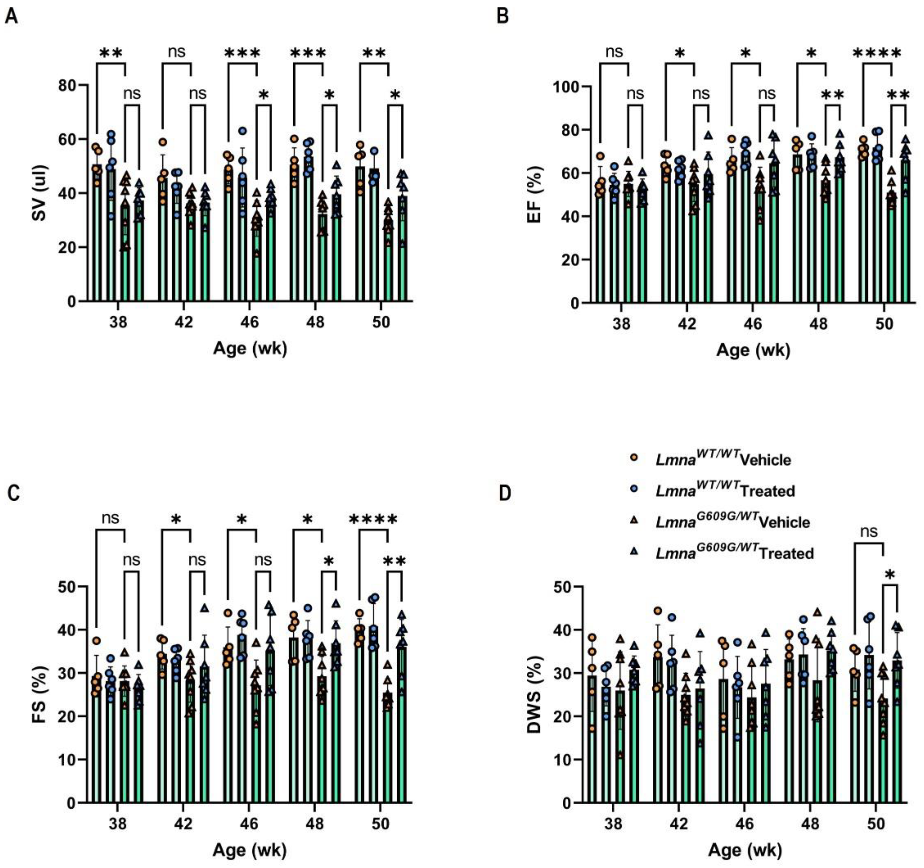
3.1. Progerinin Improves Cardiac Dysfunction in LmnaG609G/WT Mice

To measure cardiac physiology and architecture in HGPS mice, we examined cardiac function in 38- to 50-week-old LmnaG609G/WT mice using echocardiography. Progerinin mixed in LmnaG609G mouse dough or vehicle alone was orally administered at 50 mg/kg/day, which has been shown previously to be an effective dose to reduce progerin level in the HGPS mouse model (Supplementary Figure S1). Progerinin intake delayed the reduction in body weight in LmnaG609G/WT mice compared to the vehicle group (Supplementary Figure S2). In vivo, cardiac parameters by echocardiography revealed that HGPS mice displayed systolic and diastolic dysfunction compared to wild-type mice (Table S1). Echocardiography revealed no significant between-diet differences in heart rate (HR) (Supplementary Figure S3A). Comparing the stroke volume of LmnaWT/WT mice with LmnaG609G/WT mice before and after feeding, stroke volume was remarkably reduced in LmnaG609G/WT mice fed with the control diet (Figure 1A). However, significant improvements were confirmed from 8 weeks (46-week-old) in the Progerinin-fed group. There was no significant difference in cardiac output between the vehicle group and the Progerinin-fed group (Supplementary Figure S3B). As the most common measure of cardiac function, ejection fraction (EF) is the ratio of blood flow that has been released from the left ventricle during the heart cycle (EF = Cardiac output/Left ventricle volume at diastolic end*100%). The LmnaG609G/WT mice displayed overt left ventricle systolic dysfunction, with a significantly lower ejection fraction and fractional shortening (FS) than LmnaWT/WT mice. However, the Progerinin-fed LmnaG609G/WT group showed improvement in systolic function after 8 weeks (Figure 1B,C).
We next analyzed the function of left ventricle relaxation. The LmnaG609G/WT mice showed a reduced level of left ventricular posterior wall thickness at end-systole (LVPWs) and end-diastole (LVPWd). Comparing the LVPWs and LVPWd between the vehicle group and the Progerinin-fed group, LmnaG609G/WT mice showed no outstanding difference. Diastolic wall strain (DWS) has recently been used as a left ventricle function marker, indicating the relaxation of the left ventricle (DWS = [LVPWs − LVPWd]/LVPWs × 100 (%)). Although the difference in DWS between LmnaWT/WT mice and LmnaG609G/WT mice in the vehicle group was not significant, it showed a declining trend. Comparing the DWS between the vehicle group and the Progerinin-fed group, LmnaG609G/WT mice showed remarkable differences in long-term feeding, even though the DWS variation between mice is large (Figure 1D). These results demonstrate the effective improvement of cardiac function of HGPS mice by the long-term treatment of Progerinin.

3.2. Progerinin Reduces Histological Aortic Defects in LmnaG609G/WT Mice

Next, we performed microscopy studies in the aorta and heart to investigate the histological alteration. Arterial sections obtained after necropsy were analyzed by Masson Trichrome (MT) staining which is used in histology to differentiate collagen and muscle fibers on tissue sections. To simplify the complex staining and overlapping stains, images of aortic lesions were separated by using ‘color deconvolution’ of ImageJ/FIJI. MT staining in the aorta revealed significantly higher collagen decomposition (blue) in HGPS mice, accompanied by loss of muscle tissue (red). However, treatment with Progerinin reduced aortic fibrosis by restoring the composition of collagen and muscle tissue as in wild-type mice (Figure 2A and Figure S4). To visualize elastic fibers in the aorta, the sections from the three groups were stained with Verhoeff-Van Gieson’s stain. Verhoeff-Van Gieson staining in the aorta of LmnaG609G/WT mice and showed an increased number of elastin fiber breaks. However, Progerinin intake decreased the number of elastin fiber breaks (Figure 2B,C and Figure S5). Loss of smooth muscle cells is a hallmark of HGPS vessels. We also observed a lack of vascular smooth muscle cells (VSMCs) by α-smooth muscle actin staining in the aorta in LmnaG609G/WT mice. However, compared with the vehicle LmnaG609G/WT mouse group, the composition of smooth muscle cells was remarkably restored in the Progerinin-fed group (Supplementary Figure S6).

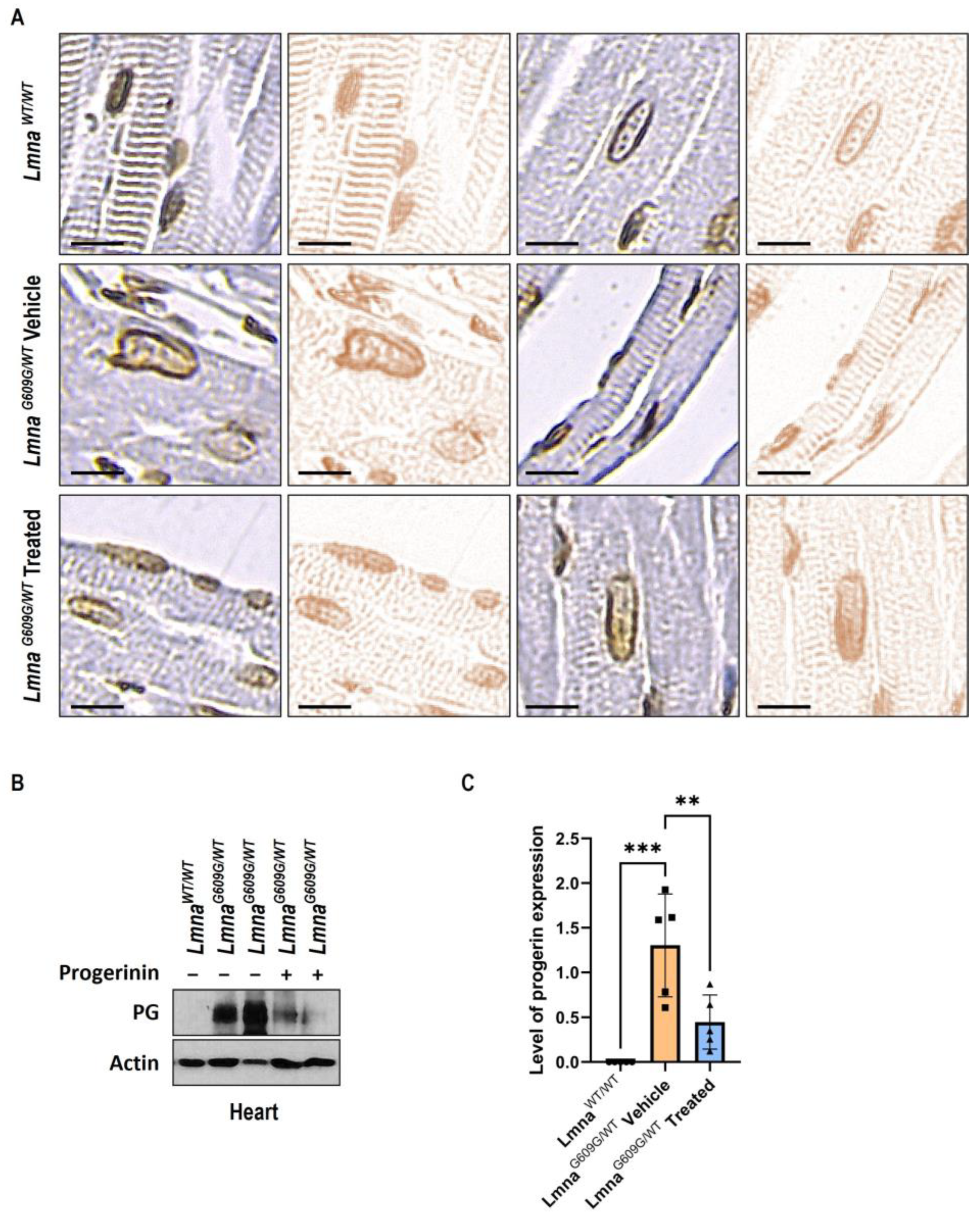
3.3. Progerinin Reduces Nuclear Deformation and Progerin Expression in Heart Tissue of LmnaG609G/WT Mice

Nuclear deformation is a hallmark of HGPS [5,12,13]. Consistent with these findings, immunochemistry studies demonstrated severely deformed nuclei in heart tissues in LmnaG609G/WT mice. However, deformed nuclear shapes in heart tissues were restored following the oral administration of Progerinin (Figure 3A and Figure S7A). To identify abnormal nuclei clearly, images of heart lesions were digitally separated by ImageJ/FIJI. Quantification of abnormal nuclei in heart tissue also shows that Progerinin prevents nuclear deformation in LmnaG609G/WT mice (Supplementary Figure S7B).
Next, in vivo, Progerinin activity was judged by Western blots of progerin. The graph reveals a decrease in progerin protein in the heart tissues of LmnaG609G/WT mice by more than 50% after treatment with Progerinin (Figure 3B). This result supports that cardiac dysfunction and histological aberration were restored by the reduction in progerin levels after treatment with Progerinin.

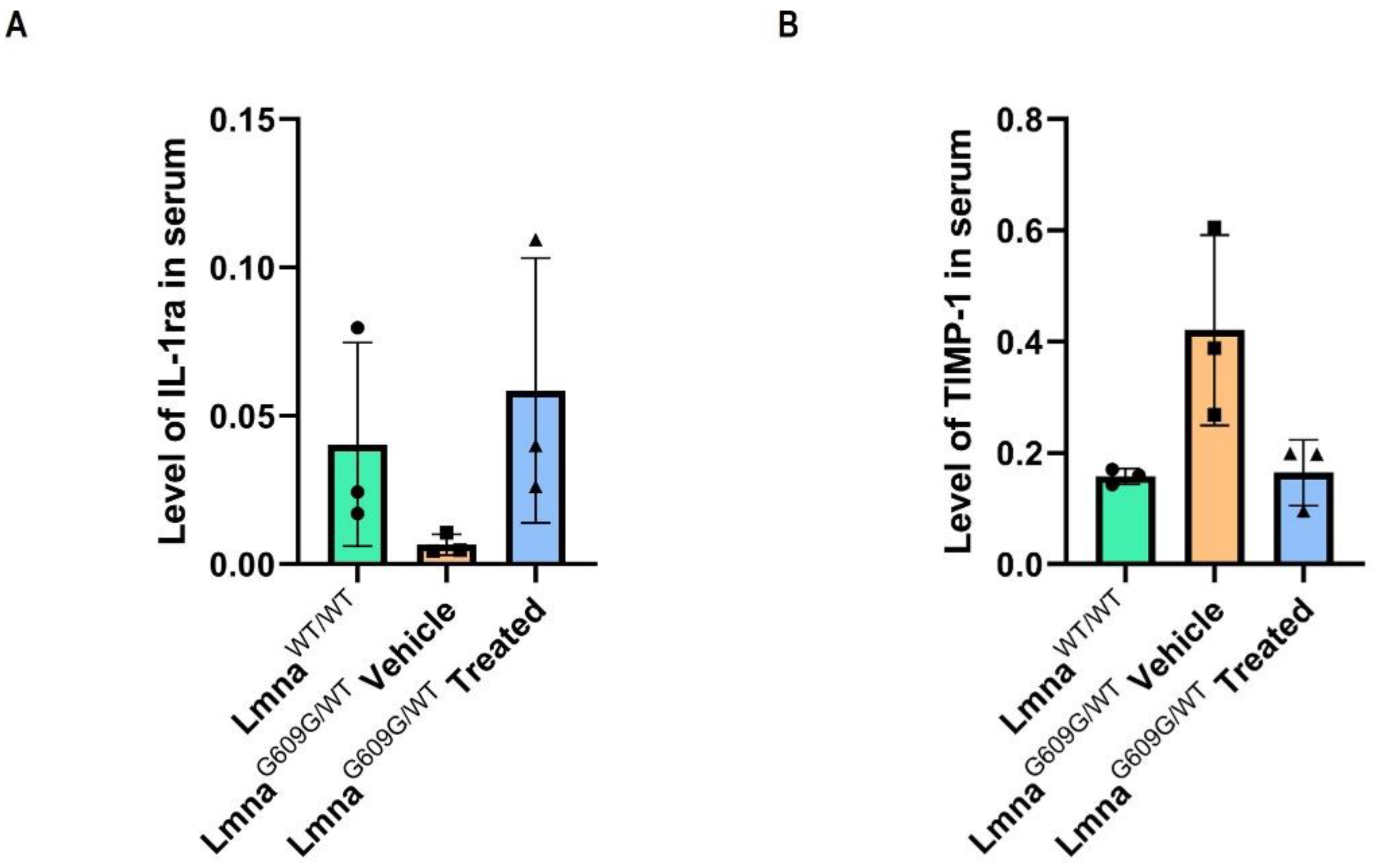
3.4. Progerinin Reverses the Levels of Cytokines in Serum of LmnaG609G/WT Mice

After 3-month treatments, we analyzed 40 different proteins in blood serum by mouse cytokine array kit to explore Progerinin’s potential impact on the levels of cytokines and chemokines which are related to cardiovascular function. We randomly selected three different mice from each group of LmnaWT/WT, LmnaG609G/WT vehicle, and LmnaG609G/WT treated mice. Two of 40 cytokines were significantly altered in LmnaG609G/WT mice compared to the wild-type model, including IL-1ra (IL-1 receptor antagonist) and TIMP-1 (Tissue inhibitor of matrix metalloproteinase-1) (Supplementary Figure S8). A low level of IL-1ra in LmnaG609G/WT mice was significantly upregulated after the Progerinin intake (Figure 4A). Conversely, TIMP-1 was a high level of cytokines in LmnaG609G/WT mice and was significantly down-regulated by Progerinin intake (Figure 4B). IL-1ra counter-regulates IL-1β, a key cytokine in the development of cardiovascular diseases [14,15]. Circulating IL-1ra expression is decreased in aged C57BL/6 mice [16]. Expression of IL-1ra was strongly reduced in blood serum from LmnaG609G/WT mice compared to wild-type mice of the same age (Figure 4A). However, IL-1α and IL-1β levels were not detectable. TIMP-1 is consistently upregulated in myocardial fibrosis and is used as a marker of fibrosis [17,18]. Expression of TIMP-1 was significantly increased in LmnaG609G/WT mice. After a 3-month Progerinin intake, elevated serum TIMP-1 level was markedly reduced (Figure 4B). Collectively, these data support that the Progerinin-mediated pharmacological inhibition of progerin ameliorates myocardial fibrosis and dysfunction in the HGPS mouse model.

4. Discussion

We have shown the efficiency of Progerinin on classical HGPS patient-derived fibroblasts and LmnaG609G mice: Progerinin leads to progerin degradation by interrupting Lamin A-progerin interaction and rejuvenates the aging phenotypes of HGPS fibroblasts, which are the increased-senescence and the decreased-proliferation. Oral administration of Progerinin to HGPS model mice improved progeroid phenotypes including life span expansion and reduced progerin expression levels in several organs including the lungs, liver, and kidneys [11]. We, therefore, hypothesized that Progerinin could also have a beneficial impact on cardiac function and phenotypes of LmnaG609G/WT mice.
Premature cardiovascular disease is a major cause of death in HGPS patients [19,20]. It is known that progerin causes extensive atherosclerosis and cardiac electrophysiological alterations that invariably result in premature aging and death [20,21,22,23,24,25]. The LmnaG609G knock-in mouse model has histological signs of cardiovascular alterations [9,26]. The progeroid signs were more evident and severe in LmnaG609G/G609G than in LmnaG609G/WT mice [9]. However, in this study, all experiments were performed by using LmnaG609G/WT mice because of the difficulty in producing enough homozygous individuals at the same time. Here, we provide a relevant analysis of echocardiographic abnormalities and histological changes in LmnaG609G/WT mice and show the efficacy of Progerinin that the changes are restored by its treatment. Investigation of HGPS cardiac dysfunction with the LmnaG609G model reveals a reduction in stroke volume, cardiac output, and diastolic function of the left ventricle. The echocardiographic evaluation showed overall reduced systolic function in LmnaG609G/WT mice compared to LmnaWT/WT mice. The small body size of LmnaG609G/WT mice can reduce systolic functions because the change in body weight is related to systolic blood pressure [27,28]. Although Progerinin shows no significant increase in weight gain, it helps maintain body weight longer and improves systolic function in LmnaG609G/WT mice. In addition, in the case of humans, there are several ways to evaluate the relaxation function of the left ventricle system [29,30]. However, in the case of mice, it is difficult to measure the relaxation function of the left ventricular system because the heart is smaller than a human heart, and the heart rate is very high [31,32]. However, changes in DWS calculated by LVPWs and LVPWd measurements are meaningful in mice in evaluating the diastolic function of the left ventricle. By the long-term treatment of Progerinin, the key cardiac function indicators such as stroke volume, ejection fraction, and fractional shortening are improved without showing side effects.
Myocardial fibrosis is essential to cardiac remodeling, leading to heart failure and premature death. Excessive deposition of the extracellular matrix such as collagen induces myocardial fibrosis [33,34]. The elastic fibers, made of elastin and fibrillin-containing microfibrils, have an important role in providing elasticity and resilience to the tissues. Histologically, aging phenotypes in elastic fibers are characterized by thinning and fragmentation of elastic structures [35]. In our previous study, we showed that myocardial fibrosis was increased in the heart of LmnaG609G/WT mice compared to wild-type mice. However, fibrosis in heart tissue was reduced by treatment with Progerinin in LmnaG609G/WT mice [11]. Here, we show a significant increase in collagen composition and elastic fiber breaks in the aorta of LmnaG609G/WT mice. Interestingly, Progerinin improves aortic fibrosis and loss of elastic fiber integrity to similar levels as wild-type mice. The reduction in smooth muscle cells in vessels is associated with aging and HGPS [23,24,36,37]. HGPS patients who had died of myocardial infarction had severe depletion of vascular smooth muscle cells (VSMCs) in the aortic media. The patients’ VSMCs were replaced by collagen fibrils increasing arterial fibrosis [26]. The LmnaG609G model, used in our study, also shows prominent VSMC depletion. We observed a significant loss of α-smooth muscle actin in the aorta of LmnaG609G/WT mice at 50 weeks of age and these phenotypes were recovered by Progerinin treatment (Supplementary Figure S6). These histological observations reinforce the findings that LmnaG609G/WT mice have progressive cardiac defects compared to wild-type mice.
Several strategies have been developed to treat HGPS, targeting the abnormal splicing of progerin, its farnesylation, clearance, or downstream signals [7,38,39,40,41]. Progerinin targets the progerin protein directly, and it inhibits the pathological interaction between Lamin A and progerin. Finally, the interruption of the binding between the two proteins makes the progerin level clear in human fibroblasts and several organs of LmnaG609G mice [11]. However, in heart tissue, the progerin level and its clearance by Progerinin treatment have not been examined yet. Therefore, it was evaluated whether Progerinin could reduce the levels of progerin and confirmed that its oral administration to LmnaG609G/WT mice efficiently reduces the level of progerin in the heart tissue (Figure 3B,C). This suggests that a significant improvement in cardiac dysfunction and histological alterations might be mediated by the clearance of progerin in heart tissues.
Interestingly, Progerinin could also reduce the level of TIMP-1 and induce the level of IL-1ra in the blood serum of LmnaG609G/WT mice (Figure 4). TIMP-1 is consistently upregulated in myocardial fibrosis and is used as a marker of fibrosis [17,42,43]. Myocardial fibrosis, overaccumulation of the extracellular matrix fibrillar collagens, is one of the main features of various cardiomyopathies and accommodates cardiac systolic and diastolic performance [33,34,43]. Therefore, we suggest that the alteration of TIMP-1 levels may be related to cardiac dysfunction and excess collagen content in the aortic wall in HGPS mice. On the other hand, IL-1ra, a natural regulator of interleukin 1 (IL-1) cytokines, is mechanistically linked to cardiovascular risk. IL-1ra blocks signals of IL-1β, a potent proinflammatory cytokine that is implicated in the development of chronic inflammatory disorders such as type 2 diabetes, rheumatoid arthritis, and cardiovascular diseases [14,44,45]. However, the relationship between IL-1ra and cardiovascular defects remains incompletely understood [46]. In our present study, we observed that the reduction in IL-1ra levels in LmnaG609G/WT mice is recovered by Progerinin intake, although we could not detect other IL-1 cytokines (Supplementary Figure S7). It might be suggested from our results that low levels of IL-1ra in HGPS model mice could be related to cardiac dysfunction. Progerinin may have a favorable impact on the IL-1 system and cardiovascular events.
In this report, we have provided evidence that Progerinin intake has therapeutic potential for HGPS-related cardiovascular dysfunction and histological defects by targeting progerin suggesting that the cardiovascular disease of HGPS patients can be improved. Several studies show that other therapeutic approaches also improve cardiac phenotypes in progeria model mice [47,48,49,50]. Especially, lonafarnib, the only drug currently in the market approved for HGPS treatment, improves arterial structure, and left ventricular diastolic function [51]. Like lonafarnib, Progerinin also shows favorable effects on cardiac function and phenotypes. However, in our previous study, we showed that treatment with Progerinin is more non-toxic and effective than lonafarnib [11]. Along with the previous study, the present findings may imply that Progerinin treatments could be better at reducing the risk of premature death in HGPS.

5. Conclusions

The evaluation of in vivo cardiac parameters by echocardiography reveals that Progerinin diet-fed LmnaG609G/WT mice have significant improvement in the key cardiac function including stroke volume, ejection fraction, and fractional shortening compared with vehicle diet-fed LmnaG609G/WT mice. Moreover, cardiovascular phenotypes of LmnaG609G/WT mice are ameliorated by Progerinin intake. These results demonstrate that Progerinin has a possibility of therapeutic strategy on the cardiovascular system in HGPS patients.

Supplementary Materials

The following supporting information can be downloaded at https://www.mdpi.com/article/10.3390/cells12091232/s1, Figure S1: Experimental protocol of echocardiography. Timeline of the administration of Progerinin to mice and the experimental protocols. Echocardiography was performed at 5 time points from 38 weeks to 50 weeks of age; Figure S2: Percentage change in body weight of experimental mouse groups over the 12-week treatment period; Figure S3: Cardiac parameters of heart rate (HR; (A)); and cardiac output (CO; (B)) measured at 38, 42, 46, 48, and 50 weeks of age in vehicle and progerinin-treated mice; Figure S4: Aortic fibrosis in wild type mice and LmnaG609G/WT mice; Figure S5: Analysis of elastic fiber of aorta in wild type mice and LmnaG609G/WT mice; Figure S6: Composition of smooth muscle cells in wild type mice and LmnaG609G/WT mice; Figure S7: Abnormal nuclei in heart tissue are restored by Progerinin administration; Figure S8: Examples of Mouse Cytokine Antibody Array blots probed with the isolated blood serum samples; Table S1: Analysis of cardiac parameters (heart rate (HR; bmp); stroke volume (SV; µL); ejection fraction (EF; %); fractional shortening (FS; %); cardiac output (CO; ml/min); left ventricular posterior wall thickness at end systole (LVPWs; mm); left ventricular posterior wall thickness at end diastole (LVPWd; mm); and diastolic wall strain (DWS; %)) by echocardiography.

Author Contributions

Conceptualization, B.-J.P.; Formal analysis, S.-m.K., S.S., E.J.S., O.K., A.-h.J. and G.T.O.; Investigation, S.-m.K., E.J.S., S.P. and T.-G.W.; Writing—original draft, S.-m.K.; Writing—review and editing, B.-H.K.; interpretation of the data, B.-H.K. and B.-J.P.; Visualization, S.-m.K.; Supervision, G.T.O. and B.-J.P.; Project administration, B.-J.P. All authors have read and agreed to the published version of the manuscript.

Funding

This research was supported by National Research Foundation of Korea (NRF) grant funded by the Korea government (MSIT) (NRF-2020R1A4A1019322 to B.J.P.; and NRF-2020R1F1A1075370 to B.J.P.), 2023 Regional Industry-linked University Open-Lab Development Support Program through the Commercializations Promotion Agency for R&D Outcomes (COMPA) funded by Ministry of Science and ICT (2023openlab(RnD)_02), and the Progeria Research Foundation (Grant #PRF 2019-75 to B.J.P.).

Institutional Review Board Statement

The study was conducted in a facility certified by the Association for Assessment and Accreditation of Laboratory Animal Care in compliance with animal polices approved by Ewha Womans University. The mouse work was performed under the study protocol (approval code: 19-042), as approved by the Institutional Animal Care and Use Committee.

Informed Consent Statement

Not applicable.

Data Availability Statement

All data that support the findings of this study are available from the corresponding author upon reasonable request.

Conflicts of Interest

The authors declare no conflict of interest.

References

  1. Gonzalo, S.; Kreienkamp, R.; Askjaer, P. Hutchinson-Gilford Progeria Syndrome: A premature aging disease caused by LMNA gene mutations. Ageing Res. Res. Rev. 2017, 33, 18–29. [Google Scholar] [CrossRef] [PubMed]
  2. Lamis, A.; Siddiqui, S.W.; Ashok, T.; Patni, N.; Fatima, M.; Aneef, A.N. Hutchinson-Gilford Progeria Syndrome: A Literature Review. Cureus 2022, 14, e28629. [Google Scholar] [CrossRef] [PubMed]
  3. Gordon, L.B.; Rothman, F.G.; López-Otín, C.; Misteli, T. Progeria: A paradigm for translational medicine. Cell 2014, 156, 400–407. [Google Scholar] [CrossRef] [PubMed]
  4. Ahmed, M.S.; Ikram, S.; Bibi, N.; Mir, A. Hutchinson–Gilford progeria syndrome: A premature aging disease. Mol. Neurobiol. 2018, 55, 4417–4427. [Google Scholar] [CrossRef] [PubMed]
  5. Hennekam, R.C. Hutchinson–Gilford progeria syndrome: Review of the phenotype. Am. J. Med. Genet. Part A 2006, 140, 2603–2624. [Google Scholar] [CrossRef] [PubMed]
  6. Kang, S.; Yoon, M.; Park, B. Laminopathies; Mutations on single gene and various human genetic diseases. BMB Rep. 2018, 51, 327. [Google Scholar] [CrossRef]
  7. Rahman, M.M.; Ferdous, K.S.; Ahmed, M.; Islam, M.T.; Khan, M.; Perveen, A.; Ashraf, G.M.; Uddin, M. Hutchinson-Gilford progeria syndrome: An overview of the molecular mechanism, pathophysiology and therapeutic approach. Curr. Gene Ther. 2021, 21, 216–229. [Google Scholar]
  8. Osorio, F.G.; Navarro, C.L.; Cadiñanos, J.; López-Mejía, I.C.; Quirós, P.M.; Bartoli, C.; Rivera, J.; Tazi, J.; Guzmán, G.; Varela, I. Splicing-directed therapy in a new mouse model of human accelerated aging. Sci. Transl. Med. 2011, 3, 106ra107. [Google Scholar] [CrossRef]
  9. Zaghini, A.; Sarli, G.; Barboni, C.; Sanapo, M.; Pellegrino, V.; Diana, A.; Linta, N.; Rambaldi, J.; D’Apice, M.R.; Murdocca, M. Long term breeding of the Lmna G609G progeric mouse: Characterization of homozygous and heterozygous models. Exp. Gerontol. 2020, 130, 110784. [Google Scholar] [CrossRef]
  10. Lee, S.; Jung, Y.; Yoon, M.; Kang, S.; Oh, A.; Lee, J.; Jun, S.; Woo, T.; Chun, H.; Kim, S.K. Interruption of progerin–lamin A/C binding ameliorates Hutchinson-Gilford progeria syndrome phenotype. J. Clin. Invest 2016, 126, 3879–3893. [Google Scholar] [CrossRef]
  11. Kang, S.; Yoon, M.; Ahn, J.; Kim, J.; Kim, S.Y.; Kang, S.Y.; Joo, J.; Park, S.; Cho, J.; Woo, T. Progerinin, an optimized progerin-lamin A binding inhibitor, ameliorates premature senescence phenotypes of Hutchinson-Gilford progeria syndrome. Commun. Biol. 2021, 4, 5. [Google Scholar] [CrossRef] [PubMed]
  12. Scaffidi, P.; Misteli, T. Lamin A-dependent nuclear defects in human aging. Science 2006, 312, 1059–1063. [Google Scholar] [CrossRef] [PubMed]
  13. Hah, J.; Kim, D. Deciphering nuclear mechanobiology in laminopathy. Cells 2019, 8, 231. [Google Scholar] [CrossRef]
  14. Bujak, M.; Frangogiannis, N.G. The role of IL-1 in the pathogenesis of heart disease. Arch Immunol. Ther. Exp. 2009, 57, 165–176. [Google Scholar] [CrossRef] [PubMed]
  15. Pfeiler, S.; Winkels, H.; Kelm, M.; Gerdes, N. IL-1 family cytokines in cardiovascular disease. Cytokine 2019, 122, 154215. [Google Scholar] [CrossRef]
  16. Böni-Schnetzler, M.; Méreau, H.; Rachid, L.; Wiedemann, S.J.; Schulze, F.; Trimigliozzi, K.; Meier, D.T.; Donath, M.Y. IL-1beta promotes the age-associated decline of beta cell function. Iscience 2021, 24, 103250. [Google Scholar] [CrossRef]
  17. Sundström, J.; Evans, J.C.; Benjamin, E.J.; Levy, D.; Larson, M.G.; Sawyer, D.B.; Siwik, D.A.; Colucci, W.S.; Wilson, P.W.; Vasan, R.S. Relations of plasma total TIMP-1 levels to cardiovascular risk factors and echocardiographic measures: The Framingham heart study. Eur. Heart J. 2004, 25, 1509–1516. [Google Scholar] [CrossRef]
  18. Lindsay, M.M.; Maxwell, P.; Dunn, F.G. TIMP-1: A marker of left ventricular diastolic dysfunction and fibrosis in hypertension. Hypertension 2002, 40, 136–141. [Google Scholar] [CrossRef]
  19. Xu, S.; Jin, Z. Hutchinson–Gilford progeria syndrome: Cardiovascular pathologies and potential therapies. Trends Biochem. Sci. 2019, 44, 561–564. [Google Scholar] [CrossRef]
  20. Hamczyk, M.R.; Villa-Bellosta, R.; Gonzalo, P.; Andrés-Manzano, M.J.; Nogales, P.; Bentzon, J.F.; López-Otín, C.; Andrés, V. Vascular smooth muscle–specific progerin expression accelerates atherosclerosis and death in a mouse model of Hutchinson-Gilford progeria syndrome. Circulation 2018, 138, 266–282. [Google Scholar] [CrossRef]
  21. Gerhard-Herman, M.; Smoot, L.B.; Wake, N.; Kieran, M.W.; Kleinman, M.E.; Miller, D.T.; Schwartzman, A.; Giobbie-Hurder, A.; Neuberg, D.; Gordon, L.B. Mechanisms of premature vascular aging in children with Hutchinson-Gilford progeria syndrome. Hypertension 2012, 59, 92–97. [Google Scholar] [CrossRef] [PubMed]
  22. Hamczyk, M.R.; del Campo, L.; Andrés, V. Aging in the cardiovascular system: Lessons from Hutchinson-Gilford progeria syndrome. Annu. Rev. Physiol. 2018, 80, 27–48. [Google Scholar] [CrossRef] [PubMed]
  23. Hamczyk, M.R.; Nevado, R.M. Vascular smooth muscle cell aging: Insights from Hutchinson-Gilford progeria syndrome. Clínica E Investig. En Arterioscler. 2023, 35, 42–51. [Google Scholar] [CrossRef] [PubMed]
  24. Olive, M.; Harten, I.; Mitchell, R.; Beers, J.K.; Djabali, K.; Cao, K.; Erdos, M.R.; Blair, C.; Funke, B.; Smoot, L. Cardiovascular pathology in Hutchinson-Gilford progeria: Correlation with the vascular pathology of aging. Arterioscler. Thromb. Vasc. Biol. 2010, 30, 2301–2309. [Google Scholar] [CrossRef] [PubMed]
  25. Prakash, A.; Gordon, L.B.; Kleinman, M.E.; Gurary, E.B.; Massaro, J.; D’Agostino, R.; Kieran, M.W.; Gerhard-Herman, M.; Smoot, L. Cardiac abnormalities in patients with Hutchinson-Gilford progeria syndrome. JAMA Cardiol. 2018, 3, 326–334. [Google Scholar] [CrossRef]
  26. Benedicto, I.; Dorado, B.; Andrés, V. Molecular and Cellular Mechanisms Driving Cardiovascular Disease in Hutchinson-Gilford Progeria Syndrome: Lessons Learned from Animal Models. Cells 2021, 10, 1157. [Google Scholar] [CrossRef]
  27. Harsha, D.W.; Bray, G.A. Weight loss and blood pressure control (Pro). Hypertension 2008, 51, 1420–1425. [Google Scholar] [CrossRef]
  28. Karason, K.; Wallentin, I.; Larsson, B.; Sjöströmt, L. Effects of obesity and weight loss on cardiac function and valvular performance. Obes. Res. 1998, 6, 422–429. [Google Scholar] [CrossRef]
  29. Constable, P.; III, W.M.; Sisson, D. Clinical assessment of left ventricular relaxation. J. Vet. Intern. Med. 1999, 13, 5–13. [Google Scholar] [CrossRef]
  30. Chengode, S. Left ventricular global systolic function assessment by echocardiography. Ann. Card. Anaesth. 2016, 19, S26. [Google Scholar] [CrossRef]
  31. Wessels, A.; Sedmera, D. Developmental anatomy of the heart: A tale of mice and man. Physiol. Genom. 2003, 15, 165–176. [Google Scholar] [CrossRef] [PubMed]
  32. Papadimitriou, D.; Xanthos, T.; Dontas, I.; Lelovas, P.; Perrea, D. The use of mice and rats as animal models for cardiopulmonary resuscitation research. Lab. Anim. 2008, 42, 265–276. [Google Scholar] [CrossRef] [PubMed]
  33. Nikolov, A.; Popovski, N. Extracellular matrix in heart disease: Focus on circulating collagen type I and III derived peptides as biomarkers of myocardial fibrosis and their potential in the prognosis of heart failure: A concise review. Metabolites 2022, 12, 297. [Google Scholar] [CrossRef]
  34. Liu, T.; Song, D.; Dong, J.; Zhu, P.; Liu, J.; Liu, W.; Ma, X.; Zhao, L.; Ling, S. Current understanding of the pathophysiology of myocardial fibrosis and its quantitative assessment in heart failure. Front. Physiol. 2017, 8, 238. [Google Scholar] [CrossRef] [PubMed]
  35. Heinz, A. Elastic fibers during aging and disease. Ageing Res. Rev. 2021, 66, 101255. [Google Scholar] [CrossRef]
  36. North, B.J.; Sinclair, D.A. The intersection between aging and cardiovascular disease. Circ. Res. 2012, 110, 1097–1108. [Google Scholar] [CrossRef] [PubMed]
  37. Riches-Suman, K.; Hussain, A. Identifying and targeting the molecular signature of smooth muscle cells undergoing early vascular ageing. Biochim. Et Biophys. Acta (BBA) Mol. Basis Dis. 2022, 1868, 166403. [Google Scholar] [CrossRef]
  38. Harhouri, K.; Frankel, D.; Bartoli, C.; Roll, P.; De Sandre-Giovannoli, A.; Lévy, N. An overview of treatment strategies for Hutchinson-Gilford Progeria syndrome. Nucleus 2018, 9, 265–276. [Google Scholar] [CrossRef]
  39. Harhouri, K.; Cau, P.; Casey, F.; Guedenon, K.M.; Doubaj, Y.; Van Maldergem, L.; Mejia-Baltodano, G.; Bartoli, C.; De Sandre-Giovannoli, A.; Lévy, N. MG132 Induces Progerin Clearance and Improves Disease Phenotypes in HGPS-like Patients’ Cells. Cells 2022, 11, 610. [Google Scholar] [CrossRef]
  40. Macicior, J.; Marcos-Ramiro, B.; Ortega-Gutiérrez, S. Small-molecule therapeutic perspectives for the treatment of progeria. Int. J. Mol. Sci. 2021, 22, 7190. [Google Scholar] [CrossRef]
  41. Mojiri, A.; Walther, B.K.; Jiang, C.; Matrone, G.; Holgate, R.; Xu, Q.; Morales, E.; Wang, G.; Gu, J.; Wang, R. Telomerase therapy reverses vascular senescence and extends lifespan in progeria mice. Eur. Heart J. 2021, 42, 4352–4369. [Google Scholar] [CrossRef] [PubMed]
  42. Picard, F.; Brehm, M.; Fassbach, M.; Pelzer, B.; Scheuring, S.; Küry, P.; Strauer, B.E.; Schwartzkopff, B. Increased cardiac mRNA expression of matrix metalloproteinase-1 (MMP–1) and its inhibitor (TIMP–1) in DCM patients. Clin. Res. Cardiol. 2006, 95, 261–269. [Google Scholar] [CrossRef]
  43. Takawale, A.; Zhang, P.; Patel, V.B.; Wang, X.; Oudit, G.; Kassiri, Z. Tissue inhibitor of matrix metalloproteinase-1 promotes myocardial fibrosis by mediating CD63–integrin β1 interaction. Hypertension 2017, 69, 1092–1103. [Google Scholar] [CrossRef] [PubMed]
  44. Almeida-Santiago, C.; Quevedo-Abeledo, J.C.; Hernández-Hernández, V.; de Vera-González, A.; Gonzalez-Delgado, A.; González-Gay, M.Á.; Ferraz-Amaro, I. Interleukin 1 receptor antagonist relation to cardiovascular disease risk in patients with rheumatoid arthritis. Sci. Rep. 2022, 12, 13698. [Google Scholar] [CrossRef] [PubMed]
  45. Herder, C.; De Las Heras Gala, T.; Carstensen-Kirberg, M.; Huth, C.; Zierer, A.; Wahl, S.; Sudduth-Klinger, J.; Kuulasmaa, K.; Peretz, D.; Ligthart, S. Circulating levels of interleukin 1-receptor antagonist and risk of cardiovascular disease: Meta-analysis of six population-based cohorts. Arterioscler. Thromb. Vasc. Biol. 2017, 37, 1222–1227. [Google Scholar] [CrossRef]
  46. Herder, C.; Donath, M.Y. Interleukin-1 receptor antagonist: Friend or foe to the heart? Lancet Diabetes Endocrinol. 2015, 3, 228–229. [Google Scholar] [CrossRef]
  47. Chen, X.; Yao, H.; Kashif, M.; Revêchon, G.; Eriksson, M.; Hu, J.; Wang, T.; Liu, Y.; Tüksammel, E.; Strömblad, S. A small-molecule ICMT inhibitor delays senescence of Hutchinson-Gilford progeria syndrome cells. Elife 2021, 10, e63284. [Google Scholar] [CrossRef]
  48. Koblan, L.W.; Erdos, M.R.; Wilson, C.; Cabral, W.A.; Levy, J.M.; Xiong, Z.; Tavarez, U.L.; Davison, L.M.; Gete, Y.G.; Mao, X. In vivo base editing rescues Hutchinson–Gilford progeria syndrome in mice. Nature 2021, 589, 608–614. [Google Scholar] [CrossRef]
  49. Puttaraju, M.; Jackson, M.; Klein, S.; Shilo, A.; Bennett, C.F.; Gordon, L.; Rigo, F.; Misteli, T. Systematic screening identifies therapeutic antisense oligonucleotides for Hutchinson–Gilford progeria syndrome. Nat. Med. 2021, 27, 526–535. [Google Scholar] [CrossRef]
  50. Marcos-Ramiro, B.; Gil-Ordóñez, A.; Marín-Ramos, N.I.; Ortega-Nogales, F.J.; Balabasquer, M.; Gonzalo, P.; Khiar-Fernández, N.; Rolas, L.; Barkaway, A.; Nourshargh, S. Isoprenylcysteine carboxylmethyltransferase-based therapy for Hutchinson–Gilford progeria syndrome. ACS Cent. Sci. 2021, 7, 1300–1310. [Google Scholar] [CrossRef]
  51. Murtada, S.; Mikush, N.; Wang, M.; Ren, P.; Kawamura, Y.; Ramachandra, A.B.; Li, D.S.; Braddock, D.T.; Tellides, G.; Gordon, L.B. Lonafarnib improves cardiovascular function and survival in a mouse model of Hutchinson-Gilford Progeria Syndrome. eLife 2023, 12, e82728. [Google Scholar] [CrossRef] [PubMed]
Figure 1. Analysis of cardiac function in HGPS model mice by therapeutic drug, Progerinin. Cardiac parameters of stroke volume (SV; (A)); ejection fraction (EF; (B)); fractional shortening (FS; (C)); and diastolic wall strain (DWS; (D)) measured at 38, 42, 46, 48, and 50 weeks of age in the vehicle and Progerinin-treated wild-type mice and LmnaG609G/+ mice (LmnaWT/WT Vehicle: n = 5; LmnaWT/WT Treated: n = 6; LmnaG609G/WT Vehicle: n = 8; LmnaG609G/WT Treated: n = 7). Statistical analysis was performed using two-way ANOVA followed by Fisher’s LSD test. * p < 0.05, ** p < 0.01, *** p < 0.001, **** p < 0.0001, and ns: not significant.
Cells 12 01232 g001
Figure 2. Improvement of aortic fibrosis and elastic fiber breaks by Progerinin in LmnaG609G/WT mice. (A) Masson’s trichrome staining of the aorta of wild-type mice and LmnaG609G/WT mice. Images of aortic lesions were digitally separated into three stains (red, blue, and green) by using ‘color deconvolution’ of ImageJ/FIJI. The blue indicates collagen decomposition, and the red indicates muscle tissue. Progerinin restored the composition of collagen and muscle tissue in LmnaG609G/WT mice. The scale bar represents 100 µm. Images of Verhoeff-Van Gieson staining (B) and the number of elastic fiber breaks (C) in the aorta of wild-type mice and LmnaG609G/WT mice (LmnaWT/WT: n = 11; LmnaG609G/WT Vehicle: n = 11; LmnaG609G/WT Treated: n = 11). The scale bar represents 25 µm. Statistical analysis was performed using one-way ANOVA followed by Tukey’s test, * p < 0.05, *** p < 0.001.
Cells 12 01232 g002
Figure 3. Nuclear deformation in heart tissue is prevented by Progerinin intake. (A) Immunohistochemistry (IHC) of heart tissue samples stained with an anti-lamin A/C antibody (LmnaWT/WT: n = 8; LmnaG609G/WT Vehicle: n = 8; LmnaG609G/WT Treated: n = 8). Images of heart lesions were separated by color deconvolution. The scale bar represents 10 µm. (B) Western blot analysis of progerin in the heart tissues of wild-type mice and LmnaG609G/WT mice. (C) Quantitative analysis of progerin expression in the heart tissues. Progerinin intake reduces the levels of progerin expression in LmnaG609G/WT mice (LmnaWT/WT: n = 5; LmnaG609G/WT Vehicle: n = 5; LmnaG609G/WT Treated: n = 5). Statistical analysis was performed using one-way ANOVA followed by Tukey’s test, ** p < 0.01, *** p < 0.001.
Cells 12 01232 g003
Figure 4. Analysis of cytokines in blood serum by Progerinin treatment. The expression level of IL-1ra protein (A) and TIMP-1 (B) from blood serum of wild-type mice and LmnaG609G/+ mice. Samples of blood serum were collected from 3 different mice of each group (LmnaWT/WT; LmnaG609G/WT Vehicle; and LmnaG609G/WT Treated). Statistical analysis was performed by ordinary one-way ANOVA (ANOVA summary p < 0.05).
Cells 12 01232 g004
Disclaimer/Publisher’s Note: The statements, opinions and data contained in all publications are solely those of the individual author(s) and contributor(s) and not of MDPI and/or the editor(s). MDPI and/or the editor(s) disclaim responsibility for any injury to people or property resulting from any ideas, methods, instructions or products referred to in the content.

