Review

Controlling the Expression Level of the Neuronal Reprogramming Factors for a Successful Reprogramming Outcome

1
Department of Neuroscience & Regenerative Medicine, Medical College of Georgia, Augusta University, Augusta, GA 30912, USA
2
Department of Biological Sciences, College of Science & Mathematics, Augusta University, Augusta, GA 30912, USA
*
Author to whom correspondence should be addressed.
Cells 2024, 13(14), 1223; https://doi.org/10.3390/cells13141223 (registering DOI)
Submission received: 24 June 2024 / Revised: 16 July 2024 / Accepted: 19 July 2024 / Published: 20 July 2024

Abstract

:
Neuronal reprogramming is a promising approach for making major advancement in regenerative medicine. Distinct from the approach of induced pluripotent stem cells, neuronal reprogramming converts non-neuronal cells to neurons without going through a primitive stem cell stage. In vivo neuronal reprogramming brings this approach to a higher level by changing the cell fate of glial cells to neurons in neural tissue through overexpressing reprogramming factors. Despite the ongoing debate over the validation and interpretation of newly generated neurons, in vivo neuronal reprogramming is still a feasible approach and has the potential to become clinical treatment with further optimization and refinement. Here, we discuss the major neuronal reprogramming factors (mostly pro-neurogenic transcription factors during development), especially the significance of their expression levels during neurogenesis and the reprogramming process focusing on NeuroD1. In the developing central nervous system, these pro-neurogenic transcription factors usually elicit distinct spatiotemporal expression patterns that are critical to their function in generating mature neurons. We argue that these dynamic expression patterns may be similarly needed in the process of reprogramming adult cells into neurons and further into mature neurons with subtype identities. We also summarize the existing approaches and propose new ones that control gene expression levels for a successful reprogramming outcome.

1. Introduction

Significant advancement has been made over the past decades in regenerative medicine for repairing the diseased/injured central nervous system (CNS), which has very limited regenerative capacity. One of the major milestones of such advancement is the development of induced pluripotent stem cells (iPSCs) [1,2]. While traditional stem cell therapy that involves transplantation of foreign cells faces immunorejection and ethical concerns, the promise of iPSC strategy is to generate a substantial quantity of neural cells from patients’ own skin fibroblasts, which are readily assessable and self-renewable, to replenish the lost neurons and other cell types in disease/injury conditions of the CNS [3]. In general, once iPSC lines are established, they can be either expanded and stored or differentiated into somatic cells of different lineages [4]. However, the protocols of iPSC generation are usually lengthy and require extensive culturing and colony selection.
Neuronal reprogramming is a revolutionary concept of changing the cell fate of developmentally committed non-neuronal somatic cells to neuronal phenotype by intrinsic and/or extrinsic manipulations [5,6,7,8]. In contrast to the scenario of iPSCs where somatic cells are induced to “dedifferentiate” into pluripotent stem cells, which can be subsequently differentiated into neurons, neuronal reprogramming directly generates neurons from somatic cells without going through a primitive stem cell stage. More recently, in vivo neuronal reprogramming has brought the reprogramming approach to another level [9,10]. Converting non-neuronal cells to neurons in situ in CNS tissues by overexpressing reprogramming factors avoids the detrimental effects associated with cell culturing, such as oxidative stress and DNA damage [11,12]. Thus far, neuronal reprogramming has been successfully demonstrated in numerous injury/disease models [9,13,14,15,16,17,18,19,20,21]. Somewhat dampening the enthusiasm in the field is the recent controversy over the validation of reprogrammed neurons by Neuronal differentiation 1 (NeuroD1) vs endogenous neurons [22,23]. This is arguably due to the use of the adeno-associated virus (AAV) gene delivery system that causes gene expression leakage in the endogenous neurons, especially with high titers of AAVs injected into the brain [24,25]. Although the percentage of NeuroD1-reprogrammed neurons may have been overestimated previously, neuronal reprogramming does occur with AAV-mediated gene delivery, as recently demonstrated by the two-photon imaging analysis [26]. Therefore, in vivo neuronal reprogramming is still a feasible approach and holds promise in repairing CNS disease and injury, but stringent validation methodologies, such as cell-lineage tracing, would need to be applied for convincing interpretations [27]. In this review, we discuss the major reprogramming factors, focusing on their dynamic expression levels during development and how we may apply them for a successful reprogramming outcome. We highlight the importance of controlling expression levels of the reprogramming factors during the neuronal reprogramming process, and provide potential ways to achieve that.

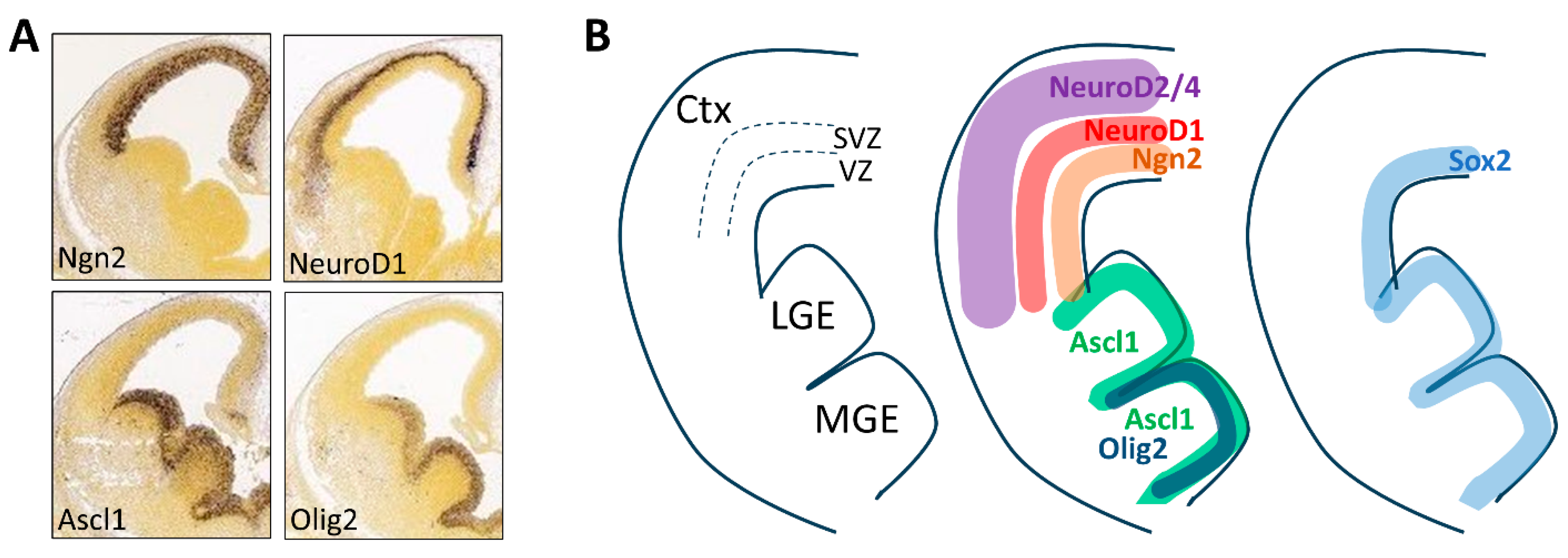
2. Reprogramming Factors during Development

The frequently applied neuronal reprogramming factors are mostly pro-neurogenic transcription factors that play key roles during CNS development. These include the basic helix-loop-helix (bHLH) gene family members Neurogenin1/2 (Ngn1/2), Achaete-scute family bHLH transcription factor 1 (Ascl1), and NeuroD1, all of which have unique spatiotemporal expression patterns during development and regulate neurogenesis in different regions of the CNS [28]. The reprogramming factor SRY-box transcription factor 2 (Sox2) belongs to the high mobility group (HMG) domain-containing gene family, and is a transcription factor mainly expressed in neural progenitors [29,30]. Among others, all these reprogramming factors have been demonstrated to reprogram somatic cells to neurons, especially in in vivo settings. We believe that understanding how these factors behave during neurogenesis will aid in designing and improving strategies for the reprogramming approach.

2.1. Expression Pattern and Function

Besides their expression and function in other tissues, these reprogramming factors have been thoroughly characterized in the developing CNS. Taking the embryonic neocortex as an example, Ngn1/2 are expressed mainly in the ventricular zone (VZ) of the dorsal neocortex, where they promote cell cycle exit of the neural progenitors as they migrate toward the outer layers of the neocortex [28]. NeuroD1 is a direct downstream target of Ngn1/2 and turns on its expression in the subventricular zone (SVZ), where neural progenitors differentiate to post-mitotic neurons [31]. As the newborn neurons further migrate outwards and differentiate, NeuroD1 expression diminishes, while its downstream effectors NeuroD2/4 highly express in the outer cortical layers [32]. Therefore, as development progresses in the neocortex, there are waves of pro-neurogenic transcription factors that act in sequence to coordinate neurogenesis (Figure 1A,B). Both Ngn1/2 and NeuroD1 are major cell fate determinants for glutamatergic neurons of the dorsal neocortex [33]. In contrast, Ascl1 is mainly expressed in the ventral telencephalon [34], namely the lateral and medial ganglionic eminence (LGE and MGE), where it instructs the neurogenesis of GABAergic interneurons [35] (Figure 1A,B). Ascl1 turns on the expression of Dlx2 [31], a homeobox-containing transcription factor that substantiates GABAergic interneuron phenotype [36] and facilitates their tangential migration into the dorsal neocortex [37]. In the MGE, Ascl1 and Dlx2 also interact with another bHLH transcription factor, Olig2, for the generation of oligodendrocytes [38]. Sox2 is expressed in the VZ of both dorsal and ventral telencephalon, and is important for maintaining neural progenitor properties [29,30] (Figure 1B). Therefore, the spatial expression patterns of these transcription factors also correlate with the temporal progression of neurogenesis, since cells that migrate to different layers of the neocortex represent distinct neurogenic states.

2.2. Expression Regulation

The expression of these transcription factors is tightly regulated by the dorsoventral gradients of the morphogenic factors through distinct receptor signaling pathways [39]. In some cases, they also regulate each other and compete to create boundaries of distinct CNS domains. For example, the discrete expression patterns of Ngn1/2 and Ascl1 separate dorsal and ventral telencephalon. Thus, Ngn1/2 double-knockout results in the misexpression of Ascl1 in the dorsal cortex and ectopic generation of GABAergic neurons [40]. In addition, the relative level of one transcription factor to others may determine the output of neuronal subtypes. During forebrain development, Ngn2 and NeuroD1 have been shown to compete with Ascl1 in the determination of neuronal subtypes [41]. The dynamic regulation of expression is further manifested by the interesting phenomenon of an oscillating expression level of Ngn2 in neocortical progenitors [42]. This is mainly achieved by the combination of precise transcriptional regulation and rapid protein degradation. Indeed, Ngn2 protein has been estimated to have a 30 min half-life in culture, and is subject to rapid ubiquitination and proteasome-mediated degradation [43]. Interestingly, Ngn2 oscillates out of phase with another oscillating transcription factor Hes1 in the dorsal cortical progenitors, a pattern that is important to control the timing of neurogenesis [42]. The oscillatory expression pattern of the transcription factors is unlikely to be present in the neuronal reprogramming settings, since their expression is usually under strong and constitutive promoters in the vector constructs. However, protein degradation would likely occur during the reprogramming process to affect their protein levels through mechanisms such as ubiquitination, which is largely an unexplored research area and a potential target for improving neuronal reprogramming outcome. Another level of gene expression regulation is the post-transcriptional regulation mediated by miRNAs. Although miRNAs are important gene expression regulators during neurogenesis [44], we will not discuss them here, since the 3′-untranslated regions (UTRs) of mRNAs, which contain their target sites, are usually omitted in the expression vectors for neuronal reprogramming. Therefore, expression levels of the reprogramming factors are not directly regulated by miRNAs unless their target sites are included in the vector (see below for an example). Taken together, the expression of the reprogramming factors is dynamically regulated throughout neurogenesis. Although the molecular context in adult cells may be very different from neural progenitors during development, we argue that a more sophisticated gene expression regulation, rather than simple overexpression, is warranted for improving reprogramming outcome. Constitutively high expression of these factors may be necessary for reprogramming to occur, but may also cause problems such as cell death [45,46].

3. Reprogramming Factors during Reprogramming

3.1. Mechanisms of Action

Reprogramming factors are usually “pioneer” transcription factors, i.e., they have the ability to access closed chromatins, recruit facilitating factors, and initiate downstream gene expression programs for a new cell identity [47,48,49,50]. These properties appear to be essential to cell reprogramming, since differentiated somatic cell types possess distinct chromatin states, with some regions being active and others silenced. As a pioneer factor, NeuroD1 reprograms the chromatin landscape to elicit neuronal programming in embryonic stem (ES) cells [51] and microglia [52] when overexpressed. Mechanistically, ectopic expression of NeuroD1 can induce the expression of endogenous NeuroD1, as well as other downstream target genes such as Hes6 and NeuroD4 [51], which may be important effectors for NeuroD1-mediated neuronal conversion. MicroRNAs (miRNAs) also play a role during NeuroD1-mediated neuronal reprogramming. Our recent research showed that miR-375 is drastically induced by NeuroD1 in human astrocyte cultures and promotes survival of the reprogrammed neurons by modulating the expression of its targets, the RNA-binding protein family genes nELAVLs, which are also highly induced by NeuroD1 [46]. Interestingly, miR-375 and nELAVLs are also upregulated by Ngn2 and Ascl1, suggesting a conserved mechanism by this interacting pair during neuronal reprogramming [46]. Cell reprogramming may share many conserved mechanisms during development, but can also assign new roles to the existing mechanisms. For example, to improve reprogramming efficiency by Sox2, a selective screen was conducted and showed that impairing the p53-p21 pathway inhibits cell cycle exit and significantly boosts the overall production of reprogrammed neuroblasts [53]. In addition, molecular characterization at single cell level has greatly contributed to our understanding of neuronal reprogramming, a unique biological process. Single-cell RNAseq not only provides mechanistic insights of the reprogramming process, but also facilitates the confirmation of neuronal identity of the reprogrammed cells [54,55].

3.2. Expression Level

While much effort has been given in searching for transcription factors that are capable of neuronal reprogramming, the level of expression of these factors is less determined to break the cell identity barrier and achieve neuronal reprogramming. A good example of testing expression level of the reprogramming factor is the neuronal reprogramming from microglia by the overexpression of NeuroD1. NeuroD1 has been a controversial factor for astrocyte-to-neuron (AtN) reprogramming, as mentioned above. There is also a controversy about whether NeuroD1 can convert microglia to neurons in the brain [56,57]. The Nakashima group first demonstrated that overexpression of NeuroD1 can convert microglia to a neuronal phenotype both in culture and in the brain [52]. Later, the Peng group presented contradictory data that NeuroD1 cannot convert microglia to neurons, but rather induces apoptosis in microglia, and that the “reprogrammed neurons” are in fact endogenous neurons that have been mislabeled by lentivirus infection [58]. This discrepancy could be due to the difference in the relative expression level of NeuroD1 between the two experiments. Although the conclusion is still not final, the Nakashima group did provide strong evidence claiming that the expression level of NeuroD1 is critical for neuronal reprogramming to occur [59]. In that report, they applied a tetracycline-inducible (i.e., Tet-on) promoter to quantitatively control NeuroD1 expression level, and demonstrated that doxycycline (1 µg/mL) is the minimal dose to induce significant microglia-to-neuron reprogramming in culture [59]. They also increased NeuroD1 expression level by repetitive lentivirus infection, arguing that a higher number of viral particles may enter the cells by repetitive infection, thereby increasing the overall NeuroD1 expression level per cell [59]. Regardless of whether NeuroD1 can convert microglia to neurons in the brain, these Tet-on experiments in culture are convincing in that a certain level of NeuroD1 must be reached to break down the cell identity barrier and initiate the neuronal reprogramming process. Most overexpression vectors utilize strong promoters such as universal ones (i.e., CAG [16,58]) or cell-type-specific ones (i.e., GFAP [16], CD68 [21,58], and NG2 [14,16]) to achieve a high expression level of the transgenes. However, whether this high expression level is high enough for reprogramming to occur is a question to be tested. In the case of microglia-to-neuron reprogramming, this high level of NeuroD1 may not be enough, and an even higher level by repetitive lentivirus infection is required for successful reprogramming. Consistent with this notion, a recent study supports a “binary switch” mechanism of reprogramming factors, in that cell fate conversion occurs only when the expression of the reprogramming factors reaches a critical expression threshold [60].
One thing worthy of pointing out is that repetitive virus infection may not only increase the transgene expression level by increasing the number of viral particles per cell as suggested [59], but may also change the cellular state of the infected cells. This has been demonstrated in astrocytes that high titers of AAV infection activate astrocytes and make them more like reactive astrocytes [61]. Reports have shown that reactive astrocytes acquire a gene expression profile that is more like that of stem/progenitor cells [62] and may be more easily reprogrammed to other cell types. Therefore, the expression level threshold of reprogramming factors could be different for different cell types and their activation state. Consistently, while NeuroD1 reprograms reactive astrocytes in disease/injury conditions with high efficiency [16,17], NeuroD1-mediated neuronal conversion from non-reactive astrocytes in different regions of the adult brain has been difficult to achieve [63]. Specifically, intravascularly delivered NeuroD1-expressing AAV induced only 2.42% AtN conversion in the striatum, but none in the cortex of adult mice [63].

3.3. Expression Duration

While a sufficient expression level of the reprogramming factors is required to break down the genomic barrier and change the cellular identity, the expression duration of these factors may also play a role in the reprogramming process. The question is how long the high expression of the reprogramming factors is required and whether it is necessary to turn off their expression after reprogramming is completed. Persistent expression of the reprogramming factors in the newly generated neurons may have unwanted effects on their maturation and integration into the existing neuronal circuitry. In the case of NeuroD1, a report has shown that transient NeuroD1 expression as achieved by a tetracycline-inducible system is sufficient to initiate neuronal programs in ES cells and generate functional mature neurons [51]. Whether a similar mechanism exists during the reprogramming process of adult cells is a question to be addressed. On the other hand, persistent reprogramming factor expression may be beneficial in generating specific neuronal subtypes of the reprogrammed neurons. For instance, NeuroD1 is a glutamatergic neuronal lineage transcription factor during neurogenesis [64]. While most mature glutamatergic neurons turn off NeuroD1 expression, the glutamatergic neurons that still maintain high levels of NeuroD1 expression even postnatally are granule neurons in the cerebellum and hippocampus [65]. Therefore, persistent NeuroD1 expression in the reprogrammed neurons from astrocytes may contribute to the fact that they are mostly of glutamatergic phenotype [16]. Sox2 is a neural stem cell transcription factor that is important for maintaining stem cell properties [29,30]. Sox2 is also a reprogramming factor that has been successfully demonstrated to reprogram multiple glial cell types into neuroblasts [15,53]. Most reprogramming studies that involve Sox2 overexpression utilize glial cell-specific promoters such as GFAP and NG2 [14,15,53,66]. The cell-type-specific promoters ensure a high expression level in the target glial cells and diminished expression in the reprogrammed neurons due to the inactivity of these promoters in neurons. In fact, persistent Sox2 expression under a universal promoter restricts its reprogramming capacity [67], and may even raise the concern of potential tumorigenicity. Even with diminished Sox2 expression in neurons by glial cell-specific promoters, the acquisition of mature neuronal phenotype with Sox2-mediated reprogramming is limited unless additional maturation strategies such as neurotrophic factors are applied [14,15,53]. Therefore, the dynamic expression pattern of the reprogramming factors is an important variable in neuronal reprogramming studies, and can be optimized to improve the reprogramming outcome.

4. Potential Strategies to Control Expression of the Reprogramming Factors

4.1. Cell-Type-Specific Promoters

To target non-neuronal cells for neuronal reprogramming, cell-type-specific promoters are often applied. This approach takes advantage of the fact that certain genes are expressed in a cell-type-specific manner. The GFAP gene promoter is a classic promoter to drive gene expression in astrocytes [68]. Over the years, modifications have been made to increase gene expression specificity and/or reduce the length of the promoter sequence to fit into small vector systems such as AAV [69]. For example, combinations of different enhancer elements have been demonstrated to alter cell type specificity of the GFAP basal promoter [70,71]. Considering the limited expression pattern of GFAP in the brain, other promoters that target astrocytes for gene expression have been developed, including GLAST and ADL1H1, most of which have also been combined with the Cre-ERT2 line [72] to add timing control with tamoxifen treatment [73,74]. By far, the most stringent astrocyte-specific gene expression is achieved with the ADL1H1-Cre-ERT2 [74]. Another thing to keep in mind is that different AAV serotypes have some degree of preference in infection to different cell types, and that the rational engineering of AAV capsids and the development of novel synthetic AAV serotypes will further facilitate cell-type targeting in combination with the promoter-based approach [75,76,77]. Thus far, cell-type-specific promoters have been used to target microglia [52,58] and NG2 glia [14] for neuronal reprogramming with success.

4.2. Drug-Controllable Promoters

To have more flexibility in controlling the timing of gene expression, numerous drug-controllable promoters have been developed [78]. Apart from the Cre-ERT2 system mentioned above, the most used of such promoters is the tetracycline-controllable promoter [79,80], which is derived from the bacterial tetracycline resistance operon. This promoter has two versions (Tet-On and Tet-off), and depending on which version is used, it either turns on or turns off gene expression when tetracycline/doxycycline is applied. The Tet-On/Off system usually contains two components, with one being the Tet responsive element (TRE), and the other being the tetracycline trans-activator (tTA) or reverse tetracycline trans-activator (rtTA) that can bind the drug and regulate gene expression [81]. The tetracycline-controllable gene expression system not only can turn on and off gene expression, but can also control expression level in a dose-dependent manner [79]. Another advantage of this system is the reversibility, i.e., one can reverse the effect using drug removal. Cell type specificity of gene expression can also be achieved with the tetracycline-controllable system by incorporating cell-type-specific gene promoters so that spatiotemporal gene expression control is feasible both in vitro and in vivo [82,83]. However, the leakiness of the system (i.e., the relatively high level of basal expression in the absence of the drug) has limited its use [84], although continuous effort has been made to improve both drug sensitivity and tightness of gene expression control [85,86,87,88]. In the neuronal reprogramming field, the Tet-On/Off system is more often applied in culture experiments where purified cell types are tested. For example, the Tet-on system has been applied to initiate neuronal differentiation from ES cells and induce neuronal reprogramming from a variety of cell types, including fibroblasts [5,6], hepatocytes [89], astrocytes [55], and microglia [59], in culture, thus making it a rather universal system for direct neuronal conversion. No reports with Tet-on system have been published for in vivo neuronal reprogramming thus far.

4.3. MiRNA-Mediated Inhibition

MiRNAs are a class of short non-coding RNAs that regulate gene expression in a post-transcriptional manner. MiRNAs usually bind to the 3′-UTR of the target mRNAs in a sequence-dependent manner and reduce gene expression level [90,91,92,93,94,95,96]. Like the cell-type-specific genes, numerous miRNAs have been identified to show cell-type-specific expression patterns. Given the inhibitory nature of their mechanism of action, miRNAs are often incorporated in the expression constructs for a “de-targeting” strategy. For instance, miR-124 is highly expressed in mature neurons, but not in other cell types of the CNS [90,91,92,93,94,95,96]. Therefore, miR-124 target sites can be included in the expression constructs that target astrocytes using a GFAP promoter to further minimize any possible “leaky” expression in neurons [97,98,99]. A great example of utilizing miRNAs as a de-targeting strategy is a recent report on the development of a toolbox expression system for targeting astrocytes in different brain regions [100]. The authors engineered a cassette of four copies of six miRNA targeting sequences that suppress transgene expression specifically in neurons and endothelial cells. By this extensive de-targeting strategy combined with a GFAP promoter (GfaABC1D), the resulting vector showed the highest gene expression specificity (>99%) in astrocytes of the mouse brain, which will for certain maximize the accuracy and resolution of cell type manipulating approaches such as neuronal reprogramming. Furthermore, it has been shown that the miRNA-mediated de-targeting approach can be even more effective than promoter-mediated ones in targeting the liver for gene expression [101].
Another potential application of miRNA target sites is to create a dynamic expression pattern of the transgenes. In the NeuroD1-mediated neuronal reprogramming model, cells experience a change in cell type, for example, from astrocytes to neurons, with highly expressed NeuroD1. As mentioned above, miR-124 is neuron-specific and highly expressed in mature neurons. Therefore, if we integrate the miR-124 target site into the NeuroD1 expression vector, we can achieve a high level of NeuroD1 expression in astrocytes (low in miR-124) for neuronal conversion to occur, and yet a reduced level of NeuroD1 in converted neurons (high in miR-124) (Figure 2A). We believe that a continuously high level of NeuroD1 in converted neurons drives them into the glutamatergic subtype since NeuroD1 is a glutamatergic neuronal lineage transcription factor during neurogenesis [64]. Thus, reducing the NeuroD1 level in the converted neurons may allow for the generation of diversified neuronal subtypes and improved functional recovery in disease/injury models (Figure 2B). In line with this hypothesis, the expression level of NeuroD1 has been related to the preference of distinct subtypes among the newly reprogrammed neurons in the retina [102].

5. Additional Notes

Thus far, we have mostly discussed gene expression control in the scenario of single reprogramming factors. However, a combination of different reprogramming factors has often been applied in neuronal reprogramming to improve efficiency, and sometimes is required for reprogramming to occur. For example, the BAM factors (Brn2, Ascl1, and Myt1l) must work together to efficiently reprogram fibroblasts to neurons [5,6]. In other cases, additional factors may facilitate neuronal reprogramming by improving cell survival [46,103] and neuronal subtype output [104,105,106]. In all these studies, the reprogramming factors and facilitating factors are expressed under strong promoters and are expected to be at a high expression level during the reprogramming process. However, factors may interact with each other at different molecular levels and affect the “functional level” of each factor. For example, NeuroD1 has been shown to compete with Ascl1 in the determination of neuronal subtypes during forebrain development [41]. Therefore, both the expression level and functional level of the reprogramming factors would need to be considered when a combination of factors is applied.
Another way of combining reprogramming factors is to allow for sequential expression. This can be achieved by combining two inducible expression systems (for example, Tet-on and Cumate-on) [78]. As discussed above, Sox2, as a stem cell maintenance transcription factor [29,30], can efficiently reprogram reactive glial cells to proliferative neuroblasts but generate mature neurons with less efficiency on its own [53,66]. On the other hand, NeuroD1 is a transcription factor of terminal differentiation and essential to cell cycle exit and neuron maturation. An interesting strategy would be to sequentially overexpress Sox2 and NeuroD1 using two inducible expression systems so that Sox2 can reprogram one glial cell into multiple neuroblasts, which then can be efficiently induced by NeuroD1 to become mature neurons. This way, a higher number of mature neurons would be reprogrammed per glial cell, which is hard to achieve by either factor alone.
Lastly, transgene silencing is an important aspect of gene expression regulation, especially for long-term studies. Both genome-integrative (retroviral and lentiviral) and non-integrative (AAV) vectors can be silenced, at least partially, by the host cells through complex mechanisms that are still largely elusive [107]. Whether transgene silencing plays a significant role in controlling the expression level of the reprogramming factors during and after neuronal reprogramming would be an interesting question to address, but it is beyond the scope of this review.

6. Conclusions and Future Perspectives

In sum, neuronal reprogramming, especially in vivo neuronal reprogramming, holds great promise to advance regenerative medicine. While considerable efforts have been made to identify the reprogramming factors in the past, the expression level of these factors has emerged as an important parameter for successful reprogramming. A high expression level of the reprogramming factors is usually required to break the genomic barrier of cell types. However, we believe that the dynamically controlled expression of the reprogramming factors will fine-tune the reprogramming process to improve reprogramming outcomes.
Future directions along this line of research lie in testing this strategy in different types of cell culture, as well as different injury/disease models to move towards clinical applications. Although this review focuses on the intrinsic properties of the reprogramming process, environmental cues such as injury/disease milieu are also critical to the reprogramming outcome. The hostile tissue conditions resulting from injury/disease will impose a substantial impact on neuronal reprogramming, including the survival of the reprogrammed neurons [108]. Therefore, the manipulation of local environment to improve neuronal reprogramming efficiency has been pursued in CNS injury models [15]. Furthermore, the injury mode can also influence the neuronal subtype identity of the reprogrammed neurons, as demonstrated in the retina [109]. On the other hand, the reprogrammed neurons, if substantial in number, can “reverse” the injured tissue environment back to a less hostile one [110]. Thus, both intrinsic and extrinsic factors would need to be considered when optimizing the reprogramming outcome in future investigations.

Author Contributions

N.M.-J. and H.L. made the Figures. All authors contributed to reviewing the literature and writing the paper. All authors have read and agreed to the published version of the manuscript.

Funding

This work was supported by National Institutes of Health grants (R01NS117918, R21NS104394, R21NS119732) (to HL) and Ann L. Jones Spinal Cord Regeneration Research Fund (to HL).

Acknowledgments

Due to the focus of this review, we apologize for not citing many significant papers in the neuronal reprogramming field.

Conflicts of Interest

The authors declare no conflicts of interest.

References

  1. Takahashi, K.; Tanabe, K.; Ohnuki, M.; Narita, M.; Ichisaka, T.; Tomoda, K.; Yamanaka, S. Induction of pluripotent stem cells from adult human fibroblasts by defined factors. Cell 2007, 131, 861–872. [Google Scholar] [CrossRef] [PubMed]
  2. Takahashi, K.; Yamanaka, S. Induction of pluripotent stem cells from mouse embryonic and adult fibroblast cultures by defined factors. Cell 2006, 126, 663–676. [Google Scholar] [CrossRef] [PubMed]
  3. Matsui, T.; Akamatsu, W.; Nakamura, M.; Okano, H. Regeneration of the damaged central nervous system through reprogramming technology: Basic concepts and potential application for cell replacement therapy. Exp. Neurol. 2014, 260, 12–18. [Google Scholar] [CrossRef] [PubMed]
  4. Konstantinides, N.; Desplan, C. Neuronal differentiation strategies: Insights from single-cell sequencing and machine learning. Development 2020, 147, dev193631. [Google Scholar] [CrossRef] [PubMed] [PubMed Central]
  5. Pang, Z.P.; Yang, N.; Vierbuchen, T.; Ostermeier, A.; Fuentes, D.R.; Yang, T.Q.; Citri, A.; Sebastiano, V.; Marro, S.; Südhof, T.C.; et al. Induction of human neuronal cells by defined transcription factors. Nature 2011, 476, 220–223. [Google Scholar] [CrossRef] [PubMed] [PubMed Central]
  6. Vierbuchen, T.; Ostermeier, A.; Pang, Z.P.; Kokubu, Y.; Südhof, T.C.; Wernig, M. Direct conversion of fibroblasts to functional neurons by defined factors. Nature 2010, 463, 1035–1041. [Google Scholar] [CrossRef] [PubMed]
  7. Yoo, A.S.; Sun, A.X.; Li, L.; Shcheglovitov, A.; Portmann, T.; Li, Y.; Lee-Messer, C.; Dolmetsch, R.E.; Tsien, R.W.; Crabtree, G.R. MicroRNA-mediated conversion of human fibroblasts to neurons. Nature 2011, 476, 228–231. [Google Scholar] [CrossRef] [PubMed] [PubMed Central]
  8. Li, X.; Zuo, X.; Jing, J.; Ma, Y.; Wang, J.; Liu, D.; Zhu, J.; Du, X.; Xiong, L.; Du, Y.; et al. Small-Molecule-Driven Direct Reprogramming of Mouse Fibroblasts into Functional Neurons. Cell Stem Cell 2015, 17, 195–203. [Google Scholar] [CrossRef] [PubMed]
  9. Li, H.; Chen, G. In Vivo Reprogramming for CNS Repair: Regenerating Neurons from Endogenous Glial Cells. Neuron 2016, 91, 728–738. [Google Scholar] [CrossRef]
  10. Gascón, S.; Masserdotti, G.; Russo, G.L.; Götz, M. Direct Neuronal Reprogramming: Achievements, Hurdles, and New Roads to Success. Cell Stem Cell 2017, 21, 18–34. [Google Scholar] [CrossRef]
  11. Turinetto, V.; Orlando, L.; Giachino, C. Induced Pluripotent Stem Cells: Advances in the Quest for Genetic Stability during Reprogramming Process. Int. J. Mol. Sci. 2017, 18, 1952. [Google Scholar] [CrossRef] [PubMed] [PubMed Central]
  12. Lund, R.J.; Närvä, E.; Lahesmaa, R. Genetic and epigenetic stability of human pluripotent stem cells. Nat. Rev. Genet. 2012, 13, 732–744. [Google Scholar] [CrossRef] [PubMed]
  13. Chen, X.; Li, H. Neuronal reprogramming in treating spinal cord injury. Neural Regen. Res. 2022, 17, 1440–1445. [Google Scholar] [PubMed] [PubMed Central]
  14. Tai, W.; Wu, W.; Wang, L.L.; Ni, H.; Chen, C.; Yang, J.; Zang, T.; Zou, Y.; Xu, X.M.; Zhang, C.L. In vivo reprogramming of NG2 glia enables adult neurogenesis and functional recovery following spinal cord injury. Cell Stem Cell 2021, 28, 923–937.e4. [Google Scholar] [CrossRef] [PubMed]
  15. Niu, W.; Zang, T.; Zou, Y.; Fang, S.; Smith, D.K.; Bachoo, R.; Zhang, C.L. In Vivo reprogramming of astrocytes to neuroblasts in the adult brain. Nat. Cell Biol. 2013, 15, 1164–1175. [Google Scholar] [CrossRef] [PubMed] [PubMed Central]
  16. Guo, Z.; Zhang, L.; Wu, Z.; Chen, Y.; Wang, F.; Chen, G. In vivo direct reprogramming of reactive glial cells into functional neurons after brain injury and in an Alzheimer’s disease model. Cell Stem Cell 2014, 14, 188–202. [Google Scholar] [CrossRef] [PubMed] [PubMed Central]
  17. Puls, B.; Ding, Y.; Zhang, F.; Pan, M.; Lei, Z.; Pei, Z.; Jiang, M.; Bai, Y.; Forsyth, C.; Metzger, M.; et al. Regeneration of Functional Neurons After Spinal Cord Injury via in situ NeuroD1-Mediated Astrocyte-to-Neuron Conversion. Front. Cell Dev. Biol. 2020, 8, 591883. [Google Scholar] [CrossRef] [PubMed] [PubMed Central]
  18. Todd, L.; Jenkins, W.; Finkbeiner, C.; Hooper, M.J.; Donaldson, P.C.; Pavlou, M.; Wohlschlegel, J.; Ingram, N.; Mu, X.; Rieke, F.; et al. Reprogramming Müller glia to regenerate ganglion-like cells in adult mouse retina with developmental transcription factors. Sci. Adv. 2022, 8, eabq7219. [Google Scholar] [CrossRef] [PubMed] [PubMed Central]
  19. Todd, L.; Hooper, M.J.; Haugan, A.K.; Finkbeiner, C.; Jorstad, N.; Radulovich, N.; Wong, C.K.; Donaldson, P.C.; Jenkins, W.; Chen, Q.; et al. Efficient stimulation of retinal regeneration from Müller glia in adult mice using combinations of proneural bHLH transcription factors. Cell Rep. 2021, 37, 109857. [Google Scholar] [CrossRef] [PubMed Central]
  20. Livingston, J.M.; Lee, T.T.; Enbar, T.; Daniele, E.; Phillips, C.M.; Krassikova, A.; Bang, K.W.A.; Kortebi, I.; Donville, B.W.; Ibragimov, O.S.; et al. Ectopic Expression of Neurod1 Is Sufficient for Functional Recovery following a Sensory-Motor Cortical Stroke. Biomedicines 2024, 12, 663. [Google Scholar] [CrossRef] [PubMed Central]
  21. Irie, T.; Matsuda, T.; Hayashi, Y.; Matsuda-Ito, K.; Kamiya, A.; Masuda, T.; Prinz, M.; Isobe, N.; Kira, J.I.; Nakashima, K. Direct neuronal conversion of microglia/macrophages reinstates neurological function after stroke. Proc. Natl. Acad. Sci. USA 2023, 120, e2307972120. [Google Scholar] [CrossRef] [PubMed] [PubMed Central]
  22. Wang, L.L.; Serrano, C.; Zhong, X.; Ma, S.; Zou, Y.; Zhang, C.L. Revisiting astrocyte to neuron conversion with lineage tracing in vivo. Cell 2021, 184, 5465–5481.e16. [Google Scholar] [CrossRef] [PubMed] [PubMed Central]
  23. Xiang, Z.; Xu, L.; Liu, M.; Wang, Q.; Li, W.; Lei, W.; Chen, G. Lineage tracing of direct astrocyte-to-neuron conversion in the mouse cortex. Neural Regen. Res. 2021, 16, 750–756. [Google Scholar] [PubMed] [PubMed Central]
  24. Chen, G. In vivo confusion over in vivo conversion. Mol. Ther. 2021, 29, 3097–3098. [Google Scholar] [CrossRef] [PubMed] [PubMed Central]
  25. Wang, L.L.; Zhang, C.L. Reply to In vivo confusion over in vivo conversion. Mol. Ther. 2022, 30, 986–987. [Google Scholar] [CrossRef] [PubMed] [PubMed Central]
  26. Xiang, Z.; He, S.; Chen, R.; Liu, S.; Liu, M.; Xu, L.; Zheng, J.; Jiang, Z.; Ma, L.; Sun, Y.; et al. Two-photon live imaging of direct glia-to-neuron conversion in the mouse cortex. Neural Regen. Res. 2024, 19, 1781–1788. [Google Scholar] [CrossRef] [PubMed] [PubMed Central]
  27. Wang, L.L.; Zhang, C.L. In vivo glia-to-neuron conversion: Pitfalls and solutions. Dev. Neurobiol. 2022, 82, 367–374. [Google Scholar] [CrossRef] [PubMed] [PubMed Central]
  28. Wilkinson, G.; Dennis, D.; Schuurmans, C. Proneural genes in neocortical development. Neuroscience 2013, 253, 256–273. [Google Scholar] [CrossRef] [PubMed]
  29. Bani-Yaghoub, M.; Tremblay, R.G.; Lei, J.X.; Zhang, D.; Zurakowski, B.; Sandhu, J.K.; Smith, B.; Ribecco-Lutkiewicz, M.; Kennedy, J.; Walker, P.R.; et al. Role of Sox2 in the development of the mouse neocortex. Dev. Biol. 2006, 295, 52–66. [Google Scholar] [CrossRef]
  30. Graham, V.; Khudyakov, J.; Ellis, P.; Pevny, L. SOX2 functions to maintain neural progenitor identity. Neuron 2003, 39, 749–765. [Google Scholar] [CrossRef]
  31. Fode, C.; Ma, Q.; Casarosa, S.; Ang, S.L.; Anderson, D.J.; Guillemot, F. A role for neural determination genes in specifying the dorsoventral identity of telencephalic neurons. Genes Dev. 2000, 14, 67–80. [Google Scholar] [CrossRef] [PubMed] [PubMed Central]
  32. Tutukova, S.; Tarabykin, V.; Hernandez-Miranda, L.R. The Role of Neurod Genes in Brain Development, Function, and Disease. Front. Mol. Neurosci. 2021, 14, 662774. [Google Scholar] [CrossRef] [PubMed] [PubMed Central]
  33. Mattar, P.; Langevin, L.M.; Markham, K.; Klenin, N.; Shivji, S.; Zinyk, D.; Schuurmans, C. Basic helix-loop-helix transcription factors cooperate to specify a cortical projection neuron identity. Mol. Cell. Biol. 2008, 28, 1456–1469. [Google Scholar] [CrossRef] [PubMed]
  34. Guillemot, F.; Joyner, A.L. Dynamic expression of the murine Achaete-Scute homologue Mash-1 in the developing nervous system. Mech. Dev. 1993, 42, 171–185. [Google Scholar] [CrossRef] [PubMed]
  35. Casarosa, S.; Fode, C.; Guillemot, F. Mash1 regulates neurogenesis in the ventral telencephalon. Development 1999, 126, 525–534. [Google Scholar] [CrossRef] [PubMed]
  36. Anderson, S.A.; Qiu, M.; Bulfone, A.; Eisenstat, D.D.; Meneses, J.; Pedersen, R.; Rubenstein, J.L. Mutations of the homeobox genes Dlx-1 and Dlx-2 disrupt the striatal subventricular zone and differentiation of late born striatal neurons. Neuron 1997, 19, 27–37. [Google Scholar] [CrossRef]
  37. Anderson, S.A.; Eisenstat, D.D.; Shi, L.; Rubenstein, J.L. Interneuron migration from basal forebrain to neocortex: Dependence on Dlx genes. Science 1997, 278, 474–476. [Google Scholar] [CrossRef]
  38. Petryniak, M.A.; Potter, G.B.; Rowitch, D.H.; Rubenstein, J.L. Dlx1 and Dlx2 control neuronal versus oligodendroglial cell fate acquisition in the developing forebrain. Neuron 2007, 55, 417–433. [Google Scholar] [CrossRef] [PubMed] [PubMed Central]
  39. Wilson, S.W.; Rubenstein, J.L. Induction and dorsoventral patterning of the telencephalon. Neuron 2000, 28, 641–651. [Google Scholar] [CrossRef]
  40. Schuurmans, C.; Armant, O.; Nieto, M.; Stenman, J.M.; Britz, O.; Klenin, N.; Brown, C.; Langevin, L.M.; Seibt, J.; Tang, H.; et al. Sequential phases of cortical specification involve Neurogenin-dependent and -independent pathways. EMBO J. 2004, 23, 2892–2902. [Google Scholar] [CrossRef] [PubMed Central]
  41. Roybon, L.; Mastracci, T.L.; Ribeiro, D.; Sussel, L.; Brundin, P.; Li, J.Y. GABAergic differentiation induced by Mash1 is compromised by the bHLH proteins Neurogenin2, NeuroD1, and NeuroD2. Cereb. Cortex 2010, 20, 1234–1244. [Google Scholar] [CrossRef] [PubMed]
  42. Shimojo, H.; Ohtsuka, T.; Kageyama, R. Oscillations in notch signaling regulate maintenance of neural progenitors. Neuron 2008, 58, 52–64. [Google Scholar] [CrossRef] [PubMed]
  43. Vosper, J.M.; Fiore-Heriche, C.S.; Horan, I.; Wilson, K.; Wise, H.; Philpott, A. Regulation of neurogenin stability by ubiquitin-mediated proteolysis. Biochem. J. 2007, 407, 277–284. [Google Scholar] [CrossRef] [PubMed]
  44. Rajman, M.; Schratt, G. MicroRNAs in neural development: From master regulators to fine-tuners. Development 2017, 144, 2310–2322. [Google Scholar] [CrossRef] [PubMed]
  45. Cai, L.; Morrow, E.M.; Cepko, C.L. Misexpression of basic helix-loop-helix genes in the murine cerebral cortex affects cell fate choices and neuronal survival. Development 2000, 127, 3021–3030. [Google Scholar] [CrossRef] [PubMed]
  46. Chen, X.; Sokirniy, I.; Wang, X.; Jiang, M.; Mseis-Jackson, N.; Williams, C.; Mayes, K.; Jiang, N.; Puls, B.; Du, Q.; et al. MicroRNA-375 Is Induced during Astrocyte-to-Neuron Reprogramming and Promotes Survival of Reprogrammed Neurons when Overexpressed. Cells 2023, 12, 2202. [Google Scholar] [CrossRef] [PubMed] [PubMed Central]
  47. Zaret, K.S. Pioneer Transcription Factors Initiating Gene Network Changes. Annu. Rev. Genet. 2020, 54, 367–385. [Google Scholar] [CrossRef] [PubMed Central]
  48. Iwafuchi-Doi, M.; Zaret, K.S. Cell fate control by pioneer transcription factors. Development 2016, 143, 1833–1837. [Google Scholar] [CrossRef] [PubMed] [PubMed Central]
  49. Wapinski, O.L.; Vierbuchen, T.; Qu, K.; Lee, Q.Y.; Chanda, S.; Fuentes, D.R.; Giresi, P.G.; Ng, Y.H.; Marro, S.; Neff, N.F.; et al. Hierarchical mechanisms for direct reprogramming of fibroblasts to neurons. Cell 2013, 155, 621–635. [Google Scholar] [CrossRef] [PubMed Central]
  50. Morris, S.A. Direct lineage reprogramming via pioneer factors; a detour through developmental gene regulatory networks. Development 2016, 143, 2696–2705. [Google Scholar] [CrossRef] [PubMed Central]
  51. Pataskar, A.; Jung, J.; Smialowski, P.; Noack, F.; Calegari, F.; Straub, T.; Tiwari, V.K. NeuroD1 reprograms chromatin and transcription factor landscapes to induce the neuronal program. EMBO J. 2016, 35, 24–45. [Google Scholar] [CrossRef] [PubMed] [PubMed Central]
  52. Matsuda, T.; Irie, T.; Katsurabayashi, S.; Hayashi, Y.; Nagai, T.; Hamazaki, N.; Adefuin, A.M.D.; Miura, F.; Ito, T.; Kimura, H.; et al. Pioneer Factor NeuroD1 Rearranges Transcriptional and Epigenetic Profiles to Execute Microglia-Neuron Conversion. Neuron 2019, 101, 472–485 e477. [Google Scholar] [CrossRef] [PubMed]
  53. Wang, L.L.; Su, Z.; Tai, W.; Zou, Y.; Xu, X.M.; Zhang, C.L. The p53 Pathway Controls SOX2-Mediated Reprogramming in the Adult Mouse Spinal Cord. Cell Rep. 2016, 17, 891–903. [Google Scholar] [CrossRef] [PubMed] [PubMed Central]
  54. Wohlschlegel, J.; Finkbeiner, C.; Hoffer, D.; Kierney, F.; Prieve, A.; Murry, A.D.; Haugan, A.K.; Ortuño-Lizarán, I.; Rieke, F.; Golden, S.A.; et al. ASCL1 induces neurogenesis in human Müller glia. Stem Cell Rep. 2023, 18, 2400–2417. [Google Scholar] [CrossRef] [PubMed Central]
  55. Pereira, A.; Diwakar, J.; Masserdotti, G.; Beşkardeş, S.; Simon, T.; So, Y.; Martín-Loarte, L.; Bergemann, F.; Vasan, L.; Schauer, T.; et al. Direct neuronal reprogramming of mouse astrocytes is associated with multiscale epigenome remodeling and requires Yy1. Nat. Neurosci. 2024, 27, 1260–1273. [Google Scholar] [CrossRef] [PubMed]
  56. Matsuda, T.; Nakashima, K. Clarifying the ability of NeuroD1 to convert mouse microglia into neurons. Neuron 2021, 109, 3912–3913. [Google Scholar] [CrossRef] [PubMed]
  57. Rao, Y.; Peng, B. Failure of observing NeuroD1-induced microglia-to-neuron conversion in vitro is not attributed to the low NeuroD1 expression level. Mol. Brain 2022, 15, 31. [Google Scholar] [CrossRef] [PubMed] [PubMed Central]
  58. Rao, Y.; Du, S.; Yang, B.; Wang, Y.; Li, Y.; Li, R.; Zhou, T.; Du, X.; He, Y.; Wang, Y.; et al. NeuroD1 induces microglial apoptosis and cannot induce microglia-to-neuron cross-lineage reprogramming. Neuron 2021, 109, 4094–4108.e5. [Google Scholar] [CrossRef]
  59. Matsuda-Ito, K.; Matsuda, T.; Nakashima, K. Expression level of the reprogramming factor NeuroD1 is critical for neuronal conversion efficiency from different cell types. Sci. Rep. 2022, 12, 17980. [Google Scholar] [CrossRef] [PubMed Central]
  60. Hersbach, B.A.; Fischer, D.S.; Masserdotti, G.; Deeksha; Mojžišová, K.; Waltzhöni, T.; Rodriguez-Terrones, D.; Heinig, M.; Theis, F.J.; Götz, M.; et al. Probing cell identity hierarchies by fate titration and collision during direct reprogramming. Mol. Syst. Biol. 2022, 18, e11129. [Google Scholar] [CrossRef]
  61. Ortinski, P.I.; Dong, J.; Mungenast, A.; Yue, C.; Takano, H.; Watson, D.J.; Haydon, P.G.; Coulter, D.A. Selective induction of astrocytic gliosis generates deficits in neuronal inhibition. Nat. Neurosci. 2010, 13, 584–591. [Google Scholar] [CrossRef] [PubMed] [PubMed Central]
  62. Gotz, M.; Sirko, S.; Beckers, J.; Irmler, M. Reactive astrocytes as neural stem or progenitor cells: In Vivo lineage, In vitro potential, and Genome-wide expression analysis. Glia 2015, 63, 1452–1468. [Google Scholar] [CrossRef] [PubMed] [PubMed Central]
  63. Brulet, R.; Matsuda, T.; Zhang, L.; Miranda, C.; Giacca, M.; Kaspar, B.K.; Nakashima, K.; Hsieh, J. NEUROD1 Instructs Neuronal Conversion in Non-Reactive Astrocytes. Stem Cell Rep. 2017, 8, 1506–1515. [Google Scholar] [CrossRef] [PubMed] [PubMed Central]
  64. Hevner, R.F.; Hodge, R.D.; Daza, R.A.; Englund, C. Transcription factors in glutamatergic neurogenesis: Conserved programs in neocortex, cerebellum, and adult hippocampus. Neurosci. Res. 2006, 55, 223–233. [Google Scholar] [CrossRef] [PubMed]
  65. Miyata, T.; Maeda, T.; Lee, J.E. NeuroD is required for differentiation of the granule cells in the cerebellum and hippocampus. Genes. Dev. 1999, 13, 1647–1652. [Google Scholar] [CrossRef] [PubMed] [PubMed Central]
  66. Su, Z.; Niu, W.; Liu, M.L.; Zou, Y.; Zhang, C.L. In Vivo conversion of astrocytes to neurons in the injured adult spinal cord. Nat. Commun. 2014, 5, 3338. [Google Scholar] [CrossRef] [PubMed] [PubMed Central]
  67. Heinrich, C.; Bergami, M.; Gascon, S.; Lepier, A.; Vigano, F.; Dimou, L.; Sutor, B.; Berninger, B.; Gotz, M. Sox2-mediated conversion of NG2 glia into induced neurons in the injured adult cerebral cortex. Stem Cell Rep. 2014, 3, 1000–1014. [Google Scholar] [CrossRef] [PubMed] [PubMed Central]
  68. Brenner, M. Structure and transcriptional regulation of the GFAP gene. Brain Pathol. 1994, 4, 245–257. [Google Scholar] [CrossRef] [PubMed]
  69. Borodinova, A.A.; Balaban, P.M.; Bezprozvanny, I.B.; Salmina, A.B.; Vlasova, O.L. Genetic Constructs for the Control of Astrocytes’ Activity. Cells 2021, 10, 1600. [Google Scholar] [CrossRef] [PubMed Central]
  70. Lee, Y.; Messing, A.; Su, M.; Brenner, M. GFAP promoter elements required for region-specific and astrocyte-specific expression. Glia 2008, 56, 481–493. [Google Scholar] [CrossRef]
  71. de Leeuw, B.; Su, M.; ter Horst, M.; Iwata, S.; Rodijk, M.; Hoeben, R.C.; Messing, A.; Smitt, P.S.; Brenner, M. Increased glia-specific transgene expression with glial fibrillary acidic protein promoters containing multiple enhancer elements. J. Neurosci. Res. 2006, 83, 744–753. [Google Scholar] [CrossRef] [PubMed]
  72. Indra, A.K.; Warot, X.; Brocard, J.; Bornert, J.M.; Xiao, J.H.; Chambon, P.; Metzger, D. Temporally-controlled site-specific mutagenesis in the basal layer of the epidermis: Comparison of the recombinase activity of the tamoxifen-inducible Cre-ERT and Cre-ERT2 recombinases. Nucleic Acids Res. 1999, 27, 4324–4327. [Google Scholar] [CrossRef] [PubMed] [PubMed Central]
  73. Hu, N.Y.; Chen, Y.T.; Wang, Q.; Jie, W.; Liu, Y.S.; You, Q.L.; Li, Z.L.; Li, X.W.; Reibel, S.; Pfrieger, F.W.; et al. Expression Patterns of Inducible Cre Recombinase Driven by Differential Astrocyte-Specific Promoters in Transgenic Mouse Lines. Neurosci. Bull. 2020, 36, 530–544. [Google Scholar] [CrossRef] [PubMed] [PubMed Central]
  74. Srinivasan, R.; Lu, T.Y.; Chai, H.; Xu, J.; Huang, B.S.; Golshani, P.; Coppola, G.; Khakh, B.S. New Transgenic Mouse Lines for Selectively Targeting Astrocytes and Studying Calcium Signals in Astrocyte Processes In Situ and In Vivo. Neuron 2016, 92, 1181–1195. [Google Scholar] [CrossRef] [PubMed] [PubMed Central]
  75. Koerber, J.T.; Klimczak, R.; Jang, J.H.; Dalkara, D.; Flannery, J.G.; Schaffer, D.V. Molecular evolution of adeno-associated virus for enhanced glial gene delivery. Mol. Ther. 2009, 17, 2088–2095. [Google Scholar] [CrossRef] [PubMed] [PubMed Central]
  76. Deverman, B.E.; Pravdo, P.L.; Simpson, B.P.; Kumar, S.R.; Chan, K.Y.; Banerjee, A.; Wu, W.L.; Yang, B.; Huber, N.; Pasca, S.P.; et al. Cre-dependent selection yields AAV variants for widespread gene transfer to the adult brain. Nat. Biotechnol. 2016, 34, 204–209. [Google Scholar] [CrossRef] [PubMed] [PubMed Central]
  77. O’Carroll, S.J.; Cook, W.H.; Young, D. AAV Targeting of Glial Cell Types in the Central and Peripheral Nervous System and Relevance to Human Gene Therapy. Front. Mol. Neurosci. 2020, 13, 618020. [Google Scholar] [CrossRef] [PubMed] [PubMed Central]
  78. Kallunki, T.; Barisic, M.; Jäättelä, M.; Liu, B. How to Choose the Right Inducible Gene Expression System for Mammalian Studies? Cells 2019, 8, 796. [Google Scholar] [CrossRef] [PubMed Central]
  79. Gossen, M.; Bujard, H. Tight control of gene expression in mammalian cells by tetracycline-responsive promoters. Proc. Natl. Acad. Sci. USA 1992, 89, 5547–5551. [Google Scholar] [CrossRef] [PubMed Central]
  80. Gossen, M.; Freundlieb, S.; Bender, G.; Müller, G.; Hillen, W.; Bujard, H. Transcriptional activation by tetracyclines in mammalian cells. Science 1995, 268, 1766–1769. [Google Scholar] [CrossRef]
  81. Das, A.T.; Tenenbaum, L.; Berkhout, B. Tet-On Systems For Doxycycline-inducible Gene Expression. Curr. Gene Ther. 2016, 16, 156–167. [Google Scholar] [CrossRef] [PubMed] [PubMed Central]
  82. Knopf, F.; Schnabel, K.; Haase, C.; Pfeifer, K.; Anastassiadis, K.; Weidinger, G. Dually inducible TetON systems for tissue-specific conditional gene expression in zebrafish. Proc. Natl. Acad. Sci. USA 2010, 107, 19933–19938. [Google Scholar] [CrossRef] [PubMed Central]
  83. Sullivan, K.A.; Vitko, I.; Blair, K.; Gaykema, R.P.; Failor, M.J.; San Pietro, J.M.; Dey, D.; Williamson, J.M.; Stornetta, R.L.; Kapur, J.; et al. Drug-Inducible Gene Therapy Effectively Reduces Spontaneous Seizures in Kindled Rats but Creates Off-Target Side Effects in Inhibitory Neurons. Int. J. Mol. Sci. 2023, 24, 11347. [Google Scholar] [CrossRef] [PubMed] [PubMed Central]
  84. Costello, A.; Lao, N.T.; Gallagher, C.; Capella Roca, B.; Julius, L.A.N.; Suda, S.; Ducrée, J.; King, D.; Wagner, R.; Barron, N.; et al. Leaky Expression of the TET-On System Hinders Control of Endogenous miRNA Abundance. Biotechnol. J. 2019, 14, e1800219. [Google Scholar] [CrossRef] [PubMed]
  85. Shaikh, S.; Nicholson, L.F. Optimization of the Tet-On system for inducible expression of RAGE. J. Biomol. Tech. 2006, 17, 283–292. [Google Scholar] [PubMed] [PubMed Central]
  86. Khor, J.M.; Ettensohn, C.A. An optimized Tet-On system for conditional control of gene expression in sea urchins. Development 2023, 150, dev201373. [Google Scholar] [CrossRef] [PubMed]
  87. Ali Hosseini Rad, S.M.; Poudel, A.; Tan, G.M.Y.; McLellan, A.D. Optimisation of Tet-On inducible systems for Sleeping Beauty-based chimeric antigen receptor (CAR) applications. Sci. Rep. 2020, 10, 13125. [Google Scholar] [CrossRef]
  88. Loew, R.; Heinz, N.; Hampf, M.; Bujard, H.; Gossen, M. Improved Tet-responsive promoters with minimized background expression. BMC Biotechnol. 2010, 10, 81. [Google Scholar] [CrossRef] [PubMed] [PubMed Central]
  89. Marro, S.; Pang, Z.P.; Yang, N.; Tsai, M.C.; Qu, K.; Chang, H.Y.; Südhof, T.C.; Wernig, M. Direct lineage conversion of terminally differentiated hepatocytes to functional neurons. Cell Stem Cell 2011, 9, 374–382. [Google Scholar] [CrossRef] [PubMed Central]
  90. Bartel, D.P. MicroRNAs: Genomics, biogenesis, mechanism, and function. Cell 2004, 116, 281–297. [Google Scholar] [CrossRef]
  91. Stefani, G.; Slack, F.J. Small non-coding RNAs in animal development. Nat. Rev. Mol. Cell Biol. 2008, 9, 219–230. [Google Scholar] [CrossRef] [PubMed]
  92. Cao, X.; Yeo, G.; Muotri, A.R.; Kuwabara, T.; Gage, F.H. Noncoding RNAs in the mammalian central nervous system. Annu. Rev. Neurosci. 2006, 29, 77–103. [Google Scholar] [CrossRef] [PubMed]
  93. Ambros, V. microRNAs: Tiny regulators with great potential. Cell 2001, 107, 823–826. [Google Scholar] [CrossRef] [PubMed]
  94. Ambros, V. The functions of animal microRNAs. Nature 2004, 431, 350–355. [Google Scholar] [CrossRef] [PubMed]
  95. Bartel, D.P. MicroRNAs: Target recognition and regulatory functions. Cell 2009, 136, 215–233. [Google Scholar] [CrossRef] [PubMed] [PubMed Central]
  96. Jonas, S.; Izaurralde, E. Towards a molecular understanding of microRNA-mediated gene silencing. Nat. Rev. Genet. 2015, 16, 421–433. [Google Scholar] [CrossRef] [PubMed]
  97. Colin, A.; Faideau, M.; Dufour, N.; Auregan, G.; Hassig, R.; Andrieu, T.; Brouillet, E.; Hantraye, P.; Bonvento, G.; Deglon, N. Engineered lentiviral vector targeting astrocytes in vivo. Glia 2009, 57, 667–679. [Google Scholar] [CrossRef] [PubMed]
  98. Delzor, A.; Escartin, C.; Deglon, N. Lentiviral vectors: A powerful tool to target astrocytes in vivo. Curr. Drug Targets 2013, 14, 1336–1346. [Google Scholar] [PubMed]
  99. Taschenberger, G.; Tereshchenko, J.; Kügler, S. A MicroRNA124 Target Sequence Restores Astrocyte Specificity of gfaABC1D-Driven Transgene Expression in AAV-Mediated Gene Transfer. Mol. Ther.-Nucleic Acids 2017, 8, 13–25. [Google Scholar] [CrossRef]
  100. Gleichman, A.J.; Kawaguchi, R.; Sofroniew, M.V.; Carmichael, S.T. A toolbox of astrocyte-specific, serotype-independent adeno-associated viral vectors using microRNA targeting sequences. Nat. Commun. 2023, 14, 7426. [Google Scholar] [CrossRef]
  101. Geisler, A.; Jungmann, A.; Kurreck, J.; Poller, W.; Katus, H.A.; Vetter, R.; Fechner, H.; Müller, O.J. microRNA122-regulated transgene expression increases specificity of cardiac gene transfer upon intravenous delivery of AAV9 vectors. Gene Ther. 2011, 18, 199–209. [Google Scholar] [CrossRef]
  102. Xu, D.; Zhong, L.T.; Cheng, H.Y.; Wang, Z.Q.; Chen, X.M.; Feng, A.Y.; Chen, W.Y.; Chen, G.; Xu, Y. Overexpressing NeuroD1 reprograms Müller cells into various types of retinal neurons. Neural Regen. Res. 2023, 18, 1124–1131. [Google Scholar] [PubMed Central]
  103. Gascon, S.; Murenu, E.; Masserdotti, G.; Ortega, F.; Russo, G.L.; Petrik, D.; Deshpande, A.; Heinrich, C.; Karow, M.; Robertson, S.P.; et al. Identification and Successful Negotiation of a Metabolic Checkpoint in Direct Neuronal Reprogramming. Cell Stem Cell 2016, 18, 396–409. [Google Scholar] [CrossRef] [PubMed]
  104. Abernathy, D.G.; Kim, W.K.; McCoy, M.J.; Lake, A.M.; Ouwenga, R.; Lee, S.W.; Xing, X.; Li, D.; Lee, H.J.; Heuckeroth, R.O.; et al. MicroRNAs Induce a Permissive Chromatin Environment that Enables Neuronal Subtype-Specific Reprogramming of Adult Human Fibroblasts. Cell Stem Cell 2017, 21, 332–348 e339. [Google Scholar] [CrossRef] [PubMed] [PubMed Central]
  105. Caiazzo, M.; Dell’Anno, M.T.; Dvoretskova, E.; Lazarevic, D.; Taverna, S.; Leo, D.; Sotnikova, T.D.; Menegon, A.; Roncaglia, P.; Colciago, G.; et al. Direct generation of functional dopaminergic neurons from mouse and human fibroblasts. Nature 2011, 476, 224–227. [Google Scholar] [CrossRef] [PubMed]
  106. Heinrich, C.; Blum, R.; Gascon, S.; Masserdotti, G.; Tripathi, P.; Sanchez, R.; Tiedt, S.; Schroeder, T.; Gotz, M.; Berninger, B. Directing astroglia from the cerebral cortex into subtype specific functional neurons. PLoS Biol. 2010, 8, e1000373. [Google Scholar] [CrossRef] [PubMed Central]
  107. Cabrera, A.; Edelstein, H.I.; Glykofrydis, F.; Love, K.S.; Palacios, S.; Tycko, J.; Zhang, M.; Lensch, S.; Shields, C.E.; Livingston, M.; et al. The sound of silence: Transgene silencing in mammalian cell engineering. Cell Syst. 2022, 13, 950–973. [Google Scholar] [CrossRef]
  108. Clifford, T.; Finkel, Z.; Rodriguez, B.; Joseph, A.; Cai, L. Current Advancements in Spinal Cord Injury Research-Glial Scar Formation and Neural Regeneration. Cells 2023, 12, 853. [Google Scholar] [CrossRef] [PubMed Central]
  109. Pavlou, M.; Probst, M.; Blasdel, N.; Prieve, A.R.; Reh, T.A. The impact of timing and injury mode on induced neurogenesis in the adult mammalian retina. Stem Cell Rep. 2024, 19, 239–253. [Google Scholar] [CrossRef] [PubMed Central]
  110. Zhang, L.; Lei, Z.; Guo, Z.; Pei, Z.; Chen, Y.; Zhang, F.; Cai, A.; Mok, G.; Lee, G.; Swaminathan, V.; et al. Development of Neuroregenerative Gene Therapy to Reverse Glial Scar Tissue Back to Neuron-Enriched Tissue. Front. Cell Neurosci. 2020, 14, 594170. [Google Scholar] [CrossRef] [PubMed Central]
Figure 1. Distinct expression pattern of the reprogramming factors in the developing telencephalon. (A) Images from the Allen Institute for Brain Science [https://portal.brain-map.org (accessed on 24 May 2024)] showing mRNA expression of some reprogramming factors in the coronal sections of E13.5 mouse telencephalon. (B) Simplified schematic drawings showing anatomical structures of the E13.5 mouse telencephalon and spatial expression patterns of the reprogramming factors. Ctx, cortex; LGE, lateral ganglionic eminence; MGE, medial ganglionic eminence; VZ, ventricular zone; SVZ, sub-ventricular zone.
Cells 13 01223 g001
Figure 2. A potential vector design using miR-124 target sites to create a dynamic expression pattern of NeuroD1. (A) Including miR-124 target sites in the expression vector allows for the different expression levels of NeuroD1 in astrocytes and neurons. (B) The dynamic expression pattern of NeuroD1 may allow for the generation of neuronal subtype diversity, which could lead to improved functional recovery. ND1, NeuroD1; miR-124T, miR-124 target site.
Cells 13 01223 g002
Disclaimer/Publisher’s Note: The statements, opinions and data contained in all publications are solely those of the individual author(s) and contributor(s) and not of MDPI and/or the editor(s). MDPI and/or the editor(s) disclaim responsibility for any injury to people or property resulting from any ideas, methods, instructions or products referred to in the content.

