Article

ERβ Regulation of Indian Hedgehog Expression in the First Wave of Ovarian Follicles

1
Department of Pathology and Laboratory Medicine, University of Kansas Medical Center (KUMC), Kansas City, KS 66160, USA
2
Obstetrics and Gynecology, University of Kansas Medical Center (KUMC), Kansas City, KS 66160, USA
*
Author to whom correspondence should be addressed.
These authors contributed equally to this work.
Cells 2024, 13(7), 644; https://doi.org/10.3390/cells13070644
Submission received: 27 December 2023 / Revised: 4 April 2024 / Accepted: 4 April 2024 / Published: 6 April 2024
(This article belongs to the Special Issue Hedgehog Signaling: Advances in Development and Cancer)

Abstract

:
Increased activation of ovarian primordial follicles in Erβ knockout (ErβKO) rats becomes evident as early as postnatal day 8.5. To identify the ERβ-regulated genes that may control ovarian primordial follicle activation, we analyzed the transcriptome profiles of ErβKO rat ovaries collected on postnatal days 4.5, 6.5, and 8.5. Compared to wildtype ovaries, ErβKO ovaries displayed dramatic downregulation of Indian hedgehog (Ihh) expression. IHH-regulated genes, including Hhip, Gli1, and Ptch1, were also downregulated in ErβKO ovaries. This was associated with a downregulation of steroidogenic enzymes Cyp11a1, Cyp19a1, and Hsd17b1. The expression of Ihh remained very low in ErβKO ovaries despite the high levels of Gdf9 and Bmp15, which are known upregulators of Ihh expression in the granulosa cells of activated ovarian follicles. Strikingly, the downregulation of the Ihh gene in ErβKO ovaries began to disappear on postnatal day 16.5 and recovered on postnatal day 21.5. In rat ovaries, the first wave of primordial follicles is rapidly activated after their formation, whereas the second wave of primordial follicles remains dormant in the ovarian cortex and slowly starts activating after postnatal day 12.5. We localized the expression of Ihh mRNA in postnatal day 8.5 wildtype rat ovaries but not in the age-matched ErβKO ovaries. In postnatal day 21.5 ErβKO rat ovaries, we detected Ihh mRNA mainly in the activated follicles in the ovaries’ peripheral regions. Our findings indicate that the expression of Ihh in the granulosa cells of the activated first wave of ovarian follicles depends on ERβ.

1. Introduction

Primordial follicle assembly following oocyte nest breakdown is the first step in ovarian folliculogenesis [1]. In mice and rats, primordial follicles develop in two distinct waves [2,3]. The first wave of primordial follicles is formed in the ovarian medulla during the first three days after birth and is rapidly activated after formation [2,3]. In contrast, the second wave of primordial follicles is assembled in the ovarian cortex during postnatal days 4.5 to 7.5, and most of these primordial follicles remain dormant and serve as the ovarian reserve [2,3]. A few second wave primordial follicles are selectively activated through a highly regulated intraovarian mechanism known as primordial follicle activation. The second wave of primordial follicles undergoes primordial follicle activation starting from postnatal day 12.5 and gradually replenishes the loss of activated first wave follicles [2,3]. The first wave follicles are completely depleted in three months, and the second wave follicles perform the ovarian functions [2,3]. Thus, the regulation of primordial follicle activation is a crucial mechanism for maintaining ovarian reserves and the female reproductive lifespan [4]. However, the role of the rapidly activated and short-lived first wave of primordial follicles in ovarian biology remains unknown [5]. It is suggested to play a role in the establishment of early reproductive functions and the hypothalamic–pituitary axis of endocrine regulation of ovarian functions. Recently, it has been reported that XO female mice, a model of Turner syndrome, lack the first wave of primordial follicles [6]. Further follow-up of XO mice has shown that they develop premature ovarian insufficiency [7]. These findings suggest that the first wave of ovarian follicles may play an important role in maintaining the second wave of primordial follicle reserve [7].
Estrogen receptor β (ERβ) is the predominant estrogen receptor in the mammalian ovary and is essential for follicle development and ovulation [8,9,10]. We previously identified that ERβ plays an important role in controlling the normal rate of primordial follicle activation [11]. Loss of ERβ increased primordial follicle activation, leading to premature ovarian senescence in Erβ knockout (ErβKO) rats [11]. Regulation of primordial follicle activation is a complex mechanism involving several negative regulators within the PI3-kinase and mTOR pathways [12]. Loss of ERβ was associated with increased activation of AKT, ERK, and mTOR pathways in ErβKO ovaries [11]. ERβ may also inhibit the induction of primordial follicle activation like the transcriptional regulators FOXO3A and FOXL2 [11,12,13]. However, the precise mechanism of the ERβ-mediated regulation of primordial follicle activation remains unclear [11]. In this study, we analyzed the ERβ-regulated genes in developing rat ovaries to understand the underlying mechanisms. We demonstrate that the expression of Indian hedgehog (Ihh) in neonatal rat ovaries is entirely ERβ dependent.
During folliculogenesis, granulosa cells of activated follicles express hedgehog proteins, which are essential for the development, growth, and differentiation of theca cells [14,15,16,17,18]. The loss of hedgehog signaling or aberrant activation of hedgehog signaling disrupts theca cell differentiation, leading to failed ovulation [16,19]. The Ihh and desert hedgehog (Dhh) genes are expressed in granulosa cells, whereas the hedgehog receptor Ptch1/Ptch2 and the signal transducer Smo, as well as hedgehog target genes like Gli1/Gli2/Gli3 transcription factors, are expressed in theca cells [20,21]. It is also important that ERβ is the predominant estrogen receptor in granulosa cells, while ERα plays a dominant role in TCs [22,23]. However, whether ERβ plays any regulatory role in Ihh or Dhh gene expression in granulosa cells or ERα in the expression of hedgehog receptors or downstream signaling molecules in theca cells is unknown.

2. Materials and Methods

2.1. Animal Model

ErβKO Holtzman Sprague Dawley rats were included in this study. An ErβKO rat model was generated by targeting the exon 3 in the Erβ gene, causing a frameshift and null mutation [8]. The rats were screened for the presence of mutations via genotyping PCR using tail-tip DNA samples as described previously [8]. All procedures were performed following the protocols approved by the University of Kansas Medical Center (KUMC) Animal Care and Use Committee.

2.2. Histological Evaluation of Ovarian Phenotypes

Ovaries were collected from 4.5-to-8.5-day-old ErβKO and age-matched wild-type rats. One ovary from each rat was embedded in OCT (Fisher Scientific, Hampton, NH, USA), frozen immediately, and preserved at −80 °C until sectioning. The other ovary was snap-frozen in liquid nitrogen and preserved at −80 °C until processing for RNA extraction. Histological sections were prepared using a cryotome. The frozen sections were prepared from whole ovaries at a thickness of 6 µm and placed on charged glass slides (Fisher Scientific). The ovary sections were stained with hematoxylin and eosin following standard procedures [24]. The H&E-stained sections were thoroughly examined for follicle morphology and counted for follicles in each stage of development, as we have described previously [11]. Histological analyses were performed on one ovary from at least 3 independent wildtype rats or 3 independent ErβKO rats.

2.3. Total Follicle Counting in the Serial Sections of the Whole Ovary

The ovaries were collected and fixed in 4% formaldehyde overnight, processed, and embedded in paraffin following standard procedures [25,26,27]. Whole ovaries were serially sectioned at 6 µM thickness and stained with hematoxylin and eosin [25,26,27]. The stages of follicle development, including primordial, primary, secondary, early antral, and antral, were determined as described previously [28]. The primordial follicles were recognized as small oocytes surrounded by a few flattened granulosa cells, the primary follicles contained larger oocytes surrounded by a single layer of cuboidal granulosa cells, and the oocytes in the secondary follicles were surrounded by multiple layers of granulosa cells. The tertiary follicles were categorized into early antral and antral follicles based on the appearance and extent of the cavities within the follicles. Atretic follicles were recognized by pyknotic granulosa cells surrounding degenerated oocytes. The follicles were counted on every fifth section under light microscopy [25,26]. To avoid counting the same follicle more than once, primordial, and primary follicles were counted if they exhibited a nucleus, whereas the secondary, early antral, and antral follicles were counted only in the presence of a nuclei with prominent nucleoli [25,26,27]. The counts were multiplied by 5 to obtain the total count of follicles in the whole ovary. Ovarian follicle counting was performed on both ovaries collected from at least 3 independent wildtype rats or 3 independent ErβKO rats.

2.4. Detection of Differentially Expressed Genes

Gene expression at the mRNA level was evaluated through RNA sequencing (RNA-Seq), and RT-qPCR analyses. Total RNA was extracted from the whole ovary with TRI Reagent (Millipore-Sigma, St. Louis, MO, USA), and RNA samples with an RIN value ≥ 9 were considered for the RNA-Seq library preparation. Next, 500 ng of total RNA from each sample was used for the RNA-Seq library preparation using the TruSeq Stranded mRNA kit (Illumina, San Diego, CA, USA) following the manufacturer’s instructions. The cDNA libraries were evaluated for quality at the KUMC Genomics Core and then sequenced on an Illumina HiSeq X sequencer at Novogene Corporation (Sacramento, CA, USA). All RNA-Seq data have been submitted to the Sequencing Read Archive (SRX6955095-6955104). RNA-Seq data were analyzed using CLC Genomics Workbench (Qiagen Bioinformatics, Redwood City, CA, USA) as described in our previous publications [9,10]. Gene ontology analysis of differentially expressed genes was performed using https://pantherdb.org, accessed on 30 November 2023 and the molecular functional pathways were determined. Ingenuity Pathway Analysis (IPA, Qiagen Bioinformatics) of the differentially expressed genes revealed functional pathways and different upstream or downstream signaling pathways.

2.5. Evaluation of Gene Expression Using RT-qPCR

Total RNA was extracted from the PND 8.5 ovaries using TRI Reagents (Sigma-Aldrich). Next, 1000 ng of total RNA from each sample was used to prepare cDNA using High-Capacity Reverse Transcription Kits (Applied Biosystems, Foster City, CA, USA). RT-qPCR amplification of cDNAs was carried out in a 10 µL reaction mixture containing Applied Biosystems Power SYBR Green PCR Master Mix (Thermo Fisher Scientific). Amplification and fluorescence detection of RT-qPCR was carried out on an Applied Biosystems QuantStudio Flex 7 Real-Time PCR System (Thermo Fisher Scientific). The ΔΔCT method was used to quantify the target mRNA expression level normalized to Rn18s (18S rRNA), as described in our previous publications [9,10]. For RT-qPCR analyses, any group of cDNAs at each time point included cDNAs prepared from 6 independent rat ovaries.

2.6. RNAScope In Situ Hybridization

Ten µm thick frozen sections of postnatal day 8.5 and postnatal day 21.5 ErβKO and age-matched wildtype rat ovaries were hybridized with RNAScope probes for rat Ihh (ACD Bio, Newark, CA, USA). In situ hybridization was performed on the frozen sections following the manufacturer’s instructions. After hybridization, the sections were reacted with HD brown and red assay reagents. The slides were mounted on Eco Mount (BioCare Medical, Pacheco, CA, USA), and images were captured using a Nikon 80i light microscope. In situ hybridization for each gene (mRNA) target was performed on ovary sections prepared from at least from three independent rats of each genotype (wildtype or ErβKO).

2.7. Statistical Analysis

Each RNA-Seq library was prepared using pooled RNA samples from three individual wildtype or ErβKO rats. Each group of RNA sequencing data consisted of three to four different libraries. For the RT-PCR experiments, each cDNA was prepared from pooled RNA from three rat ovaries of the same genotype. Either wildtype or the ErβKO group consisted of 6 cDNAs. Follicle counting was performed on ovaries collected from at least three wildtype or ErβKO rats. All of the laboratory investigations were repeated to ensure reproducibility. The data are presented as the mean ± standard error (SE). The results were analyzed using one-way ANOVA, and the significance of the mean differences was determined by Duncan’s post hoc test, with p ≤ 0.05. The statistical calculations were performed through the use of SPSS 22 (IBM, Armonk, NY, USA).

3. Results

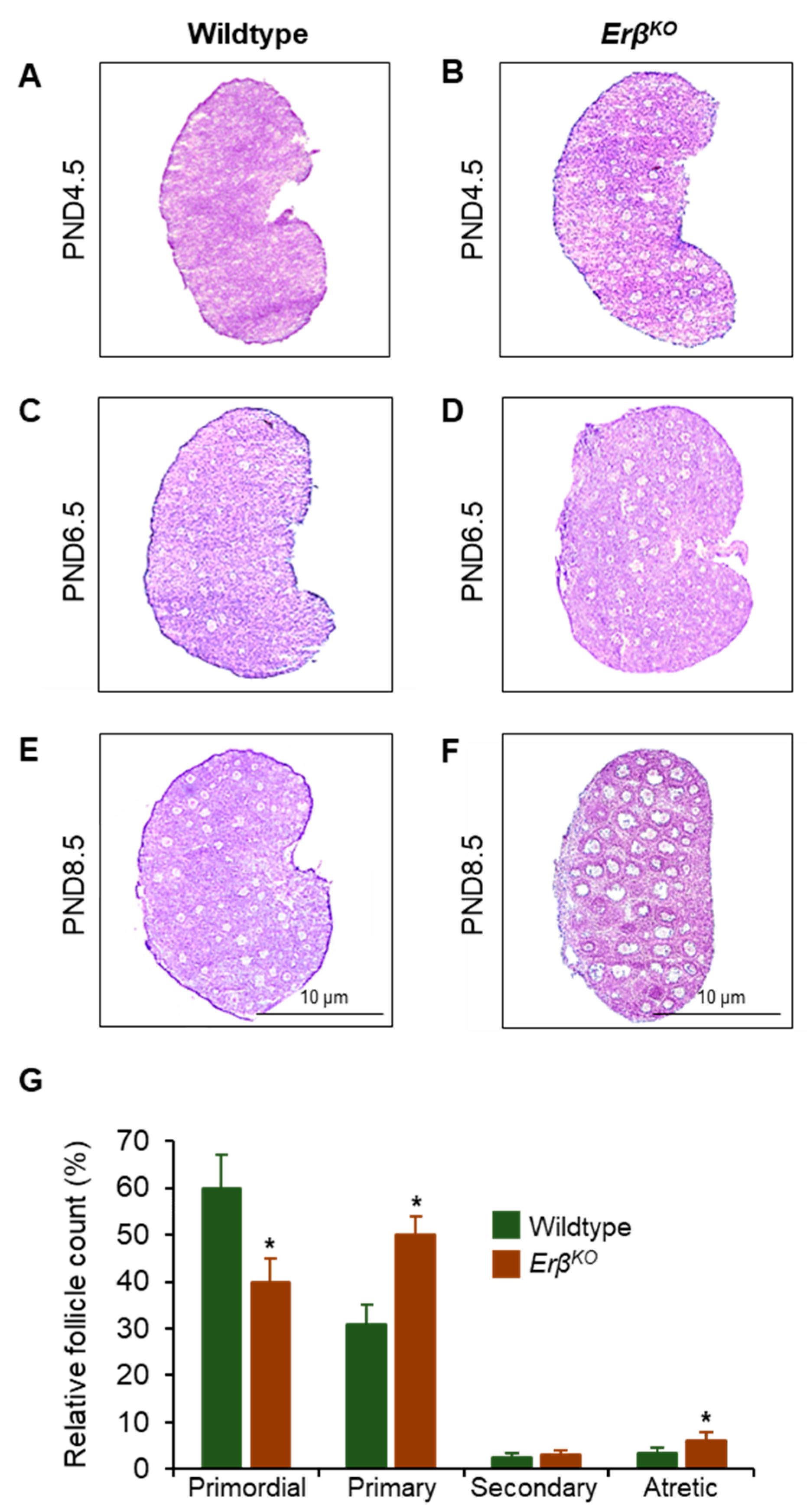
3.1. Increased Primordial Follicle Growth Activation in ErβKO Rats

Upon histological examination, the activation of medullary follicles appeared to be accelerated in ErβKO rat ovaries. However, the number of activated follicles in ErβKO was not significantly different from that in wildtype ovaries on postnatal day 4.5 and postnatal day 6.5 (Figure 1). In contrast, there were remarkable increases in primordial follicle activation in postnatal day 8.5 ErβKO ovaries (Figure 1E–G and Figure S1). On postnatal day 8.5, wildtype rat ovaries contained approximately 60% of the total follicles in the primordial stage, and approximately 30% were in the activated primary stage (Figure 1G). In contrast, on postnatal day 8.5 ErβKO ovaries, the number of activated follicles was increased to approximately 50%, and the number of primordial follicles was reduced to 40% (Figure 1G). The number of healthy secondary follicles and atretic secondary follicles also increased in postnatal day 8.5 ErβKO ovaries (Figure 1G). However, the total follicular counts of ovarian follicles in various stages of development remained similar in wildtype and ErβKO ovaries.

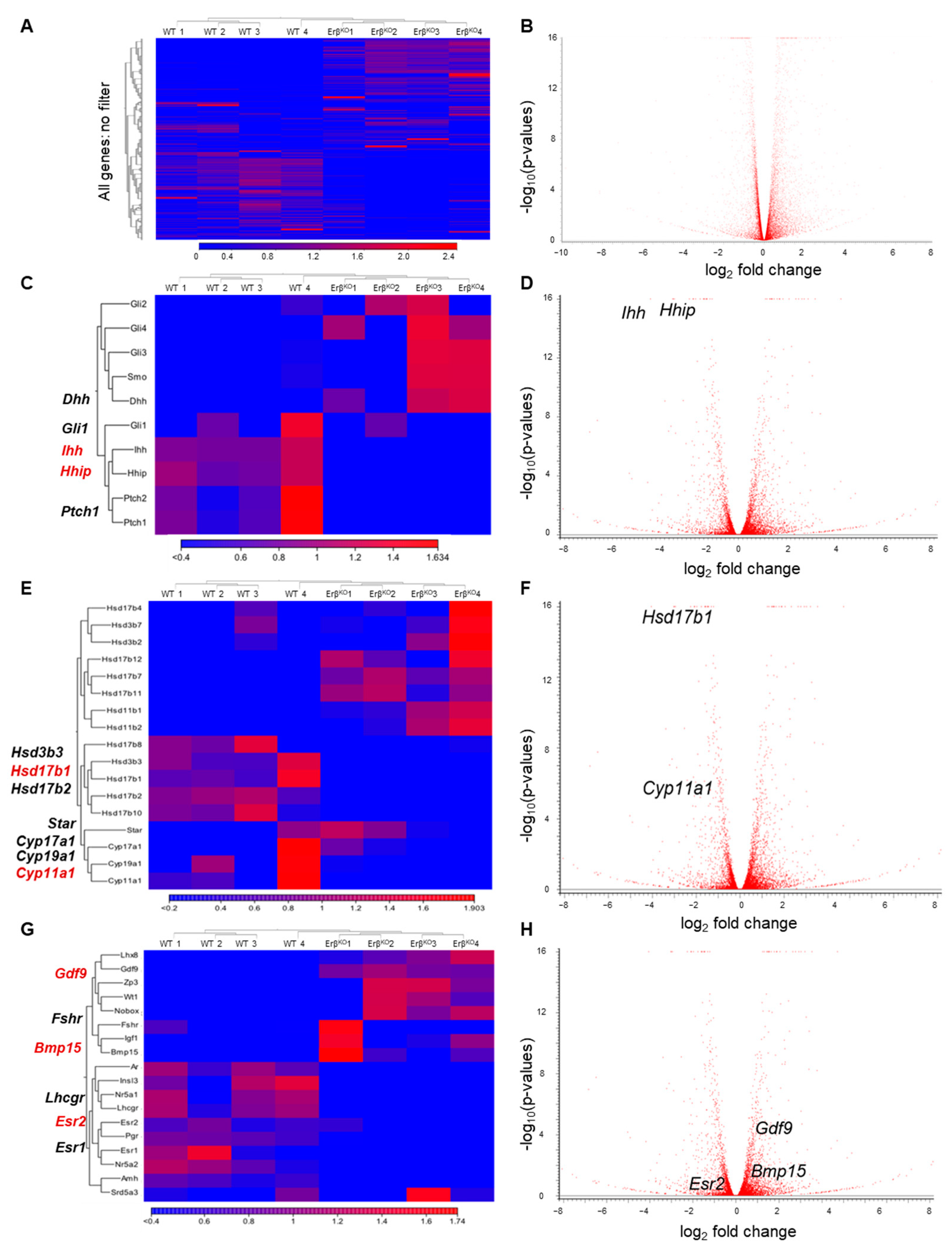
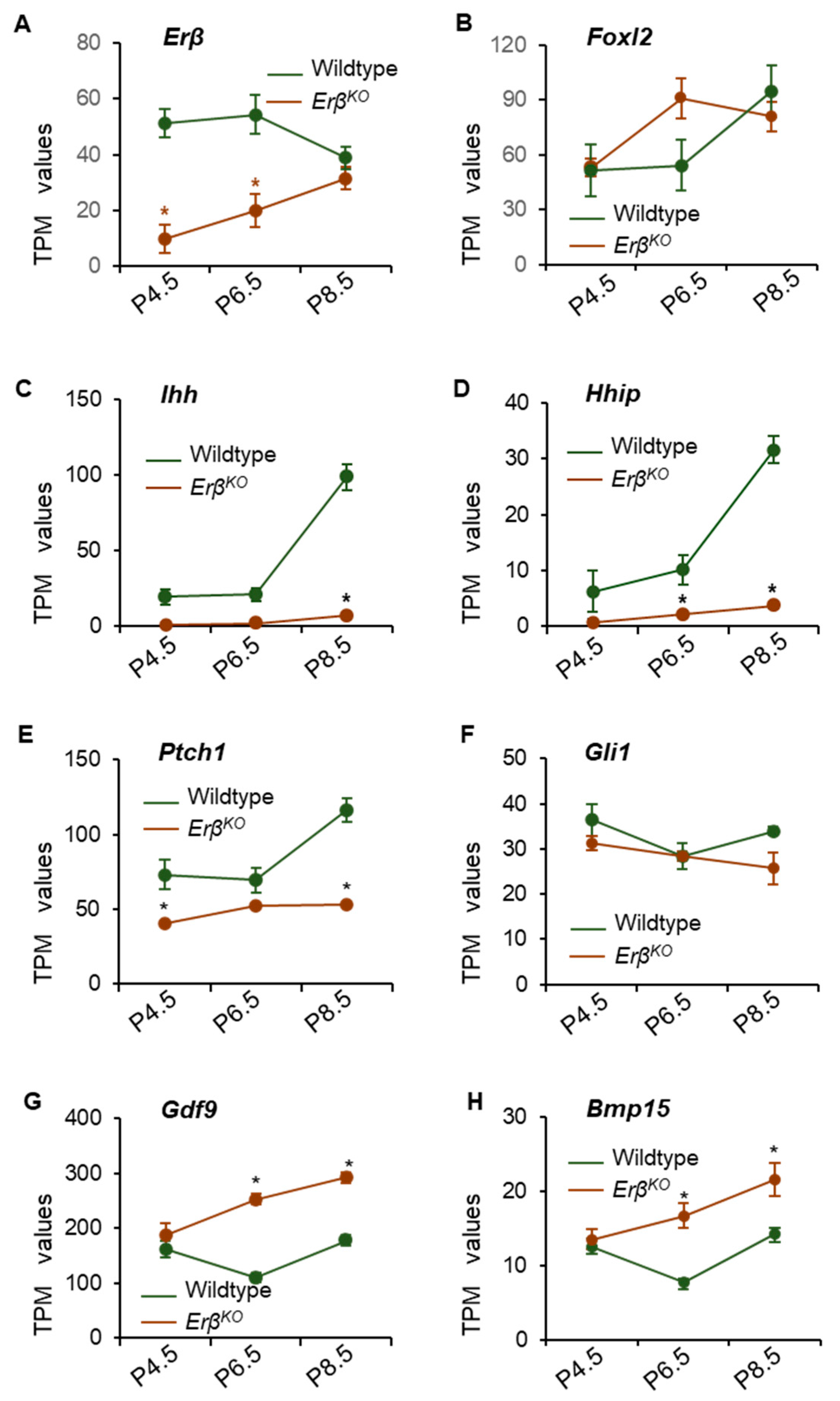
3.2. ERβ Regulates Genes in Neonatal Rat Ovaries

To identify the transcriptional targets of ERβ that are involved in regulating the rate of primordial follicle activation, we performed RNA-Seq analyses of postnatal day 4.5, 6.5, and 8.5 ErβKO ovaries and age-matched wildtype rat ovaries. RNA-Seq analyses of postnatal day 8.5 ovaries detected 19,620 genes out of 29,541 genes in the rat reference library (mRatBN7.2.110). Of the 19,620 genes, 14,403 genes had TPM values ≥ 1 and 11,933 genes had TPM values ≥ 5. We observed that 642 genes were differentially expressed in ErβKO postnatal day 8.5 ovaries (FDR p-value ≤ 0.05; absolute fold change 2, TPM ≥ 1). When we analyzed the high-copy genes (e.g., TPM ≥ 5), we found that 295 genes were differentially expressed (FDR p-value ≤ 0.05; absolute fold change 2, TPM ≥ 5). We observed that Ihh, an ovary-specific abundantly expressed gene, was one of the top downregulated genes in ErβKO ovaries. It was also associated with similar downregulation of an IHH target gene (Hhip), and the differential expression data were reproducible through the use of RT-qPCR analyses (Table 1, Figure 2, Figure 3 and Figure S2). Therefore, for further analysis, we focused on differentially expressed genes that are related to hedgehog signaling, ovarian steroidogenesis, and regulators of ovarian folliculogenesis. Differential expression of these genes was detected in postnatal day 4.5 ovaries (Supplementary Figure S3) and it became more distinct in day 6.5 (Supplementary Figure S4) and 8.5 ovaries (Figure 2A–H).
Among the differentially expressed genes with TPM values ≥ 5, we also identified 10 transcriptional regulators as being downregulated and 8 transcriptional regulators as being upregulated (Table 2). The top 5 upregulated transcription factors were Rbpjl, Dbx2, Dmrt1, Npas2, and Pou5f1. In contrast many of the downregulated transcription factors, including Pparg, Mycn, Fosl2, Egr1, Osr2, Nr5a1, Fox1, Fos, Myc, and Nr4a1, are known for their role in regulating ovarian folliculogenesis [29,30,31,32,33].
Gene ontology analysis revealed that the differentially expressed genes in postnatal day 8.5 ErβKO ovaries involved many molecular and functional pathways including ATP-dependent activity, antioxidant activity, molecular adaptor activity, transporter activity, transcription, and translation regulator activity (Supplementary Figure S5A). Ingenuity Pathway Analysis of the differentially expressed revealed different upstream and downstream signaling mechanisms such as Esr1, Fgf2, Egf, Egfr, Vegf, Edn1, Agt, Csf2, and Il1β, which are involved in the regulation of the development of vasculature or vasculogenic or angiogenesis, the growth of ovarian follicles, the proliferation of ovarian granulosa cells, and the growth of the whole ovary (Supplementary Figure S5B).

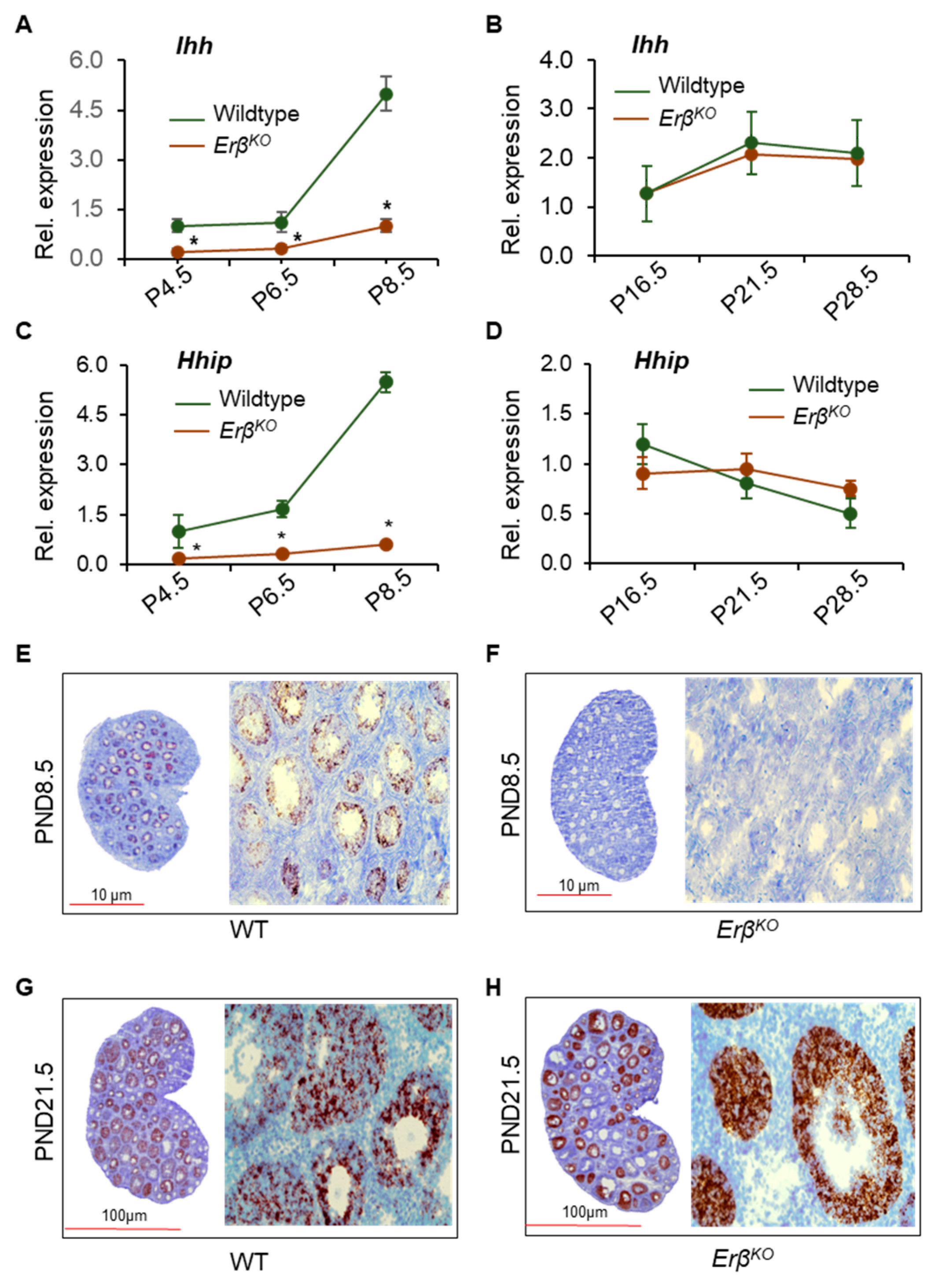
3.3. ERβ Regulates Indian Hedgehog Signaling

We observed a low level of mutant Erβ mRNA expression in postnatal day 4.5, 6.5, and 8.5 ErβKO ovaries (Figure 3A). Although we did not detect any change in the expression of Foxl2 (Figure 3B), we found that the loss of ERβ signaling disrupted Ihh expression in early neonatal ovaries (Figure 2C,D). Of the differentially expressed transcripts with TPM values ≥ 5.0, Ihh was one of most downregulated genes among the ovary-specific genes (Table 1, Figure 3C). Similar downregulation was also observed for the expression of IHH-target gene Hhip (Figure 3D). In addition to Ihh and Hhip, the expression of hedgehog receptor Patch1 and the downstream transcriptional regulator Gli1 was downregulated in ErβKO ovaries (Figure 3E,F). Strikingly, the expression levels of Gdf9 and Bmp15, which are known positive regulators of Ihh expression, were significantly higher in ErβKO ovaries (Figure 3G,H). The RNA-Seq data were verified through the use of RT-qPCR, which showed similar results (Supplementary Figure S2).

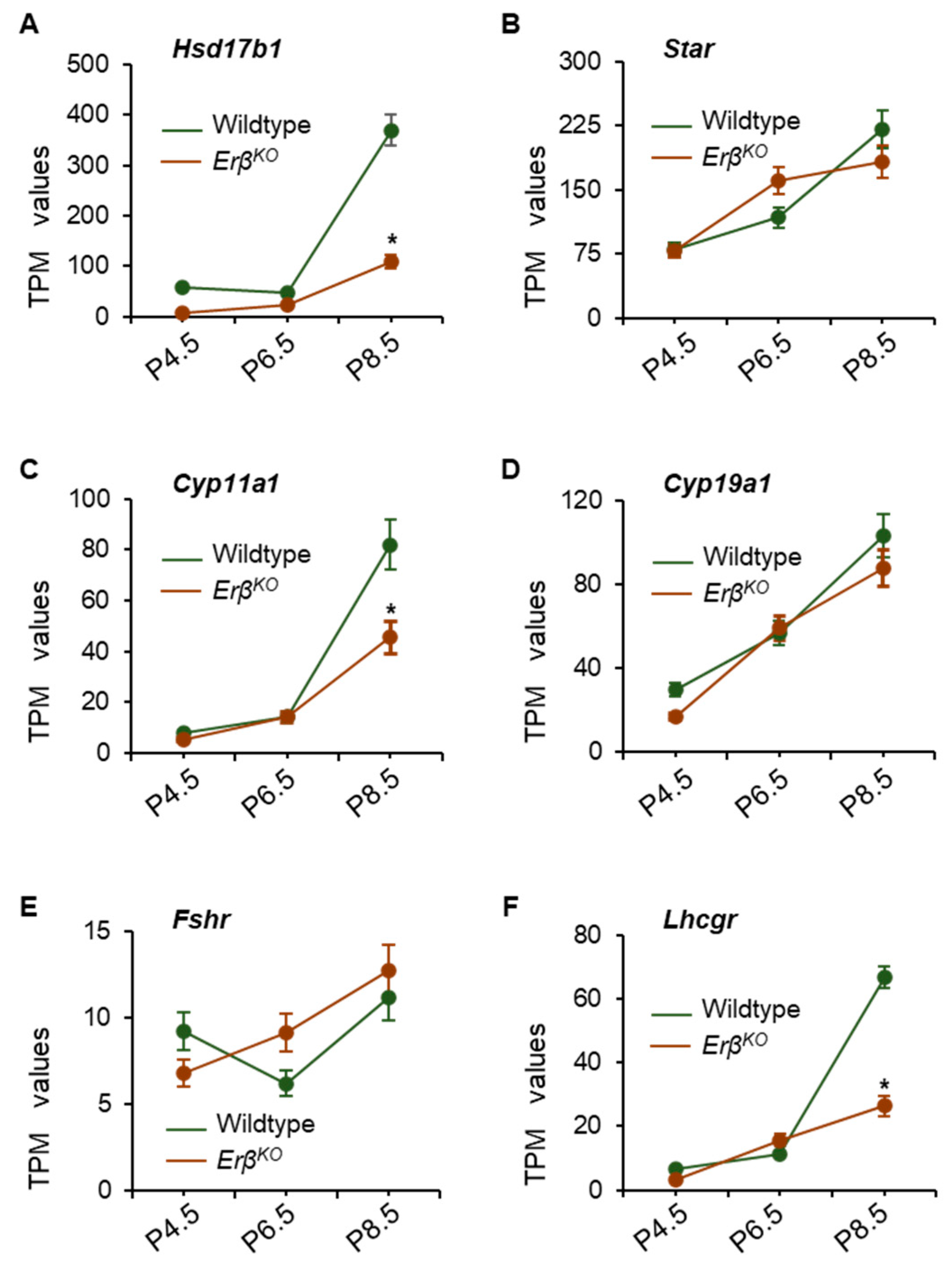
3.4. Altered Expression of Steroidogenic Enzyme Genes in ErβKO Ovaries

IHH signaling plays an important role in regulating the expression of steroidogenic enzymes. While analyzing the RNA-Seq data, we observed that the loss of ERβ disrupted the expression of Hsd17b1 and Cyp11a1 in ErβKO rat ovaries (Figure 2E,F and Figure 4A,C). The expression of the FSH responsive gene, Star, and gonadotropin receptor, Fshr, remained unchanged in ErβKO rat ovaries (Figure 4B,E). However, the expression of Lhcgr was significantly downregulated in postnatal day 8.5 ErβKO rat ovaries (Figure 4F). The RNA-Seq results were further confirmed through the use of RT-qPCR, which showed comparable results (Supplementary Figure S6).

3.5. ERβ Regulates Ihh Only during the Early Neonatal Period

The downregulation of both Ihh and Hhip in ErβKO rat ovaries was persistent, and this downregulation increased significantly from postnatal day 4.5 through to postnatal day 8.5 (p = 0.000) (Figure 3A,B). The data were confirmed through the use of RT-qPCR analysis (Figure 5A,C). Interestingly, the expression of both Ihh and Hhip was upregulated in postnatal day 16.5 ErβKO rat ovaries and remained high, with it becoming similar to that of wildtype ovaries through to postnatal day 21.5 and postnatal day 28.5 (Figure 5B,D). To identify the cells that express Ihh in ErβKO rat ovaries from postnatal day 16.5 onwards, we performed RNAScope in situ hybridization for rat Ihh using postnatal day 8.5 and postnatal day 21.5 ErβKO and age-matched wildtype ovaries. We observed that Ihh mRNA is expressed only in the granulosa cells of activated follicles, mostly secondary and early antral follicles in postnatal day 8.5 wildtype ovaries (Figure 5E). No Ihh mRNA was detected in postnatal day 8.5 ErβKO ovaries that contained the activated follicles of first wave origin (Figure 5F). As expected, both medullary and cortical follicles showed the expression of Ihh mRNA in postnatal day 21.5 wildtype ovaries (Figure 5G). However, there was no detection of Ihh mRNA in the medullary follicles of postnatal day 21.5 ErβKO ovaries but strong Ihh signals in the activated large cortical follicles (Figure 5H).

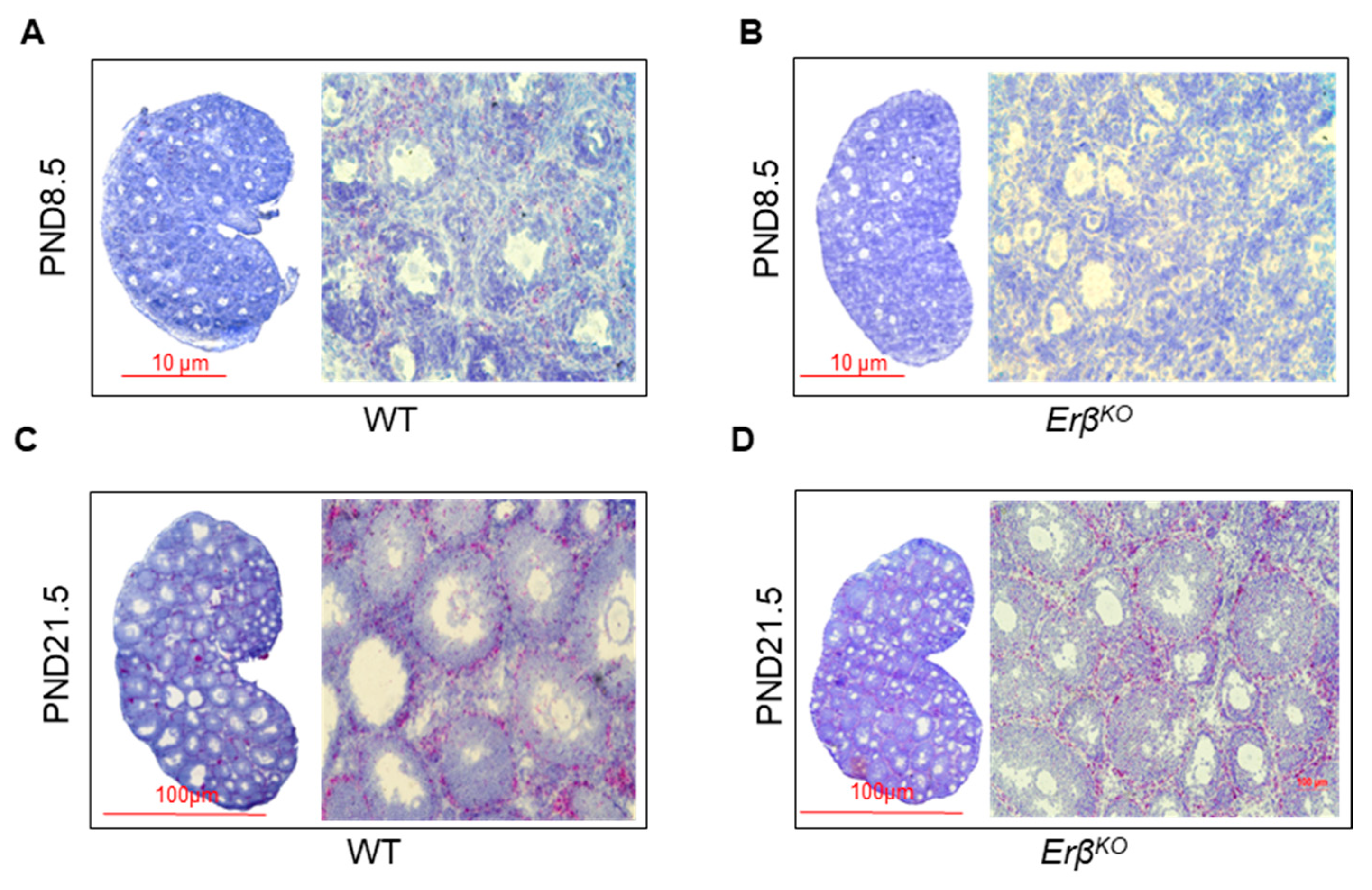
3.6. ERβ-Regulated Ihh Impacts Theca Cell Development

IHH plays an important role in the development and differentiation of TCs in activated ovarian follicles. While ERβ is a major transcriptional regulator in granulosa cells, ERα is a crucial factor in theca cells. As the expression of IHH was affected in ErβKO rat ovaries, we examined the expression of Erα mRNA in those (Figure 6). As expected, we detected no expression of Erα mRNA expression in postnatal day 8.5 ErβKO rat ovaries (Figure 6B). In contrast, Erα mRNA was present in the postnatal day 21.5 ErβKO rat ovaries, with it being a bit lower than in the wildtype follicles (Figure 6D).

4. Discussion

Pregranulosa cells in primordial follicles do not express Ihh; it is expressed in the granulosa cells of activated ovarian follicles [17]. The loss of Ihh expression in neonatal rat ovary was associated with the loss of Hhip expression in a similar fashion to that reported in an IhhKO mouse ovary [17]. Previous studies have suggested that GDF9 and BMP15 expressed by the oocytes in activated follicles induce the expression of Ihh in mouse ovaries [16,34]. In this study, we observed a very low level of Ihh and Hhip gene expression in neonatal ErβKO rat ovaries despite high levels of Gdf9 or Bmp15 expression. The presence of GDF9 and BMP15 cannot induce Ihh expression in the absence of ERβ in neonatal rat ovaries. We demonstrate that signaling mediated through ERβ is essential for Ihh expression in the granulosa cells of the activated ovarian follicles. Such an important role of estrogen signaling in the regulation of Ihh expression is a novel finding, which may have a significant impact on future studies.
RNA-Seq analyses identified altered expression of several steroidogenic enzymes in ErβKO rat ovaries, which can be either of granulosa cell or theca cell origin. It is well known that the loss of ERβ affects the expression of granulosa cell genes involved in steroidogenesis, including Cyp11a1 and Cyp19a1 [8,9]. However, in IhhKO mouse ovaries, a similar impact on steroidogenic enzymes was reported [17]. Taken together, we may conclude that the defective expression of Ihh from granulosa cells may also affect steroidogenesis in theca cells. Steroidogenesis during early life does not have any role in reproductive development; however, it may have a crucial role in the regulation of folliculogenesis.
We observed that the downregulation of Ihh expression in ErβKO rat ovaries was prominent during the first week of the rat’s life, when the majority of the activated follicles are derived from the first wave of primordial follicles [2,3]. We observed that the differential expression disappeared in the second week of life, when the activated follicles started developing from the second wave of primordial follicles [2,3]. It has been shown that while the first wave of primordial follicles develop in the medullary region, the second wave of primordial follicles develop in the ovarian cortex [2,3]. It is also well established that the granulosa cells of the first wave of primordial follicles and the second wave of primordial follicles have a distinct origin [35]. The granulosa cells in the two waves of ovarian follicles thus express differentially regulated genes. While the pregranulosa cells that are present in the first wave of primordial follicles initially express high levels of Foxl2, [36,37], the pregranulosa cells in the second wave of primordial follicles express high levels of Lgr5 [38,39,40]. Thus, it is not unlikely that the regulation of Ihh expression is different in the granulosa cells of two waves of activated follicles.
Our in situ hybridization data clearly indicate that the activated medullary follicles of first wave origin do not express Ihh even in the third week of life. Accordingly, we can conclude that the expression of Ihh is dependent on ERβ in the first wave of follicles but not in the second wave of follicles. Further understanding of such a differential regulation of Ihh expression may have an impact on extraovarian tissues where hedgehog signaling and estrogen signaling play an important role, e.g., bone formation or tumorigenesis.
Granulosa cells possess a high level of ERβ expression [22,23]. Our findings suggest that Ihh expression in granulosa cells is regulated by this ligand activated transcription factor. Granulosa cells in the activated wildtype ovarian follicles express the Ihh gene, which in turn acts on premature theca cells for the induction of their development and differentiation [16]. We observed that the IHH target gene Hhip, which is of theca cell origin, is dramatically downregulated in ErβKO rat ovaries. Thus, it is likely that one will observe the downregulation of the steroidogenic enzyme genes in theca cells, particularly those regulated by IHH signaling [18]. Our in situ hybridization data also suggest the defective development of theca cells in ErβKO rat ovaries, particularly during the first week of life. Moreover, neonatal ovaries possess mechanisms to detect steroidogenesis and respond dynamically to regulate the process of primordial follicle formation and activation [41].
Although mice harboring ovary-specific IhhKO did not exhibit any ovulatory dysfunction, this does not exclude a possible role of IHH signaling in regulating primordial follicle activation. Moreover, the representative images of IhhKO mouse ovaries appeared smaller than that of wildtype mouse ovaries, comparable to the smaller sizes of ErβKO mouse or ErβKO rat ovaries [8]. We hypothesize that ERβ-regulated expression of Ihh from the activated follicles may act on dormant primordial follicles to control excessive primordial follicle activation, which has been observed in ErβKO rat ovaries [11]. However, further studies are required to prove the mechanistic role of IHH in regulating primordial follicle activation.

5. Conclusions

It is widely accepted that Ihh is expressed in the granulosa cells of activated ovarian follicles. We observed that ERβ regulates the expression of Ihh in granulosa cells. However, such ERβ-dependent expression of Ihh was limited to the first wave of ovarian follicles. The expression of Ihh in the granulosa cells of second wave follicles did not require the presence of ERβ. Further studies are required to understand the significance of ERβ-regulated Ihh expression in the first wave of ovarian follicles.

Supplementary Materials

The following supporting information can be downloaded at: https://www.mdpi.com/article/10.3390/cells13070644/s1. Supplementary Figures S1–S6. Figure S1. Hematoxylin and Eosin stained histological sections of postnatal day 8.5 ErβKO rat ovaries; Figure S2. Verification of hedgehog signaling genes. RNA-Seq data were validated using RT-qPCR analyses; Figure S3. RNA-Seq analysis of postnatal day 4.5 rat ovaries. RNA-Seq analysis; Figure S4. RNA-Seq analysis of postnatal day 6.5 rat ovaries. RNA-Seq analysis; Figure S5. Gene Ontology and IPA analysis of differentially expressed genes in ErβKO rat ovaries; Figure S6. Verification of the expression of steroidogenic genes. RNA-Seq data were validated using RT-qPCR.

Author Contributions

V.P.C., I.D., S.G. and S.B. performed the experiments and prepared the figures. R.M., V.D., K.V., E.B.L., A.R. and V.K. helped with the data analysis, preparation of the figures, and the final manuscript. M.A.K.R. planned the studies, wrote the manuscript, and edited the figures. C.A.M. and P.E.F. critically read and edited the manuscript. All authors have read and agreed to the published version of the manuscript.

Funding

This work was partly supported by NIH R21 HD105095 grant funding.

Institutional Review Board Statement

Ethics Committee Name: The University of Kansas Medical Center Animal Care and Use Committee. Approval code: 2021-2603; approval date: 5 July 2021.

Informed Consent Statement

Not applicable.

Data Availability Statement

All RNA-Seq data have been submitted to the Sequencing Read Archive (SRX6955095-6955104).

Acknowledgments

The authors acknowledge the Cells Editorial Office for waiving the publication fees.

Conflicts of Interest

The authors declare that this research article was prepared without any commercial or financial relationships that could be construed as a potential conflict of interest.

References

  1. McGee, E.A.; Hsueh, A.J. Initial and cyclic recruitment of ovarian follicles. Endocr. Rev. 2000, 21, 200–214. [Google Scholar] [CrossRef] [PubMed]
  2. Zheng, W.; Zhang, H.; Liu, K. The two classes of primordial follicles in the mouse ovary: Their development, physiological functions and implications for future research. Mol. Hum. Reprod. 2014, 20, 286–292. [Google Scholar] [CrossRef] [PubMed]
  3. Zheng, W.; Zhang, H.; Gorre, N.; Risal, S.; Shen, Y.; Liu, K. Two classes of ovarian primordial follicles exhibit distinct developmental dynamics and physiological functions. Hum. Mol. Genet. 2014, 23, 920–928. [Google Scholar] [CrossRef] [PubMed]
  4. Zhang, T.; He, M.; Zhang, J.; Tong, Y.; Chen, T.; Wang, C.; Pan, W.; Xiao, Z. Mechanisms of primordial follicle activation and new pregnancy opportunity for premature ovarian failure patients. Front. Physiol. 2023, 14, 1113684. [Google Scholar] [CrossRef] [PubMed]
  5. Chen, Y.; Liu, Q.; Liu, R.; Yang, C.; Wang, X.; Ran, Z.; Zhou, S.; Li, X.; He, C. A Prepubertal Mice Model to Study the Growth Pattern of Early Ovarian Follicles. Int. J. Mol. Sci. 2021, 22, 5130. [Google Scholar] [CrossRef] [PubMed]
  6. Miura, K.; Murata, C.; Harikae, K.; Suzuki, H.; Kanai-Azuma, M.; Kurohmaru, M.; Tsunekawa, N.; Kanai, Y. Defects in the first wave of folliculogenesis in mouse XO ovaries. J. Reprod. Dev. 2017, 63, 333–338. [Google Scholar] [CrossRef] [PubMed]
  7. Vaz, B.; El Mansouri, F.; Liu, X.; Taketo, T. Premature ovarian insufficiency in the XO female mouse on the C57BL/6J genetic background. Mol. Hum. Reprod. 2020, 26, 678–688. [Google Scholar] [CrossRef] [PubMed]
  8. Rumi, M.A.K.; Singh, P.; Roby, K.F.; Zhao, X.; Iqbal, K.; Ratri, A.; Lei, T.; Cui, W.; Borosha, S.; Dhakal, P.; et al. Defining the Role of Estrogen Receptor beta in the Regulation of Female Fertility. Endocrinology 2017, 158, 2330–2343. [Google Scholar] [CrossRef] [PubMed]
  9. Chakravarthi, V.P.; Ratri, A.; Masumi, S.; Borosha, S.; Ghosh, S.; Christenson, L.K.; Roby, K.F.; Wolfe, M.W.; Rumi, M.A.K. Granulosa cell genes that regulate ovarian follicle development beyond the antral stage: The role of estrogen receptor β. Mol. Cell Endocrinol. 2021, 528, 111212. [Google Scholar] [CrossRef]
  10. Khristi, V.; Chakravarthi, V.P.; Singh, P.; Ghosh, S.; Pramanik, A.; Ratri, A.; Borosha, S.; Roby, K.F.; Wolfe, M.W.; Rumi, M.A.K. ESR2 regulates granulosa cell genes essential for follicle maturation and ovulation. Mol. Cell Endocrinol. 2018, 474, 214–226. [Google Scholar] [CrossRef]
  11. Chakravarthi, V.P.; Ghosh, S.; Roby, K.F.; Wolfe, M.W.; Rumi, M.K. A Gatekeeping Role of ESR2 to Maintain the Primordial Follicle Reserve. Endocrinology 2020, 161, bqaa037. [Google Scholar] [CrossRef] [PubMed]
  12. Adhikari, D.; Liu, K. Molecular mechanisms underlying the activation of mammalian primordial follicles. Endocr. Rev. 2009, 30, 438–464. [Google Scholar] [CrossRef]
  13. Ford, E.; Beckett, E.L.; Roman, S.; McLaughlin, E.A.; Sutherland, J. Advances in human primordial follicle activation and premature ovarian insufficiency. Reproduction 2019, 159, R15–R29. [Google Scholar] [CrossRef]
  14. Russell, M.C.; Cowan, R.G.; Harman, R.M.; Walker, A.L.; Quirk, S.M. The hedgehog signaling pathway in the mouse ovary. Biol. Reprod. 2007, 77, 226–236. [Google Scholar] [CrossRef]
  15. Wijgerde, M.; Ooms, M.; Hoogerbrugge, J.W.; Grootegoed, J.A. Hedgehog signaling in mouse ovary: Indian hedgehog and desert hedgehog from granulosa cells induce target gene expression in developing theca cells. Endocrinology 2005, 146, 3558–3566. [Google Scholar] [CrossRef] [PubMed]
  16. Liu, C.; Peng, J.; Matzuk, M.M.; Yao, H.H. Lineage specification of ovarian theca cells requires multicellular interactions via oocyte and granulosa cells. Nat. Commun. 2015, 6, 6934. [Google Scholar] [CrossRef]
  17. Liu, C.; Rodriguez, K.F.; Brown, P.R.; Yao, H.H. Reproductive, Physiological, and Molecular Outcomes in Female Mice Deficient in Dhh and Ihh. Endocrinology 2018, 159, 2563–2575. [Google Scholar] [CrossRef] [PubMed]
  18. Finco, I.; LaPensee, C.R.; Krill, K.T.; Hammer, G.D. Hedgehog signaling and steroidogenesis. Annu. Rev. Physiol. 2015, 77, 105–129. [Google Scholar] [CrossRef]
  19. Ren, Y.; Cowan, R.G.; Harman, R.M.; Quirk, S.M. Dominant activation of the hedgehog signaling pathway in the ovary alters theca development and prevents ovulation. Mol. Endocrinol. 2009, 23, 711–723. [Google Scholar] [CrossRef]
  20. Varjosalo, M.; Taipale, J. Hedgehog: Functions and mechanisms. Genes Dev. 2008, 22, 2454–2472. [Google Scholar] [CrossRef]
  21. Franco, H.L.; Yao, H.H. Sex and hedgehog: Roles of genes in the hedgehog signaling pathway in mammalian sexual differentiation. Chromosome Res. Int. J. Mol. Supramol. Evol. Asp. Chromosome Biol. 2012, 20, 247–258. [Google Scholar] [CrossRef] [PubMed]
  22. Drummond, A.E.; Fuller, P.J. The importance of ERbeta signalling in the ovary. J. Endocrinol. 2010, 205, 15–23. [Google Scholar] [CrossRef] [PubMed]
  23. Chakravarthi, V.P.; Khristi, V.; Ghosh, S.; Yerrathota, S.; Dai, E.; Roby, K.F.; Wolfe, M.W.; Rumi, M.A.K. ESR2 Is Essential for Gonadotropin-Induced Kiss1 Expression in Granulosa Cells. Endocrinology 2018, 159, 3860–3873. [Google Scholar] [CrossRef] [PubMed]
  24. Picut, C.A.; Swanson, C.L.; Scully, K.L.; Roseman, V.C.; Parker, R.F.; Remick, A.K. Ovarian follicle counts using proliferating cell nuclear antigen (PCNA) and semi-automated image analysis in rats. Toxicol. Pathol. 2008, 36, 674–679. [Google Scholar] [CrossRef] [PubMed]
  25. Roby, K.F.; Son, D.S.; Terranova, P.F. Alterations of events related to ovarian function in tumor necrosis factor receptor type I knockout mice. Biol. Reprod. 1999, 61, 1616–1621. [Google Scholar] [CrossRef]
  26. Roby, K.F. Alterations in follicle development, steroidogenesis, and gonadotropin receptor binding in a model of ovulatory blockade. Endocrinology 2001, 142, 2328–2335. [Google Scholar] [CrossRef] [PubMed]
  27. Roby, K.F.; Son, D.S.; Taylor, C.C.; Montgomery-Rice, V.; Kirchoff, J.; Tang, S.; Terranova, P.F. Alterations in reproductive function in SRC tyrosine kinase knockout mice. Endocrine 2005, 26, 169–176. [Google Scholar] [CrossRef] [PubMed]
  28. Pedersen, T.; Peters, H. Proposal for a classification of oocytes and follicles in the mouse ovary. J. Reprod. Fertil. 1968, 17, 555–557. [Google Scholar] [CrossRef] [PubMed]
  29. Dupont, J.; Reverchon, M.; Cloix, L.; Froment, P.; Ramé, C. Involvement of adipokines, AMPK, PI3K and the PPAR signaling pathways in ovarian follicle development and cancer. Int. J. Dev. Biol. 2012, 56, 959–967. [Google Scholar] [CrossRef]
  30. Hughes, C.H.K.; Smith, O.E.; Meinsohn, M.C.; Brunelle, M.; Gévry, N.; Murphy, B.D. Steroidogenic factor 1 (SF-1; Nr5a1) regulates the formation of the ovarian reserve. Proc. Natl. Acad. Sci. USA 2023, 120, e2220849120. [Google Scholar] [CrossRef]
  31. Russell, D.L.; Doyle, K.M.; Gonzales-Robayna, I.; Pipaon, C.; Richards, J.S. Egr-1 induction in rat granulosa cells by follicle-stimulating hormone and luteinizing hormone: Combinatorial regulation by transcription factors cyclic adenosine 3′,5′-monophosphate regulatory element binding protein, serum response factor, sp1, and early growth response factor-1. Mol. Endocrinol. 2003, 17, 520–533. [Google Scholar] [CrossRef] [PubMed]
  32. Schmidt, D.; Ovitt, C.E.; Anlag, K.; Fehsenfeld, S.; Gredsted, L.; Treier, A.C.; Treier, M. The murine winged-helix transcription factor Foxl2 is required for granulosa cell differentiation and ovary maintenance. Development 2004, 131, 933–942. [Google Scholar] [CrossRef] [PubMed]
  33. Guzmán, A.; Hughes, C.H.K.; Murphy, B.D. Orphan nuclear receptors in angiogenesis and follicular development. Reproduction 2021, 162, R35–R54. [Google Scholar] [CrossRef] [PubMed]
  34. Otsuka, F.; Yao, Z.; Lee, T.; Yamamoto, S.; Erickson, G.F.; Shimasaki, S. Bone morphogenetic protein-15. Identification of target cells and biological functions. J. Biol. Chem. 2000, 275, 39523–39528. [Google Scholar] [CrossRef] [PubMed]
  35. Niu, W.; Spradling, A.C. Two distinct pathways of pregranulosa cell differentiation support follicle formation in the mouse ovary. Proc. Natl. Acad. Sci. USA 2020, 117, 20015–20026. [Google Scholar] [CrossRef] [PubMed]
  36. Pisarska, M.D.; Barlow, G.; Kuo, F.T. Minireview: Roles of the forkhead transcription factor FOXL2 in granulosa cell biology and pathology. Endocrinology 2011, 152, 1199–1208. [Google Scholar] [CrossRef]
  37. Nicol, B.; Grimm, S.A.; Chalmel, F.; Lecluze, E.; Pannetier, M.; Pailhoux, E.; Dupin-De-Beyssat, E.; Guiguen, Y.; Capel, B.; Yao, H.H. RUNX1 maintains the identity of the fetal ovary through an interplay with FOXL2. Nat. Commun. 2019, 10, 5116. [Google Scholar] [CrossRef]
  38. Mork, L.; Maatouk, D.M.; McMahon, J.A.; Guo, J.J.; Zhang, P.; McMahon, A.P.; Capel, B. Temporal differences in granulosa cell specification in the ovary reflect distinct follicle fates in mice. Biol. Reprod. 2012, 86, 37. [Google Scholar] [CrossRef]
  39. Ng, A.; Tan, S.; Singh, G.; Rizk, P.; Swathi, Y.; Tan, T.Z.; Huang, R.Y.; Leushacke, M.; Barker, N. Lgr5 marks stem/progenitor cells in ovary and tubal epithelia. Nat. Cell Biol. 2014, 16, 745–757. [Google Scholar] [CrossRef]
  40. Rastetter, R.H.; Bernard, P.; Palmer, J.S.; Chassot, A.A.; Chen, H.; Western, P.S.; Ramsay, R.G.; Chaboissier, M.C.; Wilhelm, D. Marker genes identify three somatic cell types in the fetal mouse ovary. Dev. Biol. 2014, 394, 242–252. [Google Scholar] [CrossRef]
  41. Fowler, P.A.; Anderson, R.A.; Saunders, P.T.; Kinnell, H.; Mason, J.I.; Evans, D.B.; Bhattacharya, S.; Flannigan, S.; Franks, S.; Monteiro, A. Development of steroid signaling pathways during primordial follicle formation in the human fetal ovary. J. Clin. Endocrinol. Metab. 2011, 96, 1754–1762. [Google Scholar] [CrossRef] [PubMed]
Figure 1. Loss of ErβKO results in increased primordial follicle activation in immature rat ovaries. We detected increased growth of activated medullary follicles in ErβKO rat ovaries compared to age-matched wildtype ovaries (AG). An increased number of activated primary, secondary, and atretic follicles was evident in postnatal day 8.5 ErβKO rat ovaries (EG). While the wildtype rat ovaries contained more primordial follicles, they were replaced by primary follicles in ErβKO rat ovaries (EG). Data shown as the mean ± SE, * p < 0.05, n > 3.
Cells 13 00644 g001
Figure 2. RNA-Seq analysis of postnatal day 8.5 rat ovaries. RNA-Seq analysis (heat maps in the left panel and volcano plots in the right panel) of postnatal day 8.5 rat ovaries showing the differential expression of genes in the whole ovary (no filter) (A,B), related to the hedgehog pathway (C,D), steroidogenesis (E,F), and the key genes involved in the process of folliculogenesis (G,H). Data are shown as CLC Genomic Workbench Analysis.
Cells 13 00644 g002
Figure 3. Loss of ERβ disrupts Indian hedgehog signaling in ErβKO rat ovaries (A). RNA-Seq analysis of postnatal day 4.5, 6.5, and 8.5 showed downregulation of Erβ but not another grnaulosa cell-specific transcription factor Foxl2 (B). We observed a marked downregulation of Ihh, and its downstream targets Hhip and Gli1, as well as its receptor Ptch1 in ErβKO ovaries (CF). Ihh expression was remarkably low (C) despite its known regulators Gdf9 (G) and Bmp15 (H) being significantly upregulated in ErβKO ovaries. Data shown as the mean ± SE TPM values; * p ≤ 0.05; n = 4 (RNA-Seq).
Cells 13 00644 g003
Figure 4. Loss of ERβ dysregulated the expression of steroidogenic enzymes in ErβKO rat ovaries. RNA-Seq analysis of postnatal day (P) 4.5, 6.5, and 8.5 ovaries showed marked downregulation of Hsd17b1, and Cyp11a1 (A,C) in ErβKO ovaries. However, the expression of Star and Cyp19a1 did not show any significant changes in ErβKO ovaries (B,D). While the expression of Fshr remained unchanged, the expression of Lhcgr was significantly downregulated in postnatal day 8.5 ErβKO ovaries (E,F). Data shown as the mean ± SE TPM values; * p ≤ 0.05; n = 4.
Cells 13 00644 g004
Figure 5. ERβ regulates Ihh gene expression only in the first wave of ovarian follicles. RT-qPCR analysis of postnatal day (P) 4.5, 6.5, and 8.5 ovaries showed increasing downregulation of Ihh and Hhip expression in ErβKO ovaries (A,C). However, the downregulation of Ihh and Hhip expression disappeared in ErβKO ovaries starting from postnatal day 16.5 onwards (B,D). RNAScope in situ hybridization detected the expression of Ihh mRNA in the activated medullary follicles in postnatal day 8.5 wildtype (WT) ovaries (E) but not in the age-matched ErβKO ovaries (F). Granulosa cells in all activated follicles within the medullary as well as the cortical region of postnatal day 21.5 WT ovaries showed the expression of Ihh mRNA (G). In contrast, prominent expression of Ihh mRNA was detected only in the activated peripheral (cortical) follicles in PND 21.5 ErβKO ovaries but not in the activated medullary follicles (H). RT-qPCR data are shown as relative expression according to mean ± SE values; * p ≤ 0.05; n = 6.
Cells 13 00644 g005
Figure 6. Loss of Ihh expression affected theca cell development in ErβKO ovaries. In situ hybridization was performed for the detection of Erα mRNA (a marker of theca cells) in postnatal day 8.5 and postnatal day 21.5 wildtype (WT) and age-matched ErβKO ovaries. The histology sections showed the presence of Erα mRNA in the peripheral regions surrounding the secondary follicles in postnatal day 8.5 wildtype ovaries (A). However, the postnatal day 8.5 ErβKO ovaries did not show any expression of Erα mRNA (B). In contrast, Erα mRNA was abundantly expressed in postnatal day 21.5 ErβKO ovaries, comparable to that of age-matched WT ovaries (C,D).
Cells 13 00644 g006
Table 1. Top 10 differentially expressed genes in ErβKO postnatal day 8.5 rat ovaries (absolute fold change ≥ 2, FDR p-value ≤ 0.05, and maximum TPM ≥ 5).
1 A. Top 10 Downregulated Genes
NameChromENSEMBLRegionMax TPMFold ChangeFDR p-Value
ENSRNOG
00000065112
1ENSRNOG00000065112167210874..1672131497.4−902.740.00
Lce1m2ENSRNOG00000009581complement (178637495..178638697)6.26−280.110.00
ENSRNOG
00000065518
2ENSRNOG00000065518complement (178666387..178668404)6.7−149.270.00
Abcb1b4ENSRNOG00000066042complement (25242798..25325199)24.2−28.710.00
Snrpc-ps310ENSRNOG00000017586complement (14191662..14192401)6.69−16.780.00
Ihh9ENSRNOG00000018059complement (76504315..76510532)118.52−15.200.00
Fam111a1ENSRNOG00000012067209640953..209656547156.28−8.390.00
Hhip19ENSRNOG0000001826827863213..2795252839.48−7.870.00
Gal1ENSRNOG00000015156complement (200650439..200654959)9.89−6.630.00
Kcns27ENSRNOG0000006611166022352..660284229.77−6.520.00
1 B. Top 10 Upregulated Genes
NameChromENSEMBLRegionMax TPMFold ChangeFDR p-Value
LOC68571611ENSRNOG0000005440455280418..5530382710.84315.030.00
LOC1003607911ENSRNOG0000003351719107386..19108232470.9817.110.00
Slc27a51ENSRNOG00000019626complement (73616564..73627172)5.1313.920.00
Aldh1a71ENSRNOG00000017878complement (218201472..218248906)9.8511.660.00
ENSRNOG
00000066616
3ENSRNOG00000066616149220737..1492282795.059.670.00
Actc13ENSRNOG00000008536complement (100811987..100817523)7.049.550.00
Adcyap1r14ENSRNOG0000001209884593892..8464270025.869.290.00
Adh6_12ENSRNOG00000012436226797303..22680889213.716.900.00
Gabrr311ENSRNOG00000001679complement (40902812..40955263)10.626.760.00
Cpa24ENSRNOG0000002809259160357..59183674250.636.740.00
Table 2. Differentially expressed transcription factors in ErβKO postnatal day 8.5 rat ovaries.
NameChromENSEMBLRegionMax TPMFold ChangeFDR p-Value
Rbpjl3ENSRNOG00000026295153134140..15314651310.905.790.00
Dbx27ENSRNOG00000006885complement (126772227..126802564)5.185.360.00
Dmrt11ENSRNOG00000016075223142859..2232413336.225.130.00
Npas29ENSRNOG0000001340841463830..4164232036.434.070.00
Pou5f120ENSRNOG00000046487complement (3223129..3227891)20.602.590.00
Setbp118ENSRNOG00000016208complement (72191035..72552556)5.242.180.00
Hoxc67ENSRNOG00000063956134135502..1341483926.502.070.00
Scx7ENSRNOG00000021812108176608..10817862613.022.040.00
Pparg4ENSRNOG00000008839148423194..14854846814.06−2.040.00
Mycn6ENSRNOG00000051372complement (35717764..35723590)58.93−2.050.00
Fosl26ENSRNOG00000068412complement (24300956..24320034)36.83−2.100.00
Egr118ENSRNOG0000001942226462981..2646676667.29−2.170.00
Osr27ENSRNOG0000001113666487839..6649522492.84−2.260.00
Nr5a13ENSRNOG00000012682complement (22465502..22486328)173.03−2.350.00
Foxo12ENSRNOG00000013397136312168..13638779097.01−2.380.00
Fos6ENSRNOG00000008015105121170..10512403623.72−2.520.00
Myc7ENSRNOG0000000450093593705..9359863047.62−2.600.00
Nr4a17ENSRNOG00000007607132374840..13238929787.54−4.970.00
Disclaimer/Publisher’s Note: The statements, opinions and data contained in all publications are solely those of the individual author(s) and contributor(s) and not of MDPI and/or the editor(s). MDPI and/or the editor(s) disclaim responsibility for any injury to people or property resulting from any ideas, methods, instructions or products referred to in the content.

