Review

Origin, Function, and Implications of Intestinal and Hepatic Macrophages in the Pathogenesis of Alcohol-Associated Liver Disease

1
Laboratory of Hepato-Gastroenterology, Institute of Clinical and Experimental Research, Université Catholique de Louvain, 1200 Brussels, Belgium
2
Department of Medicine, University of California San Diego, La Jolla, CA 92161, USA
3
Department of Medicine, VA San Diego Healthcare System, San Diego, CA 92161, USA
4
Cliniques Universitaires Saint-Luc, 1200 Brussels, Belgium
*
Author to whom correspondence should be addressed.
Cells 2025, 14(3), 207; https://doi.org/10.3390/cells14030207
Submission received: 15 November 2024 / Revised: 12 January 2025 / Accepted: 15 January 2025 / Published: 30 January 2025

Abstract

:
Macrophages are members of the human innate immune system, and the majority reside in the liver. In recent years, they have been recognized as essential players in the maintenance of liver and intestinal homeostasis as well as key guardians of their respective immune systems, and they are increasingly being recognized as such. Paradoxically, they are also likely involved in chronic pathologies of the gastrointestinal tract and potentially in the alteration of the gut–liver axis in alcohol use disorder (AUD) and alcohol-associated liver disease (ALD). To date, the causal relationship between macrophages, the pathogenesis of ALD, and the immune dysregulation of the gut remains unclear. In this review, we will discuss our current understanding of the heterogeneity of intestinal and hepatic macrophages, their ontogeny, the potential factors that regulate their origin, and the evidence of how they are associated with the manifestation of chronic inflammation. We will also illustrate how the micro-environment of the intestine shapes the phenotypes and functionality of the macrophage compartment in both the intestines and liver and how they change during chronic alcohol abuse. Finally, we highlight the obstacles to current research and the prospects for this field.

Graphical Abstract

1. Introduction

1.1. History of Alcohol Use

The first evidence of the consumption of alcohol as a drink dates back to 7000 BC in China, where human communities settled and agriculture started to flourish [1]. Evidence of the use of fermented rice, millet, grapes, and honey to make alcoholic beverages after this period has been found across the world, such as in Mesopotamia, ancient Egypt, and Syria [2]. Today, alcohol is largely consumed in all cultures as a social glue for recreational and ceremonial purposes because of its psychoactive, rewarding, and anxiolytic effects. However, the consumption of alcohol has begun to decline due to concerns over increased calories in peoples’ daily diets [3]. Culture and religious beliefs play the main role in the patterns of alcohol consumption in different regions and different races.

1.2. Epidemiology and Drinking Patterns

According to data from the World Health Organization (WHO), alcohol consumption varies significantly and depends on age and geographic location. The European region consumes more than 10.26 L of pure alcohol on average, which makes it the region that consumes alcohol the most. North Africa and the Middle East have the lowest alcohol consumption per capita (source: WHO data). According to the latest data, beer is the most consumed form of alcohol (396.76 billion L), followed by wine (25.76 billion L) and spirits (23.11 billion L) [4,5]. The total amount of alcohol consumed per capita has decreased by 5.5 L globally since 2015. In Europe, alcohol use has declined by 17% since 2000, whereas the use of alcohol in the Americas has been stagnating. By contrast, alcohol consumption has been steadily increasing in the Western Pacific and South-East by 40% and 112%, respectively. Alcohol use disorder (AUD) remains a significant issue worldwide. Although the exact circumstances that cause AUD to occur are not fully understood, several factors contribute to its development, such as environmental factors, genetic predisposition, peer interaction, and certain personality disorders.

1.3. Alcohol-Associated Liver Disease

Alcohol abuse is a major risk factor for multiple preventable causes of death and diseases such as alcohol addiction, alcohol-associated liver disease (ALD), diabetes, cancers, and fetal alcohol syndrome [6]. ALD contributes to 10% of deaths globally. ALD consists of a broad spectrum of disorders ranging from isolated steatosis to alcohol-associated steatohepatitis and cirrhosis. Although the underlying mechanisms remain poorly understood, the prevalence of patients with a history of chronic heavy alcohol intake that develop advanced fibrosis is around 30–35% [7]. In subjects with an AUD, the prevalence of alcohol-related hepatitis (AH), which is an acute deterioration that occurs in people with an underlying ALD, has been estimated to be 20–40% [8]. AH encompasses steatohepatitis, pericellular fibrosis, hepatocellular injury, and bilirubinostasis.
A previous study showed that the number of intestinal macrophages in AUD patients is significantly lower than in control subjects [9]; however, the reason behind this remains unclear. Overall, the study of macrophages is heavily based on animal models. Due to the fundamental differences in terms of anatomy, physiology, and metabolism between humans and other animals, many findings cannot be readily translated into clinical significance. As it is important to understand the role of macrophages in alcohol-associated liver disease, this review will stratify the current information, focus on the available clinical findings of how macrophages could participate in the pathogenesis of ALD, and highlight the obstacles and perspectives for future studies.

2. Macrophage Heterogeneity

Macrophages were first discovered by Metchnikoff in the 19th century and later classified as a part of the mononuclear phagocyte system along with monocytes and dendritic cells [10,11]. The early concept of mononuclear phagocytes led people to believe that the tissue-resident macrophages in adulthood rely on monocytes from peripheral blood to replenish the population. However, this belief about an inherent developmental link between tissue-resident macrophages and blood monocytes has been disproved. It is now well accepted that the long-living tissue-resident macrophages rely on in situ self-renewal. Moreover, certain macrophage populations in the embryonic stage are established before the emergence of circulating monocytes. The final discovery that reshaped the macrophage pedigree came from monocyte-specific knockout mice that demonstrated seemingly normal tissue macrophage compartments [12,13] despite the inability of the bone marrow to generate monocytes. Moreover, tissue macrophage numbers remain unaffected in human patients with monocytopenia [14]. These experiments and observations suggested an alternative, non-monocytic origin of tissue-resident macrophages and therefore led to a revision of the previous monocyte–macrophage axis. Current concepts postulate that yolk sac (YS)-derived macrophages are the main precursors for most tissue-resident macrophages. Fate-mapping studies have shown that late erythron-myeloid precursors that arise in the YS are the principal source of tissue-resident macrophages involving a monocytic intermediate. Alternatively, it has been demonstrated that fetal hematopoietic stem cells might also contribute to tissue-resident macrophage populations [14].

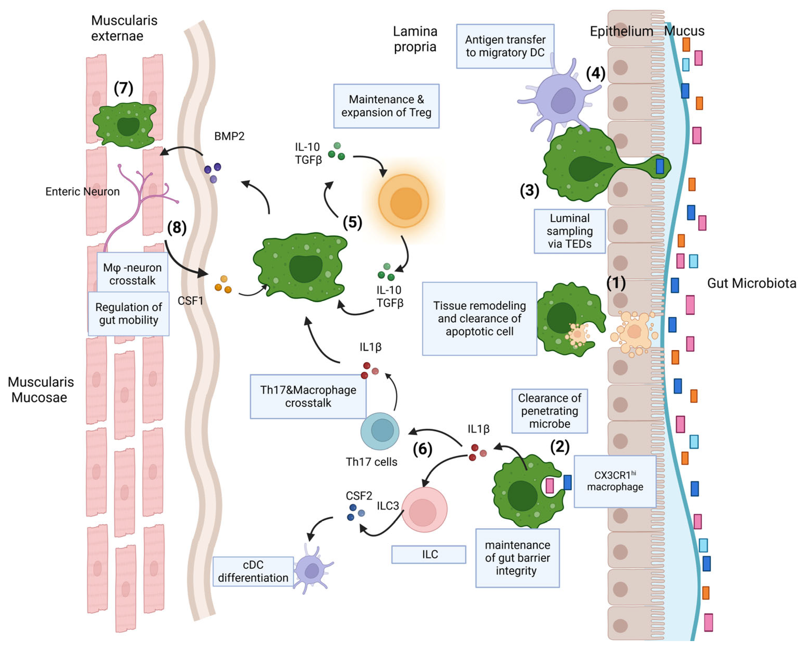
3. The Dynamics of Intestinal Macrophages

The specific cause of the variation in macrophage origins within different tissues is still not fully understood. Specifically, it is not yet clear why and how macrophages derived from embryos continue to exist in the central nervous system and epidermis, while their presence in substantial quantities does not persist in the intestinal mucosa in adulthood. Intestinal macrophages have a limited capacity for self-maintenance, and their expected half-life does not exceed 6 weeks. They are primarily replenished by bone-marrow- derived monocytes, which are CCR2-dependent [15]. One of many hypotheses in this regard is related to niche accessibility and availability. Indeed, the mucosa can be easily reached at all times, while the brain and epidermis are isolated from vasculature by the blood–brain barrier and the basal membrane, respectively. On the other hand, tissue macrophages that are not separated from the vasculature by a basal membrane, such as Kupffer cells and microglia, also function autonomously, which may indicate that proximity to the tissue might not be the reason behind both the monocyte infiltration of tissues and the reinforcement of the local immune system [16,17]. New evidence has indicated that microbiota and diet are capable of modifying macrophage profiles and how they both influence the dynamics of mucosal macrophages. Studies have illustrated that germ-free and SPF mice have a significantly lower number of intestinal macrophages [18,19,20]. The administration of antibiotics can reduce macrophage numbers, suggesting that the microbiota plays a role in augmenting macrophage turnover [21]. Gut dysbiosis, the intestinal mucosal microenvironment, dietary metabolites, and mechanical stress such as cyclic stretching have been suggested to play roles in upregulating pro-inflammatory cytokine release [22]. These gut-produced inflammatory signals potentially exacerbate monocyte recruitment and macrophage activation [23]. Additionally, the presence of commensal microbiota in the colon significantly alters macrophages’ gene expression profiles and surface markers promoting the development of CD11c+CD121b+ and CD11c−CD206hi macrophages, particularly when the differentiation of monocytes starts. A differential turnover rate of tissue macrophages has been observed in different anatomical locations of the gut wall and in the liver, which could indicate that particular niches do not support macrophage self-renewal. However, differential rates of tissue macrophage subpopulations have not been assessed experimentally. Although progress has been made in understanding the dynamics of macrophage turnover, more tailored projects are still needed to fully understand this process and the functions of different subtypes of macrophages.

3.1. Function of Gut Macrophages

Like their counterparts in the liver, intestinal macrophages are involved in pathogen clearance. Consistent with their phagocytic nature, intestinal macrophages express many phagocytosis genes, such as Mertk, cd36, cd206, ltgav, ltgb5, and many others [24,25]. Macrophages also actively participate in tissue remodeling and apoptotic cell clearance, known as efferocytosis [25], which is mediated by integrin αvβ3. They also play a role in various activities implicated in maintaining gut homeostasis (Figure 1). Although intestinal macrophages are avidly phagocytic, they do not lead to overt inflammation in response to the commensal gut microbiome, which mainly occurs through the regulation of surface-receptor expression and reduced secretion of pro-inflammatory cytokines such as CCL2 and IL-8 [26,27]. Intestinal macrophages can also produce IL-10 (CX3CR1hi intestinal macrophages) and transforming growth factor β (TGF-β) [27], two prominent cytokines that predominantly contribute to reduced inflammation and homeostasis of the GI tract. IL-10 from various sources is sensed via the IL-10 receptor (IL-10R) to inhibit the synthesis of proinflammatory cytokines [27]. IL-10 not only limits the inflammatory state of macrophages but also regulates the killing of intracellular bacteria by those cells. Human-induced pluripotent-stem-cell-derived macrophages without a functional IL-10R display impaired bacterial killing due to dysregulated PGE2 production [28].
The sub-epithelial positioning of lamina propria (LP) macrophages indicates that they are placed to capture any foreign substance that penetrates the epithelial barrier. Murine models show that LP macrophages can also form transepithelial dendrites (TEDs) to sample luminal bacteria, constituting a cellular process that allows macrophages to penetrate the gut barrier without disrupting the tight junctions and the integrity of the epithelium. This process is highly dependent on the CX3CL1-CX3CR1 axis [29,30,31] (Figure 1). In particular, when encountering Gram-negative bacteria, intestinal macrophages downregulate expression of CD14, a glycoprotein receptor for lipopolysaccharide (LPS), compared to the levels expressed by conventional macrophages to reduce the inflammatory response [32]. Similarly, it has been noticed that CX3CR1 macrophages in the upper intestine are capable of capturing food particles, and it is hypothesized that they are involved in oral tolerance to food antigens [33].
However, in inflammatory conditions, the mature intestinal macrophages, along with other cells, release pro-inflammatory cytokines and monocyte chemotactic proteins such as CCL2, CCL7, CCL8, and CCL12 [34,35,36]. In chronic alcohol abuse conditions, the upregulation of CCL3 and CCL4 is especially common in the gut [37]. The consumption of alcohol increases gut permeability, disrupts intestinal tight junctions, and promotes microbial translocation [38]. In addition, chronic alcohol exposure directly alters the population and composition of the microbiota by decreasing the population of Gram-positive bacteria and promoting a number of Gram-negative bacteria [39,40].
Among the macrophages located in the lamina propria, it has been suggested that CD11c+ macrophages are monocyte-derived short-lived macrophages [27]. Research based on mice chronically given alcohol has also indicated that CD11c+ macrophages are concentrated close to the tips of the villi and express a pro-inflammatory profile, whereas CD206+ macrophages are located at the bases of crypts and fulfill the function of tissue macrophages. These mechanisms likely contribute to the dysregulation of macrophage numbers and macrophage immunoregulatory functions in AUD patients.

3.2. Gut Macrophages and Crosstalk with Other Immune Cells

The induction of gut-specific T-reg cells plays an important role in gut homeostasis [41]. These cells are regulatory cells for immune responses and have homing properties in the mesenteric lymph nodes [42,43] (Figure 1). It is believed that this process also involves macrophage-secreted IL-10 since Cx3cr1-induced deletion of IL-10 leads to a reduction in the frequency of antigen-specific Tregs. On the contrary, the deletion of IL-10 in macrophages does not have an observable impact on the global abundance of Tregs [44,45]. A murine model was used to demonstrate that the deletion of IL-10R hinders the differentiation of monocytes into pro-fibrogenic macrophages, which ultimately leads to an increase in the number of pro-inflammatory macrophages in the intestine. Additionally, macrophages can secrete IL-1β to facilitate the induction and maintenance of commensal-specific Th17 T cells [46,47]. On the other hand, T-cell-derived IL-10 is required for the regulation of intestinal homeostasis by the T cell–macrophage IL-10 axis (Figure 1). Indeed, appropriate IL-10 signaling levels are important for regulating different macrophage functions [28].
Compared to conventional dendritic cells (cDCs), macrophages are scarcely involved in the activation of naïve T cells [48]. However, high expression of MCH class II could mean that they are more likely to be involved in antigen presentation for previously activated T cells in the liver and intestines. Macrophages may influence T cell priming by modulating the cDC differentiation process. For example, macrophage IL-1β has been demonstrated to enhance CSF2 release by type 3 innate lymphoid cells (ILC3) [49], which is the principal cytokine for cDC differentiation in the tissue. The upregulation of CSF2 in macrophages is also a common hallmark for chronic-alcohol-abuse-induced inflammation in ALD [50]. However, most of the findings regarding ALD and tissue macrophages (see below) are largely based on murine or other animal models, and the exact mechanisms and immune responses still need to be clarified in humans.

3.3. Gut Macrophage Crosstalk with Enteric Nervous System

While it has been known for many years that macrophages are present in deeper layers of the gut wall [51], only recently has work begun to investigate their roles in intestinal homeostasis. Macrophages in the muscularis are intimately associated with the enteric nervous system and, in mice, appear to be morphologically and transcriptionally distinct [52]. Apart from LP macrophages, the macrophages in the muscularis also participate in bidirectional cross talk with enteric neurons. When an enteric neuron expressing the BMP receptor is activated by the macrophage-derived bone morphogenic protein 2 (BMP2), it starts producing CSF1, which can maintain the muscularis macrophage pool and further BMP2 expression [53,54] (Figure 1). These multi-system interactions regulate enteric smooth muscle contraction, thereby controlling peristalsis. This process can be disrupted by the administration of broad-spectrum antibiotics [20], which indirectly suggests that the microbiota plays a role in intestine contraction to a certain extent [52].
By generating metabolites of alcohol and bacteria-derived products, the microbiota also contributes to immunological changes in the intestine affecting the various macrophage niches. A study using a murine model has shown that one of the consequences of alcohol use is that the gut microbiota produces neuroactive metabolites that can trigger vagus nerve activation and directly excite the central nervous system via the systemic circulation or, alternatively, through the modulation of the immune system, e.g., via altering the macrophage population associated with the enteric nervous system [53]. Furthermore, intestinal macrophages might also serve as signal-transmitting cells in neuro-immune-stem-cell regulatory circuits, modulating intestinal stem cell self-renewal via upstream enteric serotonergic neurons. This process implicates the production of the neurotransmitter serotonin in enteric neurons, a process that can be promoted by gut microbiota metabolite valeric acid.
Overall, intestinal macrophages, which initially originate from embryonic stem cells, play a role in maintaining gut homeostasis and carry out important immunoregulatory functions towards commensal microbes while also protecting against invading pathogens. Given their limited half-life, their renewal depends on bone-marrow-derived monocytes. Intestinal macrophages can modulate the overall immune state by sampling the luminal microbiome to prevent excess inflammation. It is likely that macrophages located in the various niches of the intestine (villi, crypts, muscularis mucosae, the vascular bed, etc.) have different functions.
As the sentinels of the GI tract, intestinal macrophages extensively communicate not only with other immune cells but also neurons and the epithelium to ensure gut barrier integrity. Chronic alcohol consumption interferes with the gut microbiota, the epithelium, and the intestinal immune system. However, how alcohol disrupts physiological processes in the gut in AUD patients is not fully understood.

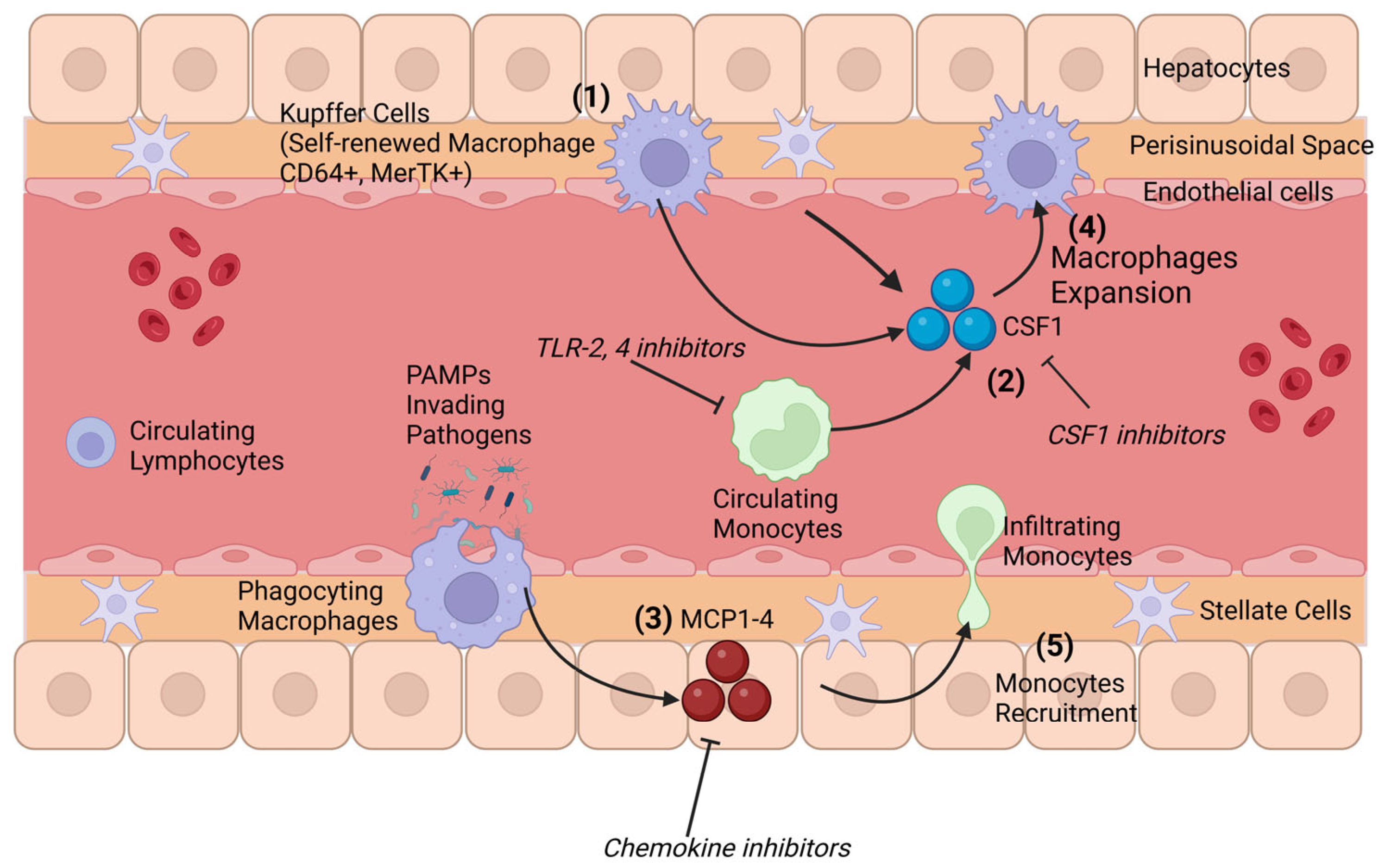
4. Origin and Composition of Hepatic Macrophages

The liver encounters constant blood flow from arterial circulation and the portal vein. Hence, the liver plays a vital role in host defense. It hosts the majority of all the macrophages in the body and is patrolled by circulating monocytes [54]. Kupffer cells (KCs) are liver-resident macrophages that originate from yolk sac progenitors during embryonic development in mammals. Although Kupffer cells reside in the liver, they are not primarily found in liver parenchyma. Indeed, the blood stream goes through the liver parenchyma using a network of tiny vessels, the sinusoids, where the KCs are located. This anatomical signature means that the liver is constantly exposed to microbial products, xenobiotics, and antigens from the GI tract and ensures effective antigen capture and presentation in the liver microenvironment [55]. In the absence of a liver injury, the macrophage pool primarily contains KCs, which have the ability to engage in self-renewal from intrahepatic precursors and, to a much lesser extent, bone marrow (BM)-derived macrophages [56,57,58]. However, the situation changes when a liver injury occurs. Some data suggest that severe liver injuries are associated with depletion of KCs. This depletion creates an intrahepatic niche that allows monocytes to infiltrate the liver. After 2–8 weeks, the infiltrated monocytes and monocyte-derived macrophages slowly adopt the transcriptional profile of KCs and become long-lived tissue-resident macrophages [56,59]. In the steady stage, KCs comprise the vast majority of the hepatic macrophages, yet another population of macrophages with a dendritic cell morphology that reside in the liver capsule has been identified in an animal model [60]. These cells are phenotypically and developmentally different from KCs but function as macrophages in the event of a liver injury to facilitate inflammatory responses (Figure 2).

Hepatic Macrophages and Crosstalk with the Gut Microbiota

Much like gut macrophages, KCs are directly influenced by the gut since food metabolites and bacterial products are passed from the gut to the liver through the portal vein. They can modulate immune tolerance under physiological conditions [28] and are able to eliminate a small number of bacterial products translocated from the gut to the liver under physiological conditions without triggering inflammation in the liver [59,61]. This delicate homeostasis can be disrupted at different levels, such as via alterations in the gut microbiome, increased gut permeability, excessive exposure of the liver to microbial products, and alteration of hepatic macrophage activity, leading to a pro-inflammatory environment in the liver. Among all the molecules that can influence the polarization of hepatic macrophages, Gram-negative-bacteria-derived LPS, which is delivered to the liver via portal blood circulation, is constantly stimulating macrophages. Hepatic macrophages are able to clear LPS in circulating blood without being activated themselves, constituting a phenomenon called LPS tolerance. Some studies have shown that LPS-TLR4/MD2-TNFα signaling on hepatic macrophages via sensing endotoxin translocation induces various inflammation responses [62,63,64]. In addition, receptor-interacting protein kinase 3 (RIP3), regulated by the gut microbiota and endotoxin translocation, also plays an important role in the pathogenesis of liver inflammation. It has been suggested that in alcohol-associated liver disease, RIP3 is important to hepatic macrophage pro-inflammatory cytokines’ responses and that the absence of RIP3 inhibits alcohol-induced liver injury [65,66,67]. Indeed, it has been shown that CGIg+ (complement receptor of the immunoglobulin superfamily) macrophages are responsible for the clearance of microbial DNA and pathobionts and that alcohol consumption reduces microbial DNA clearance by Kupffer cells, which in turn exacerbates liver inflammation and damage [68,69]. In AUD and ALD patients, there is a rise in systemic endotoxin levels together with an increase in sensitivity to LPS [70,71]. The traditional proposition in this regard is that in the progression of ALD, the constant exposure of LPS converts KCs from an immune tolerance state to an immune activation state [61,72,73]. However, recent findings instead suggest that hepatic macrophages behave as a whole pool, with KCs maintaining their immune tolerance, but monocyte-derived macrophages are either proinflammatory or profibrogenic [74,75]. Moreover, it has been postulated that the activation of monocytes/macrophages not only depends on signals from the gut microbiota but also on endogenous messengers from the hepatic microenvironment [61,76]. The dogma behind the different roles of KCs and monocytes/macrophages still needs to be further investigated.
Hepatic-tissue-resident macrophages, Kupffer cells, primarily derive from the YS. In the steady state, the liver can be considered a closed system with slow macrophage turnover and a negligible need for replacement in adulthood. Kupffer cells are constantly exposed to low levels of food particles and microbiome derivatives delivered by the portal vein from the GI tract without necessarily inducing liver inflammation. It is not surprising that hepatic macrophages are reshaped by the gut microbiome. Under pathological conditions, monocytes might infiltrate the liver and transform into pro-inflammatory macrophages. In the context of chronic alcohol abuse, where the composition of the gut microbiome is changed and gut permeability is increased, Kuppfer cells might adopt a different, pro-inflammatory phenotype in ALD patients, thus contributing to disease progression.

5. Identifying Macrophages in the Liver and Intestines

One of the major issues that has stifled our progress in understanding the immunology and functionality of hepatic and intestinal macrophages is their inaccurate identification. For example, although in murine models macrophages have been identified based on the expression of the pan-macrophage marker F4/80 [77], it has been proven that cDC and eosinophiles can express F4/80 to some extent [77,78]. In human studies, CD68 is used as a pan-macrophage marker, but a cytological study has shown that non-myeloid cells, such as fibroblasts, can express less-abundant CD68 to some extent [79]. Moreover, many macrophages, including those in the liver and especially in the intestine, express high levels of CD11c and MCH-II, which have been used as classical markers for identifying dendritic cells [80,81,82]. Thus, the identification and subsequent phenotyping of hepatic and intestinal macrophages require a multi-parameter approach. CD64 (FcγR1) and Mer tyrosine kinase (MerTK) have emerged as the most promising markers for identifying tissue macrophages [83,84,85]. CD64 can also be used across species as a macrophage marker. More recently, in an immunology study, CD163L was proposed as the marker for tissue-resident macrophages both in lymphoid tissue and in the colon, and it was determined that CLEC5A may be used as a monocyte-derived pro-inflammatory macrophage marker. However, the results are not entirely conclusive [86]. When applying the combination of CD11c and MCH-II, the expression of CD64 can distinguish bona fide cDC in the intestine [84,87,88]. CD64 cannot be the only macrophage marker used since CD64+ macrophages are dependent on colony stimulating factor 1 (CSF1), which is released by enteric neurons, whereas CD64− CD11c+ MHC-II+ Mϕs are highly dependent on the cDC-specific growth factor Flt3L [87,88]. Some research has shown that CD64− CD11c+MHC-II+ Mϕs constantly migrate to mesenteric lymph nodes to maintain and activate T cells, a CCR7-dependent process [87,89,90,91]. However, in the intestine, CD64+ Mϕs cannot migrate into adjacent tissues, [27,48,92]. In a Cx3cr1+/gfp knock-in murine model, CX3CR1hi macrophages can be observed and localized in the gut, including in the submucosa, where mature macrophages express high levels of CD11b, and the submucosal muscularis [52,53,93,94,95,96].
A set of studies on multiple tissue macrophages using multiple analysis approaches has demonstrated an alignment of tissue macrophages between humans and mice. The use of markers such as CD14 and CD64 suggests that monocytic macrophage markers are preserved across species [26,36,78,91]. The expression of CD4, CD163, CD172a, and CD206 is conserved between humans and mice [24,26,27,97,98]. Despite the presence of major similarities, there are important differences between murine and human intestinal macrophages. The major difference is that human mature intestinal macrophages only express low levels of CX3CR1 and CD11c, unlike their murine counterparts. Another work has demonstrated phenotypic heterogeneity between macrophages in the lamina propria and muscularis, where muscularis macrophages express higher levels of CD14 and CD11b compared to the ones in the lamina propria [32].
Blood monocytes, the circulating precursors of macrophages and dendritic cells, have subtypes as well, which are characterized by different CD14 expression levels in humans. CD14hi CD16− classical monocytes express the chemokine receptor CCR2 and rapidly infiltrate tissue upon injury, while CD14low CD16hi monocytes express higher levels of CX3CR1 and exhibit a patrolling behavior in vivo [99]. Classical and patrolling monocytes crawl along the liver endothelium, and it is possible that they exhibit this behavior to orchestrate the disposal of apoptotic cells and debris. However, this behavior is not observed in intermediate monocytes, indicating that monocyte migration is type-specific and dependent on adhesive molecules [100].

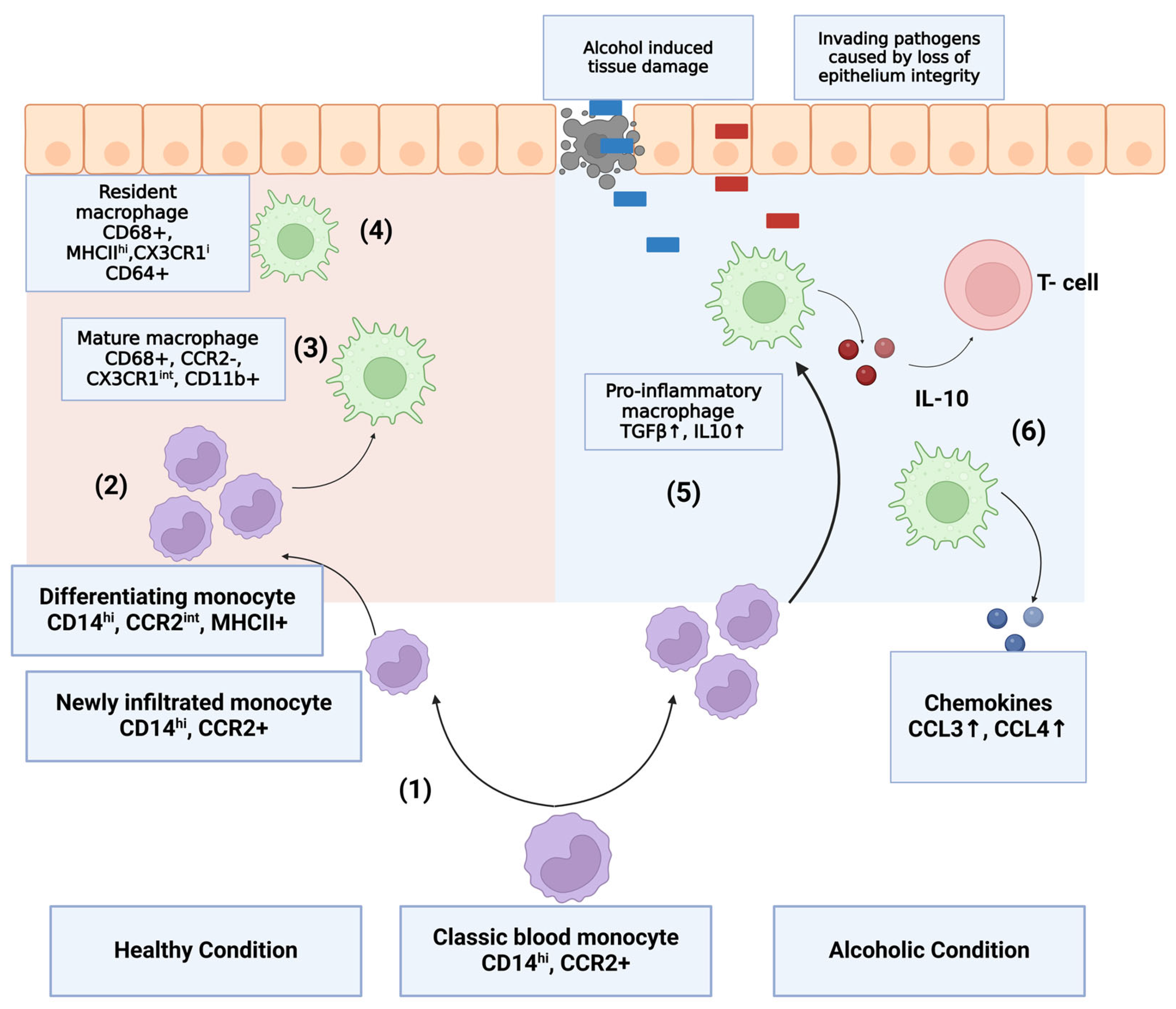
6. Monocytes and Macrophages in Alcohol Use Disorder and Alcohol-Induced Liver Disease

6.1. Implication in Intestinal and Hepatic Inflammation

Circulating monocytes are the precursors of macrophages and dendritic cells. Compared to their homeostatic counterparts, the classical CD14hi monocytes in the liver produce a wider range of pro-inflammatory cytokines and chemokines, such as TNFα, IL1β, IL-6, IL-8 MCP-1, and ROS [101,102,103,104]. By contrast, in the gut, these macrophages produce a wider range of pro-inflammatory cytokines and chemokines, like TNFα, IL-6, IL1β, IL-8, TLR-2, TLR-4, CX3CR1, and MCP-1-4 [104,105,106], upon stimulation of toll-like receptors (TLR) by their appropriate ligands, mirroring the observation in IDB studies on humans [27,84,93]. Patients with alcohol use disorder show increased monocyte numbers, especially with respect to intermediate and non-classical monocytes [104,107] (Figure 3). Serum levels of LPS are positively correlated with circulating pro-inflammatory cytokines, which further indicates the activation of monocytes occurs in a TLR-dependent manner [71]. Apart from the existing evidence of the role of TLR-4 in ALD, it has been demonstrated that TLR-2 plays an important role in monocyte activation during the inflammatory response induced by alcohol. The activation of TLR-2, which is dependent on peptidoglycan but not LPS, is associated with an exacerbation of liver damage [104]. In animal models of chronic alcohol exposure, an influx of monocytes often indicates tissue injury and subsequent disease [108]. However, in the setting of binge drinking, monocyte TLR-4 expression is rapidly downregulated, whereas systemic IL-1β levels are increased. A similar upregulation was observed after the ex vivo LPS stimulation of CD14+ monocytes obtained from control, non-alcohol-fed mice. The levels of circulating non-classical monocytes, which are considered anti-inflammatory, are also elevated. After 24 h of alcohol abstinence, the serum level of IL-1β and the number of non-classical monocytes return to their baseline levels [109]. Although CX3CR1hi-resident macrophages retain their anti-inflammatory signatures [27,97,110], which suggests that they might play a role in immunomodulation, the differentiation of CX3CR1 monocytes is delayed. This relative depletion leads to subsequent inflammation. Reduced CX3CR1 is also associated with increased gut permeability [111,112,113,114].
The CCR2-CCL2 axis also governs monocyte migration in humans. Classical monocytes express CCR2 [115], and an upregulation of its ligand, CCL2, has been found in the livers and intestines of AUD patients [104]. Furthermore, radio-labeled CD14hi monocytes have been shown to migrate to inflamed tissue of IBD mucosa [116]. In a healthy gut, resident macrophages and other lymphocytes contribute to the recruitment of monocytes by secreting ligands such as CCL2, CCL7, CCL8, etc. [117]. Nonetheless, it is important to note that CCR2 is not the only receptor, nor does it govern monocyte migration in all contexts, as the recruitment of Ly6Chi monocytes and the accumulation of its progeny are unaffected in H. hepaticus-induced colitis [118]. CCR1 and CCR5 have specialized roles in monocyte recruitment, as demonstrated by transmigration studies and in some cases of inflammation [119,120]. A recent animal study demonstrated that levels of hepatic-monocyte-derived macrophages increased in mice chronically given alcohol due to the increase in both Ly6Chi and Ly6Clo subsets, the migration of which is mediated by the CX3CR1-CX3CL1 axis [113,121].
The CX3CR1int Mϕ are localized beneath the epithelial barrier. This positioning, coupled with their high phagocytic capacity, indicates that they are designed to capture and clear invading microbes or pathogens as well as apoptotic and senescent cells. When homeostasis is perturbed by inflammation or infection caused by alcohol abuse, CD14+ monocytes and their CX3CR1int derivatives accumulate in large numbers and display enhanced pro-inflammatory characteristics. They may also be able to directly sample the luminal contents through their extending processes between the cells of the intestinal epithelium. CX3CR1int Mϕ also constitutively produce interleukin-10 (IL-10), which facilitates the secondary expansion of regulatory T cells in the mucosa and may also condition newly arrived monocytes. However, in the setting of alcohol-associated liver disease, the activation of monocytes/macrophages is changed due to a different microenvironment.

6.2. Macrophage Activation in Alcohol-Induced Liver Disease

One of the important elements of disease development and progression is the macrophage. The number of macrophages in the liver is higher during the early stage of fatty liver disease as well as in end-stage hepatitis and cirrhosis [122]. In one study, it was observed that patients with alcohol-associated steatohepatitis had hepatic macrophages expressing both anti-inflammatory and pro-inflammatory cytokines, indicating that the macrophage can adopt both phenotypes simultaneously [116]. By contrast, the number of intestinal macrophages in AUD patients is reduced [9]. Yet, the reason for the depletion of intestinal macrophages and their role in liver disease progression remain unknown.
The literature has shown that the expression levels of inflammatory genes in hepatic macrophages are upregulated [123]. Indicators of macrophage activation, such as CD58, neopterin, and leukocyte-function-associated antigen 3, as well as cytokines such as IL-6, IL-8, IL-18, and chemokines, are all elevated in ALD patients [124,125,126]. Research on a murine model has suggested that acute and chronic exposure to alcohol elevate TNF-α, IL-6, MCP1, and ROS, consistent with the activation of hepatic macrophages [99,127]. Gadolinium-chloride-induced macrophage depletion has been shown to lead to attenuated liver inflammation [128,129]. Murine KCs subjected to chronic alcohol exposure are sensitive to LPS and show an increased immune response to it [102,103]. One study also found that chronic alcohol exposure can lead to an accumulation of infiltrated monocytes in the liver [108]. Intriguingly, circulating monocytes from ALD patients produce more TNF-α and are more sensitive to LPS [127,130] compared to healthy subjects. Liver-infiltrating monocytes fall under two groups: one is composed of classical monocytes, and the other is composed of non-classical monocytes. Classical-monocyte-derived macrophages tend to exhibit a pro-inflammatory phenotype, and non-classical-monocyte-derived macrophages tend to have a pro-fibrogenic phenotype [108]. Apart from activation by macromolecules, miRNA also plays a role as a modulator of macrophage activation and has been studied extensively in recent years.

6.3. Micro RNA as a Modulator of Alcohol Response in Macrophages

miRNAs are small non-coding RNAs, mostly transcribed from DNA into primary miRNA and later processed into precursor miRNA and mature miRNA. miRNA plays an important role in the regulation of inflammatory pathways and macrophage functions in ALD [49]. Some studies have shown that a set of miRNAs can modulate the alcohol-induced hyperinflammatory response though the inhibition of TLR pathways in the liver and intestines [131,132]. Exposure to alcohol can upregulate the Kupffer-cell-derived miRNAs miR-155, miR-217, and miR132 [133,134,135]. miR-155 has an inhibitory effect on the TLR4 pathway, which in turn results in hyper-sensitivity to LPS and the upregulation of TNFα in the liver [133]. Moreover, miR-155 upregulates the release of pro-inflammatory Kupffer-cell-derived cytokines such as TNF-α and IL-1β and downregulates IL-10 in the meantime [136,137]. miR-132, on the other hand, upregulates the expression of TGF-β, IL-1β, and MCP-1 in hepatic macrophages [138]. This upregulation of pro-inflammatory cytokines shows a close tie to chronic liver disease and inflammation, as the levels of these cytokines are increased in murine models of alcohol intake. In a human study, higher levels of miR-132 were associated with fibrosis and cirrhosis [138]. Other miRNAs can also contribute to the downregulation of the monocyte/macrophage cytokine response. miR-217, miR-291b, and miR-181b-3b were shown to be responsible for the regulation of TFG-β in Kupffer cells in murine models and negatively regulate TLR signaling pathways [134,139,140]. Apart from altering cytokine and chemokine secretion, alcohol-induced miRNAs can also affect the function of monocytes/macrophages by inducing oxidative stress and phagocytosis. miR-214 expression in the liver is exacerbated by alcohol-associated oxidative stress [141]. Overexpression of miR-212 has been found in intestinal biopsies of ALD patients with intestinal barrier dysfunction and in whom ZO-1 was downregulated [142]. Upregulated miR-217 also leads to increased ROS production in KCs [134]. LPS-stimulated KCs from rats given alcohol show an increased amount of miR-125a-5p, which inhibits TLR4 signaling and leads to the polarization of macrophages to a pro-fibrogenic phenotype [143]. An early study on non-human primates showed downregulation of miR-27a in peripheral blood mononuclear cells (PBMCs), a process that drives the polarization of macrophages towards a pro-inflammatory state by secreting IL-10. This in turn indicates that alcohol can induce a phenotypic change in macrophages [144].
Under normal conditions, circulating monocytes will respond to chemokines during inflammation and subsequently penetrate tissue and differentiate into macrophages. In the steady state, their responses are tightly regulated, and these cells will further polarize into different subsets of macrophages by responding to different cytokines and miRNAs to properly attenuate the inflammation in the gut and liver. However, under pathological conditions, this delicate homeostasis is disrupted. Both monocyte-derived macrophages in the intestine as well as Kuppfer cells in the liver undergo phenotypic changes. Although all aspects of this process are not fully understood, chronic alcohol exposure seems to drive the polarization of macrophages towards a pro-inflammatory and pro-fibrotic phenotype, which ultimately contributes to tissue damage and disease progression.

7. Limitation of Animal Models in ALD Studies

Murine models have been widely used in many fields. However, murine models are suboptimal tools for ALD research due to fundamental differences between mice and humans. For instance, the basal metabolism rate per gram of body weight in mice is seven times higher than that in humans [145]. Specifically, the alcohol metabolism rate is five times higher than that in humans [146]. There is also a huge difference regarding immune cells. The blood of mice has a greater preponderance of lymphocytes, whereas human blood predominantly contains neutrophiles [147]. The human immune system is considered resistance-induction-dominant, which means it tends to directly deactivate and eliminate invading pathogens, whereas the mouse immune system tends to exhibit tolerance induction and not be responsive to a specific pathogen or set of pathogens [148]. Moreover, during alcohol-induced inflammation, the human immune system is challenged by TLR ligand/agonists such as LPS and PGN, whereas the murine immune system hardly responds to TLR ligands [149,150,151]. Additionally, mice respond to IFNγ by releasing 18 different kinds of p47 immunity-related GTPase (IRG) proteins to induce a series of cellular activities to clear out the pathogens [152]. By contrast, humans only express two kinds of IRG proteins, neither of whose activity is induced by IFNγ. One of them does not even display any antimicrobial properties apart from participating in elimination of mycobacteria by macophages [149,153]. As mentioned before, the main challenge in this regard is the lack of adequate markers with which to properly identify macrophage populations and their functionality. Many markers in mice, such as arginase 1 and Fizz1, are not found in humans, whereas human markers such as fibrinoligase and Ykl39 are not expressed in mice [154]. Additionally, cytokines such as iNOS display a striking difference. In murine macrophages, iNOS plays an important role in NO and L-citrulline synthesis [155], whereas human macrophages do not produce any iNOS, and NO is not synthesized by them [156,157]. Murine gut microbiota compositions are also different from those of humans, with humans having a higher Firmicutes-to-Bacteroidetes ratio (F/B ratio) [158]. Additionally, both humans and mice carry distinct species, which makes the composition largely different from one specimen to another [159]. Ad libitum alcohol feeding in mouse models are the most common models for the study of ALD. In one study, after eight weeks to up to 70 weeks of alcohol feeding, no significant change in mortality was observed [160]. In most cases, ab libitum feeding can induce a liver injury with elevated levels of aspartate aminotransferase and alanine aminotransferase, but no liver fibrosis would be observed [160,161], and therefore this model is unsuitable for reproducing the complexity of human disease. Although studies on murine models have given us a rough understanding of the innate immune system and macrophages in the pathogenesis of ALD, this research is hard to transpose to humans due to the different physiology and immune responses to alcohol between mice and humans. More in-depth clinical studies are needed to acquire a better understanding of ALD progression and the correlation with macrophages. Alternatively, PBMCs have emerged as a novel target for the study of pathogenesis and as a therapeutic target for alcohol-associated liver disease, specifically targeting mitochondrial oxidative function and modulating the immune response to alcohol-induced inflammation in ALD [18,19]. Liver explant studies have shown a correlation between monocyte chemotactic protein levels and MELD scores primarily in non-alcohol etiologies [162]. Nonetheless, macrophage accumulation is also observed in liver explants [163]. However, these observations in liver explants indicate end-stage liver disease, which cannot be extrapolated to the condition of chronic alcohol consumption in less advanced disease stages.

8. Therapeutic Interventions and Future Research

In ALD, the current standard treatment consists of alcohol abstinence and the use of medications such as disulfiram, naltrexone [164,165], and acamprosate [166] to eventually prevent relapses. It is not uncommon to observe a disrupted microbiota and increased intestinal permeability in ALD patients [9]. There are treatments available that can restore eubiosis in the gut, such as dietary regimens, probiotics, and prebiotics. Some clinical studies have shown that in ALD patients, probiotic treatment reduces the levels of aspartate aminotransferase, alanine transferase, lipopolysaccharides, and TNFα [167]. However, these treatments have not been proven to have long-term benefits. Apart from medical treatment, fecal microbiome transplantation has effectively been used to decrease gut dysbiosis and reduce gut permeability in animal models [166]. Also, in recent years, clinical trials have suggested that a fecal microbiome transplant can improve gut immunity and alleviate gut dysbiosis in humans (see https://clinicaltrials.gov/ct2/show/NCT02862249 accessed on 30 September 2019, https://clinicaltrials.gov/ct2/show/NCT02400216, and https://clinicaltrials.gov/ct2/show/NCT02496390 accessed on 11 December 2018). Although the gut microbiota might be a promising therapeutic and diagnostic target in AUD and ALD, convincing evidence of a benefit for humans is still lacking. According to a recent human study, an upregulation of TLR-2 signaling in circulating monocytes and liver-infiltrating monocytes is associated with inflammation in AUD and ALD. Hence, inhibiting TLR-2 could be a therapeutic target for reducing systemic inflammation and mitigating liver damage in ALD. In addition, the IL-1β- and IL-8-driven inflammatory response in monocytes is related to the activation of the NLRP-3 inflammasome, indicating that the inflammasome is another potential therapeutic target [104]. It is also conceivable that inflammation and monocyte recruitment could be mitigated by using chemokines inhibitors, as suggested in a recent human study targeting myometrial myocyte–macrophage communication [160]. In addition, targeting CSF1 has already been applied in cancer therapy [168]. It might be a potential alternative approach to, for example, alleviating liver inflammation in ALD. Macrophage-targeted therapy is often used in many cancer therapies. In recent studies, it has been pointed out that switching hepatic macrophages from a pro-inflammatory to pro-fibrogenic phenotype can alleviate inflammation and promote tissue repair in individuals with non-alcoholic fatty liver disease (NAFLD). This phenomenon is mainly driven by the upregulation of anti-inflammatory cytokines related to tissue repair and regeneration [169,170,171]. Currently, no such investigation has been conducted in relation to ALD. Even though we have gained some insight into the relationship between alcohol and the gut microbiota, many questions remain open. A better understanding of how alcohol affects bacterial communities, bacterial translocation into blood or the lymphatic system, the stimulation of the vagus nerve, the endocrine system, and alterations of immune systems will allow us to fill in the missing pieces in the pathophysiology of AUD and its remote organ damage such as in the liver. Ongoing debate about the potential role of tissue-specific macrophages is raising controversies in the field of immunology. Recently, demographic data regarding adipose tissue macrophages were investigated in an NAFLD study, demonstrating transcriptomic differences between adipose-resident vascular macrophages and metabolically active macrophages. In animal research, a series of studies have investigated the subsets of macrophages and their roles regarding spatial distribution. Additionally, resident macrophages are not exclusively embryonically derived: they can also be replenished by circulating monocytes [166]. However, little is known about the subsets of macrophages in the intestines and liver and their metabolic and functional differences in terms of spatial distribution in humans. This gap needs to be filled by more in-depth studies on humans, given the fundamental difference between humans and animal models in alcohol research.
Despite our growing understanding of how macrophages are an important member of the mononuclear phagocyte system and the innate immune system, little is known about the phenotypes and the different functions of residential macrophages and monocyte-derived macrophages. Furthermore, better markers need to be identified in order to gain a better understanding of the functions and phenotypes of different subsets of intestinal and hepatic macrophages. More studies about human tissue macrophages should be carried out using cutting-edge technology such as scRNA sequencing and omic technology. Additionally, since findings obtained using animal models are difficult to translate into clinical implications, better in vitro or ex vivo systems need to be developed, such as organoids, to study the pathogenesis of alcohol-associated liver disease and the effect of alcohol on the intestines.

Author Contributions

Y.H. is responsible for writing this manuscript. P.S. and B.S. provided scientific input and proofread and edited the manuscript. All authors have read and agreed to the published version of the manuscript.

Funding

This study was supported by services provided by NIH centers P30 DK120515 and P50 AA011999. P.S. received grant support from Fond National de Recherche Scientifique Belgium (J.0146.17 and T.0217.18) and Action de Recherche Concertée (ARC), Université Catholique de Louvain, Belgium.

Institutional Review Board Statement

Not applicable.

Informed Consent Statement

Not applicable.

Data Availability Statement

Not applicable.

Acknowledgments

We would like to thank Sebastian Joseph for proofreading the article.

Conflicts of Interest

B.S. consults for Ambys Medicines, Boehringer Ingelheim Pharma, FerringResearch Institute, Gelesis, HOST Therabiomics, Intercept Pharmaceuticals, Mabwell Therapeutics, Patara Pharmaceuticals, Surrozen, and Takeda. B.S. is the founder of Nterica Bio. UC San Diego has filed several patents with B.S. as an inventor in relation to this work. B.S.’s institution, UC San Diego, has received research support from Axial Biotherapeutics, BiomX, ChromoLogic, CymaBay Therapeutics, Intercept Pharmaceuticals, NGM Biopharmaceuticals, Prodigy Biotech, and Synlogic Operating Company.

References

  1. Wang, J.; Jiang, L.; Sun, H. Early evidence for beer drinking in a 9000-year-old platform mound in southern China. PLoS ONE 2021, 16, e0255833. [Google Scholar] [CrossRef]
  2. Joffe, A.H. Alcohol and Social Complexity in Ancient Western Asia. Curr. Anthropol. 1998, 39, 297–322. [Google Scholar] [CrossRef]
  3. Khaderi, S.A. Introduction: Alcohol and Alcoholism. Clin. Liver Dis. 2019, 23, 1–10. [Google Scholar] [CrossRef]
  4. The Alcohol Industry in Data. Available online: https://alcohol.org/guides/the-alcohol-industry-in-data/ (accessed on 9 March 2023).
  5. Bosron, W.F.; Crabb, D.W.; Ting-Kai, L.i. Relationship between kinetics of liver alcohol dehydrogenase and alcohol metabolism. Pharmacol. Biochem. Behav. 1983, 18 (Suppl. 1), 223–227. [Google Scholar] [CrossRef]
  6. Hingson, R.W.; Heeren, T.; Winter, M.R. Age at drinking onset and alcohol dependence: Age at onset, duration, and severity. Arch. Pediatr. Adolesc. Med. 2006, 160, 739–746. [Google Scholar] [CrossRef]
  7. Chrostek, L.; Panasiuk, A. Liver fibrosis markers in alcoholic liver disease. World J. Gastroenterol. 2014, 20, 8018. [Google Scholar] [CrossRef]
  8. Agarwal, K.; Kontorinis, N.; Dieterich, D.T. Alcoholic Hepatitis. Curr. Treat. Options Gastroenterol. 2023, 7, 451–458. [Google Scholar] [CrossRef]
  9. Maccioni, L.; Gao, B.; Leclercq, S.; Pirlot, B.; Horsmans, Y.; De Timary, P.; Leclercq, I.; Fouts, D.; Schnabl, B.; Stärkel, P. Intestinal permeability, microbial translocation, changes in duodenal and fecal microbiota, and their associations with alcoholic liver disease progression in humans. Gut Microbes 2020, 12, 1782157. [Google Scholar] [CrossRef]
  10. Van Furth, R.; Cohn, Z.A.; Hirsch, J.G.; Humphrey, J.H.; Spector, W.G.; Langevoort, H.L. The mononuclear phagocyte system: A new classification of macrophages, monocytes, and their precursor cells. Bull. World Health Organ. 1972, 46, 845. [Google Scholar]
  11. Van Furth, R.; Cohn, Z.A. The Origin and Kinetics of Mononuclear Phagocytes. J. Exp. Med. 1968, 128, 415–435. [Google Scholar] [CrossRef]
  12. Kuziel, W.A.; Morgan, S.J.; Dawson, T.C.; Griffin, S.; Smithies, O.; Ley, K.; Maeda, N. Severe reduction in leukocyte adhesion and monocyte extravasation in mice deficient in CC chemokine receptor 2. Proc. Natl. Acad. Sci. USA 1997, 94, 12053–12058. [Google Scholar] [CrossRef]
  13. Takahashi, K. Development and Differentiation of Macrophages and Related Cells: Historical Review and Current Concepts. J. Clin. Exp. Hematop. 2001, 41, 1–31. [Google Scholar] [CrossRef]
  14. Ginhoux, F.; Jung, S. Monocytes and macrophages: Developmental pathways and tissue homeostasis. Nat. Rev. Immunol. 2014, 14, 392–404. [Google Scholar] [CrossRef]
  15. Ginhoux, F.; Guilliams, M. Tissue-Resident Macrophage Ontogeny and Homeostasis. Immunity 2016, 44, 439–449. [Google Scholar] [CrossRef]
  16. Hattori, Y. The microglia-blood vessel interactions in the developing brain. Neurosci. Res. 2023, 187, 58–66. [Google Scholar] [CrossRef]
  17. Gomez Perdiguero, E.; Klapproth, K.; Schulz, C.; Busch, K.; Azzoni, E.; Crozet, L.; Garner, H.; Trouillet, C.; de Bruijn, M.F.; Geissmann, F.; et al. Tissue-resident macrophages originate from yolk-sac-derived erythro-myeloid progenitors. Nature 2014, 518, 547–551. [Google Scholar] [CrossRef]
  18. Kang, B.; Alvarado, L.J.; Kim, T.; Lehmann, M.; Cho, H.; He, J.; Li, P.; Larochelle, A.; Kelsall, B.L. Commensal microbiota drive the functional diversification of colon macrophages. Mucosal Immunol. 2020, 13, 216. [Google Scholar] [CrossRef]
  19. Zhang, H.; Wang, X.; Zhang, J.; He, Y.; Yang, X.; Nie, Y.; Sun, L. Crosstalk between gut microbiota and gut resident macrophages in inflammatory bowel disease. J. Transl. Intern. Med. 2023, 11, 382. [Google Scholar] [CrossRef]
  20. Chen, Q.; Nair, S.; Ruedl, C. Microbiota regulates the turnover kinetics of gut macrophages in health and inflammation. Life Sci. Alliance 2022, 5, e202101178. [Google Scholar] [CrossRef]
  21. Scott, N.A.; Andrusaite, A.; Andersen, P.; Lawson, M.; Alcon-Giner, C.; Leclaire, C.; Caim, S.; Le Gall, G.; Shaw, T.; Connolly, J.P.R.; et al. Antibiotics induce sustained dysregulation of intestinal T cell immunity by perturbing macrophage homeostasis. Sci. Transl. Med. 2018, 10, eaao4755. [Google Scholar] [CrossRef]
  22. Bander ZAl Nitert, M.D.; Mousa, A.; Naderpoor, N. The Gut Microbiota and Inflammation: An Overview. Int. J. Environ. Res. Public Health 2020, 17, 7618. [Google Scholar] [CrossRef]
  23. Zhou, Z.; Pan, X.; Li, L. Crosstalk between liver macrophages and gut microbiota: An important component of inflammation-associated liver diseases. Front. Cell Dev. Biol. 2022, 10, 1070208. [Google Scholar] [CrossRef]
  24. Schridde, A.; Bain, C.C.; Mayer, J.U.; Montgomery, J.; Pollet, E.; Denecke, B.; Milling, S.W.F.; Jenkins, S.J.; Dalod, M.; Henri, S.; et al. Tissue-specific differentiation of colonic macrophages requires TGFβ receptor-mediated signaling. Mucosal Immunol. 2017, 10, 1387–1399. [Google Scholar] [CrossRef]
  25. Kumawat, A.K.; Yu, C.; Mann, E.A.; Schridde, A.; Finnemann, S.C.; Mowat, A.M.I. Expression and characterization of αvβ5 integrin on intestinal macrophages. Eur. J. Immunol. 2018, 48, 1181–1187. [Google Scholar] [CrossRef]
  26. Bujko, A.; Atlasy, N.; Landsverk, O.J.B.; Richter, L.; Yaqub, S.; Horneland, R.; Øyen, O.; Aandahl, E.M.; Aabakken, L.; Stunnenberg, H.G.; et al. Transcriptional and functional profiling defines human small intestinal macrophage subsets. J. Exp. Med. 2018, 215, 441–458. [Google Scholar] [CrossRef]
  27. Bain, C.C.; Scott, C.L.; Uronen-Hansson, H.; Gudjonsson, S.; Jansson, O.; Grip, O.; Guilliams, M.; Malissen, B.; Agace, W.W.; Mowat, A.M. Resident and pro-inflammatory macrophages in the colon represent alternative context-dependent fates of the same Ly6Chi monocyte precursors. Mucosal Immunol. 2013, 6, 498–510. [Google Scholar] [CrossRef]
  28. Branchett, W.J.; Saraiva, M.; O’Garra, A. Regulation of inflammation by Interleukin-10 in the intestinal and respiratory mucosa. Curr. Opin. Immunol. 2024, 91, 102495. [Google Scholar] [CrossRef]
  29. Niess, J.H.; Brand, S.; Gu, X.; Landsman, L.; Jung, S.; McCormick, B.A.; Vyas, J.M.; Boes, M.; Ploegh, H.L.; Fox, J.G.; et al. CX3CR1-mediated dendritic cell access to the intestinal lumen and bacterial clearance. Science 2005, 307, 254–258. [Google Scholar] [CrossRef]
  30. Chieppa, M.; Rescigno, M.; Huang, A.Y.C.; Germain, R.N. Dynamic imaging of dendritic cell extension into the small bowel lumen in response to epithelial cell TLR engagement. J. Exp. Med. 2006, 203, 2841–2852. [Google Scholar] [CrossRef]
  31. Kim, K.W.; Vallon-Eberhard, A.; Zigmond, E.; Farache, J.; Shezen, E.; Shakhar, G.; Ludwig, A.; Lira, S.A.; Jung, S. In vivo structure/function and expression analysis of the CX3C chemokine fractalkine. Blood 2011, 118, e156–e167. [Google Scholar] [CrossRef]
  32. Ulevitch, R.J.; Tobias, P.S. Receptor-dependent mechanisms of cell stimulation by bacterial endotoxin. Annu. Rev. Immunol. 1995, 13, 437–457. [Google Scholar] [CrossRef]
  33. Mazzini, E.; Massimiliano, L.; Penna, G.; Rescigno, M. Oral Tolerance Can Be Established via Gap Junction Transfer of Fed Antigens from CX3CR1+ Macrophages to CD103+ Dendritic Cells. Immunity 2014, 40, 248–261. [Google Scholar] [CrossRef]
  34. He, J.; Song, Y.; Li, G.; Xiao, P.; Liu, Y.; Xue, Y.; Cao, Q.; Tu, X.; Pan, T.; Jiang, Z.; et al. Fbxw7 increases CCL2/7 in CX3CR1hi macrophages to promote intestinal inflammation. J. Clin. Investig. 2019, 129, 3877. [Google Scholar] [CrossRef]
  35. Asano, K.; Takahashi, N.; Ushiki, M.; Monya, M.; Aihara, F.; Kuboki, E.; Moriyama, S.; Iida, M.; Kitamura, H.; Qiu, C.; et al. Intestinal CD169+ macrophages initiate mucosal inflammation by secreting CCL8 that recruits inflammatory monocytes. Nat. Commun. 2015, 6, 7802. [Google Scholar] [CrossRef]
  36. Viola, M.F.; Boeckxstaens, G. Niche-specific functional heterogeneity of intestinal resident macrophages. Gut 2021, 70, 1383. [Google Scholar] [CrossRef]
  37. Lewis, S.A.; Sureshchandra, S.; Doratt, B.; Jimenez, V.A.; Stull, C.; Grant, K.A.; Messaoudi, I. Transcriptional, Epigenetic, and Functional Reprogramming of Monocytes from Non-Human Primates Following Chronic Alcohol Drinking. Front. Immunol. 2021, 12, 724015. [Google Scholar] [CrossRef]
  38. Maccioni, L.; Gao, B.; Leclercq, S.; Stärkel, P. Host Factors in Dysregulation of the Gut Barrier Function during Alcohol-Associated Liver Diseasent. J. Mol. Sci. 2021, 22, 12687. [Google Scholar] [CrossRef]
  39. Purohit, V.; Bode, J.C.; Bode, C.; Brenner, D.A.; Choudhry, M.A.; Hamilton, F.; Kang, Y.J.; Keshavarzian, A.; Rao, R.; Sartor, R.B.; et al. Alcohol, Intestinal Bacterial Growth, Intestinal Permeability to Endotoxin, and Medical Consequences: Summary of a Symposium. Alcohol 2008, 42, 349. [Google Scholar] [CrossRef]
  40. Tuomisto, S.; Pessi, T.; Collin, P.; Vuento, R.; Aittoniemi, J.; Karhunen, P.J. Changes in gut bacterial populations and their translocation into liver and ascites in alcoholic liver cirrhotics. BMC Gastroenterol. 2014, 14, 1–8. [Google Scholar] [CrossRef]
  41. Cosovanu, C.; Neumann, C. The Many Functions of Foxp3+ Regulatory T Cells in the Intestine. Front. Immunol. 2020, 11, 600973. [Google Scholar] [CrossRef]
  42. Worbs, T.; Bode, U.; Yan, S.; Hoffmann, M.W.; Hintzen, G.; Bernhardt, G.; Förster, R.; Pabst, O. Oral tolerance originates in the intestinal immune system and relies on antigen carriage by dendritic cells. J. Exp. Med. 2006, 203, 519–527. [Google Scholar] [CrossRef]
  43. Hadis, U.; Wahl, B.; Schulz, O.; Hardtke-Wolenski, M.; Schippers, A.; Wagner, N.; Müller, W.; Sparwasser, T.; Förster, R.; Pabst, O. Intestinal Tolerance Requires Gut Homing and Expansion of FoxP3+ Regulatory T Cells in the Lamina Propria. Immunity 2011, 34, 237–246. [Google Scholar] [CrossRef]
  44. Kim, M.; Galan, C.; Hill, A.A.; Wu, W.J.; Fehlner-Peach, H.; Song, H.W.; Schady, D.; Bettini, M.L.; Simpson, K.W.; Longman, R.S.; et al. Critical Role for the Microbiota in CX3CR1+ Intestinal Mononuclear Phagocyte Regulation of Intestinal T Cell Responses. Immunity 2018, 49, 151–163. [Google Scholar] [CrossRef]
  45. Zigmond, E.; Bernshtein, B.; Friedlander, G.; Walker, C.R.; Yona, S.; Kim, K.W.; Brenner, O.; Krauthgamer, R.; Varol, C.; Müller, W.; et al. Macrophage-Restricted Interleukin-10 Receptor Deficiency, but Not IL-10 Deficiency, Causes Severe Spontaneous Colitis. Immunity 2014, 40, 720–733. [Google Scholar] [CrossRef]
  46. Shaw, M.H.; Kamada, N.; Kim, Y.G.; Núñez, G. Microbiota-induced IL-1β, but not IL-6, is critical for the development of steady-state TH17 cells in the intestine. J. Exp. Med. 2012, 209, 251–258. [Google Scholar] [CrossRef]
  47. Panea, C.; Farkas, A.M.; Goto, Y.; Abdollahi-Roodsaz, S.; Lee, C.; Koscsó, B.; Gowda, K.; Hohl T., M.; Bogunovic, M.; Ivanov, I.I. Intestinal Monocyte-Derived Macrophages Control Commensal-Specific Th17 Responses. Cell Rep. 2015, 12, 1314–1324. [Google Scholar] [CrossRef]
  48. Schulz, O.; Jaensson, E.; Persson, E.K.; Liu, X.; Worbs, T.; Agace, W.W.; Pabst, O. Intestinal CD103+, but not CX3CR1+, antigen sampling cells migrate in lymph and serve classical dendritic cell functions. J. Exp. Med. 2009, 206, 3101–3114. [Google Scholar] [CrossRef]
  49. Gao, B.; Ahmad, M.F.; Nagy, L.E.; Tsukamoto, H. Inflammatory pathways in alcoholic steatohepatitis. J. Hepatol. 2019, 70, 249–259. [Google Scholar] [CrossRef]
  50. Mikkelsen, H.B.; Rumessen, J.J. Characterization of macrophage-like cells in the external layers of human small and large intestine. Cell Tissue Res. 1992, 270, 273–279. [Google Scholar] [CrossRef]
  51. De Schepper, S.; Verheijden, S.; Aguilera-Lizarraga, J.; Viola, M.F.; Boesmans, W.; Stakenborg, N.; Voytyuk, I.; Schmidt, I.; Boeckx, B.; Dierckx de Casterlé, I.; et al. Self-maintaining gut macrophages are essential for intestinal homeostasis. Cell 2018, 175 (Suppl. 1), 400–415.e13. [Google Scholar] [CrossRef]
  52. Gabanyi, I.; Muller, P.A.; Feighery, L.; Oliveira, T.Y.; Costa-Pinto, F.A.; Mucida, D. Neuro-immune interactions drive tissue programming in intestinal macrophages. Cell 2016, 164, 378–391. [Google Scholar] [CrossRef]
  53. Muller, P.A.; Koscsó, B.; Rajani, G.M.; Viola, M.F.; Boesmans, W.; Stakenborg, N.; Voytyuk, I.; Schmidt, I.; Boeckx, B.; Dierckx de Casterlé, I.; et al. Crosstalk between muscularis macrophages and enteric neurons regulates gastrointestinal motility. Cell 2014, 158, 300–313. [Google Scholar] [CrossRef]
  54. Blériot, C.; Ginhoux, F. Understanding the Heterogeneity of Resident Liver Macrophages. Front. Immunol. 2019, 10, 2694. [Google Scholar] [CrossRef]
  55. Ishibashi, H.; Nakamura, M.; Komori, A.; Migita, K.; Shimoda, S. Liver architecture, cell function, and disease. Semin. Immunopathol. 2009, 31, 399–409. [Google Scholar] [CrossRef]
  56. Scott, C.L.; Zheng, F.; De Baetselier, P.; Martens, L.; Saeys, Y.; De Prijck, S.; Lippens, S.; Abels, C.; Schoonooghe, S.; Raes, G.; et al. Bone marrow-derived monocytes give rise to self-renewing and fully differentiated Kupffer cells. Nat. Commun. 2016, 7, 10321. [Google Scholar] [CrossRef]
  57. Yona, S.; Kim, K.W.; Wolf, Y.; Mildner, A.; Varol, D.; Breker, M.; Strauss-Ayali, D.; Viukov, S.; Guilliams, M.; Misharin, A.; et al. Fate Mapping Reveals Origins and of Monocytes and Tissue Macrophages under Homeostasis. Immunity 2013, 38, 79–91. [Google Scholar] [CrossRef]
  58. Schulz, C.; Perdiguero, E.G.; Chorro, L.; Szabo-Rogers, H.; Cagnard, N.; Kierdorf, K.; Prinz, M.; Wu, B.; Jacobsen, S.E.; Pollard, J.W.; et al. A lineage of myeloid cells independent of myb and hematopoietic stem cells. Science 2012, 335, 86–90. [Google Scholar] [CrossRef]
  59. David, B.A.; Rezende, R.M.; Antunes, M.M.; Santos, M.M.; Freitas Lopes, M.A.; Diniz, A.B.; Sousa Pereira, R.V.; Marchesi, S.C.; Alvarenga, D.M.; Nakagaki, B.N.; et al. Combination of Mass Cytometry and Imaging Analysis Reveals Origin, Location, and Functional Repopulation of Liver Myeloid Cells in Mice. Gastroenterology 2016, 151, 1176–1191. [Google Scholar] [CrossRef]
  60. Sierro, F.; Evrard, M.; Rizzetto, S.; Melino, M.; Mitchell, A.J.; Florido, M.; Beattie, L.; Walters, S.B.; Tay, S.S.; Lu, B.; et al. A Liver Capsular Network of Monocyte-Derived Macrophages Restricts Hepatic Dissemination of Intraperitoneal Bacteria by Neutrophil Recruitment. Immunity 2017, 47, 374–388. [Google Scholar] [CrossRef]
  61. Nolan, J.P. The role of intestinal endotoxin in liver injury: A long and evolving history. Hepatology 2010, 52, 1829–1835. [Google Scholar] [CrossRef]
  62. Soares, J.B.; Pimentel-Nunes, P.; Roncon-Albuquerque, R.; Leite-Moreira, A. The role of lipopolysaccharide/toll-like receptor 4 signaling in chronic liver diseases. Hepatol. Int. 2010, 4, 659. [Google Scholar] [CrossRef]
  63. Kawaratani, H.; Tsujimoto, T.; Douhara, A.; Takaya, H.; Moriya, K.; Namisaki, T.; Noguchi, R.; Yoshiji, H.; Fujimoto, M.; Fukui, H. The effect of inflammatory cytokines in alcoholic liver disease. Mediat. Inflamm. 2013, 2013, 495156. [Google Scholar] [CrossRef]
  64. Slevin, E.; Baiocchi, L.; Wu, N.; Ekser, B.; Sato, K.; Lin, E.; Ceci, L.; Chen, L.; Lorenzo, S.R.; Xu, W.; et al. Kupffer Cells: Inflammation Pathways and Cell-Cell Interactions in Alcohol-Associated Liver Disease. Am. J. Pathol. 2020, 190, 2185–2193. [Google Scholar] [CrossRef]
  65. Singh, V.; Huang, E.; Pathak, V.; Willard, B.B.; Allende, D.S.; Nagy, L.E. Phosphoproteomics identifies pathways underlying the role of receptor-interaction protein kinase 3 in alcohol-associated liver disease and uncovers apoptosis signal-regulating kinase 1 as a target. Hepatol. Commun. 2022, 6, 2022–2041. [Google Scholar] [CrossRef]
  66. Roychowdhury, S.; McMullen, M.R.; Pisano, S.G.; Liu, X.; Nagy, L.E. Absence of receptor-interacting protein kinase 3 prevents ethanol-induced liver injury. Hepatology 2013, 57, 1773. [Google Scholar] [CrossRef]
  67. Wu, X.; Fan, X.; McMullen, M.R.; Miyata, T.; Kim, A.; Pathak, V.; Wu, J.; Day, L.Z.; Hardesty, J.E.; Welch, N.; et al. Macrophage-derived MLKL in alcohol-associated liver disease: Regulation of phagocytosis. Hepatology 2023, 77, 902. [Google Scholar] [CrossRef]
  68. Duan, Y.; Chu, H.; Brandl, K.; Jiang, L.; Zeng, S.; Meshgin, N.; Papachristoforou, E.; Argemi, J.; Mendes, B.G.; Wang, Y.; et al. CRIg on liver macrophages clears pathobionts and protects against alcoholic liver disease. Nat. Commun. 2021, 12, 7172. [Google Scholar] [CrossRef]
  69. Luo, Z.; Ji, Y.; Gao, H.; Gomes Dos Reis, F.C.; Bandyopadhyay, G.; Jin, Z.; Ly, C.; Chang, Y.J.; Zhang, D.; Kumar, D.; et al. CRIg+ Macrophages Prevent Gut Microbial DNA-Containing Extracellular Vesicle-Induced Tissue Inflammation and Insulin Resistance. Gastroenterology 2021, 160, 863–874. [Google Scholar] [CrossRef]
  70. Enomoto, N.; Ikejima, K.; Bradford, B.; Rivera, C.; Kono, H.; Brenner, D.A.; Thurman, R.G. Alcohol causes both tolerance and sensitization of rat Kupffer cells via mechanisms dependent on endotoxin. Gastroenterology 1998, 115, 443–451. [Google Scholar] [CrossRef]
  71. Liangpunsakul, S.; Toh, E.; Ross, R.A.; Heathers, L.E.; Chandler, K.; Oshodi, A.; McGee, B.; Modlik, E.; Linton, T.; Mangiacarne, D.; et al. Quantity of alcohol drinking positively correlates with serum levels of endotoxin and markers of monocyte activation. Sci. Rep. 2017, 7, 4462. [Google Scholar] [CrossRef]
  72. Mandrekar, P.; Bala, S.; Catalano, D.; Kodys, K.; Szabo, G. The Opposite Effects of Acute and Chronic Alcohol on Lipopolysaccharide-Induced Inflammation Are Linked to IRAK-M in Human Monocytes. J. Immunol. 2009, 183, 1320–1327. [Google Scholar] [CrossRef]
  73. Bala, S.; Tang, A.; Catalano, D.; Petrasek, J.; Taha, O.; Kodys, K.; Szabo, G. Induction of Bcl-3 by acute binge alcohol results in Toll-like receptor 4/LPS tolerance. J. Leukoc. Biol. 2012, 92, 611–620. [Google Scholar] [CrossRef]
  74. Ju, C.; Mandrekar, P. Macrophages and Alcohol-Related Liver Inflammation. Alcohol. Res. 2015, 37, 251. Available online: https://pubmed.ncbi.nlm.nih.gov/26717583/ (accessed on 3 April 2024).
  75. Saha, B.; Momen-Heravi, F.; Kodys, K.; Szabo, G. MicroRNA Cargo of Extracellular Vesicles from Alcohol-exposed Monocytes Signals Naive Monocytes to Differentiate into M2 Macrophages. J. Biol. Chem. 2016, 291, 149–159. [Google Scholar] [CrossRef]
  76. Szabo, G.; Petrasek, J. Inflammasome activation and function in liver disease. Nat. Rev. Gastroenterol. Hepatol. 2015, 12, 387–400. [Google Scholar] [CrossRef]
  77. Hume, D.A.; Perry, V.H.; Gordon, S. The mononuclear phagocyte system of the mouse defined by immunohistochemical localisation of antigen F4/80: Macrophages associated with epithelia. Anat. Rec. 1984, 210, 503–512. [Google Scholar] [CrossRef]
  78. McGarry, M.P.; Stewart, C.C. Murine eosinophil granulocytes bind the murine macrophage-monocyte specific monoclonal antibody F4/80. J. Leukoc. Biol. 1991, 50, 471–478. [Google Scholar] [CrossRef]
  79. Gottfried, E.; Kunz-Schughart, L.A.; Weber, A.; Rehli, M.; Peuker, A.; Müller, A.; Kastenberger, M.; Brockhoff, G.; Andreesen, R.; Kreutz, M. Expression of CD68 in Non-Myeloid Cell Types. Scand. J. Immunol. 2008, 67, 453–463. [Google Scholar] [CrossRef]
  80. Itoh, M.; Suganami, T.; Kato, H.; Kanai, S.; Shirakawa, I.; Sakai, T.; Goto, T.; Asakawa, M.; Hidaka, I.; Sakugawa, H.; et al. CD11c+ resident macrophages drive hepatocyte death-triggered liver fibrosis in a murine model of nonalcoholic steatohepatitis. JCI Insight 2017, 2, e92902. [Google Scholar] [CrossRef]
  81. Ju, C.; Tacke, F. Hepatic macrophages in homeostasis and liver diseases: From pathogenesis to novel therapeutic strategies. Cell. Mol. Immunol. 2016, 13, 316–327. [Google Scholar] [CrossRef]
  82. Pabst, O.; Bernhardt, G. The puzzle of intestinal lamina propria dendritic cells and macrophages. Eur. J. Immunol. 2010, 40, 2107–2111. [Google Scholar] [CrossRef]
  83. Gautiar, E.L.; Shay, T.; Miller, J.; Kanai, S.; Shirakawa, I.; Sakai, T.; Goto, T.; Asakawa, M.; Hidaka, I.; Sakugawa, H.; et al. Gene-expression profiles and transcriptional regulatory pathways that underlie the identity and diversity of mouse tissue macrophages. Nat. Immunol. 2012, 13, 1118–1128. [Google Scholar] [CrossRef]
  84. Tamoutounour, S.; Henri, S.; Lelouard, H.; de Bovis, B.; de Haar, C.; van der Woude, C.J.; Woltman, A.M.; Reyal, Y.; Bonnet, D.; Sichien, D.; et al. CD64 distinguishes macrophages from dendritic cells in the gut and reveals the Th1-inducing role of mesenteric lymph node macrophages during colitis. Eur. J. Immunol. 2012, 42, 3150–3166. [Google Scholar] [CrossRef]
  85. Langlet, C.; Tamoutounour, S.; Henri, S.; de Bovis, B.; de Haar, C.; van der Woude, C.J.; Woltman, A.M.; Reyal, Y.; Bonnet, D.; Sichien, D.; et al. CD64 Expression Distinguishes Monocyte-Derived and Conventional Dendritic Cells and Reveals Their Distinct Role during Intramuscular Immunization. J. Immunol. 2012, 188, 1751–1760. [Google Scholar] [CrossRef]
  86. González-Domínguez érika Samaniego, R.; Flores-Sevilla, J.L.; Campos-Campos, S.F.; Gómez-Campos, G.; Salas, A.; Campos-Peña, V.; Corbí, Á.L.; Sánchez-Mateos, P.; Sánchez-Torres, C. CD163L1 and CLEC5A discriminate subsets of human resident and inflammatory macrophages in vivo. J. Leukoc. Biol. 2015, 98, 453–466. [Google Scholar] [CrossRef]
  87. Scott, C.L.; Bain, C.C.; Wright, P.B.; Sichien, D.; Kotarsky, K.; Persson, E.K.; Luda, K.; Guilliams, M.; Lambrecht, B.N.; Agace, W.W.; et al. CCR2+CD103− intestinal dendritic cells develop from DC-committed precursors and induce interleukin-17 production by T cells. Mucosal Immunol. 2015, 8, 327–339. [Google Scholar] [CrossRef]
  88. Schlitzer, A.; McGovern, N.; Teo, P.; Zelante, T.; Atarashi, K.; Low, D.; Ho, A.W.; See, P.; Shin, A.; Wasan, P.S.; et al. IRF4 Transcription Factor-Dependent CD11b+ Dendritic Cells in Human and Mouse Control Mucosal IL-17 Cytokine Responses. Immunity 2013, 38, 970–983. [Google Scholar] [CrossRef]
  89. Bohaud, C.; Johansen, M.D.; Jorgensen, C.; Kremer, L.; Ipseiz, N.; Djouad, F. The Role of Macrophages During Mammalian Tissue Remodeling and Regeneration Under Infectious and Non-Infectious Conditions. Front. Immunol. 2021, 12, 707856. [Google Scholar] [CrossRef]
  90. Cerovic, V.; Houston, S.A.; Scott, C.L.; Aumeunier, A.; Yrlid, U.; Mowat, A.M.; Milling, S.W. Intestinal CD103− dendritic cells migrate in lymph and prime effector T cells. Mucosal Immunol. 2013, 6, 104–113. [Google Scholar] [CrossRef]
  91. Coombes, J.L.; Siddiqui, K.R.R.; Arancibia-Cárcamo, C.V.; Hall, J.; Sun, C.M.; Belkaid, Y.; Powrie, F. A functionally specialized population of mucosal CD103+ DCs induces Foxp3+ regulatory T cells via a TGF-β– and retinoic acid–dependent mechanism. J. Exp. Med. 2007, 204, 1757–1764. [Google Scholar] [CrossRef]
  92. Bain, C.C.; Bravo-Blas, A.; Scott, C.L.; Perdiguero, E.G.; Geissmann, F.; Henri, S.; Malissen, B.; Osborne, L.C.; Artis, D.; Mowat, A.M. Constant replenishment from circulating monocytes maintains the macrophage pool in the intestine of adult mice. Nat. Immunol. 2014, 15, 929–937. [Google Scholar] [CrossRef]
  93. Varol, C.; Vallon-Eberhard, A.; Elinav, E.; Aychek, T.; Shapira, Y.; Luche, H.; Fehling, H.J.; Hardt, W.D.; Shakhar, G.; Jung, S. Intestinal Lamina Propria Dendritic Cell Subsets Have Different Origin and Functions. Immunity 2009, 31, 502–512. [Google Scholar] [CrossRef]
  94. Bogunovic, M.; Ginhoux, F.; Helft, J.; Shang, L.; Hashimoto, D.; Greter, M.; Liu, K.; Jakubzick, C.; Ingersoll, M.A.; Leboeuf, M.; et al. Origin of the Lamina Propria Dendritic Cell Network. Immunity 2009, 31, 513–525. [Google Scholar] [CrossRef]
  95. Kamada, N.; Hisamatsu, T.; Okamoto, S.; Chinen, H.; Kobayashi, T.; Sato, T.; Sakuraba, A.; Kitazume, M.T.; Sugita, A.; Koganei, K.; et al. Unique CD14+ intestinal macrophages contribute to the pathogenesis of Crohn disease via IL-23/IFN-γ axis. J. Clin. Investig. 2008, 118, 2269–2280. [Google Scholar] [CrossRef]
  96. Zigmond, E.; Varol, C.; Farache, J.; Elmaliah, E.; Satpathy, A.T.; Friedlander, G.; Mack, M.; Shpigel, N.; Boneca, I.G.; Murphy, K.M.; et al. Ly6Chi Monocytes in the Inflamed Colon Give Rise to Proinflammatory Effector Cells and Migratory Antigen-Presenting Cells. Immunity 2012, 37, 1076–1090. [Google Scholar] [CrossRef]
  97. Shaw, T.N.; Houston, S.A.; Wemyss, K.; Bridgeman, H.M.; Barbera, T.A.; Zangerle-Murray, T.; Strangward, P.; Ridley, A.J.L.; Wang, P.; Tamoutounour, S.; et al. Tissue-resident macrophages in the intestine are long lived and defined by Tim-4 and CD4 expression. J. Exp. Med. 2018, 215, 1507–1518. [Google Scholar] [CrossRef]
  98. Hume, D.A.; Allan, W.; Hogan, P.G.; Doe, W.F. Immunohistochemical Characterisation of Macrophages in Human Liver and Gastrointestinal Tract: Expression of CD4, HLA-DR, OKM1, and the Mature Macrophage Marker 25F9 in Normal and Diseased Tissue. J. Leukoc. Biol. 1987, 42, 474–484. [Google Scholar] [CrossRef]
  99. Landsman, L.; Liat, B.O.; Zernecke, A.; Kim, K.W.; Krauthgamer, R.; Shagdarsuren, E.; Lira, S.A.; Weissman, I.L.; Weber, C.; Jung, S. CX3CR1 is required for monocyte homeostasis and atherogenesis by promoting cell survival. Blood 2009, 113, 963–972. [Google Scholar] [CrossRef]
  100. Collison, J.L.; Carlin, L.M.; Eichmann, M.; Geissmann, F.; Peakman, M. Heterogeneity in the Locomotory Behavior of Human Monocyte Subsets over Human Vascular Endothelium In Vitro. J. Immunol. 2015, 195, 1162–1170. [Google Scholar] [CrossRef]
  101. Enomoto, N.; Ikejima, K.; Yamashina, S.; Hirose, M.; Shimizu, H.; Kitamura, T.; Takei, Y.; Sato And, N.; Thurman, R.G. Kupffer Cell Sensitization by Alcohol Involves Increased Permeability to Gut-Derived Endotoxin. Alcohol Clin. Exp. Res. 2001, 25 (Suppl. 6), S51–S54. [Google Scholar] [CrossRef]
  102. Nagy, L.E. Recent Insights into the Role of the Innate Immune System in the Development of Alcoholic Liver Disease. Exp. Biol. Med. 2003, 228, 882–890. [Google Scholar] [CrossRef]
  103. Mandrekar, P.; Ambade, A.; Lim, A.; Szabo, G.; Catalano, D. An essential role for MCP-1 in alcoholic liver injury: Regulation of pro-inflammatory cytokines and hepatic steatosis. Hepatology 2011, 54, 2185. [Google Scholar] [CrossRef]
  104. Maccioni, L.; Kasavuli, J.; Leclercq, S.; Pirlot, B.; Laloux, G.; Horsmans, Y.; Leclercq, I.; Schnabl, B.; Stärkel, P. Toll-like receptor 2 activation in monocytes contributes to systemic inflammation and alcohol-associated liver disease in humans. Hepatol. Commun. 2023, 7, e0107. [Google Scholar] [CrossRef]
  105. Antón, M.; Rodríguez-González, A.; Ballesta, A.; González, N.; Del Pozo, A.; de Fonseca, F.R.; Gómez-Lus, M.L.; Leza, J.C.; García-Bueno, B.; Caso, J.R.; et al. Alcohol binge disrupts the rat intestinal barrier: The partial protective role of oleoylethanolamide. Br. J. Pharmacol. 2018, 175, 4464–4479. [Google Scholar] [CrossRef]
  106. Li, X.; Wang, C.; Nie, J.; Lv, D.; Wang, T.; Xu, Y. Toll-like receptor 4 increases intestinal permeability through up-regulation of membrane PKC activity in alcoholic steatohepatitis. Alcohol 2013, 47, 459–465. [Google Scholar] [CrossRef]
  107. Voss, J.K.; Li, Z.; Weinman, S.A. Elevated blood monocyte counts in alcohol-associated hepatitis. J. Gastroenterol. Hepatol. 2022, 6, 148. [Google Scholar] [CrossRef]
  108. Wang, M.; You, Q.; Lor, K.; Chen, F.; Gao, B.; Ju, C. Chronic alcohol ingestion modulates hepatic macrophage populations and functions in mice. J. Leukoc. Biol. 2014, 96, 657. [Google Scholar] [CrossRef]
  109. Janicova, A.; Haag, F.; Xu, B.; Garza, A.P.; Dunay, I.R.; Neunaber, C.; Nowak, A.J.; Cavalli, P.; Marzi, I.; Sturm, R.; et al. Acute Alcohol Intoxication Modulates Monocyte Subsets and Their Functions in a Time-Dependent Manner in Healthy Volunteers. Front. Immunol. 2021, 12, 652488. [Google Scholar] [CrossRef]
  110. Platt, A.M.; Bain, C.C.; Bordon, Y.; Sester, D.P.; Mowat, A.M. An Independent Subset of TLR Expressing CCR2-Dependent Macrophages Promotes Colonic Inflammation. J. Immunol. 2010, 184, 6843–6854. [Google Scholar] [CrossRef]
  111. Muzaki, A.R.B.M.; Tetlak, P.; Sheng, J.; Loh, S.C.; Setiagani, Y.A.; Poidinger, M.; Zolezzi, F.; Karjalainen, K.; Ruedl, C. Intestinal CD103+ CD11b- dendritic cells restrain colitis via IFN-γ-induced anti-inflammatory response in epithelial cells. Mucosal Immunol. 2016, 9, 336–351. [Google Scholar] [CrossRef]
  112. Medina-Contreras, O.; Geem, D.; Laur, O.; Williams, I.R.; Lira, S.A.; Nusrat, A.; Parkos, C.A.; Denning, T.L. CX3CR1 regulates intestinal macrophage homeostasis, bacterial translocation, and colitogenic Th17 responses in mice. J. Clin. Investig. 2011, 121, 4787. [Google Scholar] [CrossRef]
  113. Alharshawi, K.; Fey, H.; Vogle, A.; Klenk, T.; Kim, M.; Aloman, C. Alcohol Consumption Accumulation of Monocyte Derived Macrophages in Female Mice Liver Is Interferon Alpha Receptor Dependent. Front. Immunol. 2021, 12, 663548. [Google Scholar] [CrossRef]
  114. Yip, J.L.K.; Balasuriya, G.K.; Spencer, S.J.; Hill-Yardin, E.L. The Role of Intestinal Macrophages in Gastrointestinal Homeostasis: Heterogeneity and Implications in Disease. Cell. Mol. Gastroenterol. Hepatol. 2021, 12, 1701. [Google Scholar] [CrossRef]
  115. Shroka, T.M.; Kufareva, I.; Salanga, C.L.; Handel, T.M. The dual function chemokine receptor CCR2 drives migration and chemokine scavenging through distinct pathways. Sci. Signal 2023, 16, eabo4314. [Google Scholar] [CrossRef]
  116. Grimm, M.; Pullman, W.; Bennett, G.; Sullivan, P.; Pavli, P.; Doe, W. Direct evidence of monocyte recruitment to inflammatory bowel disease mucosa. J. Gastroenterol. Hepatol. 1995, 10, 387–395. [Google Scholar] [CrossRef]
  117. She, S.; Ren, L.; Chen, P.; Wang, M.; Chen, D.; Wang, Y.; Chen, H. Functional Roles of Chemokine Receptor CCR2 and Its Ligands in Liver Disease. Front. Immunol. 2022, 13, 812431. [Google Scholar] [CrossRef]
  118. Hoshi, N.; Schenten, D.; Nish, S.A.; Walther, Z.; Gagliani, N.; Flavell, R.A.; Reizis, B.; Shen, Z.; Fox, J.G.; Iwasaki, A.; et al. MyD88 signalling in colonic mononuclear phagocytes drives colitis in IL-10-deficient mice. Nat. Commun. 2012, 3, 1120. [Google Scholar] [CrossRef]
  119. Weber, C.; Weber, K.S.C.; Klier, C.; Gu, S.; Wank, R.; Horuk, R.; Nelson, P.J. Specialized roles of the chemokine receptors CCR1 and CCR5 in the recruitment of monocytes and TH1-like/CD45RO+T cells. Blood 2001, 97, 1144–1146. [Google Scholar] [CrossRef]
  120. Tacke, F.; Alvarez, D.; Kaplan, T.J.; Jakubzick, C.; Spanbroek, R.; Llodra, J.; Garin, A.; Liu, J.; Mack, M.; van Rooijen, N.; et al. Monocyte subsets differentially employ CCR2, CCR5, and CX3CR1 to accumulate within atherosclerotic plaques. J. Clin. Investig. 2007, 117, 185–194. [Google Scholar] [CrossRef]
  121. Johnson, L.A.; Jackson, D.G. The chemokine CX3CL1 promotes trafficking of dendritic cells through inflamed lymphatics. J. Cell Sci. 2013, 126, 5259–5270. [Google Scholar] [CrossRef]
  122. Ahmed, Y.A.; Lafdil, F.; Tacke, F. Ambiguous Pathogenic Roles of Macrophages in Alcohol-Associated Liver Diseases. Hepatic. Med. Evid. Res. 2023, 15, 113–127. [Google Scholar] [CrossRef]
  123. Tapia-Abellán, A.; Martínez-Esparza, M.; Ruiz-Alcaraz, A.J.; Hernández-Caselles, T.; Martínez-Pascual, C.; Miras-López, M.; Such, J.; Francés, R.; García-Peñarrubia, P. The peritoneal macrophage inflammatory profile in cirrhosis depends on the alcoholic or hepatitis C viral etiology and is related to ERK phosphorylation. BMC Immunol. 2012, 13, 1–9. [Google Scholar] [CrossRef]
  124. Luna-Casado, L.; Diez-Ruiz, A.; Gutierrez-Gea, F.; Santos-Perez, J.L.; Rico-Irles, J.; Wachter, H.; Fuchs, D. Increased peripheral mononuclear cells expression of adhesion molecules in alcoholic cirrhosis: Its relation to immune activation. J. Hepatol. 1997, 27, 477–483. [Google Scholar] [CrossRef]
  125. Fisher, N.C.; Neil, D.A.H.; Williams, A.; Adams, D.H. Serum concentrations and peripheral secretion of the beta chemokines monocyte chemoattractant protein 1 and macrophage inflammatory protein 1alpha in alcoholic liver disease. Gut 1999, 45, 416–420. [Google Scholar] [CrossRef]
  126. McClain, C.J.; Barve, S.; Deaciuc, I.; Kugelmas, M.; Hill, D. Cytokines in alcoholic liver disease. Semin. Liver Dis. 1999, 19, 205–219. [Google Scholar] [CrossRef]
  127. Zhang, Z.; Bagby, G.J.; Stoltz, D.; Oliver, P.; Schwarzenberger, P.O.; Kolls, J.K. Prolonged Ethanol Treatment Enhances Lipopolysaccharide/Phorbol Myristate Acetate-Induced Tumor Necrosis Factor-α Production in Human Monocytic Cells. Alcohol Clin. Exp. Res. 2001, 25, 444–449. [Google Scholar] [CrossRef]
  128. Petrasek, J.; Bala, S.; Csak, T.; Lippai, D.; Kodys, K.; Menashy, V.; Barrieau, M.; Min, S.Y.; Kurt-Jones, E.A.; Szabo, G. IL-1 receptor antagonist ameliorates inflammasome-dependent alcoholic steatohepatitis in mice. J. Clin. Investig. 2012, 122, 3476–3489. [Google Scholar] [CrossRef]
  129. Koop, D.R.; Klopfenstein, B.; Iimuro, Y.; Thurman, R.G. Gadolinium chloride blocks alcohol-dependent liver toxicity in rats treated chronically with intragastric alcohol despite the induction of CYP2E1. Mol. Pharmacol. 1997, 51, 944–950. [Google Scholar] [CrossRef]
  130. Gobejishvili, L.; Barve, S.; Joshi-Barve, S.; Uriarte, S.; Song, Z.; McClain, C. Chronic ethanol-mediated decrease in cAMP primes macrophages to enhanced LPS-inducible NF-kappaB activity and TNF expression: Relevance to alcoholic liver disease. Am. J. Physiol. Gastrointest. Liver Physiol. 2006, 291, G681–G688. [Google Scholar] [CrossRef]
  131. Herrnreiter, C.J.; Luck, M.E.; Cannon, A.R.; Li, X.; Choudhry, M.A. Reduced Expression of miR-146a Potentiates Intestinal Inflammation following Alcohol and Burn Injury. J. Immunol. 2024, 212, 881–893. [Google Scholar] [CrossRef]
  132. Torres, J.L.; Novo-Veleiro, I.; Manzanedo, L.; Alvela-Suárez, L.; Macías, R.; Laso, F.J.; Marcos, M. Role of microRNAs in alcohol-induced liver disorders and non-alcoholic fatty liver disease. World J. Gastroenterol. 2018, 24, 4104. [Google Scholar] [CrossRef]
  133. Bala, S.; Marcos, M.; Kodys, K.; Csak, T.; Catalano, D.; Mandrekar, P.; Szabo, G. Up-regulation of MicroRNA-155 in Macrophages Contributes to Increased Tumor Necrosis Factor α (TNFα) Production via Increased mRNA Half-life in Alcoholic Liver Disease. J. Biol. Chem. 2011, 286, 1436–1444. [Google Scholar] [CrossRef]
  134. Yin, H.; Liang, X.; Jogasuria, A.; Davidson, N.O.; You, M. miR-217 Regulates Ethanol-Induced Hepatic Inflammation by Disrupting Sirtuin 1–Lipin-1 Signaling. Am. J. Pathol. 2015, 185, 1286. [Google Scholar] [CrossRef]
  135. Momen-Heravi, F.; Catalano, D.; Talis, A.; Szabo, G.; Bala, S. Protective effect of LNA-anti-miR-132 therapy on liver fibrosis in mice. Mol. Ther. Nucleic Acids. 2021, 25, 155. [Google Scholar] [CrossRef]
  136. Bala, S.; Csak, T.; Kodys, K.; Catalano, D.; Ambade, A.; Furi, I.; Lowe, P.; Cho, Y.; Iracheta-Vellve, A.; Szabo, G. Alcohol-induced miR-155 and HDAC11 inhibit negative regulators of the TLR4 pathway and lead to increased LPS responsiveness of Kupffer cells in alcoholic liver disease. J. Leukoc. Biol. 2017, 102, 487. [Google Scholar] [CrossRef]
  137. Bala, S.; Petrasek, J.; Csak, T.; Catalano, D.; Kodys, K.; Mundkur, S.; Szabo, G. MicroRNA-155 regulates inflammation in alcoholic liver disease via targeting SOCS1 and SHIP1 (54.15). J. Immunol. 2012, 188 (Suppl. 1), 15–54. [Google Scholar] [CrossRef]
  138. Bala, S.; Szabo, G. MicroRNA Signature in Alcoholic Liver Disease. Int. J. Hepatol. 2012, 2012, 498232. [Google Scholar] [CrossRef]
  139. Saikia, P.; Roychowdhury, S.; Bellos, D.; Pollard, K.A.; McMullen, M.R.; McCullough, R.L.; McCullough, A.J.; Gholam, P.; de la Motte, C.; Nagy, L.E. Hyaluronic acid 35 normalizes TLR4 signaling in Kupffer cells from ethanol-fed rats via regulation of microRNA291b and its target Tollip. Sci. Rep. 2017, 7, 15671. [Google Scholar] [CrossRef]
  140. Saikia, P.; Bellos, D.; McMullen, M.R.; Pollard, K.A.; de la Motte, C.; Nagy, L.E. miR181b-3p and its target importin α5 regulate TLR4 signaling in Kupffer cells and liver injury in mice in response to ethanol. Hepatology 2017, 66, 602. [Google Scholar] [CrossRef]
  141. Klieser, E.; Mayr, C.; Kiesslich, T.; Wissniowski, T.; Fazio, P.D.; Neureiter, D.; Ocker, M. The Crosstalk of miRNA and Oxidative Stress in the Liver: From Physiology to Pathology and Clinical Implications. Int. J. Mol. Sci. 2019, 20, 5266. [Google Scholar] [CrossRef]
  142. Tang, Y.; Zhang, L.; Forsyth, C.B.; Shaikh, M.; Song, S.; Keshavarzian, A. The Role of miR-212 And iNOS in Alcohol-Induced Intestinal Barrier Dysfunction and Steatohepatitis. Alcohol Clin. Exp. Res. 2015, 39, 1632. [Google Scholar] [CrossRef]
  143. Kim, A.; Saikia, P.; Nagy, L.E. MiRNAs involved in M1/M2 hyperpolarization are clustered and coordinately expressed in alcoholic hepatitis. Front. Immunol. 2019, 10, 457889. [Google Scholar] [CrossRef]
  144. Sureshchandra, S.; Rais, M.; Stull, C.; Grant, K.; Messaoudi, I. Transcriptome Profiling Reveals Disruption of Innate Immunity in Chronic Heavy Ethanol Consuming Female Rhesus Macaques. PLoS ONE 2016, 11, e0159295. [Google Scholar] [CrossRef]
  145. Demetrius, L. Of mice and men. EMBO Rep. 2005, 6 (Suppl. 1), S39. [Google Scholar] [CrossRef]
  146. Cederbaum, A.I. Alcohol Metabolism. Clin. Liver Dis. 2012, 16, 667. [Google Scholar] [CrossRef]
  147. Mestas, J.; Hughes, C.C.W. Of Mice and Not Men: Differences between Mouse and Human Immunology. J. Immunol. 2004, 172, 2731–2738. [Google Scholar] [CrossRef]
  148. Zschaler, J.; Schlorke, D.; Arnhold, J. Differences in innate immune response between man and mouse. Crit. Rev. Immunol. 2014, 34, 433–454. [Google Scholar] [CrossRef]
  149. Seok, J.; Warren, H.S.; Cuenca, A.G.; Mindrinos, M.N.; Baker, H.V.; Xu, W.; Richards, D.R.; McDonald-Smith, G.P.; Gao, H.; Hennessy, L.; et al. Genomic responses in mouse models poorly mimic human inflammatory diseases. Proc. Natl. Acad. Sci. USA 2013, 110, 3507–3512. [Google Scholar] [CrossRef]
  150. Chen, J.Y.J.; Qiao, Y.; Komisar, J.L.; Baze, W.B.; Hsu, I.C.; Tseng, J. Increased susceptibility to staphylococcal enterotoxin B intoxication in mice primed with actinomycin D. Infect. Immun. 1994, 62, 4626. [Google Scholar] [CrossRef]
  151. Warren, H.S.; Fitting, C.; Hoff, E.; Adib-Conquy, M.; Beasley-Topliffe, L.; Tesini, B.; Liang, X.; Valentine, C.; Hellman, J.; Hayden, D.; et al. Resilience to bacterial infection: Difference between species could be due to proteins in serum. J. Infect. Dis. 2010, 201, 223. [Google Scholar] [CrossRef]
  152. Coers, J.; Starnbach, M.N.; Howard, J.C. Modeling Infectious Disease in Mice: Co-Adaptation and the Role of Host-Specific IFNγ Responses. PLoS Pathog. 2009, 5, e1000333. [Google Scholar] [CrossRef]
  153. Singh, S.B.; Davis, A.S.; Taylor, G.A.; Deretic, V. Human IRGM induces autophagy to eliminate intracellular mycobacteria. Science 2006, 313, 1438–1441. [Google Scholar] [CrossRef]
  154. Martinez, F.O.; Gordon, S.; Locati, M.; Mantovani, A. Transcriptional Profiling of the Human Monocyte-to-Macrophage Differentiation and Polarization: New Molecules and Patterns of Gene Expression. J. Immunol. 2006, 177, 7303–7311. [Google Scholar] [CrossRef]
  155. Barish, G.D.; Downes, M.; Alaynick, W.A.; Yu, R.T.; Ocampo, C.B.; Bookout, A.L.; Mangelsdorf, D.J.; Evans, R.M. A Nuclear Receptor Atlas: Macrophage Activation. Mol. Endocrinol. 2005, 19, 2466–2477. [Google Scholar] [CrossRef]
  156. Murray, P.J.; Wynn, T.A. Protective and pathogenic functions of macrophage subsets. Nat. Rev. Immunol. 2011, 11, 723–737. [Google Scholar] [CrossRef]
  157. Schneemann, M.; Schoeden, G. Macrophage biology and immunology: Man is not a mouse. J. Leukoc. Biol. 2007, 81, 579. [Google Scholar] [CrossRef]
  158. Randal Bollinger, R.; Barbas, A.S.; Bush, E.L.; Lin, S.S.; Parker, W. Biofilms in the large bowel suggest an apparent function of the human vermiform appendix. J. Theor. Biol. 2007, 249, 826–831. [Google Scholar] [CrossRef]
  159. Nagpal, R.; Wang, S.; Solberg Woods, L.C.; Seshie, O.; Chung, S.T.; Shively, C.A.; Register, T.C.; Craft, S.; McClain, D.A.; Yadav, H. Comparative microbiome signatures and short-chain fatty acids in mouse, rat, non-human primate, and human feces. Front. Microbiol. 2018, 9, 2897. [Google Scholar] [CrossRef]
  160. Song, M.; Chen, T.; Prough, R.A.; Cave, M.C.; Mcclain, C.J. Chronic alcohol consumption causes liver injury in high-fructose-fed male mice through enhanced hepatic inflammatory response. Alcohol Clin. Exp. Res. 2016, 40, 518. [Google Scholar] [CrossRef]
  161. Guo, F.; Zheng, K.; Benedé-Ubieto, R.; Cubero, F.J.; Nevzorova, Y.A. The Lieber-DeCarli diet—a flagship model for experimental alcoholic liver disease. Alcohol Clin. Exp. Res. 2018, 42, 1828–1840. [Google Scholar] [CrossRef]
  162. Fujimoto, M.; Uemura, M.; Nakatani, Y.; Tsujita, S.; Hoppo, K.; Tamagawa, T.; Kitano, H.; Kikukawa, M.; Ann, T.; Ishii, Y.; et al. Plasma endotoxin and serum cytokine levels in patients with alcoholic hepatitis: Relation to severity of liver disturbance. Alcohol Clin. Exp. Res. 2000, 24 (Suppl. S4), 48S–54S. [Google Scholar] [CrossRef]
  163. Marrs, T.; Walter, J. Pros and cons: Is faecal microbiota transplantation a safe and efficient treatment option for gut dysbiosis? Allergy 2021, 76, 2312–2317. [Google Scholar] [CrossRef]
  164. Bertolotti, M.; Ferrari, A.; Vitale, G.; Stefani, M.; Trenti, T.; Loria, P.; Carubbi, F.; Carulli, N.; Sternieri, E. Effect of liver cirrhosis on the systemic availability of naltrexone in humans. J. Hepatol. 1997, 27, 505–511. [Google Scholar] [CrossRef]
  165. Anton, R.F.; O’Malley, S.S.; Ciraulo, D.A.; Cisler, R.A.; Couper, D.; Donovan, D.M.; Gastfriend, D.R.; Hosking, J.D.; Johnson, B.A.; LoCastro, J.S.; et al. Combined pharmacotherapies and behavioral interventions for alcohol dependence: The COMBINE study: A randomized controlled trial. JAMA 2006, 295, 2003–2017. [Google Scholar] [CrossRef]
  166. Mason, B.J.; Heyser, C.J. Acamprosate: A prototypic neuromodulator in the treatment of alcohol dependence. CNS Neurol. Disord. Drug Targets 2010, 9, 23. [Google Scholar] [CrossRef]
  167. Bellar, A.; Welch, N.; Dasarathy, J.; Attaway, A.; Musich, R.; Kumar, A.; Sekar, J.; Mishra, S.; Sandlers, Y.; Streem, D.; et al. Peripheral blood mononuclear cell mitochondrial dysfunction in acute alcohol-associated hepatitis. Clin. Transl. Med. 2023, 13, e1276. [Google Scholar] [CrossRef]
  168. Ren, A.; He, W.; Rao, J.; Ye, D.; Cheng, P.; Jian, Q.; Fu, Z.; Zhang, X.; Deng, R.; Gao, Y.; et al. Dysregulation of innate cell types in the hepatic immune microenvironment of alcoholic liver cirrhosis. Front. Immunol. 2023, 14, 1034356. [Google Scholar] [CrossRef]
  169. Fan, N.; Zhang, X.; Zhao, W.; Zhao, J.; Luo, D.; Sun, Y.; Li, D.; Zhao, C.; Wang, Y.; Zhang, H.; et al. Covalent inhibition of pyruvate kinase M2 reprograms metabolic and inflammatory pathways in hepatic macrophages against non-alcoholic fatty liver disease. Int. J. Biol. Sci. 2022, 18, 5260–5275. [Google Scholar] [CrossRef]
  170. Lee, S.; Usman, T.O.; Yamauchi, J.; Chhetri, G.; Wang, X.; Coudriet, G.M.; Zhu, C.; Gao, J.; McConnell, R.; Krantz, K. Myeloid FoxO1 depletion attenuates hepatic inflammation and prevents nonalcoholic steatohepatitis. J. Clin. Investig. 2022, 132, e154333. [Google Scholar] [CrossRef]
  171. Pant, R.; Kabeer, S.W.; Sharma, S.; Kumar, V.; Patra, D.; Pal, D.; Tikoo, K. Pharmacological inhibition of DNMT1 restores macrophage autophagy and M2 polarization in Western diet–induced nonalcoholic fatty liver disease. J. Biol. Chem. 2023, 299, 104779. [Google Scholar] [CrossRef]
Figure 1. Homeostatic functions of intestinal macrophages. Intestinal lamina propria (LP) macrophages are highly phagocytic, and they are responsible for (1) clearing apoptotic and senescent epithelial cells. LP macrophages’ position under the epithelial barrier and highly phagocytic nature indicate they are ideally placed (2) to capture and clear any pathogens that penetrate the barrier. They may also (3) initiate cellular processes across the epithelial barrier to sample luminal contents. Macrophages can (4) transfer acquired antigens to migratory dendritic cells (DCs) for presentation to T cells in draining mesenteric lymph nodes. (5) Through their production of immunoregulatory cytokines, such as IL-10 and TGFβ, they maintain and facilitate secondary expansion of regulatory T cells (Tregs) locally in the LP, which in turn regulate the inflammatory statuses of macrophages. In a similar manner, (6) they support Th17 cells and ILC3s through their production of IL1β, which is induced by exposure to the microbiota or its derivatives. (7) Macrophages are also present in deeper layers of the gut wall, including the submucosa and muscularis externae. Muscularis macrophages participate in (8) bidirectional crosstalk with sympathetic neurons of the enteric nervous system and influence gut motility.
Cells 14 00207 g001
Figure 2. Liver macrophage heterogeneity. Hepatic macrophages are composed of different populations. (1) The most abundant one is composed of embryonically derived Kupffer cells (KC), which reside in liver sinusoids. In chronic alcohol abuse, (2) CSF1 and (3) MCP1-4 levels are elevated because of monocytes and macrophage activation. This in turn promotes the additional (4) macrophages’ expansion and (5) the recruitment of monocytes into the tissue, which might contribute to excessive systemic or local inflammation. Monocyte-derived macrophages can also adopt KCs’ phenotype and become long-lived tissue-resident macrophages. The potential therapeutic targets indicated in italic in the figure above have not been tested in the alcohol setting.
Cells 14 00207 g002
Figure 3. Under healthy conditions, (1) CD14+ monocytes constitutively enter the intestinal mucosa and differentiate into (3) mature CX3CR1int and CD68+ macrophages (Mϕ) through a series of (2) short-lived CCR2int MHC-II+ intermediates. (4) CX3CR1int Mϕ macrophages are localized beneath the epithelial barrier. This positioning, coupled with their high phagocytic capacity, indicates that they are designed to capture and clear invading microbiome or pathogens as well as apoptotic and senescent cells. When homeostasis is perturbed by inflammation or infection caused by alcohol abuse, (5) CD14+ monocytes and their CX3CR1int derivatives accumulate in large numbers and display enhanced pro-inflammatory characteristics. (6) CX3CR1int Mϕ macrophages also constitutively produce interleukin-10 (IL-10), which facilitates the secondary expansion of regulatory T cells in the mucosa and may also condition newly arrived monocytes along with upregulated chemokines CCL3 and CCL4.
Cells 14 00207 g003
Disclaimer/Publisher’s Note: The statements, opinions and data contained in all publications are solely those of the individual author(s) and contributor(s) and not of MDPI and/or the editor(s). MDPI and/or the editor(s) disclaim responsibility for any injury to people or property resulting from any ideas, methods, instructions or products referred to in the content.

