Article

DNA Methylation Mediates the Transcription of STAT4 to Regulate KISS1 During Follicular Development

1
State Key Laboratory of Swine and Poultry Breeding Industry, National Engineering Research Center for Breeding Swine Industry, Guangdong Provincial Key Laboratory of Agro-Animal Genomics and Molecular Breeding, College of Animal Science, South China Agricultural University, Guangzhou 510642, China
2
Centre for Healthy Ageing, Health Futures Institute, Murdoch University, Murdoch, WA 6150, Australia
3
National Center of Technology Innovation for Pigs, Chongqing 402460, China
*
Authors to whom correspondence should be addressed.
These authors contributed equally to this work.
Cells 2025, 14(7), 523; https://doi.org/10.3390/cells14070523
Submission received: 11 February 2025 / Revised: 20 March 2025 / Accepted: 28 March 2025 / Published: 1 April 2025

Abstract

:
Maturation of follicles is the primary condition for the initiation of puberty, and excessive apoptosis of granulosa cells (GCs) will hinder the normal development of follicles in pigs. Signal Transducer and Activator of Transcription 4 (STAT4) plays an important role in cell proliferation and apoptosis. However, the mechanism of DNA methylation regulating STAT4 transcription and affecting follicle development in pigs remains unclear. To resolve this problem, we constructed a STAT4 overexpression vector and interference fragment to explore the effects of STAT4 on GC function and investigate the effects of changes in methylation status of the STAT4 promoter region on cell function and kisspeptin-1 (KISS1) expression, as well as the STAT4 effects on the development of the follicles of pigs and mice in vitro. We found that the expression of STAT4 decreased, while DNA methylation of the STAT4 promoter region increased with the growth of the follicles. After overexpression of STAT4, the apoptosis of GCs was increased but the proliferation, cell cycle and estrogen secretion of GCs were inhibited. When GCs were treated with DNA methyltransferase inhibitor (5-Aza-CdR), the methylation of the STAT4 promoter region decreased, resulting in a significant increase in the expression of STAT4. Consequently, the expression of KISS1 was inhibited. At the same time, the expressions of genes related to cell proliferation, cell cycle and estrogen secretion signaling pathways decreased, while the expressions of genes related to the apoptosis signaling pathway increased. After infection with the STAT4 lentiviral vector (LV-STAT4) in follicles of mice, the expression of STAT4 in ovaries of mice significantly increased, and the expression of KISS1 was significantly decreased. The capillaries on the surface of follicles were constricted, the age of puberty onset in mice was delayed while the levels of GnRH, LH, FSH and E2 in serum were decreased. In conclusion, we found that reduced methylation status of the STAT4 promoter region promoted the transcription of STAT4 and then inhibited the expression of KISS1, as well as promoted the apoptosis of GCs and ultimately inhibited the normal development of follicles in mammals.

1. Introduction

The development and maturation of ovarian follicles are essential conditions for the initiation of puberty in mammals [1,2,3]. However, about 99% of follicles become atretic during follicular growth, with only about 1% reaching full maturation [4,5,6]. This low maturation rate significantly impacts reproductive efficiency in pigs [7]. Follicular development is influenced by multiple factors, including insulin-like growth factors [8] and epigenetic regulation [9]. For instance, the accumulation of the protein p53 induces apoptosis of granulosa cells (GCs) and follicular atresia by upregulating the Fas/Fas ligand [10]. Additionally, aquaporins participate in the estrogen-mediated regulation of follicular development by modulating cell cycle progression and apoptosis of GCs in buffalo follicles [11]. Overexpression of FOXO3 has been shown to induce the apoptosis of GCs, leading to follicular atresia in porcine follicles [12]. In summary, follicular atresia is closely associated with the apoptosis of GCs, thereby influencing follicular development in pigs. Nevertheless, the regulatory mechanisms underlying follicular atresia mediated by the apoptosis of GCs in pigs remain unclear.
DNA methylation, an epigenetic chemical modification, affects gene expression by altering the methylation levels around the promoter [13,14,15]. Previous studies have demonstrated that DNA methylation plays a role in follicle development in mammals [16,17,18]. For instance, the DNA methylation of promoter regions is negatively correlated with lncRNA expression during puberty in goats [19]. In mammalian follicles, SLCO3A1 enhances the proliferation of GCs and promotes follicle development through the knockdown of DNMT1 [20]. Furthermore, DNA methylation may influence the PI3K-AKT signaling pathway, the GnRH signaling pathway and the secretion of estradiol (E2) during puberty in gilts [21]. In addition, demethylation of the RSPO2 promoter facilitates proliferation and inhibits the apoptosis of GCs by upregulating RSPO2 expression in pigs [22]. The methylation states of the inhibitory factor of follicular development (IFFD) are regulated by DNMT1, which suppresses proliferation of GCs and E2 secretion while promoting the apoptosis of GCs [23]. Above all, alterations in the DNA methylation of key genes in GCs are likely to play a crucial role in follicle development and puberty in mammals.
The Signal Transducer and Activator of Transcription (STAT) family of proteins plays a critical role in cellular processes such as apoptosis, proliferation and immune response by interacting with DNA [24]. In adult mare ovaries, the mRNA expression of STAT3 is significantly upregulated, and the JAK/STAT signaling pathways are actively involved in folliculogenesis [25]. In cattle, follicular atresia due to insufficient follicle-stimulating hormone (FSH) is associated with activated leukemia inhibitory factor (LIF)-STAT3 signaling in GCs [26]. Additionally, during follicular deviation in bovines, an increased abundance of phosphorylated STAT3 in GCs indicates its involvement in the apoptosis of GCs and subsequent follicular atresia [27]. In porcine follicular development, CCAAT/enhancer-binding protein beta (C/EBPβ) enhances the anti-apoptotic and pro-proliferative effects of STAT3 in GCs [28]. Furthermore, STAT4 has been cloned in porcine tissues [29] and has been shown to facilitate the apoptosis of GCs under hypoxia conditions, thereby blocking follicular development [30]. It is also suggested that STAT4 may negatively regulate the transcriptional activity and biological functions of KISS1, leading to reduced E2 synthesis in GCs and arrested follicular development [31]. IL-11 upregulates the expression of prostaglandin endoperoxide synthase 2 (PTGS2) by activating the JAK1/STAT3 signaling pathway, thereby promoting the production of prostaglandin E2 (PGE2) in bovine GCs [32]. The necroptosis of GCs in premature ovarian failure is triggered by up- and downregulating the reticulophagy receptor CCPG1 through active STAT1/STAT3 [33]. The other research found that miR-520h inhibited the development of polycystic ovary syndrome by targeting IL6R, potentially through activation of the JAK/STAT pathway, thereby regulating granulosa cell proliferation and apoptosis [34]. These studies demonstrated that STAT4 may regulate the function of GCs, and the activation of the STAT family may be affected by ligands such as IL-11. However, the mechanism by which DNA methylation of the STAT4 promoter regulates apoptosis of GCs and the onset of puberty in pigs remains to be elucidated.
In this study, we utilized the GCs model to modulate the methylation status of the STAT4 promoter region using 5-Aza-CdR. Our aim was to explore how changes in STAT4 expression and promoter methylation status affect the growth and development of follicles. Additionally, we also aimed to elucidate the regulatory mechanism by which STAT4 modulates the expression of KISS1. This involved investigating the impact of altered STAT4 methylation status on its transcription and subsequently examining how changes in STAT4 expression influence the development of follicles in mammals.

2. Materials and Methods

2.1. Animals

Three-week-old female C57BL/6 mice were purchased from South Medical University (Guangzhou, China) for the in vivo experimental study. The mice were allocated into four groups randomly: LV-STAT4 (n = 15), LV-NC (n = 10), sh-STAT4 (n = 15) and sh-NC (n = 10). Following a three-day acclimatization feeding period in the mouse facility with a temperature of 21~26 °C, the mice were intraperitoneally injected with lentivirus 1 × 10⁷ TU once per week for three consecutive weeks. The lentiviral vectors used for STAT4 overexpression and knockdown were synthesized by Guangdong Dong ze Biological (Guangzhou, China). The vaginal openings of the mice were observed daily to ascertain their estrous cycle stage.

2.2. Culture of GCs and Follicles

The GCs were cultured in Dulbecco’s modified Eagle’s medium (DMEM; Hyclone, Logan, UT, USA) supplemented with 10% fetal bovine serum (FBS; Hyclone, Logan, UT, USA) and 1% penicillin/streptomycin (Invitrogen, Shanghai, China). Fresh, healthy porcine ovaries were obtained from a local slaughterhouse and rinsed twice with pre-chilled phosphate-buffered saline (PBS) containing 1% penicillin/streptomycin. Follicular fluid was aspirated from the follicles using a 1 mL syringe and centrifuged in DMEM. The cells isolated from the follicles were rinsed twice with PBS, resuspended and seeded into 75 cm2 culture flasks. The cells were cultivated in culture solution that contained 10% fetal bovine serum and 1% penicillin/streptomycin at 37 °C and in a 5% CO2 atmosphere.
For the follicle culture, porcine ovaries were gathered from a local slaughterhouse; then, ovarian follicles (3–4 mm diameter) were isolated after being rinsed twice with PBS. The follicles were then incubated in a 5% CO2 atmosphere at 38.5 °C for 24 h to assess their morphology and contamination status. Subsequently, the follicles were subjected to in vitro treatment with either an overexpression or knockdown lentiviral vector targeting STAT4 (1 × 10⁷ TU). After three days of culture, the follicles were collected, and their status was documented.

2.3. Real-Time Quantitative PCR (qRT-PCR)

We extracted total RNA with Trizol reagent (TaKaRa, Tokyo, Japan). The obtained RNA was then reversely transcribed into cDNA with a reverse transcription kit (TaKaRa), following this reaction procedure: 15 min at 37 °C, 5 s at 85 °C and an indefinite hold at 4 °C. qPCR was conducted using Maxima SYBR Green qPCR Master Mix (2×) (YEASEN, Shanghai, China) on the CFX96 Touch Real-Time PCR System (Bio-Rad, Berkeley, CA, USA). GAPDH served as the housekeeping gene, and the 2−ΔΔCT method was employed to determine the relative expression of target mRNAs.

2.4. EdU Assay

To evaluate cell proliferation, EdU assays were conducted using the Cell-LightTM EdU Apollo 567 kit (RiboBio, Guangzhou, China). Cells were seeded into 48-well plates and then transfected with either overexpression or interfering plasmids. Following transfection for 48 h, we added 80% acetone into the culture plate for 30 min to fix the cells. The cells were then rinsed twice with PBS and permeabilized using 0.5% Triton X-100 for 10 min. Subsequently, the cells were treated with 1× Apollo for 30 min and then stained with 1× Hoechst for an additional 30 min. The whole process was conducted under conditions that minimized light exposure. The microscopy fluorescence was performed within 48 h to visualize EdU incorporation. The proliferation rate was calculated by determining the ratio of EDU-positive cells to Hoechst-positive cells within the same view.

2.5. Cell Counting Kit-8 Viability Assay (CCK-8)

The cell proliferation and viability were assessed using the Cell Counting Kit-8 (BioSharp, Chengdu, China). Cells were seeded into 96-well plates and transfected with either overexpression or interference plasmids. Subsequently, 5 μL of plasmids of the STAT4 overexpression vector and interference fragment was added to each well to stimulate the cells separately. The CCK8 assay was conducted at 12 h, 24 h, 36 h and 48 h after treatment. The viability of cells was determined by quantifying the absorbance at 450 nm using a microplate reader (IMark; Bio-Rad Laboratories, CA, USA). The measured absorbance at this wavelength is directly correlated with the quantity of viable cells. The formula was as follows:
c e l l   v i a b i l i t y % = O D s a m p l e O D b l a c k O D c o n t r o l O D b l a c k × 100 %

2.6. Flow Cytometry

We used the Annexin V-FITC Apoptosis Detection Kit (BioVision, Milpitas, CA, USA) to assess the apoptosis rate. Following culture in 6-well plates, cells were harvested and rinsed twice with PBS. Subsequently, the cells were gently resuspended in 500 μL of 1× Annexin V buffer. Then, we added 5 μL of Annexin V-FITC and 5 μL of PI staining solution to the cells, mixed gently and incubated at room temperature in the dark for 15 min. We used flow cytometry to measure the apoptosis rate of cells, and the data were analyzed using Flowjo software 7.6 (BD, Baltimore, MD, USA).
Otherwise, we used a cell cycle kit (KeyGEN, Nanjing, China) to detect the cycle distribution of cells. The cells were transferred into 15 mL centrifuge tubes and washed with pre-cooled PBS. Following this, they were fixed using 75% ethanol and kept at 4 °C for 4 h. The cells were treated with PI/RNase staining buffer and incubated at 37 °C for 30 min in the dark after fixation. The cell cycle distribution was then examined using flow cytometry.

2.7. ELISA

To measure the E2, luteinizing hormone (LH), FSH and gonadotropin-releasing hormone (GnRH) concentrations in this research, an ELISA kit from Jianglai Biological Co., Ltd. (Shanghai, China). was utilized. According to the instructions of the kit, we added the varying concentrations of standard samples to the standard wells, while the supernatant samples were allocated to designated sample wells. Antibody reagent was then added to both the standard and sample wells. The plate was incubated at 37 °C for 1 h. The wells were rinsed with double-distilled water after incubation, and the substrate solution was added subsequently. The plate was then incubated in the dark at 37 °C for 15 min. The reaction was terminated by the addition of a stop solution, then the optical density (OD) was measured at 450 nm.

2.8. Hematoxylin and Eosin Staining (HE)

The mouse ovaries and pig follicles were assessed using HE. Firstly, we used 4% paraformaldehyde to fix the tissues and then embedded them in paraffin; then, we sectioned them to obtain the largest cross-sections of the follicles, with each section being 3 μm thick. The paraffin sections were then stained with hematoxylin for 1 min and briefly counterstained with eosin. Following a rinse with running water, the stained sections were examined under a Nikon ECLIPSE Ti2 fluorescence microscope (Nikon, Tokyo, Japan).

2.9. Bisulfite Sequencing PCR (BSP)

Genomic DNA was isolated from GCs using a tissue DNA extraction kit (D3396-02, Omega Bio-Tek, Norcross, GA, USA). The purified DNA underwent bisulfite conversion using the EZ DNA Methylation-GoldTM Kit (D5006, ZYMO RESEARCH, Tustin, CA, USA). Bisulfite-specific PCR (BSP) primers were then utilized to amplify the target DNA fragments. The amplified products were cloned into pMD-18T vector (Takara, Kyoto, Japan), and 8 randomly selected clones per group were sequenced. The methylation status of the sequencing results was analyzed by aligning with the original genomic sequences. Data analysis and visualization of methylation patterns were performed using the QUMA online tool (http://quma.cdb.riken.jp/).

2.10. Chromatin Immunoprecipitation Assay (ChIP)

The ChIP assay was performed with the Pierce Agarose ChIP Kit (Thermo Scientific, Shanghai, China). The specific operation referred to the instruction manual of the kit, which was roughly divided into cell cross-linking with complete medium containing 1% formaldehyde for 10 min and collection and then terminating with glycine solution. After cross-linking, the cell precipitate was collected, and the granulocytes were lysed and digested with the STAT4 and KISS1 antibody for the immunoprecipitation reaction. Finally, the immunoprecipitated complexes were analyzed via PCR, and the results were visualized using agarose gel electrophoresis.

2.11. Dual-Luciferase Reporter Gene Assay

The dual-luciferase reporter assay was conducted using the Dual-Luciferase Reporter Assay Kit (Shanghai Yisheng, Shanghai, China). Briefly, transfected GCs were lysed with the provided lysis buffer. The lysates were then subjected to sequential addition of the firefly luciferase assay reagent and Renilla luciferase assay reagent. Luminescence was measured using a luminometer, with the activity of firefly luciferase serving as the primary reporter and Renilla luciferase as the internal control. The relative luciferase activity was determined by calculating the ratio of firefly luminescence to Renilla luminescence.

2.12. Western Blot Analysis

The total proteins were extracted from GCs and follicles using RIPA lysis buffer (Thermo Scientific, Waltham, MA, USA), and the protein concentrations were determined using a BCA assay kit (BioSharp, Chengdu, China). The protein samples were separated by electrophoresis on 4–20% SDS polyacrylamide gel (Solabrio, Beijing, China). The separated proteins were then transferred to polyvinylidene fluoride (PVDF) membrane using the eBlotTM L1 membrane converter (GenScript, Nanjing, China). The PVDF membranes were blocked with 5% skim milk powder in Tris-buffered saline containing Tween-20 (TBST) for 2 h at room temperature, followed by incubated with diluted primary antibodies at 4 °C overnight. The primary antibody used included the following: anti-STAT4 (13028-1-AP, Affinity, 1:1000), anti-KISS1 (36939, Affinity, 1:1000), anti-GAPDH (10494-1-AP, Proteintech, 1:10,000), anti-α-Tubulin (AF7010, Affinity, 1:5000), anti-CCNE1 (AF4713, Affinity, 1:2000), anti- Caspase9 (AF6348, Affinity, 1:2000), anti-BIM (21280-1, Signalway, 1:1000), anti-p65 (10745-1-AP, Proteintech, 1:3000), anti-FSHR (AF5242, Affinity, 1:2000), anti-STAR (DF6192, Affinity, 1:2000), anti-Caspase8 (AF6442, Affinity, 1:2000), anti-CDK4 (DF6102, Affinity, 1:2000), anti-CCNB2 (bs-6656R, Affinity, 1:2000) and anti-CYP19A1 (40809, Affinity, 1:2000). After washing, the membranes were incubated with goat anti-rabbit IgG H&L (HRP) (abl50079, Abcam, 1:10,000) for 2 h at room temperature [35]. Protein bands were visualized using a BCL color kit and detected using the Odyssey Fc Imaging System (LI-COR Biosciences, Lincoln, NE, USA). The gray values of the protein bands were quantified using Image J 1 software.

2.13. Statistical Analysis

Statistical analysis was performed using GraphPad Prism 8.0 (GraphPad Software, Chicago, IL, USA). The experiment was set up with at least three biological replicates for each group. Significant differences were assessed using Student’s t-test, and all data were expressed as mean ± standard deviation (SD). Statistical significance was defined as *** p < 0.001, ** p < 0.01 and * p < 0.05.

3. Results

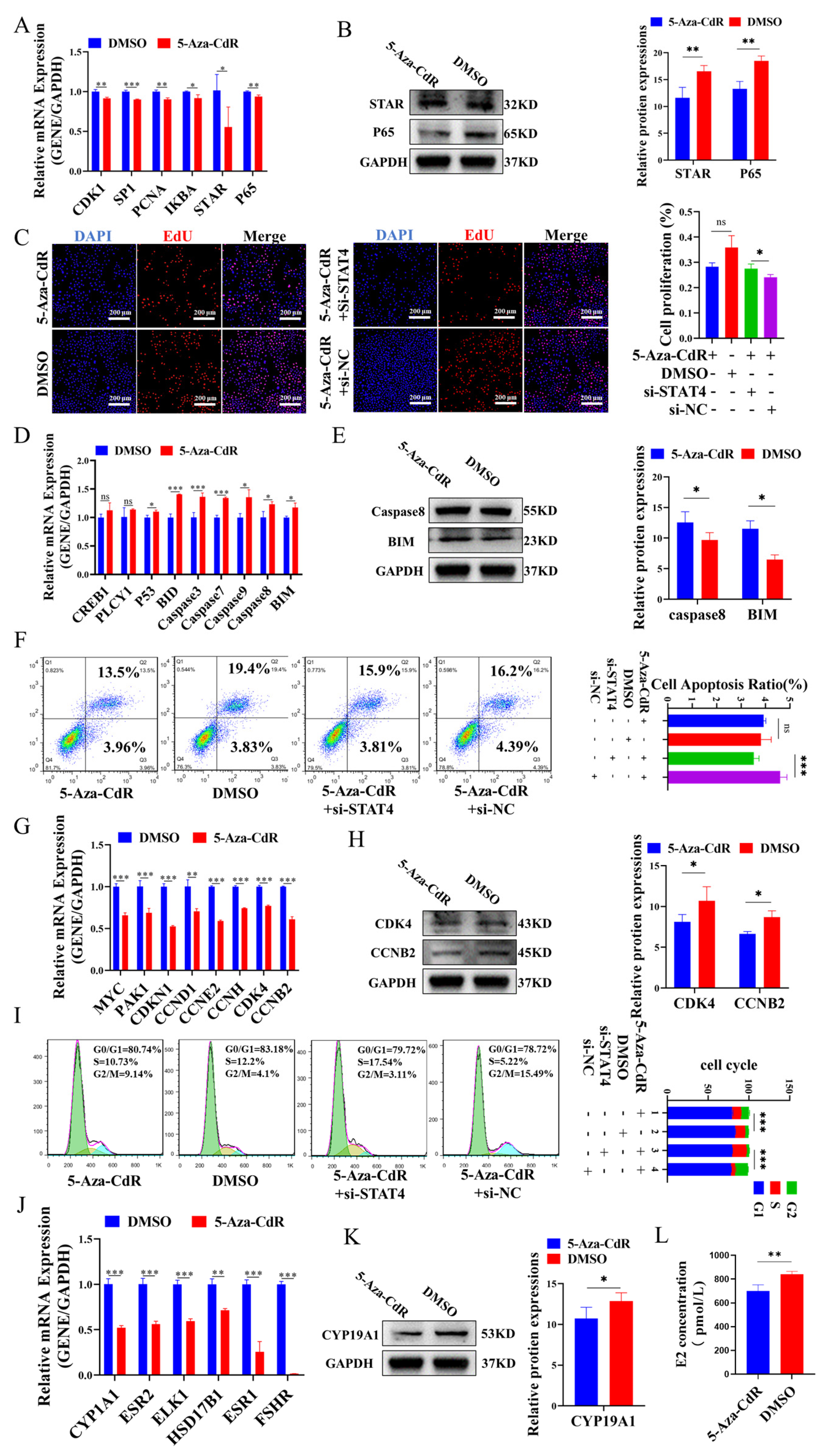
3.1. DNA Methylation May Regulate STAT4 to Increase Apoptosis in GCs

The results indicated that both mRNA (Figure 1A) and protein (Figure 1B) expression of STAT4 were significantly elevated in small follicles (1–3 mm) compared to medium (3–5 mm) and large follicles (5–7 mm). According to the CpG island prediction website (https://www.methprimer.com/), we found that the region −1220 bp/−1420 bp had more CG sites in the promoter of STAT4, including three specific CG sites (−1219 bp, −1256 bp and −1292 bp). The DNA methylation of STAT4 was obviously increased in the large follicles when contrasted with small and medium follicles (Figure 1C). After treating with 5-Aza-CdR, it was found that 5-Aza-CdR could cause demethylation at the CG site of STAT4, and the effect was most significant at a concentration of 1 µM (Figure 1D). Moreover, the expression of STAT4 also showed a similar trend after 5-Aza-CdR treatment, and the effect was most significant at a concentration of 1 µM (Figure 1E). Therefore, further methylation experiments were conducted, and the concentration of 1 µM 5-Aza-CdR was selected for subsequent experiments.
To investigate the impact of STAT4 on apoptosis in GCs, STAT4 overexpression plasmids and three STAT4-siRNAs (STAT4-siRNA1, STAT4-siRNA2 and STAT4-siRNA3) were transfected into GCs. The results showed a significant upregulation of both mRNA (Figure 1G) and protein levels (Figure 1H) of STAT4 in GCs with STAT4 overexpression (OE-STAT4) at a concentration of 500 ng/mL. In contrast, transfection with STAT4-siRNA1 remarkably reduced the mRNA (Figure 1I) and protein levels of STAT4 (Figure 1J). Subsequently, we revealed that STAT4 overexpression significantly enhanced the mRNA expressions of apoptosis-related genes, including CREB1, PLCγ1, PLCγ2, Casp3, Casp8 and Casp9 (Figure 1K), and remarkably upregulated the protein levels of Casp8 (Figure 1L). Conversely, STAT4 knockdown produced the opposite effects in GCs. In addition, OE-STAT4 substantially elevated the apoptosis in GCs (Figure 1M), whereas this effect was significantly attenuated by STAT4 knockdown (Figure 1N). These findings indicated that DNA hypomethylation may upregulate STAT4 expression to induce apoptosis in GCs.

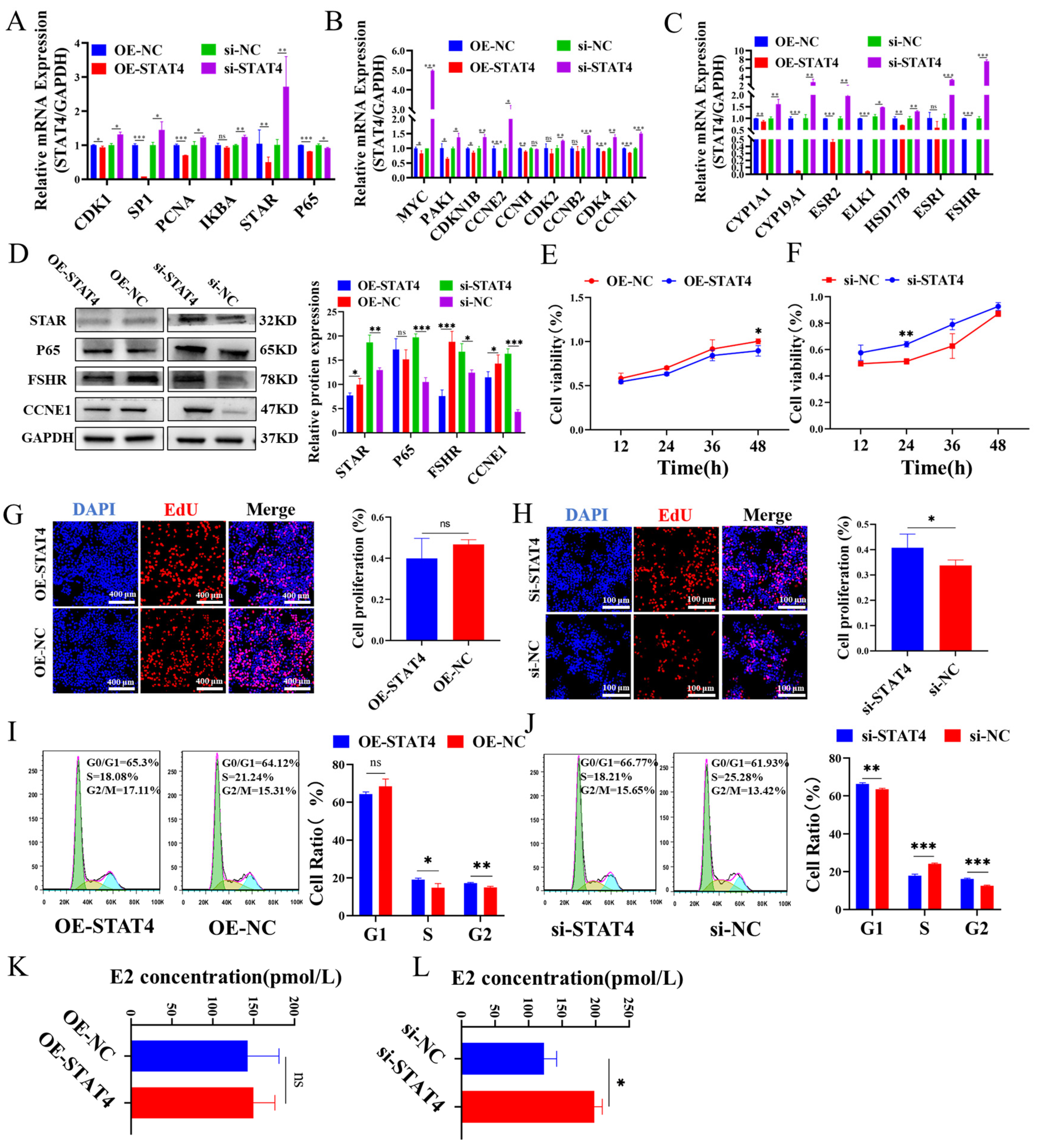
3.2. STAT4 Inhibits the Cellular Proliferation and E2 Secretion in GCs

Subsequently, the effect of STAT4 on the proliferation and E2 secretion in GCs was detected. Results showed that STAT4 overexpression significantly reduced the mRNA levels of proliferation-related genes, including CDK1, SP1, PCNA and STAR (Figure 2A), as well as cell cycle-related genes such as MYC, PAK1, CDKN1B, CCNE2, CDK4 and CCNE1 (Figure 2B). Additionally, genes involved in estrogen biosynthesis such as CYP19A1, ESR2 and FSHR were also markedly reduced (Figure 2C). Correspondingly, the protein levels of STAR, FSHR and CCNE1 were significantly suppressed by STAT4 overexpression (Figure 2D). Conversely, STAT4 knockdown notably promoted the expressions of genes associated with proliferation, cell cycle and estrogen secretion. Moreover, STAT4 overexpression and knockdown notably restrained and enhanced the viability of GCs, respectively (Figure 2E,F). Correspondingly, STAT4 overexpression inhibited GC proliferation (Figure 2G), whereas si-STAT4 markedly enhanced it (Figure 2H). Meanwhile, STAT4 overexpression significantly decreased the number of GCs that progressed to the S-phase (Figure 2I), while si-STAT4 significantly prompted the progression of GCs into the S-phase (Figure 2J). Additionally, STAT4 overexpression suppressed E2 secretion in GCs (Figure 2K), whereas si-STAT4 exhibited the opposite effects (Figure 2L). In summary, STAT4 acted as an inhibitor of GC proliferation, arrested cell cycle in the S phase and decreased E2 secretion.

3.3. DNA Hypomethylation of STAT4 Induces Apoptosis in GCs

To further explore the role of DNA hypomethylation of STAT4 in regulating GC function, cellular apoptosis, proliferation, cell cycle and estrogen secretion were detected in GCs treated with 5-Aza-CdR. We revealed that 5-Aza-CdR significantly inhibited the mRNA of CDK1, SP1, PCNA, IKBA and P65 (Figure 3A), as well as the protein expressions of STAR and P65 (Figure 3B). Correspondingly, 5-Aza-CdR reduced the proliferation of GCs, while co-treatment with 5-Aza-CdR and si-STAT4 significantly restored their proliferation (Figure 3C). Additionally, we found that 5-Aza-CdR significantly enhanced the mRNA (Figure 3D) and protein levels (Figure 3E) of Caspase8 and BIM, resulting in elevated GC apoptosis. However, co-treatment with 5-Aza-CdR and si-STAT4 significantly decreased the apoptosis of GCs (Figure 3F). Moreover, 5-Aza-CdR markedly restrained the mRNA (Figure 3G) and protein levels (Figure 3H) of CDK4 and CCNB2 and notably reduced the number of GCs that progressed to the S-phase and increased the number of GCs that progressed to the G2/M-phase (Figure 3I), with opposite effects observed upon co-treatment with si-STAT4. Similarly, 5-Aza-CdR significantly downregulated the mRNA (Figure 3J) and protein levels (Figure 3K) of CYP19A1 and inhibited E2 secretion in GCs (Figure 3L). Taken together, our results suggested that DNA hypomethylation of STAT4 enhanced apoptosis, inhibited cellular proliferation, arrested cell cycle progress and decreased E2 secretion in GCs.

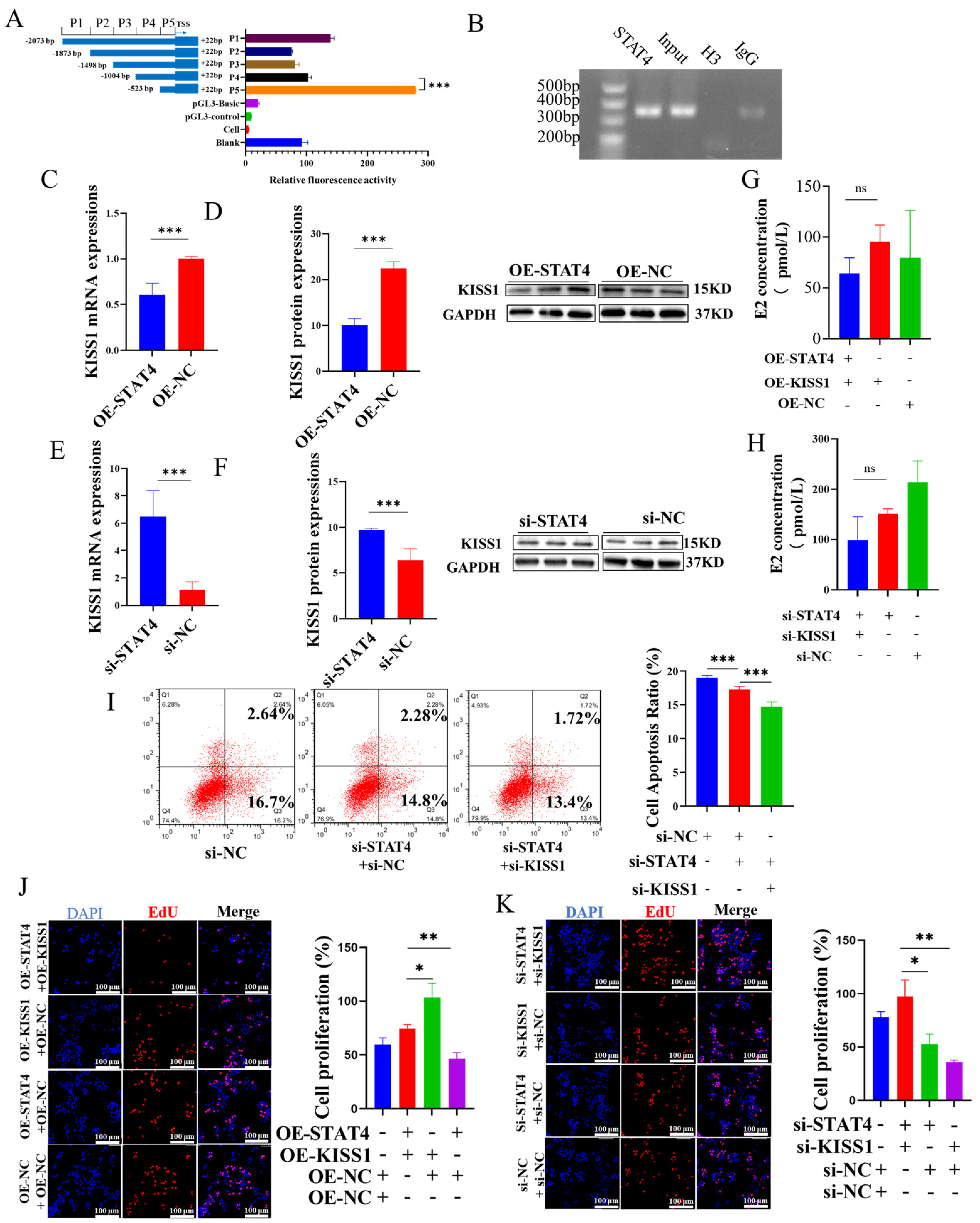
3.4. STAT4 Induces Apoptosis in GCs While Inhibiting the Expression of KISS1

Preliminary software prediction (https://tfbind.hgc.jp/) identified five potential STAT4 binding sites within the KISS1 promoter region, suggesting that STAT4 might directly target KISS1 to regulate its expression in GCs. To elucidate the molecular regulatory mechanism between STAT4 and KISS1, recombinant plasmids containing five deletion fragments (P1: −2073/+22, P2: −1873/+22, P3: −1498/+22, P4: −1004/+22 and P5: −523/+22) of the KISS1 promoter, along with the pGL3-TK internal reference, were sequentially transfected into GCs. We found that all fragments exhibited varying degrees of fluorescence activity, with the P5 fragment exhibiting the highest activity compared to the control. Using a pull-down assay with the STAT4 antibody and the site vector (−1004/−523 bp), ChIP analysis confirmed significant binding of STAT4 to this region within the KISS1 promoter (−1004/−523 bp; Figure 4B). Subsequently, we found that the mRNA expression and protein levels of KISS1 decreased (Figure 4C,D) when overexpression of STAT4 occurred, and we obtained the opposite results (Figure 4E,F) when interference of STAT4 occurred. Co-treatment of si-STAT4 and si-KISS1 significantly reduced GC apoptosis (Figure 4I) and reduced the E2 secretion, but not significantly (Figure 4H). Subsequently, OE-KISS1 promoted GC proliferation, but simultaneous overexpression of both KISS1 and STAT4 (OE-KISS1 and OE-STAT4) notably inhibited this proliferative effect (Figure 4J). Conversely, co-silencing of STAT4 and KISS1 yielded opposite outcomes (Figure 4K). These findings indicated that STAT4 binds to the KISS1 promoter, thereby suppressing KISS1 expression and promoting apoptosis in GCs.

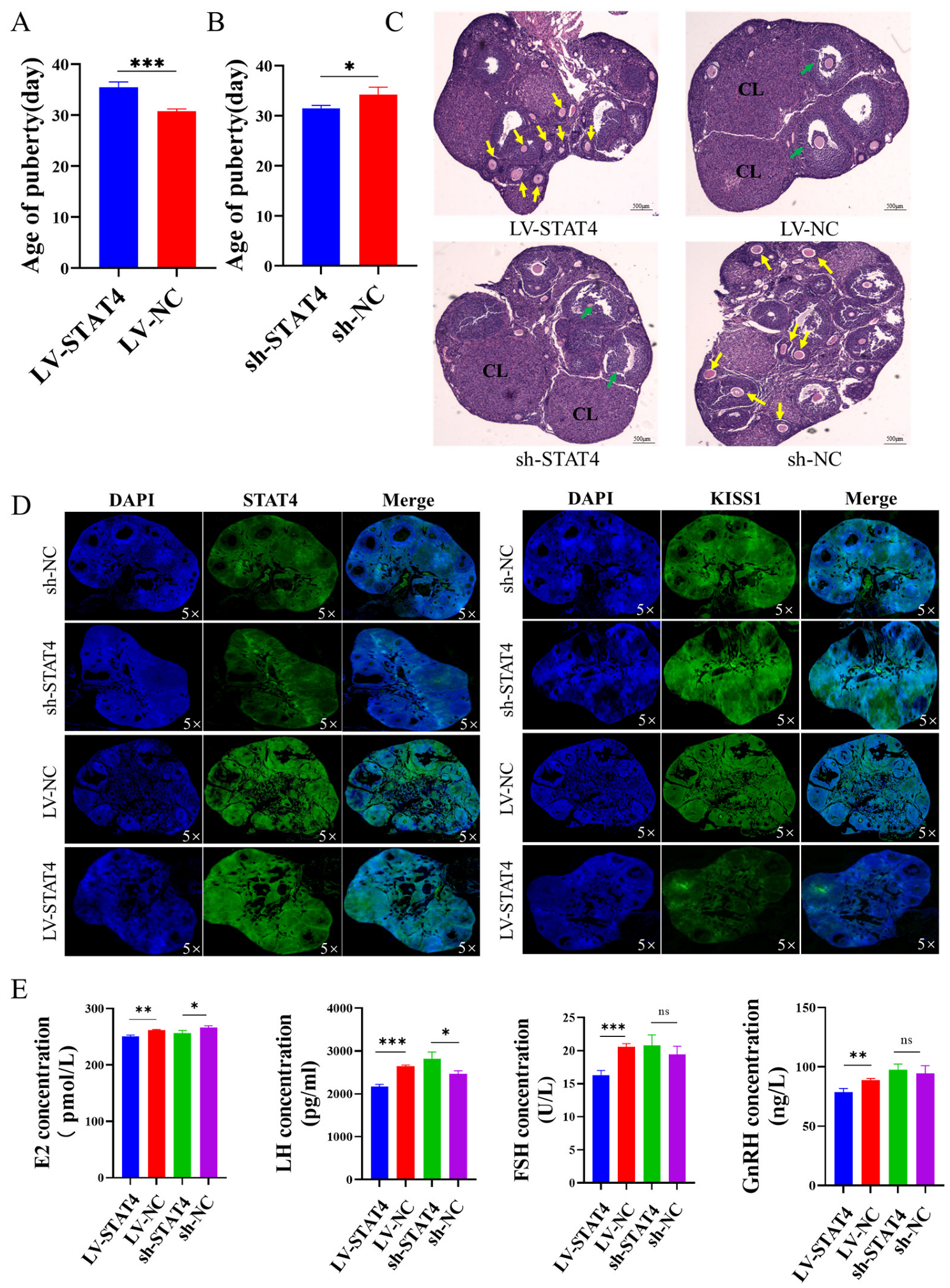
3.5. STAT4 Inhibits Follicular Development in Pigs

To further investigate the biological function of STAT4 in follicular development, lentiviral vectors for STAT4 overexpression and knockdown (LV-STAT4 and sh-STAT4, respectively), along with their respective negative controls (LV-NC and sh-NC, respectively), were constructed and transfected into porcine follicles cultured in vitro. Overexpression of STAT4 led to the loss of follicular blood vessels and the opacity of follicular fluid (Figure 5A). Consistently, LV-STAT4 significantly upregulated both the mRNA and protein levels of STAT4 while significantly suppressing the mRNA and protein levels of KISS1. In contrast, sh-STAT4 exhibited the opposite effects (Figure 5B–F). Immunofluorescence analysis further confirmed that sh-STAT4 enhanced KISS1 protein expression, whereas LV-STAT4 suppressed KISS1 protein in porcine follicles (Figure 5G). These results indicate that STAT4 inhibited follicular development by repressing KISS1 transcription.

3.6. STAT4 Blocks Follicular Development in Mice

To further verify the role of KISS1 mediated by STAT4 on follicular development, LV-STAT4 and sh-STAT4 were injected into the ovaries of 4-week-old C57BL/6 mice. It was found that LV-STAT4 inhibited sexual maturity (Figure 6A), while sh-STAT4 promoted sexual maturity in these mice (Figure 6B). Moreover, the antral follicles and corpus luteum were increased by sh-STAT4. Conversely, the results obtained with LV-STAT4 were opposite to this observation (Figure 6C). Furthermore, immunofluorescence analysis of ovarian tissues revealed that sh-STAT4 upregulated the protein levels of KISS1, whereas LV-STAT4 downregulated them (Figure 6D). Additionally, the ELISA assay indicated that LV-STAT4 significantly reduced the serum concentrations of key reproductive hormones, including E2, FSH, LH and GnRH (Figure 6E). These results indicate that STAT4 inhibited follicular development and sexual maturity by suppressing the transcription of KISS1.

4. Discussion

Numerous studies have demonstrated that GCs undergo excessive apoptosis [36], leading to follicle atresia and impaired follicular development, which in turn leads to delayed sexual maturation and reduced reproductive performance in mammals [37,38], and the apoptosis of GCs is affected by many growth factors. For example, FSH promotes the proliferation and estradiol secretion of follicular cells by binding to granulosa cell receptors and upregulating the expressions of intracellular anti-apoptotic proteins XIAP and FLIP to inhibit apoptosis, thus promoting the survival and growth of follicular cells [39,40,41]. Notch signaling, an evolutionarily conserved pathway, is involved in ovarian follicle development by regulating the proliferation of GCs [42]. In the bovine estrous cycle, the apoptosis of GCs caused dominant follicle atresia during the non-ovulation period [43]. Research about follicular atresia in pigs indicated that during follicular development, apoptotic cell death was involved in the degeneration of GCs [44]. In this study, we found that overexpression of STAT4 promoted GC apoptosis (Figure 1M) and hindered granulosa cell proliferation (Figure 2G), cyclic processes (Figure 2I) and estrogen secretion (Figure 2K). Meanwhile, overexpression of STAT4 promoted mRNA expression and protein levels of genes related to the apoptosis pathway (CREB1, PLCY1, PLCY2, P53, Casp3, Casp7, Casp9, Casp8, BIM) (Figure 1K), inhibited the expression of genes related to cell proliferation (CDK1, SP1, PCNA, IKBA, STAR, P65) (Figure 2A), cyclic processes (MYC, PAK1, CDKN1B, CCNH, CCNE2, CDK2, CCNB2, CDK4, CCNE1) (Figure 2B) and estrogen secretion (CYP1A1, CYP19A1, ESR2, ELK1, HSD17B, ESR1, FSHR) (Figure 2C). These results show that STAT4 promotes apoptosis and inhibits proliferation of GCs.
DNA methylation is a widely studied epigenetic modification that typically suppresses gene expression without altering the gene’s sequence by DNA methyltransferases, including DNMT1, DNMT3a and DNMT3b [45,46,47]. 5-Aza-CdR, a derivative of 2′-deoxycytidine, is a DNA methyltransferase inhibitor that substitutes cytosine in DNA molecules, reducing DNA methylation by inhibiting DNMTs’ activity [48]. Studies have indicated that the demethylation of the Lhcgr promoter region is a key mechanism regulating cell type-specific differentiation during follicular development [49]. In polycystic ovary syndrome, lnc-MAP3K13-7:1 inhibited the proliferation of ovarian GCs via DNMT1 downregulation mediated by CDKN1A promoter hypomethylation [50]. Moreover, in human T cells, the expression of STAT4 is regulated by DNA methylation [51]. In patients with inflammatory bowel diseases (IBDs), DNA methylation of STAT4 promoter is lower than in healthy individuals [52]. During mouse follicular development, large-scale DNA methylation occurs in proliferating ovarian GCs [53]. Another study showed that DNA promoter methylation is negatively correlated with lncRNA during puberty onset, and the methylation regulated the initiation of puberty via lncRNA [19]. In our study, we found that with the growth of follicles, the methylation of the STAT4 promoter region gradually increases, but not significantly (Figure 1C), and the expression of STAT4 gradually decreases (Figure 1A,B). Although the methylation status in small size follicles is not significantly different compared with medium size follicles, we consider that the expression of STAT4 in follicles is regulated by multiple factors, such as histone acetylation [54] and DNA methylation [55]. DNA methylation changed the chromatin conformation and transcription factor binding, possibly. Otherwise, we found some transcription factors combined with the region −1220 bp/−1420 bp of the STAT4 promoter by tfbind (https://tfbind.hgc.jp/), including SP1, GATA and P53, and these transcription factors played important roles in ovarian development [56]. Changes in DNA methylation might affect the binding of these transcription factors. For instance, changes in DNA methylation of the STAT4 promoter disrupted the binding of SP1 and significantly reduced STAT4 promoter activity in humans [51]. Knockdown p53 significantly reduced the expression of STAT4 in mice tissue [57]. Moreover, in cows diagnosed with mastitis, the methylation of the STAT5A promoter was lowest compared with JAK2 and CD4, about 9–11%, but the expression of STAT5A still changed significantly [58]. Therefore, although the change in DNA methylation is small, it still significantly regulates STAT4 expression. In the regulation of gene expression, small DNA methylation changes may also regulate STAT4 expression by affecting chromatin structure or the transcription factor binding sites [59]. Our results showed that changes in the methylation status of the STAT4 promoter may be one of the reasons for this regulation. Therefore, to further investigate whether the methylation status of the STAT4 promoter region influences STAT4 expression, we treated GCs with the methyltransferase inhibitor 5-Aza-CdR and analyzed the methylation status of the STAT4 promoter region and STAT4 expression levels. We found that at a concentration of 1 µM 5-Aza-CdR, the methylation level of the STAT4 promoter region was significantly reduced, and STAT4 expression was highest. These results suggest that changes in the methylation status of the STAT4 promoter region may regulate STAT4 expression levels (Figure 1D). The mRNA and protein expression of STAT4 were detected when the content of 5-Aza-CdR was 1 μm (Figure 1C), and it was found that the mRNA and protein expression of STAT4 were significantly decreased at this time (Figure 1E,F). Then, we co-treated GCs in vitro with 5-Aza-CdR and si-RNA and found that after co-treatment, apoptosis and related pathway genes of GCs were inhibited (Figure 3F), and the proliferation (Figure 3C), cycle process (Figure 3I) and expressions of related proteins of GCs were promoted.
Moreover, we explored the effect of STAT4 on transcriptional regulation of KISS1. According to the information provided on the website (https://tfbind.hgc.jp/), STAT4 is identified as a transcription factor for KISS1. Furthermore, the results of the dual-luciferase reporter gene assay demonstrated that STAT4 binds to the promoter region −1004 bp/−523 bp of KISS1. On the other hand, the expression of KISS1 was decreased when we overexpressed STAT4 in GCs. Otherwise, there are numerous studies showing that STAT4 regulates the transcription of KISS1 [60,61]. The research showed that STAT4 regulated the transcription of KISS1 to inhibit the oxidative damage, inflammation and neuronal apoptosis in the model of Parkinson’s disease [60]. Our team has previously also discovered that STAT4 acts as a transcription factor for KISS1 using a different website [31]. In this study, we further investigated how changes in DNA methylation in the STAT4 promoter region alter the expression of KISS1 and enhance the apoptosis of GCs. Additionally, our findings reveal that the co-expression of STAT4 and KISS1 significantly influences apoptosis, proliferation, cell cycle progression and hormone secretion, as well as the underlying signaling pathways. To further investigate the effects of STAT4 on ovarian follicle growth and development, we conducted in vitro follicle culture experiments (Figure 5) and in vivo mice studies (Figure 6). These results provide a foundation for further research into factors affecting follicular development in female mammals.
The primary regulatory pathway for the onset of puberty in mammals is the hypothalamic–pituitary–ovarian (HPO) axis [62]. E2 plays a dual role in the regulation of this HPO axis [63]. E2 promotes the release of GnRH and LH through KISS1 neurons in the hypothalamus during puberty onset in mammals [64], which is the positive feedback mechanism. In our study, we hypothesized that STAT4 affected E2 secretion by regulating the expression of KISS1 (Figure 4G,H), and it might be the positive feedback to promote follicle development. As an important experimental animal model, C57BL/6 mice have been widely used in ovarian-related studies. Some studies have analyzed the levels of luteinizing hormone (LH), follicle-stimulating hormone (FSH) and prolactin in the plasma and pituitary of mice at different ages and stages of the estrous cycle [65]. Additionally, age-related changes in estrogen receptor expression have been observed in middle-aged mice, independent of estrous cycle status, suggesting that the regulation of estrogen receptors may influence follicular development [66]. In this experiment, C57BL/6 mice were infected with LV-STAT4 and sh-STAT4 and their control group with LV-NC and sh-NC. After the treatment with STAT4, the degree of apoptosis of GCs in follicles increased (Figure 6C). Then, the estrus of mice, the levels of serum hormones GnRH, FSH, LH and E2 and the expression of KISS1 were measured. It was found that, compared with the control group, LV-STAT4 inhibited the estrus of mice, and the levels of serum hormones GnRH, FSH, LH and E2 decreased. sh-STAT4 promoted estrus in mice, and the levels of GnRH, FSH, LH and E2 in serum increased, indicating that the STAT4 gene could inhibit estrus in mice. Otherwise, we also found that LV-STAT4 decreased blood vessels in the follicles of pigs (Figure 5A) and suppressed KISS1 protein in porcine follicles (Figure 5G). Above all, we hypothesized that STAT4 may regulate KISS1 transcription, thereby influencing estrogen secretion by GCs in sow follicles and subsequently affecting follicular development. However, this study had some limitations. On the one hand, due to the post-translational modification, alternative splicing and proteolytic cleavage of KISS1 and the KISS1 proteins being short peptides that are easily degraded, these may lead to extra bands. On the other hand, the change in DNA methylation induced by 5-Aza-CdR is small in −1220 bp/−1420 bp of the STAT4 promoter, but the expression of STAT4 is significant. We guess that 5-Aza-CdR changes the methylation of the other expected region −1220 bp/−1420 bp in the STAT4 promoter. The research showed that the change in DNA methylation in −2225 bp/+605 bp of the STAT4 promoter significantly reduced STAT4 promoter activity in humans [51]. In subsequent research, we will try to use more accurate experimental methods, such as CRISPR/dCas9, to change the DNA methylation of specific CG sites in the STAT4 promoter and investigate the influence of DNA methylation on STAT4 expression.

5. Conclusions

Based on the above findings, it is evident that in normally developing ovarian follicles, methylation of the STAT4 promoter in mature follicles is significantly higher compared to immature follicles. STAT4 inhibits the expression of KISS1, induces apoptosis of GCs and suppresses both proliferation and estrogen secretion of GCs. Moreover, the results showed that STAT4 inhibits follicular development, sexual maturation and hormone secretion in mice by suppressing the transcription of KISS1. Therefore, we speculate that DNA methylation regulates the transcription of STAT4, thereby promoting KISS1 expression, fostering follicular development and ultimately facilitating puberty onset in female mammals.

Author Contributions

D.C., Y.H., X.Z., X.Y., B.M. and J.L. designed the experiments; D.C. and M.F. performed functional verification assays in vitro and contributed equally to this research; D.C., E.H. and L.Z. performed in vivo experiments; D.C. evaluated results and wrote the manuscript, with revisions by H.Q., Y.H. and X.Z. All authors have read and agreed to the published version of the manuscript.

Funding

This study was supported by the National Center of Technology Innovation for Pigs (NCTIP-XDB14), the Breed Industry Innovation Park of Guangdong Xiaoerhua Pig (2023-4408X1-29060200), an earmarked fund from the China Agriculture Research System (CARS-35), the Guangdong Basic and Applied Basic Research Foundation (2024B1515020112, 2024A1515012999, 2023A1515030054), the National Natural Science Foundation of China (31902131 and 32072694) and the Science and Technology Program of Guangdong province, China (2023B1212060057).

Institutional Review Board Statement

This study was conducted according to the guidelines of the Regulations for Administration of Affairs Concerning Experimental Animals and approved by the ethics committee of the Guangdong Laboratory Animals Monitoring Institute (IACUC2021168, 25 August 2021).

Informed Consent Statement

Not applicable.

Data Availability Statement

The data underlying this research will be provided by the corresponding authors upon reasonable request.

Conflicts of Interest

The authors declare no conflicts of interest.

Abbreviations

The following abbreviations are used in this manuscript:
STAT4Signal transducer and activator of transcription 4
KISS1Kisspeptin-1
GCsGranulosa cells
5-Aza-CdR5-azacytidine
E2Estradiol
LHLuteinizing hormone
FSHFollicle-stimulating hormone
GnRHGonadotropin-releasing hormone

References

  1. Christenson, R.K.; Ford, J.J. Puberty and estrus in confinement-reared gilts. J. Anim. Sci. 1979, 49, 743–751. [Google Scholar] [PubMed]
  2. Stringer, J.M.; Alesi, L.R.; Winship, A.L.; Hutt, K.J. Beyond apoptosis: Evidence of other regulated cell death pathways in the ovary throughout development and life. Hum. Reprod. Update 2023, 29, 434–456. [Google Scholar] [PubMed]
  3. Field, S.L.; Dasgupta, T.; Cummings, M.; Orsi, N.M. Cytokines in ovarian folliculogenesis, oocyte maturation and luteinisation. Mol. Reprod. Dev. 2014, 81, 284–314. [Google Scholar] [PubMed]
  4. Jiang, J.Y.; Cheung, C.K.; Wang, Y.; Tsang, B.K. Regulation of cell death and cell survival gene expression during ovarian follicular development and atresia. Front. Biosci. 2003, 8, d222–d237. [Google Scholar]
  5. Manabe, N.; Goto, Y.; Matsuda-Minehata, F.; Inoue, N.; Maeda, A.; Sakamaki, K.; Miyano, T. Regulation mechanism of selective atresia in porcine follicles: Regulation of granulosa cell apoptosis during atresia. J. Reprod. Dev. 2004, 50, 493–514. [Google Scholar]
  6. Wang, X.; Jiang, Y.L.; Wang, Z.L.; Kang, B. Mechanism of programmed cell death in follicular atresia. Sheng Li Xue Bao 2023, 75, 82–90. [Google Scholar]
  7. Engblom, L.; Lundeheim, N.; Dalin, A.; Andersson, K. Sow removal in Swedish commercial herds. Livest. Sci. 2007, 106, 76–86. [Google Scholar]
  8. Silva, J.R.; Figueiredo, J.R.; van den Hurk, R. Involvement of growth hormone (GH) and insulin-like growth factor (IGF) system in ovarian folliculogenesis. Theriogenology 2009, 71, 1193–1208. [Google Scholar]
  9. Pan, Z.; Zhang, J.; Li, Q.; Li, Y.; Shi, F.; Xie, Z.; Liu, H. Current advances in epigenetic modification and alteration during mammalian ovarian folliculogenesis. J. Genet. Genom. 2012, 39, 111–123. [Google Scholar]
  10. Kim, J.M.; Yoon, Y.D.; Tsang, B.K. Involvement of the Fas/Fas ligand system in p53-mediated granulosa cell apoptosis during follicular development and atresia. Endocrinology 1999, 140, 2307–2317. [Google Scholar]
  11. Cao, L.; Li, S.; Huang, S.; Shi, D.; Li, X. AQP8 participates in oestrogen-mediated buffalo follicular development by regulating apoptosis of granulosa cells. Reprod. Domest. Anim. 2021, 56, 812–820. [Google Scholar] [PubMed]
  12. Matsuda, F.; Inoue, N.; Maeda, A.; Cheng, Y.; Sai, T.; Gonda, H.; Goto, Y.; Sakamaki, K.; Manabe, N. Expression and function of apoptosis initiator FOXO3 in granulosa cells during follicular atresia in pig ovaries. J. Reprod. Dev. 2011, 57, 151–158. [Google Scholar]
  13. Mattei, A.L.; Bailly, N.; Meissner, A. DNA methylation: A historical perspective. Trends Genet. 2022, 38, 676–707. [Google Scholar]
  14. Lyko, F. The DNA methyltransferase family: A versatile toolkit for epigenetic regulation. Nat. Rev. Genet. 2018, 19, 81–92. [Google Scholar] [PubMed]
  15. Qureshi, M.Z.; Sabitaliyevich, U.Y.; Rabandiyarov, M.; Arystanbekuly, A.T. Role of DNA Methyltransferases (DNMTs) in metastasis. Cell. Mol. Biol. 2022, 68, 226–236. [Google Scholar] [PubMed]
  16. Lomniczi, A.; Wright, H.; Ojeda, S.R. Epigenetic regulation of female puberty. Front. Neuroendocrinol. 2015, 36, 90–107. [Google Scholar]
  17. Rehnitz, J.; Youness, B.; Nguyen, X.P.; Dietrich, J.E.; Roesner, S.; Messmer, B.; Strowitzki, T.; Vogt, P.H. FMR1 expression in human granulosa cells and variable ovarian response: Control by epigenetic mechanisms. Mol. Hum. Reprod. 2021, 27, gaab001. [Google Scholar]
  18. Yang, C.; Ye, J.; Liu, Y.; Ding, J.; Liu, H.; Gao, X.; Li, X.; Zhang, Y.; Zhou, J.; Zhang, X.; et al. Methylation pattern variation between goats and rats during the onset of puberty. Reprod. Domest. Anim. 2018, 53, 793–800. [Google Scholar]
  19. Yang, C.; Gao, X.; Ye, J.; Ding, J.; Liu, Y.; Liu, H.; Li, X.; Zhang, Y.; Zhou, J.; Huang, W.; et al. The interaction between DNA methylation and long non-coding RNA during the onset of puberty in goats. Reprod. Domest. Anim. 2018, 53, 1287–1297. [Google Scholar]
  20. Li, S.; Zeng, L.; Miao, F.; Li, N.; Liao, W.; Zhou, X.; Chen, Y.; Quan, H.; He, Y.; Zhang, H.; et al. Knockdown of DNMT1 Induces SLCO3A1 to Promote Follicular Growth by Enhancing the Proliferation of Granulosa Cells in Mammals. Int. J. Mol. Sci. 2024, 25, 2468. [Google Scholar] [CrossRef]
  21. Yuan, X.; Ye, S.; Chen, Z.; Pan, X.; Huang, S.; Li, Z.; Zhong, Y.; Gao, N.; Zhang, H.; Li, J.; et al. Dynamic DNA methylation of ovaries during pubertal transition in gilts. BMC Genom. 2019, 20, 510. [Google Scholar]
  22. Zhou, X.; He, Y.; Li, N.; Bai, G.; Pan, X.; Zhang, Z.; Zhang, H.; Li, J.; Yuan, X. DNA methylation mediated RSPO2 to promote follicular development in mammals. Cell Death Dis. 2021, 12, 653. [Google Scholar] [PubMed]
  23. Zhou, X.; Zhou, X.; He, Y.; Pan, X.; Quan, H.; He, B.; Li, Y.; Bai, G.; Li, N.; Zhang, Z.; et al. DNMT1-mediated lncRNA IFFD controls the follicular development via targeting GLI1 by sponging miR-370. Cell Death Differ. 2023, 30, 576–588. [Google Scholar]
  24. Chen, W.; Daines, M.O.; Khurana, H.G. Turning off signal transducer and activator of transcription (STAT): The negative regulation of STAT signaling. J. Allergy Clin. Immunol. 2004, 114, 476–489; quiz 490. [Google Scholar]
  25. Hall, S.E.; Upton, R.M.O.; McLaughlin, E.A.; Sutherland, J.M. Phosphoinositide 3-kinase/protein kinase B (PI3K/AKT) and Janus kinase/signal transducer and activator of transcription (JAK/STAT) follicular signalling is conserved in the mare ovary. Reprod. Fertil. Dev. 2018, 30, 624–633. [Google Scholar]
  26. Ilha, G.F.; Rovani, M.T.; Gasperin, B.G.; Antoniazzi, A.Q.; Gonçalves, P.B.; Bordignon, V.; Duggavathi, R. Lack of FSH support enhances LIF-STAT3 signaling in granulosa cells of atretic follicles in cattle. Reproduction 2015, 150, 395–403. [Google Scholar]
  27. Gasperin, B.G.; Rovani, M.T.; Ferreira, R.; Ilha, G.F.; Bordignon, V.; Gonçalves, P.B.; Duggavathi, R. Functional status of STAT3 and MAPK3/1 signaling pathways in granulosa cells during bovine follicular deviation. Theriogenology 2015, 83, 353–359. [Google Scholar]
  28. Yuan, X.; Zhou, X.; He, Y.; Zhong, Y.; Zhang, A.; Zhang, Z.; Zhang, H.; Li, J. C/EBPbeta Promotes STAT3 Expression and Affects Cell Apoptosis and Proliferation in Porcine Ovarian Granulosa Cells. Genes 2018, 9, 295. [Google Scholar]
  29. Huang, J.; Ma, G.; Zhu, M.; Pan, J.; Zhang, W.; Zhao, S.H. Molecular characterization of the porcine STAT4 and STAT6 genes. Mol. Biol. Rep. 2012, 39, 6959–6965. [Google Scholar]
  30. Liu, Z.; Li, C.; Chen, Q.; Bai, C.; Wu, G.; Fu, C.; He, T.; Shen, M.; Feng, C.; Liu, H. Follicular fluid meiosis-activating sterol prevents porcine ovarian granulosa cells from hypoxia-induced apoptosis via inhibiting STAT4 expression. J. Anim. Sci. 2024, 102, skae125. [Google Scholar]
  31. Jiang, Y.; Xin, X.; Pan, X.; Zhang, A.; Zhang, Z.; Li, J.; Yuan, X. STAT4 targets KISS1 to promote the apoptosis of ovarian granulosa cells. J. Ovarian Res. 2020, 13, 135. [Google Scholar] [CrossRef] [PubMed]
  32. Wu, H.; Zheng, X.; Zhou, S.; Sun, P.; Liu, M.; Yang, L.; Liang, A. IL-11 regulates the biosynthesis of PGE2 through JAK1/STAT3 signaling pathway in bovine granulosa cells. Theriogenology 2025, 237, 85–92. [Google Scholar] [CrossRef] [PubMed]
  33. Li, H.; Jing, Y.; Qu, X.; Yang, J.; Pan, P.; Liu, X.; Gao, H.; Pei, X.; Zhang, C.; Yang, Y. The Activation of Reticulophagy by ER Stress through the ATF4-MAP1LC3A-CCPG1 Pathway in Ovarian Granulosa Cells Is Linked to Apoptosis and Necroptosis. Int. J. Mol. Sci. 2023, 24, 2749. [Google Scholar] [CrossRef] [PubMed]
  34. Huang, Y.; Dong, L.P.; Cui, Y.G.; Lu, H.Y. MiR-520h inhibits viability and facilitates apoptosis of KGN cells through modulating IL6R and the JAK/STAT pathway. Reprod. Biol. 2022, 22, 100607. [Google Scholar] [CrossRef]
  35. Li, N.; Yun, B.; Zeng, L.; Lv, Y.; Zhou, Y.; Fang, M.; Li, S.; Chen, Y.; Huang, E.; Zhang, L.; et al. The antisense lncRNA of TAB2 that prevents oxidative stress to enhance the follicular growth in mammals. Commun. Biol. 2024, 7, 1246. [Google Scholar] [CrossRef]
  36. Son, W.Y.; Das, M.; Shalom-Paz, E.; Holzer, H. Mechanisms of follicle selection and development. Minerva Ginecol. 2011, 63, 89–102. [Google Scholar]
  37. Zhang, T.; BaSang, W.D.; Chang, W.; Huo, S.; Ma, X.; Ju, X.; Yu, S.; Cui, S. Dynamics of apoptosis-related gene expression during follicular development in yak. J. Anim. Physiol. Anim. Nutr. 2021, 105, 1002–1013. [Google Scholar]
  38. Yang, M.Y.; Rajamahendran, R. Involvement of apoptosis in the atresia of nonovulatory dominant follicle during the bovine estrous cycle. Biol. Reprod. 2000, 63, 1313–1321. [Google Scholar]
  39. Macklon, N.S.; Fauser, B.C. Follicle-stimulating hormone and advanced follicle development in the human. Arch. Med. Res. 2001, 32, 595–600. [Google Scholar] [CrossRef]
  40. McNeilly, A.S. The ovarian follicle and fertility. J. Steroid Biochem. Mol. Biol. 1991, 40, 29–33. [Google Scholar] [CrossRef]
  41. Adriaens, I.; Cortvrindt, R.; Smitz, J. Differential FSH exposure in preantral follicle culture has marked effects on folliculogenesis and oocyte developmental competence. Hum. Reprod. 2004, 19, 398–408. [Google Scholar] [CrossRef] [PubMed]
  42. Zhang, C.P.; Yang, J.L.; Zhang, J.; Li, L.; Huang, L.; Ji, S.Y.; Hu, Z.Y.; Gao, F.; Liu, Y.X. Notch signaling is involved in ovarian follicle development by regulating granulosa cell proliferation. Endocrinology 2011, 152, 2437–2447. [Google Scholar] [CrossRef] [PubMed]
  43. Jolly, P.D.; Tisdall, D.J.; Heath, D.A.; Lun, S.; McNatty, K.P. Apoptosis in bovine granulosa cells in relation to steroid synthesis, cyclic adenosine 3′,5′-monophosphate response to follicle-stimulating hormone and luteinizing hormone, and follicular atresia. Biol. Reprod. 1994, 51, 934–944. [Google Scholar] [CrossRef] [PubMed]
  44. Guthrie, H.D.; Grimes, R.W.; Cooper, B.S.; Hammond, J.M. Follicular atresia in pigs: Measurement and physiology. J. Anim. Sci. 1995, 73, 2834–2844. [Google Scholar] [CrossRef]
  45. Edwards, J.R.; Yarychkivska, O.; Boulard, M.; Bestor, T.H. DNA methylation and DNA methyltransferases. Epigenetics Chromatin 2017, 10, 23. [Google Scholar] [CrossRef]
  46. Li, E.; Zhang, Y. DNA methylation in mammals. Cold Spring Harb. Perspect. Biol. 2014, 6, a019133. [Google Scholar] [CrossRef]
  47. Bestor, T.H. The DNA methyltransferases of mammals. Hum. Mol. Genet. 2000, 9, 2395–2402. [Google Scholar] [CrossRef]
  48. Schermelleh, L.; Spada, F.; Easwaran, H.P.; Zolghadr, K.; Margot, J.B.; Cardoso, M.C.; Leonhardt, H. Trapped in action: Direct visualization of DNA methyltransferase activity in living cells. Nat. Methods 2005, 2, 751–756. [Google Scholar] [CrossRef]
  49. Kawai, T.; Richards, J.S.; Shimada, M. The Cell Type-Specific Expression of Lhcgr in Mouse Ovarian Cells: Evidence for a DNA-Demethylation-Dependent Mechanism. Endocrinology 2018, 159, 2062–2074. [Google Scholar] [CrossRef]
  50. Geng, X.; Zhao, J.; Huang, J.; Li, S.; Chu, W.; Wang, W.-S.; Chen, Z.-J.; Du, Y. lnc-MAP3K13-7:1 Inhibits Ovarian GC Proliferation in PCOS via DNMT1 Downregulation-Mediated CDKN1A Promoter Hypomethylation. Mol. Ther. 2021, 29, 1279–1293. [Google Scholar] [CrossRef]
  51. Shin, H.J.; Park, H.Y.; Jeong, S.J.; Park, H.W.; Kim, Y.K.; Cho, S.H.; Kim, Y.Y.; Cho, M.L.; Kim, H.Y.; Min, K.U.; et al. STAT4 expression in human T cells is regulated by DNA methylation but not by promoter polymorphism. J. Immunol. 2005, 175, 7143–7150. [Google Scholar] [PubMed]
  52. Kim, S.W.; Kim, E.S.; Moon, C.M.; Kim, T.I.; Kim, W.H.; Cheon, J.H. Abnormal Genetic and Epigenetic Changes in Signal Transducer and Activator of Transcription 4 in the Pathogenesis of Inflammatory Bowel Diseases. Dig. Dis. Sci. 2012, 57, 2600–2607. [Google Scholar] [CrossRef] [PubMed]
  53. Kawai, T.; Richards, J.S.; Shimada, M. Large-scale DNA demethylation occurs in proliferating ovarian granulosa cells during mouse follicular development. Commun. Biol. 2021, 4, 1334. [Google Scholar]
  54. Wieczorek, M.; Ginter, T.; Brand, P.; Heinzel, T.; Krämer, O.H. Acetylation modulates the STAT signaling code. Cytokine Growth Factor Rev. 2012, 23, 293–305. [Google Scholar]
  55. Yu, Q.; Thieu, V.T.; Kaplan, M.H. Stat4 limits DNA methyltransferase recruitment and DNA methylation of the IL-18Ralpha gene during Th1 differentiation. EMBO J. 2007, 26, 2052–2060. [Google Scholar] [CrossRef]
  56. Zhang, B.; Nie, X.; Miao, X.; Wang, S.; Li, J.; Wang, S. Development and verification of an immune-related gene pairs prognostic signature in ovarian cancer. J. Cell. Mol. Med. 2021, 25, 2918–2930. [Google Scholar]
  57. Yan, W.; Wei, J.; Deng, X.; Shi, Z.; Zhu, Z.; Shao, D.; Li, B.; Wang, S.; Tong, G.; Ma, Z. Transcriptional analysis of immune-related gene expression in p53-deficient mice with increased susceptibility to influenza A virus infection. BMC Med. Genom. 2015, 8, 52. [Google Scholar]
  58. Usman, T.; Ali, N.; Wang, Y.; Yu, Y. Association of Aberrant DNA Methylation Level in the CD4 and JAK-STAT-Pathway-Related Genes with Mastitis Indicator Traits in Chinese Holstein Dairy Cattle. Animals 2021, 12, 65. [Google Scholar] [CrossRef]
  59. Smith, Z.D.; Hetzel, S.; Meissner, A. DNA methylation in mammalian development and disease. Nat. Rev. Genet. 2025, 26, 7–30. [Google Scholar]
  60. Zhang, X.; Wang, Y.; Lv, J. STAT4 targets KISS1 to inhibit the oxidative damage, inflammation and neuronal apoptosis in experimental PD models by inactivating the MAPK pathway. Neurochem. Int. 2024, 175, 105683. [Google Scholar]
  61. Bohlen, T.M.; de Paula, D.G.; Teixeira, P.D.S.; da Silva Mansano, N.; Andrade Alves, G.; Donato, J., Jr.; Frazao, R. Socs3 ablation in kisspeptin cells partially prevents lipopolysaccharide-induced body weight loss. Cytokine 2022, 158, 155999. [Google Scholar] [PubMed]
  62. Cui, P.; Yang, C.; Zhang, K.; Gao, X.; Luo, L.; Tian, Y.; Song, M.; Liu, Y.; Zhang, Y.; Li, Y.; et al. Effect of estrogen on the expression of GnRH and kisspeptin in the hypothalamus of rats during puberty. Theriogenology 2015, 84, 1556–1564. [Google Scholar] [PubMed]
  63. Marques, P.; De Sousa Lages, A.; Skorupskaite, K.; Rozario, K.S.; Anderson, R.A.; George, J.T. Physiology of GnRH and Gonadotrophin Secretion; National Library of Medicine: Bethesda, MD, USA, 2000. [Google Scholar]
  64. Xie, Q.; Kang, Y.; Zhang, C.; Xie, Y.; Wang, C.; Liu, J.; Yu, C.; Zhao, H.; Huang, D. The Role of Kisspeptin in the Control of the Hypothalamic-Pituitary-Gonadal Axis and Reproduction. Front. Endocrinol. 2022, 13, 925206. [Google Scholar]
  65. Parkening, T.A.; Collins, T.J.; Smith, E.R. Plasma and pituitary concentrations of LH, FSH, and prolactin in aging C57BL/6 mice at various times of the estrous cycle. Neurobiol. Aging 1982, 3, 31–35. [Google Scholar]
  66. Bergman, M.D.; Karelus, K.; Felicio, L.S.; Nelson, J.F. Age-related alterations in estrogen receptor dynamics are independent of cycling status in middle-aged C57BL/6J mice. J. Steroid Biochem. Mol. Biol. 1991, 38, 127–133. [Google Scholar]
Figure 1. DNA methylation of STAT4 elevates while the expression of STAT4 decreases during the development of follicles. The mRNA (A) and protein (B) levels of STAT4 in large (5–7 mm in diameter), medium (3–5 mm) and small (1–3 mm) follicles. (C) BSP showed the methylation status of STAT4 in large (5–7 mm), medium (3–5 mm) and small (1–3 mm) follicles. (D) BSP assay showed the methylation status of STAT4 in GCs treated with 5-Aza-CdR. The mRNA (E) and protein (F) levels of STAT4 in GCs treated with 5-Aza-CdR. The mRNA (G) and protein (H) levels of STAT4 in GCs transfected with STAT4 overexpression plasmid. The mRNA (I) and protein (J) levels of STAT4 after transfection with STAT4-siRNAs in GCs. (K) The mRNA levels of apoptosis-related genes with STAT4 overexpression and knockdown. (L) The protein levels of Casp8 with STAT4 overexpression and knockdown. The apoptosis rates in GCs with STAT4 overexpression (M) and knockdown (N) assessed via flow cytometry. ns: not significant, *** p < 0.001, ** p < 0.01 and * p < 0.05.
Cells 14 00523 g001
Figure 2. Effects of STAT4 on the function of GCs. The mRNA of cellular proliferation (A), cell cycle (B) and estrogen secretion-related genes (C) detected in GCs with STAT4 overexpression and knockdown. (D) The protein levels of STAR, P65, FSHR and CCNE1 in GCs with STAT4 overexpression and knockdown. The CCK-8 assay shows the viability of GCs with STAT4 overexpression (E) and knockdown (F). The EDU assay shows the proliferation of GCs with STAT4 overexpression (G) and knockdown (H). The measurements of cell cycle distributions in GCs with STAT4 overexpression (I) and knockdown (J). The measurements of E2 secretions in GCs with STAT4 overexpression (K) and knockdown (L). ns: not significant, *** p < 0.001, ** p < 0.01 and * p < 0.05.
Cells 14 00523 g002
Figure 3. Changes in DNA methylation in STAT4 promoter regulate the functions of GCs. The mRNA (A) and protein (B) levels of proliferation-related genes in GCs treated with 5-Aza-CdR. (C) EdU assay showing the proliferation rates of GCs with 5-Aza-CdR and si-STAT4 treatments. (D,E) The mRNA (D) and protein (E) levels of apoptosis-related genes in GCs treated with 5-Aza-CdR. The apoptosis rates in GCs treated with 5-Aza-CdR and si-STAT4 assessed via flow cytometry (F). The mRNA (G) and protein (H) levels of cell cycle-related genes in GCs treated with 5-Aza-CdR. (I) The cell cycle distribution of GCs treated with 5-Aza-CdR and si-STAT4. The mRNA (J) and protein (K) levels of estrogen secretion-related genes in GCs treated with 5-Aza-CdR. (L) The measurements of E2 secretions in GCs treated with 5-Aza-CdR. ns: not significant, *** p < 0.001, ** p < 0.01 and * p < 0.05.
Cells 14 00523 g003
Figure 4. STAT4 regulates the expression of KISS1, and thus promotes apoptosis in GCs. (A) Analysis of the activity of the KISS1 promoter region specific segment via dual-luciferase reporter gene assay. (B) The ChIP experiment validates the binding of STAT4 to the KISS1 promoter region. Overexpression (C,D) and interference (E,F) of STAT4 affected the mRNA and protein levels of KISS1. Effects of E2 in GCs with co-transfection of OE-STAT4 and OE-KISS1 (G), si-STAT4 and si-KISS1 (H) via ELISA. (I) The apoptosis rates with co-transfection of si-STAT4 and si-KISS1 via flow cytometry. (J) The effects of co-transfection of overexpression of STAT4 and KISS1 via EdU. (K) The effects of co-transfection with si-STAT4 and si-KISS1 on the proliferation of GCs. ns: not significant, *** p < 0.001, ** p < 0.01 and * p < 0.05.
Cells 14 00523 g004
Figure 5. STAT4 inhibits the development of porcine follicles. (A) Photos of porcine follicles cultured in vitro after transfection with LV-STAT4 and sh-STAT4 on the first and third days. (B,C) The efficiency of LV-STAT4 (B) and sh-STAT4 (C). (D,E) The efficiency of LV-KISS1 (D) and sh-KISS1 (E). (F) The protein levels of STAT4 and KISS1 in GCs were assessed after transfection with LV-STAT4 and sh-STAT4. (G) The protein fluorescence intensities of STAT4 and KISS1 in follicles were assessed on the third day after LV-STAT4 and sh-STAT4 transduction via immunofluorescence. ns: not significant, *** p < 0.001, ** p < 0.01 and * p < 0.05.
Cells 14 00523 g005
Figure 6. STAT4 hinders follicular development in mice. (A,B) The effects of LV-STAT4 (A) and sh-STAT4 (B) on the initiation of puberty in mice. (C) Example of the ovaries from LV-STAT4- and sh-STAT4-injected mice collected at age 49 days and stained with HE. Yellow arrows indicate primordial follicles, green arrows indicate antral follicles, and CL points to an example of corpora lutea. (D) The protein fluorescence intensities of STAT4 and KISS1 in mice ovaries after LV-STAT4 and sh-STAT4 transduction via immunofluorescence. (E) The effects of LV-STAT4 and sh-STAT4 on the concentrations of E2, LH, FSH and GnRH in mice serum via ELISA. ns: not significant, *** p < 0.001, ** p < 0.01 and * p < 0.05.
Cells 14 00523 g006
Disclaimer/Publisher’s Note: The statements, opinions and data contained in all publications are solely those of the individual author(s) and contributor(s) and not of MDPI and/or the editor(s). MDPI and/or the editor(s) disclaim responsibility for any injury to people or property resulting from any ideas, methods, instructions or products referred to in the content.

