Intracellular Signals Activated by Canonical Wnt Ligands Independent of GSK3 Inhibition and β-Catenin Stabilization


Abstract
:1. Introduction: Wnt Signaling Promotes β-catenin Stabilization
2. STAT3 Transcriptional Activity is Stimulated by Canonical and Non-Canonical Wnts
3. JNK2 and PAK Dependent Activation of β-Catenin Nuclear Transport
4. Canonical Wnts Prevent the Action of the Kaiso Inhibitor
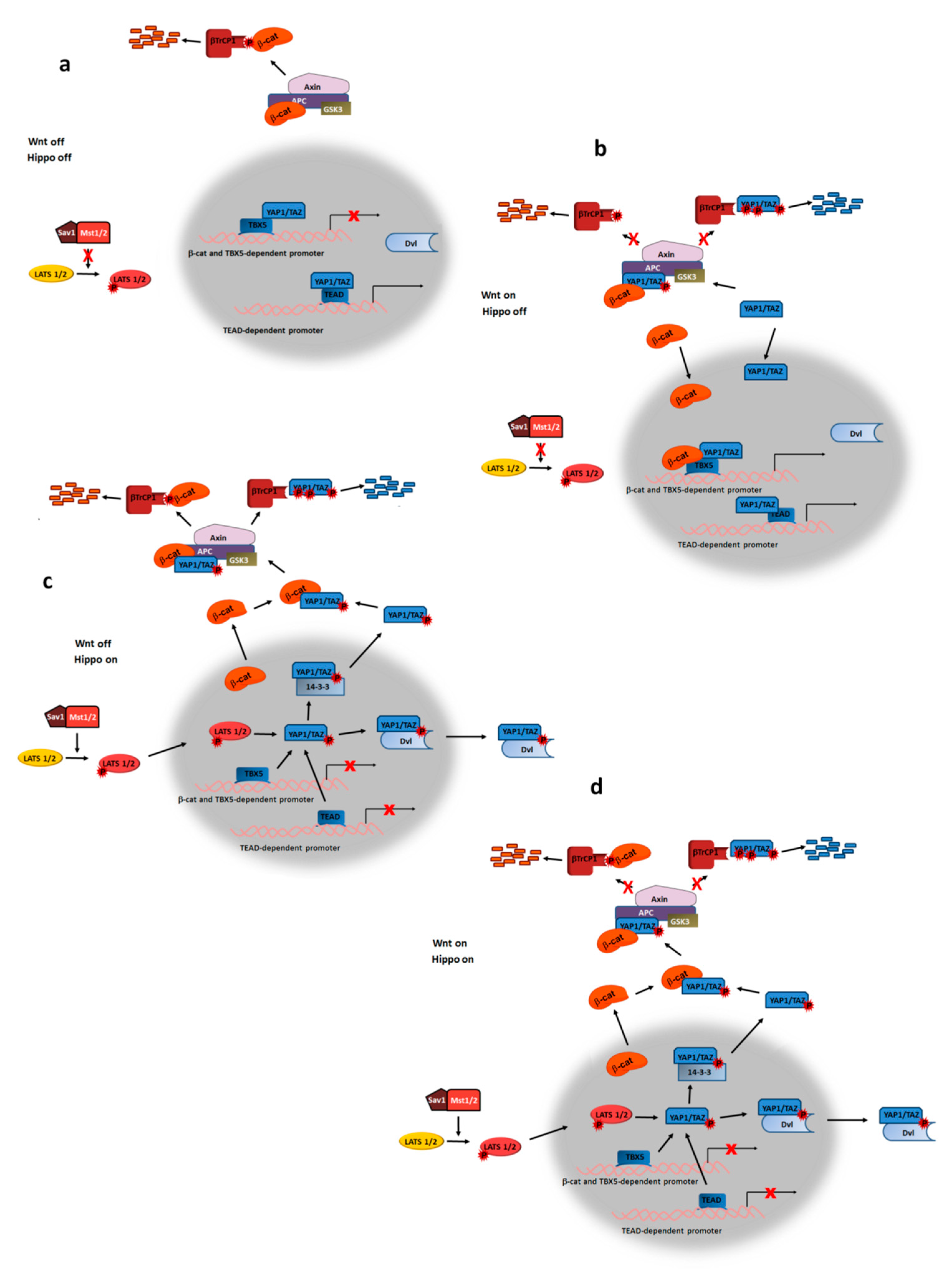
5. Wnt Ligands Modulate YAP/TAZ Transcriptional Activity
6. Concluding Remarks
Author Contributions
Funding
Acknowledgments
Conflicts of Interest
References
- Nusse, R.; Clevers, H. Wnt/β-catenin signaling, disease and emerging therapeutic modalities. Cell 2017, 169, 985–999. [Google Scholar] [CrossRef] [PubMed]
- Valenta, T.; Hausmann, G.; Basler, K. The many faces and functions of β-catenin. EMBO J. 2012, 31, 2714–2736. [Google Scholar] [CrossRef] [PubMed]
- van de Wetering, M.; Cavallo, R.; Dooijes, D.; van Beest, M.; van Es, J.; Loureiro, J.; Ypma, A.; Hursh, D.; Jones, T.; Bejsovec, A.; et al. Armadillo coactivates transcription driven by the product of the Drosophila segment polarity gene dTCF. Cell 1997, 88, 789–799. [Google Scholar] [CrossRef]
- Tenbaum, S.P.; Ordóñez-Morán, P.; Puig, I.; Chicote, I.; Arqués, O.; Landolfi, S.; Fernández, Y.; Herance, J.R.; Gispert, J.D.; Mendizabal, L.; et al. β-catenin confers resistance to PI3K and AKT inhibitors and subverts FOXO3a to promote metastasis in colon cancer. Nat. Med. 2012, 18, 892–901. [Google Scholar] [CrossRef] [PubMed]
- Botrugno, O.A.; Fayard, E.; Annicotte, J.S.; Haby, C.; Brennan, T.; Wendling, O.; Tanaka, T.; Kodama, T.; Thomas, W.; Auwerx, J.; et al. Synergy between LRH-1 and beta-catenin induces G1 cyclin-mediated cell proliferation. Mol. Cell. 2004, 15, 499–509. [Google Scholar] [CrossRef] [PubMed]
- Topol, L.; Jiang, X.; Choi, H.; Garrett-Beal, L.; Carolan, P.J.; Yang, Y. Wnt-5a inhibits the canonical Wnt pathway by promoting GSK-3-independent beta-catenin degradation. J. Cell Biol. 2003, 162, 899–908. [Google Scholar] [CrossRef] [PubMed]
- Vinyoles, M.; Del Valle-Pérez, B.; Curto, J.; Padilla, M.; Villarroel, A.; Yang, J.; García de Herreros, A.; Duñach, M. Activation of CK1ε by PP2A/PR61ε is required for the initiation of Wnt signaling. Oncogene 2017, 36, 429–438. [Google Scholar] [CrossRef]
- Duñach, M.; Del Valle-Pérez, B.; García de Herreros, A. p120-catenin in canonical Wnt signaling. Crit. Rev. Biochem. Mol. Biol. 2017, 52, 327–339. [Google Scholar] [CrossRef]
- Cruciat, C.M.; Dolde, C.; de Groot, R.E.; Ohkawara, B.; Reinhard, C.; Korswagen, H.C.; Niehrs, C. RNA helicase DDX3 is a regulatory subunit of casein kinase 1 in Wnt-β-catenin signaling. Science 2013, 339, 1436–1441. [Google Scholar] [CrossRef]
- Dolde, C.; Bischof, J.; Grüter, S.; Montada, A.; Halekotte, J.; Peifer, C.; Kalbacher, H.; Baumann, U.; Knippschild, U.; Suter, B. A CK1 FRET biosensor reveals that DDX3X is an essential activator of CK1ε. J. Cell Sci. 2018, 131, pii: jcs207316. [Google Scholar] [CrossRef]
- Klein, T.J.; Jenny, A.; Djiane, A.; Mlodzik, M. CKIepsilon/discs overgrown promotes both Wnt-Fz/beta-catenin and Fz/PCP signaling in Drosophila. Curr. Biol. 2006, 16, 1337–1343. [Google Scholar] [CrossRef] [PubMed]
- Strutt, H.; Price, M.A.; Strutt, D. Planar polarity is positively regulated by casein kinase epsilon in Drosophila. Curr. Biol. 2006, 16, 1329–1336. [Google Scholar] [CrossRef] [PubMed]
- Curto, J.; Del Valle-Pérez, B.; Villarroel, A.; Fuertes, G.; Vinyoles, M.; Peña, R.; García de Herreros, A.; Duñach, M. CK1ε and p120-catenin control Ror2 function in noncanonical Wnt signaling. Mol. Oncol. 2018, 12, 611–629. [Google Scholar] [CrossRef] [PubMed]
- Wong, H.C.; Bourdelas, A.; Krauss, A.; Lee, H.J.; Shao, Y.; Wu, D.; Mlodzik, M.; Shi, D.L.; Zheng, J. Direct binding of the PDZ domain of Dishevelled to a conserved internal sequence in the C-terminal region of Frizzled. Mol. Cell 2003, 12, 1251–1260. [Google Scholar] [CrossRef]
- Tauriello, D.V.F.; Jordens, I.; Kirchner, K.; Slootstra, J.W.; Kruitwagen, T.; Bouwman, B.A.; Noutsou, M.; Rüdiger, S.G.; Schwamborn, K.; Schambony, A.; et al. Wnt/β-catenin signaling requires interaction of the Dishevelled DEP domain and C terminus with a discontinuous motif in Frizzled. Proc. Natl. Acad. Sci. USA 2012, 109, 812–820. [Google Scholar] [CrossRef] [PubMed]
- Schwarz-Romond, T.; Fiedler, M.; Shibata, N.; Butler, P.J.; Kikuchi, A.; Higuchi, Y.; Bienz, M. The DIX domain of Dishevelled confers Wnt signaling by dynamic polymerization. Nat. Struct. Mol. Biol. 2007, 14, 484–492. [Google Scholar] [CrossRef] [PubMed]
- Gerlach, J.P.; Jordens, I.; Tauriello, D.V.F.; van ‘t Land-Kuper, I.; Bugter, J.M.; Noordstra, I.; van der Kooij, J.; Low, T.Y.; Pimentel-Muiños, F.X.; Xanthakis, D.; et al. TMEM59 potentiates Wnt signaling by promoting signalosome formation. Proc. Natl. Acad. Sci. USA 2018, 115, E3996–E4005. [Google Scholar] [CrossRef] [PubMed] [Green Version]
- Pan, W.; Choi, S.C.; Wang, H.; Qin, Y.; Volpicelli-Daley, L.; Swan, L.; Lucast, L.; Khoo, C.; Zhang, X.; Li, L.; et al. Wnt3a-mediated formation of phosphatidylinositol 4,5-bisphosphate regulates LRP6 phosphorylation. Science 2008, 321, 1350–1353. [Google Scholar] [CrossRef] [PubMed]
- Kim, I.; Pan, W.; Jones, S.A.; Zhang, Y.; Zhuang, X.; Wu, D. Clathrin and AP2 are required for PtdIns(4,5)P2-mediated formation of LRP6 signalosomes. J. Cell Biol. 2013, 200, 419–428. [Google Scholar] [CrossRef] [PubMed]
- Tanneberger, K.; Pfister, A.S.; Brauburger, K.; Schneikert, J.; Hadjihannas, M.V.; Kriz, V.; Schulte, G.; Bryja, V.; Behrens, J. Amer1/WTX couples Wnt-induced formation of PtdIns(4,5)P2 to LRP6 phosphorylation. EMBO J. 2011, 30, 1433–1443. [Google Scholar] [CrossRef]
- Gault, W.J.; Olguin, P.; Weber, U.; Mlodzik, M. Drosophila CK1-γ, gilgamesh, controls PCP-mediated morphogenesis through regulation of vesicle trafficking. J. Cell Biol. 2012, 196, 605–621. [Google Scholar] [CrossRef] [PubMed]
- Mao, J.; Wang, J.; Liu, B.; Pan, W.; Farr, G.H.; Flynn, C.; Yuan, H.; Takada, S.; Kimelman, D.; Li, L.; et al. Low-density lipoprotein receptor-related protein-5 binds to Axin and regulates the canonical Wnt signaling pathway. Mol. Cell 2001, 7, 801–809. [Google Scholar] [CrossRef]
- Davidson, G.; Wu, W.; Shen, J.; Bilic, J.; Fenger, U.; Stannek, P.; Glinka, A.; Niehrs, C. Casein kinase 1 gamma couples Wnt receptor activation to cytoplasmic signal transduction. Nature 2005, 438, 867–872. [Google Scholar] [CrossRef] [PubMed]
- Tortelote, G.G.; Reis, R.R.; de Almeida Mendes, F.; Abreu, J.G. Complexity of the Wnt/β-catenin pathway: Searching for an activation model. Cell Signal. 2017, 40, 30–43. [Google Scholar] [CrossRef] [PubMed]
- Gammons, M.; Bienz, M. Multiprotein complexes governing Wnt signal transduction. Curr. Opin. Cell Biol. 2018, 51, 42–49. [Google Scholar] [CrossRef] [PubMed]
- Kishida, M.; Koyama, S.; Kishida, S.; Matsubara, K.; Nakashima, S.; Higano, K.; Takada, R.; Takada, S.; Kikuchi, A. Axin prevents Wnt-3a-induced accumulation of beta-catenin. Oncogene 1999, 18, 979–985. [Google Scholar] [CrossRef]
- Li, L.; Yuan, H.; Weaver, C.D.; Mao, J.; Farr, G.H., 3rd; Sussman, D.J.; Jonkers, J.; Kimelman, D.; Wu, D. Axin and Frat1 interact with Dvl and GSK, bridging Dvl to GSK in Wnt-mediated regulation of LEF-1. EMBO J. 1999, 18, 4233–4240. [Google Scholar] [CrossRef] [Green Version]
- Liu, X.; Rubin, J.S.; Kimmel, A.R. Rapid, Wnt-induced changes in GSK3beta associations that regulate beta-catenin stabilization are mediated by Galpha proteins. Curr. Biol. 2005, 15, 1989–1997. [Google Scholar] [CrossRef]
- Kim, S.E.; Huang, H.; Zhao, M.; Zhang, X.; Zhang, A.; Semonov, M.V.; MacDonald, B.T.; Zhang, X.; Garcia Abreu, J.; Peng, L.; et al. Wnt stabilization of β-catenin reveals principles for morphogen receptor-scaffold assemblies. Science 2013, 340, 867–870. [Google Scholar] [CrossRef]
- Fiedler, M.; Mendoza-Topaz, C.; Rutherford, T.J.; Mieszczanek, J.; Bienz, M. Dishevelled interacts with the DIX domain polymerization interface of Axin to interfere with its function in down-regulating β-catenin. Proc. Natl. Acad. Sci. USA 2011, 108, 1937–1942. [Google Scholar] [CrossRef]
- Mi, K.; Dolan, P.J.; Johnson, G.V. The low density lipoprotein receptor-related protein 6 interacts with glycogen synthase kinase 3 and attenuates activity. J. Biol. Chem. 2006, 281, 4787–4794. [Google Scholar] [CrossRef] [PubMed]
- Cselenyi, C.S.; Jernigan, K.K.; Tahinci, E.; Thorne., C.A.; Lee, L.A.; Lee, E. LRP6 transduces a canonical Wnt signal independently of Axin degradation by inhibiting GSK3’s phosphorylation of beta-catenin. Proc. Natl. Acad. Sci.USA 2008, 105, 8032–8037. [Google Scholar] [CrossRef] [PubMed]
- Piao, S.; Lee, S.H.; Kim, H.; Yum, S.; Stamos, J.L.; Xu, Y.; Lee, S.J.; Lee, J.; Oh, S.; Han, J.K.; et al. Direct inhibition of GSK3beta by the phosphorylated cytoplasmic domain of LRP6 in Wnt/beta-catenin signaling. PLoS ONE 2008, 3, e4046. [Google Scholar] [CrossRef] [PubMed]
- Wu, G.; Huang, H.; Garcia Abreu, J.; He, X. Inhibition of GSK3 phosphorylation of beta-catenin via phosphorylated PPPSPXS motifs of Wnt coreceptor LRP6. PLoS ONE 2009, 4, e4926. [Google Scholar] [CrossRef] [PubMed]
- Li, V.S.; Ng, S.S.; Boersema, P.J.; Low, T.Y.; Karthaus, W.R.; Gerlach, J.P.; Mohammed, S.; Heck, A.J.; Maurice, M.M.; Mahmoudi, T.; et al. Wnt signaling through inhibition of β-catenin degradation in an intact Axin1 complex. Cell 2012, 149, 1245–1256. [Google Scholar] [CrossRef] [PubMed]
- Taelman, V.; Dobrowolski, R.; Plouhinec, J.L.; Fuentealba, L.C.; Vorwald, P.P.; Gumper, I.; Sabatini, D.D.; De Robertis, E.M. Wnt signaling requires sequestration of glycogen synthase kinase 3 inside multivesicular endosomes. Cell 2010, 143, 1136–1148. [Google Scholar] [CrossRef] [PubMed]
- Yamamoto, H.; Komekado, H.; Kikuchi, A. Caveolin is necessary for Wnt-3a-dependent internalization of LRP6 and accumulation of beta-catenin. Dev. Cell 2006, 11, 213–223. [Google Scholar] [CrossRef]
- Yamamoto, H.; Sakane, H.; Yamamoto, H.; Michiue, T.; Kikuchi, A. Wnt3a and Dkk1 regulate distinct internalization pathways of LRP6 to tune the activation of β-catenin signaling. Dev. Cell 2008, 15, 37–48. [Google Scholar] [CrossRef]
- Jiang, Y.; He, X.; Howe, P.H. Disabled-2 (Dab2) inhibits Wnt/β-catenin signalling by binding LRP6 and promoting its internalization through clathrin. EMBO J. 2012, 31, 2336–2349. [Google Scholar] [CrossRef]
- Vinyoles, M.; Del Valle-Pérez, B.; Curto, J.; Viñas-Castells, R.; Alba-Castellón, L.; García de Herreros, A.; Duñach, M. Multivesicular GSK3 sequestration upon Wnt signaling is controlled by p120-catenin/cadherin interaction with LRP5/6. Mol. Cell 2014, 53, 444–457. [Google Scholar] [CrossRef]
- Acebron, S.P.; Karaulanov, E.; Berger, B.S.; Huang, Y.L.; Niehrs, C. Mitotic Wnt signaling promotes protein stabilization and regulates cell size. Mol. Cell 2014, 54, 663–674. [Google Scholar] [CrossRef] [PubMed]
- Acebron, S.P.; Niehrs, C. β-catenin-independent roles of Wnt/LRP6 signaling. Trends Cell Biol. 2016, 26, 956–967. [Google Scholar] [CrossRef] [PubMed]
- Inoki, K.; Ouyang, H.; Zhu, T.; Lindvall, C.; Wang, Y.; Zhang, X.; Yang, Q.; Bennett, C.; Harada, Y.; Stankunas, K.; et al. TSC2 integrates Wnt and energy signals via a coordinated phosphorylation by AMPK and GSK3 to regulate cell growth. Cell 2006, 126, 955–968. [Google Scholar] [CrossRef] [PubMed]
- Gujral, T.S.; Chan, M.; Peshkin, L.; Sorger, P.K.; Kirschner, M.W.; MacBeath, G.A. Noncanonical Frizzled2 pathway regulates epithelial-mesenchymal transition and metastasis. Cell 2014, 159, 844–856. [Google Scholar] [CrossRef] [PubMed]
- Villarroel, A.; Del Valle-Pérez, B.; Fuertes, G.; Curto, J.; Ontiveros, N.; Garcia de Herreros, A.; Duñach, M. Src and Fyn define a new signaling cascade activated by canonical and non-canonical Wnt ligands and required for gene transcription and cell invasion. Cell. Mol. Life Sci. 2019. [Google Scholar] [CrossRef] [PubMed]
- Almeida, M.; Han, L.; Bellido, T.; Manolagas, S.C.; Kousteni, S. Wnt proteins prevent apoptosis of both uncommitted osteoblast progenitors and differentiated osteoblasts by beta-catenin-dependent and -independent signaling cascades involving Src/ERK and phosphatidylinositol 3-kinase/AKT. J. Biol. Chem. 2005, 280, 41342–41351. [Google Scholar] [CrossRef] [PubMed]
- Yokoyama, N.; Malbon, C.C. Dishevelled-2 docks and activates Src in a Wnt-dependent manner. J. Cell Sci. 2009, 122, 4439–4451. [Google Scholar] [CrossRef] [PubMed] [Green Version]
- Akbarzadeh, S.; Wheldon, L.M.; Sweet, S.M.; Talma, S.; Mardakheh, F.K.; Heath, J.K. The deleted in brachydactyly B domain of ROR2 is required for receptor activation by recruitment of Src. PLoS ONE 2008, 3, e1873. [Google Scholar] [CrossRef]
- Chen, Q.; Su, Y.; Wesslowski, J.; Hagemann, A.I.; Ramialison, M.; Wittbrodt, J.; Scholpp, S.; Davidson, G. Tyrosine phosphorylation of LRP6 by Src and Fer inhibits Wnt/β-catenin signalling. EMBO Rep. 2014, 15, 1254–1267. [Google Scholar] [CrossRef] [PubMed]
- Nile, A.H.; Mukund, S.; Stanger, K.; Wang, W.; Hannoush, R.N. Unsaturated fatty acyl recognition by Frizzled receptors mediates dimerization upon Wnt ligand binding. Proc. Natl. Acad. Sci. USA 2017, 114, 4147–4152. [Google Scholar] [CrossRef] [Green Version]
- DeBruine, Z.J.; Ke, J.; Harikumar, K.G.; Gu, X.; Borowsky, P.; Williams, B.O.; Xu, W.; Miller, L.J.; Xu, H.E.; Melcher, K. Wnt5a promotes Frizzled-4 signalosome assembly by stabilizing cysteine-rich domain dimerization. Genes Dev. 2017, 31, 916–926. [Google Scholar] [CrossRef] [PubMed] [Green Version]
- Piedra, J.; Miravet, S.; Castaño, J.; Pálmer, H.G.; Heisterkamp, N.; García de Herreros, A.; Duñach, M. p120 Catenin-associated Fer and Fyn tyrosine kinases regulate beta-catenin Tyr-142 phosphorylation and beta-catenin-alpha-catenin interaction. Mol. Cell. Biol. 2003, 23, 2287–2297. [Google Scholar] [CrossRef] [PubMed]
- Brembeck, F.H.; Schwarz-Romond, T.; Bakkers, J.; Wilhelm, S.; Hammerschmidt, M.; Birchmeier, W. Essential role of BCL9-2 in the switch between beta-catenin’s adhesive and transcriptional functions. Genes Dev. 2004, 18, 2225–2230. [Google Scholar] [CrossRef] [PubMed]
- Fiedler, M.; Graeb, M.; Mieszczanek, J.; Rutherford, T.J.; Johnson, C.M.; Bienz, M. An ancient Pygo-dependent Wnt enhanceosome integrated by Chip/LDB-SSDP. Elife 2015, 4, e09073. [Google Scholar] [CrossRef] [PubMed]
- Fragoso, M.A.; Patel, A.K.; Nakamura, R.E.; Yi, H.; Surapaneni, K.; Hackam, A.S. The Wnt/β-catenin pathway cross-talks with STAT3 signaling to regulate survival of retinal pigment epithelium cells. PLoS ONE 2012, 7, e46892. [Google Scholar] [CrossRef]
- Dijksterhuis, J.P.; Baljinnyam, B.; Stanger, K.; Sercan, H.O.; Ji, Y.; Andres, O.; Rubin, J.S.; Hannoush, R.N.; Schulte, G. Systematic mapping of WNT-FZD protein interactions reveals functional selectivity by distinct WNT-FZD pairs. J. Biol. Chem. 2015, 290, 6789–6798. [Google Scholar] [CrossRef]
- Park, H.W.; Kim, Y.C.; Yu, B.; Moroishi, T.; Mo, J.S.; Plouffe, S.W.; Meng, Z.; Lin, K.C.; Yu, F.X.; Alexander, C.M.; et al. Alternative Wnt signaling activates YAP/TAZ. Cell 2015, 162, 780–794. [Google Scholar] [CrossRef]
- Habas, R.; Dawid, I.B.; He, X. Coactivation of Rac and Rho by Wnt/Frizzled signaling is required for vertebrate gastrulation. Genes Dev. 2003, 17, 295–309. [Google Scholar] [CrossRef] [Green Version]
- Wu, X.; Tu, X.; Joeng, K.S.; Hilton, M.J.; Williams, D.; Long, F. Rac1 activation controls nuclear localization of beta-catenin during canonical Wnt signaling. Cell 2008, 133, 340–353. [Google Scholar] [CrossRef]
- Phelps, R.A.; Chidester, S.; Dehghanizadeh, S.; Phelps, J.; Sandoval, I.T.; Rai, K.; Broadbent, T.; Sarkar, S.; Burt, R.W.; Jones, D.A. A two-step model for colon adenoma initiation and progression caused by APC loss. Cell 2009, 37, 623–634. [Google Scholar] [CrossRef]
- Eaton, S.; Wepf, R.; Simons, K. Roles for Rac1 and Cdc42 in planar polarization and hair outgrowth in the wing of Drosophila. J. Cell Biol. 1996, 135, 1277–1289. [Google Scholar] [CrossRef] [PubMed]
- Zhu, G.; Wang, Y.; Huang, B.; Liang, J.; Ding, Y.; Xu, A.; Wu, W. A Rac1/PAK1 cascade controls β-catenin activation in colon cancer cells. Oncogene 2012, 31, 1001–1012. [Google Scholar] [CrossRef]
- Jamieson, C.; Lui, C.; Brocardo, M.G.; Martino-Echarri, E.; Henderson, B.R. Rac1 augments Wnt signaling by stimulating β-catenin-lymphoid enhancer factor-1 complex assembly independent of β-catenin nuclear import. J. Cell Sci. 2015, 128, 3933–3946. [Google Scholar] [CrossRef] [PubMed]
- Roura, S.; Miravet, S.; Piedra, J.; García de Herreros, A.; Duñach, M. Regulation of E-cadherin/catenin association by tyrosine phosphorylation. J. Biol. Chem. 1999, 274, 36734–36740. [Google Scholar] [CrossRef]
- Cajánek, L.; Ganji, R.S.; Henriques-Oliveira, C.; Theofilopoulos, S.; Koník, P.; Bryja, V.; Arenas, E. Tiam1 regulates the Wnt/Dvl/Rac1 signaling pathway and the differentiation of midbrain dopaminergic neurons. Mol. Cell. Biol. 2013, 33, 59–70. [Google Scholar] [CrossRef]
- Takagishi, M.; Sawada, M.; Ohata, S.; Asai, N.; Enomoto, A.; Takahashi, K.; Weng, L.; Ushida, K.; Ara, H.; Matsui, S.; et al. Daple Coordinates Planar Polarized Microtubule Dynamics in Ependymal Cells and Contributes to Hydrocephalus. Cell Rep. 2017, 20, 960–972. [Google Scholar] [CrossRef] [PubMed] [Green Version]
- Valls, G.; Codina, M.; Miller, R.K.; Del Valle-Pérez, B.; Vinyoles, M.; Caelles, C.; McCrea, P.D.; García de Herreros, A.; Duñach, M. Upon Wnt stimulation, Rac1 activation requires Rac1 and Vav2 binding to p120-catenin. J. Cell Sci. 2012, 125, 5288–5301. [Google Scholar] [CrossRef]
- Casagolda, D.; Del Valle, B.; Valls, G.; Lugilde, E.; Vinyoles, M.; Casado-Vela, J.; Solanas, G.; Batlle, E.; Reynolds, A.B.; Casal, J.I.; et al. A p120-catenin-CK1ε complex regulates Wnt signaling. J. Cell. Sci. 2010, 123, 2621–2631. [Google Scholar] [CrossRef]
- Del Valle-Pérez, B.; Arqués, O.; Vinyoles, M.; Garcia de Herreros, A.; Duñach, M. Coordinated action of CK1 isoforms in canonical Wnt signaling. Mol. Cell. Biol. 2011, 31, 2877–2888. [Google Scholar] [CrossRef]
- Noren, N.K.; Liu, B.P.; Burridge, K.; Kreft, B. p120 catenin regulates the actin cytoskeleton via Rho family GTPases. J. Cell. Biol. 2000, 150, 567–580. [Google Scholar] [CrossRef]
- Grosheva, I.; Shtutman, M.; Elbaum, M.; Bershadsky, A.D. p120-catenin affects cell motility via modulation of Rho-family GTPases: A link between cell-cell contact formation and regulation of cell locomotion. J. Cell Sci. 2001, 114, 695–707. [Google Scholar] [PubMed]
- Yu, B.; Martins, I.R.S.; Li, P.; Amarasinghe, G.K.; Umetani, J.; Fernandez-Zapico, M.E.; Billadeau, D.D.; Machius, M.; Tomchick, D.R.; Rosen, M.K. Structural and energetic mechanisms of cooperative autoinhibition and activation of Vav1. Cell 2010, 140, 246–256. [Google Scholar] [CrossRef] [PubMed]
- Fang, X.; Ji, H.; Kim, S.W.; Park, J.I.; Vaught, T.G.; Anastasiadis, P.Z.; Ciesiolka, M.; McCrea, P.D. Vertebrate development requires ARVCF and p120 catenins and their interplay with RhoA and Rac. J. Cell. Biol. 2004, 165, 87–98. [Google Scholar] [CrossRef] [PubMed] [Green Version]
- Nateri, A.S.; Spencer-Dene, B.; Behrens, A. Interaction of phosphorylated c-Jun with TCF4 regulates intestinal cancer development. Nature 2005, 437, 281–285. [Google Scholar] [CrossRef] [PubMed]
- Gan, X.Q.; Wang, J.Y.; Xi, Y.; Wu, Z.L.; Li, Y.P.; Li, L. Nuclear Dvl, c-Jun, beta-catenin, and TCF form a complex leading to stabilization of beta-catenin-TCF interaction. J. Cell Biol. 2008, 180, 1087–1100. [Google Scholar] [CrossRef] [PubMed]
- Goto, T.; Sato, A.; Shimizu, M.; Adachi, S.; Satoh, K.; Iemura, S.; Natsume, T.; Shibuya, H. IQGAP1 functions as a modulator of dishevelled nuclear localization in Wnt signaling. PLoS ONE 2013, 8, e60865. [Google Scholar] [CrossRef] [PubMed]
- Wang, W.; Li, X.; Lee, M.; Jun, S.; Aziz, K.E.; Feng, L.; Tran, M.K.; Li, N.; McCrea, P.D.; Park, J.I.; et al. FOXKs promote Wnt/β-catenin signaling by translocating DVL into the nucleus. Dev. Cell 2015, 32, 707–718. [Google Scholar] [CrossRef] [PubMed]
- Deng, N.; Ye, Y.; Wang, W.; Li, L. Dishevelled interacts with p65 and acts as a repressor of NF-κB-mediated transcription. Cell Res. 2010, 20, 1117–1127. [Google Scholar] [CrossRef]
- Rodova, M.; Kelly, K.F.; VanSaun, M.; Daniel, J.M.; Werle, M.J. Regulation of the rapsyn promoter by kaiso and delta-catenin. Mol. Cell Biol. 2004, 24, 7188–7196. [Google Scholar] [CrossRef]
- Park, J.I.; Kim, S.W.; Lyons, J.P.; Ji, H.; Nguyen, T.T.; Cho, K.; Barton, M.C.; Deroo, T.; Vleminckx, K.; Moon, R.T.; et al. Kaiso/p120-catenin and TCF/β-catenin complexes coordinately regulate canonical Wnt gene targets. Dev. Cell. 2005, 8, 843–854. [Google Scholar] [CrossRef]
- Spring, C.M.; Kelly, K.F.; O’Kelly, I.; Graham, M.; Crawford, H.C.; Daniel, J.M. The catenin p120ctn inhibits Kaiso-mediated transcriptional repression of the beta-catenin/TCF target gene matrilysin. Exp. Cell Res. 2005, 305, 253–265. [Google Scholar] [CrossRef] [PubMed]
- Ruzov, A.; Hackett, J.A.; Prokhortchouk, A.; Reddington, J.P.; Madek, M.J.; Dunican, D.S.; Prokhortchouk, E.; Pennings, S.; Meehan, R.R. The interaction of xKaiso with xTcf3: A revised model for integration of epigenetic and Wnt signaling pathways. Development 2009, 136, 723–727. [Google Scholar] [CrossRef] [PubMed]
- Del Valle-Pérez, B.; Casagolda, D.; Lugilde, E.; Valls, G.; Codina, M.; Dave, N.; García de Herreros, A.; Duñach, M. Wnt controls the transcriptional activity of Kaiso through CK1ε-dependent phosphorylation of p120-catenin. J. Cell Sci. 2011, 124, 2298–2309. [Google Scholar] [CrossRef] [PubMed]
- Ruzov, A.; Savitskaya, E.; Hackett, J.A.; Reddington, J.P.; Prokhortchouk, A.; Madek, M.J.; Chekanov, N.; Li, M.; Dunican, D.S.; Prokhortchouk, E.; et al. The non-methylated DNA-binding function of Kaiso is not required in early Xenopus laevis development. Development 2009, 136, 729–738. [Google Scholar] [CrossRef] [PubMed]
- Prokhortchouk, A.; Hendrich, B.; Jorgensen, H.; Ruzov, A.; Wilm, M.; Georgiev, G.; Bird, A.; Prokhortchouk, E. The p120-catenin partner Kaiso is a DNA methylation-dependent transcriptional repressor. Genes Dev. 2001, 15, 1613–1618. [Google Scholar] [CrossRef] [PubMed]
- Yoon, H.G.; Chan, D.W.; Reynolds, A.B.; Qin, J.; Wong, J. N-CoR mediates DNA methylation-dependent repression through a methyl CpG binding protein Kaiso. Mol. Cell. 2003, 12, 723–734. [Google Scholar] [CrossRef] [PubMed]
- Roczniak-Ferguson, A.; Reynolds, A.B. Regulation of p120-catenin nucleocytoplasmic shuttling activity. J. Cell Sci. 2003, 116, 4201–4212. [Google Scholar] [CrossRef] [PubMed] [Green Version]
- Kelly, K.F.; Spring, C.M.; Otchere, A.A.; Daniel, J.M. NLS-dependent nuclear localization of p120ctn is necessary to relieve Kaiso-mediated transcriptional repression. J. Cell Sci. 2004, 117, 2675–2686. [Google Scholar] [CrossRef] [PubMed]
- Zhenilo, S.; Deyev, I.; Litvinova, E.; Zhigalova, N.; Kaplun, D.; Sokolov, A.; Mazur, A.; Prokhortchouk, E. DeSUMOylation switches Kaiso from activator to repressor upon hyperosmotic stress. Cell Death Differ. 2018, 25, 1938–1951. [Google Scholar] [CrossRef]
- Hosking, C.R.; Ulloa, F.; Hogan, C.; Ferber, E.C.; Figueroa, A.; Gevaert, K.; Birchmeier, W.; Briscoe, J.; Fujita, Y. The transcriptional repressor Glis2 is a novel binding partner for p120 catenin. Mol. Biol. Cell 2007, 18, 1918–1927. [Google Scholar] [CrossRef]
- Lee, M.; Ji, H.; Furuta, Y.; Park, J.I.; McCrea, P.D. p120-catenin regulates REST/CoREST and modulates mouse embryonic stem cell differentiation. J. Cell Sci. 2014, 127, 4037–4051. [Google Scholar] [CrossRef] [PubMed]
- Park, J.I.; Ji, H.; Jun, S.; Gu, D.; Hikasa, H.; Li, L.; Sokol, S.Y.; McCrea, P.D. Frodo links disheveled to the p120-catenin/Kaiso pathway: Distinct subfamilies promote Wnt signals. Dev. Cell 2006, 11, 683–695. [Google Scholar] [CrossRef] [PubMed]
- Hong, J.Y.; Park, J.I.; Cho, K.; Gu, D.; Ji, H.; Artandi, S.E.; McCrea, P.D. Shared molecular mechanisms regulate multiple catenin proteins: Canonical Wnt signals and components modulate p120-catenin isoform-1 and additional p120 subfamily members. J. Cell Sci. 2010, 123, 4351–4365. [Google Scholar] [CrossRef] [PubMed]
- Hong, J.Y.; Park, J.I.; Lee, M.; Muñoz, W.A.; Miller, R.K.; Ji, H.; Gu, D.; Ezan, J.; Sokol, S.Y.; McCrea, P.D. Down’s-syndrome-related kinase Dyrk1A modulates the p120-catenin-Kaiso trajectory of the Wnt signaling pathway. J. Cell Sci. 2011, 25, 561–569. [Google Scholar]
- Zheng, Y.; Pan, D. The Hippo signaling pathway in development and disease. Dev. Cell 2019, 50, 264–282. [Google Scholar] [CrossRef] [PubMed]
- Kriz, V.; Korinek, V. Wnt, RSPO and Hippo Signalling in the Intestine and Intestinal Stem Cells. Genes 2018, 9, 20. [Google Scholar] [CrossRef]
- Rosenbluh, J.; Nijhawan, D.; Cox, A.G.; Li, X.; Neal, J.T.; Schafer, E.J.; Zack, T.I.; Wang, X.; Tsherniak, A.; Schinzel, A.C.; et al. β-Catenin-driven cancers require a YAP1 transcriptional complex for survival and tumorigenesis. Cell 2012, 151, 1457–1473. [Google Scholar] [CrossRef]
- Gruber, R.; Panayiotou, R.; Nye, E.; Spencer-Dene, B.; Stamp, G.; Behrens, A. YAP1 and TAZ Control Pancreatic Cancer Initiation in Mice by Direct Up-regulation of JAK-STAT3 Signaling. Gastroenterology 2016, 151, 526–539. [Google Scholar] [CrossRef]
- Kim, W.; Khan, S.K.; Gvozdenovic-Jeremic, J.; Kim, Y.; Dahlman, J.; Kim, H.; Park, O.; Ishitani, T.; Jho, E.H.; Gao, B.; et al. Hippo signaling interactions with Wnt/β-catenin and Notch signaling repress liver tumorigenesis. J. Clin. Invest. 2017, 127, 137–152. [Google Scholar] [CrossRef]
- Oudhoff, M.J.; Braam, M.J.S.; Freeman, S.A.; Wong, D.; Rattray, D.G.; Wang, J.; Antignano, F.; Snyder, K.; Refaeli, I.; Hughes, M.R.; et al. SETD7 Controls Intestinal Regeneration and Tumorigenesis by Regulating Wnt/β-Catenin and Hippo/YAP Signaling. Dev. Cell 2016, 37, 47–57. [Google Scholar] [CrossRef]
- Imajo, M.; Miyatake, K.; Iimura, A.; Miyamoto, A.; Nishida, E. A molecular mechanism that links Hippo signalling to the inhibition of Wnt/β-catenin signalling. EMBO J. 2012, 31, 1109–1122. [Google Scholar] [CrossRef] [PubMed]
- Cai, J.; Maitra, A.; Anders, R.A.; Taketo, M.M.; Pan, D. β-Catenin destruction complex-independent regulation of Hippo-YAP signaling by APC in intestinal tumorigenesis. Genes Dev. 2015, 29, 1493–1506. [Google Scholar] [CrossRef] [PubMed]
- Azzolin, L.; Zanconato, F.; Bresolin, S.; Forcato, M.; Basso, G.; Bicciato, S.; Cordenonsi, M.; Piccolo, S. Role of TAZ as mediator of Wnt signaling. Cell 2012, 151, 1443–1456. [Google Scholar] [CrossRef] [PubMed]
- Azzolin, L.; Panciera, T.; Soligo, S.; Enzo, E.; Bicciato, S.; Dupont, S.; Bresolin, S.; Frasson, C.; Basso, G.; Guzzardo, V.; et al. YAP/TAZ incorporation in the β-catenin destruction complex orchestrates the Wnt response. Cell 2014, 158, 157–170. [Google Scholar] [CrossRef]
- Barry, E.R.; Morikawa, T.; Butler, B.L.; Shrestha, K.; de la Rosa, R.; Yan, K.S.; Fuchs, C.S.; Magness, S.T.; Smits, R.; Ogino, S.; et al. Restriction of intestinal stem cell expansion and the regenerative response by YAP. Nature 2013, 493, 106–110. [Google Scholar] [CrossRef] [PubMed]
- Lee, Y.; Kim, N.H.; Cho, E.S.; Yang, J.H.; Cha, Y.H.; Kang, H.E.; Yun, J.S.; Cho, S.B.; Lee, S.H.; Paclikova, P.; et al. Dishevelled has a YAP nuclear export function in a tumor suppressor context-dependent manner. Nat. Commun. 2018, 9, 2301. [Google Scholar] [CrossRef] [PubMed]
- Yu, F.X.; Zhao, B.; Panupinthu, N.; Jewell, J.L.; Lian, I.; Wang, L.H.; Zhao, J.; Yuan, H.; Tumaneng, K.; Li, H.; et al. Regulation of the Hippo-YAP pathway by G-protein-coupled receptor signaling. Cell 2012, 150, 780–791. [Google Scholar] [CrossRef] [PubMed]
- Yu, F.X.; Luo, J.; Mo, J.S.; Liu, G.; Kim, Y.C.; Meng, Z.; Zhao, L.; Peyman, G.; Ouyang, H.; Jiang, W.; et al. Mutant Gq/11 promote uveal melanoma tumorigenesis by activating YAP. Cancer Cell 2014, 25, 822–830. [Google Scholar] [CrossRef] [PubMed]
- Regard, J.B.; Cherman, N.; Palmer, D.; Kuznetsov, S.A.; Celi, F.S.; Guettier, J.M.; Chen, M.; Bhattacharyya, N.; Wess, J.; Coughlin, S.R.; et al. Wnt/β-catenin signaling is differentially regulated by Gα proteins and contributes to fibrous dysplasia. Proc. Natl. Acad. Sci. USA 2011, 108, 20101–20106. [Google Scholar] [CrossRef] [PubMed]
- Ma, B.; Chen, Y.; Chen, L.; Cheng, H.; Mu, C.; Li, J.; Gao, R.; Zhou, C.; Cao, L.; Liu, J.; et al. Hypoxia regulates Hippo signalling through the SIAH2 ubiquitin E3 ligase. Nat. Cell Biol. 2015, 17, 95–103. [Google Scholar] [CrossRef] [PubMed]
- Ji, L.; Jiang, B.; Jiang, X.; Charlat, O.; Chen, A.; Mickanin, C.; Bauer, A.; Xu, W.; Yan, X.; Cong, F. The SIAH E3 ubiquitin ligases promote Wnt/β-catenin signaling through mediating Wnt-induced Axin degradation. Genes Dev. 2017, 31, 904–915. [Google Scholar] [CrossRef] [PubMed]


| Name | Function |
|---|---|
| 14-3-3 | Chaperone proteins binding YAP/TAZ after phosphorylation by LATS1/2 and promoting their nuclear export |
| α-catenin | Linking protein between β-catenin-E-cadherin complex that regulates actin filament assembly. |
| AMER/WTX (APC Membrane Recruitment Protein 1) (Wilms Tumor On The X) | Protein acting in Wnt pathway that facilitates the formation of complex composed by Axin, GSK3 and LRP. |
| AP2 (Adaptor protein 2) | Adaptor protein which contributes to clathrin-mediated endocytosis. |
| APC (Adematous polyposis coli) | Negative regulator of the Wnt pathway that controls β-catenin degradation. Also regulates the Hippo pathway controlling TAZ degradation. |
| ARVCF (Armadillo Repeat Protein Deleted In Velo-Cardio-Facial Syndrome) | Member of the armadillo protein family related to p120-catenin and involved in Rac1 activation |
| Axin | Member of the β-catenin and TAZ destruction complex. Negative regulator of the canonical Wnt pathway. |
| BCL9 | β-catenin cofactor relevant for the transcription of canonical Wnt target genes |
| β-catenin | Protein involved in the regulation of cellular adhesion and gene transcription. Acts as an intracellular signal transducer in the Wnt signaling cascade. |
| βTrCP1 (Beta-Transducin Repeat Containing E3 Ubiquitin Protein Ligase) | Ubiquitin ligase that regulates the Wnt and Hippo pathways through β-catenin and TAZ degradation, respectively. |
| Caveolin | Membrane protein involved in Wnt-dependent LRP5/6/Dvl2/Axin/GSK3 internalization and required for β-catenin accumulation. |
| CK1α (Casein Kinase 1 alpha) | Protein kinase that associates to Axin and participates in the β-catenin degradation complex phosphorylating this protein and Axin |
| CK1γ (Casein Kinase 1 gamma) | Protein Kinase that is recruited to the Wnt signalosome and promotes LRP5/6 phosphorylation |
| CK1ε (Casein Kinase 1 epsilon) | Protein kinase rapidly activated canonical and non-canonical Wnt ligands and required for Dvl2 association to the receptor complex. |
| Daple | Protein involved in Rac1 activation by non-canonical Wnt |
| DDX3 (DEAD-Box Helicase 3 X-Linked) | RNA helicase activated by Wnt signaling relevant for CK1ε activation |
| DKK1 (Dickkopf WNT Signaling Pathway Inhibitor 1) | Extracellular protein inhibitor of the canonical Wnt pathway. |
| Dvl2 (Dishevelled 2) | Cytosolic adaptor protein involved in canonical and non-canonical Wnt pathway. Wnt activation recruits Dvl to the membrane. It participates in assembling the signalosome. It is also detected in the nucleus bound to p65, FOXK and c-Jun |
| DYRK1 (Dual Specificity Tyrosine Phosphorylation Regulated Kinase 1A) | Protein tyrosine kinase that enhances p120-catenin stability and relieves Kaiso repression |
| E- and N-cadherin | Transmembrane proteins directly bound to β-catenin, p120-catenin and LRP5/6 at the adherens junctions essential for intercellular interactions |
| FOXK1/2 (Forkhead Box K1) | Transcriptional factor involved in the nuclear localization of Dvl2 |
| FOXO3 (Forkhead Box O3) | Transcriptional factor that upon binding to β-catenin behaves as a transcriptional activator |
| Frodo | Dvl2-associated protein that also interacts with p120-catenin |
| Fyn | Member of the Src family of protein kinases activated by canonical and non-canonical Wnts and required for Stat3 phosphorylation and transcriptional action |
| Fz (Frizzled) | Family of membrane receptors involved in canonical and non-canonical Wnt signaling. |
| Gα12/13 (G Protein Subunits Alpha 12 and 13) | Family of heterotrimeric G proteins required for Rho activation and the inhibition of Lats1/2 in the Hippo pathway. |
| Glis2 | Transcriptional repressor bound to p120-catenin in the nucleus |
| GSK3 (Glycogen Synthase Kinase 3) | Serine/threonine protein kinase which phosphorylates β-catenin targeting it for degradation. Inhibited by canonical Wnt |
| IQGAP1 (IQ Motif Containing GTPase Activating Protein 1) | Modulator of Dvl2 nuclear localization in Wnt signaling. |
| JNK2 (c-Jun N-Terminal Kinase 2) | Protein kinase activated by canonical Wnt that modifies β-catenin and c-Jun facilitating gene transcription |
| c-Jun | Substrate of JNK2 that interacts with Tcf4/β-catenin to regulate gene transcription. |
| Kaiso | Transcriptional repressor of canonical Wnt target genes. Interacts with TCF4, β-catenin and p120-catenin. |
| LATS1/2 (Large Tumor Suppressor Kinase 1 and 2) | Protein kinase that phosphorylates YAP1 and TAZ upon activation of the Hippo pathway promotes their nuclear export. |
| LRH1 (Liver Receptor Homolog-1) | Nuclear orphan receptor that interacts with β-catenin independent of TCF4. Is activated by β-catenin; also promotes activation of β-catenin/TCF4 complex. |
| LRP5/6 (LDL Receptor Related Protein 5 and 6) | Membrane co-receptor involved in canonical Wnt signaling |
| Mst1/2 (Mammalian STE20-Like Protein Kinase 1 and 2) | Protein kinase stimulated by the Hippo pathway that activates Lats1/2. |
| p120-catenin | Protein of the armadillo family essential for canonical and non-canonical Wnt signaling. Interacts and stabilizes in the plasma membrane Cadherin/LRP and Ror2. Also binds and regulates CK1ε, Rac1 and Kaiso |
| p65 (Rel A proto-oncogene) | Transcription factor member of the NF- B complex. Binds Dvl2 in the nucleus. |
| PAK1 (P21 (RAC1) Activated Kinase 1) | Protein kinase stimulated by Rac1 in canonical and non-canonical Wnt. Activates JNK2 and promotes β-catenin translocation to the nucleus. |
| PKC-λ (Protein Kinase C-λ) | Atypical PKC that interacts with Dvl2 |
| PR61ε (Protein Phosphatase 2, Regulatory Subunit B (B56), Epsilon Isoform) | Regulatory subunit of the PP2A phosphatase. Required for the activation of CK1ε at the initiation of the canonical and non-canonical Wnt signaling. |
| Pygo (Pygopus Family PHD Finger) | Transcriptional factor that binds to Bcl9 enhancing β-catenin transcriptional activation |
| Rac1 | Small GTPase activated by canonical and non-canonical Wnts. Activates PAK. Inhibits LATS1/2 up-regulating YAP1/TAZ transcriptional activity |
| REST/CoREST (RE1 Silencing Transcription Factor) | Transcriptional complex modulated by p120-catenin |
| RhoA | Small GTPases that inhibits LATS1/2 activity in the Hippo pathway |
| Ror1,2 (Retinoid-Related Orphan Receptor) | Tyrosine kinase-like orphan transmembrane co-receptor required for non-canonical Wnt signaling |
| Ryk | Transmembrane co-receptor involved in the non-canonical Wnt pathway. |
| Sav1 (Salvador Family WW Domain Containing Protein 1) | Regulator of Mst1 in the Hipo pathway required for LATS1 phosphorylation and activation |
| SIAH2 (Seven In Absentia (Drosophila) Homolog 2) | E3 ubiquitin-protein ligase stimulated by non-canonical Wnt involved in β-catenin degradation independent on GSK3 |
| Snail1 | Transcription factor activated by canonical and non-canonical Wnt that induces epithelial to mesenchymal transition. |
| Src | Non-receptor tyrosine protein kinase that binds to Wnt co-receptors LRP5/6 and Ror1 and is stimulated by canonical and non-canonical Wnts. |
| STAT3 (Signal Transducer And Activator Of Transcription 3) | Transcription factor stimulated by canonical and non-canonical Wnts through the Fz/Fyn branch that activates genes involved in EMT and cell invasion. |
| TAZ | Transcriptional factor repressed by the Hippo pathway. |
| TBX5 (T-Box 5) | Transcriptional factor that interacts with β-catenin and YAP1 in the nucleus and promotes transcription of genes related to colon tumorigenesis |
| TCF4 (T-cell factor 4) | Transcription factor involved in canonical Wnt signaling which binds to DNA and recruits β-catenin. |
| TEAD (TEA Domain Transcription Factor) | Transcription factor. Forms a complex with YAP1 and TAZ in the nucleus and promotes the expression of proliferative genes. |
| TIAM1 (T Cell Lymphoma Invasion And Metastasis 1) | Rac1 Guanosine exchange factor involved in Wnt-dependent Rac1 activation. |
| TMEM59 (Transmembrane Protein 59) | Protein that potentiates the formation of the Wnt signalosome interacting with Fz and LRP. |
| TORC1 (mTOR complex 1) | Complex involved in the regulation of protein synthesis. Wnt-induced GSK3 inhibition activates TORC1 and protein translation |
| TSC2 (TS complex subunit2) | GTPase activating protein that modulates Rheb and TORC1 activity; Wnt-induced GSK3 inhibition blocks TSC2 phosphorylation and activates TORC1. |
| Vav2 | Rac1 Guanosine exchange factor (GEF) involved in canonical Wnt-dependent Rac1 activation. |
| Wnt (Wingless-Type MMTV Integration Site Family) | Family of extracellular factors that bind to specific membrane receptor complexes to activate canonical or non-canonical signaling pathways |
| YAP (Yes Associated Protein) | Transcriptional factor negatively regulated by the Hippo pathway that promotes its nuclear export. Binds to β-catenin. |
| Yes | Protein kinase of the Src family. It phosphorylates YAP1 regulating the activity of the YAP1/TBX5/β-catenin complex. |
© 2019 by the authors. Licensee MDPI, Basel, Switzerland. This article is an open access article distributed under the terms and conditions of the Creative Commons Attribution (CC BY) license (http://creativecommons.org/licenses/by/4.0/).
Share and Cite
García de Herreros, A.; Duñach, M. Intracellular Signals Activated by Canonical Wnt Ligands Independent of GSK3 Inhibition and β-Catenin Stabilization. Cells 2019, 8, 1148. https://doi.org/10.3390/cells8101148
García de Herreros A, Duñach M. Intracellular Signals Activated by Canonical Wnt Ligands Independent of GSK3 Inhibition and β-Catenin Stabilization. Cells. 2019; 8(10):1148. https://doi.org/10.3390/cells8101148
Chicago/Turabian StyleGarcía de Herreros, Antonio, and Mireia Duñach. 2019. "Intracellular Signals Activated by Canonical Wnt Ligands Independent of GSK3 Inhibition and β-Catenin Stabilization" Cells 8, no. 10: 1148. https://doi.org/10.3390/cells8101148