15 pages, 780 KiB  
Review
DAF-16/FoxO in Caenorhabditis elegans and Its Role in Metabolic Remodeling
by Aleksandra Zečić and Bart P. Braeckman
Cells 2020, 9(1), 109; https://doi.org/10.3390/cells9010109 - 2 Jan 2020
Cited by 136 | Viewed by 18006
Abstract
DAF-16, the only forkhead box transcription factors class O (FoxO) homolog in Caenorhabditis elegans, integrates signals from upstream pathways to elicit transcriptional changes in many genes involved in aging, development, stress, metabolism, and immunity. The major regulator of DAF-16 activity is the [...] Read more.
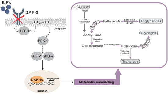
DAF-16, the only forkhead box transcription factors class O (FoxO) homolog in Caenorhabditis elegans, integrates signals from upstream pathways to elicit transcriptional changes in many genes involved in aging, development, stress, metabolism, and immunity. The major regulator of DAF-16 activity is the insulin/insulin-like growth factor 1 (IGF-1) signaling (IIS) pathway, reduction of which leads to lifespan extension in worms, flies, mice, and humans. In C. elegans daf-2 mutants, reduced IIS leads to a heterochronic activation of a dauer survival program during adulthood. This program includes elevated antioxidant defense and a metabolic shift toward accumulation of carbohydrates (i.e., trehalose and glycogen) and triglycerides, and activation of the glyoxylate shunt, which could allow fat-to-carbohydrate conversion. The longevity of daf-2 mutants seems to be partially supported by endogenous trehalose, a nonreducing disaccharide that mammals cannot synthesize, which points toward considerable differences in downstream mechanisms by which IIS regulates aging in distinct groups. Full article
(This article belongs to the Special Issue The FoxO Transcription Factors and Metabolic Regulation)
Show Figures

Figure 1

22 pages, 3920 KiB  
Article
MGL1 Receptor Plays a Key Role in the Control of T. cruzi Infection by Increasing Macrophage Activation through Modulation of ERK1/2, c-Jun, NF-κB and NLRP3 Pathways
by Tonathiu Rodriguez, Thalia Pacheco-Fernández, Alicia Vázquez-Mendoza, Oscar Nieto-Yañez, Imelda Juárez-Avelar, José L. Reyes, Luis I. Terrazas and Miriam Rodriguez-Sosa
Cells 2020, 9(1), 108; https://doi.org/10.3390/cells9010108 - 1 Jan 2020
Cited by 10 | Viewed by 3896
Abstract
Macrophage galactose-C type lectin (MGL)1 receptor is involved in the recognition of Trypanosoma cruzi (T. cruzi) parasites and is important for the modulation of the innate and adaptive immune responses. However, the mechanism by which MGL1 promotes resistance to T. cruzi [...] Read more.
Macrophage galactose-C type lectin (MGL)1 receptor is involved in the recognition of Trypanosoma cruzi (T. cruzi) parasites and is important for the modulation of the innate and adaptive immune responses. However, the mechanism by which MGL1 promotes resistance to T. cruzi remains unclear. Here, we show that MGL1 knockout macrophages (MGL1−/− Mφ) infected in vitro with T. cruzi were heavily parasitized and showed decreased levels of reactive oxygen species (ROS), nitric oxide (NO), IL-12 and TNF-α compared to wild-type macrophages (WT Mφ). MGL1−/− Mφ stimulated in vitro with T. cruzi antigen (TcAg) showed low expression of TLR-2, TLR-4 and MHC-II, which resulted in deficient splenic cell activation compared with similar co-cultured WT Mφ. Importantly, the activation of p-ERK1/2, p-c-Jun and p-NF-κB p65 were significantly reduced in MGL1−/− Mφ exposed to TcAg. Similarly, procaspase 1, caspase 1 and NLRP3 inflammasome also displayed a reduced expression that was associated with low IL-β production. Our data reveal a previously unappreciated role for MGL1 in Mφ activation through the modulation of ERK1/2, c-Jun, NF-κB and NLRP3 signaling pathways, and to the development of protective innate immunity against experimental T. cruzi infection. Full article
(This article belongs to the Section Cell Signaling)
Show Figures

Figure 1

27 pages, 9477 KiB  
Article
Monocytes as Endothelial Progenitor Cells (EPCs), Another Brick in the Wall to Disentangle Tumor Angiogenesis
by Filipa Lopes-Coelho, Fernanda Silva, Sofia Gouveia-Fernandes, Carmo Martins, Nuno Lopes, Germana Domingues, Catarina Brito, António M Almeida, Sofia A Pereira and Jacinta Serpa
Cells 2020, 9(1), 107; https://doi.org/10.3390/cells9010107 - 1 Jan 2020
Cited by 35 | Viewed by 5883
Abstract
Bone marrow contains endothelial progenitor cells (EPCs) that, upon pro-angiogenic stimuli, migrate and differentiate into endothelial cells (ECs) and contribute to re-endothelialization and neo-vascularization. There are currently no reliable markers to characterize EPCs, leading to their inaccurate identification. In the past, we showed [...] Read more.
Bone marrow contains endothelial progenitor cells (EPCs) that, upon pro-angiogenic stimuli, migrate and differentiate into endothelial cells (ECs) and contribute to re-endothelialization and neo-vascularization. There are currently no reliable markers to characterize EPCs, leading to their inaccurate identification. In the past, we showed that, in a panel of tumors, some cells on the vessel wall co-expressed CD14 (monocytic marker) and CD31 (EC marker), indicating a putative differentiation route of monocytes into ECs. Herein, we disclosed monocytes as potential EPCs, using in vitro and in vivo models, and also addressed the cancer context. Monocytes acquired the capacity to express ECs markers and were able to be incorporated into blood vessels, contributing to cancer progression, by being incorporated in tumor neo-vasculature. Reactive oxygen species (ROS) push monocytes to EC differentiation, and this phenotype is reverted by cysteine (a scavenger and precursor of glutathione), which indicates that angiogenesis is controlled by the interplay between the oxidative stress and the scavenging capacity of the tumor microenvironment. Full article
(This article belongs to the Special Issue Angiogenesis in Cancer)
Show Figures

Figure 1

16 pages, 3658 KiB  
Article
Is Autophagy Involved in Pepper Fruit Ripening?
by Omar López-Vidal, Adela Olmedilla, Luisa María Sandalio, Francisca Sevilla and Ana Jiménez
Cells 2020, 9(1), 106; https://doi.org/10.3390/cells9010106 - 1 Jan 2020
Cited by 19 | Viewed by 4530
Abstract
Autophagy is a universal self-degradation process involved in the removal and recycling of cellular constituents and organelles; however, little is known about its possible role in fruit ripening, in which the oxidation of lipids and proteins and changes in the metabolism of different [...] Read more.
Autophagy is a universal self-degradation process involved in the removal and recycling of cellular constituents and organelles; however, little is known about its possible role in fruit ripening, in which the oxidation of lipids and proteins and changes in the metabolism of different cellular organelles occur. In this work, we analyzed several markers of autophagy in two critical maturation stages of pepper (Capsicum annuum L.) fruits where variations due to ripening become clearly visible. Using two commercial varieties that ripen to yellow and red fruits respectively, we studied changes in the gene expression and protein content of several autophagy (ATG) components, ATG4 activity, as well as the autophagy receptor NBR1 and the proteases LON1 and LON2. Additionally, the presence of intravacuolar vesicles was analyzed by electron microscopy. Altogether, our data reveal that autophagy plays a role in the metabolic changes which occur during ripening in the two studied varieties, suggesting that this process may be critical to acquiring final optimal quality of pepper fruits. Full article
(This article belongs to the Special Issue Advances in the Plant Autophagy)
Show Figures

Figure 1

24 pages, 4359 KiB  
Article
Csf1 Deficiency Dysregulates Glial Responses to Demyelination and Disturbs CNS White Matter Remyelination
by Bartosz Wylot, Jakub Mieczkowski, Sylwia Niedziolka, Bozena Kaminska and Malgorzata Zawadzka
Cells 2020, 9(1), 99; https://doi.org/10.3390/cells9010099 - 31 Dec 2019
Cited by 22 | Viewed by 5017
Abstract
Remyelination, a highly efficient central nervous system (CNS) regenerative process, is performed by oligodendrocyte progenitor cells (OPCs), which are recruited to the demyelination sites and differentiate into mature oligodendrocytes to form a new myelin sheath. Microglia, the specialized CNS-resident phagocytes, were shown to [...] Read more.
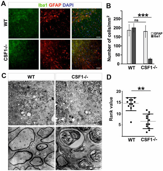
Remyelination, a highly efficient central nervous system (CNS) regenerative process, is performed by oligodendrocyte progenitor cells (OPCs), which are recruited to the demyelination sites and differentiate into mature oligodendrocytes to form a new myelin sheath. Microglia, the specialized CNS-resident phagocytes, were shown to support remyelination through secretion of factors stimulating OPC recruitment and differentiation, and their pharmacological depletion impaired remyelination. Macrophage colony-stimulating factor (Csf1) has been implicated in the control of recruitment and polarization of microglia/macrophages in injury-induced CNS inflammation. However, it remains unclear how Csf1 regulates a glial inflammatory response to demyelination as well as axonal survival and new myelin formation. Here, we have investigated the effects of the inherent Csf1 deficiency in a murine model of remyelination. We showed that remyelination was severely impaired in Csf1-/- mutant mice despite the fact that reduction in monocyte/microglia accumulation affects neither the number of OPCs recruited to the demyelinating lesion nor their differentiation. We identified a specific inflammatory gene expression signature and found aberrant astrocyte activation in Csf1-/- mice. We conclude that Csf1-dependent microglia activity is essential for supporting the equilibrium between microglia and astrocyte pro-inflammatory vs. regenerative activation, demyelinated axons integration and, ultimately, reconstruction of damaged white matter. Full article
(This article belongs to the Special Issue Microglia in Neurodegenerative Diseases)
Show Figures

Figure 1

21 pages, 1289 KiB  
Review
Extracellular Vesicles from Mesenchymal Stem Cells as Novel Treatments for Musculoskeletal Diseases
by María José Alcaraz, Alvaro Compañ and María Isabel Guillén
Cells 2020, 9(1), 98; https://doi.org/10.3390/cells9010098 - 31 Dec 2019
Cited by 67 | Viewed by 10240
Abstract
Mesenchymal stem/stromal cells (MSCs) represent a promising therapy for musculoskeletal diseases. There is compelling evidence indicating that MSC effects are mainly mediated by paracrine mechanisms and in particular by the secretion of extracellular vesicles (EVs). Many studies have thus suggested that EVs may [...] Read more.
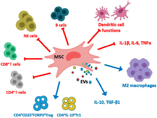
Mesenchymal stem/stromal cells (MSCs) represent a promising therapy for musculoskeletal diseases. There is compelling evidence indicating that MSC effects are mainly mediated by paracrine mechanisms and in particular by the secretion of extracellular vesicles (EVs). Many studies have thus suggested that EVs may be an alternative to cell therapy with MSCs in tissue repair. In this review, we summarize the current understanding of MSC EVs actions in preclinical studies of (1) immune regulation and rheumatoid arthritis, (2) bone repair and bone diseases, (3) cartilage repair and osteoarthritis, (4) intervertebral disk degeneration and (5) skeletal muscle and tendon repair. We also discuss the mechanisms underlying these actions and the perspectives of MSC EVs-based strategies for future treatments of musculoskeletal disorders. Full article
(This article belongs to the Section Intracellular and Plasma Membranes)
Show Figures

Figure 1

30 pages, 3745 KiB  
Review
Insights into Drought Stress Signaling in Plants and the Molecular Genetic Basis of Cotton Drought Tolerance
by Tahir Mahmood, Shiguftah Khalid, Muhammad Abdullah, Zubair Ahmed, Muhammad Kausar Nawaz Shah, Abdul Ghafoor and Xiongming Du
Cells 2020, 9(1), 105; https://doi.org/10.3390/cells9010105 - 31 Dec 2019
Cited by 265 | Viewed by 17373
Abstract
Drought stress restricts plant growth and development by altering metabolic activity and biological functions. However, plants have evolved several cellular and molecular mechanisms to overcome drought stress. Drought tolerance is a multiplex trait involving the activation of signaling mechanisms and differentially expressed molecular [...] Read more.
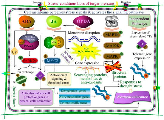
Drought stress restricts plant growth and development by altering metabolic activity and biological functions. However, plants have evolved several cellular and molecular mechanisms to overcome drought stress. Drought tolerance is a multiplex trait involving the activation of signaling mechanisms and differentially expressed molecular responses. Broadly, drought tolerance comprises two steps: stress sensing/signaling and activation of various parallel stress responses (including physiological, molecular, and biochemical mechanisms) in plants. At the cellular level, drought induces oxidative stress by overproduction of reactive oxygen species (ROS), ultimately causing the cell membrane to rupture and stimulating various stress signaling pathways (ROS, mitogen-activated-protein-kinase, Ca2+, and hormone-mediated signaling). Drought-induced transcription factors activation and abscisic acid concentration co-ordinate the stress signaling and responses in cotton. The key responses against drought stress, are root development, stomatal closure, photosynthesis, hormone production, and ROS scavenging. The genetic basis, quantitative trait loci and genes of cotton drought tolerance are presented as examples of genetic resources in plants. Sustainable genetic improvements could be achieved through functional genomic approaches and genome modification techniques such as the CRISPR/Cas9 system aid the characterization of genes, sorted out from stress-related candidate single nucleotide polymorphisms, quantitative trait loci, and genes. Exploration of the genetic basis for superior candidate genes linked to stress physiology can be facilitated by integrated functional genomic approaches. We propose a third-generation sequencing approach coupled with genome-wide studies and functional genomic tools, including a comparative sequenced data (transcriptomics, proteomics, and epigenomic) analysis, which offer a platform to identify and characterize novel genes. This will provide information for better understanding the complex stress cellular biology of plants. Full article
(This article belongs to the Special Issue Molecular and Cellular Mechanisms of Stress Responses)
Show Figures

Figure 1

13 pages, 2381 KiB  
Article
Inhibition of Transcription Induces Phosphorylation of YB-1 at Ser102 and Its Accumulation in the Nucleus
by Dmitry A. Kretov, Daria A. Mordovkina, Irina A. Eliseeva, Dmitry N. Lyabin, Dmitry N. Polyakov, Vandana Joshi, Bénédicte Desforges, Loic Hamon, Olga I. Lavrik, David Pastré, Patrick A. Curmi and Lev P. Ovchinnikov
Cells 2020, 9(1), 104; https://doi.org/10.3390/cells9010104 - 31 Dec 2019
Cited by 25 | Viewed by 5581
Abstract
The Y-box binding protein 1 (YB-1) is an RNA/DNA-binding protein regulating gene expression in the cytoplasm and the nucleus. Although mostly cytoplasmic, YB-1 accumulates in the nucleus under stress conditions. Its nuclear localization is associated with aggressiveness and multidrug resistance of cancer cells, [...] Read more.
The Y-box binding protein 1 (YB-1) is an RNA/DNA-binding protein regulating gene expression in the cytoplasm and the nucleus. Although mostly cytoplasmic, YB-1 accumulates in the nucleus under stress conditions. Its nuclear localization is associated with aggressiveness and multidrug resistance of cancer cells, which makes the understanding of the regulatory mechanisms of YB-1 subcellular distribution essential. Here, we report that inhibition of RNA polymerase II (RNAPII) activity results in the nuclear accumulation of YB-1 accompanied by its phosphorylation at Ser102. The inhibition of kinase activity reduces YB-1 phosphorylation and its accumulation in the nucleus. The presence of RNA in the nucleus is shown to be required for the nuclear retention of YB-1. Thus, the subcellular localization of YB-1 depends on its post-translational modifications (PTMs) and intracellular RNA distribution. Full article
(This article belongs to the Section Cell Nuclei: Function, Transport and Receptors)
Show Figures

Figure 1

17 pages, 7043 KiB  
Article
Novel Mechanistic Insight into the Anticancer Activity of Cucurbitacin D against Pancreatic Cancer (Cuc D Attenuates Pancreatic Cancer)
by Mohammed Sikander, Shabnam Malik, Sheema Khan, Sonam Kumari, Neeraj Chauhan, Parvez Khan, Fathi T. Halaweish, Bhavin Chauhan, Murali M. Yallapu, Meena Jaggi and Subhash C. Chauhan
Cells 2020, 9(1), 103; https://doi.org/10.3390/cells9010103 - 31 Dec 2019
Cited by 22 | Viewed by 6105
Abstract
Pancreatic cancer (PanCa) is one of the leading causes of death from cancer in the United States. The current standard treatment for pancreatic cancer is gemcitabine, but its success is poor due to the emergence of drug resistance. Natural products have been widely [...] Read more.
Pancreatic cancer (PanCa) is one of the leading causes of death from cancer in the United States. The current standard treatment for pancreatic cancer is gemcitabine, but its success is poor due to the emergence of drug resistance. Natural products have been widely investigated as potential candidates in cancer therapies, and cucurbitacin D (Cuc D) has shown excellent anticancer properties in various models. However, there is no report on the therapeutic effect of Cuc D in PanCa. In the present study, we investigated the effects of the Cuc D on PanCa cells in vitro and in vivo. Cuc D inhibited the viability of PanCa cells in a dose and time dependent manner, as evident by MTS assays. Furthermore, Cuc D treatment suppressed the colony formation, arrest cell cycle, and decreased the invasion and migration of PanCa cells. Notably, our findings suggest that mucin 13 (MUC13) is down-regulated upon Cuc D treatment, as demonstrated by Western blot and qPCR analyses. Furthermore, we report that the treatment with Cuc D restores miR-145 expression in PanCa cells/tissues. Cuc D treatment suppresses the proliferation of gemcitabine resistant PanCa cells and inhibits RRM1/2 expression. Treatment with Cuc D effectively inhibited the growth of xenograft tumors. Taken together, Cuc D could be utilized as a novel therapeutic agents for the treatment/sensitization of PanCa. Full article
(This article belongs to the Special Issue Molecular and Cellular Mechanisms of Cancers: Pancreatic Cancer)
Show Figures

Figure 1

21 pages, 8838 KiB  
Article
Spontaneous Ultraslow Na+ Fluctuations in the Neonatal Mouse Brain
by Lisa Felix, Daniel Ziemens, Gerald Seifert and Christine R. Rose
Cells 2020, 9(1), 102; https://doi.org/10.3390/cells9010102 - 31 Dec 2019
Cited by 6 | Viewed by 3460 | Correction
Abstract
In the neonate forebrain, network formation is driven by the spontaneous synchronized activity of pyramidal cells and interneurons, consisting of bursts of electrical activity and intracellular Ca2+ oscillations. By employing ratiometric Na+ imaging in tissue slices obtained from animals at postnatal [...] Read more.
In the neonate forebrain, network formation is driven by the spontaneous synchronized activity of pyramidal cells and interneurons, consisting of bursts of electrical activity and intracellular Ca2+ oscillations. By employing ratiometric Na+ imaging in tissue slices obtained from animals at postnatal day 2–4 (P2–4), we found that 22% of pyramidal neurons and 43% of astrocytes in neonatal mouse hippocampus also exhibit transient fluctuations in intracellular Na+. These occurred at very low frequencies (~2/h), were exceptionally long (~8 min), and strongly declined after the first postnatal week. Similar Na+ fluctuations were also observed in the neonate neocortex. In the hippocampus, Na+ elevations in both cell types were diminished when blocking action potential generation with tetrodotoxin. Neuronal Na+ fluctuations were significantly reduced by bicuculline, suggesting the involvement of GABAA-receptors in their generation. Astrocytic signals, by contrast, were neither blocked by inhibition of receptors and/or transporters for different transmitters including GABA and glutamate, nor of various Na+-dependent transporters or Na+-permeable channels. In summary, our results demonstrate for the first time that neonatal astrocytes and neurons display spontaneous ultraslow Na+ fluctuations. While neuronal Na+ signals apparently largely rely on suprathreshold GABAergic excitation, astrocytic Na+ signals, albeit being dependent on neuronal action potentials, appear to have a separate trigger and mechanism, the source of which remains unclear at present. Full article
(This article belongs to the Special Issue Astrocytes in Space and Time)
Show Figures

Figure 1

30 pages, 1943 KiB  
Review
COUP-TFII in Health and Disease
by Simone Polvani, Sara Pepe, Stefano Milani and Andrea Galli
Cells 2020, 9(1), 101; https://doi.org/10.3390/cells9010101 - 31 Dec 2019
Cited by 45 | Viewed by 6580
Abstract
The nuclear receptors (NRs) belong to a vast family of evolutionary conserved proteins acting as ligand-activated transcription factors. Functionally, NRs are essential in embryogenesis and organogenesis and in adulthood they are involved in almost every physiological and pathological process. Our knowledge of NRs [...] Read more.
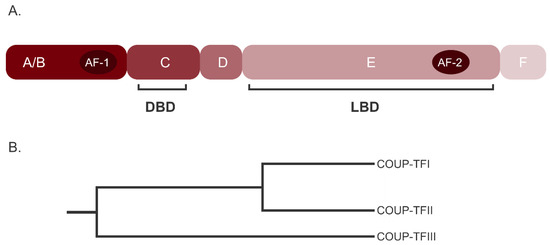
The nuclear receptors (NRs) belong to a vast family of evolutionary conserved proteins acting as ligand-activated transcription factors. Functionally, NRs are essential in embryogenesis and organogenesis and in adulthood they are involved in almost every physiological and pathological process. Our knowledge of NRs action has greatly improved in recent years, demonstrating that both their expression and activity are tightly regulated by a network of signaling pathways, miRNA and reciprocal interactions. The Chicken Ovalbumin Upstream Promoter Transcription Factor II (COUP-TFII, NR2F2) is a NR classified as an orphan due to the lack of a known natural ligand. Although its expression peaks during development, and then decreases considerably, in adult tissues, COUP-TFII is an important regulator of differentiation and it is variably implicated in tissues homeostasis. As such, alterations of its expression or its transcriptional activity have been studied and linked to a spectrum of diseases in organs and tissues of different origins. Indeed, an altered COUP-TFII expression and activity may cause infertility, abnormality in the vascular system and metabolic diseases like diabetes. Moreover, COUP-TFII is actively investigated in cancer research but its role in tumor progression is yet to be fully understood. In this review, we summarize the current understanding of COUP-TFII in healthy and pathological conditions, proposing an updated and critical view of the many functions of this NR. Full article
(This article belongs to the Collection Functions of Nuclear Receptors)
Show Figures

Figure 1

15 pages, 748 KiB  
Review
Signaling Network of Forkhead Family of Transcription Factors (FOXO) in Dietary Restriction
by Yizhou Jiang, Fengxia Yan, Zhongping Feng, Philip Lazarovici and Wenhua Zheng
Cells 2020, 9(1), 100; https://doi.org/10.3390/cells9010100 - 31 Dec 2019
Cited by 35 | Viewed by 5889
Abstract
Dietary restriction (DR), which is defined as a reduction of particular or total nutrient intake without causing malnutrition, has been proved to be a robust way to extend both lifespan and health-span in various species from yeast to mammal. However, the molecular mechanisms [...] Read more.
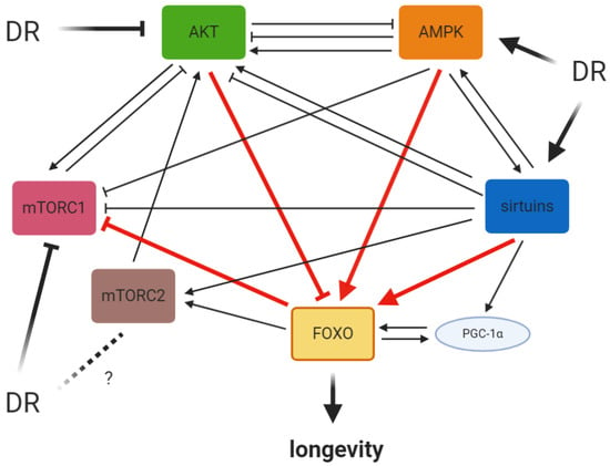
Dietary restriction (DR), which is defined as a reduction of particular or total nutrient intake without causing malnutrition, has been proved to be a robust way to extend both lifespan and health-span in various species from yeast to mammal. However, the molecular mechanisms by which DR confers benefits on longevity were not yet fully elucidated. The forkhead box O transcription factors (FOXOs), identified as downstream regulators of the insulin/IGF-1 signaling pathway, control the expression of many genes regulating crucial biological processes such as metabolic homeostasis, redox balance, stress response and cell viability and proliferation. The activity of FOXOs is also mediated by AMP-activated protein kinase (AMPK), sirtuins and the mammalian target of rapamycin (mTOR). Therefore, the FOXO-related pathways form a complex network critical for coordinating a response to environmental fluctuations in order to maintain cellular homeostasis and to support physiological aging. In this review, we will focus on the role of FOXOs in different DR interventions. As different DR regimens or calorie (energy) restriction mimetics (CRMs) can elicit both distinct and overlapped DR-related signaling pathways, the benefits of DR may be maximized by combining diverse forms of interventions. In addition, a better understanding of the precise role of FOXOs in different mechanistic aspects of DR response would provide clear cellular and molecular insights on DR-induced increase of lifespan and health-span. Full article
(This article belongs to the Special Issue The FoxO Transcription Factors and Metabolic Regulation)
Show Figures

Figure 1

15 pages, 1126 KiB  
Article
Expression of Adenoviral E1A in Transformed Cells as an Additional Factor of HDACi-Dependent FoxO Regulation
by Alisa Morshneva, Olga Gnedina, Tamara Marusova and Maria Igotti
Cells 2020, 9(1), 97; https://doi.org/10.3390/cells9010097 - 30 Dec 2019
Cited by 3 | Viewed by 3807
Abstract
The adenoviral early region 1A (E1A) protein has proapoptotic and angiogenic activity, along with its chemosensitizing effect, making it the focus of increased interest in the context of cancer therapy. It was previously shown that E1A-induced chemosensitization to different drugs, including histone deacetylases [...] Read more.
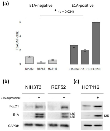
The adenoviral early region 1A (E1A) protein has proapoptotic and angiogenic activity, along with its chemosensitizing effect, making it the focus of increased interest in the context of cancer therapy. It was previously shown that E1A-induced chemosensitization to different drugs, including histone deacetylases inhibitors (HDACi), appears to be mediated by Forkhead box O (FoxO) transcription factors. In this study, we explore the relationship between E1A expression and the modulation of FoxO activity with HDACi sodium butyrate (NaBut). We show here that the basal FoxO level is elevated in E1A-expressing cells. Prolonged NaBut treatment leads to the inhibition of the FoxO expression and activity in E1A-expressing cells. However, in E1A-negative cells, NaBut promotes the transactivation ability of FoxO over time. A more detailed investigation revealed that the NaBut-induced decrease of FoxO activity in E1A-expressing cells is due to the NaBut-dependent decrease in E1A expression. Therefore, NaBut-induced inhibition of FoxO in E1A-positive cells can be overcome under unregulated overexpression of E1A. Remarkably, the CBP/p300-binding domain of E1Aad5 is responsible for stabilization of the FoxO protein. Collectively, these data show that the expression of E1A increases the FoxO stability but makes the FoxO level more sensitive to HDACi treatment. Full article
(This article belongs to the Special Issue The FoxO Transcription Factors and Metabolic Regulation)
Show Figures

Figure 1

13 pages, 1986 KiB  
Review
Extracellular Vesicle-Mediated Communication between the Glioblastoma and Its Microenvironment
by Esperanza R. Matarredona and Angel M. Pastor
Cells 2020, 9(1), 96; https://doi.org/10.3390/cells9010096 - 30 Dec 2019
Cited by 73 | Viewed by 6407
Abstract
The glioblastoma is the most malignant form of brain cancer. Glioblastoma cells use multiple ways of communication with the tumor microenvironment in order to tune it for their own benefit. Among these, extracellular vesicles have emerged as a focus of study in the [...] Read more.
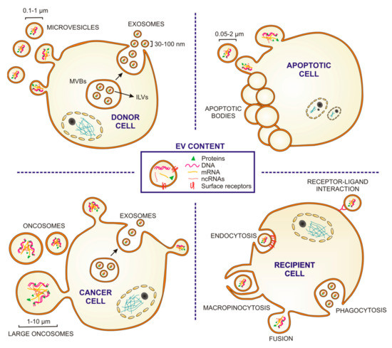
The glioblastoma is the most malignant form of brain cancer. Glioblastoma cells use multiple ways of communication with the tumor microenvironment in order to tune it for their own benefit. Among these, extracellular vesicles have emerged as a focus of study in the last few years. Extracellular vesicles contain soluble proteins, DNA, mRNA and non-coding RNAs with which they can modulate the phenotypes of recipient cells. In this review we summarize recent findings on the extracellular vesicles-mediated bilateral communication established between glioblastoma cells and their tumor microenvironment, and the impact of this dialogue for tumor progression and recurrence. Full article
(This article belongs to the Special Issue Molecular and Cellular Mechanisms of Cancers: Glioblastoma)
Show Figures

Figure 1

16 pages, 4754 KiB  
Article
Angiogenic Activity of Cytochalasin B-Induced Membrane Vesicles of Human Mesenchymal Stem Cells
by Marina O. Gomzikova, Margarita N. Zhuravleva, Vyacheslav V. Vorobev, Ilnur I. Salafutdinov, Alexander V. Laikov, Sevindzh K. Kletukhina, Ekaterina V. Martynova, Leysan G. Tazetdinova, Atara I. Ntekim, Svetlana F. Khaiboullina and Albert A. Rizvanov
Cells 2020, 9(1), 95; https://doi.org/10.3390/cells9010095 - 30 Dec 2019
Cited by 28 | Viewed by 5899
Abstract
The cytochalasin B-induced membrane vesicles (CIMVs) are suggested to be used as a vehicle for the delivery of therapeutics. However, the angiogenic activity and therapeutic potential of human mesenchymal stem/stromal cells (MSCs) derived CIMVs (CIMVs-MSCs) remains unknown. Objectives: The objectives of this study [...] Read more.
The cytochalasin B-induced membrane vesicles (CIMVs) are suggested to be used as a vehicle for the delivery of therapeutics. However, the angiogenic activity and therapeutic potential of human mesenchymal stem/stromal cells (MSCs) derived CIMVs (CIMVs-MSCs) remains unknown. Objectives: The objectives of this study were to analyze the morphology, size distribution, molecular composition, and angiogenic properties of CIMVs-MSCs. Methods: The morphology of CIMVs-MSC was analyzed by scanning electron microscopy. The proteomic analysis, multiplex analysis, and immunostaining were used to characterize the molecular composition of the CIMVs-MSCs. The transfer of surface proteins from a donor to a recipient cell mediated by CIMVs-MSCs was demonstrated using immunostaining and confocal microscopy. The angiogenic potential of CIMVs-MSCs was evaluated using an in vivo approach of subcutaneous implantation of CIMVs-MSCs in mixture with Matrigel matrix. Results: Human CIMVs-MSCs retain parental MSCs content, such as growth factors, cytokines, and chemokines: EGF, FGF-2, Eotaxin, TGF-α, G-CSF, Flt-3L, GM-CSF, Fractalkine, IFNα2, IFN-γ, GRO, IL-10, MCP-3, IL-12p40, MDC, IL-12p70, IL-15, sCD40L, IL-17A, IL-1RA, IL-1a, IL-9, IL-1b, IL-2, IL-4, IL-5, IL-6, IL-7, IL-8, IP-10, MCP-1, MIP_1a, MIP-1b, TNF-α, TNF-β, VEGF. CIMVs-MSCs also have the expression of surface receptors similar to those in parental human MSCs (CD90+, CD29+, CD44+, CD73+). Additionally, CIMVs-MSCs could transfer membrane receptors to the surfaces of target cells in vitro. Finally, CIMVs-MSCs can induce angiogenesis in vivo after subcutaneous injection into adult rats. Conclusions: Human CIMVs-MSCs have similar content, immunophenotype, and angiogenic activity to those of the parental MSCs. Therefore, we believe that human CIMVs-MSCs could be used for cell free therapy of degenerative diseases. Full article
(This article belongs to the Section Intracellular and Plasma Membranes)
Show Figures

Figure 1