Article

FMO1 Is Involved in Excess Light Stress-Induced Signal Transduction and Cell Death Signaling

by
Weronika Czarnocka
1,2,*,
Yosef Fichman
3,
Maciej Bernacki
2,4,
Elżbieta Różańska
1,
Izabela Sańko-Sawczenko
1,
Ron Mittler
3,5 and
Stanisław Karpiński
2
1
Department of Botany, Institute of Biology, Warsaw University of Life Sciences, Nowoursynowska 159, 02-776 Warsaw, Poland
2
Department of Plant Genetics, Breeding and Biotechnology, Institute of Biology, Warsaw University of Life Sciences, Nowoursynowska 159, 02-776 Warsaw, Poland
3
The Division of Plant Sciences and Interdisciplinary Plant Group, College of Agriculture, Food and Natural Resources, Christopher S. Bond Life Sciences Center University of Missouri, Columbia, MO 65211, USA
4
Institute of Technology and Life Sciences, Falenty, Al. Hrabska 3, 05-090 Raszyn, Poland
5
Department of Surgery, University of Missouri School of Medicine, Columbia, MO 65212, USA
*
Author to whom correspondence should be addressed.
Cells 2020, 9(10), 2163; https://doi.org/10.3390/cells9102163
Submission received: 29 August 2020 / Revised: 22 September 2020 / Accepted: 24 September 2020 / Published: 24 September 2020
(This article belongs to the Special Issue Programmed Cell Death Regulation in Plants)

Abstract

:
Because of their sessile nature, plants evolved integrated defense and acclimation mechanisms to simultaneously cope with adverse biotic and abiotic conditions. Among these are systemic acquired resistance (SAR) and systemic acquired acclimation (SAA). Growing evidence suggests that SAR and SAA activate similar cellular mechanisms and employ common signaling pathways for the induction of acclimatory and defense responses. It is therefore possible to consider these processes together, rather than separately, as a common systemic acquired acclimation and resistance (SAAR) mechanism. Arabidopsis thaliana flavin-dependent monooxygenase 1 (FMO1) was previously described as a regulator of plant resistance in response to pathogens as an important component of SAR. In the current study, we investigated its role in SAA, induced by a partial exposure of Arabidopsis rosette to local excess light stress. We demonstrate here that FMO1 expression is induced in leaves directly exposed to excess light stress as well as in systemic leaves remaining in low light. We also show that FMO1 is required for the systemic induction of ASCORBATE PEROXIDASE 2 (APX2) and ZINC-FINGER OF ARABIDOPSIS 10 (ZAT10) expression and spread of the reactive oxygen species (ROS) systemic signal in response to a local application of excess light treatment. Additionally, our results demonstrate that FMO1 is involved in the regulation of excess light-triggered systemic cell death, which is under control of LESION SIMULATING DISEASE 1 (LSD1). Our study indicates therefore that FMO1 plays an important role in triggering SAA response, supporting the hypothesis that SAA and SAR are tightly connected and use the same signaling pathways.

1. Introduction

Because of their sessile nature, in the course of evolution plants have evolved systemic defense mechanisms in response to many different stresses, including pathogens, wounding and abiotic stresses, which are essential for plant survival in unfavorable conditions. One of these mechanisms is systemic acquired resistance (SAR), in which, upon local pathogen attack, plants induce protection against a subsequent infection, leading to broad-spectrum disease resistance at distal, uninfected tissues. SAR mechanism involves transcriptional reprogramming of various defense-related genes (such as pathogenesis-related (PR) genes) [1], rapid production of reactive oxygen species (ROS) and nitric oxide [2], accumulation of the salicylic acid (SA) [3], and subsequent hypersensitive death of infected cells in order to restrain pathogens spread [4].
However, not only pathogens can evoke systemic signal transduction. For instance, excess- or high-light treatment of one leaf is able to induce the systemic signal that is rapidly transmitted to distal leaves, resulting in an acclimatory response and enhanced tolerance to the following stress, called systemic acquired acclimation (SAA) [5]. Recent studies identified several different signaling processes responsible for SAA, such as transcriptomic reprograming [6], systemic changes in ROS and phytohormone levels [7,8], changes in Ca2+ [9], in electrical signaling [10] and in non-photochemical quenching (NPQ) [11,12]. It was also shown that spatial-temporal SAA induction is dependent on direct vascular connections [13]. There is growing evidence that the SAA signal is transduced by bundle sheath parenchyma cells [14]. For instance, high-light treatment is able to induce specific changes in the bundle sheath parenchyma cells plasma membrane electrical potential, called photoelectrophysiological signaling (PEPS) and physical interruption of bundle sheath cells layer blocked PEPS and SAA induction [10]. PEPS was shown to be involved not only in SAA, but also in the immune defense in response to pathogens [10]. In Arabidopsis thaliana, robust molecular markers of SAA, ASCORBATE PEROXIDASE 2 (APX2, AT3G09640) and ZINC-FINGER OF ARABIDOPSIS 10 (ZAT10, AT1G27730) [5,15], are expressed in bundle sheath parenchyma cells [16,17]. Moreover, the speed of the PEPS between different leaves depends on APX2 gene expression [10]. Both APX2 and ZAT10 respond to ROS and are key regulators of plant stress signaling pathways [16,18,19]. Moreover, the transcription factor ZAT10 plays a key role in the specific activation of the ROS-related antioxidant system, including APX2 [20].
Although SAR and SAA were previously considered as separate processes, more and more evidence indicates several common elements between them. For instance, the systemic ROS wave, SA, ethylene (ET), and brassinosteroid signaling, as well as PEPS, are suggested as joint mechanisms of SAA and SAR [21]. Light stress “priming” treatment was shown to confer long-term stress acclimation not just to a second high-light exposure, but is also able to induce cross-tolerance against bacteria [10,22]. Initiation of SAR has been shown to occur in a light-dependent manner and the SAR mechanism differ under variable light regimes [23]. Both SAR and SAA induction is normally associated with programmed cell death (PCD) induction in some cells. PCD is a highly regulated and organized process of cell suicide, which plays an important role in the cell homeostasis maintenance, tissue specialization, removal of damaged or infected cells and the acclimation response [24]. Cell death in plants is preceded by the production and accumulation of ROS, which causes redox homeostasis disturbance and damage to cellular components, such as membrane lipids, nucleic acids and proteins. However, during SAR and SAA ROS play also an important role as signaling molecules [8]. Moreover, cell death was proven to be regulated by LESION SIMULATING DISEASE 1 (LSD1), ENHANCED DISEASE SUSCEPTIBILITY 1 (EDS1) and PHYTOALEXIN DEFICIENT 4 (PAD4) [25,26]. These proteins were originally defined as immune defense response regulators [25]. Later on, it was demonstrated that they also regulate the acclimation to high-light, UV, photorespiration and lysigenous aerenchyma formation [26,27,28].
FLAVIN-DEPENDENT MONOOXYGENASE 1 (FMO1, AT1G19250) belongs to the family of flavin containing monooxygenases (FMO), consisting of 29 members in Arabidopsis [29,30]. FMOs are present in all kingdoms of life, all possessing a flavin–adenine dinucleotide (FAD) prosthetic group and use NAD(P)H and O2 to oxidize low-molecular-weight molecules [31]. FMO1 is a positive defense regulator and was firstly described to be involved in response to bacterial and oomycete pathogens in Arabidopsis and powdery mildew fungus in barley [29,32,33,34]. More recent works showed that Arabidopsis FMO1 is also needed in the response to oviposition by the Large White butterfly [35] and belowground plant-to-plant signaling mechanism in response to oviposition to prepare for potential pathogen invasion [36]. The Arabidopsis knockout line of FMO1 (fmo1) is impaired in the establishment of SAR, unable to initiate systemic accumulation of SA and systemic expression of defense genes [34]. The transcription reprogramming of systemic leaves during SAR is also depends on a functional FMO1 [1,37]. Upon bacterial infection, fmo1 mutant fails to form lesions and allows pathogen invasive growth, but introducing wild type FMO1 complements the fmo1 defects in pathogen resistance [32]. However, the responses at the site of pathogen attack, including increases in the SA, jasmonic acid and camalexin levels, and expression of defense related genes, are activated in fmo1 in a similar manner to that of wild type [34]. Induction of FMO1 expression at the site of pathogen infection is attenuated by the recessive mutations in EDS1 and PAD4 genes [34]. The FMO1-dependent promotion of SAR relies on EDS1/PAD4 activities and FMO1 positively regulates the EDS1-driven cell death pathway [32]. While the fmo1 mutant shows enhanced susceptibility to pathogens and loss of SAR [32,34], FMO1 overexpression increases the resistance against bacterial and oomycete pathogens [33,38]. There is also evidence that expression of FMO1 is induced by many stress factors, such as hypoxia [39] or upon superoxide generation [29]. Therefore, FMO1 was proposed as a marker gene for defense and cell death in plants [29]. Furthermore, it was shown that FMO1 expression is reduced in non-stressed plants deficient in the positive regulators of cell death, EDS1 and PAD4 [40,41]. Meanwhile, plants with a mutation in the negative regulator of cell death, LSD1, demonstrated elevated FMO1 expression [29,40,42], which suggests that FMO1 is involved in the LSD1-, EDS1- and PAD4-dependent cell death regulation.
In the work by Mishina and Zeier (2006) it was suggested that FMO1 is involved in the synthesis of a yet unknown metabolite required for the transduction or amplification of a signal during the early phases of systemic signal transduction [34]. Such biochemical function of FMO1 was recently discovered independently by two research groups. FMO1 was shown to function as a pipecolic acid (Pip) N-hydroxylase, catalyzing the biochemical conversion of Pip to a previously undescribed plant metabolite, N-hydroxy-pipecolic acid (N-OH-Pip, NHP) in the presence of FAD and NADH as cofactors [43,44]. NHP is a mobile molecule initiating SAR signal transduction and exogenously applied NHP moves systemically in Arabidopsis and rescues the SAR-deficiency of fmo1 mutant [43,44]. However, the signaling pathway or molecule responsible for the activation of FMO1 expression is yet to be specified.
Because the mechanisms of SAR and SAA employ several common molecular elements and pathways, it is possible that FMO1, considered as exclusively SAR regulator, is also an important regulator of SAA. Moreover, since the expression of FMO1 is highly induced in the lsd1 mutant [40], it is further possible that LSD1 and FMO1 regulate at least some common pathways, such as ROS accumulation and cell death. Our results demonstrate that indeed FMO1 is involved in SAA and is a positive regulator of cell death propagation in lsd1 mutant. Thus, we report that LSD1 and FMO1 are interconnected, as well as provide evidence that FMO1 is yet another common element of SAR and SAA, collectively called SAAR [14].

2. Materials and Methods

2.1. Plant Material and Growth Conditions

Arabidopsis thaliana fmo1-1 (SALK_026163) [32], lsd1-1, lsd1-1/fmo1-1 mutants and a line overexpressing FMO1 gene, all in Columbia-0 (Col-0) background were used in this study along with Col-0 wild type plants. The expression of FMO1 in fmo1, lsd1, lsd1/fmo1 and FMO1-OE was checked with real-time quantitative PCR (qPCR) prior experiments (Supplementary Figure S1). Seeds were sown on Jiffy pots, stratified for two days in 4 °C and germinated in standard laboratory conditions (8 h/16 h day/night, photosynthetic photon flux density 100 ± 25 µmol photons m2 s¹, 50% relative air humidity and temperature 22/18 °C day/night).

2.2. Plasmid Construction

In order to obtain genetic construct for FMO1 overexpression, total plant RNA was extracted from three-week-old wild type plants using the TRIzol reagent (Invitrogen, Life Technologies, Carlsbad, CA, USA) and purified from residual DNA with a DNA-free™ DNA Removal Kit (Ambion, Life Technologies, Carlsbad, CA, USA). The total RNA concentration was measured at 260 nm using a UV–VIS spectrophotometer (NanoDrop, Thermo Fisher Scientific, Waltham, MA, USA). cDNA synthesis was performed on 2 μg of RNA using a High Capacity cDNA Reverse Transcription Kit (Life Technologies, Carlsbad, CA, USA). Full-length FMO1 (AT1G19250) coding sequence was amplified by a polymerase chain reaction (PCR) using a Phusion HighFidelity DNA Polymerase (Thermo Fisher Scientific) on cDNA template with specific primers, extended with the attB sites for Gateway cloning (Invitrogen). Primer sequences are provided in Supplementary Table S1. The genetic construct, in which GFP-FMO1 fusion was expressed under the control of the cauliflower mosaic virus 35S promoter (p35S: GFP-FMO1) was obtained by recombinational Gateway cloning (Invitrogen) using pK7WGF2 vector [45].

2.3. Stable Transformation of Arabidopsis

In order to generate Arabidopsis plants overexpressing FMO1, the construct was transformed into plants using Agrobacterium tumefaciens strain C58C1 by floral dip protocol [46]. Selection for kanamycin-resistance and homozygous plant production were performed as previously described [47].

2.4. Excess Light (EL) Treatment

Four-week-old rosettes were taken for analysis. For qPCR analysis, the EL (1700 µmol photons m−2 s−1) was applied for 2 min on 6th leaf (designated as local leaf, LL) without generation of radiant heat, using a KL 2500 LED white light source and a fiber optic cable (SCHOTT AG, Mainz, Germany). Local and systemic leaves (3rd and 9th), having vascular connection [13] were harvested before stress, immediately after EL treatment as well as after 10 and 20 min of recovery. For cell death analysis local leaves were exposed to 10-minute-long EL stress, and local and systemic leaves were collected before and 1 day after stress (for micro-lesion analysis) and, additionally, 3 and 6 days after EL treatment (for ion leakage analysis).

2.5. RNA Extraction and cDNA Synthesis

RNA was extracted from local and systemic leaves separately in five biological replicates. Each biological replicate consisted of leaves harvested from three individual rosettes. Total RNA extraction was performed using GeneMATRIX Universal RNA Purification Kit (EURX, Gdańsk, Poland) with additional step of on-column DNaseI digestion. RNA concentration and purity were analyzed spectrophotometrically with Eppendorf BioSpectrometer (Eppendorf, Hamburg, Germany). The RNA quality was tested by electrophoretic separation in 1% agarose gel. cDNA synthesis was performed for equimolar RNA amounts of each sample using a High Capacity cDNA Reverse Transcription Kit (Thermo Fisher Scientific).

2.6. Relative Gene Expression Measurement by Real-Time qPCR

Real-time qPCR was performed for cDNA obtained from non-treated plants, excess-light treated local leaves and systemic leaves. qPCR was performed in duplicates for each of five biological replicates, using the CFX Connect Real-Time PCR Detection System (Bio-Rad Laboratories, Hercules, CA, USA) and Power SYBR Green Master Mix (Thermo Fisher Scientific). The following cycling program was used in qPCR: 95 °C for 10 min, followed by 40 cycles of denaturation in 95 °C for 15 s and annealing/extension in 60 °C for 30 s. Primers were designed with Primer3Plus software (Primer3Plus, Free Software Foundation, Inc., Boston, MA, USA) and sequences are provided in Supplementary Table S1. Two reference genes were used, according to the RefGenes tool incorporated in Genevestigator [48], 5-FORMYLTETRAHYDROFOLATE CYCLOLIGASE (5-FCL, AT5G13050) and PROTEIN PHOSPHATASE 2A SUBUNIT A2 (PP2AA2, AT3G25800). The specificity of each primer pair was analyzed by melting curve. The efficiency of real-time qPCR was calculated using LinRegPCR tool [49]. Calculation of relative gene expression levels and the significance of difference between tested samples was performed using CFX Maestro Software (Bio-Rad Laboratories).

2.7. ROS Imaging

Four-week-old plants were fumigated for 30 min in a glass container using a nebulizer (Punasi Direct, Hong Kong, China) with a solution of 50 µM H2DCFDA (Millipore-Sigma, St. Louis, MO, USA) in 0.05 M phosphate buffer, pH 7.4, 0.01% (v/v) Silwet L-77 [50,51]. Following the fumigation, a local EL stress treatment (1700 µmol photons m−2 s−1) was applied to a single local leaf for 2 min. Oxidized DCF fluorescence was detected at excitation/emission 490 nm/520 nm using IVIS Lumina S5 (PerkinElmer, Waltham, MA, USA). Images were acquired every 2 min up to 30 min after the stress. ROS accumulation was analyzed using Living Image 4.7.2 software (PerkinElmer). Time-course images were generated and radiant efficiency of regions of interest (ROI) was calculated. Each data set includes standard error for 32 biological repeats and was assigned a Student t-test score.

2.8. Cell Death Quantification by Electrolyte Leakage Analysis

Cell death was quantified by ion leakage from whole rosettes for 6 plants per genotype in two independent experiments (n = 12). The 6th leaf of four-week-old plants was treated with EL for 10 min. Plants 3 days post treatment (3 dpt) and 6 days post treatment (6 dpt) together with non-treated counterparts were harvested and transferred to 50 mL Falcon tubes filled with 35 mL of deionized water. The conductivity was measured with InoLab Cond Level 1 conductivity meter (WTW Wissenschaftlich-Technische Werkstätten GmbH, Weilheim, Germany) and calculated as previously described [26,52].

2.9. Trypan Blue Staining and Micro-Lesions Counting

Trypan blue (TB) stock (30 µmol trypan blue; Sigma Aldrich St. Louis, Mo, USA) in a mixture of lactic acid, glycerol and water (10 mL: 10 mL: 20 mL) was diluted with 96% ethanol (1:2) to obtain TB working solution. Leaves were harvested from non-treated plants and plants 24 h after EL treatment. Twelve non-treated, local and systemic leaves per each genotype, from two independent experiments (n = 24), were harvested and dipped separately in TB working solution in 50 mL Falcon tubes. Leaves were incubated in TB working solution for 30 min in room temperature and gently shaken from time to time. After incubation, the TB working solution was removed and replaced witch methanol. The leaves were incubated in methanol for 24 h. During the incubation, methanol was changed several times for the fresh one. Leaves devoid of chlorophyll were visualized using Nikon SMZ18 stereomicroscopes (Nikon Inc., Melville, NY, USA) with an adapted camera Nikon d5100 (Nikon Inc.). Photos of individual leaves were analyzed in ImageJ software version 1.8.0 (http://rsb.info.nih.gov/ij) and blue spots (micro-lesions) were counted per mm2 of leaf area.

3. Results

3.1. FMO1 Expression Is Induced in Both Local EL-Treated and Systemic Leaves

To test whether the FMO1 gene is induced upon EL treatment in local (LL) and/or untreated (systemic, SL) leaves we performed a time-course qPCR analysis for non-treated, as well as EL-treated, local leaves and systemic leaves remaining in low light. Our results indicated that FMO1 expression was induced, although not statistically significantly, both locally and systemically already after the 2 min long EL treatment (Figure 1). The highest induction of FMO1 expression was observed 10 min after recovery in both local and systemic leaves, whereas, after 20 min of recovery, the FMO1 expression level in both local and systemic leaves dropped. Nevertheless, after 20 min of recovery the systemic leaves remained statistically significant induction of FMO1 expression, when compared to non-treated systemic leaves. These results indicate that FMO1 expression is rapidly induced upon EL treatment in both local and systemic leaves.

3.2. FMO1, Together with LSD1, Is Required for the Spread of the ROS Systemic Signal

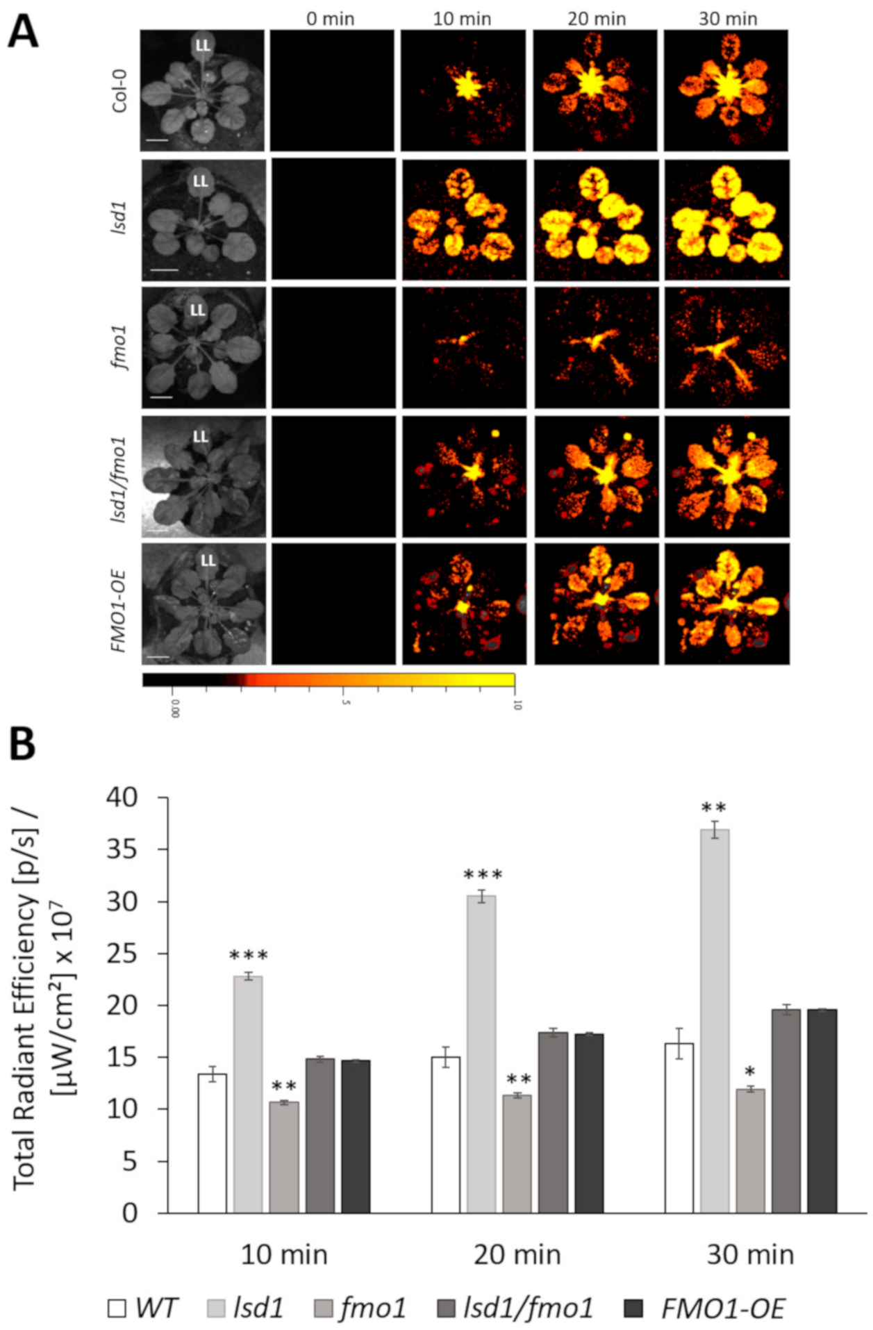
We next wanted to determine the role of FMO1 in spreading the EL stress signal to systemic leaves. Because ROS are among the fastest signal molecules spreading from leaf to leaf [8,53], we employed local EL treatment and subsequent whole-plant ROS accumulation imaging. ROS levels were monitored over the course of 30 min in the wild type, fmo1 mutant and FMO1 overexpressing plants (FMO1-OE). Since the LSD1 is known to control ROS accumulation upon stress [26], we also used lsd1 mutant and lsd1/fmo1 double mutant in order to analyze the FMO1 and LSD1 interdependence (Figure 2).
As shown in Figure 2, two-minute-long illumination of a single leaf resulted in a time-dependent ROS accumulation in both EL-treated and systemic leaves. As expected, lsd1 mutant demonstrated faster ROS accumulation in both treated and untreated leaves, compared to the wild type. lsd1 plants had significantly higher level of ROS, while fmo1 mutant showed significantly lower ROS accumulation 10, 20 and 30 min after EL exposure. Interestingly, lsd1/fmo1 double mutant proved to have similar levels of ROS, as the wild type plants, which means that fmo1 mutation was able to revert lsd1 hyper-ROS accumulating phenotype. These results indicate that FMO1 is a positive regulator of ROS propagation in the lsd1 background, which means that FMO1 is required for the systemic spread of ROS that lies at the same signaling pathway as LSD1.
In order to assess which specific ROS are engaged in the systemic ROS spread, we performed a qPCR experiment in which we tested the relative expression levels of genes encoding specific ROS markers in non-stressed plants, EL-treated leaves (2 min EL + 10 min recovery) and corresponding systemic leaves. Telomere-binding protein TRF-like 4 (TRFL4, AT3G53790), beta-glucosidase BGLU23 (AT3G09260) and DMR6-LIKE OXYGENASE 1 (DLO1, AT4G10500) were used as marker genes specifically responsive to singlet oxygen (1O2), superoxide anion (O2) and hydrogen peroxide (H2O2), respectively [54]. Our results indicated that 2-minute-long EL exposure of local leaf was sufficient to significantly induce the expression of TRFL4 in lsd1 mutant (Supplementary Figure S2). Although not statistically significant, the expression levels of TRFL4 were lower in fmo1 mutant in both local and systemic leaves. After EL treatment, we observed an increase in BGLU23 expression levels in all tested genotypes, which indicates that local EL exposure caused O2 generation. In systemic leaves, the BGLU23 expression levels were significantly elevated in lsd1 mutant and reduced in fmo1 plants, when compared to systemic leaves of the wild type. The transcription of H2O2, marker gene, DLO1, proved to be elevated in all non-treated, local and systemic leaved of lsd1 mutant. Meanwhile, DLO1 expression was significantly reduced in EL-treated local and systemic leaves of fmo1 and lsd1/fmo1 double mutant, in comparison to wild type plants. Interestingly, systemic leaves of FMO1-OE line showed elevated DLO1 transcript level, when compared to the wild type. These results indicate that the marker genes of all 1O2, O2 and H2O2 were to some extent involved in the local response to EL. However, the most responding marker gene in tested genetic system was DLO1, indicating that H2O2 may be the main ROS form engaged in ROS signal propagation dependent on FMO1.

3.3. Functional FMO1 Is Required for the Systemic Induction of APX2 and ZAT10 Expression upon Local EL Treatment

APX2 and ZAT10 are well-described regulators of systemic acquired acclimation (SAA) [10,55]. The expression of APX2 and ZAT10 is induced upon excess-light treatment in both local and systemic leaves [5,15]. Therefore, we decided to test the expression of APX2 and ZAT10 in non-treated, EL-treated (2 min EL + 10 min recovery) and corresponding systemic leaves in the genotypes tested in the current study.
There was no difference in APX2 transcript levels among non-treated genotypes (Figure 3A). Upon EL treatment, APX2 transcription was highly induced in local leaves of all tested genotypes. However, the levels of APX2 transcript abundance were significantly higher in lsd1, fmo1, lsd1/fmo1 mutants and FMO1-OE line, compared to the wild type plants. Interestingly, in the systemic leaves of fmo1 and lsd1/fmo1 mutants the increase in APX2 expression was significantly lower, in relation to the wild type.
ZAT10 expression levels in non-stress conditions were significantly decreased in lsd1 and fmo1 mutants (Figure 3B). After EL treatment ZAT10 transcription was elevated in all the genotypes, except lsd1/fmo1 double mutant and FMO1-OE line. In systemic leaves we observed a similar pattern to APX2 expression, since ZAT10 transcript abundance was lower in fmo1 and lsd1/fmo1 mutants, and additionally in FMO1-OE line. These results indicate that functional FMO1 is needed for systemic signaling inducing APX2 and ZAT10 expression.

3.4. FMO1 Is Involved in LSD1-Dependent Spread of Systemic Cell Death

LSD1 has been broadly described as a negative regulator of cell death spread, called runaway cell death (RCD) [8,24]. Upon stress, the lsd1 mutant accumulates high levels of ROS (Figure 2 and [40]) and cannot restrain cell death propagation once it has been initiated [26,27,56]. Because we observed a reversion in the lsd1 phenotype in terms of ROS accumulation, by introducing fmo1 mutation into lsd1 background (lsd1/fmo1 double mutant), we set out to examine whether FMO1 is involved in a cell death pathway, negatively regulated by LSD1.
We quantified the cell death in two ways. In the first method we used the measurement of total ion leakage from the rosettes [26,52,57] before stress, as well as 3 and 6 days after local EL treatment. Our results indicated that in non-stress conditions there were no significant changes in cell death among tested genotypes (Figure 4A). However, EL treatment of local leaf caused noticeable increase in ion leakage 3 days post treatment (dpt) in all tested lines. As expected, lsd1 mutant showed significantly increased cell death, in comparison to wild type counterparts. However, most importantly, lsd1/fmo1 double mutant demonstrated reduced ion leakage, when compared to the lsd1 single mutant. The cell death spread 6 dpi was most visible in lsd1 mutant (Figure 4A and Supplementary Figure S3). Nevertheless, we observed slower spread of cell death in fmo1 and lsd1/fmo1 mutants, when compared to the lsd1 single mutant (Figure 4A and Supplementary Figure S3). These results indicate that the lack of functional FMO1 was able to revert the cell death spread in lsd1 background.
The second method for cell death assessment we used was the staining of non-treated, EL-treated local leaves and systemic leaves with trypan blue in order to analyze the micro-lesion formation. Micro-lesions constitute small lesion area within the leaf tissue, comprising one or a few dead cells [58]. The development of micro-lesions in systemic leaves was previously shown only for SAR in response to pathogen attack [58,59]. So far, there was no experimental proof of the existence of micro-lesion formation during SAA. Our results demonstrate that a 10-minute-long local EL stress was generally able to induce micro-lesion formation 24 h after treatment in both local and systemic leaves (Figure 4B,C). Interestingly, we observed higher numbers of micro-lesions in systemic leaves than in local leaves. Nevertheless, lsd1 mutant had significantly increased numbers of micro-lesions in both local and systemic leaves, while FMO1-OE line demonstrated higher micro-lesion number only in local leaves. However, most importantly, the formation of systemic micro-lesions in fmo1 mutant was significantly inhibited, when compared to the wild type and again lsd1/fmo1 double mutant manifested reversed micro-lesion formation phenotype in relation to the lsd1 single mutant. These results suggest that FMO1 is a positive regulator of systemic cell death signal transmission and takes part in the LSD1-dependent cell death regulatory pathway.

3.5. Proposed Model of FMO1 Involvement in ROS Signaling, SAAR and Cell Death

Based on the results presented in this work, we propose the following model of the FMO1 involvement in the ROS signaling, SAAR and cell death upon EL treatment (Figure 5). LSD1 negatively influences the FMO1-dependent signaling pathways. Among these pathways there is a propagation of ROS signals from local, exposed to EL leaves, to systemic leaves, as well as APX2- and ZAT10-dependent signaling. ROS spread, positively regulated by FMO1, leads to cell death and simultaneously to SAAR.

4. Discussion

The role of FMO1 has been well described thus far in the SAR pathway [32,33,34]. Taking into consideration many common signaling pathways between SAR and SAA [10,14,60], our aim was to determine whether FMO1 is also engaged in SAA.
Our findings suggest that the expression of FMO1 is induced shortly after EL treatment, and that 2-minute-long exposure to EL was sufficient to induce FMO1 expression in both local and systemic leaves. The analysis of light-induced FMO1 expression kinetics demonstrated that its highest expression in local leaves was observed 10 minutes after the end of EL treatment. In contrast, 20 min after EL treatment the level of FMO1 expression in local leaves dropped, but remained elevated in systemic leaves. The induction of FMO1 expression in both local and systemic leaves was also shown for pathogen-treated plants [32,34]. These results indicate that the expression of FMO1 is induced both locally and systemically not only during SAR, but also SAA. Interestingly, the systemic expression of the FMO1 gene depends on phytochromes during SAR establishment, since phyAphyB double mutant did not increase the expression of FMO1 in systemic leaves upon pathogen infection [61]. All these results strongly suggest the role of FMO1 in the integration of SAA and SAR signals. It has been previously postulated that in fact high-light induced SAA and SAR processes should be considered as common mechanism, for which the SAAR abbreviation has been suggested (Figure 5 and [14]).
ROS are important signaling molecules in SAA [5,8]. Therefore, in the next step we wanted to investigate if FMO1 activity has any role in the systemic propagation of ROS. Our results indicated that in wild type plants a 2-minute-long EL treatment of local leaves resulted in ROS level increase already 10 min after EL exposure. The lsd1 mutant, known to accumulate higher level of ROS under both abiotic and biotic stress [25,26], demonstrated in our study quicker ROS propagation after local EL treatment, in relation to Col-0. This result was also confirmed by the expression level of ROS marker genes, TRFL4, BGLU23 and DLO1 that were significantly elevated in local and/or systemic leaves of lsd1 mutant, in comparison to the wild type. In contrast, the fmo1 mutant showed less intense ROS accumulation, compared to the wild type, which was also reflected by the expression of ROS marker genes in local and/or systemic leaves. The expression level of DLO1, a marker gene for H2O2 accumulation was reduced in both local and systemic leaves of fmo1 mutant. On the contrary, the DLO1 transcript abundance was elevated in FMO1-OE line. These results indicate that H2O2, which is a relatively long-lived molecule and can diffuse over long distances [62], may be the main ROS form engaged in ROS signal propagation by FMO1. However, other ROSs should not be excluded. Despite 1O2 and O2 are rather short-lived, they can function as signal molecules, affecting many signaling pathways [8,63]. Importantly, the lsd1/fmo1 double mutant showed similar ROS level to that of wild type, which indicates that FMO1 is a positive regulator of ROS propagation in the lsd1 background. These results indicate that FMO1 is engaged in the LSD1-dependent ROS signaling pathway, promoting ROS accumulation (Figure 5). In this term, our results are in some way different from the study on the role of FMO1 on ROS signaling during SAR, where FMO1 seemed not to play a critical role in the regulation of the oxidative burst at the site of pathogen attack [34]. However other works examining the FMO1 involvement in biotic stresses [33,64] confirmed the role of FMO1 in ROS accumulation, observed in this study.
Taking into account that ROS are among the fastest signaling molecules, which accumulation is induced already after 2-minute-long EL treatment and the fact that FMO1 expression can be highly induced upon O2 generation [29], we suggest that ROS are the primary signal for activation of FMO1-dependent SAAR. However, a signaling pathway or a molecule responsible for the activation of FMO1 expression is yet to be specified. Recent works demonstrated that the FMO1 promoter is bound by SAR DEFICIENT 1 (SARD1) transcription factor [65], possessing redox-regulated cysteines [66]. Moreover, SUPPRESSOR OF GAMMA RADIATION 1 (SOG1) transcription factor binds FMO1 promoter to induce its expression under oxidative stress [64]. Enhanced FMO1 expression in response to phytotoxin inducing plant cell necrosis, thaxtomin A, in rbohd/f mutant, compared with wild type plants, may indicate that stress-induced extracellular ROS production is downstream of FMO1 activity [41].
Oxidative-stress induced APX2 and ZAT10 are expressed in bundle sheath cells of both local and systemic leaves in response to high or excess light and are marker genes for early SAA events [5,13,16,17]. Our results showed that the expression of APX2 was highly elevated in EL-treated local leaves. The APX2 induction in systemic leaves was lower than in local counterparts, but still, the APX2 transcript abundance was increased, when compared to non-treated plants. Importantly, the levels of APX2 expression were significantly lower in the systemic leaves of fmo1 and lsd1/fmo1 mutant, compared to the wild type. A similar pattern was revealed for ZAT10. The expression levels of ZAT10 in systemic leaves of fmo1 and lsd1/fmo1 mutants were significantly reduced in relation to wild type plants. Our data are consistent with the study by Mishina and Zeier (2006), who showed that the expression levels of pathogenesis-related (PR) genes and a marker for ROS generation GLUTATHIONE S-TRANSFERASE 1 (GST1) was abolished in fmo1 systemic leaves upon pathogen attack [34]. These results indicate that functional FMO1 is needed for the induction of SAA marker gene expression in systemic leaves. Interestingly, the FMO1-OE line exhibited reduced ZAT10 transcript abundance, which suggests that both lack and excess of FMO1 activity may affect transcriptional reprograming in systemic leaves.
It was demonstrated that loss of FMO1 function did not block EDS1-dependent cell death associated with the mutation in ACCELERATED CELL DEATH 11 (ACD11) [67]. The acd11/fmo1 double mutant showed similar extent of cell death to the acd11 single mutant and it was concluded that the contribution of FMO1 to cell death initiation or execution is minimal [29]. However, the results indicating that FMO1 is a component of LSD1-dependent ROS signaling pathway prompted us to test if FMO1 is also engaged in LSD1-dependent negative regulation of cell death. In order to define FMO1 role in EL-induced cell death, we used two methods of cell death assessment, by electrolyte leakage from whole rosettes and micro-lesions counting preceded by trypan blue staining. Our results demonstrated that 3 and 6 days post-treatment the lsd1 mutant exhibited visible symptoms of runaway cell death and significantly increased ion leakage from damaged tissue. LSD1 has been already shown to negatively regulate cell death, triggered by both biotic and abiotic stresses in the past [25,26,27]. Interestingly, our current analysis reveals that a mutation in fmo1 reduces ion leakage in lsd1 mutant, which indicates that FMO1 is a positive regulator of lsd1-dependent cell death. The expression level of FMO1 was shown to be elevated in lsd1 background previously and confirmed in the current study (Supplementary Figure S1), which strengthens the demonstrated contribution of FMO1 to the LSD1-dependent cell death pathway (Figure 5). LSD1 has been recently demonstrated as a transcriptional regulator [47]. Although the FMO1 promoter was not among LSD1-bound sequences [47], it is possible that LSD1 inhibits FMO1 expression in an indirect manner, through another transcription regulator.
Micro-lesions constitute small lesion areas within the leaf tissue, comprising one or a few dead cells [58]. Even though the development of micro-lesions in systemic leaves was previously shown during SAR in response to pathogen attack [58,59], so far there was no evidence of micro-lesion formation in the development of SAA. Here we show that micro-lesions could be observed already 24 h after 10-minute-long EL treatment and interestingly, they were generally more expressed in systemic than in local leaves. As expected, the number of micro-lesions was significantly higher in both local and systemic leaves of lsd1 mutant, when compared to the wild type. Even though the fmo1 mutant did not show any changes in the number of micro-lesions in local leaves, which was also shown for pathogen treatment study [34], it demonstrated significantly reduced cell death in systemic leaves. It indicates that the functional FMO1 is needed for the progression of cell death in systemic leaves. Moreover, the mutation in fmo1 reverted the cell death in local and systemic leaves of lsd1, once again confirming the role of FMO1 as a positive regulator of lsd1-dependent runaway cell death. Interestingly, the FMO1 transcript levels were not changed in the constitutive defense mutants, engaged in ET (CONSTITUTIVE TRIPLE RESPONSE 1, CTR1) or SA signal transduction pathway (MAP KINASE 4, MPK4), indicating that FMO1 is not activated simply by deregulated ET or SA signaling [29]. Instead, FMO1 may influence the components downstream of the LSD1-dependent signaling pathway.
In this study, we revealed that FMO1 plays an important role in SAA, which adds another evidence that EL stress-triggered SAA and pathogen-induced SAR are tightly connected and use the same signaling pathways. FMO1 functions in the induction of both SAA and SAR in treated tissue and in the propagation of a signal to distant tissues. However, its role in the perception of some long-distance signal in systemic tissue and the potentiation of defense response in systemic tissues should not be excluded. FMO1 was shown to be expressed in the tissues adjoining the vasculature [29]. Taking into account that the bundle sheath cells, surrounding vascular bundles are strongly suggested to transduce SAA signal [10], the FMO1-dependent perception and signal transmission is likely to occur in bundle sheath parenchyma cells.

Supplementary Materials

The following are available online at https://www.mdpi.com/2073-4409/9/10/2163/s1, Figure S1: Relative FMO1 expression level, Figure S2: Relative expression of different ROS forms markers, Figure S3: Rosette morphology of the wild type (Col-0), lsd1, fmo1, lsd1/fmo1 mutants and FMO1-OE line before, 3 and 6 days post EL treatment, Table S1: Primers used for FMO1 cloning and qPCR.

Author Contributions

W.C. planned the experiments and postulated the hypotheses tested in this paper. W.C. performed RNA isolation, cDNA synthesis and qPCRs together with E.R. W.C. prepared the genetic construct for the cloning, stably transformed Arabidopsis plants and together with I.S.-S. selected the best FMO1-OE T4 line. Y.F. performed the ROS imaging. W.C. and M.B. did the ion leakage and micro-lesion analysis experiments. W.C. performed all the in-silico analyses and the FMO1 localization experiment using confocal microscopy. W.C. wrote the manuscript and prepared the figures. S.K. and R.M. reviewed the manuscript. All authors have read and agreed to the published version of the manuscript.

Funding

This work was supported by the Maestro 6 project (2014/14/A/NZ1/00218) granted to S.K. by the National Science Centre.

Conflicts of Interest

The authors declare no conflict of interest. The funders had no role in the design of the study; in the collection, analyses, or interpretation of data; in the writing of the manuscript, or in the decision to publish the results.

References

  1. Gruner, K.; Griebel, T.; Návarová, H.; Attaran, E.; Zeier, J. Reprogramming of plants during systemic acquired resistance. Front. Plant Sci. 2013, 4, 252. [Google Scholar] [CrossRef] [PubMed] [Green Version]
  2. El-Shetehy, M.; Wang, C.; Shine, M.B.; Yu, K.; Kachroo, A.; Kachroo, P. Nitric oxide and reactive oxygen species are required for systemic acquired resistance in plants. Plant Signal. Behav. 2015, 10, e998544. [Google Scholar] [CrossRef] [PubMed] [Green Version]
  3. Cameron, R.K.; Paiva, N.L.; Lamb, C.J.; Dixon, R.A. Accumulation of salicylic acid and PR-1 gene transcripts in relation to the systemic acquired resistance (SAR) response induced by Pseudomonas syringae pv. tomato in Arabidopsis. Physiol. Mol. Plant Pathol. 1999, 55, 121–130. [Google Scholar] [CrossRef]
  4. Balint-Kurti, P. The plant hypersensitive response: Concepts, control and consequences. Mol. Plant Pathol. 2019, 20, 1163–1178. [Google Scholar] [CrossRef] [Green Version]
  5. Karpinski, S.; Reynolds, H.; Karpinska, B.; Wingsle, G.; Creissen, G.; Mullineaux, P. Systemic signaling and acclimation in response to excess excitation energy in Arabidopsis. Science 1999, 284, 654–657. [Google Scholar] [CrossRef]
  6. Zandalinas, S.I.; Sengupta, S.; Burks, D.; Azad, R.K.; Mittler, R. Identification and characterization of a core set of ROS wave-associated transcripts involved in the systemic acquired acclimation response of Arabidopsis to excess light. Plant J. 2019, 98, 126–141. [Google Scholar] [CrossRef]
  7. Mittler, R.; Blumwald, E. The Roles of ROS and ABA in Systemic Acquired Acclimation[OPEN]. Plant Cell 2015, 27, 64–70. [Google Scholar] [CrossRef] [Green Version]
  8. Czarnocka, W.; Karpiński, S. Friend or foe? Reactive oxygen species production, scavenging and signaling in plant response to environmental stresses. Free Radic. Biol. Med. 2018, 122, 4–20. [Google Scholar] [CrossRef]
  9. Gilroy, S.; Białasek, M.; Suzuki, N.; Górecka, M.; Devireddy, A.R.; Karpiński, S.; Mittler, R. ROS, Calcium, and Electric Signals: Key Mediators of Rapid Systemic Signaling in Plants1[OPEN]. Plant Physiol. 2016, 171, 1606–1615. [Google Scholar] [CrossRef]
  10. Szechyńska-Hebda, M.; Kruk, J.; Górecka, M.; Karpińska, B.; Karpiński, S. Evidence for Light Wavelength-Specific Photoelectrophysiological Signaling and Memory of Excess Light Episodes in Arabidopsis[W][OA]. Plant Cell 2010, 22, 2201–2218. [Google Scholar] [CrossRef] [Green Version]
  11. Ciszak, K.; Kulasek, M.; Barczak, A.; Grzelak, J.; Maćkowski, S.; Karpiński, S. PsbS is required for systemic acquired acclimation and post-excess-light-stress optimization of chlorophyll fluorescence decay times in Arabidopsis. Plant Signal. Behav. 2015, 10, e982018. [Google Scholar] [CrossRef] [PubMed] [Green Version]
  12. Białasek, M.; Górecka, M.; Mittler, R.; Karpiński, S. Evidence for the Involvement of Electrical, Calcium and ROS Signaling in the Systemic Regulation of Non-Photochemical Quenching and Photosynthesis. Plant Cell Physiol. 2017, 58, 207–215. [Google Scholar] [CrossRef] [PubMed]
  13. Carmody, M.; Crisp, P.A.; d’Alessandro, S.; Ganguly, D.; Gordon, M.; Havaux, M.; Albrecht-Borth, V.; Pogson, B.J. Uncoupling High Light Responses from Singlet Oxygen Retrograde Signaling and Spatial-Temporal Systemic Acquired Acclimation1[OPEN]. Plant Physiol. 2016, 171, 1734–1749. [Google Scholar] [CrossRef] [PubMed] [Green Version]
  14. Karpinski, S.; Szechynska-Hebda, M. Secret life of plants. Plant Signal. Behav. 2010, 5, 1391–1394. [Google Scholar] [CrossRef] [Green Version]
  15. Rossel, J.B.; Wilson, P.B.; Hussain, D.; Woo, N.S.; Gordon, M.J.; Mewett, O.P.; Howell, K.A.; Whelan, J.; Kazan, K.; Pogson, B.J. Systemic and Intracellular Responses to Photooxidative Stress in Arabidopsis. Plant Cell 2007, 19, 4091–4110. [Google Scholar] [CrossRef] [PubMed] [Green Version]
  16. Fryer, M.J.; Ball, L.; Oxborough, K.; Karpinski, S.; Mullineaux, P.M.; Baker, N.R. Control of Ascorbate Peroxidase 2 expression by hydrogen peroxide and leaf water status during excess light stress reveals a functional organisation of Arabidopsis leaves. Plant J. 2003, 33, 691–705. [Google Scholar] [CrossRef]
  17. Galvez-Valdivieso, G.; Fryer, M.J.; Lawson, T.; Slattery, K.; Truman, W.; Smirnoff, N.; Asami, T.; Davies, W.J.; Jones, A.M.; Baker, N.R.; et al. The High Light Response in Arabidopsis Involves ABA Signaling between Vascular and Bundle Sheath Cells. Plant Cell 2009, 21, 2143–2162. [Google Scholar] [CrossRef] [Green Version]
  18. Mittler, R.; Kim, Y.; Song, L.; Coutu, J.; Coutu, A.; Ciftci-Yilmaz, S.; Lee, H.; Stevenson, B.; Zhu, J.-K. Gain- and loss-of-function mutations in Zat10 enhance the tolerance of plants to abiotic stress. FEBS Lett. 2006, 580, 6537–6542. [Google Scholar] [CrossRef] [Green Version]
  19. Bechtold, U.; Richard, O.; Zamboni, A.; Gapper, C.; Geisler, M.; Pogson, B.; Karpinski, S.; Mullineaux, P.M. Impact of chloroplastic- and extracellular-sourced ROS on high light-responsive gene expression in Arabidopsis. J. Exp. Bot. 2008, 59, 121–133. [Google Scholar] [CrossRef]
  20. Miller, G.; Shulaev, V.; Mittler, R. Reactive oxygen signaling and abiotic stress. Physiol. Plant. 2008, 133, 481–489. [Google Scholar] [CrossRef]
  21. Baxter, A.; Mittler, R.; Suzuki, N. ROS as key players in plant stress signalling. J. Exp. Bot. 2014, 65, 1229–1240. [Google Scholar] [CrossRef] [PubMed]
  22. Zhang, S.; Apel, K.; Kim, C. Singlet oxygen-mediated and EXECUTER-dependent signalling and acclimation of Arabidopsis thaliana exposed to light stress. Philos. Trans. R. Soc. B Biol. Sci. 2014, 369. [Google Scholar] [CrossRef] [PubMed] [Green Version]
  23. Zeier, J.; Pink, B.; Mueller, M.J.; Berger, S. Light conditions influence specific defence responses in incompatible plant-pathogen interactions: Uncoupling systemic resistance from salicylic acid and PR-1 accumulation. Planta 2004, 219, 673–683. [Google Scholar] [CrossRef]
  24. Wituszyńska, W.; Karpiński, S. Programmed Cell Death as a Response to High Light, UV and Drought Stress in Plants. Abiotic Stress Plant Responses Appl. Agric. 2013, 207–246. [Google Scholar] [CrossRef] [Green Version]
  25. Rustérucci, C.; Aviv, D.H.; Holt, B.F.; Dangl, J.L.; Parker, J.E. The Disease Resistance Signaling Components EDS1 and PAD4 Are Essential Regulators of the Cell Death Pathway Controlled by LSD1 in Arabidopsis. Plant Cell 2001, 13, 2211–2224. [Google Scholar] [CrossRef] [Green Version]
  26. Wituszyńska, W.; Szechyńska-Hebda, M.; Sobczak, M.; Rusaczonek, A.; Kozłowska-Makulska, A.; Witoń, D.; Karpiński, S. Lesion simulating disease 1 and enhanced disease susceptibility 1 differentially regulate UV-C-induced photooxidative stress signalling and programmed cell death in Arabidopsis thaliana. Plant Cell Environ. 2015, 38, 315–330. [Google Scholar] [CrossRef]
  27. Mateo, A.; Mühlenbock, P.; Rustérucci, C.; Chang, C.C.-C.; Miszalski, Z.; Karpinska, B.; Parker, J.E.; Mullineaux, P.M.; Karpinski, S. LESION SIMULATING DISEASE 1 Is Required for Acclimation to Conditions That Promote Excess Excitation Energy. Plant Physiol. 2004, 136, 2818–2830. [Google Scholar] [CrossRef] [Green Version]
  28. Mühlenbock, P.; Plaszczyca, M.; Plaszczyca, M.; Mellerowicz, E.; Karpinski, S. Lysigenous aerenchyma formation in Arabidopsis is controlled by LESION SIMULATING DISEASE1. Plant Cell 2007, 19, 3819–3830. [Google Scholar] [CrossRef] [Green Version]
  29. Olszak, B.; Malinovsky, F.G.; Brodersen, P.; Grell, M.; Giese, H.; Petersen, M.; Mundy, J. A putative flavin-containing mono-oxygenase as a marker for certain defense and cell death pathways. Plant Sci. 2006, 170, 614–623. [Google Scholar] [CrossRef]
  30. Schlaich, N.L. Flavin-containing monooxygenases in plants: Looking beyond detox. Trends Plant Sci. 2007, 12, 412–418. [Google Scholar] [CrossRef]
  31. Rossner, R.; Kaeberlein, M.; Leiser, S.F. Flavin-containing monooxygenases in aging and disease: Emerging roles for ancient enzymes. J. Biol. Chem. 2017, 292, 11138–11146. [Google Scholar] [CrossRef] [PubMed] [Green Version]
  32. Bartsch, M.; Gobbato, E.; Bednarek, P.; Debey, S.; Schultze, J.L.; Bautor, J.; Parker, J.E. Salicylic Acid–Independent ENHANCED DISEASE SUSCEPTIBILITY1 Signaling in Arabidopsis Immunity and Cell Death Is Regulated by the Monooxygenase FMO1 and the Nudix Hydrolase NUDT7. Plant Cell 2006, 18, 1038–1051. [Google Scholar] [CrossRef] [PubMed] [Green Version]
  33. Koch, M.; Vorwerk, S.; Masur, C.; Sharifi-Sirchi, G.; Olivieri, N.; Schlaich, N.L. A role for a flavin-containing mono-oxygenase in resistance against microbial pathogens in Arabidopsis. Plant J. Cell Mol. Biol. 2006, 47, 629–639. [Google Scholar] [CrossRef] [PubMed]
  34. Mishina, T.E.; Zeier, J. The Arabidopsis flavin-dependent monooxygenase FMO1 is an essential component of biologically induced systemic acquired resistance. Plant Physiol. 2006, 141, 1666–1675. [Google Scholar] [CrossRef] [Green Version]
  35. Hilfiker, O.; Groux, R.; Bruessow, F.; Kiefer, K.; Zeier, J.; Reymond, P. Insect eggs induce a systemic acquired resistance in Arabidopsis. Plant J. Cell Mol. Biol. 2014, 80, 1085–1094. [Google Scholar] [CrossRef] [Green Version]
  36. Orlovskis, Z.; Reymond, P. Pieris brassicae eggs trigger interplant systemic acquired resistance against a foliar pathogen in Arabidopsis. New Phytol. 2020. [Google Scholar] [CrossRef]
  37. Bernsdorff, F.; Döring, A.-C.; Gruner, K.; Schuck, S.; Bräutigam, A.; Zeier, J. Pipecolic Acid Orchestrates Plant Systemic Acquired Resistance and Defense Priming via Salicylic Acid-Dependent and -Independent Pathways. Plant Cell 2016, 28, 102–129. [Google Scholar] [CrossRef] [Green Version]
  38. Ding, P.; Rekhter, D.; Ding, Y.; Feussner, K.; Busta, L.; Haroth, S.; Xu, S.; Li, X.; Jetter, R.; Feussner, I.; et al. Characterization of a Pipecolic Acid Biosynthesis Pathway Required for Systemic Acquired Resistance. Plant Cell 2016, 28, 2603–2615. [Google Scholar] [CrossRef] [Green Version]
  39. Yang, C.-Y.; Hsu, F.-C.; Li, J.-P.; Wang, N.-N.; Shih, M.-C. The AP2/ERF transcription factor AtERF73/HRE1 modulates ethylene responses during hypoxia in Arabidopsis. Plant Physiol. 2011, 156, 202–212. [Google Scholar] [CrossRef] [Green Version]
  40. Wituszyńska, W.; Ślesak, I.; Vanderauwera, S.; Szechyńska-Hebda, M.; Kornaś, A.; Kelen, K.V.D.; Mühlenbock, P.; Karpińska, B.; Maćkowski, S.; Breusegem, F.V.; et al. LESION SIMULATING DISEASE1, ENHANCED DISEASE SUSCEPTIBILITY1, and PHYTOALEXIN DEFICIENT4 Conditionally Regulate Cellular Signaling Homeostasis, Photosynthesis, Water Use Efficiency, and Seed Yield in Arabidopsis. Plant Physiol. 2013, 161, 1795–1805. [Google Scholar] [CrossRef] [Green Version]
  41. Joglekar, S.; Suliman, M.; Bartsch, M.; Halder, V.; Maintz, J.; Bautor, J.; Zeier, J.; Parker, J.E.; Kombrink, E. Chemical Activation of EDS1/PAD4 Signaling Leading to Pathogen Resistance in Arabidopsis. Plant Cell Physiol. 2018, 59, 1592–1607. [Google Scholar] [CrossRef] [PubMed]
  42. Li, Y.; Chen, L.; Mu, J.; Zuo, J. LESION SIMULATING DISEASE1 Interacts with Catalases to Regulate Hypersensitive Cell Death in Arabidopsis1[C][W]. Plant Physiol. 2013, 163, 1059–1070. [Google Scholar] [CrossRef] [PubMed] [Green Version]
  43. Chen, Y.-C.; Holmes, E.C.; Rajniak, J.; Kim, J.-G.; Tang, S.; Fischer, C.R.; Mudgett, M.B.; Sattely, E.S. N-hydroxy-pipecolic acid is a mobile metabolite that induces systemic disease resistance in Arabidopsis. Proc. Natl. Acad. Sci. USA. 2018, 115, E4920–E4929. [Google Scholar] [CrossRef] [PubMed] [Green Version]
  44. Hartmann, M.; Zeier, T.; Bernsdorff, F.; Reichel-Deland, V.; Kim, D.; Hohmann, M.; Scholten, N.; Schuck, S.; Bräutigam, A.; Hölzel, T.; et al. Flavin Monooxygenase-Generated N-Hydroxypipecolic Acid Is a Critical Element of Plant Systemic Immunity. Cell 2018, 173, 456–469. [Google Scholar] [CrossRef] [PubMed] [Green Version]
  45. Karimi, M.; Inzé, D.; Depicker, A. GATEWAY vectors for Agrobacterium-mediated plant transformation. Trends Plant Sci. 2002, 7, 193–195. [Google Scholar] [CrossRef]
  46. Clough, S.J.; Bent, A.F. Floral dip: A simplified method for Agrobacterium -mediated transformation of Arabidopsis thaliana. Plant J. 1998, 16, 735–743. [Google Scholar] [CrossRef] [Green Version]
  47. Czarnocka, W.; Van Der Kelen, K.; Willems, P.; Szechyńska-Hebda, M.; Shahnejat-Bushehri, S.; Balazadeh, S.; Rusaczonek, A.; Mueller-Roeber, B.; Van Breusegem, F.; Karpiński, S. The dual role of LESION SIMULATING DISEASE 1 as a condition-dependent scaffold protein and transcription regulator. Plant Cell Environ. 2017, 40, 2644–2662. [Google Scholar] [CrossRef] [Green Version]
  48. Hruz, T.; Laule, O.; Szabo, G.; Wessendorp, F.; Bleuler, S.; Oertle, L.; Widmayer, P.; Gruissem, W.; Zimmermann, P. Genevestigator V3: A Reference Expression Database for the Meta-Analysis of Transcriptomes. Available online: https://www.hindawi.com/journals/abi/2008/420747/ (accessed on 4 August 2020).
  49. Ramakers, C.; Ruijter, J.M.; Deprez, R.H.L.; Moorman, A.F.M. Assumption-free analysis of quantitative real-time polymerase chain reaction (PCR) data. Neurosci. Lett. 2003, 339, 62–66. [Google Scholar] [CrossRef]
  50. Fichman, Y.; Miller, G.; Mittler, R. Whole-Plant Live Imaging of Reactive Oxygen Species. Mol. Plant 2019, 12, 1203–1210. [Google Scholar] [CrossRef] [Green Version]
  51. Fichman, Y.; Mittler, R. Noninvasive Live ROS Imaging of Whole Plants Grown in Soil. Trends Plant Sci. 2020, S1360–1385. [Google Scholar] [CrossRef]
  52. Czarnocka, W.; Rusaczonek, A.; Willems, P.; Sujkowska-Rybkowska, M.; Van Breusegem, F.; Karpiński, S. Novel Role of JAC1 in Influencing Photosynthesis, Stomatal Conductance, and Photooxidative Stress Signalling Pathway in Arabidopsis thaliana. Front. Plant Sci. 2020, 11, 1124. [Google Scholar] [CrossRef]
  53. Miller, G.; Schlauch, K.; Tam, R.; Cortes, D.; Torres, M.A.; Shulaev, V.; Dangl, J.L.; Mittler, R. The Plant NADPH Oxidase RBOHD Mediates Rapid Systemic Signaling in Response to Diverse Stimuli. Sci. Signal. 2009, 2, ra45. [Google Scholar] [CrossRef] [PubMed] [Green Version]
  54. Gadjev, I.; Vanderauwera, S.; Gechev, T.S.; Laloi, C.; Minkov, I.N.; Shulaev, V.; Apel, K.; Inzé, D.; Mittler, R.; Van Breusegem, F. Transcriptomic footprints disclose specificity of reactive oxygen species signaling in Arabidopsis. Plant Physiol. 2006, 141, 436–445. [Google Scholar] [CrossRef] [PubMed] [Green Version]
  55. Hahn, A.; Kilian, J.; Mohrholz, A.; Ladwig, F.; Peschke, F.; Dautel, R.; Harter, K.; Berendzen, K.W.; Wanke, D. Plant Core Environmental Stress Response Genes Are Systemically Coordinated during Abiotic Stresses. Int. J. Mol. Sci. 2013, 14, 7617–7641. [Google Scholar] [CrossRef] [PubMed] [Green Version]
  56. Mühlenbock, P.; Szechyńska-Hebda, M.; Płaszczyca, M.; Baudo, M.; Mateo, A.; Mullineaux, P.M.; Parker, J.E.; Karpińska, B.; Karpiński, S. Chloroplast Signaling and LESION SIMULATING DISEASE1 Regulate Crosstalk between Light Acclimation and Immunity in Arabidopsis. Plant Cell 2008, 20, 2339–2356. [Google Scholar] [CrossRef] [Green Version]
  57. Rusaczonek, A.; Czarnocka, W.; Kacprzak, S.; Witoń, D.; Ślesak, I.; Szechyńska-Hebda, M.; Gawroński, P.; Karpiński, S. Role of phytochromes A and B in the regulation of cell death and acclimatory responses to UV stress in Arabidopsis thaliana. J. Exp. Bot. 2015, 66, 6679–6695. [Google Scholar] [CrossRef] [Green Version]
  58. Alvarez, M.E.; Pennell, R.I.; Meijer, P.J.; Ishikawa, A.; Dixon, R.A.; Lamb, C. Reactive oxygen intermediates mediate a systemic signal network in the establishment of plant immunity. Cell 1998, 92, 773–784. [Google Scholar] [CrossRef] [Green Version]
  59. Clarke, J.D.; Aarts, N.; Feys, B.J.; Dong, X.; Parker, J.E. Constitutive disease resistance requires EDS1 in the Arabidopsis mutants cpr1 and cpr6 and is partially EDS1-dependent in cpr5. Plant J. Cell Mol. Biol. 2001, 26, 409–420. [Google Scholar] [CrossRef]
  60. Karpiński, S.; Szechyńska-Hebda, M.; Wituszyńska, W.; Burdiak, P. Light acclimation, retrograde signalling, cell death and immune defences in plants. Plant Cell Environ. 2013, 36, 736–744. [Google Scholar] [CrossRef]
  61. Griebel, T.; Zeier, J. Light regulation and daytime dependency of inducible plant defenses in Arabidopsis: Phytochrome signaling controls systemic acquired resistance rather than local defense. Plant Physiol. 2008, 147, 790–801. [Google Scholar] [CrossRef] [Green Version]
  62. Bienert, G.P.; Møller, A.L.B.; Kristiansen, K.A.; Schulz, A.; Møller, I.M.; Schjoerring, J.K.; Jahn, T.P. Specific aquaporins facilitate the diffusion of hydrogen peroxide across membranes. J. Biol. Chem. 2007, 282, 1183–1192. [Google Scholar] [CrossRef] [PubMed] [Green Version]
  63. Laloi, C.; Havaux, M. Key players of singlet oxygen-induced cell death in plants. Front. Plant Sci. 2015, 6, 39. [Google Scholar] [CrossRef] [PubMed] [Green Version]
  64. Chen, P.; Umeda, M. DNA double-strand breaks induce the expression of flavin-containing monooxygenase and reduce root meristem size in Arabidopsis thaliana. Genes Cells Dev. Mol. Cell. Mech. 2015, 20, 636–646. [Google Scholar] [CrossRef] [PubMed]
  65. Sun, T.; Zhang, Y.; Li, Y.; Zhang, Q.; Ding, Y.; Zhang, Y. ChIP-seq reveals broad roles of SARD1 and CBP60g in regulating plant immunity. Nat. Commun. 2015, 6, 10159. [Google Scholar] [CrossRef] [Green Version]
  66. Budimir, J.; Treffon, K.; Nair, A.; Thurow, C.; Gatz, C. Redox-active cysteines in TGACG-BINDING FACTOR 1 (TGA1) do not play a role in salicylic acid or pathogen-induced expression of TGA1-regulated target genes in Arabidopsis thaliana. New Phytol. 2020. [Google Scholar] [CrossRef]
  67. Brodersen, P.; Petersen, M.; Pike, H.M.; Olszak, B.; Skov, S.; Ødum, N.; Jørgensen, L.B.; Brown, R.E.; Mundy, J. Knockout of Arabidopsis ACCELERATED-CELL-DEATH11 encoding a sphingosine transfer protein causes activation of programmed cell death and defense. Genes Dev. 2002, 16, 490–502. [Google Scholar] [CrossRef] [Green Version]
Figure 1. Relative expression levels of FMO1 in wild type (WT) Arabidopsis thaliana in non-treated (NT) plants, after 2 min of EL treatment, and after 10 and 20 min of recovery. Values ± SD are averages for 5 independent biological replicates, for which qPCRs were performed in duplicate (n = 10). Stars above the bars indicate statistically significant difference, in comparison to the wild type, according to the t-test at a level of p < 0.05 (*), p < 0.005 (**) or p < 0.001 (***).
Cells 09 02163 g001
Figure 2. Time-lapse imaging of systemic ROS accumulation in wild type, lsd1, fmo1, lsd1/fmo1 and FMO1-OE Arabidopsis thaliana plants subjected to a 2-minute-long local EL stress treatment, applied to local leaf (LL) only (A). Statistical analysis of ROS accumulation in local and systemic leaves at 10, 20 and 30 min (B). Values ± SD are averages of 8 rosettes for four independent experiments (n = 32). Stars above the bars indicate statistically significant difference, in comparison to the wild type, according to the t-test at a level of p < 0.05 (*), p < 0.005 (**) or p < 0.001 (***). Scale bar = 1 cm.
Cells 09 02163 g002
Figure 3. Relative expression of SAA marker genes, APX2 (A) and ZAT10 (B) in wild type, lsd1, fmo1, lsd1/fmo1 and FMO1-OE Arabidopsis thaliana plants, performed for non-treated (NT) leaves, local leaves exposed to EL stress (LL) and systemic leaves (SL). Values ± SD are averages for 5 independent biological replicates, for which qPCRs were performed in duplicate (n = 10). Stars above the bars indicate statistically significant difference, in comparison to the wild type, according to the t-test at a level of p < 0.05 (*), p < 0.005 (**) or p < 0.001 (***).
Cells 09 02163 g003
Figure 4. Cell death in wild type, lsd1, fmo1, lsd1/fmo1 and FMO1-OE Arabidopsis thaliana plants, quantified as cellular electrolyte leakage from the whole rosettes in non-treated (NT) plants, plants 3 days post treatment (3 dpt) and 6 days post treatment (6 dpt). Values ± SD are averages of 6 rosettes for two independent experiments (n = 12) (A). Cell death quantified as micro-lesion number in non-treated (NT), local leaves exposed to EL stress (LL) and systemic leaves (SL). Values ± SD are averages of 12 leaves for two independent experiments (n = 24) (B). Symbols above the bars indicate the significant difference from the wild type (Col-0) at the level of p < 0.05 (*), p < 0.005 (**) or p < 0.001(***) or from lsd1 mutant at the level of p < 0.05 (#), p < 0.005 (##) or p < 0.001(###) as indicated by the Tukey’s multiple comparison test. The representative pictures of micro-lesions in the wild type (Col-0), lsd1, fmo1, lsd1/fmo1 mutants and FMO1-OE line in non-stress as well as local EL-treated leaves (LL) and systemic leaves (SL) (C).
Cells 09 02163 g004
Figure 5. Proposed model of FMO1 involvement in ROS signaling, systemic acquired acclimation and resistance (SAAR), and cell death.
Cells 09 02163 g005