Review

Peroxisome Proliferator-Activated Receptors and Their Novel Ligands as Candidates for the Treatment of Non-Alcoholic Fatty Liver Disease

1
Institut National de la Recherche Agronomique (INRAE), ToxAlim, UMR1331 Toulouse, France
2
Institut National de la Santé et de la Recherche Médicale (Inserm), Institute of Metabolic and Cardiovascular Diseases, UMR1048 Toulouse, France
3
Institute of Metabolic and Cardiovascular Diseases, University of Toulouse, UMR1048 Toulouse, France
4
Lee Kong Chian School of Medicine, Nanyang Technological University Singapore, Clinical Sciences Building, 11 Mandalay Road, Singapore 308232, Singapore
5
Center for Integrative Genomics, Université de Lausanne, Le Génopode, CH-1015 Lausanne, Switzerland
*
Authors to whom correspondence should be addressed.
Cells 2020, 9(7), 1638; https://doi.org/10.3390/cells9071638
Submission received: 29 May 2020 / Revised: 26 June 2020 / Accepted: 4 July 2020 / Published: 8 July 2020
(This article belongs to the Special Issue The Role of PPARs in Disease)

Abstract

:
Non-alcoholic fatty liver disease (NAFLD) is a major health issue worldwide, frequently associated with obesity and type 2 diabetes. Steatosis is the initial stage of the disease, which is characterized by lipid accumulation in hepatocytes, which can progress to non-alcoholic steatohepatitis (NASH) with inflammation and various levels of fibrosis that further increase the risk of developing cirrhosis and hepatocellular carcinoma. The pathogenesis of NAFLD is influenced by interactions between genetic and environmental factors and involves several biological processes in multiple organs. No effective therapy is currently available for the treatment of NAFLD. Peroxisome proliferator-activated receptors (PPARs) are nuclear receptors that regulate many functions that are disturbed in NAFLD, including glucose and lipid metabolism, as well as inflammation. Thus, they represent relevant clinical targets for NAFLD. In this review, we describe the determinants and mechanisms underlying the pathogenesis of NAFLD, its progression and complications, as well as the current therapeutic strategies that are employed. We also focus on the complementary and distinct roles of PPAR isotypes in many biological processes and on the effects of first-generation PPAR agonists. Finally, we review novel and safe PPAR agonists with improved efficacy and their potential use in the treatment of NAFLD.

1. Introduction

The aim of this review is to provide information for a better understanding of the factors that impact the development, progression and complications of liver steatosis, nonalcoholic fatty liver disease (NAFLD) and nonalcoholic steatohepatitis (NASH). We discuss the roles of the nuclear receptors peroxisome proliferator-activated receptors (PPARs) in the regulation of biological processes that are participating in NAFLD, which include energy metabolism, inflammation, and fibrosis. PPARs are ligand activated transcription factors; we present new agonists that are currently in clinical trials for their potential to treat NAFLD for which no effective therapy is available.

2. NAFLD

2.1. Epidemiology

NAFLD is the most common chronic hepatic disease. It comprises a spectrum of liver conditions that can eventually lead to cirrhosis and liver cancer. Hepatic steatosis in the absence of excessive alcohol consumption is the hallmark of NAFLD, which is characterized by abnormal accumulation of triglycerides (TGs) in hepatocytes, a condition called non-alcoholic fatty liver (NAFL). The condition is considered to be benign, but can evolve into NASH, which is accompanied by hepatocyte damage and inflammation, with or without fibrosis, resulting in an increased risk of progression to cirrhosis and hepatocellular carcinoma (HCC).
The worldwide prevalence of NAFLD is constantly increasing in parallel with the global obesity pandemic. The prevalence of NAFLD is currently estimated to be approximately 25% in the general population, with the highest rates reported in South America and the USA, and the lowest in Africa [1]. A rapid and massive increase in NAFLD prevalence has also been observed in China as a result of an increase in obesity due to urbanization and lifestyle changes [2]. The trends in NAFLD incidence were followed for 17 years in a US community, finding that the incidence of NAFLD increased 5-fold, and even more (7-fold) in young adults [3]. Importantly, due to the growing increase in childhood obesity and children presenting greater vulnerability to genetic and environmental factors, NAFLD is now affecting up to 20% of the general pediatric population [4,5]. NAFLD in non-obese patients, so-called lean NAFLD, is also increasing, particularly in Asian patients [6]. Lean NAFLD is not fully understood, but possible determinants may include genetic background, different fat distribution, high fructose intake, and altered gut microbiota. Both epidemiological and preclinical studies have shown that NAFLD is more common in men than in women before menopause [7]. However, the incidence of NAFLD increases in women after menopause, suggesting a protective role of estrogens [8]. Sex-specific NASH signatures were recently identified in human liver, suggesting that NASH is a sexually dimorphic disease [9].

2.2. Etiology

The etiology of NAFLD is complex and involves ethnic, genetic, metabolic, and environmental factors (Figure 1).

2.2.1. Ethnicity

Ethnic differences have been reported to be associated with the risk of NAFLD. For example, Hispanic individuals have a higher prevalence and severity of NAFLD [10]. Ethnic disparities are not yet well understood, but genetic and environmental factors are likely to influence the conditions associated with NAFLD, such as insulin resistance [11].

2.2.2. Genetic Factors

Genome-wide association studies have identified a number of genetic factors that influence NAFLD initiation and/or progression [12,13]. The most validated genes are involved in hepatic lipid metabolism and include PNPLA3, TM6SF2, MBOAT7, and GCKR. The most common and well-described polymorphism is in PNPLA3. Patatin-like phospholipase domain containing protein 3 (PNPLA3) is an enzyme highly expressed in the liver that hydrolyzes TGs in hepatocytes and retinyl esters in hepatic stellate cells (HSCs). The I198M variant (rs738409, isoleucine to methionine substitution at position 148) of PNPLA3 is strongly associated with the development and progression of NAFLD [14]. This variant has decreased hydrolase activity, resulting in an accumulation of TGs and retinyl esters in lipid droplets [15,16]. At the molecular level, PNPLA3 (I148M) accumulates on lipid droplets due to defective ubiquitylation, resulting in reduced proteasome degradation [17]. In preclinical studies, overexpression of mutant PNPLA3 (I148M) in mouse liver was shown to promote hepatic TG accumulation [18]. PNPLA3 (I148M) is present at high levels in Hispanics and may represent a major determinant of ethnicity-related differences in hepatic fat accumulation [19]. However, this variant increases the risk of severe hepatic fat accumulation, inflammation, fibrosis, and HCC in different ethnicities around the world [20]. A variant of the transmembrane 6 superfamily 2 (TM6SF2) protein has also been described as a major risk factor for NAFLD [21]. TM6SF2 is predominantly expressed in hepatocytes and enterocytes and localized in the endoplasmic reticulum and Golgi. The protein’s exact function remains elusive, but it may be involved in very low-density lipoprotein (VLDL) formation in hepatocytes. The E167K (rs58542926, glutamate to lysine substitution at postion 167) loss of function variant of the protein causes higher liver fat content and fibrosis, but reduced secretion of VLDL and serum TGs [22,23,24]. This variant is also associated with reduced cardiovascular risk due to lower levels of circulating VLDL [25]. Results obtained from mouse studies, though often controversial, clearly indicate that the level of TM6SF2 protein is an important determinant of lipoprotein metabolism and NAFLD [26]. More recently, a polymorphism (rs641738) in the locus carrying the membrane bound O-acyltransferase domain-containing 7 (MBOAT7) gene has been associated with the risk and severity of NAFLD [27]. MBOAT7, also known as lysophosphatidylinositol acyltransferase (LPIAT1) is an enzyme involved in hepatic phospholipid remodeling by transferring polyunsaturated fatty acids to lysophospholipids. The variant rs641738 results in suppression of MBOAT7 at the messenger RNA and protein levels, altered phosphatidylinositol profiles, and was recently associated not only with steatosis development, but with more severe liver damage and advanced stages of fibrosis [28], as well as HCC in patients without cirrhosis [29]. In mice, downregulation of MBOAT7 leads to hepatic steatosis associated with obesity [30]. The rs1260326 polymorphism in the glucokinase regulator (GCKR) gene is a loss-of-function mutation that has also been linked to NAFLD development [31]. GCKR negatively regulates glucokinase in response to fructose-1-phosphate, modulating glucose uptake in the liver. The rs1260326 variant results in increased hepatic glucose uptake and malonyl-CoA concentration, providing more substrates for de novo lipogenesis [32]. This GCKR variant is also highly associated with fatty liver in obese youths [33]. Recent genetic and epidemiological studies have identified other polymorphisms associated with NAFLD progression in several genes involved in retinol metabolism (hydroxysteroid 17-beta dehydrogenase 3 (HSD17B3)), glycogen synthesis (protein phosphatase 1 regulatory subunit 3B (PPP1R3B)), bile acid homeostasis (beta-klotho (KLB)), oxidative stress (uncoupling protein 2 (UCP2), superoxide dismutase 2 (SOD2)), insulin signaling pathway (tribbles pseudokinase 1 (TRIB1)), and inflammation (suppressor of cytokine signaling 1 (SOCS1), interferon lambda 3 (IFNL3), MER proto-oncogene tyrosine kinase (MERTK)) [12,34,35,36,37,38,39]. In addition, epigenetic mechanisms, including post-translational histone modifications, DNA methylation, and micro-RNAs, are important in disease development [12]. A recent review presented a new prediction model that describes enriched genetic pathways in NAFLD, defined as the NAFLD-reactome [40]. Yet another layer of complexity has emerged with several genetic polymorphisms associated with both NAFLD and other liver diseases and metabolic disorders [41].

2.2.3. Metabolic Factors

In addition to ethnic and genetic factors, several metabolic and environmental factors contribute to NAFLD (Figure 1). Metabolic syndrome is defined as the presence of three of the five following conditions: high serum TGs, low serum high-density lipoprotein (HDL), elevated systemic blood pressure, hyperglycemia, and central obesity. Metabolic syndrome is recognized as a strong risk factor of NAFLD development and progression [42]. In a large cohort study including different ethnic groups, the prevalence of NAFLD increased in subjects with more metabolic syndrome criteria, reaching 98% when all five criteria were present [43]. NAFLD is not only associated with metabolic syndrome in general, but also with its individual conditions. Among NAFLD patients, the prevalence of metabolic syndrome is 42%, obesity 51%, type 2 diabetes mellitus (T2DM) 22%, dyslipidemia 69%, and hypertension 39% [1]. In patients with T2DM and normal circulating aminotransferase levels, the prevalence of NAFLD has been estimated to be 50% [44]. A recent meta-analysis including more than 35000 T2DM patients reported a pooled prevalence (24 studies) of NAFLD of 60% [45]. Based on histopathological assessment, T2DM patients also have a high risk of developing NASH and advanced fibrosis [46,47]. Obesity has been identified as an independent risk factor, with a 3.5-fold increased risk of developing NAFLD [48], and a linear relationship exists between body mass index (BMI) and NAFLD/NASH prevalence [49]. Several studies have highlighted the importance of fat distribution in showing that the amount of visceral fat is higher in NAFLD patients [50] and correlates with the severity of the disease [51], whereas large subcutaneous fat areas are associated with regression of NAFLD. These findings suggest that different types of body fat can increase or reduce the risk of NAFLD [52]. Several dyslipidemia phenotypes have been described in NAFLD patients [53], and are characterized by an increase in small dense low density lipoprotein (LDL) particles [54], higher postprandial lipemia after an oral fat meal [55], and HDL dysfunction [56]. Several studies have also reported that hypertension increases the risk of NAFLD [57] and the risk of NAFLD progression to fibrosis [58].

2.2.4. Environmental Factors

Environmental factors, especially dietary factors, also contribute to NAFLD development and progression [59,60]. The Western diet, which is particularly rich in added fructose, is associated with a greater risk of NAFLD, whereas the Mediterranean diet, which is high in polyunsaturated fatty acids (PUFAs), monounsaturated fatty acids (MUFAs), and fiber, has a beneficial effect on NAFLD [61,62]. Several nutrients impact the metabolic pathways leading to the lipid accumulation that characterizes the NAFLD initiation step, whereas others modulate key features in the pathogenesis of NASH, such as oxidative stress and inflammation.
NAFLD patients have been shown to have higher fructose intake due to sweetened beverage consumption [63], which is associated with the progression of fibrosis and inflammation [64]. Fructose consumption has dramatically increased in the last few decades, in parallel with the increased use of added sugars in the form of sucrose and high-fructose corn syrup in processed foods and beverages [65]. Mechanistically, fructose stimulates de novo lipogenesis, a central mechanism of hepatic lipid accumulation in NAFLD (see Section 2.3). Fructose metabolism rapidly induces precursors of lipogenesis, leads to ATP depletion, the suppression of mitochondrial fatty acid oxidation, and the production of carbohydrate metabolites, which activate the lipogenesis transcriptional program via the transcription factors carbohydrate-responsive element-binding protein (ChREBP) and sterol regulatory element binding protein 1c (SREBP1c) [65,66]. Recently, a novel pathway of lipogenesis activation by fructose, in which fructose is converted to acetate by the gut microbiota, was described. This pathway results in lipogenic pools of acetyl-CoA [67]. Fructose metabolism also leads to uric acid production, which has pro-oxidative and pro-inflammatory effects [68].
High-fat diets (HFDs) induce obesity and insulin resistance, which are strongly associated with NAFLD. A meta-analysis including 1400 NAFLD patients suggested that omega-3 PUFA supplementation has a beneficial effect on liver fat [69]. In contrast, no effect of omega-3 supplementation on NASH has been reported [70]. Omega-3 PUFAs, which are particularly abundant in fish oil, impact the activity of transcription factors, such as PPARα [71], liver X receptor (LXR) [72], ChREBP [73], SREBP1c [74], and peroxisome proliferator-activated receptor-gamma coactivator 1β (PGC1β) [75], which control the expression of genes involved in fatty acid homeostasis [76]. In a cohort of T2DM patients, a MUFA-rich diet induced a reduction in liver fat content [77], potentially through an increase in hepatic beta-oxidation [78]. In preclinical studies, dietary cholesterol has been shown to promote NASH and fibrosis, and contribute to HCC progression [79,80]. In human studies, high cholesterol levels have been associated mostly with cirrhosis and liver cancer [81].
High-protein diets have prevented hepatic lipid accumulation in animal studies [82,83,84]. In a small cohort of healthy men, a high-protein diet rich in glutamate increased plasma short-chain TG levels, which was interpreted as having resulted from increased de novo lipogenesis [85]. In contrast, alterations in plasma amino acid concentrations are clearly associated with the occurrence and severity of NAFLD [86], especially amino acids that are involved in glutathione synthesis, such as glycine, serine, and glutamate [87,88]. The current literature also suggests that branched chain amino acids (BCAAs) are increased in the plasma of NAFLD patients [89,90]. Interestingly, plasma BCAA levels correlate with NAFLD severity in a sex-dependent manner, increasing with disease severity in women, but decreasing in men [91]. Increased BCAA levels may be due to impaired BCAA catabolism by the gut microbiota [90]. Preclinical studies have proposed that BCAAs promote steatosis by increasing adipocyte lipolysis and decreasing the conversion of free fatty acids (FFAs) into TGs [92]. Micronutrients, such as vitamins, also play an important role in NAFLD. Plasma vitamin D levels are inversely associated with the severity of NAFLD [93]. In adult patients with NAFLD, vitamin E supplementation improves steatosis and hepatic inflammation, but has no effect on fibrosis [94]. Animal studies suggest that vitamin E ameliorates NAFLD/NASH by attenuating oxidative stress and inflammation [95].
Increasing epidemiological and experimental evidence suggests that exposure to some environmental contaminants could contribute to NAFLD progression [96,97,98,99]. Pesticides, insecticides, fungicides, and herbicides have demonstrated hepatotoxic effects by modulating lipid metabolism, inflammation, and oxidative stress [100].

2.2.5. Gut Microbiota

In recent years, gut microbiota and microbiota-derived compounds have emerged as important players in the pathogenesis of NAFLD in mice and humans [101,102]. Gut microbiota have been shown to cause NAFLD in animal studies. In humans, NAFLD severity is associated with gut dysbiosis, with an enrichment of Bacteroides in NASH patients compared to matched healthy individuals [103]. A recent review described the microbiome signature in human NAFLD according to the different stages of disease severity [104]. Suggested mechanisms by which the gut microbiota impact NAFLD and its progression include increased intestinal permeability [105], leading to the release of bacterial endotoxins (lipopolysaccharide (LPS)), and microbiota-derived factors (short-chain fatty acids), which may trigger inflammatory responses and affect hepatic metabolism via the modulation of metabolic gene expression [106]. As mentioned above, the gut microbiota converts fructose into acetate, which fuels hepatic lipogenesis [67]. Human NAFLD studies have some limiting factors that have not always been considered, such as possible confounding effects of obesity, insulin resistance, and T2DM on dysbiosis, as well as the variable demographic characteristics of the analyzed cohorts. Together with the use of different sequencing tools and NAFLD diagnostic methods, they may have been responsible for the discrepancy observed in microbiome signatures [104].

2.3. Pathophysiology

The pathogenesis of NAFLD and its complications are complex and not fully understood. As described previously, several factors acting in collaboration or synergy contribute to NAFLD development and its progression to NASH, leading to the multiple parallel hit hypothesis of NAFLD progression [107] (Figure 2).
Hepatic steatosis is characterized by excessive accumulation of TGs in hepatocytes due to an imbalance between FFA influx and export, and/or catabolism. Increased FFAs within the livers of NAFLD patients originate primarily from adipose lipolysis (59%), followed by de novo lipogenesis (26%) and diet (15%) [108,109]. Both adipose lipolysis and de novo lipogenesis are normally regulated by insulin. However, NAFLD patients are usually insulin-resistant, and insulin is not able to suppress lipolysis, leading to increased circulating FFAs arriving to the liver. Adipose tissue contributes to NAFLD by modulating the lipid flux to the liver and via production of hormones and cytokines that impact hepatocyte physiology [110,111]. In the liver, insulin also fails to inhibit hepatic glucose production, but continues to stimulate lipid synthesis, leading to hyperglycemia, hyperlipidemia, and steatosis. This paradox of hepatic insulin resistance, which is also associated with obesity and T2DM, is still not fully understood. Current hypotheses to explain the selective hepatic insulin resistance involve extrahepatic pathways from peripheral metabolic organs [112], which underscores the multi-organ dimension of NAFLD pathogenesis. Insulin stimulates lipogenesis through the transcription factor SREBP1c, which regulates the expression of genes encoding enzymes involved in de novo lipogenesis [113,114]. The resulting hyperglycemia activates the glucose-responsive transcription factor ChREBP, which is also an important regulator of lipogenic gene expression [115,116]. Both SREBP1c and ChREBP are required for the maximal postprandial enhancement of lipogenesis [117]. The lipogenesis product malonyl-CoA inhibits fatty acids from associating with carnitine by down-regulating the enzyme carnitine acyltransferase, which reduces their entry into mitochondria and their beta-oxidation, thereby contributing to the overall increase in hepatic lipids. Adipose insulin resistance also leads to adipose tissue defects, including decreased secretion of adiponectin, an adipokine that increases beta-oxidation and decreases de novo lipogenesis in the liver [118]. When both fatty acid catabolism and export via VLDL secretion are not sufficient to compensate for the hepatic lipid overload, toxic fatty acid derivatives are produced that promote steatosis progression to NASH [119]. NASH is characterized by fat deposition, inflammation, ballooned hepatocytes, hepatocyte apoptosis and necrosis, and a variable rate of fibrotic progression. In hepatocytes, candidate lipotoxic lipids include saturated fatty acids, lysophosphatidylcholine, ceramides, sphingolipids, and diacylglycerol [120]. Hepatic free cholesterol levels are also elevated in NASH patients and contribute to liver toxicity [121]. A specific lipid signature that discriminates between control, steatotic, and NASH patients has been established and highlights dysregulation in the long-chain fatty acid (LCFA) synthesis pathway in NASH, leading to accumulation of LCFA and a decrease in phospholipids [122]. In response to lipid-induced hepatocellular injury, inflammasomes become activated, and endoplasmic reticulum (ER) and oxidative stress increase, leading to pro-inflammatory cytokine production, lipid peroxidation, hepatocyte cell death (apoptosis and necrosis), and aggravated liver damage. Chronic hepatocyte injury induces the recruitment and Toll-like receptor (TLR)-dependent activation of inflammatory cells, mainly liver macrophages or Kupffer cells, which amplifies inflammation and apoptosis. Kupffer cells also produce activating factors (platelet-derived growth factor [PDGF] and transforming growth factor β [TGFβ]) for the activation of HSCs, which proliferate and secrete collagen, as well as other extracellular matrix proteins, leading to fibrosis [123].

2.4. Progression and Associated Diseases

NAFLD progression is still not clearly understood due, in part, to its heterogeneity. Data indicate that all NAFLD patients have a risk of developing progressive liver disease over time. However, fibrosis is currently the best histopathological predictor of hepatic complications and disease-related mortality [124,125], and stage 2–4 fibrosis is predictive of cirrhosis-related issues [126]. In general, NAFLD is a slowly progressive disease, and many patients will develop cirrhosis or liver-related mortality; among NAFL patients who are considered to suffer from a benign condition, approximately 25% may progress to liver fibrosis. Identifying these patients and providing effective treatment remains a challenge [124]. Other patients will develop NASH, and these patients are more prone to progress to advanced stages of the disease. Overall, it means that some patients will remain at a stable steatosis stage, some will progress to NASH with or without fibrosis, and others will develop fibrosis without NASH (Figure 2). Using paired biopsies, McPherson et al. reported that 44% of patients with NAFL developed NASH, but that fibrosis progression was not different between patients with NASH and patients with NAFL at baseline [127]. A meta-analysis of paired liver biopsy studies in patients with NAFLD confirmed that fibrosis progression does not differ between NAFL and NASH patients, with an overall 35–40% of patients developing fibrosis [58]. Compared to matched controls, NAFLD patients are at higher risk of HCC [128], and the incidence of HCC was higher in NAFLD patients with cirrhosis than in those without cirrhosis [129]. Collectively, these data confirm the heterogeneous nature of NAFLD and led to classifying patients as fast and slow progressors. Slow progressors may develop NASH but have a low risk of fibrosis, whereas fast progressors rapidly progress from steatosis to advanced fibrosis [130].
Due to its culmination in cirrhosis and HCC, NAFLD is becoming the major cause of liver transplantation. In addition to liver-related complications, NAFLD is also highly associated with an increased risk of extra-hepatic cancer [131], as well as cardiovascular and metabolic diseases. As described above, T2DM, hypertension, and cardiovascular disease (CVD) are major risk factors for NAFLD, but the link between these cardiometabolic diseases and NAFLD is more complex than initially thought. Clinical and experimental evidence now suggests a bi-directional relationship and indicate that NAFLD may precede and promote T2DM, hypertension, and CVD, rather than being the result of these conditions [132]. The incidence of metabolic comorbidities, cardiovascular events, and mortality was studied in a cohort of NAFLD patients followed for 17 years [3]. Patients with NAFLD had more diabetes, hypertension, and hyperlipidemia, increased risk of cardiovascular events and mortality, and shorter life expectancy than patients without NAFLD.
Altogether, NAFLD is a complex, multi-factorial, metabolic disease, the development and progression of which are strongly influenced by ethnicity, genetic predisposition, and metabolic and environmental risk factors (Figure 1). In addition, interactions between all of these factors, especially gene-diet interaction, promote NAFLD development, which has boosted the emergence of nutrigenomics as a novel approach for the management of NAFLD patients [133]. The pathogenesis of NAFLD is complex and involves many hepatic mechanisms, such as defects in lipid and glucose metabolism and insulin resistance, and important cross-talk between the liver and other organs in the adipose-liver and gut-liver axes, including important roles of the microbiota (Figure 2). Moreover, in contrast to NAFL, which can easily be detected by ultrasound and plasma biochemistry, the diagnosis of NASH and fibrosis requires liver biopsy for precise staging, which remains a limitation for the diagnosis of advanced phases of the disease. Despite many drugs being in development, there is currently no U.S. Food and Drug Administration (FDA)-approved pharmacological therapy for NAFLD treatment.

3. Current Therapeutic Strategies for NAFLD

3.1. Lifestyle Modification and Bariatric Surgery

NAFLD is considered the hepatic expression of metabolic syndrome and is closely associated with morbidities, such as obesity and insulin-resistance. Thus, weight loss represents the primary effective strategy for NAFLD management. Weight loss can be achieved through different interventions, including lifestyle changes, pharmacotherapy, and surgical procedures, and improves NAFLD biomarkers, though it effect on liver fibrosis is not significant [134]. In the absence of an approved drug therapy for NAFLD/NASH, weight loss through lifestyle interventions (exercise, diet) remains the first-line treatment. Bariatric surgery, which can be performed using minimally invasive techniques, also represents an effective option.

3.1.1. Exercise

Aerobic exercise refers to physical exercise usually performed at light-to-moderate intensity over a relatively long period, during which increased breathing brings oxygen into the body to sustain aerobic metabolism. Eight weeks of aerobic exercise in different forms reduces hepatic fat independently from the dose and intensity of the exercise [135]. Liver fat content is also reduced in pre-diabetic patients with NAFLD who are subjected to Nordic walking for 8 months [136]. These results are supported by a recent meta-analysis that found that exercise training alone has a beneficial effect on liver fat content, even in the absence of significant weight loss [137].
High-intensity interval training (HIIT), which alternates short periods of intense exercise with less intense recovery periods, performed three times per week for 12 weeks has been reported to reduce liver fat by 27% in adult NAFLD patients compared to individuals on standard care, and to also improve cardiac function [138]. Eight-week HIIT also has a beneficial effect on intra-hepatic TGs in obese diabetic patients with NAFLD [139]. Twelve weeks of HIIT reduces inflammatory markers and improves hepatic stiffness in obese men with NAFLD, suggesting that HIIT may have beneficial effects in patients with NASH [140]. Collectively, these data show that HIIT regimens significantly reduce hepatic fat in NAFLD/NASH patients.
Resistance training is a form of physical activity that causes muscle contraction against an external resistance and improves strength and endurance. Resistance exercise for 8 weeks reduces hepatic lipids in NAFLD patients [141], and 3 months of resistance training reduces liver fat content in NAFLD patients, but without a significant change in weight [142]. Interestingly, combined aerobic and resistance training improves aerobic capacity and skeletal muscle strength, and may be the most effective exercise program for improving NAFLD [143].
Collectively, exercise in whatever form appears to reduce the liver fat content, even in the absence of weight loss. No significant difference has been found between aerobic or resistance training in the reduction of liver fat, whereas continuous training of moderate volume and moderate intensity seems to be more beneficial [144,145]. Interestingly, although combining an exercise program with dietary interventions augments the reduction in hepatic fat content, exercise only is also effective in reducing hepatic lipid content in NAFLD patients [136,146]. As most of the studies have been performed with diabetic and/or obese NAFLD patients, the beneficial effect of exercise still needs confirmation in large-scale prospective studies, as a recent meta-analysis showed that physical activity only slightly reduces liver fat content in non-diabetic NAFLD patients [147]. Interestingly, starting to exercise has been independently associated with NAFLD remission only in men, suggesting a sex-specific hepatic response to exercise [148].
The above studies are informative, but the mechanisms underlying the reduction in hepatic fat following exercise are poorly studied. Proposed mechanisms of action include changes in liver physiology, such as increased VLDL clearance and improved mitochondrial fatty acid oxidation, together with extra-hepatic effects, such as improved peripheral insulin sensitivity, decreased visceral fat, and improved cardiovascular function [149,150].

3.1.2. Dietary Interventions

Dietary modifications remain the most effective physiological intervention for losing weight. Therefore, several studies have analyzed the effects of different dietary patterns on NAFLD development and progression. Currently, the Mediterranean diet is recommended for the management of NAFLD [151]. The Mediterranean diet has been shown by proton magnetic resonance spectroscopy to reduces liver steatosis in obese NAFLD patients without changes in body weight [152]. Adherence to the Mediterranean diet reduces the severity of liver disease among NAFLD patients and is associated with lower insulin resistance [153]. These findings are supported by two recent systematic reviews, which reported a reduction in hepatic steatosis in patients with NAFLD following the Mediterranean diet [154,155].
Caloric restriction leading to weight loss has also been associated with improved metabolic parameters in patients with NAFLD. A 12-month hypocaloric diet improved NASH-related histological parameters (steatosis, inflammation, and ballooning) in a paired biopsy study. In addition, individuals with weight loss > 10% have better NASH resolution and present with a regression of fibrosis, reinforcing the importance of weight loss in NAFLD management [156].
Given the detrimental hepatic effects of carbohydrates, especially fructose as described above, very low-carbohydrate ketogenic diets have received attention for the management of NAFLD. However, though ketogenic diets have largely been analyzed in rodents, only a few studies have been performed in humans. A pilot study in obesity-associated fatty liver disease showed that patients on a 6-month ketogenic diet lost weight and presented with histological improvements in steatosis, inflammation, and fibrosis [157]. Recently, a short-term ketogenic diet was shown to decrease hepatic lipids in obese patients in only 6 days, despite increased plasma FFA levels. This effect is attributed to an increase in hepatic TG hydrolysis and the use of released fatty acids for ketogenesis [158]. Another recent study reported that 1 year on a carbohydrate-restricted diet reduces the risk of fatty liver and advanced fibrosis in obese diabetic patients [159]. Notably, these two studies included obese and/or diabetic patients with suspicion of NAFLD, but imaging- or biopsy-proven NAFLD was not documented.
High-protein foods for weight loss have received much attention in recent years, and have started to be tested in NAFLD patients, but still remain poorly studied. A 2-week isocaloric, low-carbohydrate diet with increased protein content promotes multiple metabolic benefits in obese NAFLD patients, including a reduction in hepatic lipids due to decreased de novo lipogenesis and increased beta-oxidation. Interestingly, these changes are associated with an alteration in the composition of the gut microbiota [160]. Ketone bodies produced in response to carbohydrate restriction can induce additional protective effects in NAFLD, such as anti-oxidant and anti-inflammatory effects [161]. Another recent study analyzed the effects of isocaloric diets rich in animal proteins or plant proteins for 6 weeks in diabetic patients with NAFLD and found that both high-protein diets reduce liver fat [162]. As several studies have highlighted the role of the gut microbiota in NAFLD pathogenesis, supplementation with probiotics has been tested in NAFLD patients. To date, clinical data from such studies are disputed, but most of them report loss of body weight, suggesting that probiotic supplementation can be used as a complementary approach for patients with NAFLD [163]. Furthermore, high intake of insoluble dietary fiber correlates with a lower prevalence of NAFLD [164], and high-fiber diets promote short-chain fatty acid producing microbiota with beneficial effects in T2DM patients [165]. Clinical data on such diets in NAFLD patients are currently lacking. Recently, 1-year administration of a symbiotic combination (one probiotic and one prebiotic) was shown to change the fecal microbiome but had no effect on liver fat or fibrosis compared to placebo in NAFLD patients [166].
Taken together, observations from dietary interventions show that the Mediterranean diet and caloric restriction are beneficial for patients with NAFLD. As mentioned above, the macronutrient composition of the diet also appears to be important; saturated fatty acids and simple sugars damage the liver, whereas MUFAs, PUFAs, and dietary fiber induce beneficial hepatic effects [167]. More recently, studies have underscored that meal timing and frequency may also be important [143]. Studies in rodents have suggested that intermittent fasting and restricted feeding can have beneficial effects on NAFLD, and the few human studies agree that regular meals combined with regular fasting periods may provide physiological benefits (inflammation, circadian rhythm, autophagy, stress resistance, and gut microbiota) [168]. Combined diet and exercise interventions may induce greater benefits, though the current data are controversial [150]. Moreover, though lifestyle interventions (diet and exercise) are effective in reducing intrahepatic lipids without changes in body weight, weight loss appears to be required for improvement in NASH and fibrosis. Interestingly, lessening of NAFLD was measured in 67% of non-obese patients following lifestyle intervention [169].
The susceptibility to developing NAFLD comprises inherited risk factors, as described earlier, such as I148M PNPLA3, E167K TM6SF2, P446L GCKR, and rs641738 in MBOAT7. As these variants are nutrient-sensing, nutritional genomics approaches can be utilized in the future as interventions that make use of beneficial nutrients suitable to the patients’ genomes and avoid those that have unhealthy effects. This avenue remains to be explored, though several ongoing clinical trials are already testing nutrigenomic diets in NAFLD patients [170].

3.1.3. Bariatric Surgery

Bariatric surgery is another effective non-pharmacological weight-loss therapy, and is indicated for patients with a BMI > 35 and severe comorbidities, such as T2DM and hypertension. Several studies have reported resolution of steatosis, as well as NASH and fibrosis, in patients who have undergone weight-loss surgery [171,172]. According to a meta-analysis of 21 studies, bariatric surgery results in histological or biochemical improvement of steatosis, NASH, and fibrosis in 88%, 59%, and 30% of NAFLD patients, respectively [173]. Furthermore, patients with NAFLD who undergo bariatric surgery have a lower risk of progression to cirrhosis compared to matched controls without surgery [174]. Bariatric surgery has beneficial effects through both weight loss and effects on metabolic pathways involved in NAFLD, including improved glucose and lipid homeostasis and decreased inflammation [175]. In a prospective study evaluating fibrosis and NASH in severely obese patients, most of the patients had low levels of NAFLD 5 years after surgery, but fibrosis had slightly increased [176]. Overall, bariatric surgery is very effective for reducing weight, but its effect on fibrosis progression is not yet clear and requires further attention. In addition, complications associated with this invasive procedure, such as sepsis and hemorrhage, limit its application.
In conclusion, lifestyle interventions and bariatric surgery are effective in NAFLD, especially through the induction of weight loss. However, studies are still needed to clarify the long-term effect of these interventions.

3.2. Pharmacotherapy

For most patients, lifestyle interventions such as diet and exercise, although effective, are difficult to achieve, and even more difficult to maintain. Thus, the development of pharmacological treatments is necessary. Most of the current pharmacological interventions aim at decreasing metabolic risk factors, such as obesity, insulin resistance, dyslipidemia, and hypertension. A systemic review of 29 randomized controlled trials testing several anti-diabetic drugs in NAFLD patients with and without T2DM reported that all anti-hyperglycemic agents have beneficial effects, at least on serum liver enzymes [177]. Among these anti-diabetic agents, pioglitazone is recommended for NAFLD patients with T2DM [178]. Vitamin E, which has anti-oxidant activity, is another current strategy for NASH management in patients without T2DM.

3.2.1. Pioglitazone

Pioglitazone is a thiazolidinedione that improves insulin resistance and glucose and lipid metabolism in T2DM. The phase 3 Pioglitazone vs Vitamin E vs Placebo for Treatment of Non-Diabetic Patients With Nonalcoholic Steatohepatitis (PIVENS) trial examined the effect of pioglitazone and vitamin E in non-diabetic patients with biopsy-proven NASH after 96 weeks of treatment. Compared to placebo, pioglitazone was associated with reduced hepatic steatosis, inflammation, and ballooning, but it did not improve fibrosis [94,179]. Several other studies have reported that pioglitazone treatment leads to histological improvement of steatosis and inflammation in subjects with NASH from 6 months of treatment onwards [180,181]. Interestingly, a retrospective analysis of data collected from the PIVENS trial suggested a strong link between the histological features of NASH resolution and improved fibrosis in NASH [182]. Nevertheless, the benefit of pioglitazone on fibrosis remains to be clarified because of divergent results. Some studies have reported an improvement in fibrosis [181,183,184], whereas others have reported no change in fibrosis [94,180].
In a recent study, patients with biopsy-proven NASH and prediabetes or T2DM were given pioglitazone or placebo for 18 months. A reduction in intrahepatic TG content and NASH resolution was observed in both groups, whereas fibrosis was reduced only in the T2DM patients [185]. Interestingly, genetic factors could contribute to the variability in the response to pioglitazone in NASH patients [186]. Adverse effects of pioglitazone include body weight gain, fluid retention, bone loss, and heart failure [187]. Furthermore, prediabetic and diabetic NASH patients treated with pioglitazone for 3 years exhibit decreased bone mineral density at the level of the spine, which is already present after 18 months of treatment compared to placebo [188]. A systematic review and meta-analysis concluded the risk of bladder cancer may be increased by pioglitazone and, therefore, recommend that patients on high-dose and long-term pioglitazone treatment be examined regularly for manifestations of bladder cancer [189]. Another systemic review of observational studies of the association between pioglitazone use and bladder cancer concluded that further research needs be conducted to clarify the role of pioglitazone use in the incidence of this cancer [190].
Preclinical studies have greatly contributed to our understanding of the mechanisms underlying the beneficial effects of pioglitazone. Pioglitazone is a ligand of PPARγ, a member of the nuclear receptor superfamily that is highly expressed in adipose tissue and plays a key role in glucose regulation and lipid metabolism [191]. Hepatoprotective effects of pioglitazone include increased insulin sensitivity, adipose TG storage, and adiponectin production, as well as decreased pro-inflammatory cytokine production by adipose tissue and macrophages [192,193]. These effects lead to a reduction in fatty acid delivery to the liver and decreased inflammation. In a murine model of NASH (high fructose and high trans fat), pioglitazone improves the toxic lipid profile by increasing the hepatic mitochondrial oxidative capacity and changing whole body BCAA metabolism [194]. Pioglitazone reduces HFD-induced steatosis in mice by stimulating the hepatic expression of genes and proteins involved in lipolysis, beta-oxidation, and autophagy [195]. In adiponectin-deficient mice, the reduction of HFD-induced steatosis by pioglitazone is blunted, revealing a role of adiponectin in this process [196].

3.2.2. Vitamin E

Vitamin E, which is known for its anti-oxidant effects, is considered the first-line treatment in NAFLD patients without T2DM. The PIVENS trial showed that vitamin E improves NASH compared to placebo (43% vs. 19%) in NAFLD patients without diabetes. As for pioglitazone, there was no improvement of fibrosis after 96 weeks of treatment [94]. Resolution of NASH in this cohort correlated with increased HDL levels, decreased TG levels, and reduced lipoprotein-related CVD risk compared to patients without an improvement in NASH [197,198]. The effect of vitamin E on NASH resolution was confirmed in non-diabetic children with NASH in the Treatment of Nonalcoholic Fatty Liver Disease in Children (TONIC) trial despite no improvement in liver enzyme levels [199]. Interestingly, the vitamin E response in non-diabetic NASH patients has been linked to the genotype of haptoglobin (Hp), an anti-oxidant protein that prevents hemoglobin-mediated oxidative injury. Two alleles of Hp (Hp 1 and Hp 2) generate three distinct genotypes (Hp 1-1, Hp 2-1, and Hp 2-2). NASH patients carrying at least one Hp 2 allele respond better to vitamin E treatment in terms of steatohepatitis resolution, histological improvement, and NAFLD activity score (NAS) compared to those with the Hp 1-1 genotype [200]. In contrast, in diabetic patients with biopsy-proven NASH, vitamin E supplementation for 18 months did not significantly reduce the NAS compared to placebo, despite resolution of NASH in 42% of patients vs. 18% with placebo. In this study, the effects of a combination of vitamin E and pioglitazone on liver histology were also examined. Though no change in fibrosis was observed, steatosis, inflammation, and ballooning were reduced by the combination therapy [201]. However, whether the combination of vitamin E and pioglitazone is more beneficial than pioglitazone alone was not examined. Others have seen differences regarding the vitamin E response between diabetic and non-diabetic individuals. The serum vitamin E concentration is higher in diabetic NAFLD patients, and there is an inverse relationship between vitamin E levels and all-cause mortality only in NAFLD patients without diabetes [202]. Clinical use of vitamin E has been limited because its long-term treatment has been associated with prostate cancer [203] and hemorrhagic stroke [204]. Vitamin E may also increase the risk of overall mortality, though this remains controversial [205,206].
The mechanisms of vitamin E action have been investigated in several rodent models of NAFLD. Well known for its anti-oxidant activities, vitamin E contributes to the scavenging of reactive oxygen species (ROS) and reactive nitrogen species (RNS), increase in the anti-oxidative enzyme superoxide dismutase (SOD), and inhibition of lipid peroxidation [207]. Recently, vitamin E supplementation for 2 weeks in HFD-fed mice showed beneficial effects on lipid accumulation and glucose homeostasis through activation of the transcription factor nuclear factor erythroid-2-related factor 2 (Nrf2) and upregulation of carboxylesterase 1 (CES1) [208]. In addition, vitamin E reduces apoptosis and inflammation through regulation of M1/M2 macrophage polarization and inhibition of T-cell recruitment [95]. Moreover, vitamin E induces adiponectin expression via a PPARγ-dependent mechanism [209].
In summary, the two classic therapies vitamin E and pioglitazone have beneficial effects on steatosis and inflammation. Vitamin E does not improve liver fibrosis, which is the strongest indicator of mortality in NAFLD patients, and the effect of pioglitazone on fibrosis varies from study to study. Furthermore, adverse effects and uncertain long-term benefits associated with both pioglitazone and vitamin E have limited their clinical use in NAFLD.

3.2.3. Other Current and Emerging Medications

Several other known molecules have been investigated or are currently under investigation in clinical trials for their effectiveness in NASH patients. Most of these medications target metabolic comorbidities and have been approved for the treatment of other diseases closely associated with NAFLD, such as obesity, dyslipidemia, and T2DM. For example, orlistat is an intestinal lipase inhibitor indicated for the treatment of obesity; statins are inhibitors of the enzyme hydroxymethylglutaryl-coenzyme A (HMG-CoA) reductase that are used to treat dyslipidemia due to their lipid-lowering effect; glucagon-like peptide (GLP-1) receptor agonists and dipeptidyl peptidase-4 (DPP-4) inhibitors increase incretins and are approved for the treatment of diabetic patients. In addition, the GLP-1 receptor agonist, liraglutide, is being investigated in a phase 2 clinical trial in NASH patients, the Liraglutide Efficacy and Action in NASH (LEAN) study [210]. All of these medications are effective in reducing hepatic steatosis, but no changes in liver inflammation or fibrosis have been reported [211]. As NAFLD is characterized by a disturbance in lipid and glucose homeostasis, drugs targeting de novo lipogenesis and glucose metabolism, such as stearoyl-CoA desaturase 1 (SCD1) and acetyl CoA carboxylase (ACC) inhibitors, sodium-glucose cotransporter-2 (SGLT2) inhibitors, and fibroblast growth factor (FGF) analogues, are currently being tested in phase 2 or 3 clinical trials. Several late-stage clinical trials are also investigating the effects of agents that target the mechanisms involved in advanced stages of NAFLD, such as inflammation (C-C chemokine receptor CCR2/CCR5 antagonist cenicriviroc), apoptosis (caspase inhibitor emricasan, apoptosis signal-regulating kinase 1 ASK1 inhibitor selonsertib), and fibrosis (galectin-3 inhibitor belapectin). Given the multiple-hit pathogenesis of NAFLD, a multifactorial approach based on combination treatments simultaneously targeting several pathways (metabolic syndrome conditions, hepatic lipid accumulation, and NASH features) should be more effective than single drug therapy [211,212,213].

3.2.4. Drugs Targeting Nuclear Receptors

Hepatic metabolic pathways, the alteration of which characterizes the first step of NAFLD, are mainly regulated at the transcriptional level. Therefore, transcription factors, and nuclear receptors in particular, may represent therapeutic targets in NAFLD. Within the nuclear receptor superfamily, PPARs, farnesoid X receptor (FXR), constitutive androstane receptor (CAR), pregnane X receptor (PXR), LXR, and thyroid hormone receptor-β (THR-β) are key regulators of the gut-liver-adipose tissue axis and control the expression of genes involved in lipid and glucose metabolism, bile acid homeostasis, and inflammation, which are all features of NAFLD/NASH [214,215,216]. Obeticholic acid is an FXR agonist that improves the histological features of NASH in patients without cirrhosis [217] and is currently being investigated in a phase 3 clinical trial [218]. An 18-month interim analysis of this ongoing study reported improved fibrosis in NASH patients treated with obeticholic acid compared to placebo [218]. A selective THR-β agonist, resmetirom, has demonstrated a highly significant reduction in hepatic fat and decreased hepatic inflammation in NASH patients following a 36-week treatment [219].
The three PPAR isotypes play distinct roles in lipid metabolism, energy homeostasis, and inflammation, which make them attractive targets in NAFLD, and they are discussed in more detail in the next section.

4. PPARs as Promising Targets for the Treatment of NAFLD

4.1. Overview of PPARs

PPARs are ligand-activated transcription factors belonging to the nuclear receptor family. Three isotypes of PPARs have been identified that are encoded by different genes: PPARα, PPARβ/δ, and PPARγ. Globally, the PPARs are activated by different ligands, have different tissue distribution, and distinct biological functions, but there is some overlap in these features (Table 1) and the three PPAR isotypes have a conserved protein structure and similar mechanisms of action (Figure 3). In addition, they all regulate energy homeostasis through lipid and glucose metabolism, and inflammation via modulation of largely specific target gene transcription.

4.1.1. Structure, Tissue Expression, and Mode of Action

PPAR proteins contain four domains. The N-terminal A/B domain contains the ligand-independent transactivation function called activation function (AF)-1. The C domain is the DNA binding domain (DBD), which consists of two zinc-finger motifs that bind a specific DNA sequence called the peroxisome proliferator response element (PPRE), which is usually localized in gene promoters. The D domain is a flexible hinge region connecting the DBD and the ligand-binding domain (LBD). The C terminus domain contains the LBD and the ligand-dependent transactivation function AF-2, which includes the region for dimerization and interaction with regulatory proteins [220] (Figure 3A).
PPARα is highly expressed in oxidative tissues, such as the liver, skeletal muscle, brown adipose tissue (BAT), heart, and kidney. PPARβ/δ is most abundant in skeletal and cardiac muscles, adipose tissue, and skin, but also in inflammatory cells and liver cells, including hepatocytes, Kupffer cells, and HSCs. PPARγ is expressed predominantly in white and brown adipose tissue and macrophages [221] (Table 1).
PPARs have a large ligand-binding pocket, which contributes to their ability to bind various endogenous and synthetic ligands, as well as xenobiotics. The receptors are activated by endogenous ligands, including fatty acids and their derivatives, such as eicosanoids, which originate from dietary lipids, de novo lipogenesis, and adipose lipolysis [222,223,224,225,226] (Table 1). The development of several synthetic PPAR ligands, including molecules used in experimental research and pharmaceutical agents, has greatly contributed to the understanding of PPAR functions. Several studies reported that environmental pollutants also activate PPARs, supporting a role of PPARs in xenobiotic-induced toxicity in several organs [227,228,229].
All PPAR isotypes have a similar mechanism of action and function as heterodimers with the 9-cis retinoic acid receptor (RXR). In the absence of ligand, PPAR and its heterodimerization partner RXR are bound to corepressor complexes, leading to the repression of some target genes. Upon ligand binding to the LBD, a conformational change occurs, leading to corepressor dissociation and recruitment of coactivators. The activated PPAR/RXR heterodimers then bind to a DNA-specific sequence in the promoter of target genes (i.e., the PPRE) and stimulate transcription of the gene [226,230]. PPARs can also negatively regulate gene transcription via a PPRE-independent mechanism involving protein-protein interactions termed transrepression. In this process, PPARs bind other transcription factors, especially inflammatory transcription factors, inhibiting their binding to DNA and repressing their target gene transcription. Transrepression is the main mechanism involved in the anti-inflammatory effect of PPARs [231] (Figure 3B).

4.1.2. PPARs in Glucose and Lipid Metabolism

Through modulation of gene transcription, the three PPAR isotypes play distinct roles in lipid and glucose metabolism, which are key processes in NAFLD pathogenesis.
PPAR expression and activity are regulated at several levels, including gene and protein expression, as well as ligand availability, post-translational modifications, and cofactor recruitment, and by different factors, such as hormones, cytokines, and growth factors [220,226,232]. Interestingly, hepatic expression of PPARs fluctuates in a circadian manner that is linked to the nutritional status [233]. For example, hepatic PPARα peaks in the early night, which corresponds to the end of the day-time fasting period in nocturnal rodents [234,235], whereas PPARβ/δ is active during the dark/feeding period [236]. Accordingly, PPARα is mainly active in the fasted state [235,237,238]. In response to fasting, hepatocyte PPARα controls the expression of several genes involved in whole-body fatty acid homeostasis, allowing the liver to use fatty acids and provide energy-rich fuel for other organs. PPARα facilitates fatty acid uptake by the liver and mitochondrial transport by controlling the transcription of genes encoding fatty acid transport proteins (fatty acid transport protein-1 [FATP1], CD36, fatty acid binding protein-1 [l-FABP]) and carnitine palmitoyltransferases (CPT1A, CPT2). PPARα is the central regulator of hepatic fatty acid catabolism, it regulates gene transcription of rate-limiting enzymes required for microsomal (cytochrome P450 family 4 subfamily A [CYP4A]), peroxisomal (acyl-CoA oxidase 1 [ACOX], enoyl-CoA hydratase and 3-hydroxyacyl CoA dehydrogenase [EHHADH]), and mitochondrial beta-oxidation (acyl-CoA dehydrogenase medium chain [ACADM], acyl-CoA dehydrogenase long chain [ACADL], acyl-CoA dehydrogenase very long chain [ACADVL]) [220,235,239]. In addition, hepatic PPARα regulates the expression of ketogenic enzymes, such as 3-hydroxy-3-methylglutaryl-CoA synthase 2 (HMGCS2), leading to the production of ketone bodies, which are a vital alternative source of energy in the absence of glucose for several organs, including the brain and heart [237,240]. Ketone bodies also act as cell signaling mediators and modulate inflammation [241]. Furthermore, PPARα is required for the hepatic expression of murine and human fibroblast growth factor 21 (FGF21) [242,243,244], an hepatokine with systemic metabolic effects and hepatoprotective properties [245]. Hepatocyte PPARα is also essential for fasting-induced angiopoietin-like protein 4 (Angptl4; inhibitor of lipoprotein lipase) expression, whereas expression of the genes encoding growth differentiation factor 15 (Gdf15) and Igfbp1 is increased in the absence of PPARα in hepatocytes [246,247]. During fasting, PPARα also increases the transcription of genes involved in autophagy, leading to lipophagy, a mechanism involved in hepatic lipid catabolism [248]. Interestingly, there is reciprocal regulation of PPARα and the autophagy-lysosomal signal [249]. Lysosomal inhibition leads to downregulation of PPARα and its target genes, decreasing peroxisomal lipid oxidation and biogenesis [250]. The class 3 PI3K, Vps15, which plays a central role in autophagy, has been shown to control PPARα activation for lipid degradation and mitochondrial biogenesis [251]. In hepatocytes, PPARα activation promotes lipoprotein TG hydrolysis by increasing the enzyme activity of lipoprotein lipase (LPL) through a direct increase in its transcription, and decreases the expression of genes encoding lipoproteins, such as apolipoprotein C3 and apolipoprotein A4, which act as inhibitors of LPL activity [220,225]. Consequently, activation of mouse and human PPARα reduces plasma TG levels, indirectly leading to increased plasma HDL-cholesterol levels and decreased plasma LDL-cholesterol levels [252]. A few studies have reported that, in the fed state, PPARα regulates hepatic lipogenesis, mainly indirectly through transcriptional upregulation of SREBP1c [253] and increased proteolytic cleavage into its active form [254]. PPARα also modulates glucose metabolism by regulating the expression of genes involved in hepatic glycerol metabolism, promoting gluconeogenesis [255], which could explain the marked hypoglycemia in fasted PPARα-deficient mice [238]. In addition to lipid and glucose metabolism, PPARα also regulates amino acid metabolism in the liver through regulation of the expression of enzymes involved in the transamination and deamination of amino acids and urea synthesis, which correlates with a modulation of the plasma urea concentration [256]. From the above information, regulation of the hepatic activity of PPARα is expected to impact liver physiology, especially lipid metabolism. One example of such regulation is that of the NAD+-dependent protein deacetylase sirtuin 1 (SIRT 1), which increases the activity of PPARα primarily through the activation of peroxisome proliferator-activated receptor-gamma coactivator 1α (PGC-1α). Deletion or overexpression of SIRT1 in hepatocytes decreases or increases the expression of PPARα target genes. Accordingly, hepatocyte-specific SIRT1-knockout mice fed a HFD develop liver steatosis, inflammation, and ER stress [257]. Lipid oxidation in the skeletal muscle [258] and white adipose tissue [259] is also controlled by PPARα. Overexpression of PPARα in the heart resulting in high PPARα-dependent fatty acid oxidation contributes to diabetic cardiomyopathy through a mechanism involving the cardiac lipoprotein lipase as a source of PPARα ligand [260]. Interestingly, PPARα-dependent regulation of fatty acid oxidation in extrahepatic tissues plays an important role during fasting and can compensate, at least in part, for the absence of PPARα in hepatocyte-specific Ppara-null mice [261]. A role for adipose PPARα in the β-adrenergic regulation of lipolysis has been suggested [262]. Overexpression of PPARα in adipose tissue is associated with improvement in HFD-induced alterations in glucose metabolism, mostly through modulation of BCAA metabolism [263]. The role of PPARα in brown adipose tissue thermogenesis and white adipose tissue browning remains unclear, as some studies have suggested that PPARα is required to maintain body temperature [235,264] and for adipocyte browning [265], whereas other studies indicate that PPARα is dispensable for cold-induced adipose browning [266] and brown adipocyte function in vivo [267]. Redundant roles of PPARα and PPARγ in brown adipose tissue may account for these discrepancies [268]. A recent study identified hepatocyte B-cell lymphoma 6 protein (BCL6) as a negative regulator of the PPARα-dependent transcription program during fasting. BCL6 interacts with a high number of the same genes as PPARα and represses lipid catabolism in the fed state [269]. Intriguingly, though PPARα is required for the adaptive response to fasting, it is dispensable during intermittent fasting, a condition that ameliorates hepatic steatosis [270]. Finally, PPARα has demonstrated interesting functions in hepatic sexual dimorphism. Its SUMOylation in the female liver causes repression of genes involved in steroid metabolism and immunity, which safeguards female mice against estrogen-induced intrahepatic cholestasis, the most common liver disease during pregnancy [271].
PPARβ/δ is well-studied in skeletal muscles [272], where its expression is induced by exercise training and promotes mitochondrial biogenesis and glucose uptake by increasing PGC-1α [273]. PPARβ/δ also increases PGC-1α expression, even after exercise cessation, by preventing its degradation [274]. In addition, PPARβ/δ is required to maintain oxidative fibers in muscles via the transcription of PGC-1α [275]. Transgenic mice overexpressing PPARβ/δ in adipose tissue are protected from HFD-induced obesity and exhibit decreased adipose lipid accumulation through thermogenic gene regulation [276]. In the liver, PPARβ/δ regulates both lipid and glucose metabolism [230]. Its expression is highly reduced by fasting and rapidly restored by refeeding [277]. PPARβ/δ activation improves insulin sensitivity in diabetic mice, mostly by regulating genes related to hepatic fatty acid synthesis and the pentose phosphate pathway [278]. Accordingly, liver PPARβ/δ overexpression through adenovirus improves glucose tolerance and insulin sensitivity in mice fed a HFD. PPARβ/δ regulates glucose utilization by increasing the transcription of genes involved in lipogenesis, glucose utilization, and glycogen synthesis through direct and indirect mechanisms [279]. Such indirect mechanisms include upregulation of the lipogenic transcription factor SREBP-1c and co-activator PGC-1β [279]. Intriguingly, hepatic PPARβ/δ overexpression leads to decreased liver damage, suggesting that it may protect from lipotoxicity by regulating MUFA synthesis [279]. In contrast, another study showed that PPARβ/δ regulates SREBP-1 activity via induction of insulin-induced gene-1 (Insig-1), which inhibits the proteolytic cleavage of SREBP-1 into its mature form and consequently leads to reduced lipogenesis [280]. PPARβ/δ also regulates the expression of genes involved in lipoprotein metabolism (APOA4, VLDLR) [281], which is consistent with the reduced plasma TG levels observed after PPARβ/δ ligand treatment [282,283]. PPARβ/δ deficiency induces an increase in VLDL receptor (VLDLR) levels and hepatic steatosis through the activating transcription factor 4 (ATF4) ER stress pathway [284]. Interestingly, Pparβ/δ deletion in CD11b+ Kupffer cells leads to hepatic lipid accumulation in early life, during the suckling period [230]. Recently, intestinal PPARβ/δ was shown to participate in reducing obesity, insulin resistance, and dyslipidemia in mice fed a HFD, but the underlying mechanism is unknown [285]. Notably, outside the scope of this review article, several aspects of PPARβ/δ function are relevant to cancer growth [286].
PPARγ is mainly active in the fed state and controls fat storage in adipose tissue. It transcriptionally regulates the expression of genes involved in adipogenesis and adipose differentiation, and in lipid metabolism, including fatty acid uptake (fatty acid binding protein 4 [FABP4], CD36) and TG lipolysis (LPL) in adipose tissues. Consequently, adipose-specific deficiency of PPARγ induces a dramatic loss of adipose tissue and severe insulin resistance, leading to hepatic fat accumulation [287,288]. PPARγ enhances insulin sensitivity not only by reducing adipose fatty acid influx into the liver, but also by inducing adipokines, such as adiponectin and leptin [192,193], as well as FGF1 [289]. A recent study indicated that adipose PPARγ also regulates the plasma levels of BCAA, which may participate in the insulin-sensitizing effects [290]. Another mechanism contributing to increased insulin sensitivity upon PPARγ activation is the induction of FGF21 in adipose tissue, which acts in an autocrine manner to reciprocally regulate PPARγ activity by suppressing its SUMOylation [291]. A more recent study indicated that PPARγ is required to maintain brown adipose tissue thermogenesis [267]. PPARγ expression in the liver is low under ordinary physiological conditions but increases during the development of steatosis in rodents. Hepatocyte-specific deletion of PPARγ in diabetic mice improves steatosis through decreased expression of lipogenic genes (fatty acid synthase (FASN), ACC, SCD1), but aggravates systemic insulin resistance, likely by decreasing insulin sensitivity in adipose tissue [292]. PPARγ also promotes hepatic lipid accumulation by regulating the expression of lipid-droplet-binding protein FSP27 [293,294]. The activator protein-1 (AP-1) complex is an important regulator of hepatic PPARγ signaling, and distinct AP-1 dimers differentially regulate human and mouse PPARγ transcription in the liver and, thus, hepatic lipid content [295]. In addition to lipid droplet formation, PPARγ is also involved in TG synthesis, which may prevent peripheral lipotoxicity by storing FFAs as TGs [296]. PPARγ activation can also promote hepatic steatosis induced by genetic insults through the upregulation of glycolytic enzymes (pyruvate kinase M2 (PKM2), hexokinase 2 (HK2)) [297].
Cross-talk between the different PPAR isotypes have been reported but are relatively little documented so far. The three PPAR isotypes contain a highly conserved DNA-binding domain and bind the same response element (PPRE) in the regulatory regions of target genes. Furthermore, they present overlapping expression patterns in several organs. Therefore, cross-talks between PPARs is likely. In fact, an interplay between PPARα and PPARγ has been reported in BAT. A set of genes involved in BAT function is activated by both a PPARα agonist (fenofibrate) and a PPARγ agonist (rosiglitazone) in mice, which suggests a functional redundancy, which may explain why some findings suggest that PPARα is dispensable for thermogenesis while others clearly indicate a role of PPARα in BAT function. As an example of redundancy, the gene coding for lysosomal protease cathepsin Z, a regulator of BAT thermogenic function, is a shared PPARα and PPARγ target gene [268]. Compensation between PPARs has also been observed. In PPARα-deficient mice fed a HFD, in which PPARγ is overexpressed in the liver, characteristic PPARα targets involved in fatty acid oxidation are up-regulated, indicating that PPARγ can compensate for PPARα in gene regulation [222]. Similarly, a compensatory role of PPARβ/δ in the repression of hepatic Cyp7b1 in female mice has been shown in the absence of PPARα [271]. Collectively, these studies reveal cross-talk and compensatory mechanisms between PPAR isotypes, which may be important to consider when testing PPAR agonists.
Overall, all three PPAR isotypes regulate lipid and glucose metabolism by regulating both overlapping and distinct genes in multiple organs [298] (Figure 4). PPARα is the master regulator of hepatic lipid catabolism in response to fasting. PPARγ promotes insulin sensitivity by controlling adipose lipid storage and adipocyte differentiation, whereas its role in the liver remains unclear. PPARβ/δ promotes hepatic glucose utilization and fatty acid synthesis, as well as fat catabolism in muscles.

4.1.3. PPARs in Inflammation and HSC Activation

All PPARs play an important role in inflammation [299]. Evidence supports a role of PPARα in the control of hepatic inflammation [220]. One of the mechanisms by which PPARα exerts anti-inflammatory effects is through the down-regulation of acute phase genes and genes such as IL-1 receptor antagonist (IL-1Ra) and the nuclear factor kappa B subunit 1 (NF-κB) inhibitor IκB [220,300]. However, PPARα regulates inflammation mostly through a transrepression mechanism in which it binds to inflammatory transcription factors, such as NF-κB components (p65 and c-Jun), AP-1, and signal transducer and activator of transcription (STAT), thereby suppressing their transcriptional activity. An elegant study found that mice with a mutation in the DBD of PPARα, which limits its transcriptional activity to transrepression, are protected against liver inflammation through downregulation of pro-inflammatory genes and do not progress to liver fibrosis in dietary-induced NASH [301]. In addition, PPARα modulates the duration of inflammation by controlling the catabolism of its ligand leukotriene B4, a chemotactic agent involved in the inflammatory response [299,302]. Interestingly, hepatic PPARα contributes to the regulation of circulating monocytes during fasting through the modulation of bone marrow C-C motif chemokine ligand 2 (CCL2) production [303]. Few studies have examined the role of PPARα in Kupffer cells. A study of macrophage-specific PPARα-deficient mice compared to wild-type mice showed that Kupffer cell PPARα activation downregulates the expression of pro-inflammatory cytokines IL-15 and IL-18, which are mainly produced by M1 macrophages and Kupffer cells. This observation suggests that PPARα activation in these cells may prevent M1 polarization and mediate the anti-inflammatory effects of PPARα agonists [304]. Although anti-fibrogenic effects of PPARα activation have been reported in mouse models of liver fibrosis [301,305], the role of PPARα in HSCs is poorly defined. One study indicated that PPARα may inhibit TGFβ-stimulated HSC activation [306].
The role of PPARβ/δ in inflammation is less studied. PPARβ/δ is required for M2 macrophage activation in both adipose tissue and the liver. Bone marrow transfer experiments have shown that hematopoietic PPARβ/δ protects against HFD-induced insulin resistance, obesity, and fat accumulation in the liver. Moreover, PPARβ/δ appears to be necessary in Kupffer cells for oxidative phosphorylation, suggesting that these liver macrophages directly influence lipid homeostasis [307]. In addition, PPARβ/δ in CD11+ Kupffer cells has been suggested to prevent lipid accumulation in hepatocytes during the suckling stage in young mice [308]. PPARβ/δ is highly expressed in HSCs, but its role in these cells is not yet completely understood [230]. Studies in a mouse model of carbon tetrachloride (CCl4)-induced liver injury reported that PPARβ/δ activation stimulates HSC proliferation and promotes liver fibrosis [309], and contributes to HSC proliferation during acute and chronic hepatic inflammation in rats [310]. Another study in a mouse model of CCl4-induced liver fibrosis indicated that PPARβ/δ activation has anti-fibrotic effects [311]. The main difference between these studies is the use of different PPARβ/δ agonists.
PPARγ is expressed in macrophages, where it inhibits the expression of activated macrophage markers by reducing the activity of other transcription factors, including AP-1, STAT1, and NF-κB [312]. PPARγ activation induces monocyte differentiation towards M2 anti-inflammatory macrophages in vitro and in human blood [313]. In addition, PPARγ in macrophages is required for M2 macrophage activation and to protect mice against diet-induced obesity [314]. In line with these observations, PPARγ activation decreases HFD-induced M1 polarization through inhibition of the NF-κB pathway, reducing local inflammation and hepatic steatosis [315]. Interestingly, PPARγ promotes T regulatory cell accumulation in adipose tissue. Moreover, the PPARγ expressed by T regulatory cells is required for the insulin-sensitizing effect of PPARγ activation [316]. PPARγ expression and activation are reduced during HSC activation in vitro and in vivo [317]. In culture-activated HSCs, restoration of PPARγ levels using an adenoviral vector induces a phenotypic switch back to quiescence associated with inhibition of HSC activation markers [318]. Accordingly, ligand activation of PPARγ was shown to reduce HSC activation and proliferation, as well as collagen deposition, in a mouse model of CCl4-induced liver fibrosis [319]. The contribution of non-parenchymal cell PPARγ to the regulation of hepatic inflammation and fibrosis has been confirmed in the CCl4 model of liver injury [320].
To summarize, all PPARs play important roles in hepatic inflammation (Figure 4). PPARα negatively regulates pro-inflammatory genes, PPARβ/δ and PPARγ control macrophage M2 polarization. In addition, PPARγ has anti-fibrotic effects, but the roles of PPARα and PPARβ/δ in HSCs are not fully elucidated and require further study.

4.1.4. PPARs in NAFLD

Human studies indicate a link between PPAR functions and NAFLD pathogenesis. In a cohort of obese patients with NAFLD, the hepatic expression of PPARβ/δ and PPARγ remained unchanged during NAFLD progression, but the expression of PPARα and its target genes negatively correlated with the histological severity of steatosis and NASH both at baseline and after 1 year of follow-up. In addition, decreased liver PPARα expression is associated with increased insulin resistance and decreased adiponectin levels [323]. More recently, reduced PPARβ/δ expression and activity were observed in patients with severe hepatic steatosis [284].
Preclinical evidence indicates a role of PPARs in mouse models of NAFLD. Hepatic expression of PPARα and its target genes is increased in mice undergoing chronic high-fat feeding. Interestingly, an increase in PPARγ expression has been observed in PPARα-deficient mice fed a HFD [222]. Whole-body PPARα-deficient mice develop obesity, which is more pronounced and associated with higher fat deposition in females [324]. PPARα deficiency in mice fed a HFD promotes hepatic steatosis and inflammatory gene expression [325,326]. In a mouse model of steatohepatitis induced by methionine and choline deficiency diet (MCD), PPARα-null mice develop more severe steatosis and steatohepatitis is associated with increased lipid peroxidation compared to control mice [327]. The systemic deletion of PPARα also leads to more severe steatosis in response to a trans fatty acid-rich diet [328]. Liver-specific PPARα-deficiency has revealed the importance of hepatocyte PPARα in protecting the animals from HFD-induced NAFLD, including steatosis and hepatic inflammation [235]. Interestingly, PPARα-null mice and hepatocyte-specific PPARα-deficient mice do not present with increased glucose intolerance when fed a HFD [329]. In addition, hepatocyte-specific deletion of PPARα induces spontaneous steatosis in aging mice and aggravates MCD-induced liver damage [235]. Interestingly, hepatocyte-specific depletion of G protein pathway suppressor 2 (Gps2), a co-repressor of PPARα, protects mice from HFD-induced steatosis and improves MCD-induced fibrosis through PPARα activation. In humans, liver Gps2 expression positively correlates with NASH and fibrosis [330]. In response to HFD, whole-body and hepatic deficiencies in PPARα differentially alter the lipid profiles, suggesting that extrahepatic PPARα is involved in lipid metabolism and the adaptive response to HFD [329]. PPARα in extrahepatic tissues also contributes to the protection of fasting-induced hepatosteatosis [261].
PPARβ/δ-deficient mice exhibit impaired thermogenesis and increased HFD-induced obesity [276]. PPARβ/δ deletion also leads to fat deposition in the liver and exacerbated hepatic steatosis induced by ER stress, which is accompanied by an increase in hepatic VLDLR levels [284]. In response to CCl4-induced liver toxicity, PPARβ/δ-deficient mice present with increased hepatotoxicity, which is associated with an increase in NF-κB signaling [331].
Systemic deletion of PPARγ induces embryonic lethality due to placental defect [332,333]. Recently, mice with whole-body PPARγ deletion except in the placenta were obtained. These mice are completely lipodystrophic, which is consistent with PPARγ being required in mature white and brown adipocytes for their survival [287], develop T2DM, and get a fatty liver [334]. As mentioned above, specific deletion of PPARγ in adipose tissue also leads to hepatic steatosis [288]. Increased hepatic expression of PPARγ is also observed in mice fed a HFD [335]. Intriguingly, hepatocyte-specific deletion of PPARγ protects mice against HFD-induced steatosis and glucose intolerance, but has no effect on insulin sensitivity, hepatic inflammation, or obesity [335,336]. In contrast, PPARγ deficiency in non-parenchymal liver cells (Kupffer cells and HSCs) aggravates acute and chronic CCl4-induced liver damage, increasing inflammatory and fibrogenic responses, whereas the deletion of PPARγ in hepatocytes does not have this effect [320]. Finally, a role of hepatic PPARγ in tumorigenesis has been shown in a mouse model of liver cancer [337].
Collectively, all PPAR isotypes regulate not only many aspects of glucose and lipid metabolism, but also contribute to anti-inflammatory responses, and potentially to HSC function (Figure 4). In addition, caloric restriction, which has beneficial effects in NAFLD patients, reduces the expression of PPARα and its target genes involved in lipid oxidation in the duodenum. Interestingly, this change in duodenum gene expression influences the microbiota composition [338]. Reciprocally, the gut microbiota appears to influence hepatic PPAR activity [339]. Moreover, some beneficial effects of gut microbiota on NAFLD were recently suggested to involve PPARs [340]. Overall, PPARs modulate the transcription of both overlapping and distinct downstream target genes involved in many NAFLD-related functions in multiple organs, including lipid and glucose metabolism and inflammation (Figure 4). Therefore, PPARs represent relevant targets for NAFLD.

4.2. Available PPAR Agonists

Several experimental and clinical studies have reported the use of PPAR agonists in the treatment of NAFLD [341,342], which we review below.

4.2.1. PPARα Agonists

Fibrates are lipid-lowering agents used in clinical practice to treat hypertriglyceridemia and atherogenic dyslipidemia [343]. In rodent models of NAFLD, fibrates have demonstrated beneficial effects on hepatic steatosis, inflammation, and fibrosis. In MCD-induced mouse steatohepatitis, the PPARα agonist Wy14643 reduces hepatic TG levels and histological inflammation [327], as well as liver fibrosis in association with a decrease in HSC activation [305]. In this model, the beneficial effect of Wy14643 on MCD-induced liver damage is independent of its impact on fat accumulation in the liver, and due to the expression of genes involved in anti-inflammatory and anti-fibrogenic pathways [301]. PPARα activation by Wy14643 also decreases steatosis and inflammatory pathways in foz/foz mice, a genetic model of NASH, fed a HFD [344]. In the thioacetamide rat model of liver cirrhosis, Wy14643 and fenofibrate reverse histological liver fibrosis, in part by reducing the activity of the hepatic anti-oxidant enzyme catalase [345]. Fenofibrate also reduces CCl4-induced hepatic fibrosis in rats [346]. In a recent study, fenofibrate prevented liver damage induced by chronic intoxication of mice with 3,5-diethoxycarbonyl-1,4-dihydrocollidine (DDC), a model that induces key morphological features of NASH [347].
Conversely, fibrates only exhibit an effect on TG levels in humans. In obese patients with NAFLD, fenofibrate reduces plasma TGs by increasing VLDL-TG clearance from plasma, but does not change intrahepatic TG levels after 8 weeks of treatment [348]. Similarly, administration of fenofibrate for 48 weeks improves TG and glucose levels, but not liver histology in NAFLD patients [349]. Liver stiffness and biochemical markers of fibrosis (hyaluronic acid, TGF-β, and tumor necrosis factor-alpha (TNFα)) were decreased after 24 weeks of fenofibrate treatment, but no data on liver histology were given in this study [350]. In the Effects of Epanova Compared to Placebo and Compared to Fenofibrate on Liver Fat Content in Hypertriglyceridemic Overweight Subjects (EFFECT) I trial, 12 weeks of fenofibrate also reduced plasma TG levels, but increased liver fat content and liver volume in overweight or obese patients with NAFLD, suggesting a complex effect of fenofibrate on human hepatic lipid metabolism that requires further investigation [351]. Gemfibrozil has also demonstrated PPARα-dependent hypolipidemic actions [352] and attenuated hepatic lipid accumulation in vitro [353]. However, in NAFLD patients, gemfibrozil has only shown beneficial effects on plasma levels of liver enzymes [354,355].

4.2.2. PPARβ/δ Agonists

Current PPARβ/δ agonists, including GW501516, GW0742, and MBX-8025 (Sedalpar), have mostly been tested in experimental models of NAFLD, and clinical studies are lacking. Though treatment of mice with GW501516 results in increased liver TG content after 4 weeks, long-term treatment (8 weeks) leads to reduced hepatic fat content. Interestingly, both PPARα and PPARβ/δ are required for the effect of GW501516 on hepatic lipid accumulation, as GW501516-dependent reduction in hepatic steatosis is abolished in PPARα-null mice. PPARβ/δ may modulate the levels of 1-palmitoyl-2-oleoyl-sn-glycero-3-phosphocholine (POPC), an endogenous activator of PPARα [356]. GW501516 treatment protects against HFD-induced obesity and insulin resistance, and reduces hepatic lipid accumulation by increasing muscle lipid oxidation [357]. GW501516 also increases the expression of hepatic VLDLR in mice fed a HFD [358]. Furthermore, GW501516 administration for 8 weeks decreases hepatic steatosis and insulin resistance in LDLR−/− mice fed a HFD via the increased expression of genes involved in hepatic fatty acid oxidation and decreased expression of hepatic fatty acid synthesis genes [359]. However, GW501516 does not improve liver injury induced by CCl4 [311]. One human study reported that administration of GW501516 to healthy individuals for 2 weeks reduced liver fat content and serum TG levels [282].
In a diabetic rat model, GW0742 decreased hepatic TGs, glucose intolerance, epididymal fat weight, and inflammatory cytokines [360]. Another study indicated that GW0742 reduces hepatic TGs, glucose intolerance, and insulin resistance in mice fed a HFD. These effects were associated with several changes in hepatic gene expression, including an increase in PPARα and beta-oxidation gene expression and decreased expression of PPARγ and lipogenic genes, as well as genes involved in inflammation and ER stress [361]. GW0742 also reduces CCl4-induced hepatotoxicity, which is associated with modulation of NF-κB signaling [362].
The more recent PPARβ/δ agonist seladelpar reduces glucose intolerance and hepatic TGs in the foz/foz mouse model of NASH when fed an atherogenic diet. Seladelpar also decreased the NAS by 50% and reversed NASH in all mice. In addition, seladelpar improved liver histology, with decreased hepatic apoptosis and fibrosis and a reduction in the number of macrophages around hepatocytes (crown-like structures) [363].

4.2.3. PPARγ Agonists

Thiazolidinediones (pioglitazone, rosiglitazone) are synthetic ligands of PPARγ that are clinically used as insulin sensitizers in the treatment of T2DM [193]. Though pioglitazone effectively improves hepatic steatosis in humans, preclinical data in rodents have been controversial, and the exact molecular mechanisms underlying the action of pioglitazone are not fully understood.
Several studies indicate that pioglitazone reduces HFD-induced steatosis in mice by increasing adiponectin production and the hepatic expression of genes involved in lipolysis, beta-oxidation, and autophagy [195,196]. In contrast, pioglitazone was shown to have no effect on liver histology in a rat dietary model of NASH (high fat, high cholesterol and cholate) [364]. A recent study showed that the effect of pioglitazone on NAFLD is influenced by CAR activity, as pioglitazone improves hepatic steatosis much better in CAR-deficient mice, suggesting an interaction between CAR and PPARγ [365]. Finally, another study indicates that pioglitazone promotes hepatic steatosis. In this study, the global expression profiles in the livers of mice fed a HFD and treated with pioglitazone reveal that pioglitazone upregulates the expression of genes involved in fatty acid uptake and de novo lipogenesis, and reduces the expression of inflammatory genes, leading to hepatic TG accumulation and improved insulin resistance [366].
As discussed above, several studies have reported that pioglitazone treatment is effective in NAFLD patients [94,180,181,183]. Pioglitazone improves the histological features of NAFLD, including steatosis and inflammation, whereas its effect on fibrosis is less clear. A recent meta-analysis reported that pioglitazone therapy is associated with an improvement in advanced fibrosis in NAFLD patients, even in non-diabetic patients. This meta-analysis also indicated that weight gain and limb edema is associated with pioglitazone treatment [367].
Another thiazolidinedione that has shown promising results in preclinical studies is rosiglitazone. In animal models, rosiglitazone protects against HFD-induced hepatic steatosis and reduces hepatic lipid content by increasing the expression of genes involved in beta-oxidation and decreasing the expression of lipogenic genes. These beneficial effects of rosiglitazone on lipid metabolism are accompanied by a decrease in hepatic M1 macrophages and modulation of the TLR4/NF-κB signaling pathways [368]. In the MCD model, rosiglitazone improves hepatic steatosis, inflammation, and fibrosis and reduces the expression of the HSC activator TGF-β [369]. In a model of liver cholestasis and fibrosis induced by bile duct ligation, rosiglitazone reduces fibrosis and hepatocyte apoptosis by inhibiting NF-κB-TNFα signaling in a PPARγ-dependent manner [370]. Interestingly, a recent study indicated that adipose PPARγ is dispensable for the whole-body insulin-sensitizing effect of rosiglitazone, suggesting the presence of PPARγ-independent targets of rosiglitazone in adipocytes, or that rosiglitazone activates PPARγ in other tissues [371]. In a small paired biopsy study, rosiglitazone treatment of NASH patients for 48 weeks results in improved hepatic steatosis, necroinflammation, and ballooning. In most patients, body weight increases during the treatment period, and the weight gain remains after a 6-month post-treatment follow-up [372]. The Fatty Liver Improvement With Rosiglitazone Therapy (FLIRT) trial assessed the effect of rosiglitazone in patients with biopsy-proven NASH. Treatment with rosiglitazone for 1 year increased adiponectin levels and reduced insulin resistance in most patients, and reduced hepatic steatosis in half of patients, but did not improve liver inflammation or fibrosis. The main adverse effect was weight gain in 40% of responders [373]. In a post hoc analysis of this cohort, patients treated with rosiglitazone presented with increased hepatic expression of PPARγ, which was associated with increased expression of several pro-inflammatory genes in the liver (monocyte chemoattractant protein-1 [MCP1], IL8, SOCS3), suggesting a potential long-term deleterious effect [374].
Although all PPAR agonists have had beneficial effects in preclinical models of NAFLD, their effectiveness in human pathology is limited. In NAFLD patients, PPARα activation only reduces plasma TG levels, whereas PPARγ agonists improve insulin sensitivity and steatosis, but do not seem to impact liver fibrosis. The efficacy of current PPARβ/δ agonists against NAFLD in humans is not known. Moreover, some PPAR agonists have adverse effects (weight gain and fluid retention following pioglitazone) or limited potency (fibrates) that limit their application.
Novel PPAR agonists, called selective PPAR modulators (SPPARMs), aim to maximize the beneficial effects and minimize the adverse effects of current agonists. Furthermore, given the multiple and distinct effects of PPARs in the liver and other organs, targeting two or three isotypes has emerged as a promising novel therapeutic strategy for treating NAFLD (Table 2).

4.3. Novel PPAR Agonists

4.3.1. Pemafibrate

Pemafibrate (K-877) is a novel selective PPARα modulator that, compared to fenofibrate, exhibits high potency for human PPARα and enhanced PPARα selectivity and activity in vitro [375]. The crystal structure of the PPARα-pemafibrate complex showed that pemafibrate is highly flexible and can change its conformation following coactivator binding. In addition, several hydrophobic interactions between PPARα and pemafibrate may improve the binding affinity for PPARα [376]. The hepatic transcriptome of primary human hepatocytes and mice treated with pemafibrate indicates a PPARα-dependent increase in the expression of genes involved in lipid catabolism and ketogenesis. Interestingly, VLDLR and FGF21 are also induced by pemafibrate in humans and mice, and at a higher level than by fenofibrate [377,378]. Pemafibrate decreases plasma TG and total cholesterol levels in the LDLR−/− mouse model of atherosclerosis, which is associated with increased expression of PPARα and its target genes in both the liver and intestine [379]. In Western diet-fed ApoE2KI mice, pemafibrate also improves lipoprotein metabolism, resulting in a greater reduction in TG and increase in HDL-cholesterol levels compared to fenofibrate. Pemafibrate also decreases atherosclerotic lesions, lesion macrophage infiltration, and inflammatory markers [380]. In mice fed a HFD, pemafibrate reduces postprandial accumulation of TGs at the same level as fenofibrate, but at lower doses [381]. In addition, pemafibrate protects against HFD-induced obesity, glucose intolerance, and insulin resistance, and decreases the cell size in white adipose tissue and brown adipose tissue, but has no effect on hepatic TG accumulation. Pemafibrate-activated PPARα in the liver increases hepatic and plasma levels of FGF21, whereas in inguinal adipose tissue, pemafibrate increases the expression of genes involved in fatty acid oxidation and thermogenesis, and the mitochondrial marker elongation of very long chain fatty acids protein 3 (Elovl3) in brown adipose tissue [382]. One study examined the effects of pemafibrate in a mouse model of NASH induced by an amylin diet that exhibits the different stages of NASH, including steatosis, inflammation, hepatocyte ballooning, and fibrosis. Pemafibrate reduces hepatic TG levels, inflammation, and fibrosis, and increases expression of PPARα and its target genes involved in beta-oxidation. In addition, pemafibrate increases the expression of lipogenic genes. However, in contrast to previous reports, fenofibrate tested in parallel with pemafibrate was also effective in reducing fibrosis [383]. Recently, the therapeutic potential of pemafibrate was tested in a mouse model of a diabetes-based NASH-HCC model. In this model, combined chemical (one low dose of streptozotocin just after birth) and dietary (continuous HFD feeding) interventions leads to diabetes in 1 week and sequential liver damage from steatosis, NASH, and HCC but did not induce obesity and insulin resistance. Pemafibrate reduces macrophage recruitment and inflammation in the liver, potentially through the downregulation of endothelial adhesion molecules. Intriguingly, hepatic TG accumulation is not improved with pemafibrate in this model [384].
In clinical studies, pemafibrate has demonstrated safety and efficacy in patients with atherogenic dyslipidemia [385] and appears to be superior to fenofibrate to reduce plasma TG levels [386,387,388]. In a phase 3 clinical trial, treatment of Japanese T2DM patients with pemafibrate for 24 weeks reduced fasting serum TG levels by 45%. Interestingly, in this cohort, pemafibrate increased plasma FGF21 [389]. The ongoing Pemafibrate to Reduce Cardiovascular Outcomes by Reducing Triglycerides in Patients with Diabetes (PROMINENT) study is a placebo-controlled trial testing the effect of pemafibrate on cardiovascular events in T2DM patients with elevated TG and low HDL-cholesterol levels [390].
Due to the multiple and distinct effects of PPARs, dual or pan-PPAR agonists represent attractive approaches for targeting the multiple biological processes involved in the pathogenesis of NAFLD. Below, we review the most promising of them.

4.3.2. PPARα and β/δ Dual Agonist Elafibranor

The PPARα and PPARβ/δ dual agonist elafibranor (GFT-505) has preferential activity on human PPARα in vitro and additional but lower activity on human PPARβ/δ [391]. Several experimental studies indicate that elafibranor has beneficial effects on NAFLD/NASH and fibrosis in rodent models. Efficiency was first demonstrated in Western diet-fed human apolipoprotein E2 transgenic mice, in which elafibranor improves lipid profiles and reduces hepatic expression of pro-inflammatory and pro-fibrotic genes. Histological examination has demonstrated that elafibranor decreases steatosis, inflammation, and fibrosis. Similar results have been reported in ob/ob mice fed MCD. Using PPARα-deficient mice, this study demonstrates the importance of PPARα in the effects of elafibranor, but also reveals PPARα-independent mechanisms [392]. In other mouse models of diet-induced NASH, elafibranor induces weight loss and improves steatosis, as well as inflammation and fibrosis. Hepatic transcriptome analysis has revealed that elafibranor modulates the expression of genes involved in lipid metabolism, inflammation, fibrogenesis, HSC activation, and apoptosis [393,394]. Elafibranor has also shown efficiency in a rapid diet-induced NASH model with additional cyclodextrin in drinking water, which induces NASH in 3 weeks without obesity [395]. Elafibranor also prevents and reverses CCl4-induced liver fibrosis and inflammation in rats [392]. Interestingly, in alcoholic steatohepatitis, elafibranor reduces adipose tissue autophagy dysfunction, leading to hepatoprotective and anti-inflammatory effects in several organs, including the liver, intestines, and adipose tissue [396].
Few clinical studies have reported the impact of elafibranor in humans. Eight-week treatment with elafibranor reduces fasting plasma TG levels and improves both hepatic and whole-body insulin sensitivity in obese insulin-resistant patients. Elafibranor also improves liver enzyme levels, suggesting beneficial effects on liver functions [397]. A phase 2 study examined the efficacy of elafibranor treatment for 1 year in NASH patients. According to the updated definition of resolution for NASH, elafibranor resolves NASH without fibrosis worsening, but only in patients with severe disease (NAS > 4). Elafibranor is not efficient in patients with mild disease (NAS < 4) and fails to demonstrate a beneficial effect on fibrosis [398]. In these human studies, elafibranor had a safety profile with no specific adverse effects.
The combination of its insulin-sensitizing and hepatoprotective effects makes elafibranor a good candidate for treating NAFLD. However, no beneficial effects on fibrosis have been demonstrated. It is currently being tested in a phase 3 clinical trial in NASH patients with fibrosis (NAS > 4).

4.3.3. PPARα and γ Dual Agonist Saroglitazar

Glitazars are dual PPARα/γ agonists developed to combine the beneficial effects of PPARα on plasma TGs and lipoproteins and PPARγ on insulin resistance. Most of these agonists have been discontinued due to adverse effects, but saroglitazar (Lipaglyn) was clinically approved in India in 2013 to treat diabetic dyslipidemia [399]. In vitro, saroglitazar has predominant activity on PPARα and moderate activity on PPARγ, reducing the adverse effects associated with PPARγ activation by pioglitazone [400].
In a diet-induced mouse model of NASH (choline-deficient, l-amino acid-defined, HFD), saroglitazar leads to a greater reduction in NAS than the PPARα agonist fenofibrate and PPARγ agonist pioglitazone. Histological examination of the liver tissue demonstrated a strong reduction in steatosis and decreased hepatocyte ballooning and inflammation, but only a trend in reduced fibrosis. Saroglitazar reduces hepatic expression of pro-inflammatory and pro-fibrotic genes. In vitro, saroglitazar decreases lipid-mediated oxidative stress and HSC activation. In addition, saroglitazar reduces CCl4-induced liver fibrosis in rats [401] and regulates adipose tissue homeostasis in mice [402]. In HFD-fed mice, saroglitazar improves serum TG levels and insulin resistance and reduces body weight and white adipose tissue mass. Histological examination of the adipose tissue has shown that saroglitazar reduces adipocyte hypertrophy by increasing the expression of thermogenic genes. In addition, saroglitazar treatment increases M2 macrophages and decreases M1 macrophages in adipose tissue, indicating that saroglitazar promotes an anti-inflammatory environment in adipose tissue [402]. In a rapid rat model of NASH induced by high-fat emulsion and small doses of LPS, saroglitazar improved adipocyte dysfunction through increased plasma adiponectin. In the liver, saroglitazar induced a decrease in TLR4 signaling upon LPS administration, with reduced NF-κB, TLR4, and TGFβ, which suggests a role of saroglitazar in response to gut endotoxins [403]. Saroglitazar also reduces thioacetamide-induced liver fibrosis in rats and decreases leptin, TGF-β, and platelet-derived growth factor (PDGF-BB) in the liver [404].
Several clinical studies have indicated that saroglitazar treatment in patients with diabetic dyslipidemia results in improved lipid and glucose parameters, including a reduction in plasma TG levels and fasting plasma glucose [405,406], and improves whole body insulin sensitivity in these patients [407]. In a review summarizing 18 studies on the effect of saroglitazar in patients with diabetic dyslipidemia, saroglitazar treatment was associated with a reduction in ALT levels and fatty liver in NAFLD patients with diabetic dyslipidemia [408]. A phase 2 study is evaluating the safety and efficacy of saroglitazar in patients with NASH. The primary endpoint is to assess the changes in NAS with no worsening of fibrosis from baseline to week 24 of treatment. The 16-week efficacy of saroglitazar in reducing serum ALT in NAFLD patients is also being tested in a phase 2 trial. Furthermore, two phase 3 clinical trials are currently evaluating saroglitazar in NAFLD with an amelioration of the fibrosis score as the primary outcome. The first study is investigating the efficacy of saroglitazar compared to pioglitazone in NAFLD patients over a period of 24 weeks. The second study is comparing the effect of combined saroglitazar and vitamin E treatment vs. vitamin E alone vs. saroglitazar alone (NCT04193982). Based on all observations thus far, saroglitazar shows promise as a potential NASH drug.

4.3.4. Pan-PPAR Agonist Lanifibranor

Lanifibranor (IVA337) is a moderately potent and well-balanced modulator of the three PPAR isotypes and has a good safety profile. Compared to glitazones, lanifibranor has demonstrated differences in co-regulator recruitment [409]. Treatment of db/db mice with lanifibranor induces a dose-dependent decrease in circulating glucose and TG levels [409]. Lanifibranor has also been tested in mouse models of NASH [410,411]. In the MCD model, lanifibranor reduces steatosis and hepatic TG levels, as well as inflammation. In foz/foz mice fed a HFD, lanifibranor attenuates steatosis, inflammation, and hepatocyte ballooning. Hepatic gene expression analysis has shown increased expression of genes involved in fatty acid beta-oxidation and decreased expression of pro-inflammatory and pro-fibrotic genes. In addition, lanifibranor treatment improves metabolic parameters, such as glucose intolerance, and increases plasma adiponectin levels [410]. Beneficial effects of lanifibranor on NASH histology, including reduced fibrosis, were confirmed recently in a preclinical model of NASH and fibrosis (choline-deficient amino acid-defined HFD mouse model). Interestingly, decreased macrophage infiltration in the liver has been observed upon lanifibranor treatment, suggesting that Kupffer cells may be important targets of lanifibranor to improve NAFLD. Similar results of NASH histology were obtained in the Western diet model, together with a reduction in plasma TG levels [411]. Lanifibranor is also effective in reducing collagen deposition and increasing plasma adiponectin in mice with CCl4-induced liver fibrosis [409,410]. In vitro results have demonstrated that lanifibranor inhibits the proliferation and activation of HSCs, as well as the activation of hepatic macrophages [410,411]. The anti-inflammatory and anti-fibrotic effects of lanifibranor were also demonstrated in preclinical mouse models of skin and pulmonary fibrosis [412,413].
Lanifibranor is currently undergoing phase 2 clinical trials in NAFLD. The first study is evaluating the efficacy and the safety of two doses of lanifibranor for 24 weeks vs. placebo in adult NASH patients with liver steatosis and moderate to severe necroinflammation without cirrhosis. The second study is designed to study lanifibranor in patients with T2DM and NAFLD.
The therapeutic potential of the novel PPAR agonists discussed above appears to be well-established in experimental models. However, none of the current preclinical models of NASH reproduce all features of human NASH [414]. In addition, differences exist between humans and mice regarding the PPARs. For example, hepatic PPARα expression is higher in rodents than in humans, which may explain why PPARα activation has stronger beneficial effects in rodent NAFLD. In this respect, “humanized” preclinical strategies, for example using transgenic mice expressing human PPARs or mice with a humanized liver may represent a relevant strategy for the evaluation of PPAR agonists [342]. In addition, a better understanding of the molecular mechanisms, especially the transcriptional coregulator network, underlying PPAR-dependent transcription in different species, tissues and diseases may be needed for designing more-specific and more-potent PPAR ligands [415]. Finally, PPAR functions are also regulated at the level of posttranslational modifications that influence protein stability and localization, ligand binding, and co-factor interaction. Understanding the role of these posttranslational modifications and their association with diseases might help in the development of novel molecules that specifically inhibit or promote such modifications [232].
In a relatively near future, the results of phase 2 and phase 3 clinical trials will determine the therapeutic potential of these novel compounds in NAFLD. Given the role of PPARs in multiple pathways involved in NAFLD and the beneficial effects of each single isotype agonist, we consider combined activation of several PPARs as a promising approach for NAFLD treatment because of potential optimization of the benefits and reduction of the side effects (Figure 5).

5. Concluding Remarks

The prevalence of NAFLD is dramatically increasing in developed countries, but no approved therapy is available. Most of the current pharmacological strategies target comorbidities, such as the manifestations of metabolic syndrome. Vitamin E and pioglitazone have beneficial effects on steatosis and inflammation, but can induce adverse effects in some patients. In addition, none of the currently used medications improve fibrosis, which is the strongest indicator of mortality in NAFLD. As highlighted in this review, the pathogenesis of NAFLD is multifactorial, which represents both a challenge and an opportunity for developing intervention strategies. As regulators of gene expression, the three PPARs impact, in some way, all currently known functions associated with NAFLD pathogenesis. The PPARs have emerged as crucial regulators of the whole organism and cellular metabolic functions. As links between lipid signaling and inflammation, they also fine-tune the crosstalk between metabolic processes and the innate immune system. All of these attributes make them relevant targets for treating NAFLD. Different approaches may be successful. One approach would be to selectively modify the pharmacological characteristics of agonists, as has been done with the PPARα selective modulator pemafibrate, to ameliorate the profile of beneficial effects with respect to issues associated with fibrate treatment. Developing molecules simultaneously targeting two or all three PPAR isotypes is another promising approach for NAFLD treatment that allows targeting of the multifaceted roles of PPARs. The dual agonists—elafibranor and saroglitazar—and the pan agonist lanifibranor have demonstrated many beneficial effects on liver histology with minimal adverse effects. Some of these novel agonists are currently in phase 3 clinical trials and appear promising for NASH treatment. As discussed herein, PPARs not only impact liver, but also other organs. In particular, they can have both positive and negative effects on heart physiology, pathology and injury [416,417]. Therefore, it is of importance that the new potential NASH drugs are evaluated for potential beneficial as well as deleterious effects on cardiac functions. In a post hoc analysis, elafibranor resolved NASH without fibrosis worsening and did not cause cardiac events [398]. Saroglitazar showed a potential to lower the cardiovascular risk in T2DM patients [418]. Pemafibrate is currently being tested for its effect on reducing cardiovascular events in diabetic patients with high TG levels in the PROMINENT study (NCT03071692) [390].
In parallel with the study of these novel promising agonists, it will be important to increase knowledge of the liver-specific functions of the PPAR isotypes, particularly in hepatocytes, Kupffer cells, and HSCs, and deepen our understanding of their roles in inflammation and fibrosis. Drugs that combine PPAR activation and other PPAR-independent pathways, which converge in ameliorating the manifestations of NAFLD, are also worth exploring. Interestingly, telmisartan, an angiotensin receptor blocker, is also a PPARα/γ dual agonist and worth exploring in the treatment of NAFLD. Inhibition of angiotensin converting enzyme (ACE) and angiotensin II type 1 receptor (AT1) improves liver fibrosis by keeping HSCs in a quiescent state through suppression of TGF-β [419]. These effects, combined with those known of PPARα/γ activation, deserve further investigation. Given the importance of circadian clock proteins in coordinating energy metabolism, the clock regulation of drug targets should also be taken into account in the development of pharmaceuticals for the treatment of NAFLD [420]. Despite recent remarkable and fast progress in the field, there are still many challenges imposed by the complex physiopathology underlying the development and progression of NAFLD, not least of all its heterogeneity, which is not fully understood. For example, why some patients will progress to advanced stages and others will not is not clear, and NAFLD in lean patients is also not completely understood.

Author Contributions

Conceptualization, A.F., W.W., H.G.; Writing—Original Draft Preparation, A.F., W.W., N.L.; Writing—Review & Editing, A.F., W.W., H.G., A.M., N.L.; Funding Acquisition, A.F., W.W., H.G., A.M., N.L. All authors have read and agreed to the published version of the manuscript.

Funding

A.F. is supported by the AgreenSkills+ fellowship program. W.W. was supported by a Start-Up Grant from the Lee Kong Chian School of Medicine, Nanyang Technological University Singapore. This work was also funded by ANR Hepadialogue and by grants from Région Occitanie (to A.F., W.W., H.G., A.M., N.L.).

Conflicts of Interest

The authors declare no conflicts of interest.

References

  1. Younossi, Z.M.; Koenig, A.B.; Abdelatif, D.; Fazel, Y.; Henry, L.; Wymer, M. Global epidemiology of nonalcoholic fatty liver disease-Meta-analytic assessment of prevalence, incidence, and outcomes. Hepatology 2016, 64, 73–84. [Google Scholar] [CrossRef] [Green Version]
  2. Zhou, F.; Zhou, J.; Wang, W.; Zhang, X.J.; Ji, Y.X.; Zhang, P.; She, Z.G.; Zhu, L.; Cai, J.; Li, H. Unexpected Rapid Increase in the Burden of NAFLD in China From 2008 to 2018: A Systematic Review and Meta-Analysis. Hepatology 2019, 70, 1119–1133. [Google Scholar] [CrossRef] [PubMed]
  3. Allen, A.M.; Therneau, T.M.; Larson, J.J.; Coward, A.; Somers, V.K.; Kamath, P.S. Nonalcoholic fatty liver disease incidence and impact on metabolic burden and death: A 20 year-community study. Hepatology 2018, 67, 1726–1736. [Google Scholar] [CrossRef] [PubMed] [Green Version]
  4. Temple, J.L.; Cordero, P.; Li, J.; Nguyen, V.; Oben, J.A. A guide to non-alcoholic fatty liver disease in childhood and adolescence. Int. J. Mol. Sci. 2016, 17, 947. [Google Scholar] [CrossRef] [PubMed]
  5. Goldner, D.; Lavine, J.E. Nonalcoholic Fatty Liver Disease in Children: Unique Considerations and Challenges. Gastroenterology 2020. [Google Scholar] [CrossRef]
  6. Younes, R.; Bugianesi, E. NASH in Lean Individuals. Semin. Liver Dis. 2019, 39, 86–95. [Google Scholar] [CrossRef] [Green Version]
  7. Lonardo, A.; Bellentani, S.; Argo, C.K.; Ballestri, S.; Byrne, C.D.; Caldwell, S.H.; Cortez-Pinto, H.; Grieco, A.; Machado, M.V.; Miele, L.; et al. Epidemiological modifiers of non-alcoholic fatty liver disease: Focus on high-risk groups. Dig. Liver Dis. 2015, 47, 997–1006. [Google Scholar] [CrossRef] [Green Version]
  8. Lonardo, A.; Nascimbeni, F.; Ballestri, S.; Fairweather, D.; Win, S.; Than, T.A.; Abdelmalek, M.F.; Suzuki, A. Sex Differences in NAFLD: State of the Art and Identification of Research Gaps. Hepatology 2019, 70, 1457–1469. [Google Scholar] [CrossRef]
  9. Vandel, J.; Dubois-Chevalier, J.; Gheeraert, C.; Derudas, B.; Raverdy, V.; Thuillier, D.; Van Gaal, L.; Francque, S.; Pattou, F.; Staels, B.; et al. Hepatic molecular signatures highlight the sexual dimorphism of Non-Alcoholic SteatoHepatitis (NASH). Hepatology 2020. [Google Scholar] [CrossRef]
  10. Rich, N.E.; Oji, S.; Mufti, A.R.; Browning, J.D.; Parikh, N.D.; Odewole, M.; Mayo, H.; Singal, A.G. Racial and Ethnic Disparities in Nonalcoholic Fatty Liver Disease Prevalence, Severity, and Outcomes in the United States: A Systematic Review and Meta-analysis. Clin. Gastroenterol. Hepatol. 2018, 16, 198–210.e2. [Google Scholar] [CrossRef] [Green Version]
  11. Younossi, Z.; Anstee, Q.M.; Marietti, M.; Hardy, T.; Henry, L.; Eslam, M.; George, J.; Bugianesi, E. Global burden of NAFLD and NASH: Trends, predictions, risk factors and prevention. Nat. Rev. Gastroenterol. Hepatol. 2018, 15, 11–20. [Google Scholar] [CrossRef] [PubMed]
  12. Eslam, M.; Valenti, L.; Romeo, S. Genetics and epigenetics of NAFLD and NASH: Clinical impact. J. Hepatol. 2018, 68, 268–279. [Google Scholar] [CrossRef]
  13. Trépo, E.; Valenti, L. Update on NAFLD genetics: From new variants to the clinic. J. Hepatol. 2020, 72, 1196–1209. [Google Scholar] [CrossRef] [PubMed]
  14. Romeo, S.; Kozlitina, J.; Xing, C.; Pertsemlidis, A.; Cox, D.; Pennacchio, L.A.; Boerwinkle, E.; Cohen, J.C.; Hobbs, H.H. Genetic variation in PNPLA3 confers susceptibility to nonalcoholic fatty liver disease. Nat. Genet. 2008, 40, 1461–1465. [Google Scholar] [CrossRef] [PubMed] [Green Version]
  15. Pingitore, P.; Pirazzi, C.; Mancina, R.M.; Motta, B.M.; Indiveri, C.; Pujia, A.; Montalcini, T.; Hedfalk, K.; Romeo, S. Recombinant PNPLA3 protein shows triglyceride hydrolase activity and its I148M mutation results in loss of function. Biochim. Biophys. Acta Mol. Cell Biol. Lipids 2014, 1841, 574–580. [Google Scholar] [CrossRef] [Green Version]
  16. Pirazzi, C.; Valenti, L.; Motta, B.M.; Pingitore, P.; Hedfalk, K.; Mancina, R.M.; Burza, M.A.; Indiveri, C.; Ferro, Y.; Montalcini, T.; et al. PNPLA3 has retinyl-palmitate lipase activity in human hepatic stellate cells. Hum. Mol. Genet. 2014, 23, 4077–4085. [Google Scholar] [CrossRef] [PubMed] [Green Version]
  17. BasuRay, S.; Smagris, E.; Cohen, J.C.; Hobbs, H.H. The PNPLA3 variant associated with fatty liver disease (I148M) accumulates on lipid droplets by evading ubiquitylation. Hepatology 2017, 66, 1111–1124. [Google Scholar] [CrossRef] [PubMed] [Green Version]
  18. Li, J.Z.; Huang, Y.; Karaman, R.; Ivanova, P.T.; Brown, H.A.; Roddy, T.; Castro-Perez, J.; Cohen, J.C.; Hobbs, H.H. Chronic overexpression of PNPLA3 I148M in mouse liver causes hepatic steatosis. J. Clin. Invest. 2012, 122, 4130–4144. [Google Scholar] [CrossRef] [Green Version]
  19. Martínez, L.A.; Larrieta, E.; Kershenobich, D.; Torre, A. The Expression of PNPLA3 Polymorphism could be the Key for Severe Liver Disease in NAFLD in Hispanic Population. Ann. Hepatol. 2017, 16, 909–915. [Google Scholar] [CrossRef]
  20. Krawczyk, M.; Liebe, R.; Lammert, F. Toward Genetic Prediction of Nonalcoholic Fatty Liver Disease Trajectories: PNPLA3 and Beyond. Gastroenterology 2020. [Google Scholar] [CrossRef]
  21. Kozlitina, J.; Smagris, E.; Stender, S.; Nordestgaard, B.G.; Zhou, H.H.; Tybjærg-Hansen, A.; Vogt, T.F.; Hobbs, H.H.; Cohen, J.C. Exome-wide association study identifies a TM6SF2 variant that confers susceptibility to nonalcoholic fatty liver disease. Nat. Genet. 2014, 46, 352–356. [Google Scholar] [CrossRef] [PubMed] [Green Version]
  22. Liu, Y.-L.; Reeves, H.L.; Burt, A.D.; Tiniakos, D.; Mcpherson, S.; Leathart, J.B.S.; Allison, M.E.D.; Alexander, G.J.; Piguet, A.-C.; Anty, R.; et al. ARTICLE TM6SF2 rs58542926 influences hepatic fibrosis progression in patients with non-alcoholic fatty liver disease. Nat. Commun. 2014, 5, 4309. [Google Scholar] [CrossRef] [PubMed] [Green Version]
  23. Kahali, B.; Liu, Y.L.; Daly, A.K.; Day, C.P.; Anstee, Q.M.; Speliotes, E.K. TM6SF2: Catch-22 in the fight against nonalcoholic fatty liver disease and cardiovascular disease? Gastroenterology 2015, 148, 679–684. [Google Scholar] [CrossRef] [PubMed]
  24. Holmen, O.L.; Zhang, H.; Fan, Y.; Hovelson, D.H.; Schmidt, E.M.; Zhou, W.; Guo, Y.; Zhang, J.; Langhammer, A.; Løchen, M.L.; et al. Systematic evaluation of coding variation identifies a candidate causal variant in TM6SF2 influencing total cholesterol and myocardial infarction risk. Nat. Genet. 2014, 46, 345–351. [Google Scholar] [CrossRef] [PubMed]
  25. Dongiovanni, P.; Petta, S.; Maglio, C.; Fracanzani, A.L.; Pipitone, R.; Mozzi, E.; Motta, B.M.; Kaminska, D.; Rametta, R.; Grimaudo, S.; et al. Transmembrane 6 superfamily member 2 gene variant disentangles nonalcoholic steatohepatitis from cardiovascular disease. Hepatology 2015, 61, 506–514. [Google Scholar] [CrossRef] [PubMed]
  26. Ehrhardt, N.; Doche, M.E.; Chen, S.; Mao, H.Z.; Walsh, M.T.; Bedoya, C.; Guindi, M.; Xiong, W.; Ignatius Irudayam, J.; Iqbal, J.; et al. Hepatic Tm6sf2 overexpression affects cellular ApoB-trafficking, plasma lipid levels, hepatic steatosis and atherosclerosis. Hum. Mol. Genet. 2017, 26, 2719–2731. [Google Scholar] [CrossRef]
  27. Mancina, R.M.; Dongiovanni, P.; Petta, S.; Pingitore, P.; Meroni, M.; Rametta, R.; Borén, J.; Montalcini, T.; Pujia, A.; Wiklund, O.; et al. The MBOAT7-TMC4 Variant rs641738 Increases Risk of Nonalcoholic Fatty Liver Disease in Individuals of European Descent. Gastroenterology 2016, 150, 1219–1230.e6. [Google Scholar] [CrossRef] [Green Version]
  28. Luukkonen, P.K.; Zhou, Y.; Hyötyläinen, T.; Leivonen, M.; Arola, J.; Orho-Melander, M.; Orešič, M.; Yki-Järvinen, H. The MBOAT7 variant rs641738 alters hepatic phosphatidylinositols and increases severity of non-alcoholic fatty liver disease in humans. J. Hepatol. 2016, 65, 1263–1265. [Google Scholar] [CrossRef] [Green Version]
  29. Donati, B.; Dongiovanni, P.; Romeo, S.; Meroni, M.; McCain, M.; Miele, L.; Petta, S.; Maier, S.; Rosso, C.; De Luca, L.; et al. MBOAT7 rs641738 variant and hepatocellular carcinoma in non-cirrhotic individuals. Sci. Rep. 2017, 7, 4492. [Google Scholar] [CrossRef] [Green Version]
  30. Helsley, R.N.; Varadharajan, V.; Brown, A.L.; Gromovsky, A.D.; Schugar, R.C.; Ramachandiran, I.; Fung, K.; Kabbany, M.N.; Banerjee, R.; Neumann, C.; et al. Obesity-linked suppression of membrane-bound o-acyltransferase 7 (MBOAT7) drives non-alcoholic fatty liver disease. Elife 2019, 8, 1–69. [Google Scholar] [CrossRef]
  31. Speliotes, E.K.; Yerges-Armstrong, L.M.; Wu, J.; Hernaez, R.; Kim, L.J.; Palmer, C.D.; Gudnason, V.; Eiriksdottir, G.; Garcia, M.E.; Launer, L.J.; et al. Genome-wide association analysis identifies variants associated with nonalcoholic fatty liver disease that have distinct effects on metabolic traits. PLoS Genet. 2011, 7. [Google Scholar] [CrossRef]
  32. Beer, N.L.; Tribble, N.D.; McCulloch, L.J.; Roos, C.; Johnson, P.R.V.; Orho-Melander, M.; Gloyn, A.L. The P446L variant in GCKR associated with fasting plasma glucose and triglyceride levels exerts its effect through increased glucokinase activity in liver. Hum. Mol. Genet. 2009, 18, 4081–4088. [Google Scholar] [CrossRef] [PubMed] [Green Version]
  33. Santoro, N.; Zhang, C.K.; Zhao, H.; Pakstis, A.J.; Kim, G.; Kursawe, R.; Dykas, D.J.; Bale, A.E.; Giannini, C.; Pierpont, B.; et al. Variant in the glucokinase regulatory protein (GCKR) gene is associated with fatty liver in obese children and adolescents. Hepatology 2012, 55, 781–789. [Google Scholar] [CrossRef] [PubMed] [Green Version]
  34. Ma, Y.; Belyaeva, O.V.; Brown, P.M.; Fujita, K.; Valles, K.; Karki, S.; de Boer, Y.S.; Koh, C.; Chen, Y.; Du, X.; et al. 17-Beta Hydroxysteroid Dehydrogenase 13 Is a Hepatic Retinol Dehydrogenase Associated With Histological Features of Nonalcoholic Fatty Liver Disease. Hepatology 2019, 69, 1504–1519. [Google Scholar] [CrossRef] [PubMed]
  35. Anstee, Q.M.; Darlay, R.; Cockell, S.; Meroni, M.; Govaere, O.; Tiniakos, D.; Burt, A.D.; Bedossa, P.; Palmer, J.; Liu, Y.-L.; et al. Genome-wide association study of non-alcoholic fatty liver and steatohepatitis in a histologically-characterised cohort. J. Hepatol. 2020. [Google Scholar] [CrossRef]
  36. Dongiovanni, P.; Meroni, M.; Mancina, R.M.; Baselli, G.; Rametta, R.; Pelusi, S.; Männistö, V.; Fracanzani, A.L.; Badiali, S.; Miele, L.; et al. Protein phosphatase 1 regulatory subunit 3B gene variation protects against hepatic fat accumulation and fibrosis in individuals at high risk of nonalcoholic fatty liver disease. Hepatol. Commun. 2018, 2, 666–675. [Google Scholar] [CrossRef] [Green Version]
  37. Liu, Q.; Xue, F.; Meng, J.; Liu, S.S.; Chen, L.Z.; Gao, H.; Geng, N.; Jin, W.W.; Xin, Y.N.; Xuan, S.Y. TRIB1 rs17321515 and rs2954029 gene polymorphisms increase the risk of non-alcoholic fatty liver disease in Chinese Han population. Lipids Health Dis. 2019, 18. [Google Scholar] [CrossRef] [Green Version]
  38. Dongiovanni, P.; Crudele, A.; Panera, N.; Romito, I.; Meroni, M.; De Stefanis, C.; Palma, A.; Comparcola, D.; Fracanzani, A.L.; Miele, L.; et al. β-Klotho gene variation is associated with liver damage in children with NAFLD. J. Hepatol. 2020, 72, 411–419. [Google Scholar] [CrossRef] [Green Version]
  39. Kempinska-Podhorodecka, A.; Wunsch, E.; Milkiewicz, P.; Stachowska, E.; Milkiewicz, M. The Association between SOCS1−1656G>A Polymorphism, Insulin Resistance and Obesity in Nonalcoholic Fatty Liver Disease (NAFLD) Patients. J. Clin. Med. 2019, 8, 1912. [Google Scholar] [CrossRef] [Green Version]
  40. Sookoian, S.; Pirola, C.J.; Valenti, L.; Davidson, N.O. Genetic pathways in nonalcoholic fatty liver disease: Insights from systems biology. Hepatology. 2020. [Google Scholar] [CrossRef]
  41. Eslam, M.; George, J. Genetic contributions to NAFLD: Leveraging shared genetics to uncover systems biology. Nat. Rev. Gastroenterol. Hepatol. 2020, 17, 40–52. [Google Scholar] [CrossRef] [PubMed]
  42. Marchesini, G.; Bugianesi, E.; Forlani, G.; Cerrelli, F.; Lenzi, M.; Manini, R.; Natale, S.; Vanni, E.; Villanova, N.; Melchionda, N.; et al. Nonalcoholic fatty liver, steatohepatitis, and the metabolic syndrome. Hepatology 2003, 37, 917–923. [Google Scholar] [CrossRef] [PubMed]
  43. Smits, M.M.; Ioannou, G.N.; Boyko, E.J.; Utzschneider, K.M. Non-alcoholic fatty liver disease as an independent manifestation of the metabolic syndrome: Results of a US national survey in three ethnic groups. J. Gastroenterol. Hepatol. 2013, 28, 664–670. [Google Scholar] [CrossRef] [PubMed]
  44. Portillo-Sanchez, P.; Bril, F.; Maximos, M.; Lomonaco, R.; Biernacki, D.; Orsak, B.; Subbarayan, S.; Webb, A.; Hecht, J.; Cusi, K. High prevalence of nonalcoholic fatty liver disease in patients with type 2 diabetes mellitus and normal plasma aminotransferase levels. J. Clin. Endocrinol. Metab. 2015, 100, 2231–2238. [Google Scholar] [CrossRef] [PubMed]
  45. Dai, W.; Ye, L.; Liu, A.; Wen, S.W.; Deng, J.; Wu, X.; Lai, Z. Prevalence of nonalcoholic fatty liver disease in patients with type 2 diabetes mellitus: A meta-analysis. Medicine (USA) 2017, 96, e8179. [Google Scholar] [CrossRef]
  46. Leite, N.C.; Villela-Nogueira, C.A.; Pannain, V.L.N.; Bottino, A.C.; Rezende, G.F.M.; Cardoso, C.R.L.; Salles, G.F. Histopathological stages of nonalcoholic fatty liver disease in type 2 diabetes: Prevalences and correlated factors. Liver Int. 2011, 31, 700–706. [Google Scholar] [CrossRef]
  47. Loomba, R.; Abraham, M.; Unalp, A.; Wilson, L.; Lavine, J.; Doo, E.; Bass, N.M. Association between diabetes, family history of diabetes, and risk of nonalcoholic steatohepatitis and fibrosis. Hepatology 2012, 56, 943–951. [Google Scholar] [CrossRef] [Green Version]
  48. Li, L.; Liu, D.W.; Yan, H.Y.; Wang, Z.Y.; Zhao, S.H.; Wang, B. Obesity is an independent risk factor for non-alcoholic fatty liver disease: Evidence from a meta-analysis of 21 cohort studies. Obes. Rev. 2016, 17, 510–519. [Google Scholar] [CrossRef] [PubMed]
  49. Katrina Loomis, A.; Kabadi, S.; Preiss, D.; Hyde, C.; Bonato, V.; Louis, M.S.; Desai, J.; Gill, J.M.R.; Welsh, P.; Waterworth, D.; et al. Body mass index and risk of nonalcoholic fatty liver disease: Two electronic health record prospective studies. J. Clin. Endocrinol. Metab. 2016, 101, 945–952. [Google Scholar] [CrossRef] [PubMed]
  50. Yu, S.J.; Kim, W.; Kim, D.; Yoon, J.H.; Lee, K.; Kim, J.H.; Cho, E.J.; Lee, J.H.; Kim, H.Y.; Kim, Y.J.; et al. Visceral obesity predicts significant fibrosis in patients with nonalcoholic fatty liver disease. Medicine (USA) 2015, 94. [Google Scholar] [CrossRef] [PubMed]
  51. van der Poorten, D.; Milner, K.L.; Hui, J.; Hodge, A.; Trenell, M.I.; Kench, J.G.; London, R.; Peduto, T.; Chisholm, D.J.; George, J. Visceral fat: A key mediator of steatohepatitis in metabolic liver disease. Hepatology 2008, 48, 449–457. [Google Scholar] [CrossRef] [PubMed]
  52. Kim, D.; Chung, G.E.; Kwak, M.S.; Seo, H.B.; Kang, J.H.; Kim, W.; Kim, Y.J.; Yoon, J.H.; Lee, H.S.; Kim, C.Y. Body Fat Distribution and Risk of Incident and Regressed Nonalcoholic Fatty Liver Disease. Clin. Gastroenterol. Hepatol. 2016, 14, 132–138.e4. [Google Scholar] [CrossRef] [PubMed]
  53. Du, T.; Sun, X.; Yuan, G.; Zhou, X.; Lu, H.; Lin, X.; Yu, X. Lipid phenotypes in patients with nonalcoholic fatty liver disease. Metabolism 2016, 65, 1391–1398. [Google Scholar] [CrossRef] [PubMed]
  54. Sugino, I.; Kuboki, K.; Matsumoto, T.; Murakami, E.; Nishimura, C.; Yoshino, G. Influence of fatty liver on plasma small, dense LDL- cholesterol in subjects with and without metabolic syndrome. J. Atheroscler. Thromb. 2011, 18, 1–7. [Google Scholar] [CrossRef] [Green Version]
  55. Musso, G.; Gambino, R.; De Michieli, F.; Cassader, M.; Rizzetto, M.; Durazzo, M.; Fagà, E.; Silli, B.; Pagano, G. Dietary habits and their relations to insulin resistance and postprandial lipemia in nonalcoholic steatohepatitis. Hepatology 2003, 37, 909–916. [Google Scholar] [CrossRef]
  56. Katsiki, N.; Mikhailidis, D.P.; Mantzoros, C.S. Non-alcoholic fatty liver disease and dyslipidemia: An update. Metabolism 2016, 65, 1109–1123. [Google Scholar] [CrossRef]
  57. Wu, S.J.; Zou, H.; Zhu, G.Q.; Wang, L.R.; Zhang, Q.; Shi, K.Q.; Han, J.B.; Huang, W.J.; Braddock, M.; Chen, Y.P.; et al. Increased levels of systolic blood pressure within the normal range are associated with significantly elevated risks of nonalcoholic fatty liver disease. Medicine (USA) 2015, 94. [Google Scholar] [CrossRef]
  58. Singh, S.; Allen, A.M.; Wang, Z.; Prokop, L.J.; Murad, M.H.; Loomba, R. Fibrosis Progression in Nonalcoholic Fatty Liver vs Nonalcoholic Steatohepatitis: A Systematic Review and Meta-analysis of Paired-Biopsy Studies. Clin. Gastroenterol. Hepatol. 2015, 13, 643–654.e9. [Google Scholar] [CrossRef] [Green Version]
  59. Barrera, F.; George, J. The role of diet and nutritional intervention for the management of patients with NAFLD. Clin. Liver Dis. 2014, 18, 91–112. [Google Scholar] [CrossRef]
  60. Berná, G.; Romero-Gomez, M. The role of nutrition in non-alcoholic fatty liver disease: Pathophysiology and management. Liver Int. 2020, 40, 102–108. [Google Scholar] [CrossRef] [Green Version]
  61. Torres, M.C.P.; Aghemo, A.; Lleo, A.; Bodini, G.; Furnari, M.; Marabotto, E.; Miele, L.; Giannini, E.G. Mediterranean diet and NAFLD: What we know and questions that still need to be answered. Nutrients 2019, 11, 2971. [Google Scholar] [CrossRef] [PubMed] [Green Version]
  62. Marjot, T.; Moolla, A.; Cobbold, J.F.; Hodson, L.; Tomlinson, J.W. Nonalcoholic Fatty Liver Disease in Adults: Current Concepts in Etiology, Outcomes, and Management. Endocr. Rev. 2020, 41, 66–117. [Google Scholar] [CrossRef]
  63. Ouyang, X.; Cirillo, P.; Sautin, Y.; McCall, S.; Bruchette, J.L.; Diehl, A.M.; Johnson, R.J.; Abdelmalek, M.F. Fructose consumption as a risk factor for non-alcoholic fatty liver disease. J. Hepatol. 2008, 48, 993–999. [Google Scholar] [CrossRef] [PubMed] [Green Version]
  64. Abdelmalek, M.F.; Suzuki, A.; Guy, C.; Unalp-Arida, A.; Colvin, R.; Johnson, R.J.; Diehl, A.M. Nonalcoholic Steatohepatitis Clinical Research Network Increased fructose consumption is associated with fibrosis severity in patients with nonalcoholic fatty liver disease. Hepatology 2010, 51, 1961–1971. [Google Scholar] [CrossRef] [PubMed] [Green Version]
  65. Softic, S.; Cohen, D.E.; Kahn, C.R. Role of Dietary Fructose and Hepatic De Novo Lipogenesis in Fatty Liver Disease. Dig. Dis. Sci. 2016, 61, 1282–1293. [Google Scholar] [CrossRef] [Green Version]
  66. Hannou, S.A.; Haslam, D.E.; McKeown, N.M.; Herman, M.A. Fructose metabolism and metabolic disease. J. Clin. Invest. 2018, 128, 545–555. [Google Scholar] [CrossRef]
  67. Zhao, S.; Jang, C.; Liu, J.; Uehara, K.; Gilbert, M.; Izzo, L.; Zeng, X.; Trefely, S.; Fernandez, S.; Carrer, A.; et al. Dietary fructose feeds hepatic lipogenesis via microbiota-derived acetate. Nature 2020, 579, 586–591. [Google Scholar] [CrossRef]
  68. Jensen, T.; Abdelmalek, M.F.; Sullivan, S.; Nadeau, K.J.; Green, M.; Roncal, C.; Nakagawa, T.; Kuwabara, M.; Sato, Y.; Kang, D.H.; et al. Fructose and sugar: A major mediator of non-alcoholic fatty liver disease. J. Hepatol. 2018, 68, 1063–1075. [Google Scholar] [CrossRef] [Green Version]
  69. Yan, J.H.; Guan, B.J.; Gao, H.Y.; Peng, X.E. Omega-3 polyunsaturated fatty acid supplementation and non-alcoholic fatty liver disease: A meta-analysis of randomized controlled trials. Medicine (USA) 2018, 97, e12271. [Google Scholar] [CrossRef]
  70. Argo, C.K.; Patrie, J.T.; Lackner, C.; Henry, T.D.; De Lange, E.E.; Weltman, A.L.; Shah, N.L.; Al-Osaimi, A.M.; Pramoonjago, P.; Jayakumar, S.; et al. Effects of n-3 fish oil on metabolic and histological parameters in NASH: A double-blind, randomized, placebo-controlled trial. J. Hepatol. 2015, 62, 190–197. [Google Scholar] [CrossRef]
  71. Martin, P.G.P.; Guillou, H.; Lasserre, F.; Déjean, S.; Lan, A.; Pascussi, J.M.; SanCristobal, M.; Legrand, P.; Besse, P.; Pineau, T. Novel aspects of PPARα-mediated regulation of lipid and xenobiotic metabolism revealed through a nutrigenomic study. Hepatology 2007, 45, 767–777. [Google Scholar] [CrossRef]
  72. Ducheix, S.; Montagner, A.; Polizzi, A.; Lasserre, F.; Marmugi, A.; Bertrand-Michel, J.; Podechard, N.; Al Saati, T.; Chétiveaux, M.; Baron, S.; et al. Essential fatty acids deficiency promotes lipogenic gene expression and hepatic steatosis through the liver X receptor. J. Hepatol. 2013, 58, 984–992. [Google Scholar] [CrossRef] [PubMed]
  73. Dentin, R.; Benhamed, F.; Pégorier, J.P.; Foufelle, F.; Viollet, B.; Vaulont, S.; Girard, J.; Postic, C. Polyunsaturated fatty acids suppress glycolytic and lipogenic genes through the inhibition of ChREBP nuclear protein translocation. J. Clin. Invest. 2005, 115, 2843–2854. [Google Scholar] [CrossRef] [PubMed] [Green Version]
  74. Sekiya, M.; Yahagi, N.; Matsuzaka, T.; Najima, Y.; Nakakuki, M.; Nagai, R.; Ishibashi, S.; Osuga, J.I.; Yamada, N.; Shimano, H. Polyunsaturated Fatty Acids Ameliorate Hepatic Steatosis in Obese Mice by SREBP-1 Suppression. Hepatology 2003, 38, 1529–1539. [Google Scholar] [CrossRef] [PubMed] [Green Version]
  75. Lin, J.; Yang, R.; Tarr, P.T.; Wu, P.H.; Handschin, C.; Li, S.; Yang, W.; Pei, L.; Uldry, M.; Tontonoz, P.; et al. Hyperlipidemic effects of dietary saturated fats mediated through PGC-1β coactivation of SREBP. Cell 2005, 120, 261–273. [Google Scholar] [CrossRef] [PubMed] [Green Version]
  76. Desvergne, B.; Michalik, L.; Wahli, W. Transcriptional regulation of metabolism. Physiol. Rev. 2006, 86, 465–514. [Google Scholar] [CrossRef]
  77. Bozzetto, L.; Prinster, A.; Annuzzi, G.; Costagliola, L.; Mangione, A.; Vitelli, A.; Mazzarella, R.; Longobardo, M.; Mancini, M.; Vigorito, C.; et al. Liver fat is reduced by an isoenergetic MUFA diet in a controlled randomized study in type 2 diabetic patients. Diabetes Care 2012, 35, 1429–1435. [Google Scholar] [CrossRef] [Green Version]
  78. Bozzetto, L.; Costabile, G.; Luongo, D.; Naviglio, D.; Cicala, V.; Piantadosi, C.; Patti, L.; Cipriano, P.; Annuzzi, G.; Rivellese, A.A. Reduction in liver fat by dietary MUFA in type 2 diabetes is helped by enhanced hepatic fat oxidation. Diabetologia 2016, 59, 2697–2701. [Google Scholar] [CrossRef] [Green Version]
  79. Liang, J.Q.; Teoh, N.; Xu, L.; Pok, S.; Li, X.; Chu, E.S.H.; Chiu, J.; Dong, L.; Arfianti, E.; Haigh, W.G.; et al. Dietary cholesterol promotes steatohepatitis related hepatocellular carcinoma through dysregulated metabolism and calcium signaling. Nat. Commun. 2018, 9, 4490. [Google Scholar] [CrossRef] [Green Version]
  80. Wang, X.; Cai, B.; Yang, X.; Sonubi, O.O.; Zheng, Z.; Ramakrishnan, R.; Shi, H.; Valenti, L.; Pajvani, U.B.; Sandhu, J.; et al. Cholesterol Stabilizes TAZ in Hepatocytes to Promote Experimental Non-alcoholic Steatohepatitis. Cell Metab. 2020, 31, 969–986.e7. [Google Scholar] [CrossRef]
  81. Ioannou, G.N.; Morrow, O.B.; Connole, M.L.; Ioannou, G.N.; Kuver, R.; Lee, S.P.; Teoh, N.C.; Farrell, G.C. Association between dietary nutrient composition and the incidence of cirrhosis or liver cancer in the United States population. Hepatology 2009, 50, 175–184. [Google Scholar] [CrossRef] [PubMed] [Green Version]
  82. Garcia-Caraballo, S.C.; Comhair, T.M.; Verheyen, F.; Gaemers, I.; Schaap, F.G.; Houten, S.M.; Hakvoort, T.B.M.; Dejong, C.H.C.; Lamers, W.H.; Koehler, S.E. Prevention and reversal of hepatic steatosis with a high-protein diet in mice. Biochim. Biophys. Acta Mol. Basis Dis. 2013, 1832, 685–695. [Google Scholar] [CrossRef] [PubMed] [Green Version]
  83. Garcia Caraballo, S.C.; Comhair, T.M.; Houten, S.M.; Dejong, C.H.C.; Lamers, W.H.; Koehler, S.E. High-protein diets prevent steatosis and induce hepatic accumulation of monomethyl branched-chain fatty acids. J. Nutr. Biochem. 2014, 25, 1263–1274. [Google Scholar] [CrossRef]
  84. Schwarz, J.; Tomé, D.; Baars, A.; Hooiveld, G.J.E.J.; Müller, M. Dietary Protein Affects Gene Expression and Prevents Lipid Accumulation in the Liver in Mice. PLoS ONE 2012, 7. [Google Scholar] [CrossRef] [PubMed]
  85. Charidemou, E.; Ashmore, T.; Li, X.; McNally, B.D.; West, J.A.; Liggi, S.; Harvey, M.; Orford, E.; Griffin, J.L. A randomized 3-way crossover study indicates that high-protein feeding induces de novo lipogenesis in healthy humans. JCI Insight 2019, 4. [Google Scholar] [CrossRef] [Green Version]
  86. Gaggini, M.; Carli, F.; Rosso, C.; Buzzigoli, E.; Marietti, M.; Della Latta, V.; Ciociaro, D.; Abate, M.L.; Gambino, R.; Cassader, M.; et al. Altered amino acid concentrations in NAFLD: Impact of obesity and insulin resistance. Hepatology 2018, 67, 145–158. [Google Scholar] [CrossRef] [Green Version]
  87. Mardinoglu, A.; Agren, R.; Kampf, C.; Asplund, A.; Uhlen, M.; Nielsen, J. Genome-scale metabolic modelling of hepatocytes reveals serine deficiency in patients with non-alcoholic fatty liver disease. Nat. Commun. 2014, 5. [Google Scholar] [CrossRef] [Green Version]
  88. Mardinoglu, A.; Bjornson, E.; Zhang, C.; Klevstig, M.; Söderlund, S.; Ståhlman, M.; Adiels, M.; Hakkarainen, A.; Lundbom, N.; Kilicarslan, M.; et al. Personal model-assisted identification of NAD + and glutathione metabolism as intervention target in NAFLD. Mol. Syst. Biol. 2017, 13, 916. [Google Scholar] [CrossRef]
  89. Lake, A.D.; Novak, P.; Shipkova, P.; Aranibar, N.; Robertson, D.G.; Reily, M.D.; Lehman-Mckeeman, L.D.; Vaillancourt, R.R.; Cherrington, N.J. Branched chain amino acid metabolism profiles in progressive human nonalcoholic fatty liver disease. Amino Acids 2015, 47, 603–615. [Google Scholar] [CrossRef]
  90. Hoyles, L.; Fernández-Real, J.-M.; Federici, M.; Serino, M.; Abbott, J.; Charpentier, J.; Heymes, C.; Luque, J.L.; Anthony, E.; Barton, R.H.; et al. Molecular phenomics and metagenomics of hepatic steatosis in non-diabetic obese women. Nat. Med. 2018, 24, 1070–1080. [Google Scholar] [CrossRef]
  91. Grzych, G.; Vonghia, L.; Bout, M.-A.; Weyler, J.; Verrijken, A.; Dirinck, E.; Joncquel, M.; Van Gaal, L.; Paumelle, R.; Francque, S.; et al. Plasma BCAA changes in Patients with NAFLD are Sex Dependent. J. Clin. Endocrinol. Metab. 2020. [Google Scholar] [CrossRef] [PubMed]
  92. Zhang, F.; Zhao, S.; Yan, W.; Xia, Y.; Chen, X.; Wang, W.; Zhang, J.; Gao, C.; Peng, C.; Yan, F.; et al. Branched Chain Amino Acids Cause Liver Injury in Obese/Diabetic Mice by Promoting Adipocyte Lipolysis and Inhibiting Hepatic Autophagy. EBioMedicine 2016, 13, 157–167. [Google Scholar] [CrossRef] [PubMed] [Green Version]
  93. Liu, S.; Liu, Y.; Wan, B.; Zhang, H.; Wu, S.; Zhu, Z.; Lin, Y.; Wang, M.; Zhang, N.; Lin, S.; et al. Association between vitamin d status and non-alcoholic fatty liver disease: A population-based study. J. Nutr. Sci. Vitaminol. (Tokyo) 2019, 65, 303–308. [Google Scholar] [CrossRef] [PubMed] [Green Version]
  94. Sanyal, A.J.; Chalasani, N.; Kowdley, K.V.; McCullough, A.; Diehl, A.M.; Bass, N.M.; Neuschwander-Tetri, B.A.; Lavine, J.E.; Tonascia, J.; Unalp, A.; et al. Pioglitazone, Vitamin E, or Placebo for Nonalcoholic Steatohepatitis. N. Engl. J. Med. 2010, 362, 1675–1685. [Google Scholar] [CrossRef] [Green Version]
  95. Nagashimada, M.; Ota, T. Role of vitamin E in nonalcoholic fatty liver disease. IUBMB Life 2019, 71, 516–522. [Google Scholar] [CrossRef]
  96. Zein, C.O.; Unalp, A.; Colvin, R.; Liu, Y.C.; McCullough, A.J. Smoking and severity of hepatic fibrosis in nonalcoholic fatty liver disease. J. Hepatol. 2011, 54, 753–759. [Google Scholar] [CrossRef] [Green Version]
  97. La Merrill, M.A.; Johnson, C.L.; Smith, M.T.; Kandula, N.R.; Macherone, A.; Pennell, K.D.; Kanaya, A.M. Exposure to Persistent Organic Pollutants (POPs) and Their Relationship to Hepatic Fat and Insulin Insensitivity among Asian Indian Immigrants in the United States. Environ. Sci. Technol. 2019, 53, 13906–13918. [Google Scholar] [CrossRef] [PubMed]
  98. al-Eryani, L.; Wahlang, B.; Falkner, K.C.; Guardiola, J.J.; Clair, H.B.; Prough, R.A.; Cave, M. Identification of Environmental Chemicals Associated with the Development of Toxicant-associated Fatty Liver Disease in Rodents. Toxicol. Pathol. 2015, 43, 482–497. [Google Scholar] [CrossRef] [PubMed]
  99. Marmugi, A.; Ducheix, S.; Lasserre, F.; Polizzi, A.; Paris, A.; Priymenko, N.; Bertrand-Michel, J.; Pineau, T.; Guillou, H.; Martin, P.G.P.; et al. Low doses of bisphenol a induce gene expression related to lipid synthesis and trigger triglyceride accumulation in adult mouse liver. Hepatology 2012, 55, 395–407. [Google Scholar] [CrossRef] [PubMed]
  100. Armstrong, L.E.; Guo, G.L. Understanding Environmental Contaminants’ Direct Effects on Non-alcoholic Fatty Liver Disease Progression. Curr. Environ. Health. Rep. 2019, 6, 95–104. [Google Scholar] [CrossRef] [PubMed]
  101. Schwenger, K.J.; Clermont-Dejean, N.; Allard, J.P. The role of the gut microbiome in chronic liver disease: The clinical evidence revised. JHEP Reports 2019, 1, 214–226. [Google Scholar] [CrossRef] [PubMed] [Green Version]
  102. Leung, C.; Rivera, L.; Furness, J.B.; Angus, P.W. The role of the gut microbiota in NAFLD. Nat. Rev. Gastroenterol. Hepatol. 2016, 13, 412–425. [Google Scholar] [CrossRef]
  103. Boursier, J.; Mueller, O.; Barret, M.; Machado, M.; Fizanne, L.; Araujo-Perez, F.; Guy, C.D.; Seed, P.C.; Rawls, J.F.; David, L.A.; et al. The severity of nonalcoholic fatty liver disease is associated with gut dysbiosis and shift in the metabolic function of the gut microbiota. Hepatology 2016, 63, 764–775. [Google Scholar] [CrossRef] [Green Version]
  104. Aron-Wisnewsky, J.; Vigliotti, C.; Witjes, J.; Le, P.; Holleboom, A.G.; Verheij, J.; Nieuwdorp, M.; Clément, K. Gut microbiota and human NAFLD: Disentangling microbial signatures from metabolic disorders. Nat. Rev. Gastroenterol. Hepatol. 2020, 17, 279–297. [Google Scholar] [CrossRef]
  105. Luther, J.; Garber, J.J.; Khalili, H.; Dave, M.; Bale, S.S.; Jindal, R.; Motola, D.L.; Luther, S.; Bohr, S.; Jeoung, S.W.; et al. Hepatic Injury in Nonalcoholic Steatohepatitis Contributes to Altered Intestinal Permeability. Cell. Mol. Gastroenterol. Hepatol. 2015, 1, 222–232.e2. [Google Scholar] [CrossRef] [Green Version]
  106. Kolodziejczyk, A.A.; Zheng, D.; Shibolet, O.; Elinav, E. The role of the microbiome in NAFLD and NASH. EMBO Mol. Med. 2019, 11. [Google Scholar] [CrossRef] [PubMed]
  107. Tilg, H.; Moschen, A.R. Evolution of inflammation in nonalcoholic fatty liver disease: The multiple parallel hits hypothesis. Hepatology 2010, 52, 1836–1846. [Google Scholar] [CrossRef] [PubMed]
  108. Donnelly, K.L.; Smith, C.I.; Schwarzenberg, S.J.; Jessurun, J.; Boldt, M.D.; Parks, E.J. Sources of fatty acids stored in liver and secreted via lipoproteins in patients with nonalcoholic fatty liver disease. J. Clin. Invest. 2005, 115. [Google Scholar] [CrossRef] [Green Version]
  109. Lambert, J.E.; Ramos–Roman, M.A.; Browning, J.D.; Parks, E.J.; Ramos-Roman, M.A.; Browning, J.D.; Parks, E.J. Increased De Novo Lipogenesis Is a Distinct Characteristic of Individuals With Nonalcoholic Fatty Liver Disease. Gastroenterology 2014, 146, 726–735. [Google Scholar] [CrossRef]
  110. Anghel, S.I.; Wahli, W. Fat poetry: A kingdom for PPARγ. Cell Res. 2007, 17, 486–511. [Google Scholar] [CrossRef] [Green Version]
  111. Azzu, V.; Vacca, M.; Virtue, S.; Allison, M.; Vidal-Puig, A. Adipose Tissue-Liver Cross Talk in the Control oef Whole-Body Metabolism: Implications in Nonalcoholic Fatty Liver Disease. Gastroenterology 2020. [Google Scholar] [CrossRef] [PubMed]
  112. Santoleri, D.; Titchenell, P.M. Resolving the Paradox of Hepatic Insulin Resistance. CMGH 2019, 7, 447–456. [Google Scholar] [CrossRef] [PubMed] [Green Version]
  113. Shimano, H.; Yahagi, N.; Amemiya-Kudo, M.; Hasty, A.H.; Osuga, J.I.; Tamura, Y.; Shionoiri, F.; Iizuka, Y.; Ohashi, K.; Harada, K.; et al. Sterol regulatory element-binding protein-1 as a key transcription factor for nutritional induction of lipogenic enzyme genes. J. Biol. Chem. 1999, 274, 35832–35839. [Google Scholar] [CrossRef] [PubMed] [Green Version]
  114. Horton, J.D.; Goldstein, J.L.; Brown, M.S. SREBPs: Activators of the complete program of cholesterol and fatty acid synthesis in the liver. J. Clin. Invest. 2002, 109, 1125–1131. [Google Scholar] [CrossRef]
  115. Iizuka, K.; Bruick, R.K.; Liang, G.; Horton, J.D.; Uyeda, K. Deficiency of carbohydrate response element-binding protein (ChREBP) reduces lipogenesis as well as glycolysis. Proc. Natl. Acad. Sci. USA 2004, 101, 7281–7286. [Google Scholar] [CrossRef] [Green Version]
  116. Dentin, R.; Benhamed, F.; Hainault, I.; Fauveau, V.; Foufelle, F.; Dyck, J.R.B.; Girard, J.; Postic, C. Liver-specific inhibition of ChREBP improves hepatic steatosis and insulin resistance in ob/ob mice. Diabetes 2006, 55, 2159–2170. [Google Scholar] [CrossRef] [Green Version]
  117. Linden, A.G.; Li, S.; Choi, H.Y.; Fang, F.; Fukasawa, M.; Uyeda, K.; Hammer, R.E.; Horton, J.D.; Engelking, L.J.; Liang, G. Interplay between ChREBP and SREBP-1c coordinates postprandial glycolysis and lipogenesis in livers of mice. J. Lipid Res. 2018, 59, 475–487. [Google Scholar] [CrossRef] [Green Version]
  118. Liu, Q.; Yuan, B.; Lo, K.A.; Patterson, H.C.; Sun, Y.; Lodish, H.F. Adiponectin regulates expression of hepatic genes critical for glucose and lipid metabolism. Proc. Natl. Acad. Sci. USA 2012, 109, 14568–14573. [Google Scholar] [CrossRef] [Green Version]
  119. Koliaki, C.; Szendroedi, J.; Kaul, K.; Jelenik, T.; Nowotny, P.; Jankowiak, F.; Herder, C.; Carstensen, M.; Krausch, M.; Knoefel, W.T.; et al. Adaptation of Hepatic Mitochondrial Function in Humans with Non-Alcoholic Fatty Liver Is Lost in Steatohepatitis. Cell Metab. 2015, 21, 739–746. [Google Scholar] [CrossRef] [Green Version]
  120. Farrell, G.C.; Haczeyni, F.; Chitturi, S. Pathogenesis of NASH: How metabolic complications of overnutrition favour lipotoxicity and pro-inflammatory fatty liver disease. In Advances in Experimental Medicine and Biology; Springer LLC: New York, NY, USA, 2018; Volume 1061, pp. 19–44. [Google Scholar]
  121. Ioannou, G.N.; Landis, C.S.; Jin, G.; Haigh, W.G.; Farrell, G.C.; Kuver, R.; Lee, S.P.; Savard, C. Cholesterol Crystals in Hepatocyte Lipid Droplets Are Strongly Associated With Human Nonalcoholic Steatohepatitis. Hepatol. Commun. 2019, 3, 776–791. [Google Scholar] [CrossRef]
  122. Chiappini, F.; Coilly, A.; Kadar, H.; Gual, P.; Tran, A.; Desterke, C.; Samuel, D.; Duclos-Vallée, J.C.; Touboul, D.; Bertrand-Michel, J.; et al. Metabolism dysregulation induces a specific lipid signature of nonalcoholic steatohepatitis in patients. Sci. Rep. 2017, 7. [Google Scholar] [CrossRef] [PubMed] [Green Version]
  123. Friedman, S.L.; Neuschwander-Tetri, B.A.; Rinella, M.; Sanyal, A.J. Mechanisms of NAFLD development and therapeutic strategies. Nat. Med. 2018, 24, 908–922. [Google Scholar] [CrossRef] [PubMed]
  124. Adams, L.A.; Ratziu, V. Journal of Hepatology; Elsevier, B.V.: Amsterdam, The Netherlands, 1 May 2015; pp. 1002–1004. [Google Scholar]
  125. Taylor, R.S.; Taylor, R.J.; Bayliss, S.; Hagström, H.; Nasr, P.; Schattenberg, J.M.; Ishigami, M.; Toyoda, H.; Wai-Sun Wong, V.; Peleg, N.; et al. Association Between Fibrosis Stage and Outcomes of Patients With Nonalcoholic Fatty Liver Disease: A Systematic Review and Meta-Analysis. Gastroenterology 2020, 158, 1611–1625.e12. [Google Scholar] [CrossRef] [PubMed] [Green Version]
  126. Angulo, P.; Kleiner, D.E.; Dam-Larsen, S.; Adams, L.A.; Bjornsson, E.S.; Charatcharoenwitthaya, P.; Mills, P.R.; Keach, J.C.; Lafferty, H.D.; Stahler, A.; et al. Liver fibrosis, but no other histologic features, is associated with long-term outcomes of patients with nonalcoholic fatty liver disease. Gastroenterology 2015, 149. [Google Scholar] [CrossRef] [Green Version]
  127. McPherson, S.; Hardy, T.; Henderson, E.; Burt, A.D.; Day, C.P.; Anstee, Q.M. Evidence of NAFLD progression from steatosis to fibrosing-steatohepatitis using paired biopsies: Implications for prognosis and clinical management. J. Hepatol. 2015, 62, 1148–1155. [Google Scholar] [CrossRef]
  128. Kanwal, F.; Kramer, J.R.; Mapakshi, S.; Natarajan, Y.; Chayanupatkul, M.; Richardson, P.A.; Li, L.; Desiderio, R.; Thrift, A.P.; Asch, S.M.; et al. Risk of Hepatocellular Cancer in Patients With Non-Alcoholic Fatty Liver Disease. Gastroenterology 2018, 155, 1828–1837.e2. [Google Scholar] [CrossRef] [Green Version]
  129. Reig, M.; Gambato, M.; Man, N.K.; Roberts, J.P.; Victor, D.; Orci, L.A.; Toso, C. Should Patients with NAFLD/NASH Be Surveyed for HCC? Transplantation 2019, 103, 39–44. [Google Scholar] [CrossRef]
  130. Haas, J.T.; Francque, S.; Staels, B. Pathophysiology and Mechanisms of Nonalcoholic Fatty Liver Disease. Annu. Rev. Physiol. 2016, 78, 181–205. [Google Scholar] [CrossRef]
  131. Sanna, C.; Rosso, C.; Marietti, M.; Bugianesi, E. Non-alcoholic fatty liver disease and extra-hepatic cancers. Int. J. Mol. Sci. 2016, 17, 717. [Google Scholar] [CrossRef] [Green Version]
  132. Lonardo, A.; Nascimbeni, F.; Mantovani, A.; Targher, G. Hypertension, diabetes, atherosclerosis and NASH: Cause or consequence? J. Hepatol. 2018, 68, 335–352. [Google Scholar] [CrossRef]
  133. Dongiovanni, P.; Valenti, L. A nutrigenomic approach to non-alcoholic fatty liver disease. Int. J. Mol. Sci. 2017, 18, 1534. [Google Scholar] [CrossRef] [PubMed] [Green Version]
  134. Koutoukidis, D.A.; Astbury, N.M.; Tudor, K.E.; Morris, E.; Henry, J.A.; Noreik, M.; Jebb, S.A.; Aveyard, P. Association of Weight Loss Interventions with Changes in Biomarkers of Nonalcoholic Fatty Liver Disease: A Systematic Review and Meta-analysis. JAMA Intern. Med. 2019, 179, 1262–1271.e12. [Google Scholar] [CrossRef] [PubMed] [Green Version]
  135. Keating, S.E.; Hackett, D.A.; George, J.; Johnson, N.A. Exercise and non-alcoholic fatty liver disease: A systematic review and meta-analysis. J. Hepatol. 2012, 57, 157–166. [Google Scholar] [CrossRef] [PubMed]
  136. Cheng, S.; Ge, J.; Zhao, C.; Le, S.; Yang, Y.; Ke, D.; Wu, N.; Tan, X.; Zhang, X.; Du, X.; et al. Effect of aerobic exercise and diet on liver fat in pre-diabetic patients with non-alcoholic-fatty-liver-disease: A randomized controlled trial. Sci. Rep. 2017, 7. [Google Scholar] [CrossRef] [Green Version]
  137. Smart, N.A.; King, N.; McFarlane, J.R.; Graham, P.L.; Dieberg, G. Effect of exercise training on liver function in adults who are overweight or exhibit fatty liver disease: A systematic review and meta-analysis. Br. J. Sports Med. 2018, 52, 834–843. [Google Scholar] [CrossRef]
  138. Hallsworth, K.; Thoma, C.; Hollingsworth, K.G.; Cassidy, S.; Anstee, Q.M.; Day, C.P.; Trenell, M.I. Modified high-intensity interval training reduces liver fat and improves cardiac function in non-alcoholic fatty liver disease: A randomized controlled trial. Clin. Sci. 2015, 129, 1097–1105. [Google Scholar] [CrossRef]
  139. Abdelbasset, W.K.; Tantawy, S.A.; Kamel, D.M.; Alqahtani, B.A.; Soliman, G.S. A randomized controlled trial on the effectiveness of 8-week high-intensity interval exercise on intrahepatic triglycerides, visceral lipids, and health-related quality of life in diabetic obese patients with nonalcoholic fatty liver disease. Medicine (USA) 2019, 98. [Google Scholar] [CrossRef]
  140. Oh, S.; So, R.; Shida, T.; Matsuo, T.; Kim, B.; Akiyama, K.; Isobe, T.; Okamoto, Y.; Tanaka, K.; Shoda, J. High-intensity aerobic exercise improves both hepatic fat content and stiffness in sedentary obese men with nonalcoholic fatty liver disease. Sci. Rep. 2017, 7, 1–12. [Google Scholar] [CrossRef] [Green Version]
  141. Hallsworth, K.; Fattakhova, G.; Hollingsworth, K.G.; Thoma, C.; Moore, S.; Taylor, R.; Day, C.P.; Trenell, M.I. Resistance exercise reduces liver fat and its mediators in non-alcoholic fatty liver disease independent of weight loss. Gut 2011, 60, 1278–1283. [Google Scholar] [CrossRef]
  142. Zelber-Sagi, S.; Buch, A.; Yeshua, H.; Vaisman, N.; Webb, M.; Harari, G.; Kis, O.; Fliss-Isakov, N.; Izkhakov, E.; Halpern, Z.; et al. Effect of resistance training on non-alcoholic fatty-liver disease a randomized-clinical trial. World J. Gastroenterol. 2014, 20, 4382–4392. [Google Scholar] [CrossRef]
  143. El-Agroudy, N.N.; Kurzbach, A.; Rodionov, R.N.; O’Sullivan, J.; Roden, M.; Birkenfeld, A.L.; Pesta, D.H. Are Lifestyle Therapies Effective for NAFLD Treatment? Trends Endocrinol. Metab. 2019, 30, 701–709. [Google Scholar] [CrossRef] [Green Version]
  144. Hashida, R.; Kawaguchi, T.; Bekki, M.; Omoto, M.; Matsuse, H.; Nago, T.; Takano, Y.; Ueno, T.; Koga, H.; George, J.; et al. Aerobic vs. resistance exercise in non-alcoholic fatty liver disease: A systematic review. J. Hepatol. 2017, 66, 142–152. [Google Scholar] [CrossRef]
  145. Katsagoni, C.N.; Georgoulis, M.; Papatheodoridis, G.V.; Panagiotakos, D.B.; Kontogianni, M.D. Effects of lifestyle interventions on clinical characteristics of patients with non-alcoholic fatty liver disease: A meta-analysis. Metabolism 2017, 68, 119–132. [Google Scholar] [CrossRef] [PubMed] [Green Version]
  146. Golabi, P.; Locklear, C.T.; Austin, P.; Afdhal, S.; Byrns, M.; Gerber, L.; Younossi, Z.M. Effectiveness of exercise in hepatic fat mobilization in nonalcoholic fatty liver disease: Systematic review. World J. Gastroenterol. 2016, 22, 6318–6327. [Google Scholar] [CrossRef] [PubMed]
  147. Wang, S.-T.; Zheng, J.; Peng, H.-W.; Cai, X.-L.; Pan, X.-T.; Li, H.-Q.; Hong, Q.-Z.; Peng, X.-E. Physical activity intervention for non-diabetic patients with non-alcoholic fatty liver disease: A meta-analysis of randomized controlled trials. BMC Gastroenterol. 2020, 20, 66. [Google Scholar] [CrossRef]
  148. Yoshioka, N.; Ishigami, M.; Watanabe, Y.; Sumi, H.; Doisaki, M.; Yamaguchi, T.; Ito, T.; Ishizu, Y.; Kuzuya, T.; Honda, T.; et al. Effect of weight change and lifestyle modifications on the development or remission of nonalcoholic fatty liver disease: Sex-specific analysis. Sci. Rep. 2020, 10. [Google Scholar] [CrossRef] [PubMed]
  149. Romero-Gómez, M.; Zelber-Sagi, S.; Trenell, M. Treatment of NAFLD with diet, physical activity and exercise. J. Hepatol. 2017, 67, 829–846. [Google Scholar] [CrossRef] [PubMed] [Green Version]
  150. Parry, S.A.; Hodson, L. Managing NAFLD in Type 2 Diabetes: The Effect of Lifestyle Interventions, a Narrative Review. Adv. Ther. 2020, 37, 1381–1406. [Google Scholar] [CrossRef] [PubMed] [Green Version]
  151. Marchesini, G.; Day, C.P.; Dufour, J.F.; Canbay, A.; Nobili, V.; Ratziu, V.; Tilg, H.; Roden, M.; Gastaldelli, A.; Yki-Jarvinen, H.; et al. EASL-EASD-EASO Clinical Practice Guidelines for the management of non-alcoholic fatty liver disease. J. Hepatol. 2016, 64, 1388–1402. [Google Scholar] [CrossRef] [PubMed] [Green Version]
  152. Ryan, M.C.; Itsiopoulos, C.; Thodis, T.; Ward, G.; Trost, N.; Hofferberth, S.; O’Dea, K.; Desmond, P.V.; Johnson, N.A.; Wilson, A.M. The Mediterranean diet improves hepatic steatosis and insulin sensitivity in individuals with non-alcoholic fatty liver disease. J. Hepatol. 2013, 59, 138–143. [Google Scholar] [CrossRef]
  153. Kontogianni, M.D.; Tileli, N.; Margariti, A.; Georgoulis, M.; Deutsch, M.; Tiniakos, D.; Fragopoulou, E.; Zafiropoulou, R.; Manios, Y.; Papatheodoridis, G. Adherence to the Mediterranean diet is associated with the severity of non-alcoholic fatty liver disease. Clin. Nutr. 2014, 33, 678–683. [Google Scholar] [CrossRef] [PubMed]
  154. Saeed, N.; Nadeau, B.; Shannon, C.; Tincopa, M. Evaluation of dietary approaches for the treatment of non-alcoholic fatty liver disease: A systematic review. Nutrients 2019, 11, 3064. [Google Scholar] [CrossRef] [PubMed] [Green Version]
  155. Moosavian, S.P.; Arab, A.; Paknahad, Z. The effect of a Mediterranean diet on metabolic parameters in patients with non-alcoholic fatty liver disease: A systematic review of randomized controlled trials. Clin. Nutr. ESPEN 2020, 35, 40–46. [Google Scholar] [CrossRef] [PubMed]
  156. Vilar-Gomez, E.; Martinez-Perez, Y.; Calzadilla-Bertot, L.; Torres-Gonzalez, A.; Gra-Oramas, B.; Gonzalez-Fabian, L.; Friedman, S.L.; Diago, M.; Romero-Gomez, M. Weight Loss Through Lifestyle Modification Significantly Reduces Features of Nonalcoholic Steatohepatitis. Gastroenterology 2015, 149, 365–367. [Google Scholar] [CrossRef]
  157. Tendler, D.; Lin, S.; Yancy, W.S.; Mavropoulos, J.; Sylvestre, P.; Rockey, D.C.; Westman, E.C. The effect of a low-carbohydrate, ketogenic diet on nonalcoholic fatty liver disease: A pilot study. Dig. Dis. Sci. 2007, 52, 589–593. [Google Scholar] [CrossRef]
  158. Luukkonen, P.K.; Dufour, S.; Lyu, K.; Zhang, X.-M.; Hakkarainen, A.; Lehtimäki, T.E.; Cline, G.W.; Petersen, K.F.; Shulman, G.I.; Yki-Järvinen, H. Effect of a ketogenic diet on hepatic steatosis and hepatic mitochondrial metabolism in nonalcoholic fatty liver disease. Proc. Natl. Acad. Sci. USA 2020, 117, 7347–7354. [Google Scholar] [CrossRef] [Green Version]
  159. Vilar-Gomez, E.; Athinarayanan, S.J.; Adams, R.N.; Hallberg, S.J.; Bhanpuri, N.H.; McKenzie, A.L.; Campbell, W.W.; McCarter, J.P.; Phinney, S.D.; Volek, J.S.; et al. Post hoc analyses of surrogate markers of non-alcoholic fatty liver disease (NAFLD) and liver fibrosis in patients with type 2 diabetes in a digitally supported continuous care intervention: An open-label, non-randomised controlled study. BMJ Open 2019, 9. [Google Scholar] [CrossRef] [Green Version]
  160. Bergentall, M.; Wu, H.; Bergh, P.-O.; Hakkarainen, A.; Nielsen, J.; Williams, K.J.; Uhlén, M.; Lee, S.; Snyder, M.; Romeo, S.; et al. An Integrated Understanding of the Rapid Metabolic Benefits of a Carbohydrate-Restricted Diet on Hepatic Steatosis in Humans. Cell Metab. 2018, 27, 559–571.e5. [Google Scholar] [CrossRef] [Green Version]
  161. Watanabe, M.; Tozzi, R.; Risi, R.; Tuccinardi, D.; Mariani, S.; Basciani, S.; Spera, G.; Lubrano, C.; Gnessi, L. Beneficial effects of the ketogenic diet on nonalcoholic fatty liver disease: A comprehensive review of the literature. Obes. Rev. 2020. [Google Scholar] [CrossRef] [Green Version]
  162. Markova, M.; Pivovarova, O.; Hornemann, S.; Sucher, S.; Frahnow, T.; Wegner, K.; Machann, J.; Petzke, K.J.; Hierholzer, J.; Lichtinghagen, R.; et al. Isocaloric Diets High in Animal or Plant Protein Reduce Liver Fat and Inflammation in Individuals With Type 2 Diabetes. Gastroenterology 2017, 152, 571–585.e8. [Google Scholar] [CrossRef] [Green Version]
  163. Xiao, M.W.; Lin, S.X.; Shen, Z.H.; Luo, W.W.; Wang, X.Y. Systematic review with meta-analysis: The effects of probiotics in nonalcoholic fatty liver disease. Gastroenterol. Res. Pract. 2019, 2019, 1484598. [Google Scholar] [CrossRef]
  164. Xia, Y.; Zhang, S.; Zhang, Q.; Liu, L.; Meng, G.; Wu, H.; Bao, X.; Gu, Y.; Sun, S.; Wang, X.; et al. Insoluble dietary fibre intake is associated with lower prevalence of newly-diagnosed non-alcoholic fatty liver disease in Chinese men: A large population-based cross-sectional study. Nutr. Metab. 2020, 17. [Google Scholar] [CrossRef] [PubMed]
  165. Zhao, L.; Zhang, F.; Ding, X.; Wu, G.; Lam, Y.Y.; Wang, X.; Fu, H.; Xue, X.; Lu, C.; Ma, J.; et al. Gut bacteria selectively promoted by dietary fibers alleviate type 2 diabetes. Science 2018, 359, 1151–1156. [Google Scholar] [CrossRef] [PubMed] [Green Version]
  166. Scorletti, E.; Afolabi, P.R.; Miles, E.A.; Smith, D.E.; Almehmadi, A.; Alshathry, A.; Childs, C.E.; Del Fabbro, S.; Bilson, J.; Moyses, H.E.; et al. Synbiotics Alter Fecal Microbiomes, But Not Liver Fat or Fibrosis, in a Randomized Trial of Patients With Nonalcoholic Fatty Liver Disease. Gastroenterology 2020, 158, 1597–1610.e7. [Google Scholar] [CrossRef] [PubMed]
  167. George, E.S.; Forsyth, A.; Itsiopoulos, C.; Nicoll, A.J.; Ryan, M.; Sood, S.; Roberts, S.K.; Tierney, A.C. Practical dietary recommendations for the prevention andmanagement of nonalcoholic fatty liver disease in adults. Adv. Nutr. 2018, 9, 30–40. [Google Scholar] [CrossRef] [Green Version]
  168. Paoli, A.; Tinsley, G.; Bianco, A.; Moro, T. The influence of meal frequency and timing on health in humans: The role of fasting. Nutrients 2019, 11, 719. [Google Scholar] [CrossRef] [Green Version]
  169. Wong, V.W.S.; Wong, G.L.H.; Chan, R.S.M.; Shu, S.S.T.; Cheung, B.H.K.; Li, L.S.; Chim, A.M.L.; Chan, C.K.M.; Leung, J.K.Y.; Chu, W.C.W.; et al. Beneficial effects of lifestyle intervention in non-obese patients with non-alcoholic fatty liver disease. J. Hepatol. 2018, 69, 1349–1356. [Google Scholar] [CrossRef]
  170. Meroni, M.; Longo, M.; Rustichelli, A.; Dongiovanni, P. Nutrition and genetics in NAFLD: The perfect binomium. Int. J. Mol. Sci. 2020, 21, 2986. [Google Scholar] [CrossRef] [Green Version]
  171. Taitano, A.A.; Markow, M.; Finan, J.E.; Wheeler, D.E.; Gonzalvo, J.P.; Murr, M.M. Bariatric Surgery Improves Histological Features of Nonalcoholic Fatty Liver Disease and Liver Fibrosis. J. Gastrointest. Surg. 2015, 19, 429–437. [Google Scholar] [CrossRef]
  172. Lassailly, G.; Caiazzo, R.; Buob, D.; Pigeyre, M.; Verkindt, H.; Labreuche, J.; Raverdy, V.; Leteurtre, E.; Dharancy, S.; Louvet, A.; et al. Bariatric surgery reduces features of nonalcoholic steatohepatitis in morbidly obese patients. Gastroenterology 2015, 149, 379–388. [Google Scholar] [CrossRef] [Green Version]
  173. Fakhry, T.K.; Mhaskar, R.; Schwitalla, T.; Muradova, E.; Gonzalvo, J.P.; Murr, M.M. Bariatric surgery improves nonalcoholic fatty liver disease: A contemporary systematic review and meta-analysis. Surg. Obes. Relat. Dis. 2019, 15, 502–511. [Google Scholar] [CrossRef] [PubMed]
  174. Wirth, K.M.; Sheka, A.C.; Kizy, S.; Irey, R.; Benner, A.; Sieger, G.; Simon, G.; Ma, S.; Lake, J.; Aliferis, C.; et al. Bariatric Surgery is Associated with Decreased Progression of Nonalcoholic Fatty Liver Disease to Cirrhosis. Ann. Surg. 2020, 1. [Google Scholar] [CrossRef] [PubMed]
  175. Laursen, T.L.; Hagemann, C.A.; Wei, C.; Kazankov, K.; Thomsen, K.L.; Knop, F.K.; Grønbæk, H. Bariatric surgery in patients with non-alcoholic fatty liver disease—From pathophysiology to clinical effects. World J. Hepatol. 2019, 11, 138–249. [Google Scholar] [CrossRef] [PubMed]
  176. Mathurin, P.; Hollebecque, A.; Arnalsteen, L.; Buob, D.; Leteurtre, E.; Caiazzo, R.; Pigeyre, M.; Verkindt, H.; Dharancy, S.; Louvet, A.; et al. Prospective Study of the Long-Term Effects of Bariatric Surgery on Liver Injury in Patients Without Advanced Disease. Gastroenterology 2009, 137, 532–540. [Google Scholar] [CrossRef]
  177. Mantovani, A.; Byrne, C.D.; Scorletti, E.; Mantzoros, C.S.; Targher, G. Efficacy and safety of anti-hyperglycaemic drugs in patients with non-alcoholic fatty liver disease with or without diabetes: An updated systematic review of randomized controlled trials. Diabetes Metab. 2020. [Google Scholar] [CrossRef]
  178. Chalasani, N.; Younossi, Z.; Lavine, J.E.; Charlton, M.; Cusi, K.; Rinella, M.; Harrison, S.A.; Brunt, E.M.; Sanyal, A.J. The diagnosis and management of nonalcoholic fatty liver disease: Practice guidance from the American Association for the Study of Liver Diseases. Hepatology 2018, 67, 328–357. [Google Scholar] [CrossRef] [PubMed]
  179. Chalasani, N.P.; Sanyal, A.J.; Kowdley, K.V.; Robuck, P.R.; Hoofnagle, J.; Kleiner, D.E.; Ünalp, A.; Tonascia, J. Pioglitazone versus vitamin E versus placebo for the treatment of non-diabetic patients with non-alcoholic steatohepatitis: PIVENS trial design. Contemp. Clin. Trials 2009, 30, 88–96. [Google Scholar] [CrossRef] [Green Version]
  180. Belfort, R.; Harrison, S.A.; Brown, K.; Darland, C.; Finch, J.; Hardies, J.; Balas, B.; Gastaldelli, A.; Tio, F.; Pulcini, J.; et al. A Placebo-Controlled Trial of Pioglitazone in Subjects with Nonalcoholic Steatohepatitis. N. Engl. J. Med. 2006, 355, 2297–2307. [Google Scholar] [CrossRef] [Green Version]
  181. Cusi, K.; Orsak, B.; Bril, F.; Lomonaco, R.; Hecht, J.; Ortiz-Lopez, C.; Tio, F.; Hardies, J.; Darland, C.; Musi, N.; et al. Long-Term Pioglitazone Treatment for Patients with Nonalcoholic Steatohepatitis and Prediabetes or Type 2 Diabetes Mellitus. Ann. Intern. Med. 2016, 165, 305. [Google Scholar] [CrossRef]
  182. Brunt, E.M.; Kleiner, D.E.; Wilson, L.A.; Sanyal, A.J.; Neuschwander-Tetri, B.A. Improvements in Histologic Features and Diagnosis Associated with Improvement in Fibrosis in Nonalcoholic Steatohepatitis: Results From the Nonalcoholic Steatohepatitis Clinical Research Network Treatment Trials. Hepatology 2019, 70, 522–531. [Google Scholar] [CrossRef]
  183. Aithal, G.P.; Thomas, J.A.; Kaye, P.V.; Lawson, A.; Ryder, S.D.; Spendlove, I.; Austin, A.S.; Freeman, J.G.; Morgan, L.; Webber, J. Randomized, Placebo-Controlled Trial of Pioglitazone in Nondiabetic Subjects With Nonalcoholic Steatohepatitis. Gastroenterology 2008, 135, 1176–1184. [Google Scholar] [CrossRef] [PubMed] [Green Version]
  184. Boettcher, E.; Csako, G.; Pucino, F.; Wesley, R.; Loomba, R. Meta-analysis: Pioglitazone improves liver histology and fibrosis in patients with non-alcoholic steatohepatitis. Aliment. Pharmacol. Ther. 2012, 35, 66–75. [Google Scholar] [CrossRef] [PubMed] [Green Version]
  185. Bril, F.; Kalavalapalli, S.; Clark, V.C.; Lomonaco, R.; Soldevila-Pico, C.; Liu, I.C.; Orsak, B.; Tio, F.; Cusi, K. Response to Pioglitazone in Patients With Nonalcoholic Steatohepatitis With vs Without Type 2 Diabetes. Clin. Gastroenterol. Hepatol. 2018, 16, 558–566.e2. [Google Scholar] [CrossRef]
  186. Kawaguchi-Suzuki, M.; Cusi, K.; Bril, F.; Gong, Y.; Langaee, T.; Frye, R.F. A genetic score associates with pioglitazone response in patients with non-alcoholic steatohepatitis. Front. Pharmacol. 2018, 9. [Google Scholar] [CrossRef]
  187. Shah, P.; Mudaliar, S. Pioglitazone: Side effect and safety profile. Expert Opin. Drug Saf. 2010, 9, 347–354. [Google Scholar] [CrossRef]
  188. Portillo-Sanchez, P.; Bril, F.; Lomonaco, R.; Barb, D.; Orsak, B.; Bruder, J.M.; Cusi, K. Effect of pioglitazone on bone mineral density in patients with nonalcoholic steatohepatitis: A 36-month clinical trial. J. Diabetes 2019, 11, 223–231. [Google Scholar] [CrossRef] [PubMed]
  189. Tang, H.; Shi, W.; Fu, S.; Wang, T.; Zhai, S.; Song, Y.; Han, J. Pioglitazone and bladder cancer risk: A systematic review and meta-analysis. Cancer Med. 2018, 7, 1070–1080. [Google Scholar] [CrossRef] [PubMed]
  190. Ripamonti, E.; Azoulay, L.; Abrahamowicz, M.; Platt, R.W.; Suissa, S. A systematic review of observational studies of the association between pioglitazone use and bladder cancer. Diabet. Med. 2019, 36, 22–35. [Google Scholar] [CrossRef] [Green Version]
  191. Kersten, S.; Desvergne, B.; Wahli, W. Roles of PPARS in health and disease. Nature 2000, 405, 421–424. [Google Scholar] [CrossRef]
  192. Czaja, M.J. Pioglitazone: More than just an insulin sensitizer. Hepatology 2009, 49, 1427–1430. [Google Scholar] [CrossRef]
  193. Soccio, R.E.; Chen, E.R.; Lazar, M.A. Thiazolidinediones and the promise of insulin sensitization in type 2 diabetes. Cell Metab. 2014, 20, 573–591. [Google Scholar] [CrossRef] [Green Version]
  194. Kalavalapalli, S.; Bril, F.; Koelmel, J.P.; Abdo, K.; Guingab, J.; Andrews, P.; Li, W.-Y.; Jose, D.; Yost, R.A.; Frye, R.F.; et al. Pioglitazone improves hepatic mitochondrial function in a mouse model of nonalcoholic steatohepatitis. Am. J. Physiol. Metab. 2018, 315. [Google Scholar] [CrossRef] [PubMed]
  195. Hsiao, P.J.; Chiou, H.Y.C.; Jiang, H.J.; Lee, M.Y.; Hsieh, T.J.; Kuo, K.K. Pioglitazone Enhances Cytosolic Lipolysis, β-oxidation and Autophagy to Ameliorate Hepatic Steatosis. Sci. Rep. 2017, 7. [Google Scholar] [CrossRef] [Green Version]
  196. de Mendonça, M.; dos Santos, B.d.A.C.; de Sousa, É.; Rodrigues, A.C. Adiponectin is required for pioglitazone-induced improvements in hepatic steatosis in mice fed a high-fat diet. Mol. Cell. Endocrinol. 2019, 493. [Google Scholar] [CrossRef] [PubMed]
  197. Corey, K.E.; Vuppalanchi, R.; Wilson, L.A.; Cummings, O.W.; Chalasani, N. NASH resolution is associated with improvements in HDL and triglyceride levels but not improvement in LDL or non-HDL-C levels. Aliment. Pharmacol. Ther. 2015, 41, 301–309. [Google Scholar] [CrossRef] [Green Version]
  198. Corey, K.E.; Wilson, L.A.; Altinbas, A.; Yates, K.P.; Kleiner, D.E.; Chung, R.T.; Krauss, R.M.; Chalasani, N.; Bringman, D.; Dasarathy, S.; et al. Relationship between resolution of non-alcoholic steatohepatitis and changes in lipoprotein sub-fractions: A post-hoc analysis of the PIVENS trial. Aliment. Pharmacol. Ther. 2019, 49, 1205–1213. [Google Scholar] [CrossRef] [PubMed]
  199. Lavine, J.E.; Schwimmer, J.B.; Van Natta, M.L.; Molleston, J.P.; Murray, K.F.; Rosenthal, P.; Abrams, S.H.; Scheimann, A.O.; Sanyal, A.J.; Chalasani, N.; et al. Effect of Vitamin e or metformin for treatment of nonalcoholic fatty liver disease in children and adolescents the tonic randomized controlled trial. JAMA J. Am. Med. Assoc. 2011, 305, 1659–1668. [Google Scholar] [CrossRef] [PubMed] [Green Version]
  200. Banini, B.A.; Cazanave, S.C.; Yates, K.P.; Asgharpour, A.; Vincent, R.; Mirshahi, F.; Le, P.; Contos, M.J.; Tonascia, J.; Chalasani, N.P.; et al. Haptoglobin 2 Allele is Associated with Histologic Response to Vitamin E in Subjects with Nonalcoholic Steatohepatitis. J. Clin. Gastroenterol. 2019, 53, 750–758. [Google Scholar] [CrossRef] [PubMed]
  201. Bril, F.; Biernacki, D.M.; Kalavalapalli, S.; Lomonaco, R.; Subbarayan, S.K.; Lai, J.; Tio, F.; Suman, A.; Orsak, B.K.; Hecht, J.; et al. Role of Vitamin E for nonalcoholic steatohepatitis in patients with type 2 diabetes: A randomized controlled trial. Diabetes Care 2019, 42, 1481–1488. [Google Scholar] [CrossRef]
  202. Tsou, P.; Wu, C.-J. Serum Vitamin E Levels of Adults with Nonalcoholic Fatty Liver Disease: An Inverse Relationship with All-Cause Mortality in Non-Diabetic but Not in Pre-Diabetic or Diabetic Subjects. J. Clin. Med. 2019, 8, 1057. [Google Scholar] [CrossRef] [Green Version]
  203. Klein, E.A.; Thompson, I.M.; Tangen, C.M.; Crowley, J.J.; Lucia, S.; Goodman, P.J.; Minasian, L.M.; Ford, L.G.; Parnes, H.L.; Gaziano, J.M.; et al. Vitamin E and the risk of prostate cancer: The selenium and vitamin E cancer prevention trial (SELECT). JAMA J. Am. Med. Assoc. 2011, 306, 1549–1556. [Google Scholar] [CrossRef] [PubMed]
  204. Schürks, M.; Glynn, R.J.; Rist, P.M.; Tzourio, C.; Kurth, T. Effects of vitamin E on stroke subtypes: Meta-analysis of randomised controlled trials. BMJ 2010, 341, c5702. [Google Scholar] [CrossRef] [Green Version]
  205. Miller, E.R.; Pastor-Barriuso, R.; Dalal, D.; Riemersma, R.A.; Appel, L.J.; Guallar, E. Meta-analysis: High-dosage vitamin E supplementation may increase all-cause mortality. Ann. Intern. Med. 2005, 142, 37–46. [Google Scholar] [CrossRef] [PubMed] [Green Version]
  206. Abner, E.L.; Schmitt, F.A.; Mendiondo, M.S.; Marcum, J.L.; Kryscio, R.J. Vitamin E and All-Cause Mortality: A Meta-Analysis. Curr. Aging Sci. 2012, 4, 158–170. [Google Scholar] [CrossRef] [PubMed]
  207. Perumpail, B.; Li, A.; John, N.; Sallam, S.; Shah, N.; Kwong, W.; Cholankeril, G.; Kim, D.; Ahmed, A. The Role of Vitamin E in the Treatment of NAFLD. Diseases 2018, 6, 86. [Google Scholar] [CrossRef] [PubMed] [Green Version]
  208. He, W.; Xu, Y.; Ren, X.; Xiang, D.; Lei, K.; Zhang, C.; Liu, D. Vitamin E Ameliorates Lipid Metabolism in Mice with Nonalcoholic Fatty Liver Disease via Nrf2/CES1 Signaling Pathway. Dig. Dis. Sci. 2019, 64, 3182–3191. [Google Scholar] [CrossRef]
  209. Landrier, J.F.; Gouranton, E.; El Yazidi, C.; Malezet, C.; Balaguer, P.; Borel, P.; Amiot, M.J. Adiponectin expression is induced by vitamin E via a peroxisome proliferator-activated receptor γ-dependent mechanism. Endocrinology 2009, 150, 5318–5325. [Google Scholar] [CrossRef]
  210. Armstrong, M.J.; Gaunt, P.; Aithal, G.P.; Barton, D.; Hull, D.; Parker, R.; Hazlehurst, J.M.; Guo, K.; Abouda, G.; Aldersley, M.A.; et al. Liraglutide safety and efficacy in patients with non-alcoholic steatohepatitis (LEAN): A multicentre, double-blind, randomised, placebo-controlled phase 2 study. Lancet 2016, 387, 679–690. [Google Scholar] [CrossRef] [Green Version]
  211. Polyzos, S.A.; Kang, E.S.; Boutari, C.; Rhee, E.J.; Mantzoros, C.S. Current and emerging pharmacological options for the treatment of nonalcoholic steatohepatitis. Metabolism. 2020. [Google Scholar] [CrossRef]
  212. Muthiah, M.D.; Sanyal, A.J. Current management of non-alcoholic steatohepatitis. Liver Int. 2020, 40, 89–95. [Google Scholar] [CrossRef] [Green Version]
  213. Neuschwander-Tetri, B.A. Therapeutic Landscape for NAFLD in 2020. Gastroenterology 2020. [Google Scholar] [CrossRef] [PubMed]
  214. Tanaka, N.; Aoyama, T.; Kimura, S.; Gonzalez, F.J. Targeting nuclear receptors for the treatment of fatty liver disease. Pharmacol. Ther. 2017, 179, 142–157. [Google Scholar] [CrossRef] [PubMed]
  215. Cave, M.C.; Clair, H.B.; Hardesty, J.E.; Falkner, K.C.; Feng, W.; Clark, B.J.; Sidey, J.; Shi, H.; Aqel, B.A.; McClain, C.J.; et al. Nuclear receptors and nonalcoholic fatty liver disease11This article is part of a Special Issue entitled: Xenobiotic nuclear receptors: New Tricks for An Old Dog, edited by Dr. Wen Xie. Biochim. Biophys. Acta Gene Regul. Mech. 2016, 1859, 1083–1099. [Google Scholar] [CrossRef] [Green Version]
  216. Yang, X.; Gonzalez, F.J.; Huang, M.; Bi, H. Nuclear receptors and non-alcoholic fatty liver disease: An update. Liver Res. 2020. [Google Scholar] [CrossRef]
  217. Neuschwander-Tetri, B.A.; Loomba, R.; Sanyal, A.J.; Lavine, J.E.; Van Natta, M.L.; Abdelmalek, M.F.; Chalasani, N.; Dasarathy, S.; Diehl, A.M.; Hameed, B.; et al. Farnesoid X nuclear receptor ligand obeticholic acid for non-cirrhotic, non-alcoholic steatohepatitis (FLINT): A multicentre, randomised, placebo-controlled trial. Lancet 2015, 385, 956–965. [Google Scholar] [CrossRef] [Green Version]
  218. Younossi, Z.M.; Ratziu, V.; Loomba, R.; Rinella, M.; Anstee, Q.M.; Goodman, Z.; Bedossa, P.; Geier, A.; Beckebaum, S.; Newsome, P.N.; et al. Obeticholic acid for the treatment of non-alcoholic steatohepatitis: Interim analysis from a multicentre, randomised, placebo-controlled phase 3 trial. Lancet 2019, 394, 2184–2196. [Google Scholar] [CrossRef] [Green Version]
  219. Harrison, S.A.; Bashir, M.R.; Guy, C.D.; Zhou, R.; Moylan, C.A.; Frias, J.P.; Alkhouri, N.; Bansal, M.B.; Baum, S.; Neuschwander-Tetri, B.A.; et al. Resmetirom (MGL-3196) for the treatment of non-alcoholic steatohepatitis: A multicentre, randomised, double-blind, placebo-controlled, phase 2 trial. Lancet 2019, 394, 2012–2024. [Google Scholar] [CrossRef]
  220. Bougarne, N.; Weyers, B.; Desmet, S.J.; Deckers, J.; Ray, D.W.; Staels, B.; De Bosscher, K. Molecular actions of PPARα in lipid metabolism and inflammation. Endocr. Rev. 2018, 39, 760–802. [Google Scholar] [CrossRef] [PubMed] [Green Version]
  221. Braissant, O.; Foufelle, F.; Scotto, C.; Dauça, M.; Wahli, W. Differential expression of peroxisome proliferator-activated receptors (PPARs): Tissue distribution of PPAR-α, -β, and -γ in the adult rat. Endocrinology 1996, 137, 354–366. [Google Scholar] [CrossRef] [PubMed] [Green Version]
  222. Patsouris, D.; Reddy, J.K.; Müller, M.; Kersten, S. Peroxisome proliferator-activated receptor α mediates the effects of high-fat diet on hepatic gene expression. Endocrinology 2006, 147, 1508–1516. [Google Scholar] [CrossRef] [PubMed] [Green Version]
  223. Chakravarthy, M.V.; Pan, Z.; Zhu, Y.; Tordjman, K.; Schneider, J.G.; Coleman, T.; Turk, J.; Semenkovich, C.F. “New” hepatic fat activates PPARα to maintain glucose, lipid, and cholesterol homeostasis. Cell Metab. 2005, 1, 309–322. [Google Scholar] [CrossRef] [Green Version]
  224. Grygiel-Górniak, B. Peroxisome proliferator-activated receptors and their ligands: Nutritional and clinical implications—A review. Nutr. J. 2014, 13. [Google Scholar] [CrossRef] [Green Version]
  225. Dubois, V.; Eeckhoute, J.; Lefebvre, P.; Staels, B. Distinct but complementary contributions of PPAR isotypes to energy homeostasis. J. Clin. Invest. 2017, 127, 1202–1214. [Google Scholar] [CrossRef] [PubMed] [Green Version]
  226. Feige, J.N.; Gelman, L.; Michalik, L.; Desvergne, B.; Wahli, W. From molecular action to physiological outputs: Peroxisome proliferator-activated receptors are nuclear receptors at the crossroads of key cellular functions. Prog. Lipid Res. 2006, 45, 120–159. [Google Scholar] [CrossRef]
  227. Takeuchi, S.; Matsuda, T.; Kobayashi, S.; Takahashi, T.; Kojima, H. In vitro screening of 200 pesticides for agonistic activity via mouse peroxisome proliferator-activated receptor (PPAR)α and PPARγ and quantitative analysis of in vivo induction pathway. Toxicol. Appl. Pharmacol. 2006, 217, 235–244. [Google Scholar] [CrossRef]
  228. Xi, Y.; Zhang, Y.; Zhu, S.; Luo, Y.; Xu, P.; Huang, Z. PPAR-Mediated Toxicology and Applied Pharmacology. Cells 2020, 9, 352. [Google Scholar] [CrossRef] [PubMed] [Green Version]
  229. Casals-Casas, C.; Desvergne, B. Endocrine Disruptors: From Endocrine to Metabolic Disruption. Annu. Rev. Physiol. 2011, 73, 135–162. [Google Scholar] [CrossRef] [PubMed] [Green Version]
  230. Chen, J.; Montagner, A.; Tan, N.S.; Wahli, W. Insights into the role of PPARβ/δ in NAFLD. Int. J. Mol. Sci. 2018, 19, 1893. [Google Scholar] [CrossRef] [Green Version]
  231. Ricote, M.; Glass, C.K. PPARs and molecular mechanisms of transrepression. Biochim. Biophys. Acta Mol. Cell Biol. Lipids 2007, 1771, 926–935. [Google Scholar] [CrossRef] [Green Version]
  232. Brunmeir, R.; Xu, F. Functional regulation of PPARs through post-translational modifications. Int. J. Mol. Sci. 2018, 19, 1738. [Google Scholar] [CrossRef] [Green Version]
  233. Bookout, A.L.; Jeong, Y.; Downes, M.; Yu, R.T.; Evans, R.M.; Mangelsdorf, D.J.; Yang, X.; Downes, M.; Yu, R.T.; Bookout, A.L.; et al. Nuclear Receptor Expression Links the Circadian Clock to Metabolism. Cell 2006, 126, 789–799. [Google Scholar] [CrossRef] [Green Version]
  234. Lemberger, T.; Saladin, R.; Vázquez, M.; Assimacopoulos, F.; Staels, B.; Desvergne, B.; Wahli, W.; Auwerx, J. Expression of the peroxisome proliferator-activated receptor α gene is stimulated by stress and follows a diurnal rhythm. J. Biol. Chem. 1996, 271, 1764–1769. [Google Scholar] [CrossRef] [PubMed] [Green Version]
  235. Montagner, A.; Polizzi, A.; Fouché, E.; Ducheix, S.; Lippi, Y.; Lasserre, F.; Barquissau, V.; Régnier, M.; Lukowicz, C.; Benhamed, F.; et al. Liver PPARα is crucial for whole-body fatty acid homeostasis and is protective against NAFLD. Gut 2016, 65, 1202–1214. [Google Scholar] [CrossRef] [PubMed] [Green Version]
  236. Liu, S.; Brown, J.D.; Stanya, K.J.; Homan, E.; Leidl, M.; Inouye, K.; Bhargava, P.; Gangl, M.R.; Dai, L.; Hatano, B.; et al. A diurnal serum lipid integrates hepatic lipogenesis and peripheral fatty acid use. Nature 2013, 502, 550–554. [Google Scholar] [CrossRef] [PubMed] [Green Version]
  237. Régnier, M.; Polizzi, A.; Lippi, Y.; Fouché, E.; Michel, G.; Lukowicz, C.; Smati, S.; Marrot, A.; Lasserre, F.; Naylies, C.; et al. Insights into the role of hepatocyte PPARα activity in response to fasting. Mol. Cell. Endocrinol. 2018, 471, 75–88. [Google Scholar] [CrossRef]
  238. Kersten, S.; Seydoux, J.; Peters, J.M.; Gonzalez, F.J.; Desvergne, B.; Wahli, W.; Seydoux, J.; Peters, J.M.; Gonzalez, F.J.; Desvergne, B.; et al. Peroxisome proliferator-activated receptor alpha mediates the adaptive response to fasting. J. Clin. Invest. 1999, 103, 1489–1498. [Google Scholar] [CrossRef] [PubMed] [Green Version]
  239. Lee, S.S.; Pineau, T.; Drago, J.; Lee, E.J.; Owens, J.W.; Kroetz, D.L.; Fernandez-Salguero, P.M.; Westphal, H.; Gonzalez, F.J. Targeted disruption of the alpha isoform of the peroxisome proliferator-activated receptor gene in mice results in abolishment of the pleiotropic effects of peroxisome proliferators. Mol. Cell. Biol. 1995, 15, 3012–3022. [Google Scholar] [CrossRef] [PubMed] [Green Version]
  240. Pawlak, M.; Baugé, E.; Lalloyer, F.; Lefebvre, P.; Staels, B. Ketone body therapy protects from lipotoxicity and acute liver failure upon Ppar α deficiency. Mol. Endocrinol. 2015, 29, 1134–1143. [Google Scholar] [CrossRef] [Green Version]
  241. Puchalska, P.; Crawford, P.A. Multi-dimensional Roles of Ketone Bodies in Fuel Metabolism, Signaling, and Therapeutics. Cell Metab. 2017, 25, 262–284. [Google Scholar] [CrossRef] [Green Version]
  242. Inagaki, T.; Dutchak, P.; Zhao, G.; Ding, X.; Gautron, L.; Parameswara, V.; Li, Y.; Goetz, R.; Mohammadi, M.; Esser, V.; et al. Endocrine Regulation of the Fasting Response by PPARα-Mediated Induction of Fibroblast Growth Factor 21. Cell Metab. 2007, 5, 415–425. [Google Scholar] [CrossRef] [Green Version]
  243. Badman, M.K.; Pissios, P.; Kennedy, A.R.; Koukos, G.; Flier, J.S.; Maratos-Flier, E. Hepatic Fibroblast Growth Factor 21 Is Regulated by PPARα and Is a Key Mediator of Hepatic Lipid Metabolism in Ketotic States. Cell Metab. 2007, 5, 426–437. [Google Scholar] [CrossRef] [PubMed] [Green Version]
  244. Gälman, C.; Lundåsen, T.; Kharitonenkov, A.; Bina, H.A.; Eriksson, M.; Hafström, I.; Dahlin, M.; Åmark, P.; Angelin, B.; Rudling, M. The Circulating Metabolic Regulator FGF21 Is Induced by Prolonged Fasting and PPARα Activation in Man. Cell Metab. 2008, 8, 169–174. [Google Scholar] [CrossRef] [PubMed] [Green Version]
  245. Kliewer, S.A.; Mangelsdorf, D.J. A Dozen Years of Discovery: Insights into the Physiology and Pharmacology of FGF21. Cell Metab. 2019, 29, 246–253. [Google Scholar] [CrossRef] [PubMed] [Green Version]
  246. Kersten, S.; Mandard, S.; Tan, N.S.; Escher, P.; Metzger, D.; Chambon, P.; Gonzalez, F.J.; Desvergne, B.; Wahli, W. Characterization of the fasting-induced adipose factor FIAF, a novel peroxisome proliferator-activated receptor target gene. J. Biol. Chem. 2000, 275, 28488–28493. [Google Scholar] [CrossRef] [Green Version]
  247. Smati, S.; Régnier, M.; Fougeray, T.; Polizzi, A.; Fougerat, A.; Lasserre, F.; Lukowicz, C.; Tramunt, B.; Guillaume, M.; Burnol, A.F.; et al. Regulation of hepatokine gene expression in response to fasting and feeding: Influence of PPAR-α and insulin-dependent signalling in hepatocytes. Diabetes Metab. 2020, 46, 129–136. [Google Scholar] [CrossRef]
  248. Lee, J.M.; Wagner, M.; Xiao, R.; Kim, K.H.; Feng, D.; Lazar, M.A.; Moore, D.D. Nutrient-sensing nuclear receptors coordinate autophagy. Nature 2014, 516, 112–115. [Google Scholar] [CrossRef] [PubMed]
  249. Sinha, R.A.; Rajak, S.; Singh, B.K.; Yen, P.M. Hepatic lipid catabolism via PPARα-lysosomal crosstalk. Int. J. Mol. Sci. 2020, 21, 2391. [Google Scholar] [CrossRef] [Green Version]
  250. Siong Tan, H.W.; Anjum, B.; Shen, H.M.; Ghosh, S.; Yen, P.M.; Sinha, R.A. Lysosomal inhibition attenuates peroxisomal gene transcription via suppression of PPARA and PPARGC1A levels. Autophagy 2019, 15, 1455–1459. [Google Scholar] [CrossRef]
  251. Iershov, A.; Nemazanyy, I.; Alkhoury, C.; Girard, M.; Barth, E.; Cagnard, N.; Montagner, A.; Chretien, D.; Rugarli, E.I.; Guillou, H.; et al. The class 3 PI3K coordinates autophagy and mitochondrial lipid catabolism by controlling nuclear receptor PPARα. Nat. Commun. 2019, 10. [Google Scholar] [CrossRef] [Green Version]
  252. Kersten, S. Peroxisome proliferator activated receptors and lipoprotein metabolism. PPAR Res. 2008, 2008. [Google Scholar] [CrossRef] [Green Version]
  253. Fernández-Alvarez, A.; Soledad Alvarez, M.; Gonzalez, R.; Cucarella, C.; Muntané, J.; Casado, M. Human SREBP1c expression in liver is directly regulated by Peroxisome Proliferator-activated Receptor α (PPARα). J. Biol. Chem. 2011, 286, 21466–21477. [Google Scholar] [CrossRef] [PubMed] [Green Version]
  254. Knight, B.L.; Hebbach, A.; Hauton, D.; Brown, A.M.; Wiggins, D.; Patel, D.D.; Gibbons, G.F. A role for PPARα in the control of SREBP activity and lipid synthesis in the liver. Biochem. J. 2005, 389, 413–421. [Google Scholar] [CrossRef] [PubMed] [Green Version]
  255. Patsouris, D.; Mandard, S.; Voshol, P.J.; Escher, P.; Tan, N.S.; Havekes, L.M.; Koenig, W.; März, W.; Tafuri, S.; Wahli, W.; et al. PPARalpha governs glycerol metabolism. J. Clin. Invest. 2004, 114, 94–103. [Google Scholar] [CrossRef] [PubMed] [Green Version]
  256. Kersten, S.; Mandard, S.; Escher, P.; Gonzalez, F.J.; Tafuri, S.; Desvergne, B.; Wahli, W. The peroxisome proliferator-activated receptor α regulates amino acid metabolism. FASEB J. 2001, 15, 1971–1978. [Google Scholar] [CrossRef] [PubMed]
  257. Purushotham, A.; Schug, T.T.; Xu, Q.; Surapureddi, S.; Guo, X.; Li, X. Hepatocyte-Specific Deletion of SIRT1 Alters Fatty Acid Metabolism and Results in Hepatic Steatosis and Inflammation. Cell Metab. 2009, 9, 327–338. [Google Scholar] [CrossRef] [Green Version]
  258. Muoio, D.M.; Way, J.M.; Tanner, C.J.; Winegar, D.A.; Kliewer, S.A.; Houmard, J.A.; Kraus, W.E.; Lynis Dohm, G. Peroxisome proliferator-activated receptor-α regulates fatty acid utilization in primary human skeletal muscle cells. Diabetes 2002, 51, 901–909. [Google Scholar] [CrossRef] [PubMed] [Green Version]
  259. Ribet, C.; Montastier, E.; Valle, C.; Bezaire, V.; Mazzucotelli, A.; Mairal, A.; Viguerie, N.; Langin, D. Peroxisome proliferator-activated receptor-α control of lipid and glucose metabolism in human white adipocytes. Endocrinology 2010, 151, 123–133. [Google Scholar] [CrossRef] [PubMed]
  260. Duncan, J.G.; Bharadwaj, K.G.; Fong, J.L.; Mitra, R.; Sambandam, N.; Courtois, M.R.; Lavine, K.J.; Goldberg, I.J.; Kelly, D.P. Rescue of cardiomyopathy in peroxisome proliferator-activated receptor-α transgenic mice by deletion of lipoprotein lipase identifies sources of cardiac lipids and peroxisome proliferator-activated receptor-α activators. Circulation 2010, 121, 426–435. [Google Scholar] [CrossRef] [PubMed]
  261. Brocker, C.N.; Patel, D.P.; Velenosi, T.J.; Kim, D.; Yan, T.; Yue, J.; Li, G.; Krausz, K.W.; Gonzalez, F.J. Extrahepatic PPAR modulates fatty acid oxidation and attenuates fasting-induced hepatosteatosis in mice. J. Lipid Res. 2018, 59, 2140–2152. [Google Scholar] [CrossRef] [Green Version]
  262. Montgomery, M.K.; Bayliss, J.; Keenan, S.; Rhost, S.; Ting, S.B.; Watt, M.J. The role of Ap2a2 in PPARα-mediated regulation of lipolysis in adipose tissue. FASEB J. 2019, 33, 13267–13279. [Google Scholar] [CrossRef] [Green Version]
  263. Takahashi, H.; Sanada, K.; Nagai, H.; Li, Y.; Aoki, Y.; Ara, T.; Seno, S.; Matsuda, H.; Yu, R.; Kawada, T.; et al. Over-expression of PPARα in obese mice adipose tissue improves insulin sensitivity. Biochem. Biophys. Res. Commun. 2017, 493, 108–114. [Google Scholar] [CrossRef] [PubMed]
  264. Hondares, E.; Rosell, M.; Diaz-Delfin, J.; Olmos, Y.; Monsalve, M.; Iglesias, R.; Villarroya, F.; Giralt, M. Peroxisome proliferator-activated receptor alpha (PPARalpha) induces PPARgamma coactivator 1alpha (PGC-1alpha) gene expression and contributes to thermogenic activation of brown fat: Involvement of PRDM16 1296. J. Biol. Chem. 2011, 286, 43112–43122. [Google Scholar] [CrossRef] [PubMed] [Green Version]
  265. Barquissau, V.; Beuzelin, D.; Pisani, D.F.; Beranger, G.E.; Mairal, A.; Montagner, A.; Roussel, B.; Tavernier, G.; Marques, M.A.; Moro, C.; et al. White-to-brite conversion in human adipocytes promotes metabolic reprogramming towards fatty acid anabolic and catabolic pathways. Mol. Metab. 2016, 5, 352–365. [Google Scholar] [CrossRef] [PubMed]
  266. Defour, M.; Dijk, W.; Ruppert, P.; Nascimento, E.B.M.; Schrauwen, P.; Kersten, S. The Peroxisome Proliferator-Activated Receptor α is dispensable for cold-induced adipose tissue browning in mice. Mol. Metab. 2018, 10, 39–54. [Google Scholar] [CrossRef] [PubMed]
  267. Lasar, D.; Rosenwald, M.; Kiehlmann, E.; Balaz, M.; Tall, B.; Opitz, L.; Lidell, M.E.; Zamboni, N.; Krznar, P.; Sun, W.; et al. Peroxisome Proliferator Activated Receptor Gamma Controls Mature Brown Adipocyte Inducibility through Glycerol Kinase. Cell Rep. 2018, 22, 760–773. [Google Scholar] [CrossRef] [Green Version]
  268. Shen, Y.; Su, Y.; Silva, F.J.; Weller, A.H.; Sostre-Colón, J.; Titchenell, P.M.; Steger, D.J.; Seale, P.; Soccio, R.E. Shared PPARα/γ Target Genes Regulate Brown Adipocyte Thermogenic Function. Cell Rep. 2020, 30, 3079–3091.e5. [Google Scholar] [CrossRef] [Green Version]
  269. Sommars, M.A.; Ramachandran, K.; Senagolage, M.D.; Futtner, C.R.; Germain, D.M.; Allred, A.L.; Omura, Y.; Bederman, I.R.; Barish, G.D. Dynamic repression by BCL6 controls the genome-wide liver response to fasting and steatosis. Elife 2019, 8. [Google Scholar] [CrossRef]
  270. Li, G.; Brocker, C.N.; Yan, T.; Xie, C.; Krausz, K.W.; Xiang, R.; Gonzalez, F.J. Metabolic adaptation to intermittent fasting is independent of peroxisome proliferator-activated receptor alpha. Mol. Metab. 2018, 7, 80–89. [Google Scholar] [CrossRef]
  271. Leuenberger, N.; Pradervand, S.; Wahli, W. Sumoylated PPARalpha mediates sex specific gene repression and protects the liver from estrogen induced toxicity in mice. J. Clin. Invest. 2009, 119, 3138–3148. [Google Scholar] [CrossRef] [Green Version]
  272. Manickam, R.; Wahli, W. Roles of Peroxisome Proliferator-Activated Receptor β/δ in skeletal muscle physiology. Biochimie 2017, 136, 42–48. [Google Scholar] [CrossRef]
  273. Koh, J.H.; Hancock, C.R.; Terada, S.; Higashida, K.; Holloszy, J.O.; Han, D.H. PPARβ Is Essential for Maintaining Normal Levels of PGC-1α and Mitochondria and for the Increase in Muscle Mitochondria Induced by Exercise. Cell Metab. 2017, 25, 1176–1185.e5. [Google Scholar] [CrossRef] [PubMed]
  274. Park, J.S.; Holloszy, J.O.; Kim, K.; Koh, J.H. Exercise training-induced PPARβ increases PGC-1α protein stability and improves insulin-induced glucose uptake in rodent muscles. Nutrients 2020, 12, 652. [Google Scholar] [CrossRef] [PubMed] [Green Version]
  275. Schuler, M.; Ali, F.; Chambon, C.; Duteil, D.; Bornert, J.M.; Tardivel, A.; Desvergne, B.; Wahli, W.; Chambon, P.; Metzger, D. PGC1α expression is controlled in skeletal muscles by PPARβ, whose ablation results in fiber-type switching, obesity, and type 2 diabetes. Cell Metab. 2006, 4, 407–414. [Google Scholar] [CrossRef] [PubMed]
  276. Wang, Y.X.; Lee, C.H.; Tiep, S.; Yu, R.T.; Ham, J.; Kang, H.; Evans, R.M. Peroxisome-proliferator-activated receptor δ activates fat metabolism to prevent obesity. Cell 2003, 113, 159–170. [Google Scholar] [CrossRef] [Green Version]
  277. Escher, P.; Braissant, O.; Basu-Modak, S.; Michalik, L.; Wahli, W.; Desvergne, B. Rat PPARs: Quantitative analysis in adult rat tissues and regulation in fasting and refeeding. Endocrinology 2001, 142, 4195–4202. [Google Scholar] [CrossRef] [PubMed]
  278. Lee, C.H.; Olson, P.; Hevener, A.; Mehl, I.; Chong, L.W.; Olefsky, J.M.; Gonzalez, F.J.; Ham, J.; Kang, H.; Peters, J.M.; et al. PPARδ regulates glucose metabolism and insulin sensitivity. Proc. Natl. Acad. Sci. USA 2006, 103, 3444–3449. [Google Scholar] [CrossRef] [PubMed] [Green Version]
  279. Liu, S.; Hatano, B.; Zhao, M.; Yen, C.C.; Kang, K.; Reilly, S.M.; Gangl, M.R.; Gorgun, C.; Balschi, J.A.; Ntambi, J.M.; et al. Role of peroxisome proliferator-activated receptor δ/β in hepatic metabolic regulation. J. Biol. Chem. 2011, 286, 1237–1247. [Google Scholar] [CrossRef] [Green Version]
  280. Qin, X.; Xie, X.; Fan, Y.; Tian, J.; Guan, Y.; Wang, X.; Zhu, Y.; Wang, N. Peroxisome proliferator-activated receptor-δ induces insulin-induced gene-1 and suppresses hepatic lipogenesis in obese diabetic mice. Hepatology 2008, 48, 432–441. [Google Scholar] [CrossRef]
  281. Sanderson, L.M.; Boekschoten, M.V.; Desvergne, B.; Müller, M.; Kersten, S. Transcriptional profiling reveals divergent roles of PPARα and PPARβ/δ in regulation of gene expression in mouse liver. Physiol. Genomics 2010, 41, 42–52. [Google Scholar] [CrossRef] [PubMed]
  282. Risérus, U.; Sprecher, D.; Johnson, T.; Olson, E.; Hirschberg, S.; Liu, A.; Fang, Z.; Hegde, P.; Richards, D.; Sarov-Blat, L.; et al. Activation of peroxisome proliferator-activated receptor (PPAR)delta promotes reversal of multiple metabolic abnormalities, reduces oxidative stress, and increases fatty acid oxidation in moderately obese men. Diabetes 2008, 57, 332–339. [Google Scholar] [CrossRef] [Green Version]
  283. Bedu, E.; Wahli, W.; Desvergne, B. Peroxisome proliferator-activated receptor β/δ as a therapeutic target for metabolic diseases. Expert Opin. Ther. Targets 2005, 9, 861–873. [Google Scholar] [CrossRef]
  284. Zarei, M.; Barroso, E.; Palomer, X.; Dai, J.; Rada, P.; Quesada-López, T.; Escolà-Gil, J.C.; Cedó, L.; Zali, M.R.; Molaei, M.; et al. Hepatic regulation of VLDL receptor by PPARβ/δ and FGF21 modulates non-alcoholic fatty liver disease. Mol. Metab. 2018, 8, 117–131. [Google Scholar] [CrossRef] [PubMed]
  285. Doktorova, M.; Zwarts, I.; Van Zutphen, T.; Van DIjk, T.H.; Bloks, V.W.; Harkema, L.; De Bruin, A.; Downes, M.; Evans, R.M.; Verkade, H.J.; et al. Intestinal PPARδ protects against diet-induced obesity, insulin resistance and dyslipidemia. Sci. Rep. 2017, 7. [Google Scholar] [CrossRef] [PubMed] [Green Version]
  286. Wagner, N.; Wagner, K.-D. PPAR Beta/Delta and the Hallmarks of Cancer. Cells 2020, 9, 1133. [Google Scholar] [CrossRef]
  287. Imai, T.; Takakuwa, R.; Marchand, S.; Dentz, E.; Bornert, J.M.; Messaddeq, N.; Wendling, O.; Mark, M.; Desvergne, B.; Wahli, W.; et al. Peroxisome proliferator-activated receptor γ is required in mature white and brown adipocytes for their survival in the mouse. Proc. Natl. Acad. Sci. USA 2004, 101, 4543–4547. [Google Scholar] [CrossRef] [Green Version]
  288. Wang, F.; Mullican, S.E.; DiSpirito, J.R.; Peed, L.C.; Lazar, M.A. Lipoatrophy and severe metabolic disturbance in mice with fat-specific deletion of PPARγ. Proc. Natl. Acad. Sci. USA 2013, 110, 18656–18661. [Google Scholar] [CrossRef] [PubMed] [Green Version]
  289. Jonker, J.W.; Suh, J.M.; Atkins, A.R.; Ahmadian, M.; Li, P.; Whyte, J.; He, M.; Juguilon, H.; Yin, Y.Q.; Phillips, C.T.; et al. A PPARγ-FGF1 axis is required for adaptive adipose remodelling and metabolic homeostasis. Nature 2012, 485, 391–394. [Google Scholar] [CrossRef] [PubMed]
  290. Blanchard, P.G.; Moreira, R.J.; Castro, É.; Caron, A.; Côté, M.; Andrade, M.L.; Oliveira, T.E.; Ortiz-Silva, M.; Peixoto, A.S.; Dias, F.A.; et al. PPARγ is a major regulator of branched-chain amino acid blood levels and catabolism in white and brown adipose tissues. Metabolism 2018, 89, 27–38. [Google Scholar] [CrossRef] [PubMed]
  291. Dutchak, P.A.; Katafuchi, T.; Bookout, A.L.; Choi, J.H.; Yu, R.T.; Mangelsdorf, D.J.; Kliewer, S.A. Fibroblast growth factor-21 regulates PPARγ activity and the antidiabetic actions of thiazolidinediones. Cell 2012, 148, 556–567. [Google Scholar] [CrossRef] [PubMed] [Green Version]
  292. Matsusue, K.; Haluzik, M.; Lambert, G.; Yim, S.-H.; Gavrilova, O.; Ward, J.M.; Brewer, B.; Reitman, M.L.; Gonzalez, F.J. Liver-specific disruption of PPARγ in leptin-deficient mice improves fatty liver but aggravates diabetic phenotypes. J. Clin. Invest. 2003, 111, 737–747. [Google Scholar] [CrossRef] [Green Version]
  293. Matsusue, K.; Kusakabe, T.; Noguchi, T.; Takiguchi, S.; Suzuki, T.; Yamano, S.; Gonzalez, F.J. Hepatic Steatosis in Leptin-Deficient Mice Is Promoted by the PPARγ Target Gene Fsp27. Cell Metab. 2008, 7, 302–311. [Google Scholar] [CrossRef] [PubMed] [Green Version]
  294. Wang, W.; Xu, M.J.; Cai, Y.; Zhou, Z.; Cao, H.; Mukhopadhyay, P.; Pacher, P.; Zheng, S.; Gonzalez, F.J.; Gao, B. Inflammation is independent of steatosis in a murine model of steatohepatitis. Hepatology 2017, 66, 108–123. [Google Scholar] [CrossRef] [PubMed] [Green Version]
  295. Hasenfuss, S.C.; Bakiri, L.; Thomsen, M.K.; Williams, E.G.; Auwerx, J.; Wagner, E.F. Regulation of steatohepatitis and PPARγ signaling by distinct AP-1 dimers. Cell Metab. 2014, 19, 84–95. [Google Scholar] [CrossRef] [PubMed] [Green Version]
  296. Lee, Y.K.; Park, J.E.; Lee, M.; Hardwick, J.P. Hepatic lipid homeostasis by peroxisome proliferator-activated receptor gamma 2. Liver Res. 2018, 2, 209–215. [Google Scholar] [CrossRef] [PubMed]
  297. Panasyuk, G.; Espeillac, C.; Chauvin, C.; Pradelli, L.A.; Horie, Y.; Suzuki, A.; Annicotte, J.S.; Fajas, L.; Foretz, M.; Verdeguer, F.; et al. PPARγ contributes to PKM2 and HK2 expression in fatty liver. Nat. Commun. 2012, 3. [Google Scholar] [CrossRef] [Green Version]
  298. Wang, Y.; Nakajima, T.; Gonzalez, F.J.; Tanaka, N. PPARs as metabolic regulators in the liver: Lessons from liver-specific PPAR-null mice. Int. J. Mol. Sci. 2020, 21, 2061. [Google Scholar] [CrossRef] [Green Version]
  299. Wahli, W.; Michalik, L. PPARs at the crossroads of lipid signaling and inflammation. Trends Endocrinol. Metab. 2012, 23, 351–363. [Google Scholar] [CrossRef]
  300. Stienstra, R.; Mandard, S.; Tan, N.S.; Wahli, W.; Trautwein, C.; Richardson, T.A.; Lichtenauer-Kaligis, E.; Kersten, S.; Müller, M. The Interleukin-1 receptor antagonist is a direct target gene of PPARα in liver. J. Hepatol. 2007, 46, 869–877. [Google Scholar] [CrossRef] [Green Version]
  301. Pawlak, M.; Baugé, E.; Bourguet, W.; De Bosscher, K.; Lalloyer, F.; Tailleux, A.; Lebherz, C.; Lefebvre, P.; Staels, B. The transrepressive activity of peroxisome proliferator-activated receptor alpha is necessary and sufficient to prevent liver fibrosis in mice. Hepatology 2014, 60, 1593–1606. [Google Scholar] [CrossRef]
  302. Devchand, P.R.; Keller, H.; Peters, J.M.; Vazquez, M.; Gonzalez, F.J.; Wahli, W. The PPARα-leukotriene B4 pathway to inflammation control. Nature 1996, 384, 39–43. [Google Scholar] [CrossRef]
  303. Jordan, S.; Tung, N.; Casanova-Acebes, M.; Chang, C.; Cantoni, C.; Zhang, D.; Wirtz, T.H.; Naik, S.; Rose, S.A.; Brocker, C.N.; et al. Dietary Intake Regulates the Circulating Inflammatory Monocyte Pool. Cell 2019, 178, 1102–1114.e17. [Google Scholar] [CrossRef] [PubMed]
  304. Brocker, C.N.; Yue, J.; Kim, D.; Qu, A.; Bonzo, J.A.; Gonzalez, F.J. Hepatocyte-specific PPARA expression exclusively promotes agonist-induced cell proliferation without influence from nonparenchymal cells. Am. J. Physiol. Gastrointest. Liver Physiol. 2017, 312, G283–G299. [Google Scholar] [CrossRef] [PubMed]
  305. Ip, E.; Farrell, G.; Hall, P.; Robertson, G.; Leclercq, I. Administration of the Potent PPARα Agonist, Wy-14,643, Reverses Nutritional Fibrosis and Steatohepatitis in Mice. Hepatology 2004, 39, 1286–1296. [Google Scholar] [CrossRef] [PubMed]
  306. Chen, L.; Li, L.; Chen, J.; Li, L.; Zheng, Z.; Ren, J.; Qiu, Y. Oleoylethanolamide, an endogenous PPAR-α ligand, attenuates liver fibrosis targeting hepatic stellate cells. Oncotarget 2015, 6, 42530–42540. [Google Scholar] [CrossRef] [Green Version]
  307. Odegaard, J.I.; Ricardo-Gonzalez, R.R.; Red Eagle, A.; Vats, D.; Morel, C.R.; Goforth, M.H.; Subramanian, V.; Mukundan, L.; Ferrante, A.W.; Chawla, A. Alternative M2 Activation of Kupffer Cells by PPARδ Ameliorates Obesity-Induced Insulin Resistance. Cell Metab. 2008, 7, 496–507. [Google Scholar] [CrossRef] [Green Version]
  308. Chen, J.; Zhuang, Y.; Sng, M.K.; Tan, N.S.; Wahli, W. The potential of the FSP1cre-Pparb/d−/− mouse model for studying Juvenile NAFLD. Int. J. Mol. Sci. 2019, 20, 5115. [Google Scholar] [CrossRef] [Green Version]
  309. Kostadinova, R.; Montagner, A.; Gouranton, E.; Fleury, S.; Guillou, H.; Dombrowicz, D.; Desreumaux, P.; Wahli, W. GW501516-activated PPARβ/δ promotes liver fibrosis via p38-JNK MAPK-induced hepatic stellate cell proliferation. Cell Biosci. 2012, 2. [Google Scholar] [CrossRef] [Green Version]
  310. Hellemans, K.; Michalik, L.; Dittie, A.; Knorr, A.; Rombouts, K.; De Jong, J.; Heirman, C.; Quartier, E.; Schuit, F.; Wahli, W.; et al. Peroxisome proliferator-activated receptor-β signaling contributes to enhanced proliferation of hepatic stellate cells. Gastroenterology 2003, 124, 184–201. [Google Scholar] [CrossRef]
  311. Iwaisako, K.; Haimerl, M.; Paik, Y.H.; Taura, K.; Kodama, Y.; Sirlin, C.; Yu, E.; Yu, R.T.; Downes, M.; Evans, R.M.; et al. Protection from liver fibrosis by a peroxisome proliferator-activated receptor δ agonist. Proc. Natl. Acad. Sci. USA 2012, 109. [Google Scholar] [CrossRef] [Green Version]
  312. Ricote, M.; Li, A.C.; Willson, T.M.; Kelly, C.J.; Glass, C.K. The peroxisome proliferator-activated receptor-γ is a negative regulator of macrophage activation. Nature 1998, 391, 79–82. [Google Scholar] [CrossRef]
  313. Bouhlel, M.A.; Derudas, B.; Rigamonti, E.; Dièvart, R.; Brozek, J.; Haulon, S.; Zawadzki, C.; Jude, B.; Torpier, G.; Marx, N.; et al. PPARγ Activation Primes Human Monocytes into Alternative M2 Macrophages with Anti-inflammatory Properties. Cell Metab. 2007, 6, 137–143. [Google Scholar] [CrossRef] [Green Version]
  314. Odegaard, J.I.; Ricardo-Gonzalez, R.R.; Goforth, M.H.; Morel, C.R.; Subramanian, V.; Mukundan, L.; Eagle, A.R.; Vats, D.; Brombacher, F.; Ferrante, A.W.; et al. Macrophage-specific PPARγ controls alternative activation and improves insulin resistance. Nature 2007, 447, 1116–1120. [Google Scholar] [CrossRef] [PubMed] [Green Version]
  315. Luo, W.; Xu, Q.; Wang, Q.; Wu, H.; Hua, J. Effect of modulation of PPAR-γ activity on Kupffer cells M1/M2 polarization in the development of non-alcoholic fatty liver disease. Sci. Rep. 2017, 7. [Google Scholar] [CrossRef] [Green Version]
  316. Cipolletta, D.; Feuerer, M.; Li, A.; Kamei, N.; Lee, J.; Shoelson, S.E.; Benoist, C.; Mathis, D. PPAR-γ is a major driver of the accumulation and phenotype of adipose tissue T reg cells. Nature 2012, 486, 549–553. [Google Scholar] [CrossRef]
  317. Miyahara, T.; Schrum, L.; Rippe, R.; Xiong, S.; Yee, J.; Motomura, K.; Anania, F.A.; Willson, T.M.; Tsukamoto, H. Peroxisome proliferator-activated receptors and hepatic stellate cell activation. J. Biol. Chem. 2000, 275, 35715–35722. [Google Scholar] [CrossRef] [PubMed] [Green Version]
  318. Hazra, S.; Xiong, S.; Wang, J.; Rippe, R.A.; Chatterjee, V.K.K.; Tsukamoto, H. Peroxisome Proliferator-activated Receptor γ Induces a Phenotypic Switch from Activated to Quiescent Hepatic Stellate Cells. J. Biol. Chem. 2004, 279, 11392–11401. [Google Scholar] [CrossRef] [PubMed] [Green Version]
  319. Galli, A.; Crabb, D.W.; Ceni, E.; Salzano, R.; Mello, T.; Svegliati-Baroni, G.; Ridolfi, F.; Trozzi, L.; Surrenti, C.; Casini, A. Antidiabetic thiazolidinediones inhibit collagen synthesis and hepatic stellate cell activation in vivo and in vitro. Gastroenterology 2002, 122, 1924–1940. [Google Scholar] [CrossRef] [PubMed]
  320. Morán-Salvador, E.; Titos, E.; Rius, B.; González-Périz, A.; García-Alonso, V.; López-Vicario, C.; Miquel, R.; Barak, Y.; Arroyo, V.; Clària, J. Cell-specific PPARγ deficiency establishes anti-inflammatory and anti-fibrogenic properties for this nuclear receptor in non-parenchymal liver cells. J. Hepatol. 2013, 59, 1045–1053. [Google Scholar] [CrossRef]
  321. Mandard, S.; Müller, M.; Kersten, S. Peroxisome proliferator-activated receptor α target genes. Cell. Mol. Life Sci. 2004, 61, 393–416. [Google Scholar] [CrossRef]
  322. Kersten, S.; Rakhshandehroo, M.; Knoch, B.; Müller, M. Peroxisome proliferator-activated receptor alpha target genes. PPAR Res. 2010, 2010. [Google Scholar] [CrossRef] [Green Version]
  323. Francque, S.; Verrijken, A.; Caron, S.; Prawitt, J.; Paumelle, R.; Derudas, B.; Lefebvre, P.; Taskinen, M.R.; Van Hul, W.; Mertens, I.; et al. PPARα gene expression correlates with severity and histological treatment response in patients with non-alcoholic steatohepatitis. J. Hepatol. 2015, 63, 164–173. [Google Scholar] [CrossRef]
  324. Costet, P.; Legendre, C.; Moré, J.; Edgar, A.; Galtier, P.; Pineau, T. Peroxisome proliferator-activated receptor alpha-isoform deficiency leads to progressive dyslipidemia with sexually dimorphic obesity and steatosis. J. Biol. Chem. 1998, 273, 29577–29585. [Google Scholar] [CrossRef] [PubMed] [Green Version]
  325. Stienstra, R.; Mandard, S.; Patsouris, D.; Maass, C.; Kersten, S.; Müller, M. Peroxisome proliferator-activated receptor α protects against obesity-induced hepatic inflammation. Endocrinology 2007, 148, 2753–2763. [Google Scholar] [CrossRef] [Green Version]
  326. Abdelmegeed, M.A.; Yoo, S.-H.; Henderson, L.E.; Gonzalez, F.J.; Woodcroft, K.J.; Song, B.-J. PPARα Expression Protects Male Mice from High Fat–Induced Nonalcoholic Fatty Liver. J. Nutr. 2011, 141, 603–610. [Google Scholar] [CrossRef] [PubMed]
  327. Ip, E.; Farrell, G.C.; Robertson, G.; Hall, P.; Kirsch, R.; Leclercq, I. Central role of PPARα-dependent hepatic lipid turnover in dietary steatohepatitis in mice. Hepatology 2003, 38, 123–132. [Google Scholar] [CrossRef] [PubMed]
  328. Hu, X.; Tanaka, N.; Guo, R.; Lu, Y.; Nakajima, T.; Gonzalez, F.J.; Aoyama, T. PPARα protects against trans-fatty-acid-containing diet-induced steatohepatitis. J. Nutr. Biochem. 2017, 39, 77–85. [Google Scholar] [CrossRef] [PubMed]
  329. Régnier, M.; Polizzi, A.; Smati, S.; Lukowicz, C.; Fougerat, A.; Lippi, Y.; Fouché, E.; Lasserre, F.; Naylies, C.; Bétoulières, C.; et al. Hepatocyte-specific deletion of Pparα promotes NAFLD in the context of obesity. Sci. Rep. 2020, 10. [Google Scholar] [CrossRef]
  330. Liang, N.; Damdimopoulos, A.; Goñi, S.; Huang, Z.; Vedin, L.L.; Jakobsson, T.; Giudici, M.; Ahmed, O.; Pedrelli, M.; Barilla, S.; et al. Hepatocyte-specific loss of GPS2 in mice reduces non-alcoholic steatohepatitis via activation of PPARα. Nat. Commun. 2019, 10. [Google Scholar] [CrossRef] [Green Version]
  331. Shan, W.; Nicol, C.J.; Ito, S.; Bility, M.T.; Kennett, M.J.; Ward, J.M.; Gonzalez, F.J.; Peters, J.M. Peroxisome proliferator-activated receptor-β/δ protects against chemically induced liver toxicity in mice. Hepatology 2008, 47, 225–235. [Google Scholar] [CrossRef] [PubMed]
  332. Barak, Y.; Nelson, M.C.; Ong, E.S.; Jones, Y.Z.; Ruiz-Lozano, P.; Chien, K.R.; Koder, A.; Evans, R.M. PPARγ is required for placental, cardiac, and adipose tissue development. Mol. Cell 1999, 4, 585–595. [Google Scholar] [CrossRef]
  333. Nadra, K.; Anghel, S.I.; Joye, E.; Tan, N.S.; Basu-Modak, S.; Trono, D.; Wahli, W.; Desvergne, B. Differentiation of Trophoblast Giant Cells and Their Metabolic Functions Are Dependent on Peroxisome Proliferator-Activated Receptor β/δ. Mol. Cell. Biol. 2006, 26, 3266–3281. [Google Scholar] [CrossRef] [PubMed] [Green Version]
  334. Gilardi, F.; Winkler, C.; Quignodon, L.; Diserens, J.G.; Toffoli, B.; Schiffrin, M.; Sardella, C.; Preitner, F.; Desvergne, B. Systemic PPARγ deletion in mice provokes lipoatrophy, organomegaly, severe type 2 diabetes and metabolic inflexibility. Metabolism 2019, 95, 8–20. [Google Scholar] [CrossRef] [PubMed] [Green Version]
  335. Morán-Salvador, E.; López-Parra, M.; García-Alonso, V.; Titos, E.; Martínez-Clemente, M.; González-Périz, A.; López-Vicario, C.; Barak, Y.; Arroyo, V.; Clària, J. Role for PPARγ in obesity-induced hepatic steatosis as determined by hepatocyte- and macrophage-specific conditional knockouts. FASEB J. 2011, 25, 2538–2550. [Google Scholar] [CrossRef] [PubMed]
  336. Greenstein, A.W.; Majumdar, N.; Yang, P.; Subbaiah, P.V.; Kineman, R.D.; Cordoba-Chacon, J. Hepatocyte-specific, PPARγ-regulated mechanisms to promote steatosis in adult mice. J. Endocrinol. 2017, 232, 107–121. [Google Scholar] [CrossRef] [PubMed]
  337. Patitucci, C.; Couchy, G.; Bagattin, A.; Cañeque, T.; De Reyniès, A.; Scoazec, J.Y.; Rodriguez, R.; Pontoglio, M.; Zucman-Rossi, J.; Pende, M.; et al. Hepatocyte nuclear factor 1α suppresses steatosisassociated liver cancer by inhibiting PPARγ transcription. J. Clin. Invest. 2017, 127, 1873–1888. [Google Scholar] [CrossRef] [PubMed] [Green Version]
  338. Duszka, K.; Ellero-Simatos, S.; Ow, G.S.; Defernez, M.; Paramalingam, E.; Tett, A.; Ying, S.; König, J.; Narbad, A.; Kuznetsov, V.A.; et al. Complementary intestinal mucosa and microbiota responses to caloric restriction. Sci. Rep. 2018, 8. [Google Scholar] [CrossRef] [PubMed]
  339. Montagner, A.; Korecka, A.; Polizzi, A.; Lippi, Y.; Blum, Y.; Canlet, C.; Tremblay-Franco, M.; Gautier-Stein, A.; Burcelin, R.; Yen, Y.-C.C.; et al. Hepatic circadian clock oscillators and nuclear receptors integrate microbiome-derived signals. Sci. Rep. 2016, 6, 20127. [Google Scholar] [CrossRef] [PubMed]
  340. Ul Hasan, A.; Rahman, A.; Kobori, H. Interactions between host PPARs and gut microbiota in health and disease. Int. J. Mol. Sci. 2019, 20, 387. [Google Scholar] [CrossRef] [Green Version]
  341. Choudhary, N.S.; Kumar, N.; Duseja, A. Peroxisome Proliferator-Activated Receptors and Their Agonists in Nonalcoholic Fatty Liver Disease. J. Clin. Exp. Hepatol. 2019, 9, 731–739. [Google Scholar] [CrossRef] [Green Version]
  342. Boeckmans, J.; Natale, A.; Rombaut, M.; Buyl, K.; Rogiers, V.; De Kock, J.; Vanhaecke, T.; Rodrigues, R.M. Anti-NASH Drug Development Hitches a Lift on PPAR Agonism. Cells 2019, 9, 37. [Google Scholar] [CrossRef] [Green Version]
  343. Okopień, B.; Bułdak, Ł.; Bołdys, A. Benefits and risks of the treatment with fibrates––a comprehensive summary. Expert Rev. Clin. Pharmacol. 2018, 11, 1099–1112. [Google Scholar] [CrossRef]
  344. Larter, C.Z.; Yeh, M.M.; van Rooyen, D.M.; Brooling, J.; Ghatora, K.; Farrell, G.C. Peroxisome proliferator-activated receptor-α agonist, Wy 14643, improves metabolic indices, steatosis and ballooning in diabetic mice with non-alcoholic steatohepatitis. J. Gastroenterol. Hepatol. 2012, 27, 341–350. [Google Scholar] [CrossRef]
  345. Toyama, T.; Nakamura, H.; Harano, Y.; Yamauchi, N.; Morita, A.; Kirishima, T.; Minami, M.; Itoh, Y.; Okanoue, T. PPARα ligands activate antioxidant enzymes and suppress hepatic fibrosis in rats. Biochem. Biophys. Res. Commun. 2004, 324, 697–704. [Google Scholar] [CrossRef] [PubMed]
  346. Rodríguez-Vilarrupla, A.; Laviña, B.; García-Calderó, H.; Russo, L.; Rosado, E.; Roglans, N.; Bosch, J.; García-Pagán, J.C. PPARα activation improves endothelial dysfunction and reduces fibrosis and portal pressure in cirrhotic rats. J. Hepatol. 2012, 56, 1033–1039. [Google Scholar] [CrossRef] [PubMed]
  347. Nikam, A.; Patankar, J.V.; Somlapura, M.; Lahiri, P.; Sachdev, V.; Kratky, D.; Denk, H.; Zatloukal, K.; Abuja, P.M. The PPARα Agonist Fenofibrate Prevents Formation of Protein Aggregates (Mallory-Denk bodies) in a Murine Model of Steatohepatitis-like Hepatotoxicity. Sci. Rep. 2018, 8. [Google Scholar] [CrossRef] [PubMed]
  348. Fabbrini, E.; Mohammed, B.S.; Korenblat, K.M.; Magkos, F.; McCrea, J.; Patterson, B.W.; Klein, S. Effect of Fenofibrate and Niacin on Intrahepatic Triglyceride Content, Very Low-Density Lipoprotein Kinetics, and Insulin Action in Obese Subjects with Nonalcoholic Fatty Liver Disease. J. Clin. Endocrinol. Metab. 2010, 95, 2727. [Google Scholar] [CrossRef]
  349. Fernández-Miranda, C.; Pérez-Carreras, M.; Colina, F.; López-Alonso, G.; Vargas, C.; Solís-Herruzo, J.A. A pilot trial of fenofibrate for the treatment of non-alcoholic fatty liver disease. Dig. Liver Dis. 2008, 40, 200–205. [Google Scholar] [CrossRef]
  350. El-Haggar, S.M.; Mostafa, T.M. Comparative clinical study between the effect of fenofibrate alone and its combination with pentoxifylline on biochemical parameters and liver stiffness in patients with non-alcoholic fatty liver disease. Hepatol. Int. 2015, 9, 471–479. [Google Scholar] [CrossRef] [PubMed]
  351. Oscarsson, J.; Önnerhag, K.; Risérus, U.; Sundén, M.; Johansson, L.; Jansson, P.A.; Moris, L.; Nilsson, P.M.; Eriksson, J.W.; Lind, L. Effects of free omega-3 carboxylic acids and fenofibrate on liver fat content in patients with hypertriglyceridemia and non-alcoholic fatty liver disease: A double-blind, randomized, placebo-controlled study. J. Clin. Lipidol. 2018, 12, 1390–1403.e4. [Google Scholar] [CrossRef] [Green Version]
  352. Roy, A.; Pahan, K. Gemfibrozil, stretching arms beyond lipid lowering. Immunopharmacol. Immunotoxicol. 2009, 31, 339–351. [Google Scholar] [CrossRef] [Green Version]
  353. Zhang, X.; Wang, S.; Hu, L.; Wang, J.; Liu, Y.; Shi, P. Gemfibrozil reduces lipid accumulation in smmc-7721 cells via the involvement of pparα and srebp1. Exp. Ther. Med. 2019, 17, 1282–1289. [Google Scholar] [CrossRef] [Green Version]
  354. Basaranoglu, M.; Acbay, O.; Sonsuz, A. A controlled trial of gemfibrozil in the treatment of patients with nonalcoholic steatohepatitis. J. Hepatol. 1999, 31, 384. [Google Scholar] [CrossRef]
  355. Yaghoubi, M.; Jafari, S.; Sajedi, B.; Gohari, S.; Akbarieh, S.; Heydari, A.H.; Jameshoorani, M. Comparison of fenofibrate and pioglitazone effects on patients with nonalcoholic fatty liver disease. Eur. J. Gastroenterol. Hepatol. 2017, 29, 1385–1388. [Google Scholar] [CrossRef]
  356. Garbacz, W.G.; Huang, J.T.J.; Higgins, L.G.; Wahli, W.; Palmer, C.N.A. PPAR Is Required for PPAR δ Action in Regulation of Body Weight and Hepatic Steatosis in Mice. PPAR Res. 2015, 2015. [Google Scholar] [CrossRef] [PubMed] [Green Version]
  357. Tanaka, T.; Yamamoto, J.; Iwasaki, S.; Asaba, H.; Hamura, H.; Ikeda, Y.; Watanabe, M.; Magoori, K.; Ioka, R.X.; Tachibana, K.; et al. Activation of peroxisome proliferator-activated receptor δ induces fatty acid β-oxidation in skeletal muscle and attenuates metabolic syndrome. Proc. Natl. Acad. Sci. USA 2003, 100, 15924–15929. [Google Scholar] [CrossRef] [Green Version]
  358. Zarei, M.; Barroso, E.; Palomer, X.; Escolà-Gil, J.C.; Cedó, L.; Wahli, W.; Vázquez-Carrera, M. Pharmacological PPARβ/δ activation upregulates VLDLR in hepatocytes. Clin. e Investig. en Arterioscler. 2019, 31, 111–118. [Google Scholar] [CrossRef] [PubMed]
  359. Bojic, L.A.; Telford, D.E.; Fullerton, M.D.; Ford, R.J.; Sutherland, B.G.; Edwards, J.Y.; Sawyez, C.G.; Gros, R.; Kemp, B.E.; Steinberg, G.R.; et al. PPAR—Activation attenuates hepatic steatosis in Ldlr Mice by enhanced fat oxidation, reduced lipogenesis, and improved insulin sensitivity. J. Lipid Res. 2014, 55, 1254–1266. [Google Scholar] [CrossRef] [Green Version]
  360. Chung, C.H.; Lee, M.Y.; Choi, R.; Kim, H.M.; Cho, E.J.; Kim, B.H.; Choi, Y.S.; Naowaboot, J.; Lee, E.Y.; Yang, Y.C.; et al. Peroxisome proliferator-activated receptor δ agonist attenuates hepatic steatosis by anti-inflammatory mechanism. Exp. Mol. Med. 2012, 44, 578–585. [Google Scholar] [CrossRef]
  361. Silva-Veiga, F.M.; Rachid, T.L.; de Oliveira, L.; Graus-Nunes, F.; Mandarim-de-Lacerda, C.A.; Souza-Mello, V. GW0742 (PPAR-beta agonist) attenuates hepatic endoplasmic reticulum stress by improving hepatic energy metabolism in high-fat diet fed mice. Mol. Cell. Endocrinol. 2018, 474, 227–237. [Google Scholar] [CrossRef]
  362. Shan, W.; Palkar, P.S.; Murray, I.A.; Mcdevitt, E.I.; Kennett, M.J.; Kang, B.H.; Isom, H.C.; Perdew, G.H.; Gonzalez, F.J.; Peters, J.M. Ligand activation of peroxisome proliferator—Activated receptor β/δ (PPARβ/δ) attenuates carbon tetrachloride hepatotoxicity by downregulating proinflammatory gene expression. Toxicol. Sci. 2008, 105, 418–428. [Google Scholar] [CrossRef] [Green Version]
  363. Haczeyni, F.; Wang, H.; Barn, V.; Mridha, A.R.; Yeh, M.M.; Haigh, W.G.; Ioannou, G.N.; Choi, Y.-J.; McWherter, C.A.; Teoh, N.C.-H.; et al. The selective peroxisome proliferator-activated receptor-delta agonist seladelpar reverses nonalcoholic steatohepatitis pathology by abrogating lipotoxicity in diabetic obese mice. Hepatol. Commun. 2017, 1, 663–674. [Google Scholar] [CrossRef] [PubMed] [Green Version]
  364. Daniels, S.J.; Leeming, D.J.; Detlefsen, S.; Bruun, M.F.; Hjuler, S.T.; Henriksen, K.; Hein, P.; Karsdal, M.A.; Brockbank, S.; Cruwys, S. Biochemical and histological characterisation of an experimental rodent model of non-alcoholic steatohepatitis – Effects of a peroxisome proliferator-activated receptor gamma (PPAR-γ) agonist and a glucagon-like peptide-1 analogue. Biomed. Pharmacother. 2019, 111, 926–933. [Google Scholar] [CrossRef]
  365. Ahn, H.Y.; Kim, H.H.; Hwang, J.Y.; Park, C.; Cho, B.Y.; Park, Y.J. Effects of pioglitazone on nonalcoholic fatty liver disease in the absence of constitutive androstane receptor expression. PPAR Res. 2018, 2018. [Google Scholar] [CrossRef] [PubMed] [Green Version]
  366. Jia, C.; Huan, Y.; Liu, S.; Hou, S.; Sun, S.; Li, C.; Liu, Q.; Jiang, Q.; Wang, Y.; Shen, Z. Effect of chronic pioglitazone treatment on hepatic gene expression profile in obese C57BL/6J mice. Int. J. Mol. Sci. 2015, 16, 12213–12229. [Google Scholar] [CrossRef] [Green Version]
  367. Musso, G.; Cassader, M.; Paschetta, E.; Gambino, R. Thiazolidinediones and advanced liver fibrosis in nonalcoholic steatohepatitis: A meta-analysis. JAMA Intern. Med. 2017, 177, 633–640. [Google Scholar] [CrossRef]
  368. Wu, H.M.; Ni, X.X.; Xu, Q.Y.; Wang, Q.; Li, X.Y.; Hua, J. Regulation of lipid-induced macrophage polarization through modulating peroxisome proliferator-activated receptor-gamma activity affects hepatic lipid metabolism via a Toll-like receptor 4/NF-κB signaling pathway. J. Gastroenterol. Hepatol. 2020. [Google Scholar] [CrossRef]
  369. Nan, Y.M.; Fu, N.; Wu, W.J.; Liang, B.L.; Wang, R.Q.; Zhao, S.X.; Zhao, J.M.; Yu, J. Rosiglitazone prevents nutritional fibrosis and steatohepatitis in mice. Scand. J. Gastroenterol. 2009, 44, 358–365. [Google Scholar] [CrossRef]
  370. Wei, Z.; Zhao, D.; Zhang, Y.; Chen, Y.; Zhang, S.; Li, Q.; Zeng, P.; Li, X.; Zhang, W.; Duan, Y.; et al. Rosiglitazone ameliorates bile duct ligation-induced liver fibrosis by down-regulating NF-κB-TNF-α signaling pathway in a PPARγ-dependent manner. Biochem. Biophys. Res. Commun. 2019, 519, 854–860. [Google Scholar] [CrossRef] [PubMed]
  371. Wang, Q.A.; Zhang, F.; Jiang, L.; Ye, R.; An, Y.; Shao, M.; Tao, C.; Gupta, R.K.; Scherer, P.E. Peroxisome Proliferator-Activated Receptor γ and Its Role in Adipocyte Homeostasis and Thiazolidinedione-Mediated Insulin Sensitization. Mol. Cell. Biol. 2018, 38. [Google Scholar] [CrossRef] [PubMed] [Green Version]
  372. Neuschwander-Tetri, B.A.; Brunt, E.M.; Wehmeier, K.R.; Oliver, D.; Bacon, B.R. Improved nonalcoholic steatohepatitis after 48 weeks of treatment with the PPAR-γ ligand rosiglitazone. Hepatology 2003, 38, 1008–1017. [Google Scholar] [CrossRef] [PubMed]
  373. Ratziu, V.; Giral, P.; Jacqueminet, S.; Charlotte, F.; Hartemann-Heurtier, A.; Serfaty, L.; Podevin, P.; Lacorte, J.M.; Bernhardt, C.; Bruckert, E.; et al. Rosiglitazone for Nonalcoholic Steatohepatitis: One-Year Results of the Randomized Placebo-Controlled Fatty Liver Improvement With Rosiglitazone Therapy (FLIRT) Trial. Gastroenterology 2008, 135, 100–110. [Google Scholar] [CrossRef] [PubMed]
  374. Lemoine, M.; Serfaty, L.; Cervera, P.; Capeau, J.; Ratziu, V. Hepatic molecular effects of rosiglitazone in human non-alcoholic steatohepatitis suggest long-term pro-inflammatory damage. Hepatol. Res. 2014, 44, 1241–1247. [Google Scholar] [CrossRef] [PubMed]
  375. Fruchart, J.C.; Santos, R.D.; Aguilar-Salinas, C.; Aikawa, M.; Al Rasadi, K.; Amarenco, P.; Barter, P.J.; Ceska, R.; Corsini, A.; Després, J.P.; et al. The selective peroxisome proliferator-activated receptor alpha modulator (SPPARMα) paradigm: Conceptual framework and therapeutic potential. Cardiovasc. Diabetol. 2019, 18, 71. [Google Scholar] [CrossRef] [PubMed]
  376. Kawasaki, M.; Kambe, A.; Yamamoto, Y.; Arulmozhiraja, S.; Ito, S.; Nakagawa, Y.; Tokiwa, H.; Nakano, S.; Shimano, H. Elucidation of molecular mechanism of a selective PPARα modulator, pemafibrate, through combinational approaches of x-ray crystallography, thermodynamic analysis, and first-principle calculations. Int. J. Mol. Sci. 2020, 21, 361. [Google Scholar] [CrossRef] [Green Version]
  377. Raza-Iqbal, S.; Tanaka, T.; Anai, M.; Inagaki, T.; Matsumura, Y.; Ikeda, K.; Taguchi, A.; Gonzalez, F.J.; Sakai, J.; Kodama, T. Transcriptome analysis of K-877 (A novel selective PPARα modulator (SPPARMα))-regulated genes in primary human hepatocytes and the mouse liver. J. Atheroscler. Thromb. 2015, 22, 754–772. [Google Scholar] [CrossRef] [Green Version]
  378. Sasaki, Y.; Raza-Iqbal, S.; Tanaka, T.; Murakami, K.; Anai, M.; Osawa, T.; Matsumura, Y.; Sakai, J.; Kodama, T. Gene expression profiles induced by a novel selective peroxisome proliferator-activated receptor α modulator (SPPARMA) pemafibrate. Int. J. Mol. Sci. 2019, 20, 5682. [Google Scholar] [CrossRef] [Green Version]
  379. Takei, K.; Nakagawa, Y.; Wang, Y.; Han, S.-I.; Satoh, A.; Sekiya, M.; Matsuzaka, T.; Shimano, H. Effects of K-877, a novel selective PPARα modulator, on small intestine contribute to the amelioration of hyperlipidemia in low-density lipoprotein receptor knockout mice. J. Pharmacol. Sci. 2017, 133, 214–222. [Google Scholar] [CrossRef] [Green Version]
  380. Hennuyer, N.; Duplan, I.; Paquet, C.; Vanhoutte, J.; Woitrain, E.; Touche, V.; Colin, S.; Vallez, E.; Lestavel, S.; Lefebvre, P.; et al. The novel selective PPARα modulator (SPPARMα) pemafibrate improves dyslipidemia, enhances reverse cholesterol transport and decreases inflammation and atherosclerosis. Atherosclerosis 2016, 249, 200–208. [Google Scholar] [CrossRef] [Green Version]
  381. Sairyo, M.; Kobayashi, T.; Masuda, D.; Kanno, K.; Zhu, Y.; Okada, T.; Koseki, M.; Ohama, T.; Nishida, M.; Sakata, Y.; et al. A novel selective PPARαmodulator (SPPARMα), K-877 (pemafibrate), attenuates postprandial hypertriglyceridemia in mice. J. Atheroscler. Thromb. 2018, 25, 142–152. [Google Scholar] [CrossRef] [Green Version]
  382. Araki, M.; Nakagawa, Y.; Oishi, A.; Han, S.I.; Wang, Y.; Kumagai, K.; Ohno, H.; Mizunoe, Y.; Iwasaki, H.; Sekiya, M.; et al. The peroxisome proliferator-activated receptor α (PPARα) agonist pemafibrate protects against diet-induced obesity in mice. Int. J. Mol. Sci. 2018, 19, 2148. [Google Scholar] [CrossRef] [Green Version]
  383. Honda, Y.; Kessoku, T.; Ogawa, Y.; Tomeno, W.; Imajo, K.; Fujita, K.; Yoneda, M.; Takizawa, T.; Saito, S.; Nagashima, Y.; et al. Pemafibrate, a novel selective peroxisome proliferator-activated receptor alpha modulator, improves the pathogenesis in a rodent model of nonalcoholic steatohepatitis. Sci. Rep. 2017, 7, 42477. [Google Scholar] [CrossRef] [PubMed]
  384. Sasaki, Y.; Asahiyama, M.; Tanaka, T.; Yamamoto, S.; Murakami, K.; Kamiya, W.; Matsumura, Y.; Osawa, T.; Anai, M.; Fruchart, J.C.; et al. Pemafibrate, a selective PPARα modulator, prevents non-alcoholic steatohepatitis development without reducing the hepatic triglyceride content. Sci. Rep. 2020, 10. [Google Scholar] [CrossRef]
  385. Yamashita, S.; Arai, H.; Yokote, K.; Araki, E.; Matsushita, M.; Nojima, T.; Suganami, H.; Ishibashi, S. Efficacy and safety of pemafibrate, a novel selective peroxisome proliferator-activated receptor α modulator (SPPARMα): Pooled analysis of phase 2 and 3 studies in dyslipidemic patients with or without statin combination. Int. J. Mol. Sci. 2019, 20, 5537. [Google Scholar] [CrossRef] [PubMed] [Green Version]
  386. Ishibashi, S.; Arai, H.; Yokote, K.; Araki, E.; Suganami, H.; Yamashita, S. Efficacy and safety of pemafibrate (K-877), a selective peroxisome proliferator-activated receptor α modulator, in patients with dyslipidemia: Results from a 24-week, randomized, double blind, active-controlled, phase 3 trial. J. Clin. Lipidol. 2018, 12, 173–184. [Google Scholar] [CrossRef] [PubMed] [Green Version]
  387. Arai, H.; Yamashita, S.; Yokote, K.; Araki, E.; Suganami, H.; Ishibashi, S. Efficacy and safety of pemafibrate versus fenofibrate in patients with high triglyceride and low HDL cholesterol levels: A multicenter, placebo-controlled, double-blind, randomized trial. J. Atheroscler. Thromb. 2018, 25, 521–538. [Google Scholar] [CrossRef] [Green Version]
  388. Ida, S.; Kaneko, R.; Murata, K. Efficacy and safety of pemafibrate administration in patients with dyslipidemia: A systematic review and meta-analysis. Cardiovasc. Diabetol. 2019, 18. [Google Scholar] [CrossRef] [Green Version]
  389. Araki, E.; Yamashita, S.; Arai, H.; Yokote, K.; Satoh, J.; Inoguchi, T.; Nakamura, J.; Maegawa, H.; Yoshioka, N.; Tanizawa, Y.; et al. Effects of pemafibrate, a novel selective PPARa modulator, on lipid and glucose metabolism in patients with type 2 diabetes and hypertriglyceridemia: A randomized, double-blind, placebo-controlled, phase 3 trial. Diabetes Care 2018, 41, 538–546. [Google Scholar] [CrossRef] [Green Version]
  390. Pradhan, A.D.; Paynter, N.P.; Everett, B.M.; Glynn, R.J.; Amarenco, P.; Elam, M.; Ginsberg, H.; Hiatt, W.R.; Ishibashi, S.; Koenig, W.; et al. Rationale and design of the Pemafibrate to Reduce Cardiovascular Outcomes by Reducing Triglycerides in Patients with Diabetes (PROMINENT) study. Am. Heart J. 2018, 206, 80–93. [Google Scholar] [CrossRef] [PubMed]
  391. Westerouen Van Meeteren, M.J.; Drenth, J.P.H.; Tjwa, E.T.T.L. Elafibranor: A potential drug for the treatment of nonalcoholic steatohepatitis (NASH). Expert Opin. Investig. Drugs 2020, 29, 117–123. [Google Scholar] [CrossRef] [PubMed]
  392. Staels, B.; Rubenstrunk, A.; Noel, B.; Rigou, G.; Delataille, P.; Millatt, L.J.; Baron, M.; Lucas, A.; Tailleux, A.; Hum, D.W.; et al. Hepatoprotective effects of the dual peroxisome proliferator-activated receptor alpha/delta agonist, GFT505, in rodent models of nonalcoholic fatty liver disease/nonalcoholic steatohepatitis. Hepatology 2013, 58. [Google Scholar] [CrossRef] [PubMed]
  393. Tølbøl, K.S.; Kristiansen, M.N.; Hansen, H.H.; Veidal, S.S.; Rigbolt, K.T.; Gillum, M.P.; Jelsing, J.; Vrang, N.; Feigh, M. Metabolic and hepatic effects of liraglutide, obeticholic acid and elafibranor in diet-induced obese mouse models of biopsy-confirmed nonalcoholic steatohepatitis. World J. Gastroenterol. 2018, 24, 179–194. [Google Scholar] [CrossRef]
  394. Roth, J.D.; Veidal, S.S.; Fensholdt, L.K.D.; Rigbolt, K.T.G.; Papazyan, R.; Nielsen, J.C.; Feigh, M.; Vrang, N.; Young, M.; Jelsing, J.; et al. Combined obeticholic acid and elafibranor treatment promotes additive liver histological improvements in a diet-induced ob/ob mouse model of biopsy-confirmed NASH. Sci. Rep. 2019, 9. [Google Scholar] [CrossRef] [PubMed]
  395. Briand, F.; Heymes, C.; Bonada, L.; Angles, T.; Charpentier, J.; Branchereau, M.; Brousseau, E.; Quinsat, M.; Fazilleau, N.; Burcelin, R.; et al. A 3-week nonalcoholic steatohepatitis mouse model shows elafibranor benefits on hepatic inflammation and cell death. Clin. Transl. Sci. 2020, 13. [Google Scholar] [CrossRef] [PubMed]
  396. Li, T.H.; Yang, Y.Y.; Huang, C.C.; Liu, C.W.; Tsai, H.C.; Lin, M.W.; Tsai, C.Y.; Huang, S.F.; Wang, Y.W.; Lee, T.Y.; et al. Elafibranor interrupts adipose dysfunction-mediated gut and liver injury in mice with alcoholic steatohepatitis. Clin. Sci. 2019, 133, 531–544. [Google Scholar] [CrossRef]
  397. Cariou, B.; Hanf, R.; Lambert-Porcheron, S.; Zair, Y.; Sauvinet, V.; Noel, B.; Flet, L.; Vidal, H.; Staels, B.; Laville, M. Dual peroxisome proliferator- activated receptor α/δ agonist gft505 improves hepatic and peripheral insulin sensitivity in abdominally obese subjects. Diabetes Care 2013, 36, 2923–2930. [Google Scholar] [CrossRef] [PubMed] [Green Version]
  398. Ratziu, V.; Harrison, S.A.; Francque, S.; Bedossa, P.; Lehert, P.; Serfaty, L.; Romero-Gomez, M.; Boursier, J.; Abdelmalek, M.; Caldwell, S.; et al. Elafibranor, an Agonist of the Peroxisome Proliferator-Activated Receptor-α and -δ, Induces Resolution of Nonalcoholic Steatohepatitis Without Fibrosis Worsening. Gastroenterology 2016, 150, 1147–1159.e5. [Google Scholar] [CrossRef] [PubMed] [Green Version]
  399. Agrawal, R. The First Approved Agent in the Glitazar’s Class: Saroglitazar. Curr. Drug Targets 2014, 15, 151–155. [Google Scholar] [CrossRef] [PubMed]
  400. Jain, M.R.; Giri, S.R.; Trivedi, C.; Bhoi, B.; Rath, A.; Vanage, G.; Vyas, P.; Ranvir, R.; Patel, P.R. Saroglitazar, a novel PPARα/γ agonist with predominant PPARα activity, shows lipid-lowering and insulin-sensitizing effects in preclinical models. Pharmacol. Res. Perspect. 2015, 3. [Google Scholar] [CrossRef] [PubMed]
  401. Jain, M.R.; Giri, S.R.; Bhoi, B.; Trivedi, C.; Rath, A.; Rathod, R.; Ranvir, R.; Kadam, S.; Patel, H.; Swain, P.; et al. Dual PPARα/γ agonist saroglitazar improves liver histopathology and biochemistry in experimental NASH models. Liver Int. 2018, 38, 1084–1094. [Google Scholar] [CrossRef] [Green Version]
  402. Kumar, D.; Goand, U.K.; Gupta, S.; Shankar, K.; Varshney, S.; Rajan, S.; Srivastava, A.; Gupta, A.; Vishwakarma, A.L.; Srivastava, A.K.; et al. Saroglitazar reduces obesity and associated inflammatory consequences in murine adipose tissue. Eur. J. Pharmacol. 2018, 822, 32–42. [Google Scholar] [CrossRef]
  403. Hassan, N.F.; Nada, S.A.; Hassan, A.; El-Ansary, M.R.; Al-Shorbagy, M.Y.; Abdelsalam, R.M. Saroglitazar Deactivates the Hepatic LPS/TLR4 Signaling Pathway and Ameliorates Adipocyte Dysfunction in Rats with High-Fat Emulsion/LPS Model-Induced Non-alcoholic Steatohepatitis. Inflammation 2019, 42, 1056–1070. [Google Scholar] [CrossRef]
  404. Makled, M.N.; Sharawy, M.H.; El-Awady, M.S. The dual PPAR-α/γ agonist saroglitazar ameliorates thioacetamide-induced liver fibrosis in rats through regulating leptin. Naunyn. Schmiedebergs. Arch. Pharmacol. 2019, 392, 1569–1576. [Google Scholar] [CrossRef] [PubMed]
  405. Jani, R.H.; Pai, V.; Jha, P.; Jariwala, G.; Mukhopadhyay, S.; Bhansali, A.; Joshi, S. A multicenter, prospective, randomized, double-blind study to evaluate the safety and efficacy of saroglitazar 2 and 4 mg compared with placebo in type 2 diabetes mellitus patients having hypertriglyceridemia not controlled with atorvastatin therapy (PRESS VI). Diabetes Technol. Ther. 2014, 16, 63–71. [Google Scholar] [CrossRef] [Green Version]
  406. Pai, V.; Paneerselvam, A.; Mukhopadhyay, S.; Bhansali, A.; Kamath, D.; Shankar, V.; Gambhire, D.; Jani, R.H.; Joshi, S.; Patel, P. A multicenter, prospective, randomized, double-blind study to evaluate the safety and efficacy of saroglitazar 2 and 4 mg compared to pioglitazone 45 mg in diabetic dyslipidemia (PRESS V). J. Diabetes Sci. Technol. 2014, 8, 132–141. [Google Scholar] [CrossRef] [PubMed] [Green Version]
  407. Jain, N.; Bhansali, S.; Kurpad, A.V.; Hawkins, M.; Sharma, A.; Kaur, S.; Rastogi, A.; Bhansali, A. Effect of a Dual PPAR α/γ agonist on Insulin Sensitivity in Patients of Type 2 Diabetes with Hypertriglyceridemia- Randomized double-blind placebo-controlled trial. Sci. Rep. 2019, 9. [Google Scholar] [CrossRef] [Green Version]
  408. Kaul, U.; Parmar, D.; Manjunath, K.; Shah, M.; Parmar, K.; Patil, K.P.; Jaiswal, A. New dual peroxisome proliferator activated receptor agonist—Saroglitazar in diabetic dyslipidemia and non-alcoholic fatty liver disease: Integrated analysis of the real world evidence. Cardiovasc. Diabetol. 2019, 18, 80. [Google Scholar] [CrossRef] [Green Version]
  409. Boubia, B.; Poupardin, O.; Barth, M.; Binet, J.; Peralba, P.; Mounier, L.; Jacquier, E.; Gauthier, E.; Lepais, V.; Chatar, M.; et al. Design, Synthesis, and Evaluation of a Novel Series of Indole Sulfonamide Peroxisome Proliferator Activated Receptor (PPAR) α/γ/δ Triple Activators: Discovery of Lanifibranor, a New Antifibrotic Clinical Candidate. J. Med. Chem. 2018, 61, 2246–2265. [Google Scholar] [CrossRef]
  410. Wettstein, G.; Luccarini, J.-M.; Poekes, L.; Faye, P.; Kupkowski, F.; Adarbes, V.; Defrêne, E.; Estivalet, C.; Gawronski, X.; Jantzen, I.; et al. The new-generation pan-peroxisome proliferator-activated receptor agonist IVA337 protects the liver from metabolic disorders and fibrosis. Hepatol. Commun. 2017, 1, 524–537. [Google Scholar] [CrossRef]
  411. Lefere, S.; Puengel, T.; Hundertmark, J.; Penners, C.; Frank, A.K.; Guillot, A.; de Muynck, K.; Heymann, F.; Adarbes, V.; Defrêne, E.; et al. Differential effects of selective- and pan-PPAR agonists on experimental steatohepatitis and hepatic macrophages. J. Hepatol. 2020. [Google Scholar] [CrossRef] [PubMed]
  412. Ruzehaji, N.; Frantz, C.; Ponsoye, M.; Avouac, J.; Pezet, S.; Guilbert, T.; Luccarini, J.M.; Broqua, P.; Junien, J.L.; Allanore, Y. Pan PPAR agonist IVA337 is effective in prevention and treatment of experimental skin fibrosis. Ann. Rheum. Dis. 2016, 75, 2175–2183. [Google Scholar] [CrossRef] [Green Version]
  413. Avouac, J.; Konstantinova, I.; Guignabert, C.; Pezet, S.; Sadoine, J.; Guilbert, T.; Cauvet, A.; Tu, L.; Luccarini, J.M.; Junien, J.L.; et al. Pan-PPAR agonist IVA337 is effective in experimental lung fibrosis and pulmonary hypertension. Ann. Rheum. Dis. 2017, 76, 1931–1940. [Google Scholar] [CrossRef]
  414. Farrell, G.; Schattenberg, J.M.; Leclercq, I.; Yeh, M.M.; Goldin, R.; Teoh, N.; Schuppan, D. Mouse Models of Nonalcoholic Steatohepatitis: Toward Optimization of Their Relevance to Human Nonalcoholic Steatohepatitis. Hepatology 2019, 69, 2241–2257. [Google Scholar] [CrossRef] [PubMed] [Green Version]
  415. Kang, Z.; Fan, R. PPARα and NCOR/SMRT corepressor network in liver metabolic regulation. FASEB J. 2020. [Google Scholar] [CrossRef] [PubMed]
  416. Kalliora, C.; Kyriazis, I.D.; Oka, S.I.; Lieu, M.J.; Yue, Y.; Area-Gomez, E.; Pol, C.J.; Tian, Y.; Mizushima, W.; Chin, A.; et al. Dual PPARα/γ activation inhibits SIRT1-PGC1α axis and causes cardiac dysfunction. JCI Insight 2019, 4. [Google Scholar] [CrossRef] [Green Version]
  417. Li, S.; Yang, B.; Du, Y.; Lin, Y.; Liu, J.; Huang, S.; Zhang, A.; Jia, Z.; Zhang, Y. Targeting PPARα for the treatment and understanding of cardiovascular diseases. Cell. Physiol. Biochem. 2019, 51, 2760–2775. [Google Scholar] [CrossRef]
  418. Krishnappa, M.; Patil, K.; Parmar, K.; Trivedi, P.; Mody, N.; Shah, C.; Faldu, K.; Maroo, S.; Parmar, D. Effect of saroglitazar 2 mg and 4 mg on glycemic control, lipid profile and cardiovascular disease risk in patients with type 2 diabetes mellitus: A 56-week, randomized, double blind, phase 3 study (PRESS XII study). Cardiovasc. Diabetol. 2020, 19, 93. [Google Scholar] [CrossRef] [PubMed]
  419. Afroze, S.H.; Munshi, M.K.; Martínez, A.K.; Uddin, M.; Gergely, M.; Szynkarski, C.; Guerrier, M.; Nizamutdinov, D.; Dostal, D.; Glaser, S. Activation of the renin-angiotensin system stimulates biliary hyperplasia during cholestasis induced by extrahepatic bile duct ligation. Am. J. Physiol. Gastrointest. Liver Physiol. 2015, 308, G691–G701. [Google Scholar] [CrossRef] [PubMed] [Green Version]
  420. Saran, A.R.; Dave, S.; Zarrinpar, A. Circadian Rhythms in the Pathogenesis and Treatment of Fatty Liver Disease. Gastroenterology 2020, 158. [Google Scholar] [CrossRef]
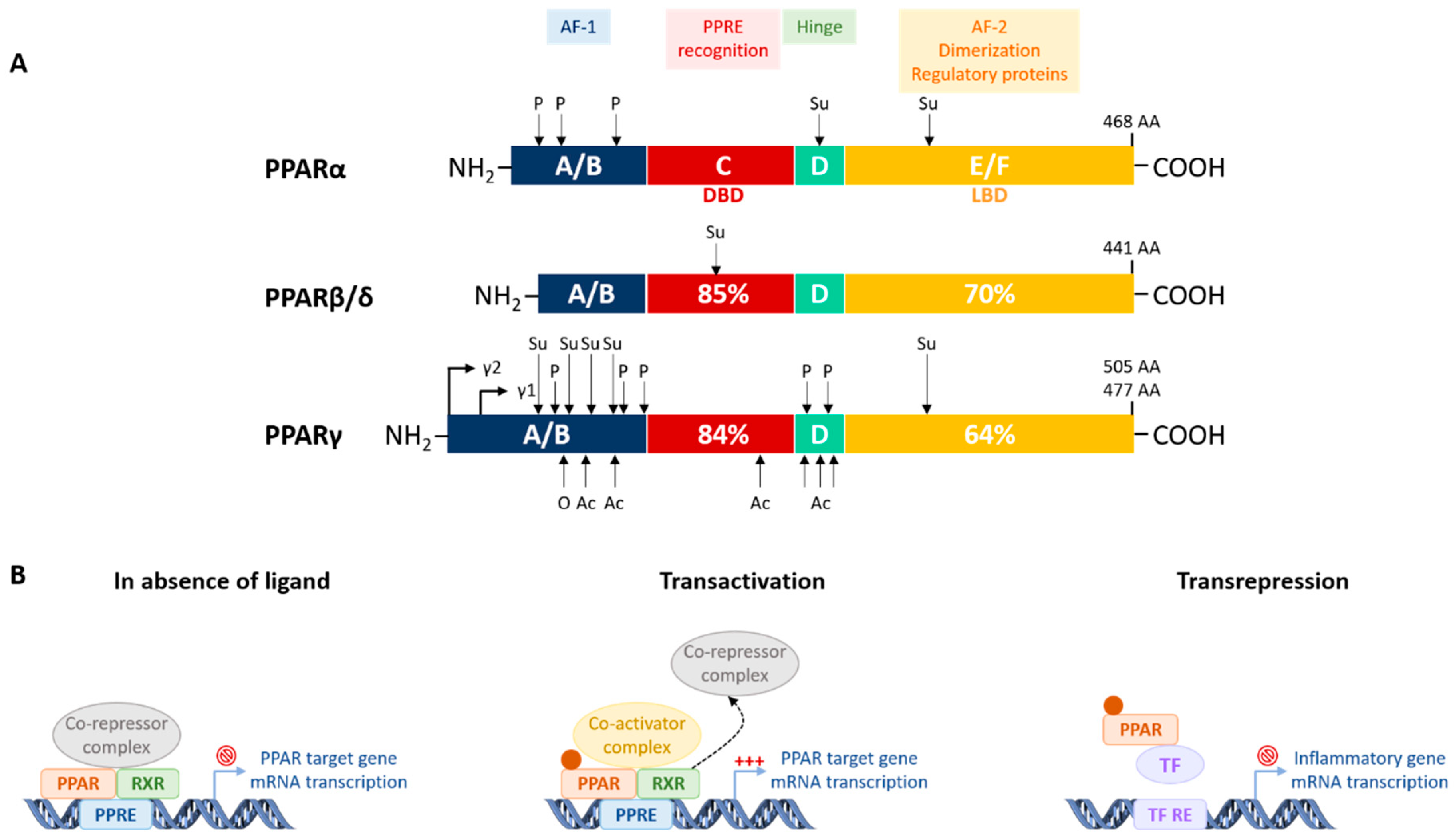
Figure 1. Non-alcoholic fatty liver disease (NAFLD) determinants. Multiple factors contribute to the development of NAFLD and its progression. Obesity and T2DM are closely associated with NAFLD, and both drive the increasing prevalence of NAFLD. The genetic background also strongly influences disease development. In addition, the progression of NAFLD depends on complex interactions between genetic and environmental factors, especially dietary factors. More recently, the gut microbiota has emerged as an important determinant of NAFLD pathogenesis. Abbreviations: NAFLD, non-alcoholic fatty liver disease; T2DM, type 2 diabetes mellitus; PNPLA3, patatin-like phospholipase domain containing protein 3; TM6SF2, transmembrane 6 superfamily 2; MBOAT7, membrane bound O-acyltransferase domain-containing 7; GCKR, glucokinase regulator.
Cells 09 01638 g001
Figure 2. NAFLD progression. NAFLD is a progressive disease characterized by fat accumulation in hepatocytes, ranging from hepatic steatosis (NAFL) to non-alcoholic steatohepatitis (NASH), with additional inflammation with or without fibrosis. The latter is the strongest histological predictor of disease-related mortality. Though steatosis has previously been considered to be benign, some NAFL patients progress to NASH with or without fibrosis, whereas others develop fibrosis without having NASH. The pathogenesis of NAFLD is complex and involves several different pathways in multiple organs, including metabolic and inflammatory pathways. Abbreviations: NAFL, non-alcoholic fatty liver; NASH, non-alcoholic steatohepatitis; HCC, hepatocellular carcinoma; T2DM, type 2 diabetes mellitus; CVD, cardiovascular disease.
Cells 09 01638 g002
Figure 3. Protein structure and mechanisms of transcriptional regulation of PPARs. (A) Functional domains and posttranslational modifications of human PPARs. PPARs contain four distinct domains: a N-terminal A/B domain (ligand-independent AF-1), a C-domain (DNA-binding domain), a D- domain (hinge domain), and a C-terminal E/F domain (ligand-binding domain). Main functions of the 4 domains are listed. The number inside each domain corresponds to the percentage of amino acid sequence identity of human PPARβ/δ and γ relative to PPARα. The number of amino acids indicated at the COOH-terminus are for the human receptors. The locations of posttranslational modification sites are indicated by arrows. The 2 splice variants of PPARγ are indicated by γ1 and γ2. (B) PPAR mechanism of action. In the absence of ligand, PPAR-RXR heterodimers are bound to corepressor complexes and prevent gene transcription. Binding of an endogenous ligand or a synthetic agonist to the PPAR LBD triggers a conformational change, leading to corepressor complex dissociation and recruitment of coactivator complex. The activated PPAR/RXR heterodimer then binds to a specific DNA sequence in the promotor region of target genes (PPRE) and stimulates target gene transcription (transactivation). Through the binding to inflammatory transcription factors such as NF-κB and AP-1 (identified by TF), PPARs inhibit their binding to DNA and negatively regulate expression of proinflammatory genes (transrepression). Abbreviations: AF-1, activation function-1; AF-2, activation function-2; PPRE, peroxisome proliferator response element; DBD, DNA binding domain; LBD, ligand binding domain; AA, amino acid; P, phosphorylation; Su, SUMOylation; Ac, acetylation; O, O-GlcNacylation; RXR, 9-cis retinoic acid receptor; TF, transcription factor; TF RE, transcription factor response element; NH2, protein N terminus; COOH, protein carboxyl terminus.
Cells 09 01638 g003
Figure 4. PPAR target genes and their implications in major functions associated with NAFLD pathogenesis. Key target genes of PPARα (A), PPARβ/δ (B), and PPARγ (C) and their association with four main biological processes driving NAFLD development and progression, i.e., lipid and glucose metabolism, inflammation, and hepatic stellate cell (HSC) activation. Genes in red, blue, and green are also regulated by PPARα, PPARβ/δ, and PPARγ, respectively. Genes whose expression is regulated by all three PPARs are underlined. A question mark (?) after a gene name indicates that PPAR may potentially regulate it. (A) PPARα promotes the expression of genes involved in fatty acid catabolism (Hmgcs2, Acox, Cyp4a, Ehhadh, Acad, Cpt1a, Cpt2, Cd36, Slc27a1, Fabp1) [235,237,321,322], autophagy (Atg, Tfeb) [248,249], and glycerol metabolism (Gyk, Gpdh) [255], and regulates lipoprotein metabolism (Lpl, ApoC3) [220] and hepatic Fgf21 expression [242,243]. It also downregulates inflammatory genes and transcription factors (Nf-κb, Ap-1, Stat, Iκb, Il1ra) [220,281,300], and may downregulate Tgfβ expression [306]. (B) PPARβ/δ increases PGC-1α in muscles [273,275], Insig1 [280] and Cd36 [279] in the liver, and regulates the expression of genes involved in lipoprotein metabolism (ApoA4, Vldlr) [281,284] and glucose utilization [279]. PPARβ/δ regulates the expression of genes induced during alternative macrophage activation (Arg1, Clec7a) [307] and may also influence HSC activation (Tgfβ, α-Sma, Col1a1) [309,310]. (C) PPARγ controls the expression of genes involved in adipogenesis (Fabp4, Cd36, Lpl, Mogat1) [296,314] and genes encoding adipokines (Adipoq, leptin, Fgf21) [192,193,289,291]. It also promotes Fsp27 expression in the liver during steatosis [293]. PPARγ downregulates inflammatory transcription factors (Nf-κb, Ap-1, Stat) [312,315] and may also reduce expression of Tgfβ [318,319]. Abbreviations: Hmgcs2, 3-hydroxy-3-methylglutaryl-CoA synthase 2; Acox, peroxisomal acyl-coenzyme A oxidase 1; Cyp4a, cytochrome P450 family 4 subfamily A; Ehhadh, enoyl-CoA hydratase and 3-hydroxyacyl CoA dehydrogenase; Acadm, acyl-CoA dehydrogenase medium chain; Acadl, acyl-CoA dehydrogenase long chain; Acadvl, acyl-CoA dehydrogenase very long chain; Cpt1a, carnitine palmitoyltransferase 1a; Cpt2, carnitine palmitoyltransferase 2; Slc27a1, solute carrier family 27 member 1; Fabp1, fatty acid binding protein 1; Fabp4, fatty acid binding protein 4; Lpl, lipoprotein lipase; ApoC3, apolipoprotein C3; ApoA4, apolipoprotein A4; Atg, autophagy-related genes; Tfeb, transcription factor EB; Fgf21, fibroblast growth factor 21; Pgc1α, peroxisome proliferator-activated receptor gamma coactivator 1-alpha; Fasn, fatty acid synthase; Acc, acetyl-CoA carboxylase; Scd1, stearoyl-CoA desaturase; Insig1, insulin-induced gene 1; Vldlr, very-low density lipoprotein receptor; Mogat1, monoacylglycerol O-acyltransferase 1; Fsp27, fat specific protein 27; Adipoq, adiponectin; Gpdh, glyceraldehyde-3-phosphate dehydrogenase; Gyk, glycerol kinase; Glut2, glucose transporter type 2; Pk, pyruvate kinase; Gk, glucokinase; Nf-κb, nuclear factor kappa B subunit 1; Ap-1, activator protein; Stat, signal transducer and activator of transcription; Iκb, Nf-κb inhibitor; Il1ra, IL-1 receptor antagonist; Arg1, arginase 1; Clec7a, C-type lectin domain containing 7A; Tgfβ, transforming growth factor beta; Col1a1, collagen type I alpha 1 chain; α-Sma, alpha-smooth muscle actin.
Cells 09 01638 g004
Figure 5. Novel PPAR agonists with potential for NAFLD treatment. The main genes regulated by the PPARα agonist pemafibrate, the dual PPARα and β/δ agonist elafibranor, the dual PPARα and γ agonist saroglitazar, and the pan-PPAR agonist lanifibranor in preclinical models of NAFLD. Through the modulation of gene expression, these novel compounds regulate several hepatic and extrahepatic pathways, including lipid and glucose metabolism, inflammation, and hepatic stellate cell activation, which are all key processes involved in NAFLD. Abbreviations: BAT, brown adipose tissue; WAT, white adipose tissue; Pdk4, pyruvate dehydrogenase kinase 4; Lpl, lipoprotein lipase; ApoC3, apolipoprotein C3; Vldlr, very-low density lipoprotein receptor; Cpt, carnitine palmitoyltransferase; Slc27a1, solute carrier family 27 member 1; Fabp, fatty acid binding protein; Cyp4a, cytochrome P450 family 4 subfamily A; Ehhadh, enoyl-CoA hydratase and 3-hydroxyacyl CoA dehydrogenase; Acad, acyl-CoA dehydrogenase; Hmgcs2, 3-hydroxy-3-methylglutaryl-CoA synthase 2; Acot1, acyl-CoA thioesterase 1; Fgf21, fibroblast growth factor 21; Nrf1, nuclear respiratory factor 1; Sod1, superoxide dismutase 1; Il1β, interleukin-1 beta; Il6, interleukin-6; Tnfα, tumor necrosis factor-alpha; Ccr2, C-C motif chemokine receptor 2; Tlr4, Toll-like receptor 4; Mcp1, monocyte chemoattractant protein 1; Ccl5, C-C motif chemokine ligand 5; Nlrp3, NLR family pyrin domain containing 3; Nfκb, nuclear factor kappa B subunit 1; Tgfβ, transforming growth factor beta; Col1a1, collagen type I alpha 1 chain; Timp1, metalloproteinase inhibitor 1; α-Sma, alpha-smooth muscle actin; Ucp1, uncoupling protein 1; Cidea, cell death inducing DFFA like effector A; Elovl3, elongation of very long chain fatty acids protein 3; Acox, peroxisomal acyl-coenzyme A oxidase 1; Abca1, ATP binding cassette subfamily A member 1; Npc1l1, Niemann-Pick C1-like protein 1; Pgc1α, peroxisome proliferator-activated receptor gamma coactivator 1-alpha; Ifnγ, interferon gamma; Il4, interleukin-4; Il10, interleukin-10; Stat6, signal transducer and activator of transcription 6.
Cells 09 01638 g005
Table 1. Expression, ligands, and functions of Peroxisome proliferator-activated receptors (PPARs) related to NAFLD and therapeutic potential.
IsotypesPPARαPPARβ/δPPARγ
Main tissue expressionLiverSkeletal & cardiac musclesWAT
Skeletal musclesBAT
HeartLiverMacrophages
KidneyWAT
BATBAT
IntestineMacrophages
Main natural ligandsFAFAFA
EicosanoidsVLDL componentsArachidonic acid metabolites
Phospholipids
Main synthetic single agonistsFenofibrateGW501516Pioglitazone
Wy14643GW0742Rosiglitazone
GemfibrazilSeladelpar
Pemafibrate
Biological functions related to NAFLDTG hydrolysisMuscle FA storageAdipogenesis
FA catabolismFA catabolismAdipose FA storage
KetogenesisLipoprotein metabolismAdipokine secretion
FGF21 productionGlucose utilizationAnti-inflammatory
Glycerol metabolismAnti-inflammatoryAnti-fibrotic
Anti-Inflammatory
Potential therapeutic target forHypertriglyceridemiaAtherogenic dyslipidemiaInsulin resistance
Atherogenic dyslipidemiaInsulin resistanceObesity
NAFLDObesityT2DM
T2DMNAFLD
NAFLD
Abbreviations: BAT, brown adipose tissue; WAT, white adipose tissue; FA, fatty acid; VLDL, very low density lipoprotein; TG, triglyceride; FGF21, fibroblast growth factor 21; T2DM, type 2 diabetes mellitus; NAFLD, non-alcoholic fatty liver disease.
Table 2. PPAR agonists currently in late-stage clinical trials (phase 2 and phase 3). Overview of new PPAR agonists: trivial name, chemical structure, and short description.
CompoundsChemical StructureDescription
Pemafibrate Cells 09 01638 i001Selective and potent PPARα modulator
Clinical application for dyslipidemia
Safety profile in clinical studies
Currently in phase 3: effect on CV events in T2DM
Elafibranor Cells 09 01638 i002High activity on PPARα, moderate activity on PPARβ/δ
Safety profile in clinical studies
Currently in phase 3: effect on fibrosis in NASH
Saroglitazar Cells 09 01638 i003High activity on PPARα, moderate activity on PPARγ
Clinical application for dyslipidemia
Safety profile in clinical studies
Currently in phase 2: effect on NAFLD/NASH, phase 3: effect on fibrosis compared to pioglitazone and vitamin E
Lanifibranor Cells 09 01638 i004Moderate and balanced activity on PPARα, PPARβ/δ, PPARγ
Currently in phase 2: effect on NAFLD/NASH
Abbreviations: CV, cardiovascular; NAFLD, non-alcoholic fatty liver disease; NASH, non-alcoholic steatohepatitis.

