Article

Identification of Chicken Transglutaminase 1 and In Situ Localization of Transglutaminase Activity in Avian Skin and Esophagus

1
Skin Biology Laboratory, Department of Dermatology, Medical University of Vienna, 1090 Vienna, Austria
2
Clinic for Poultry and Fish Medicine, Department for Farm Animals and Veterinary Public Health, University of Veterinary Medicine Vienna, 1210 Vienna, Austria
*
Author to whom correspondence should be addressed.
Genes 2021, 12(10), 1565; https://doi.org/10.3390/genes12101565
Submission received: 24 August 2021 / Revised: 27 September 2021 / Accepted: 29 September 2021 / Published: 30 September 2021
(This article belongs to the Special Issue Genomics and Evolution of Sauropsid Traits in the Genomics Era)

Abstract

:
Transglutaminase 1 (TGM1) is a membrane-anchored enzyme that cross-links proteins during terminal differentiation of epidermal and esophageal keratinocytes in mammals. The current genome assembly of the chicken, which is a major model for avian skin biology, does not include an annotated region corresponding to TGM1. To close this gap of knowledge about the genetic control of avian cornification, we analyzed RNA-sequencing reads from organotypic chicken skin and identified TGM1 mRNA. By RT-PCR, we demonstrated that TGM1 is expressed in the skin and esophagus of chickens. The cysteine-rich sequence motif required for palmitoylation and membrane anchorage is conserved in the chicken TGM1 protein, and differentiated chicken keratinocytes display membrane-associated transglutaminase activity. Expression of TGM1 and prominent transglutaminase activity in the esophageal epithelium was also demonstrated in the zebra finch. Altogether, the results of this study indicate that TGM1 is conserved among birds and suggest that chicken keratinocytes may be a useful model for the study of TGM1 in non-mammalian cornification.

1. Introduction

The outermost epidermal layer of amniotes (mammals, reptiles and birds) consists of multiple layers of cornified keratinocytes. Cornification is a mode of programmed cell death that involves the covalent cross-linking of proteins in the cell periphery to form a so-called cornified envelope [1,2,3]. A large portion of the cornified envelope proteins is encoded by genes of the epidermal differentiation complex (EDC) which was originally identified in mammals [4,5,6]. Later studies showed that an EDC is also present in other amniotes [7,8,9,10,11,12].
The cross-linking of epidermal proteins depends on disulfide bonds between cysteine residues and isopeptide bonds between glutamine and lysine residues [1,13]. The latter bond is formed under the control of transglutaminases (TGMs) [14]. In the human epidermis, TGM1, TGM3 and TGM5 are implicated in cornified envelope formation [15,16]. TGM1 is anchored to the cell membrane, whereas TGM3 and TGM5 are cytosolic enzymes [17]. The association of TGM1 with the membrane depends on the palmitoylation of a cysteine-rich sequence motif close to the amino-terminus of the TGM1 protein. Defects of the human TGM1 gene cause autosomal recessive lamellar ichthyosis, a debilitating and life-threatening skin disease [18], and targeted inactivation of TGM1 in mice leads to perinatal death due to skin barrier impairment [19,20]. Apart from the skin, TGM1 is highly abundant in the esophagus, where its expression coincides with that of the EDC gene Cornulin (CRNN) [21,22]. Esophageal keratinocytes form cornified envelopes in vitro [23], but the functions of TGM1 and transglutamination in the esophagus are not fully understood at present.
In addition to TGM1 in mammals, TGM1 orthologs were reported to be conserved in alligators, Xenopus frogs and fishes [24,25]. The existence of TGM1 in fish suggests that TGM1 originated prior to the evolution of a continuous cornified layer on the skin surface, which first appeared in amphibians. Surprisingly, there is no annotated region corresponding to TGM1 in the current release of the chicken (Gallus gallus) genome sequence (assembly: GCF_016699485.2), raising the question as to whether epidermal transglutamination might occur by an atypical mechanism in the main avian model species [26,27]. The uncertainty about the conservation of a central enzyme of epidermal transglutamination is worrying, because most of the research on avian epidermal cornification proteins was conducted in the chicken [7,8,28,29,30,31,32,33,34,35,36,37]. Recently, transglutamination of the EDC protein cornulin was proposed to influence the mechanical properties of the avian esophagus [38]. According to this hypothesis, loss of cornulin in the avian clade Passeri has led to higher elasticity of the oroesophageal cavity, a feature supporting pure-tone song in songbirds [38]. However, the enzymatic process of transglutamination has not yet been reported for the esophagus of birds.
In view of the important role of the chicken as a model for non-mammalian skin biology, we aimed at determining whether the chicken has a homolog of mammalian TGM1 and whether chicken skin and esophagus show a pattern of TGM activity similar to that in the homologous organs of mammals.

2. Materials and Methods

2.1. Animals

Tissue samples were prepared from commercial broiler chickens (21 days old; Ross-308), specified pathogen-free embryos (18 days of incubation, corresponding to Hamburger and Hamilton stage 44 [39]; VALO Biomedica, Sachsenring, Germany) and zebra finches and mice (strain C57BL/6J) immediately after killing the animals. The Ethics Committee of the Medical University of Vienna decided that, in agreement with the national laws, a permission for sacrificing animals (zebra finches and mice) for organ preparation was not required. For sampling in chickens, non-treated negative control birds from an animal trial that was performed with different objectives at the University of Veterinary Medicine Vienna were utilized during necropsy. The trial was approved by the institutional ethics and animal welfare committee and the national authority according to §§ 26ff. of Animal Experiments Act, Tierversuchsgesetz 2012–TVG 2012 (license number BMBWF GZ: 2020-0.761.569).

2.2. Transglutaminase In Situ Activity Assay

Fresh tissue samples were dissected and washed in phosphate-buffered saline (PBS). Chicken in vitro skin models were obtained from a previous study [40]. The samples were placed in cryomolds (Tissue-Tek® Cryomold®, Sakura FineTec, Torrance, CA, USA) filled with optimal cutting temperature (OCT) compound (Scigen, Paramount, Canada) and snap-frozen in liquid nitrogen. Cryo-sections of 6 µm thickness were prepared with a cryostat (Leica CM3050S) and stored at −20 °C or stained immediately. An in situ transglutaminase activity assay was performed based on published protocols [41,42,43]. Briefly, cryosections were thawed at room temperature for 10 min before they were placed in PBS for 5 min at room temperature to remove remaining OCT from the slides. The sections were encircled with a liquid blocker (Daido Sangyo Co. Ltd. Tokyo, Japan) and subsequently incubated with 1% bovine serum albumin (BSA) in 1 M Tris-HCl, pH 7.4 at room temperature for 30 min. Subsequently, the samples were incubated with 5 µM Alexa-fluor 555-cadaverine (catalog number A30677, Thermo Fisher Scientific, Waltham, MA, USA) as TGM substrate in 0.1 M Tris-HCl pH 7.4, and either 5 mM CaCl2 to facilitate transglutaminase activity or 5 mM EDTA to suppress transglutaminase activity (negative control) for 2 h at room temperature under protection from light. The reaction was stopped by incubating the sections in 25 mM EDTA in PBS for 5 min. The sections were rinsed in PBS and then incubated with 1 µg/mL nucleic acid stain Hoechst 33258 (Molecular Probes, Eugene, OR, USA) at room temperature protected from light for 5 min. Afterwards, the sections were rinsed in PBS and mounted with Permafluor (catalog number TA-030-FM, Thermo Fisher Scientific, Waltham, MA, USA). Commercially available nail polish was used to seal the slides to prevent the sections from drying. Sections were studied with an Olympus BX63 light microscope and images were taken with an Olympus UC-90 camera. Fluorescent images were taken and merged with cellSens Dimensions (version 1.16). Confocal images were taken with a Zeiss 980 confocal laser scanning microscope using the program Zen (Blue edition). Where indicated in the figure legends, the brightness of images of both activity labeling and negative controls was equally increased.

2.3. RNA Preparation and RT-PCR

Tissue samples were placed in RNA-later (AM7020, Invitrogen, Carlsbad, CA, USA) immediately after dissection and stored overnight at 4 °C, and subsequently at −80 °C for a long-term duration. RNA isolation was performed with Trizol (catalog number 30-2010 PEQ-GOLD Trifast, VWR, Radnor, PA, USA) from homogenized tissues according to published protocols [44]. cDNA synthesis was performed with the qScript® cDNA synthesis kit (catalog number 95047-025, Quantabio, Beverly, MA, USA). Chicken TGM1 was amplified with the intron-spanning primers 5′-CTGTCCTGCCCCGCATCCATC-3′ and 5′-GAGCTGCAATTCGACGCCTC-3′. Chicken TGM2 was amplified with the primers 5′-TCTGTGTATCGCTCTGCTCC-3′ and 5′-TGGGCTCCAGGGACTACATA-3′. Zebra finch TGM1 was amplified with the primer pair 5’-GTGCAGGTCGTGTTCCAGAA-3′ and 5’-CCAATCAGTGACGCCGCTC-3’. cDNA of the housekeeping gene EEF1A1 was amplified with the primer pairs 5′-GGCCCGAAGTTCCTGAAATC-3′ and 5′-CTGTTGGTGTCATCAAGGCC-3′ for chicken and 5′-AGATGGCCCCAAATTCCTGA-3’ and 5’-GTCGCTGTTGGTGTCATCAA-3′ for zebra finch. Primers were synthesized by Microsynth (Switzerland). cDNA was PCR-amplified with the Dream Taq DNA polymerase (catalog number EP0702, Thermo Scientific, Waltham, MA) according to a published protocol [45] which was modified by the addition of 5% DMSO (Merck, Darmstadt, Germany) to the reaction buffer.

2.4. Chicken TGM1 Sequence Assembly In Silico

Publicly available TGM1 protein sequences of human (GenBank accession number: NP_000350.1), zebra finch (XP_041568788) and Swainson’s thrush (XP_032940238) were used as queries for tBLASTn searches [46] in sequence reads of a chicken in vitro skin model [40] (NCBI accession number: SRX9610284). The reads were translated with the Expasy Translate tool at https://web.expasy.org/translate/ (accessed on 28 September 2021). The 3’-terminal end of the chicken TGM1 sequence was identified by tBLASTn in the whole genome shotgun sequence of the chicken (NCBI accession: JAENSL010000336). Overlapping sequence reads and exons of chicken TGM1 were assembled to obtain the complete coding sequence of chicken TGM1.

2.5. Molecular Phylogenetics

Transglutaminase sequences for the phylogenetic analysis were downloaded from NCBI GenBank (Table S1). The sequences were aligned using MAFFT (Version 7.427) [47] with the parameters --maxiterate set to 1000 and --localpair. Alignments were trimmed using BMGE, version 1.12 [48], by setting the entropy-like value of the BLOSUM matrix to -BLOSUM30, the maximum entropy threshold (-h) to 1, and the minimum length of selected regions (-b) to 1. The first three amino-terminal amino acid residues were excluded manually in AliView [49]. The model for amino acid replacement was calculated using ProtTest (Version 3.0) [50,51]. All available matrices (-all-matrices) and models with rate variation among sites (-all-distributions) were included. The likelihood of the predicted models was assessed with the Akaike information criterion (-sort A) [52]. The selected amino acid substitution model was LG [53]. Maximum likelihood tree and bootstrap analysis (-b 100) were performed using PHYML (Version 20120412) [54]. Tree topology (t), branch length (l), and rate parameters (r) were optimized (-o tlr). Visualization and annotation of phylogenetic trees was performed with FigTree (http://tree.bio.ed.ac.uk/software/figtree/, accessed on 28 September 2021).

3. Results

3.1. Identification of Chicken TGM1

To determine whether TGM1 is conserved in the chicken, we searched the genome sequence assembly of the chicken for a chromosomal segment homologous to the locus of TGM1 in other species. In the human and zebra finch (Taeniopygia guttata), TGM1 is located between RABGGTA and NEDD8 (Figure 1A). RABGGTA is not present in the current Gallus gallus genome annotation release (assembly: GCF_016699485.2), whereas NEDD8 is located on chromosome 35. On the 5′-side of the NEDD8 gene, i.e., the expected locus of the TGM1 gene, there is a sequence gap in the assembly, and the genes predicted on the other side of the gap are not homologous to genes flanking TGM1 in other species (Figure 1A).
tBLASTn searches using the amino acid sequence of zebra finch TGM1 protein (XP_041568788) as the query revealed the presence of two sequence stretches similar to the last two exons of TGM1 in the chicken genome region between NEDD8 and the genome sequence gap mentioned above (Figure 1A). Next, we performed tBLASTn searches in the RNA-sequence reads that we had previously obtained in a transcriptome analysis of chicken epidermis reconstituted in vitro [40]. Using this approach, we obtained a series of overlapping short reads which, together with the sequence of the TGM1-like exons, besides NEDD8, covered the entire coding sequence of chicken TGM1 (Figure 1A and Figure S1, Table S2).
Translation of the predicted chicken TGM1 mRNA sequence (Figure S1) and alignment of the resulting amino acid sequence with TGM1 of human, mouse and zebra finch showed a high degree of sequence conservation (Figure 1B). The palmitoylation sequence motif (Figure 1B, asterisks) and the amino acid residues (cysteine, histidine, aspartate) representing the catalytic triad (Figure 1B, yellow shading) within the transglutaminase core domain are conserved in chicken TGM1. Molecular phylogenetics confirmed that chicken TGM1 is orthologous to TGM1 of human, mouse and zebra finch (Figure 1C and Figure S2).

3.2. TGM1 Is Expressed in the Epidermis and Esophagus of the Chicken

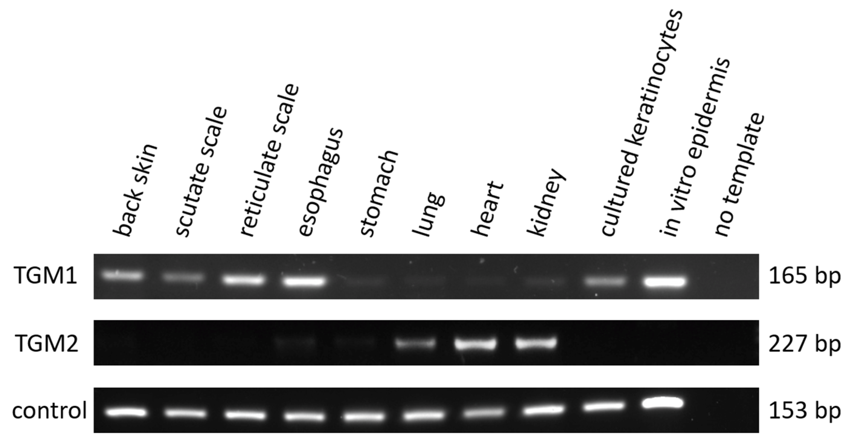
Using primers annealing to TGM1-specific sequences, we performed RT-PCR screening of chicken tissues and isolated cells. TGM1 mRNA was detected in the skin from the back, scutate scales on the legs, reticulate scales from the toes and in the esophagus, but not or only at very low levels in the stomach, lung, heart and kidney of chickens (Figure 2). TGM1 is also expressed in isolated keratinocytes and in the epidermal compartment of chicken skin reconstituted in vitro [40] (Figure 2). By contrast, TGM2 was detected only in lung, heart and kidney (Figure 2). Transcripts of the house-keeping gene EEF1A1 were detected in all samples. TGM1 expression was not only detected in adult chicken skin and esophagus tissues, but also in the tissues sampled from embryos at Hamburger and Hamilton stage 44 (Figure S3). RT-PCR analysis of zebra finch tissues demonstrated conserved expression of TGM1 in the skin and esophagus (Figure S4).

3.3. TGM Activity Is Present in Differentiated Epithelial Cells of Chicken Skin and Esophagus

To determine if TGM1 expression correlated with TGM activity, we determined tissue TGM activity in situ. Cryosections were incubated with the TGM substrate cadaverine coupled with a fluorescent label, and calcium, which is required for the catalytic activity of TGMs. TGM activity was readily detected in chicken reticulate scales when sections were labeled in the presence of 5 mM calcium ions (Figure 3A), whereas sections incubated in the presence of the same concentration of magnesium ions did not show a signal (Figure 3B). TGM was also detected in the scutate scales and back skin of adult chickens. Mouse skin, which served as positive control, was labeled in the uppermost layers of the epidermis under the stratum corneum (Figure 3C). Replacement of calcium ions with EDTA, which is a standard negative control in TGM activity labeling experiments [41,42,43], abolished the fluorescent labeling (Figure 3B). These experiments demonstrated calcium-dependent TGM activity in chicken skin and confirmed the specificity of the TGM activity assay.
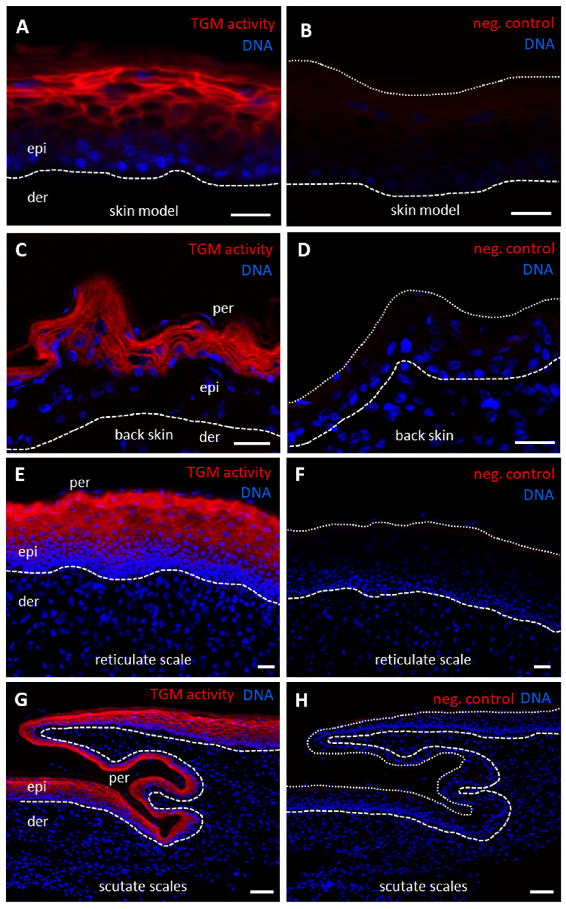
TGM activity was also detected in chicken skin models (Figure 4A), in the upper layers of the epidermis of back skin (Figure 4C), reticulate scales (Figure 4E) and scutate scales (Figure 4G) of chicken embryos. The periderm, a transient epithelial layer during embryonic development, did not contain TGM activity, as evidenced by the absence of a TGM activity signal in nucleated cells on the surface of embryonic epidermis and scales (Figure 4C,E,G). Negative control experiments in which calcium was replaced by EDTA confirmed the specificity of labeling in samples (Figure 4B,D,F,H). The labeling was most prominently detected at cell membranes, suggesting that the activity corresponded to TGM1, the only membrane-anchored TGM.
TGM activity was also detected in a membrane-associated pattern in the suprabasal layers of the chicken esophageal epithelium, whereas no activity was present in the three most basal layers and in the mesenchymal compartments of the esophagus (Figure 5A,B). This pattern was similar to the patterns of TGM activity in the esophagus of the zebra finch (Figure 5C,D) and the mouse (Figure 5E,F).

4. Discussion

Our identification of the complete coding sequence of chicken TGM1 demonstrates that this enzyme is conserved in the main model species of birds [56,57,58]. Together with the determination of the TGM activity patterns in chicken skin and esophagus, the results of this study close a significant gap in knowledge about the biology of the avian integument. Previous reports showed that epidermal TGM activity can be induced by hydrocortisone in chicken skin [25,26], chicken keratinocytes undergo cornification [27,29] and apparent substrates of transglutamination, such as loricrin and involucrin-like proteins [7,8], are conserved in the chicken. Our results strongly suggest that a major step in epidermal cornification, i.e., TGM1-dependent protein cross-linking on the inner face of the epithelial cell, is active in the chicken.
The sequence conservation of chicken TGM1 includes not only the domain and residues required for catalytic activity, but also the cysteine-rich sequence motif that is palmitoylated for membrane anchorage of the enzyme [59]. Among human and chicken TGMs, TGM1 is the only protein containing such a motif, indicating that it is also the only membrane-bound enzyme capable of protein transglutamination. The TGM activity of terminally differentiated epidermal keratinocytes of the chicken is associated with the cell membranes and thereby resembles the distribution pattern of TGM activity in human skin [41,42]. Even more clearly, TGM activity is membrane-associated in the esophageal epithelium of the chicken. There is no obvious difference in the TGM activity pattern between the chicken and the zebra finch, a representative of songbirds. Therefore, the evolutionary loss of the esophageal transglutamination substrate cornulin in songbirds [38] does not correspond to a loss of transglutamination in general, but rather suggests a change in the composition of the cross-linked proteome of the esophagus of passerines.
In conclusion, terminal differentiation of epidermal keratinocytes and esophageal epithelial cells of the chicken involves expression of TGM1 and membrane-associated TGM activity, suggesting that transglutamination substrates encoded by chicken EDC genes are cross-linked by mechanisms similar to those of transglutamination in mammalian epidermis and esophagus.

Supplementary Materials

The following are available online at https://www.mdpi.com/article/10.3390/genes12101565/s1, Table S1. Transglutaminases used for phylogenetic analysis. Table S2. Nucleotide sequences used for the assembly of the complete coding sequence of chicken TGM1. Figure S1. Chicken TGM1 sequence assembly. Figure S2. Alignment of trimmed amino acid sequence of transglutaminases used for the phylogenetic analysis. Figure S3. RT-PCR analysis of TGM1 in embryonic skin of the chicken. Figure S4. RT-PCR analysis of zebra finch TGM1 in selected tissues.

Author Contributions

Conceptualization, A.P.S. and L.E.; investigation, A.P.S. and M.S.; data curation, A.P.S. and L.E.; methodology, A.P.S., M.S., J.L., S.P. and L.E.; writing—original draft preparation, A.P.S. and L.E.; writing—review and editing, A.P.S., M.S., J.L., S.P. and L.E.; project administration, L.E.; funding acquisition, L.E. All authors have read and agreed to the published version of the manuscript.

Funding

This work was supported by the Austrian Science Fund (FWF): P28004 and P32777. Open access funding is provided by the Austrian Science Fund (FWF).

Institutional Review Board Statement

All animal procedures were conducted according to the guidelines established by the Animal Care and Use Committees of the Medical University of Vienna and the University of Veterinary Medicine Vienna, Austria. The Ethics Committee of the Medical University of Vienna decided that, in agreement with the national laws, permission for sacrificing animals for organ preparation was not required. For sampling in chickens, non-treated negative control birds from an animal trial that was performed with different objectives at the University of Veterinary Medicine Vienna were utilized during necropsy. The trial was approved by the institutional ethics and animal welfare committee and the national authority according to §§ 26ff. of Animal Experiments Act, Tierversuchsgesetz 2012–TVG 2012 (license number BMBWF GZ: 2020-0.761.569).

Informed Consent Statement

Not applicable.

Data Availability Statement

Data are contained within the article or supplementary material.

Acknowledgments

We thank Michael Hess, University of Veterinary Medicine Vienna, and Erwin Tschachler, Medical University of Vienna, for advice and helpful discussions, Veronika Mlitz and Florian Ehrlich for providing tissue samples for comparative analysis, and Bahar Golabi, Department of Dermatology, Medical University of Vienna, for excellent technical support. Open Access Funding by the Austrian Science Fund (FWF).

Conflicts of Interest

The authors declare no conflict of interest.

References

  1. Candi, E.; Schmidt, R.; Melino, G. The cornified envelope: A model of cell death in the skin. Nat. Rev. Mol. Cell Biol. 2005, 6, 328–340. [Google Scholar] [CrossRef]
  2. Eckhart, L.; Lippens, S.; Tschachler, E.; Declercq, W. Cell death by cornification. Biochim. Biophys. Acta (BBA) Bioenerg. 2013, 1833, 3471–3480. [Google Scholar] [CrossRef] [PubMed]
  3. Matsui, T.; Amagai, M. Dissecting the formation, structure and barrier function of the stratum corneum. Int. Immunol. 2015, 27, 269–280. [Google Scholar] [CrossRef] [PubMed] [Green Version]
  4. Mischke, D.; Korge, B.P.; Marenholz, I.; Volz, A.; Ziegler, A. Genes Encoding Structural Proteins of Epidermal Cornification and S100 Calcium-Binding Proteins Form a Gene Complex (“Epidermal Differentiation Complex”) on Human Chromosome 1q21. J. Investig. Dermatol. 1996, 106, 989–992. [Google Scholar] [CrossRef] [PubMed] [Green Version]
  5. Henry, J.; Toulza, E.; Hsu, C.Y.; Pellerin, L.; Balica, S.; Mazereeuw-Hautier, J.; Paul, C.; Serre, G.; Jonca, N.; Simon, M. Update on the epidermal differentiation complex. Front. Biosci. 2012, 17, 1517–1532. [Google Scholar] [CrossRef] [Green Version]
  6. Kypriotou, M.; Huber, M.; Hohl, D. The human epidermal differentiation complex: Cornified envelope precursors, S100 proteins and the ‘fused genes’ family. Exp. Dermatol. 2012, 21, 643–649. [Google Scholar] [CrossRef] [PubMed]
  7. Vanhoutteghem, A.; Djian, P.; Green, H. Ancient origin of the gene encoding involucrin, a precursor of the cross-linked envelope of epidermis and related epithelia. Proc. Natl. Acad. Sci. USA 2008, 105, 15481–15486. [Google Scholar] [CrossRef] [Green Version]
  8. Strasser, B.; Mlitz, V.; Hermann, M.; Rice, R.H.; Eigenheer, R.A.; Alibardi, L.; Tschachler, E.; Eckhart, L. Evolutionary Origin and Diversification of Epidermal Barrier Proteins in Amniotes. Mol. Biol. Evol. 2014, 31, 3194–3205. [Google Scholar] [CrossRef] [PubMed] [Green Version]
  9. Holthaus, K.B.; Strasser, B.; Sipos, W.; Schmidt, H.; Mlitz, V.; Sukseree, S.; Weissenbacher, A.; Tschachler, E.; Alibardi, L.; Eckhart, L. Comparative Genomics Identifies Epidermal Proteins Associated with the Evolution of the Turtle Shell. Mol. Biol. Evol. 2015, 33, 726–737. [Google Scholar] [CrossRef]
  10. Holthaus, K.B.; Mlitz, V.; Strasser, B.; Tschachler, E.; Alibardi, L.; Eckhart, L. Identification and comparative analysis of the epidermal differentiation complex in snakes. Sci. Rep. 2017, 7, 45338. [Google Scholar] [CrossRef] [Green Version]
  11. Holthaus, K.B.; Strasser, B.; Lachner, J.; Sukseree, S.; Sipos, W.; Weissenbacher, A.; Tschachler, E.; Alibardi, L.; Eckhart, L. Comparative Analysis of Epidermal Differentiation Genes of Crocodilians Suggests New Models for the Evolutionary Origin of Avian Feather Proteins. Genome Biol. Evol. 2018, 10, 694–704. [Google Scholar] [CrossRef] [PubMed]
  12. Holthaus, K.B.; Alibardi, L.; Tschachler, E.; Eckhart, L. Identification of epidermal differentiation genes of the tuatara pro-vides insights into the early evolution of lepidosaurian skin. Sci. Rep. 2020, 10, 12844. [Google Scholar] [CrossRef]
  13. Alibardi, L. Vertebrate keratinization evolved into cornification mainly due to transglutaminase and sulfhydryl oxidase activities on epidermal proteins: An immunohistochemical survey. Anat. Rec. Adv. Integr. Anat. Evol. Biol. 2021. [Google Scholar] [CrossRef]
  14. Eckert, R.L.; Kaartinen, M.T.; Nurminskaya, M.; Belkin, A.M.; Colak, G.; Johnson, G.V.; Mehta, K. Transglutaminase regu-lation of cell function. Physiol. Rev. 2014, 94, 383–417. [Google Scholar] [CrossRef] [PubMed] [Green Version]
  15. Cassidy, A.; van Steensel, M.; Steijlen, P.M.; van Geel, M.; van der Velden, J.; Morley, S.M.; Terrinoni, A.; Melino, G.; Candi, E.; McLean, W.I. A Homozygous Missense Mutation in TGM5 Abolishes Epidermal Transglutaminase 5 Activity and Causes Acral Peeling Skin Syndrome. Am. J. Hum. Genet. 2005, 77, 909–917. [Google Scholar] [CrossRef] [PubMed] [Green Version]
  16. Chermnykh, E.S.; Alpeeva, E.V.; Vorotelyak, E.A. Transglutaminase 3: The Involvement in Epithelial Differentiation and Cancer. Cells 2020, 9, 1996. [Google Scholar] [CrossRef] [PubMed]
  17. Grenard, P.; Bates, M.K.; Aeschlimann, D. Evolution of Transglutaminase Genes: Identification of a Transglutaminase Gene Cluster on Human Chromosome 15q15. J. Biol. Chem. 2001, 276, 33066–33078. [Google Scholar] [CrossRef] [Green Version]
  18. Russell, L.J.; DiGiovanna, J.J.; Rogers, G.R.; Steinert, P.M.; Hashem, N.; Compton, J.G.; Bale, S.J. Mutations in the gene for transglutaminase 1 in autosomal recessive lamellar ichthyosis. Nat. Genet. 1995, 9, 279–283. [Google Scholar] [CrossRef] [PubMed]
  19. Matsuki, M.; Yamashita, F.; Ishida-Yamamoto, A.; Yamada, K.; Kinoshita, C.; Fushiki, S.; Ueda, E.; Morishima, Y.; Tabata, K.; Yasuno, H.; et al. Defective stratum corneum and early neonatal death in mice lacking the gene for transglutaminase 1 (keratinocyte transglutaminase). Proc. Natl. Acad. Sci. USA 1998, 95, 1044–1049. [Google Scholar] [CrossRef] [PubMed] [Green Version]
  20. Kuramoto, N.; Takizawa, T.; Takizawa, T.; Matsuki, M.; Morioka, H.; Robinson, J.M.; Yamanishi, K. Development of ich-thyosiform skin compensates for defective permeability barrier function in mice lacking transglutaminase 1. J. Clin. Investig. 2002, 109, 243–250. [Google Scholar] [CrossRef] [PubMed]
  21. Michel, S.; Bernerd, F.; Jetten, A.M.; E Floyd, E.; Shroot, B.; Reichert, U. Expression of Keratinocyte Transglutamine mRNA Revealed by In Situ Hybridization. J. Investig. Dermatol. 1992, 98, 364–368. [Google Scholar] [CrossRef] [PubMed] [Green Version]
  22. Contzler, R.; Favre, B.; Huber, M.; Hohl, D. Cornulin, a new member of the “fused gene” family, is expressed during epi-dermal differentiation. J. Investig. Dermatol. 2005, 124, 990–997. [Google Scholar] [CrossRef] [Green Version]
  23. Heimann, R.; Rice, R.H. Rat esophageal and epidermal keratinocytes: Intrinsic differences in culture and derivation of con-tinuous lines. J. Cell Physiol. 1983, 117, 362–367. [Google Scholar] [CrossRef] [Green Version]
  24. Cruz, S.I.R.; Phillips, M.A.; Kültz, D.; Rice, R.H. Tgm1-like transglutaminases in tilapia (Oreochromis mossambicus). PLoS ONE 2017, 12, e0177016. [Google Scholar] [CrossRef]
  25. Kikuta, A.; Furukawa, E.; Ogawa, R.; Suganuma, N.; Saitoh, M.; Nishimaki, T.; Katsumura, T.; Oota, H.; Kawamoto, T.; Tatsukawa, H.; et al. Biochemical Characterization of Medaka (Oryzias latipes) Transglutaminases, OlTGK1 and OlTGK2, as Orthologues of Human Keratinocyte-Type Transglutaminase. PLoS ONE 2015, 10, e0144194. [Google Scholar] [CrossRef] [Green Version]
  26. Obinata, A.; Endo, H.; Obinata, H.E.A. Induction of epidermal transglutaminase by hydrocortisone in chick embryonic skin. Nature 1977, 270, 440–441. [Google Scholar] [CrossRef] [PubMed]
  27. Obinata, A.; Endo, H. Induction of chick epidermal transglutaminase by hydrocortisone in ovo and in vitro with reference to the differentiation of epidermal cells. J. Biol. Chem. 1979, 254, 8487–8490. [Google Scholar] [CrossRef]
  28. Vanhoutteghem, A.; Londero, T.; Ghinea, N.; Djian, P. Serial cultivation of chicken keratinocytes, a composite cell type that accumulates lipids and synthesizes a novel beta-keratin. Differentiation 2004, 72, 123–137. [Google Scholar] [CrossRef] [PubMed]
  29. Greenwold, M.J.; Sawyer, R.H. Genomic organization and molecular phylogenies of the beta (beta) keratin multigene family in the chicken (Gallus gallus) and zebra finch (Taeniopygia guttata), implications for feather evolution. BMC Evol. Biol. 2010, 10, 148. [Google Scholar] [CrossRef] [Green Version]
  30. Rice, R.H.; Winters, B.R.; Durbin-Johnson, B.P.; Rocke, D. Chicken Corneocyte Cross-Linked Proteome. J. Proteome Res. 2013, 12, 771–776. [Google Scholar] [CrossRef]
  31. Alibardi, L.; Holthaus, K.B.; Sukseree, S.; Hermann, M.; Tschachler, E.; Eckhart, L. Immunolocalization of a Histidine-Rich Epidermal Differentiation Protein in the Chicken Supports the Hypothesis of an Evolutionary Developmental Link between the Embryonic Subperiderm and Feather Barbs and Barbules. PLoS ONE 2016, 11, e0167789. [Google Scholar] [CrossRef]
  32. Davis, A.C.; Greenwold, M.J.; Sawyer, R.H. Complex Gene Loss and Duplication Events Have Facilitated the Evolution of Multiple Loricrin Genes in Diverse Bird Species. Genome Biol. Evol. 2019, 11, 984–1001. [Google Scholar] [CrossRef] [PubMed]
  33. Lachner, J.; Ehrlich, F.; Mlitz, V.; Hermann, M.; Alibardi, L.; Tschachler, E.; Eckhart, L. Immunolocalization and phylogenetic profiling of the feather protein with the highest cysteine content. Protoplasma 2019, 256, 1257–1265. [Google Scholar] [CrossRef] [PubMed] [Green Version]
  34. Alibardi, L.; Eckhart, L. Immunolocalization of epidermal differentiation complex proteins reveals distinct molecular com-positions of cells that control structure and mechanical properties of avian skin appendages. J. Morphol. 2021, 282, 917–933. [Google Scholar] [CrossRef]
  35. Mlitz, V.; Strasser, B.; Jaeger, K.; Hermann, M.; Ghannadan, M.; Buchberger, M.; Alibardi, L.; Tschachler, E.; Eckhart, L. Trichohyalin-Like Proteins Have Evolutionarily Conserved Roles in the Morphogenesis of Skin Appendages. J. Investig. Dermatol. 2014, 134, 2685–2692. [Google Scholar] [CrossRef] [Green Version]
  36. Davis, A.; Greenwold, M. Evolution of an Epidermal Differentiation Complex (EDC) Gene Family in Birds. Genes 2021, 12, 767. [Google Scholar] [CrossRef]
  37. Mlitz, V.; Hermann, M.; Buchberger, M.; Tschachler, E.; Eckhart, L. The Trichohyalin-Like Protein Scaffoldin Is Expressed in the Multilayered Periderm during Development of Avian Beak and Egg Tooth. Genes 2021, 12, 248. [Google Scholar] [CrossRef]
  38. Feng, S.; Stiller, J.; Deng, Y.; Armstrong, J.; Fang, Q.; Reeve, A.H.; Xie, D.; Chen, G.; Guo, C.; Faircloth, B.C.; et al. Dense sampling of bird diversity increases power of comparative genomics. Nature 2020, 587, 252–257. [Google Scholar] [CrossRef]
  39. Hamburger, V.; Hamilton, H.L. A series of normal stages in the development of the chick embryo. J. Morphol. 1951, 88, 49–92. [Google Scholar] [CrossRef]
  40. Lachner, J.; Derdak, S.; Mlitz, V.; Wagner, T.; Holthaus, K.B.; Ehrlich, F.; Mildner, M.; Tschachler, E.; Eckhart, L. An in vitro model of avian skin reveals evolutionarily conserved transcriptional regulation of epidermal barrier formation. J. Investig. Dermatol. 2021. [Google Scholar] [CrossRef]
  41. Raghunath, M.; Hennies, H.C.; Velten, F.; Wiebe, V.; Steinert, P.M.; Reis, A.; Traupe, H. A novel in situ method for the detection of deficient transglutaminase activity in the skin. Arch. Dermatol. Res. 1998, 290, 621–627. [Google Scholar] [CrossRef]
  42. Hohl, D.; Aeschlimann, D.; Huber, M. In vitro and rapid in situ transglutaminase assays for congenital ichthyoses—A com-parative study. J. Investig. Dermatol. 1998, 110, 268–271. [Google Scholar] [CrossRef] [Green Version]
  43. Cau, L.; Pendaries, V.; Lhuillier, E.; Thompson, P.R.; Serre, G.; Takahara, H.; Méchin, M.-C.; Simon, M. Lowering relative humidity level increases epidermal protein deimination and drives human filaggrin breakdown. J. Dermatol. Sci. 2017, 86, 106–113. [Google Scholar] [CrossRef] [Green Version]
  44. Chomczynski, P. A reagent for the single-step simultaneous isolation of RNA, DNA and proteins from cell and tissue samples. BioTechniques 1993, 15, 532–536. [Google Scholar]
  45. Lachner, J.; Mlitz, V.; Tschachler, E.; Eckhart, L. Epidermal cornification is preceded by the expression of a keratino-cyte-specific set of pyroptosis-related genes. Sci. Rep. 2017, 7, 17446. [Google Scholar] [CrossRef]
  46. Altschul, S.F.; Gish, W.; Miller, W.; Myers, E.W.; Lipman, D.J. Basic local alignment search tool. J. Mol. Biol. 1990, 215, 403–410. [Google Scholar] [CrossRef]
  47. Katoh, K.; Misawa, K.; Kuma, K.; Miyata, T. MAFFT: A novel method for rapid multiple sequence alignment based on fast Fourier transform. Nucleic Acids Res. 2002, 30, 3059–3066. [Google Scholar] [CrossRef] [PubMed] [Green Version]
  48. Criscuolo, A.; Gribaldo, S. BMGE (Block Mapping and Gathering with Entropy): A new software for selection of phylogenetic informative regions from multiple sequence alignments. BMC Evol. Biol. 2010, 10, 210. [Google Scholar] [CrossRef] [PubMed] [Green Version]
  49. Larsson, A. AliView: A fast and lightweight alignment viewer and editor for large datasets. Bioinformatics 2014, 30, 3276–3278. [Google Scholar] [CrossRef] [PubMed]
  50. Abascal, F.; Zardoya, R.; Posada, D. ProtTest: Selection of best-fit models of protein evolution. Bioinformatics 2005, 21, 2104–2105. [Google Scholar] [CrossRef] [Green Version]
  51. Darriba, D.; Taboada, G.L.; Doallo, R.; Posada, D. ProtTest 3: Fast selection of best-fit models of protein evolution. Bioinformatics 2011, 27, 1164–1165. [Google Scholar] [CrossRef] [PubMed] [Green Version]
  52. Akaike, H. Information Theory and an Extension of the Maximum Likelihood Principle. In Selected Papers of Hirotugu Akaike; Parzen, E., Tanabe, K., Kitagawa, G., Eds.; Springer Series in Statistics (Perspectives in Statistics): New York, NY, USA, 1998; pp. 199–213. [Google Scholar]
  53. Le, S.Q.; Gascuel, O. An Improved General Amino Acid Replacement Matrix. Mol. Biol. Evol. 2008, 25, 1307–1320. [Google Scholar] [CrossRef] [PubMed] [Green Version]
  54. Guindon, S.; Gascuel, O. A Simple, Fast, and Accurate Algorithm to Estimate Large Phylogenies by Maximum Likelihood. Syst. Biol. 2003, 52, 696–704. [Google Scholar] [CrossRef] [PubMed] [Green Version]
  55. Makarova, K.S.; Aravind, L.; Koonin, E.V. A superfamily of archaeal, bacterial, and eukaryotic proteins homologous to animal transglutaminases. Protein Sci. 1999, 8, 1714–1719. [Google Scholar] [CrossRef] [PubMed] [Green Version]
  56. Sawyer, R.H.; Knapp, L.W.; O’Guin, M.W. The Skin of Birds. Epidermis, dermis and appendages. In Biology of the Integument; Bereither-Hahn, J., Matoltsy, A.G., Sylvia-Richards, R., Eds.; Springer: Berlin, Germany, 1986; Volume 2 Ver-tebrates, pp. 194–238. [Google Scholar]
  57. International Chicken Genome Sequencing Consortium. Sequence and comparative analysis of the chicken genome provide unique perspectives on vertebrate evolution. Nature 2004, 432, 695–716. [Google Scholar] [CrossRef] [PubMed]
  58. Liang, Y.-C.; Wu, P.; Lin, G.-W.; Chen, C.-K.; Yeh, C.-Y.; Tsai, S.; Yan, J.; Jiang, T.-X.; Lai, Y.-C.; Huang, D.; et al. Folding Keratin Gene Clusters during Skin Regional Specification. Dev. Cell 2020, 53, 561–576.e9. [Google Scholar] [CrossRef] [PubMed]
  59. Chen, L.-Y.; Lin, K.-R.; Chen, Y.-J.; Chiang, Y.-J.; Ho, K.-C.; Shen, L.-F.; Song, I.-W.; Liu, K.-M.; Yang-Yen, H.-F.; Chen, Y.-T.; et al. Palmitoyl Acyltransferase Activity of ZDHHC13 Regulates Skin Barrier Development Partly by Controlling PADi3 and TGM1 Protein Stability. J. Investig. Dermatol. 2020, 140, 959–970.e3. [Google Scholar] [CrossRef] [PubMed]
Figure 1. Identification and sequence of chicken TGM1. (A) Gene locus of TGM1 in human, mouse, zebra finch and chicken. Synteny is indicated by the conserved genes TINF2 and NEDD8 (grey). Two TGM1-like exons were identified at the position indicated by the arrow. Genes are indicated by arrows pointing in the direction of transcription. BLAST search for RNA-sequencing reads covering the 5’ regions of chicken TGM1 is schematically shown at the bottom of the panel. (B) Alignment of amino acid sequences of TGM1 proteins from human, mouse, zebra finch and chicken. Identical residues in all sequences are shown with red fonts. Asterisks indicate the cysteine residues at the palmitoylation site. The transglutaminase core domain [55] is marked by a line above the sequences. Residues of the catalytic triad are highlighted by yellow shading. (C) Phylogenetic analysis of TGMs from human, mouse, zebra finch and chicken.
Genes 12 01565 g001
Figure 2. Expression analysis of TGM1 and TGM2 in chicken tissues and cells. RNA from the indicated tissues and cultured cells was subjected to RT-PCR with primers specific for TGM1, TGM2, and the house-keeping gene EEF1A1 (control). PCR products were analyzed by agarose gel electrophoresis, revealing bands that correspond to the predicted size of the PCR products.
Genes 12 01565 g002
Figure 3. In situ labeling of transglutaminase (TGM) activity in chicken reticulate scales and mouse skin. Reticulate scales from an adult chicken (A,B) and mouse ear skin (C,D) were cryosectioned and incubated with Alexa-fluor 555-cadaverine (red) in buffer containing either calcium (A,C), magnesium (B) or EDTA (D). Nuclear DNA was labeled with Hoechst 33258 (blue). The images were recorded with an Olympus BX63 microscope (A,B) and a Zeiss 980 confocal laser scanning microscope (C,D). White dashed lines indicate the lower border of the epidermis (AD) and white dotted lines indicate the upper border of the epidermis in images of samples incubated with magnesium (B) and EDTA (D). Scale bars = 20 µm (A,B), 10 µm (C,D).
Genes 12 01565 g003
Figure 4. In situ labeling of transglutaminase (TGM) activity in chicken skin models and chicken skin. Skin models consisting of keratinocytes and fibroblasts isolated from chicks and cultured in vitro (A,B) and back skin (C,D), reticulate scales (E,F) and scutate scales (G,H) from chicken embryos (Hamburger and Hamilton stage 44) were cryosectioned and incubated with Alexa-fluor 555-cadaverine (red) in buffer containing either calcium (A,C,E,G) or, as negative (neg.) control, EDTA (B,D,F,H). Nuclear DNA was labeled with Hoechst 33258 (blue). The images were recorded with an Olympus BX63 microscope (A,B,EH) and a Zeiss 980 confocal laser scanning microscope (C,D). The brightness of the images in panels (AD) was uniformly increased by 20%. White dashed lines indicate lower border of the epidermis (AH) and white dotted lines indicate the upper border of the epidermis in images of negative control samples (B,D,F,H). Scale bars = 20 µm (AF), 50 µm (G,H). der, dermis; epi, epidermis; per, periderm.
Genes 12 01565 g004
Figure 5. In situ labeling of transglutaminase (TGM) activity in the esophagus. The esophagus of the chicken (A,B), zebra finch (C,D) and mouse (E,F) were cryosectioned and incubated with Alexa-fluor 555-cadaverine (red) in buffer containing either calcium (A,C,E) or, as negative (neg.) control, EDTA (B,D,F). Nuclear DNA was labeled with Hoechst 33258 (blue). The images were recorded with an Olympus BX63 microscope. The brightness of the images was increased by 60% in panels (C,D) and by 30% in panels E and F. White dashed lines indicate the basement membrane of esophageal epithelia. Scale bars = 20 µm. epi, epithelium; lam, lamina propria.
Genes 12 01565 g005
Publisher’s Note: MDPI stays neutral with regard to jurisdictional claims in published maps and institutional affiliations.