Article

Telomerase (hTERT) Overexpression Reveals a Promising Prognostic Biomarker and Therapeutical Target in Different Clinical Subtypes of Pediatric Acute Lymphoblastic Leukaemia

by
Beatriz Maria Dias Nogueira
1,†,
Laudreísa da Costa Pantoja
2,†,
Emerson Lucena da Silva
1,†,
Fernando Augusto Rodrigues Mello Júnior
3,
Eliel Barbosa Teixeira
4,
Alayde Vieira Wanderley
2,
Jersey Heitor da Silva Maués
5,
Manoel Odorico de Moraes Filho
1,
Maria Elisabete Amaral de Moraes
1,
Raquel Carvalho Montenegro
1,
André Salim Khayat
4,‡ and
Caroline Aquino Moreira-Nunes
1,4,*,‡
1
Department of Medicine, Pharmacogenetics Laboratory, Drug Research and Development Center (NPDM), Federal University of Ceará, Fortaleza 60430-275, CE, Brazil
2
Department of Pediatrics, Octávio Lobo Children’s Hospital, Belém 60430-275, PA, Brazil
3
Molecular Biology Laboratory, Ophir Loyola Hospital, Belém 66063-240, PA, Brazil
4
Department of Biological Sciences, Oncology Research Center, Federal University of Pará, Belém 66073-005, PA, Brazil
5
Hematology and Transfusion Medicine Center, University of Campinas, Campinas 13083-970, SP, Brazil
*
Author to whom correspondence should be addressed.
Equal contribution as first authors.
Equal contribution as senior authors.
Genes 2021, 12(10), 1632; https://doi.org/10.3390/genes12101632
Submission received: 16 September 2021 / Revised: 6 October 2021 / Accepted: 12 October 2021 / Published: 17 October 2021
(This article belongs to the Section Human Genomics and Genetic Diseases)

Abstract

:
Acute Lymphoblastic Leukemia (ALL) is a neoplasm of the hematopoietic system defined as a clonal expansion of an abnormal lymphoid precursor cell. It mostly affects children under five years of age and is the most common tumor to afflict pediatric patients. The expression of the human telomerase gene (hTERT) in patients with ALL has been studied as a biomarker and could become a new therapeutic target. We evaluate the role of hTERT gene expression in ALL pediatric patients, through quantitative real-time PCR technique, and the possible correlation between hTERT expression and clinical variables: gender, age, white blood cells (WBC), gene fusions, and immunophenotyping. The analysis between healthy controls and ALL patients (N = 244) was statistically significant (p < 0.001), demonstrating hTERT overexpression in these patients. In comparison with the usual set of clinical variables, the data were not statistically significant (p > 0.05), indicating that hTERT is equally overexpressed among patients regardless of gender, age, gene fusions, and immunophenotyping. Moreover, patients who presented a higher hTERT expression level had a significant (p < 0.0001) lower overall survival rate. In summary, hTERT expression emerges as an important molecular pathway in leukemogenesis regardless patient’s clinical variables, thus, the data here presented pointed it as a valuable biomarker in pediatric acute lymphoblastic leukemia and a promising target for new therapeutic and prognostic measures.

1. Introduction

Acute lymphoblastic leukemia (ALL) is a hematopoietic malignant disorder characterized by the affliction of lymphoid progenitor cells leading to their transformation and abnormal proliferation of immature clonal cells, denominated as blasts. ALL composes 80% of all neoplasia cases diagnosed in children and its survival rate demonstrates that children between 1 and 9 years with B-cell ALL tend to have a better prognosis than under 1-year-old infants and children 10 years or older, that in general presents a shorter overall survival [1,2,3,4,5,6,7,8].
During leukemogenesis onset, hematopoietic cells undergo genetic mutations that may trigger chromosomal translocations due to defects on chromosome ends, denominated as telomeres, thereby conferring their malignant phenotype [1,2,9,10,11,12]. Dysfunctions afflicting the telomere complex may expose the DNA to enzymatic activity leading to degradation, breakage, and deleterious processes on the genetic material. The main consequence of loss or dysfunction of the telomeres is a chromosomal instability that further aggravates and may lead to genetic fusions, karyotype abnormalities, cancer progression, and poor clinical outcomes [13,14].
Telomere shortening may be reverted by the human telomerase gene (hTERT) activity, however, this enzyme is only present in limited amounts in human cells [15,16]. Reactivation of hTERT was identified as a common and important biomarker in cancer, and research data demonstrates that enzyme activity plays a critical role in aberrant cell proliferation, tumor progression, and immortality [17].
The altered hTERT expression in patients with hematological diseases as ALL has been widely discussed in the literature as a possible molecular biomarker that could be useful in the development new targeted therapeutics for neoplastic patients [18,19,20]. Assessing altered gene expression in patients with ALL is being studied as a possible way to detect new biomarkers, which could lead to the development of new targeted drugs [19].
Thus, the aim of this study was to assess the hTERT expression profile in a Brazilian cohort of pediatric ALL patients, to evaluate its possible role in leukemogenesis pathway, and as a prognostic biomarker and potential novel therapeutical target.

2. Patients and Methods

2.1. Biological Samples

The present study has a total number of 244 pediatric patients diagnosed with Acute Lymphoblastic Leukemia at Octávio Lobo Children’s Hospital (Belém, PA, Brazil) according to the French American British (FAB) criteria [21]. The clinical data from patients were analyzed based on risk-stratification criteria of Berlin-Franklin-Münster (BFM) [22].
Patients’ blood samples were collected at the time of diagnosis, and samples from 10 healthy volunteers were used as control. The study was approved by the Ethics Committee of the Ophir Loyola Hospital (approval number: 2,798,615), informed written assent was obtained from the patients’ legal guardians and all methods were carried out in accordance with Helsinki guidelines and regulations.

2.2. Extraction of RNA and Reverse Transcription of RNA to cDNA

RNA from samples was extracted with TRIzol Reagent® (Invitrogen, Walthan, MA, USA) according to the manufacturer’s instructions. From 25 µL of RNA, the cDNA was synthesized using High-Capacity cDNA Reverse Transcriptase kit (Life Technologies, Carlsbad, CA, USA) to convert the extracted and purified RNA to cDNA. The conversion step was performed on a Veriti® Thermal Cycler (Applied Biosystems, Foster City, CA, USA). After this step, the samples were stored in a freezer at −20 °C until use for analysis.

2.3. Genetic Fusion Identification

Multiplex Polymerase Chain Reactions were developed in order to identify fused transcripts that commonly occur in cases of ALL in the studied region, such as: E2A-PBX1, MLL-AF4, BCR-ABL, TEL-AML1, and SIL-TAL. For this purpose, the primers are presented in Supplementary File 1 (Table S1).
Multiplex PCR was performed in a final volume of 16.5 μL with 1 μL of cDNA; 4.25 μL of nuclease free water; 6.25 μL of GoTaq Colorless Master Mix (Promega, Madison, WI, USA) and 0.5 μL of each primer. After initial denaturation at 95 °C for 3 min, 35 cycles were performed, including 94 °C for 2 min, 61 °C for 1 min, and 70 °C for 2 min in each cycle. The final extension, at 70 °C for 30 min, was performed to ensure complete elongation of all PCR products. All rounds of reactions were done along with two additional reactions. One with a c-DNA-free tube (negative control), to avoid false-positive results due to possible contamination with other genetic materials; and another with a tube containing a pair of primers with annealed in an unaltered gene (H36B4), reducing the possibility of false-negative results due to poor quality of the c-DNA sample or reagents, presence of reaction inhibitors, or human error.

2.3.1. Electrophoresis in Agarose Gel

For the possible visualization and analysis of PCR products, which would characterize the occurrence of gene fusion, agarose gel electrophoresis was performed. The gel was prepared by mixing 1.5 g of powdered agarose into 100 mL of 1X TEB Buffer. After heating in a microwave until the powder had completely dissolved, 10 µL of SYBR® Safe DNA Gel Stain (Life Technologies, Carlsbad, CA, USA) was added. After polymerization, the gel was placed in the electrophoresis vat, then 1L of 1X TEB buffer was added and the samples, homogenized with BlueJuiceTM Gel Loading Buffer (Life Technologies, Carlsbad, CA, USA) at a ratio of 4 to 1, were applied one to each well. In addition to the samples, 0.5 μL of 1 Kb Plus DNA Ladder (Invitrogen, Walthan, MA, USA) was applied to the first well so that the size of the amplicons obtained could be estimated. After that, the poles of the vat were connected to an electrophoresis source, submitting a current of 100 Volts for approximately 30 min. At the end of the process, the gel was placed on a Transilluminator, Safe Imager 2.0 Blue Light Transilluminator (Invitrogen, Walthan, MA, USA), for visualization and analysis of possible bands.

2.3.2. Sequencing of Fusioned Fragments

To prove the existence of the fused genes detected by PCR-multiplex, the direct sequencing of the PCR product of the fused fragments was performed, serving, then, as validation of the PCR. The technique was performed in the automatic sequencer ABI Prism-3130 (Applied Biosystems, Waltham, MA, USA). The methodology used is based on the biochemical synthesis of the DNA using the Big Dye Terminator Cycle Sequencing kit (Applied Biosystem, Foster City, CA, USA). The nucleotide sequences produced were analyzed using the Sequence Scanner V 1.0 program (Applied Biosystem, Foster City, CA, USA).

2.4. Validation of Gene Expression by Real-Time Quantitative Polymerase Chain Reaction (qPCR)

The gene selected for evaluation of telomerase expression was hTERT (Hs_00972650_m1), and the genes ABL (Hs_01104728_m1), and GAPDH (Hs_02786624_g1) were used as an internal control. The detection method was the TaqMan® Gene expression assays system (Applied Biosystems®, Foster City, CA, USA) and qPCR was performed using QuantStudio5 Real-Time PCR system (Applied Biosystems, Foster City, CA, USA).
For each sample, the following were used: 3 μL of cDNA, 1 μL of each primer/probe, 12.5 μL of TaqMan® Gene Expression Master Mix (Life Technologies, Carlsbad, CA, USA), and 8.5 μL of ultra-pure water. The gene-expression levels were based on absolute and relative analyses and calculated using the 2−ΔΔCQ (delta delta cycle quantification) method. Using the heath samples as the calibrator/control.
Fold change data are represented as mean ± standard deviation of three independent experiments. Each sample was analyzed in triplicate for experimental and technique validation, according to the international standards for evaluation of gene expression by real-time PCR [23,24].

2.5. hTERT Protein-Protein Interaction Network and Functional Enrichment Analysis

To identify the biological functions in the group of higher hTERT expression (>5 fold change) and the pathways involved in acute lymphoblastic leukaemia, we performed a PPI (Protein-Protein Interaction) analysis. We built the prototype of the hTERT PPI with Cytoscape (v3.8.0) [25], retrieving the protein data in the STRING (http://string-db.org/, accessed on 14 September 2021). We apply a confidence score of 0.70 and test a maximum number of 50 interactions to generate a core network.
Then, the Molecular Complex Detection (MCODE) tool was applied to the generated PPI network modules with maximum score [26]. The following parameters were used: degree cutoff ≥ 2, node score cutoff ≥ 2, K-core ≥ 2 and max. depth = 100. The highest scoring PPI network topology was selected and validated in the STRING database (http://string-db.org/, accessed on 15 September 2021). In the subsequent step, the functions of the PPI genes validated in GO were made in: Gene Ontology (http://geneontology.org/, accessed on 16 September 2021), REACTOME (https://reactome.org/, accessed on 16 September 2021) and KEGG (https://www. genome.jp/kegg/, accessed on 16 September 2021).

2.6. Statistical Analysis

Data were expressed as mean or median ± dispersion measures depending on the normality of samples. The Shapiro–Wilk test was used to evaluate the sample’s distribution. Comparison tests were performed by Analysis of Variance (ANOVA) followed by the Mann–Whitney test for comparison between two groups, and Kruskal–Wallis test and Dunn’s Multiple Comparison post-test when more than two groups were compared in non-parametric tests. To parametric analysis, t-test and Bonferroni’s post-test were used, when two and more than two groups were analyzed, respectively. The Chi-Square test was applied in order to compare the likely influence between two clinical variables.
Overall patient survival was calculated from the date of diagnosis to the date of mortality or the last follow-up using the Kaplan–Meier method with the log-rank test. All statistics were performed using R (https://www.r-project.org, accessed on 14 September 2021) [27]. Significant differences were considered with an interval of confidence of 95% (p < 0.05).

3. Results

3.1. Clinical Features of Acute Lymphoblastic Leukemia Patients

From a total of 244 ALL pediatric patients included in this study, only 148 presented, in their medical records, the complete clinical and epidemiologic data to perform the statistical correlations and risk management assess. The median age of diagnosis was 6.0 years (range 0.83–18), from the total patients, 84 (56.8%) were male and 64 were female (43.2%).
Patients were sorted into different groups by age (>1, 1–9, ≥10), the white blood cells count (WBC), in groups of >50 × 103/mm3, 50–100 × 103/mm3 and >100 × 103/mm3, gender (male or female), according to their immunophenotype (B-ALL, T-ALL and biphenotypic) and classical genetic fusions. Patients who were participating in this study presented the following genetic fusions: E2A-PBX1 (30), BCR-ABL (19), TEL-AML1 (10), MLL-AF4 (7), SIL-TAL (5), and in 77 patients the classical genetic fusions were not present and could not be identified by Multiplex PCR or through molecular sequencing (Table 1).
Our analysis showed significant differences between the age group and the genetic fusion presented (p < 0.0001), where it was observed that 26.8% and 9.3% of patients aged between 1 and 9 years were positive for E2A-PBX1 and TEL-AML1, respectively. Between patients aged 10 years or more, 21.3% showed the BCR-ABL fusion and 6.4% the SIL-TAL translocation, and in patients younger than 1 year MLL-AF4 were visualized in 33.3% of diagnoses. The majority of undiagnosed genetic fusions were found in patients aged 10 years or more (61.9%).
A significant difference was also found when Immunophenotype and white blood cells count were compared (p = 0.0002), showing that patients with biphenotypic and T-ALL cell immunophenotype presented white blood cells count (WBC) greater than 100 × 103/mm3, while the B-ALL cell immunophenotype patients mostly had the count between 50–100 × 103/mm3 (94.7%). When genetic fusions were compared to the WBC, we also found a significant statistics (p = 0.0002), additionaly the results showed that patients with E2A-PBX1 and TEL-AML1 had WBC between 50 × 103/mm3 and 100 × 103/mm3, as well as in patients with BCR-ABL, MLL-AF4, and SIL-TAL fusions, WBC were greater than 100 × 103/mm3. Moreover, 57% of patients with no classical genetic fusion, presented WBC lower than 50 × 103/mm3.

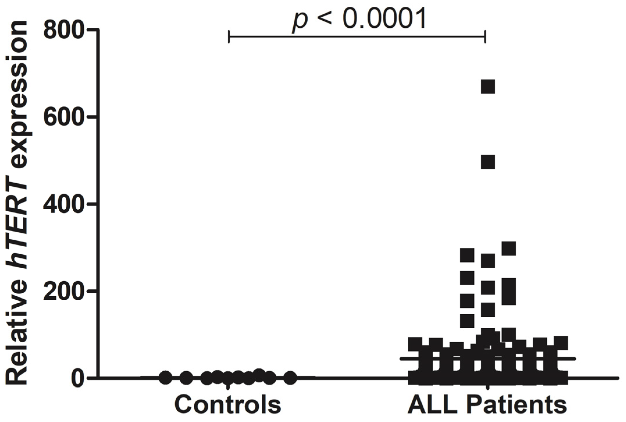
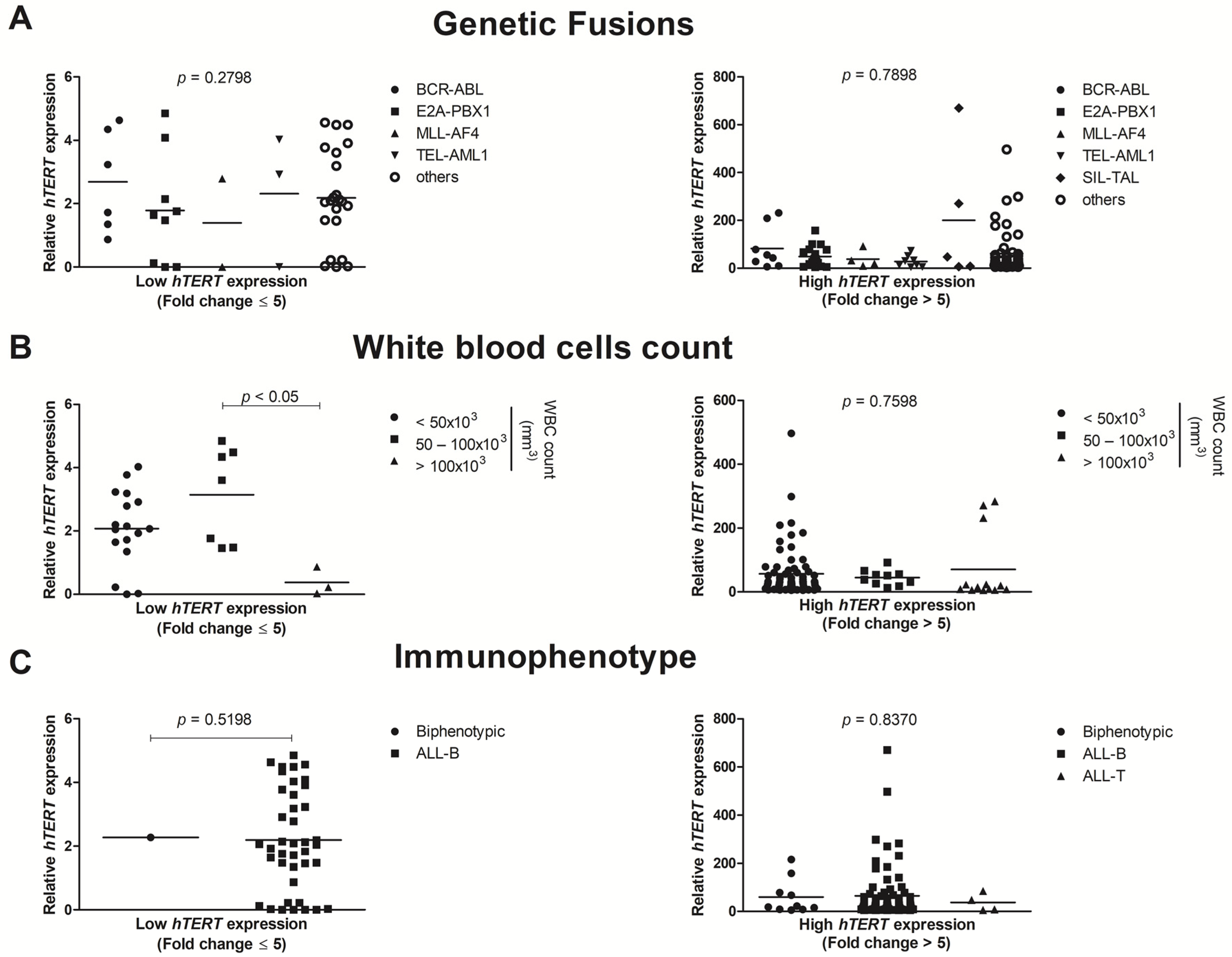
3.2. hTERT Shows Enhanced Gene Expression in ALL Pediatric Patients

We performed an analysis of gene expression of telomerase (hTERT) in 244 blood samples from pediatric patients with ALL and compared them to blood samples of 10 healthy patients (control group). hTERT was approximately 26.7-fold change overexpressed in patients with ALL when compared to the control group (p < 0.0001) (Figure 1).
Next, we investigated if hTERT expression differed among the clinical features of pediatric ALL patients. For this purpose, data were normalized and categorized assuming Log2 fold change expression levels, and patients were divided in two main groups: with “low hTERT expression” represented by patients with fold change ≤ 5 and “high hTERT expression” comprise those patients with a fold change > 5.
As shown in Figure 2A, when we compared the hTERT gene expression between each genetic fusions: E2A-PBX1, BCR-ABL, TEL-AML1, MLL-AF4, SIL-TAL, and others (patients that did not presented any classical fusions), no statistically significant differences were found in low (p = 0.2798) or high (p = 0.7898) hTERT expression groups. When the patients were categorized by the white blood cells count (WBC) into <50 × 103/mm3, 50–100 × 103/mm3 and >100 × 103/mm3, no statistical difference was seen in the group of high hTERT expression (p = 0.7598). However, in the low hTERT expression group, there was a significant statistical result between the groups of patients that presented a WBC range 50–100 × 103/mm3 and >100 × 103/mm3 (p < 0.05).
Moreover, Figure 2C shows that hTERT expression was not statistically different in ALL pediatric patients when sorted by immunophenotype (biphenotypic, ALL-B cell, and ALL-T cell) at low (p = 0.5198) or high (p = 0.8370) hTERT gene expression.
Figure 3A,B shows the hTERT expression in pediatric ALL patients sorted by gender and age (<1 year, 1–9 years and >10 years). In regards to the gender parameter, in low or high expression, no statistically significant differences were seen, between the analyzed groups (p = 0.8535, and p = 0.4886, respectively). Furthermore, for age analysis, no significant results were also found between groups of low (p = 0.6209) or high (p = 0.6088) hTERT expression.

3.3. Patient’s Overall Survival

Patient’s overall survival rate (OS) was also performed to evaluate the role of hTERT gene expression levels and its potential association with prognosis. For this analysis, patients who presented all the clinical risk parameters used for the previous statistical analysis, were included to perform the overall survival rate. A total of 83 patients were included, and the median time of follow-up was 23.9 months (range 7–56 months). We performed the OS analysis using the same normalized expression data divided in two groups: patients who presented >5-fold change and ≤5-fold change gene expression levels. The ALL patients who presented hTERT expression levels higher than 5-fold change, presented a poorer prognosis with significantly lower survival rates (p < 0.0001), as shown in Figure 4.

3.4. PPI Network and Biological Functions of hTERT

To identify and highlight which cellular and molecular pathways the participation of hTERT would be most prominent, we proposed a computational assembly of a Protein-Protein Interaction (PPI) network. The PPI generated with MCODE presented a score of 13.53 with 14 nodes and 88 edges (Figure 5A–C). We validated this topology and achieved a PPI enrichment p-value < 1 × 10⁻¹⁶. We identified a total of 14 gene hubs: ACD, CCNA1, CCNA2, CDK2, DKC1, GAR1, NHP2, NOP10, PIF1, PPOT1, PRTEL1, PTERF1, PTERT, PWRAP53, which showed the highest number of significant interactions. Furthermore, the enrichment analysis of GO for the group with stonger hTERT expression, showed these genes strongly associated in the biological process, cellular component, and molecular function of important telomerase events, which were observed with strong enrichment for GO:0000781-chromosome, telomeric region, GO:0070034-telomerase RNA binding, GO:0042162-telomeric DNA binding (Figure 5D).
To provide a broader interpretation of the hTERT biological functions and interaction with these genes, we have identified 20 REACTOME pathways, highlighting telomere extension by telomerase, telomere extension, chromosome and telomerase maintenance, cell cycle (Figure 5E) that were highly correlated with broader KEGG pathways with emphasis on the cell cycle (hsa04110) (Figure 5F).

4. Discussion

Telomerase (hTERT) is expressed in stem cells that naturally undergo self-renewal processes to maintain their constant replication without losing portions of their telomeres. Such expression occurs in hematopoietic cells, proliferating lymphocytes, the degenerative basal layer of the epidermis of human skin and can also be detected in germ and embryonic cells due to their high replication rates. The altered expression of telomerase in humans leads to cell immortalization and it is a cancer hallmark [15,16,19,28,29,30].
In the present study, we demonstrate that hTERT is significant overexpressed in all the 244 ALL pediatric patients cohort analyzed in this study, in comparison to healthy controls (p < 0.0001), and, therefore, supporting more accurately the possibility of considering it as a molecular biomarker in this cancer model. Studies have shown that in ALL tumorigenesis model, the leukemic cells use the telomerase activity to first acquire an immortalized phenotype, preventing cellular apoptosis induced by the reduction of telomeres and then fomenting their ability of self-renewal [31,32,33,34].
We also performed a correlation analysis between hTERT expression, despite low or high expressions, and clinical risk assessment variables: gene fusions (BCR–ABL; E2A–PBX1; MLL-AF4; TEL–AML1; SIL–TAL1 or other fusions), age and immunophenotyping, however, none of them alone was significantly associated with telomerase overexpression (p > 0.05). Similar findings were described in the literature regarding clinical data and telomeres/telomerase activity, showing that hTERT overexpression seems to have a prominent importance in this disease model, independently of clinical findings and variables of risk management [32,35,36,37].
In regards to leucometric parameters, patients with high hTERT expression did not present significant statistical results (p > 0.05) in any of the groups, however, the groups of patients with low hTERT expression and with a WBC ranges of 50–100 × 103/mm3 and >100 × 103/mm3, when compared, presented a significant statistical result (p < 0.05). This is a important finding, once high levels of WBC counts (>50 × 103/mm3) are described as a desfavorable prognosis in clinical risk management guidelines [22,38], and this association with hTERT expression could provide an additional biomarker to clinical prognosis assessment.
This is the first study in Brazil and South America to correlate all clinical risk assessment variables with hTERT expression in ALL pediatric patients. Therefore, our data corroborate with a previous work by Ohyashiki et al. [39] who first demonstrate that an increased telomerase activity was associated with poorer survival in pediatric patients and these usually present higher telomerase activity than adults. Thus, the data presented here are a noteworthy finding that pointed a possible common molecular biomarker to manage ALL patients despite different clinical findings. That risk management of ALL pediatric patients take into consideration clinical variables is one of the most important information to prognosis assessment and to guide better therapeutical protocols in clinical trials and medical centers [1,40,41,42].
While literature demonstrate telomerase activity to be even more overexpressed in male ALL patients, when compared to female ones [43,44], the current study did not detect any statistically significant differences (p > 0.05) in hTERT expression levels among patients of different genders. In addition, the male: female ratio found in the present study was 1.3:1, which corroborates the results of another Brazilian study, in which the pediatric patients described presented a male: female ratio of 1.7:1 [40].
Chromosomal translocations are still the main marker of genetic instability in ALL leukemogenesis, the gene fusions t(1; 19)(q23; p13) that generates E2A-PBX1 and t(9; 22)(q34; q11) that originates the chimeric gene BCR-ABL, were the two more frequent found in ALL cohort analyzed in this study, 20.27% and 12.83%, respectively. Several studies demonstrate that those two genetic fusions in particular, are the most therapeutic relevant in ALL clinical practice due to their poor prognosis and high-risk disease [1,45,46,47]. Nevertheless, most of ALL patients (52%) analyzed in this work did not present any classical gene fusions, described as main findings in this disease, which might compromise those patients to be treated with possible targeted therapies developed to specific genetical biomarkers, which have improved patient survival [48,49,50]. In this scenario, the presence of hTERT overexpression seen in those patients may arise as a potential therapeutical possibility [51,52].
In addition, the overall survival rate analysis demonstrated that patients who presented a higher hTERT expression (>5-fold change) had a poorer prognosis with significantly lower survival rates (p < 0.0001). Studies have pointed out that, telomeres and telomerase seem to have an important prognosis strength in different types of cancers and hematological malignancies [53,54,55,56,57]. Quantification of hTERT expression may assist when monitoring ALL minimal residual disease, serving as clinical follow-up for patients both in treatment and in complete remission after treatment [20,32,43].
Telomerase have been widely discussed as a potential antitumoral target in several haematological malignancies [37,58,59,60]. To identify hTERT biological interaction pathways in ALL pathogenesis, we constructed a computational PPI network, that demonstrate an important role for the hTERT involvement in pathways for chromosome maintenance, mitotic cell cycle transition, DNA double-strand break repair, cellular senescence, pathways in cancer and leukaemia. The generated PPI network also revealed a strong correlation between TERT and WRAP53 (WD40-enconding RNA antisense to p53), a protein that is widely involved in apoptosis, cell cycle regulation and the most important function is to act as an antisense regulator of p53 function and its dysfunction that could promote tumorigenesis [61,62,63]. Moreover, WRAP53 recently was found to be a telomerase holoenzyme subunit and present an essential role for telomere elongation, in addition, WRAP53 overexpression have been described in different human cancer cells, associated with a poorer prognosis suggesting that it might be an oncogene and as potential target for cancer therapy [64,65,66,67].
However, despite all scientific data reports, further studies needs to evaluate and explore, if both, the size of the telomeres and the telomerase gene expression, may assist in the investigation of hematological malignancies, other types of cancer and several diseases, as well as establishing telomerase as a potent molecular biomarker for diagnosis, prognosis, and targeted therapy [10,68,69].

5. Conclusions

The data presented in this study indicate that hTERT expression might seem to be linked to the pathogenesis of the different types of ALL found in the studied population, more precisely in the maintenance and disease persistence, a fact that may be understood in association to the multifactorial nature of leukemogenesis. In this context, hTERT expression emerges as an important molecular pathway in leukemogenesis regardless of the patient’s clinical variables; thus, the data here presented pointed it out as a valuable biomarker in pediatric acute lymphoblastic leukemia and a promising target for new therapeutic and prognostic measures.

Supplementary Materials

The following are available online at https://www.mdpi.com/article/10.3390/genes12101632/s1, Supplementary file 1: Table S1—Primers applied for gene fusion identification.

Author Contributions

Conceptualization, C.A.M.-N. and A.S.K.; Provision of data and subsequent analysis and interpretation, B.M.D.N., L.d.C.P., E.L.d.S., F.A.R.M.J., E.B.T., A.V.W., M.O.d.M.F., M.E.A.d.M., R.C.M. and C.A.M.-N.; Writing—original draft preparation, B.M.D.N., E.L.d.S., L.d.C.P. and C.A.M.-N.; Statistical analysis, B.M.D.N., E.L.d.S., F.A.R.M.J., J.H.d.S.M. and C.A.M.-N.; Writing—review and editing, E.L.d.S., A.S.K., C.A.M.-N. ; Funding acquisition, A.S.K. and C.A.M.-N. All authors have read and agreed to the published version of the manuscript.

Funding

This study was supported by Brazilian funding agencies: Coordination for the Improvement of Higher Education Personnel (CAPES; to E.B.T.), National Council of Technological and Scientific Development (CNPq; to E.L.S., M.O.d.M.F., M.E.A.d.M., R.C.M., A.S.K. and C.A.M.-N.). We also thank PROPESP/UFPA for publication payment and A.S.K is thankful to Servier do Brasil for the support.

Institutional Review Board Statement

This research was approved by the Human Research Ethics Committee of the Ophir Loyola Hospital (Consubstantiated opinion nº 2.798.615) and performed in accordance with the ethical standards as laid down in the 1964 Declaration of Helsinki and its later amendments.

Informed Consent Statement

Informed written assent was obtained from the patients’ legal guardians involved in the study.

Conflicts of Interest

The authors declare no conflict of interest. The funders had no role in the design of the study; in the collection, analyses, or data interpretation; in the writing of the manuscript, or in the decision to publish the results.

References

  1. Inaba, H.; Mullighan, C.G. Pediatric acute lymphoblastic leukemia. Haematologica 2020, 105, 2524–2539. [Google Scholar] [CrossRef]
  2. Philip, D.G.P.; Pizzo, A. Histologic Classification of Pediatric Gonadal and Extragonadal Tumors. Princ. Pract. Pediatr. Oncol. 2017, 53, 1689–1699. [Google Scholar]
  3. Dasgupta, R.K.; Marini, B.L.; Rudoni, J.; Perissinotti, A.J. A review of CD19-targeted immunotherapies for relapsed or refractory acute lymphoblastic leukemia. J. Oncol. Pharm. Pract. 2018, 24, 453–467. [Google Scholar] [CrossRef]
  4. Scharff, B.F.S.S.; Modvig, S.; Marquart, H.V.; Christensen, C. Integrin-Mediated Adhesion and Chemoresistance of Acute Lymphoblastic Leukemia Cells Residing in the Bone Marrow or the Central Nervous System. Front. Oncol. 2020, 10, 775. [Google Scholar] [CrossRef] [PubMed]
  5. Mullighan, C.G. The molecular genetic makeup of acute lymphoblastic leukemia. Hematol. Am. Soc. Hematol. Educ. Program. 2012, 2012, 389–396. [Google Scholar] [CrossRef] [Green Version]
  6. McNeer, J.L.; Bleyer, A. Acute lymphoblastic leukemia and lymphoblastic lymphoma in adolescents and young adults. Pediatr. Blood Cancer 2018, 65, e26989. [Google Scholar] [CrossRef] [PubMed]
  7. Greaves, M. A causal mechanism for childhood acute lymphoblastic leukaemia. Nat. Rev. Cancer 2018, 18, 471–484. [Google Scholar] [CrossRef] [PubMed]
  8. Sanjuan-Pla, A.; Bueno, C.; Prieto, C.; Acha, P.; Stam, R.W.; Marschalek, R.; Menéndez, P. Revisiting the biology of infant t(4;11)/MLL-AF4+ B-cell acute lymphoblastic leukemia. Blood 2015, 126, 2676–2685. [Google Scholar] [CrossRef] [PubMed] [Green Version]
  9. De Smith, A.J.; Lavoie, G.; Walsh, K.M.; Aujla, S.; Evans, E.; Hansen, H.M.; Smirnov, I.; Kang, A.Y.; Zenker, M.; Ceremsak, J.J.; et al. Predisposing germline mutations in high hyperdiploid acute lymphoblastic leukemia in children. Genes Chromosom. Cancer 2019, 58, 723–730. [Google Scholar] [CrossRef]
  10. Capraro, V.; Zane, L.; Poncet, D.; Perol, D.; Galia, P.; Preudhomme, C.; Bonnefoy-Berard, N.; Gilson, E.; Thomas, X.; El-Hamri, M.; et al. Telomere deregulations possess cytogenetic, phenotype, and prognostic specificities in acute leukemias. Exp. Hematol. 2011, 39, 195–202.e2. [Google Scholar] [CrossRef]
  11. Jones, S.; Hruban, R.H.; Kamiyama, M.; Borges, M.; Zhang, X.; Parsons, D.W.; Cheng, J.; Lin, H.; Palmisano, E.; Brune, K.; et al. Exomic Sequencing Identifies PALB2 as a Pancreatic Cancer Susceptibility Gene. Science 2009, 324, 217. [Google Scholar] [CrossRef] [PubMed] [Green Version]
  12. Jabbour, E.; O’Brien, S.; Konopleva, M.; Kantarjian, H. New insights into the pathophysiology and therapy of adult acute lymphoblastic leukemia. Cancer 2015, 121, 2517–2528. [Google Scholar] [CrossRef] [PubMed]
  13. Fiorini, E.; Santoni, A.; Colla, S. Dysfunctional telomeres and hematological disorders. Differentiation 2018, 100, 1–11. [Google Scholar] [CrossRef] [PubMed]
  14. M’Kacher, R.; Colicchio, B.; Borie, C.; Junker, S.; Marquet, V.; Heidingsfelder, L.; Soehnlen, K.; Najar, W.; Hempel, W.M.; Oudrhiri, N.; et al. Telomere and Centromere Staining Followed by M-FISH Improves Diagnosis of Chromosomal Instability and Its Clinical Utility. Genes 2020, 11, 475. [Google Scholar] [CrossRef] [PubMed]
  15. McNally, E.J.; Luncsford, P.J.; Armanios, M. Long telomeres and cancer risk: The price of cellular immortality. J. Clin. Investig. 2019, 129, 3474–3481. [Google Scholar] [CrossRef] [Green Version]
  16. Vasko, T.; Kaifie, A.; Stope, M.B.; Kraus, T.; Ziegler, P. Telomeres and Telomerase in Hematopoietic Dysfunction: Prognostic Implications and Pharmacological Interventions. Int. J. Mol. Sci. 2017, 18, 2267. [Google Scholar] [CrossRef] [Green Version]
  17. Yuan, X.; Dai, M.; Xu, D. Telomere-related Markers for Cancer. Curr. Top. Med. Chem. 2020, 20, 410–432. [Google Scholar] [CrossRef]
  18. Ivancich, M.; Schrank, Z.; Wojdyla, L.; Leviskas, B.; Kuckovic, A.; Sanjali, A.; Puri, N. Treating Cancer by Targeting Telomeres and Telomerase. Antioxidants 2017, 6, 15. [Google Scholar] [CrossRef] [PubMed]
  19. Lee, H.-S.; Carmena, M.; Liskovykh, M.; Peat, E.; Kim, J.-H.; Oshimura, M.; Masumoto, H.; Teulade-Fichou, M.-P.; Pommier, Y.; Earnshaw, W.C.; et al. Systematic Analysis of Compounds Specifically Targeting Telomeres and Telomerase for Clinical Implications in Cancer Therapy. Cancer Res. 2018, 78, 6282–6296. [Google Scholar] [CrossRef] [Green Version]
  20. Tabori, U.; Dome, J.S. Telomere Biology of Pediatric Cancer. Cancer Investig. 2007, 25, 197–208. [Google Scholar] [CrossRef]
  21. Bennett, J.M.; Catovsky, D.; Daniel, M.T.; Flandrin, G.; Galton, D.A.; Gralnick, H.R.; Sultan, C. Proposals for the classification of chronic (mature) B and T lymphoid leukaemias. French-American-British (FAB) Cooperative Group. J. Clin. Pathol. 1989, 42, 567–584. [Google Scholar] [CrossRef] [PubMed] [Green Version]
  22. Conter, V.; Bartram, C.R.; Valsecchi, M.G.; Schrauder, A.; Panzer-Grümayer, R.; Möricke, A.; Aricò, M.; Zimmermann, M.; Mann, G.; De Rossi, G.; et al. Molecular response to treatment redefines all prognostic factors in children and adolescents with B-cell precursor acute lymphoblastic leukemia: Results in 3184 patients of the AIEOP-BFM ALL 2000 study. Blood 2010, 115, 3206–3214. [Google Scholar] [CrossRef]
  23. Schmittgen, T.D.; Livak, K.J. Analyzing real-time PCR data by the comparative CT method. Nat. Protoc. 2008, 3, 1101–1108. [Google Scholar] [CrossRef] [PubMed]
  24. Bustin, S.A.; Benes, V.; Garson, J.A.; Hellemans, J.; Huggett, J.; Kubista, M.; Mueller, R.; Nolan, T.; Pfaffl, M.W.; Shipley, G.L.; et al. The MIQE Guidelines: Minimum Information for Publication of Quantitative Real-Time PCR Experiments. Clin. Chem. 2009, 55, 611–622. [Google Scholar] [CrossRef] [Green Version]
  25. Shannon, P.; Markiel, A.; Ozier, O.; Baliga, N.S.; Wang, J.T.; Ramage, D.; Amin, N.; Schwikowski, B.; Ideker, T. Cytoscape: A Software Environment for Integrated Models of Biomolecular Interaction Networks. Genome Res. 2003, 13, 2498–2504. [Google Scholar] [CrossRef]
  26. Bader, G.D.; Hogue, C.W.V. An automated method for finding molecular complexes in large protein interaction networks. BMC Bioinform. 2003, 4, 2–27. [Google Scholar] [CrossRef] [Green Version]
  27. R Core Team R: A Language and Environment for Statistical Computing. Available online: https://www.r-project.org/ (accessed on 11 September 2021).
  28. Smith, E.M.; Pendlebury, D.F.; Nandakumar, J. Structural biology of telomeres and telomerase. Cell. Mol. Life Sci. 2020, 77, 61–79. [Google Scholar] [CrossRef] [PubMed]
  29. Rubtsova, M.; Dontsova, O. Human Telomerase RNA: Telomerase Component or More? Biomolecules 2020, 10, 873. [Google Scholar] [CrossRef] [PubMed]
  30. Hanahan, D.; Weinberg, R.A. Hallmarks of Cancer: The Next Generation. Cell 2011, 144, 646–674. [Google Scholar] [CrossRef] [Green Version]
  31. Sandri, S.; De Sanctis, F.; Lamolinara, A.; Boschi, F.; Poffe, O.; Trovato, R.; Fiore, A.; Sartori, S.; Sbarbati, A.; Bondanza, A.; et al. Effective control of acute myeloid leukaemia and acute lymphoblastic leukaemia progression by telomerase specific adoptive T-cell therapy. Oncotarget 2017, 8, 86987–87001. [Google Scholar] [CrossRef] [PubMed] [Green Version]
  32. Kustanovich, A.M.; Savitskaya, T.V.; Potapnev, M.P. Telomerase activity and telomere length in malignant cells of children with acute lymphoblastic leukemia. Exp. Oncol. 2003, 25, 69–73. [Google Scholar]
  33. Borssén, M.; Cullman, I.; Norén-Nyström, U.; Sundström, C.; Porwit, A.; Forestier, E.; Roos, G. hTERT promoter methylation and telomere length in childhood acute lymphoblastic leukemia—Associations with immunophenotype and cytogenetic subgroup. Exp. Hematol. 2011, 39, 1144–1151. [Google Scholar] [CrossRef] [PubMed]
  34. Hu, Q.; Chen, X.; Liu, S.; Wen, R.; Yuan, X.; Xu, D.; Liu, G.; Wen, F. Methylation of CDKN2B CpG islands is associated with upregulated telomerase activity in children with acute lymphoblastic leukemia. Oncol. Lett. 2017, 13, 2115–2120. [Google Scholar] [CrossRef] [PubMed]
  35. Cogulu, O.; Kosova, B.; Gunduz, C.; Karaca, E.; Aksoylar, S.; Erbay, A.; Karapinar, D.; Vergin, C.; Vural, F.; Tombuloglu, M.; et al. The evaluation of hTERT mRNA expression in acute leukemia children and 2 years follow-up of 40 cases. Int. J. Hematol. 2008, 87, 276–283. [Google Scholar] [CrossRef] [PubMed]
  36. Ohyashiki, J.H.; Hisatomi, H.; Nagao, K.; Honda, S.; Takaku, T.; Zhang, Y.; Sashida, G.; Ohyashiki, K. Quantitative relationship between functionally active telomerase and major telomerase components (hTERT and hTR) in acute leukaemia cells. Br. J. Cancer 2005, 92, 1942–1947. [Google Scholar] [CrossRef] [PubMed] [Green Version]
  37. Karow, A.; Haubitz, M.; Leibundgut, E.O.; Helsen, I.; Preising, N.; Steiner, D.; Dantonello, T.M.; Ammann, R.A.; Roessler, J.; Kartal-Kaess, M.; et al. Targeting Telomere Biology in Acute Lymphoblastic Leukemia. Int. J. Mol. Sci. 2021, 22, 6653. [Google Scholar] [CrossRef] [PubMed]
  38. PDQ® Pediatric Treatment Editorial Board. PDQ Childhood Acute Lymphoblastic Leukemia Treatment. Available online: https://www.cancer.gov/types/leukemia/hp/child-all-treatment-pdq (accessed on 5 October 2021).
  39. Ohyashiki, J.H.; Ohyashiki, K.; Iwama, H.; Hayashi, S.; Toyama, K.; Shay, J.W. Clinical implications of telomerase activity levels in acute leukemia. Clin. Cancer Res. 1997, 3, 619–625. [Google Scholar]
  40. Leite, E.P.; Muniz, M.T.C.; Azevedo, A.D.C.A.C.D.; Souto, F.R.; Maia, Â.C.L.; Gondim, C.M.D.F.; Bandeira, F.M.G.C.; Melo, R.A.M. Fatores prognósticos em crianças e adolescentes com Leucemia Linfóide Aguda. Rev. Bras. Saúde Matern. Infant. 2007, 7, 413–421. [Google Scholar] [CrossRef]
  41. Kato, M.; Manabe, A. Treatment and biology of pediatric acute lymphoblastic leukemia. Pediatr. Int. 2018, 60, 4–12. [Google Scholar] [CrossRef]
  42. Ibagy, A.; Silva, D.B.; Seiben, J.; Winneshoffer, A.P.F.F.; Costa, T.E.J.B.; Dacoregio, J.S.; Costa, I.; Faraco, D. Acute Lymphoblastic Leukemia in Infants: 20 years of Experience. J. Pediatr. 2013, 89, 64–69. [Google Scholar] [CrossRef] [Green Version]
  43. Bruedigam, C.; Lane, S.W. Telomerase in hematologic malignancies. Curr. Opin. Hematol. 2016, 23, 346–353. [Google Scholar] [CrossRef]
  44. Paiva, R.M.A.; Calado, R.T. Telomere Dysfunction and Hematologic Disorders, 1st ed.; Elsevier Inc.: Amsterdam, The Netherlands, 2014; Volume 125, pp. 133–157. ISBN 9780123978981. [Google Scholar]
  45. Buitenkamp, T.D.; Izraeli, S.; Zimmermann, M.; Forestier, E.; Heerema, N.A.; Heuvel-Eibrink, M.M.V.D.; Pieters, R.; Korbijn, C.M.; Silverman, L.B.; Schmiegelow, K.; et al. Acute lymphoblastic leukemia in children with Down syndrome: A retrospective analysis from the Ponte di Legno study group. Blood 2014, 123, 70–77. [Google Scholar] [CrossRef] [PubMed] [Green Version]
  46. Pui, C.-H.; Yang, J.J.; Hunger, S.P.; Pieters, R.; Schrappe, M.; Biondi, A.; Vora, A.; Baruchel, A.; Silverman, L.B.; Schmiegelow, K.; et al. Childhood Acute Lymphoblastic Leukemia: Progress Through Collaboration. J. Clin. Oncol. 2015, 33, 2938–2948. [Google Scholar] [CrossRef] [PubMed]
  47. Kimura, S.; Mullighan, C.G. Molecular markers in ALL: Clinical implications. Best Pr. Res. Clin. Haematol. 2020, 33, 101193. [Google Scholar] [CrossRef]
  48. Terwilliger, T.; Abdul-Hay, M. Acute lymphoblastic leukemia: A comprehensive review and 2017 update. Blood Cancer J. 2017, 7, e577. [Google Scholar] [CrossRef] [Green Version]
  49. Place, A.E.; Pikman, Y.; Stevenson, K.E.; Harris, M.H.; Pauly, M.; Sulis, M.-L.; Hijiya, N.; Gore, L.; Cooper, T.M.; Loh, M.L.; et al. Phase I trial of the mTOR inhibitor everolimus in combination with multi-agent chemotherapy in relapsed childhood acute lymphoblastic leukemia. Pediatr. Blood Cancer 2018, 65, e27062. [Google Scholar] [CrossRef] [PubMed]
  50. Diaz-Flores, E.; Comeaux, E.Q.; Kim, K.L.; Melnik, E.M.; Beckman, K.; Davis, K.L.; Wu, K.; Akutagawa, J.; Bridges, O.; Marino, R.; et al. Bcl-2 Is a Therapeutic Target for Hypodiploid B-Lineage Acute Lymphoblastic Leukemia. Cancer Res. 2019, 79, 2339–2351. [Google Scholar] [CrossRef] [PubMed] [Green Version]
  51. Gómez, D.L.M.; Armando, R.G.; Cerrudo, C.S.; Ghiringhelli, P.D.; Gomez, D.E. Telomerase as a Cancer Target. Development of New Molecules. Curr. Top. Med. Chem. 2016, 16, 2432–2440. [Google Scholar] [CrossRef] [Green Version]
  52. Guterres, A.N.; Villanueva, J. Targeting telomerase for cancer therapy. Oncogene 2020, 39, 5811–5824. [Google Scholar] [CrossRef]
  53. Valls, C.; Piñol, C.; Reñé, J.M.; Buenestado, J.; Viñas, J. Telomere length is a prognostic factor for overall survival in colorectal cancer. Colorectal Dis. 2011, 13, 1265–1272. [Google Scholar] [CrossRef]
  54. Fernandez-Marcelo, T.; Gómez, A.; Pascua, I.; De Juan, C.; Head, J.; Hernando, F.; Jarabo, J.-R.; Calatayud, J.; Torres-García, A.-J.; Iniesta, P. Telomere length and telomerase activity in non-small cell lung cancer prognosis: Clinical usefulness of a specific telomere status. J. Exp. Clin. Cancer Res. 2015, 34, 78. [Google Scholar] [CrossRef] [PubMed] [Green Version]
  55. Kroupa, M.; Rachakonda, S.K.; Liska, V.; Srinivas, N.; Urbanová, M.; Jiraskova, K.; Schneiderova, M.; Vycital, O.; Vymetalkova, V.; Vodickova, L.; et al. Relationship of telomere length in colorectal cancer patients with cancer phenotype and patient prognosis. Br. J. Cancer 2019, 121, 344–350. [Google Scholar] [CrossRef] [PubMed]
  56. Mascarenhas, J.O.; Komrokji, R.S.; Cavo, M.; Martino, B.; Niederwieser, D.; Reiter, A.; Scott, B.L.; Baer, M.R.; Hoffman, R.; Odenike, O.; et al. Telomerase Activity, Telomere Length and HTERT Expression Correlate with Clinical Outcomes in Higher-Risk Myelofibrosis (MF) Relapsed/Refractory (R/R) to Janus Kinase Inhibitor Treated with Imetelstat. Hemasphere 2020, 4, 1098. [Google Scholar]
  57. Jebaraj, B.M.C.; Stilgenbauer, S. Telomere Dysfunction in Chronic Lymphocytic Leukemia. Front. Oncol. 2021, 10, 3062. [Google Scholar] [CrossRef]
  58. Bruedigam, C.; Bagger, F.O.; Heidel, F.H.; Kuhn, C.P.; Guignes, S.; Song, A.; Austin, R.; Vu, T.; Lee, E.; Riyat, S.; et al. Telomerase Inhibition Effectively Targets Mouse and Human AML Stem Cells and Delays Relapse following Chemotherapy. Cell Stem Cell 2014, 15, 775–790. [Google Scholar] [CrossRef] [Green Version]
  59. Bruedigam, C.; Wackrow, B.; Song, A.; Porter, A.H.; Lee, S.C.-W.; Moore, M.F.A.S.; Abdel-Wahab, O.; Lane, S.W. The Preclinical Efficacy of a Novel Telomerase Inhibitor, Imetelstat, in AML—A Randomized Trial in Patient-Derived Xenografts. Blood 2016, 128, 578. [Google Scholar] [CrossRef]
  60. Mascarenhas, J.; Komrokji, R.S.; Cavo, M.; Martino, B.; Niederwieser, D.; Reiter, A.; Scott, B.L.; Baer, M.R.; Hoffman, R.; Odenike, O.; et al. Imetelstat Is Effective Treatment for Patients with Intermediate-2 or High-Risk Myelofibrosis Who Have Relapsed on or Are Refractory to Janus Kinase Inhibitor Therapy: Results of a Phase 2 Randomized Study of Two Dose Levels. Blood 2018, 132, 685. [Google Scholar] [CrossRef]
  61. Farnebo, M. Wrap53, a novel regulator of p53. Cell Cycle 2009, 8, 2343–2346. [Google Scholar] [CrossRef]
  62. Mahmoudi, S.; Henriksson, S.; Corcoran, M.; Méndez-Vidal, C.; Wiman, K.G.; Farnebo, M. Wrap53, a Natural p53 Antisense Transcript Required for p53 Induction upon DNA Damage. Mol. Cell 2009, 33, 462–471. [Google Scholar] [CrossRef]
  63. Henriksson, S.; Farnebo, M. On the road with WRAP53β: Guardian of Cajal bodies and genome integrity. Front. Genet. 2015, 6, 91. [Google Scholar] [CrossRef] [PubMed] [Green Version]
  64. Venteicher, A.S.; Abreu, E.B.; Meng, Z.; McCann, K.E.; Terns, R.M.; Veenstra, T.D.; Terns, M.P.; Artandi, S.E. A Human Telomerase Holoenzyme Protein Required for Cajal Body Localization and Telomere Synthesis. Science 2009, 323, 644–648. [Google Scholar] [CrossRef] [Green Version]
  65. Mahmoudi, S.; Henriksson, S.; Farnebo, L.; Roberg, K.; Farnebo, M. WRAP53 promotes cancer cell survival and is a potential target for cancer therapy. Cell Death Dis. 2011, 2, e114. [Google Scholar] [CrossRef] [PubMed]
  66. Zhang, H.; Wang, D.-W.; Adell, G.; Sun, X.-F. WRAP53 is an independent prognostic factor in rectal cancer- a study of Swedish clinical trial of preoperative radiotherapy in rectal cancer patients. BMC Cancer 2012, 12, 294. [Google Scholar] [CrossRef] [PubMed] [Green Version]
  67. Sun, C.-K.; Luo, X.-B.; Gou, Y.-P.; Hu, L.; Wang, K.; Li, C.; Xiang, Z.-T.; Zhang, P.; Kong, X.-L.; Zhang, C.-L.; et al. TCAB1: A potential target for diagnosis and therapy of head and neck carcinomas. Mol. Cancer 2014, 13, 180. [Google Scholar] [CrossRef] [PubMed] [Green Version]
  68. Lulkiewicz, M.; Bajsert, J.; Kopczynski, P.; Barczak, W.; Rubis, B. Telomere length: How the length makes a difference. Mol. Biol. Rep. 2020, 47, 7181–7188. [Google Scholar] [CrossRef] [PubMed]
  69. Nogueira, B.M.D.; Machado, C.B.; Montenegro, R.C.; De Moraes, M.E.A.; Moreira-Nunes, C.A. Telomere Length and Hematological Disorders: A Review. In Vivo 2020, 34, 3093–3101. [Google Scholar] [CrossRef]
Figure 1. The ALL patients demonstrate enhanced hTERT expression. Gene expression analysis of hTERT in ALL blood samples from pediatric patients (n = 244). Relative hTERT expression was calculated using ABL and GAPDH as endogenous normalizers. Data are presented as the median and each dot plot represents the hTERT expression in a single patient. For statistical analysis, the normal distribution was confirmed by the Shapiro–Wilk normality test followed by the Mann–Whitney test. Comparison with control samples: p < 0.0001. ALL: acute lymphoblastic leukemia.
Genes 12 01632 g001
Figure 2. The ALL pediatric cohort (n = 148) was separated into two groups, with low (fold change ≤ 5) and high (fold change > 5) hTERT expression. (A) Those patients did not statisticaly differ despite all genetic fusions. (B) For WBC, only in the group with low hTERT expression, a significant difference was observed between 50–100 × 103 mm3 and >100 × 103 mm3 samples (p < 0.05) (C) Immunophenotype features did also not present significant results in low or high hTERT expression groups. Relative hTERT expression was calculated using ABL and GAPDH as endogenous normalizers. Data are presented as the median and each dot plot represents the hTERT expression in a single patient. For statistical analysis, the Shapiro–Wilk normality test was performed, followed by the Kruskal–Wallis test and Dunn’s Multiple Comparison post-test. WBC: white blood cells count, ALL: acute lymphoblastic leukemia.
Genes 12 01632 g002
Figure 3. The ALL patients (n = 148) were separated into two groups, with low (fold change ≤ 5) and high (fold change > 5) hTERT expression. (A) Gender and (B) Age did not imply significant alterations between groups with low or high hTERT gene expression. Relative hTERT expression was calculated using ABL and GAPDH as endogenous normalizers. Data are presented as the median and each dot plot represents the hTERT expression in a single patient. For statistical analysis, the Shapiro–Wilk normality test was performed, followed by the Kruskal–Wallis test and Dunn’s Multiple Comparison post-test for age analysis or t-test for gender.
Genes 12 01632 g003
Figure 4. Comparison of survival time for patients with different levels of hTERT expression. Survival time was statistically different between patients. Patients expressing hTERT showed a significantly lower survival rate in patients (p < 0.0001) who had higher expression levels (>5 fold-change).
Genes 12 01632 g004
Figure 5. Protein–protein interaction network for hTERT. (A) Prototype of the PPI network built with Cytoscape containing 51 nodes and 377 edges. (B) PPI generated with MCODE presented a score of 13.53 with 14 nodes and 88 edges. (C) Validation done with String-db for the PPI topology containing 14 gene hubs that was predicted by the MCODE algorithm. (D) GO functional annotation for PPI genes. (E) Identification of enzymatic reaction pathways of REACTOME and (F) Pathways of KEGG. All annotation terms were normalized with Log10 (p-value). PPI: Protein–Protein Interaction.
Genes 12 01632 g005
Table 1. Clinical features analysis of acute lymphoblastic leukemia patients.
N (%)
AgeWBC (×103)/mm3Gender
>11–9≥10p-Value>5050–100>100p-ValueMFp-Value
Immunophenotype 0.3853 0.0002 * 0.5586
Biphenotypic0 (0)11 (11.3)2 (4.3) 10 (10)1 (5.3)2 (6.9) 7 (8.3)6 (9.4)
T-ALL cell0 (0)2 (2.1)3 (6.4) 0 (0)0 (0)5 (17.2) 4 (4.8)1 (1.6)
B-ALL cell4 (100)84 (86.6)42 (89.4) 90 (90)18 (94.7)22 (75.9) 73 (86.9)57 (89.1)
Genetic Fusion <0.0001 ** 0.0002 * 0.9998
E2A-PBX10 (0)26 (26.8)4 (8.5) 19 (19)8 (42.1)3 (10.3) 17 (20.2)13 (20.3)
BCR-ABL1 (16.7)8 (8.2)10 (21.3) 12 (12)1 (5.3)6 (20.7) 11 (13.1)8 (12.5)
TEL-AML10 (0)9 (9.3)1 (2.1) 8 (8)2 (10.5)0 (0) 6 (7.1)4 (6.3)
MLL-AF42 (33.3)5 (5.2)0 (0) 4 (4)1 (5.3)2 (6.9) 4 (4.8)3 (4.7)
SIL-TAL0 (0)2 (2.1)3 (6.4) 0 (0)0 (0)5 (17.2) 3 (3.6)2 (3.1)
Others1 (16.7)47 (48.5)29 (61.7) 57 (57)7 (36.8)13 (44.8) 43 (51.2)34 (53.1)
Legend: p-value by Chi-square test; α = 0.05. F: female; M: male; WBC: White blood cell count. Statistically significant values: * p = 0.0002; ** p < 0.0001.
Publisher’s Note: MDPI stays neutral with regard to jurisdictional claims in published maps and institutional affiliations.

