Article

The Peroxiredoxin Asp f3 Acts as Redox Sensor in Aspergillus fumigatus

1
Junior Research Group Evolution of Microbial Interactions, Leibniz-Institute for Natural Product Research and Infection Biology–Hans Knöll Institute (HKI), Beutenbergstr. 11a, 07745 Jena, Germany
2
Institute of Microbiology, Friedrich Schiller University Jena, 07743 Jena, Germany
3
Systemsbiology and Bioinformatics, Leibniz-Institute for Natural Product Research and Infection Biology–Hans Knöll Institute, Beutenbergstr. 11a, 07745 Jena, Germany
*
Author to whom correspondence should be addressed.
Genes 2021, 12(5), 668; https://doi.org/10.3390/genes12050668
Submission received: 16 March 2021 / Revised: 24 April 2021 / Accepted: 27 April 2021 / Published: 29 April 2021

Abstract

:
The human pathogenic fungus Aspergillus fumigatus is readily eradicated by the innate immunity of immunocompetent human hosts, but can cause severe infections, such as invasive aspergillosis (IA), in immunocompromised individuals. During infection, the fungal redox homeostasis can be challenged by reactive oxygen species (ROS), either derived from the oxidative burst of innate immune cells or the action of antifungal drugs. The peroxiredoxin Asp f3 was found to be essential to cause IA in mice, but how Asp f3 integrates with fungal redox homeostasis remains unknown. Here, we show that in vivo, Asp f3 acts as a sensor for ROS. While global transcription in fungal hyphae under minimal growth conditions was fully independent of Asp f3, a robust induction of the oxidative stress response required the presence of the peroxiredoxin. Hyphae devoid of Asp f3 failed to activate several redox active genes, like members of the gliotoxin biosynthesis gene cluster and integral members of the Afyap1 regulon, the central activator of the ROS defense machinery in fungi. Upon deletion of the asp f3 gene Afyap1 displayed significantly reduced nuclear localization during ROS exposure, indicating that Asp f3 can act as an intracellular redox sensor for several target proteins.

1. Introduction

The ascomycete Aspergillus fumigatus is a ubiquitous fungus which is generally involved in the decomposition of (plant based) biomass, thus playing an important role during carbon and nitrogen recycling [1,2]. With its ability to adapt to a wide temperature range, different oxygen and pH levels and nutritional challenges like low iron levels, A. fumigatus thrives in many diverse environments and is also known as the most common airborne fungal pathogen. Infection occurs via the widely distributed asexual conidia which after inhalation, germinate and colonize the lung tissue of immuno-compromised patients such as those suffering from HIV, leukemia or active therapeutic immunosuppression following organ and stem cell transplantation [3,4]. Depending on the underlying condition infections range from allergic bronchopulmonary aspergillosis (ABPA) to often fatal invasive aspergillosis (IA), a disease reaching mortality rates in the range of 30–95% [5,6]. Reasons for high mortality are deficiencies in specific and timely diagnostics as well as the limited availability of effective therapeutic treatment [7]. Hence it is imperative to aim for a better understanding of the pathophysiology of A. fumigatus, enabling more targeted approaches towards the development of new therapeutic solutions.
The protein Asp f3 was originally found as a prominent allergen on the surface of fungal conidia [8]. Due to its high abundance and affinity to serum immunoglobulin E (IgE) Asp f3 was also introduced as an auspicious vaccine candidate, protecting Asp f3 immunized mice against invasive pulmonary aspergillosis [9,10]. Recently, we characterized the protein as a dimeric, two-type-cysteine peroxiredoxin Asp f3 which showed peroxidase activity in vitro and protected A. fumigatus from external oxidative stress [11]. The protein was furthermore required for virulence in a murine model of pulmonary aspergillosis, and may thus present a promising target for therapeutic applications. Whether its role as a virulence determinant directly relates to its function as a reductive reactive oxygen species (ROS) scavenging enzyme is currently unknown.
Clinical data support an essential role of reactive oxygen species in the defense against fungal infections, as patients suffering from chronic granulomatous disease (CGD) have reduced capability to produce ROS which renders them especially susceptible to A. fumigatus infections [7]. The fact that the absence of the major ROS defense activator Yap1 in A. fumigatus causes hypersensitivity to ROS, but at the same time does not attenuate its virulence, makes it unlikely that there is a direct correlation between efficient ROS scavenging and virulence. However, ROS may still impact survival of A. fumigatus in the host. Only recently host derived ROS were observed to induce the programmed cell death in conidia of the fungus after ingestion by innate immune phagocytes [12]. Here we examine the role of Asp f3 in A. fumigatus in the response towards ROS. We show that the absence of Asp f3 does not affect the fungal transcriptome under unstressed conditions but leads to a significant shift in gene expression upon challenge with oxidative stress. Surprisingly, ROS exposure to cells lacking Asp f3 did not activate the AfYap1 regulon, suggesting that Asp f3 acts as an essential redox switch to launch a potent defense against ROS or ROS mediated damages.

2. Materials and Methods

2.1. A. fumigatus Strain and Culture Conditions

All strains and plasmids used in the study are listed in Table 1. The asp f3 deletion and complemented strains were generated as described by Hillmann et al. [11]. A. fumigatus was cultured on/in Aspergillus minimal medium (AMM) with 1% Glucose as carbon source and 20 mM NaNO3 as nitrogen source by inoculation with 105 conidia if not otherwise noted [13]. Liquid cultures were kept shaking at 180 rpm at 37 °C for 24 h. Conidia were harvested with 0.1% (v/v) Tween 80 from AMM-agar plates cultivated at 37 °C for 96 h. Mutant-phenotypes were selected by either 250 µg/mL hygromycin B (Invivogen, Toulouse, France) or pyrithiamine (0.1 mg/mL, Sigma-Aldrich, Taufkirchen, Germany), depending on the resistance marker used in transformation [14]. Conidia were counted by a CASY® Modell TT (OLS OMNI Life Science, Bremen, Germany). For long–time storage, conidia were mixed with glycerol at 20% (v/v) and frozen at −80 °C.

2.2. Construction of Fluorescent Reporter Strains

For the generation of a VENUS-Fusion protein expression strain, the gene sequence of Afyap1 was cloned into plasmid pGpdA-Afyap1-VENUS containing a constitutive gpdA-promoter and VENUS gene with a nos terminator (Supplementary Figure S1). The target gene (Afyap1) was introduced as a N-terminal-fusion to VENUS via CPEC and further paired with the PtrA resistance cassette which confers resistance to the antimetabolite pyrithiamine [16]. For a list of Primers see Supplementary Table S1. Plasmids were amplified in E. coli DH5α, linear fragments for transformation were amplified via PCR (Phusion Flash Polymerase, Thermo Fisher Scientific, Bremen, Germany) and transformed into A. fumigatus D141 and Δasp f3 via protoplast formation [17,18]. Mutants were confirmed by diagnostic PCR and a detectable VENUS-signal during fluorescence microscopy.

2.3. Isolation of Chromosomal DNA

Fungal strains were cultivated for 16 h at 37 °C at 180 rpm in Sabouraud 2% Glucose Bouillon (Carl Roth, Karlsruhe, Germany). The mycelium was harvested through miracloth, washed thoroughly with H2O, dried and frozen with liquid nitrogen. Frozen mycelium was then ground to a fine powder in a mortar and stored at −20 °C until further use. Isolation of chromosomal DNA was carried out as described previously [19].

2.4. Oxidative Stress Experiments

Reactive oxygen species were produced either by addition of H2O2 or directly in vivo we with the xanthine oxidase enzymatic system generating a mixture of H2O2 and O2 as previously described [11]. Prior to treatment 105 conidia were grown in liquid Czapek Dox medium (BD, Franklin Lakes, NJ, USA) in 6-well tissue culture plates (VWR International, Leuven, Belgium) in a final volume of 3 mL and cultured for 48 h until a thin layer of mycelium was formed. A sub-lethal concentration (for Δasp f3) of 150 µM xanthine was supplied. The addition of 100 µg/mL (0.2 units/mL) xanthine oxidase (Sigma-Aldrich, Taufkirchen, Germany) started the reaction. For the transcriptome, analysis reaction was stopped after 15 min and samples were harvested, frozen with liquid nitrogen and stored at −80 °C until further use. All data analyzed originated from three biological replicates.

2.5. RNA Isolation

Total RNA was isolated from ROS treated and untreated mycelia of the wild type and the Δasp f3 strain using the RNeasy Plant Mini Kit (Qiagen, Hilden, Germany). Frozen mycelium was transferred to tubes containing glass beads (Sigma-Aldrich, Taufkirchen, Germany), after addition of resuspension buffer cells were disrupted by mechanical force applied via FastPrep (MP Biomedicals, Irvine, CA, USA) for 60 s at high-speed setting (6.0). Further processing was conducted according to the manufacturer’s protocol. Extracted RNA was stored at −80 °C. RNA concentration was determined by NanoDrop ND1000 Spectrophotometer (NanoDrop Technologies Inc., Wilmington, DE, USA).

2.6. Analysis of Transcriptome Data

The preparation of cDNA libraries from total RNA and the sequencing was performed by GATC Biotech (GATC Biotech, Konstanz, Germany). According to GATC Protocol samples were enriched for mRNA by isolation of poly(A)+ mRNA, mRNA was fragmented and cDNA synthesis was performed to generate strand specific cDNA libraries. From these libraries 1 × 50 bp single end reads were sequenced with the Genome Sequencer Illumina HiSeq (HiSeq 4000 50bp SR) (Illumina, San Diego, CA, USA). FastQC [20] and Trimmomatic v0.32 [21] were used for quality control and trimming of library adaptors. Mapping of reads was achieved with HiSat2 [22] against the reference genome of A. fumigatus A293. The normalized number of reads were analyzed with EdgeR, Limma, DESeq, DESeq2 [23,24,25,26] and genes were considered as differentially expressed gene (DEGs) when the differences in the number of reads were statistically significant according to one or more of these tests.

2.7. Gene Expression Analysis by qRT-PCR

A. fumigatus WT and Δasp f3 conidia (1 × 10⁹) were grown in 3 mL AMM and CD at 37 °C and 180 rpm for 6 h to induce swelling. After 6 h, swollen conidia were treated with 0.1 M H2O2 for 15 min. Swollen conidia were harvested from triplicate samples at 0 and 15 min after the addition of 0.1 M H2O2 by centrifugation at 800 g and 4 °C for 5 min. Swollen conidia were subsequently lysed with glass beads in the FastPrep (MP Biomedicals, Irvine, CA, USA) for 60 s at 13,000 rpm and processed for total RNA isolation using a Qiagen RNeasy plant mini kit (Qiagen, Hilden, Germany), according to manufacturer’s protocol. Extracted RNA was then stored at −20 °C. Concentration of each sample was determined with NanoDrop ND1000 spectrophotometer (NanoDrop Technologies Inc., Wilmington, DE, USA).
RNA was treated with RQ1 RNase-free DNase (Promega, Walldorf, Germany) and transcribed into cDNA (RevertAid First Strand cDNA Synthesis Kit, Thermo Fischer Scientific, Germany), according to the manufacturers protocol. Quantitative real time polymerase chain reaction (qRT-PCR) was performed using the cDNA as a template. The ΔCT method was used to analyze the relative expression of the target genes, normalized to the constitutively expressed tubA gene encoding tubulin A. Al primers are listed in Supplementary Table S1. The reactions were carried out in a total volume of 20 μL on Quantstudio3 system (Thermo Fisher Scientific, Bremen, Germany).

2.8. DAPI Staining and Fluorescence Microscopy

A total of 105 conidia were incubated at 37 °C for 10 h in 300 μL of CD in ibidi® μ-slide (ibidi, Gräfelfing, Germany) until germination. The nuclei were then stained with NucBlue Live ReadyProbes Reagent (ThermoFisher, Dreieich, Germany) according to manufacturer’s guidelines. Afterwards, 2 mM H2O2 was added and after 30 min the samples were subsequently analyzed under the microscope. Fluorescent stain and proteins were excited with 408 nm and 488 nm, respectively, to analyze the localization of AfYap1VENUS in both WT and Δasp f3 strains using a Zeiss Axio Observer Spinning Disk Confocal Microscope (ZEISS, Jena, Germany) using ZEN software (Version 2.6). Microscopic images were evenly processed and analyzed with ImageJ software [27].

2.9. Co-Localization Analysis

For the co-localization analysis of AfYap1VENUS and DAPI, the Coloc2 plugin of ImageJ was used to calculate the Pearson’s correlation coefficient to identify the intensity correlation of fluorescence signals. GraphPad9 Prism software was used to plot the graph and calculate the p-value. Error bars represent ± standard deviation from at least 3 images.

2.10. Preparation of Protein Extracts and Catalase Activity Measurements

Crude protein extracts were prepared from A. fumigatus swollen conidia incubated for 6 h in CD medium and treated with 2 mM of H2O2 for 45 min. Conidia were harvested by centrifugation and washed thoroughly with PBS. Conidia were re-suspended in assay buffer (Abcam, Cambridge, UK) and disrupted by FastPrep treatment, repeated mixing and sonication for 10 min to enhance the solubilization of proteins. Protein concentration was determined by the Bradford assay [28] and spectrophotometric measurements (UV mini 1240, Shimadzu, Kyoto, Japan). Catalase activity was determined using the catalase activity assay kit (cat. No. ab83464 Catalase Activity Assay Kit, Abcam, Cambridge, UK) according to the manufacturer’s instructions and fluorometric measurements in a fluorescence plate reader (Tecan, Männedorf, Switzerland) at excitation and emission wavelengths of 535 and 587 nm, respectively.

2.11. In-Gel Catalase Activity Assay

A. fumigatus conidia of different strains were inoculated in CD medium and grown for 20 h at 37 °C prior to a 5 mM H2O2 treatment for 45 min. The mycelium was harvested, frozen in liquid nitrogen and ground to a fine powder. Isolation of native protein occurred according to Lessing et. al. (2007) [29]. Again, protein concentration was measurured via Bradford assay [28] and spectrophotometric measurements (UV mini 1240, Shimadzu, Kyōto, Japan). A total of 30 µg of protein was loaded on an 8–16% polyacrylamide (wt/vo) Tris-Glycine Gel (Invitrogen™ Novex™ WedgeWell™, Art. Nr.: 15486814 ). The runtime was for 4 h at 60 V at 4 °C. Catalase activity in the gel was determined with the method described by Goldberg and Hochman [30]. A total of 1 µg catalase from bovine liver (C1345, 2000–5000 units/mg protein, Sigma-Aldrich, Taufkirchen, Germany) was used as a positive control.

2.12. Functional Annotation of Transcriptome Data

Enrichment analyses of genes were carried out with the FungiFun2 package [31]. Default settings were used to enrich genes according to GO and FunCat categories. Hits were deemed significant with a p-values < 0.01. Enrichment was carried out for the Go-terms “biological process” and “molecular function”. Additional analyses were performed with the AspGD Gene Ontology Term Finder (http://www.aspergillusgenome.org/cgi-bin/GO/goTermFinder, accessed on 28 February 2021) [32].

3. Results

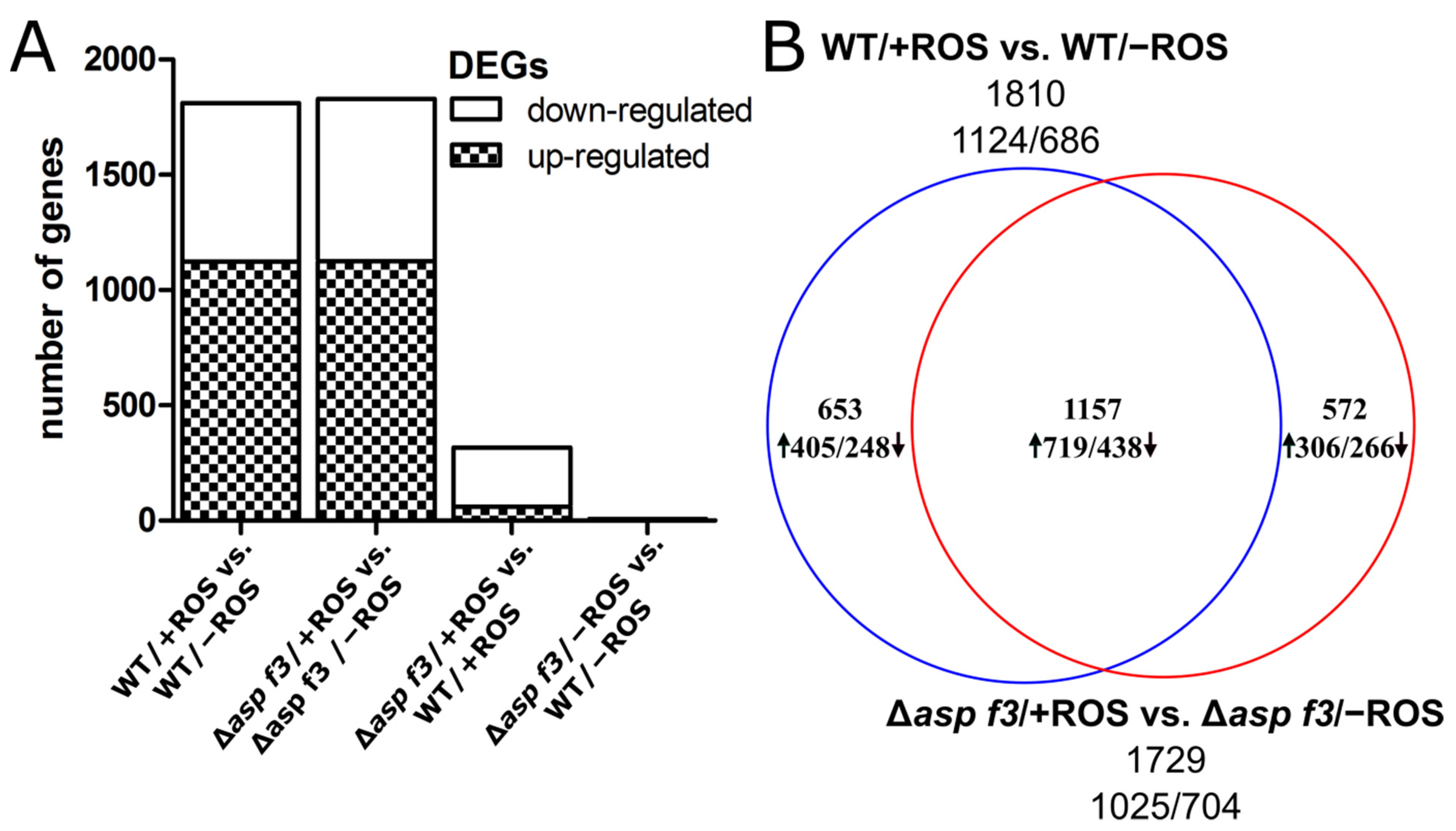
3.1. Global Transcriptome Analysis Reveals a ROS Specific Function of Asp f3

To understand the protective role of Asp f3 during ROS exposure we monitored global transcription in wild type hyphae of A. fumigatus (WT) and the ROS sensitive deletion mutant Δasp f3. Both strains were first grown in minimal medium and either left untreated (−ROS) or exposed to H2O2 and O2 (+ROS), therefore ROS were generated in vivo by the xanthine oxidase enzymatic system for 15 min in biological triplicates. A principal component analysis of the fungal transcriptomes demonstrated a comparatively low cumulative variance in untreated samples, indicating that the lack of Asp f3, despite its abundance as protein, did not significantly impact transcription under ambient growth conditions of fungal hyphae in minimal medium (Figure 1). These results corresponded to a widely indistinguishable phenotype under a wide range of growth conditions with various carbon sources (Supplementary Figure S2).
In fact, in the absence of ROS, WT and Δasp f3 showed an exceptionally high similarity in their genome expression and only seven genes were found to be differentially expressed genes (DEGs). The only DEG commonly identified by all four methods was asp f3 which remained undetected in the mutant strain, as expected. This indicated that Asp f3 plays a minor role in the absence of ROS and allowed a direct comparison of the ROS-treated samples without a background of genes that directly respond to the absence of Asp f3.
ROS exposure induced a shift in the global transcriptome with nearly identical numbers of DEGs for both strains. For the wild type and Δasp f3 mutant we identified 1810 and 1729 ROS dependent DEGs, respectively. The numbers of up- and downregulated genes are likewise similar, identifying 1124 up- and 686 downregulated genes for the WT and 1025 up- and 704 down-regulated genes in Δasp f3 (Figure 2A). However, when the DEGs were compared between the two strains, we found only about two thirds of them in congruency. This proportion was comparable for up- as well as for the down-regulated genes (Figure 2B).

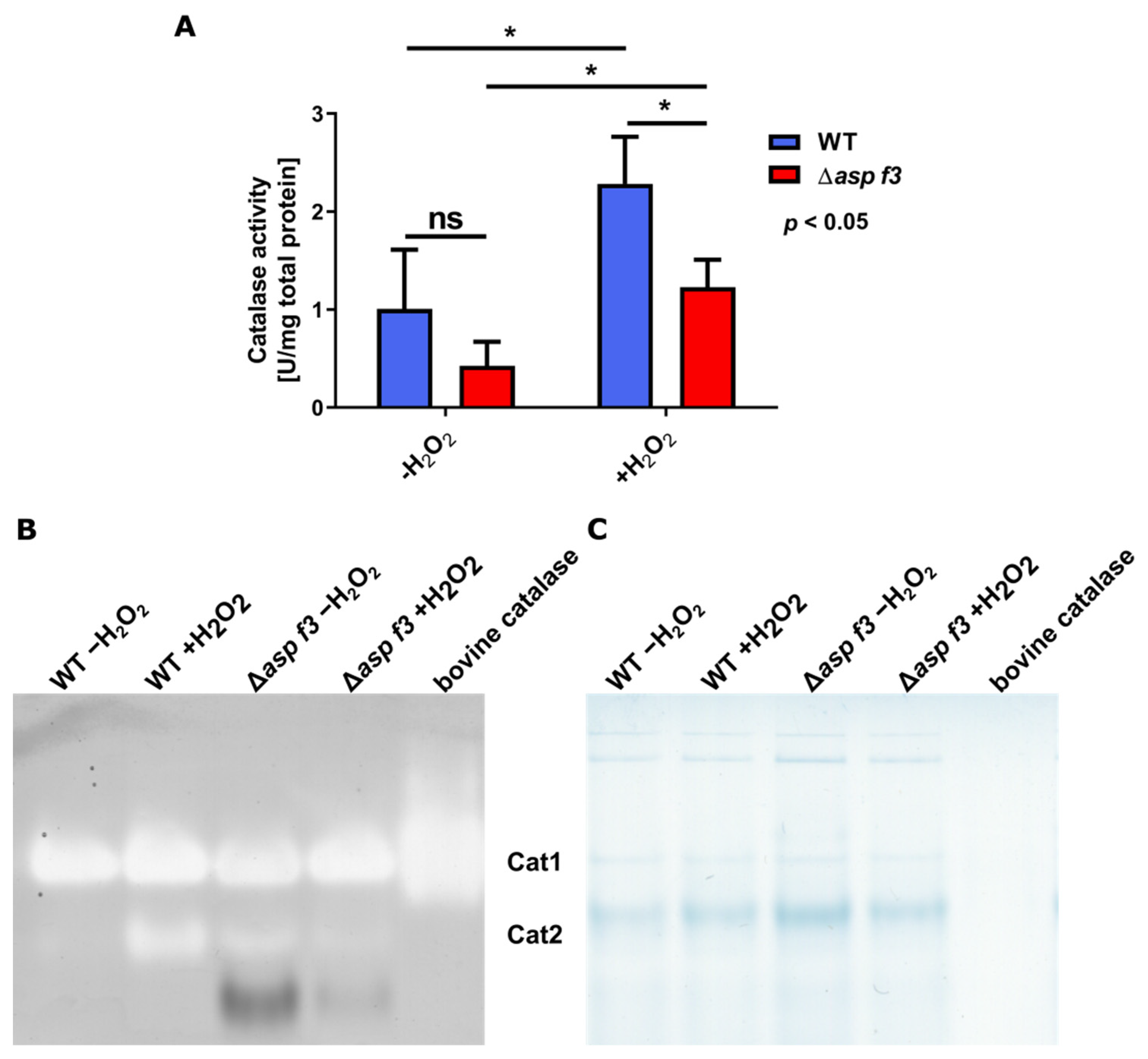
3.2. Transcriptional Induction of the ROS Defense Requires Asp f3

Although the number of ROS induced DEGs was similar in hyphae of the wild type and Δasp f3, 653 genes were differentially expressed only in the wild type, indicating that Asp f3 was required for gene expression under oxidative stress and Δasp f3 seems to react differently when challenged with ROS. To get an overview of the effects triggered by loss of the peroxiredoxin we plotted the expression of all genes in WT and Δasp f3 under ROS exposure (Figure 3A).
This direct comparison of the treated samples showed 319 DEGs of which 60 were higher expressed in hyphae of the mutant when compared to those of the wild type. The remaining 259 genes showed lower expression in the mutant during ROS exposure. The lowest was the asp f3 gene itself with RPKM values of 14 and approximately 5900 in hyphae of the mutant and the wild type, respectively.
Of the 259 genes that were specifically downregulated in Δasp f3 under ROS, 53 genes were not affected by ROS in the wild type. The remaining 205 of these DEGs were up-regulated after ROS exposure of the wild type. A gene ontology analysis for molecular functions of the group of genes that lacked ROS dependent expression in the absence of the peroxiredoxin identified primarily oxidoreductase activity, inorganic phosphate transmembrane transporter activity, peroxidase activity and FMN binding as significantly enriched categories (Figure 3B, Supplementary Table S2). Biological processes that showed attenuated expression specific to the absence of Asp f3 included categories such as phosphate ion transport, transmembrane transport and mycotoxin biosynthetic processes. The latter was in principle limited to gliotoxin biosynthesis (Figure 3C, Supplementary Table S3). Indeed, a closer look at the genes of the gliotoxin cluster identified 7 of 12 genes as downregulated, including gliP, the gene for the non-ribosomal peptide synthetase. The largest enriched biological processes were oxidation-reduction processes and transmembrane transport. More specifically, the transmembrane transport of phosphate ions and the specific response to oxidative stress was deregulated in the absence of the peroxiredoxin. To analyze whether Asp f3 is required for the activation of ROS defense, catalase activity was determined in total protein extracts from both strains before and after a treatment with H2O2 by indirect measurement of catalase activity (Figure 4A). Untreated hyphae (−H2O2) revealed insignificant differences in catalase activity. Exposure of H2O2 to the swollen spores increased catalase activity in both WT and Δasp f3, but ROS dependent upregulation in Δasp f3 led to significantly lower activity then in hyphae of the wild type. To determine which catalases were active in the samples both strains were grown for 20 h and challenged with H2O2 for 45 min. Proteins were extracted and loaded in equal amounts (30 µg) on a native polyacrylamide gel to perform an in-gel catalase activity assay (Figure 4B,C). Catalase activity was visualized by a negative staining and shows a strong induction of Cat2 in the wild type after challenge with H2O2. In contrast to the wild type, Cat2 activity was detectable in the unchallenged sample of Δasp f3. However, exposure to H2O2 did not lead to an induction of Cat2 activity. Activity of the spore borne catalase CatA was not observed.

3.3. Full ROS Dependent Activation of Several Afyap1 Target Genes Requires Asp f3

Several genes with a major role in the redox-homeostasis and the defense against oxidative stress were upregulated in the wild type (Table 2), among them three confirmed targets of the major activator AfYap1 [29]: the bifunctional catalase-peroxidase (cat2, AFUA_8g01670), the Cytochrome c peroxidase (ccp1, AFUA_4G09110), and the p-Nitroreductase family protein (pnr1, AFUA_5g09910). The latter is not only known to be strongly induced by diverse environmental stresses such as superoxide stress, osmotic stress and heat stress but is also expressed when the fungus is exposed to neutrophils [34,35]. However, other putative AfYap1-targets suggested by Lessing and colleagues [29], such as the mitochondrial peroxiredoxin (Prx1) or methionine synthase, were not significantly affected in the transcriptome data set of the wild type or the mutant. The expression of the putative Afyap1 targets, the yap1 itself, and several other ROS defense genes was analyzed by qRT-PCR in two different conditions (Figure 5). Expression of ROS defense genes in Czapek-Dox (CD) medium (Figure 5A), which was also used to generate the transcriptome data, was approximately an order of magnitude lower than in Aspergillus minimal medium (AMM) which was supplemented with added trace metals as a major difference (Figure 5B). For both wild type and Δasp f3 the expression of the Afyap1 gene was comparably lower in CD than in AMM even without the addition of ROS stress. For both strains the transcript levels were also not significantly affected by the addition of ROS in CD, while a significant induction of the regulator gene Afyap1 was detected for both strains in AMM. For several other ROS defense genes, including the putative Afyap1 targets ccp1, cat2, and pnr1, ROS dependent activation was lower in hyphae of the asp f3 deletion mutant. This lack of activation was also more pronounced in the trace metal free CD medium. ROS mediated induction of catA and gpx3 were detected for both strains in both media.

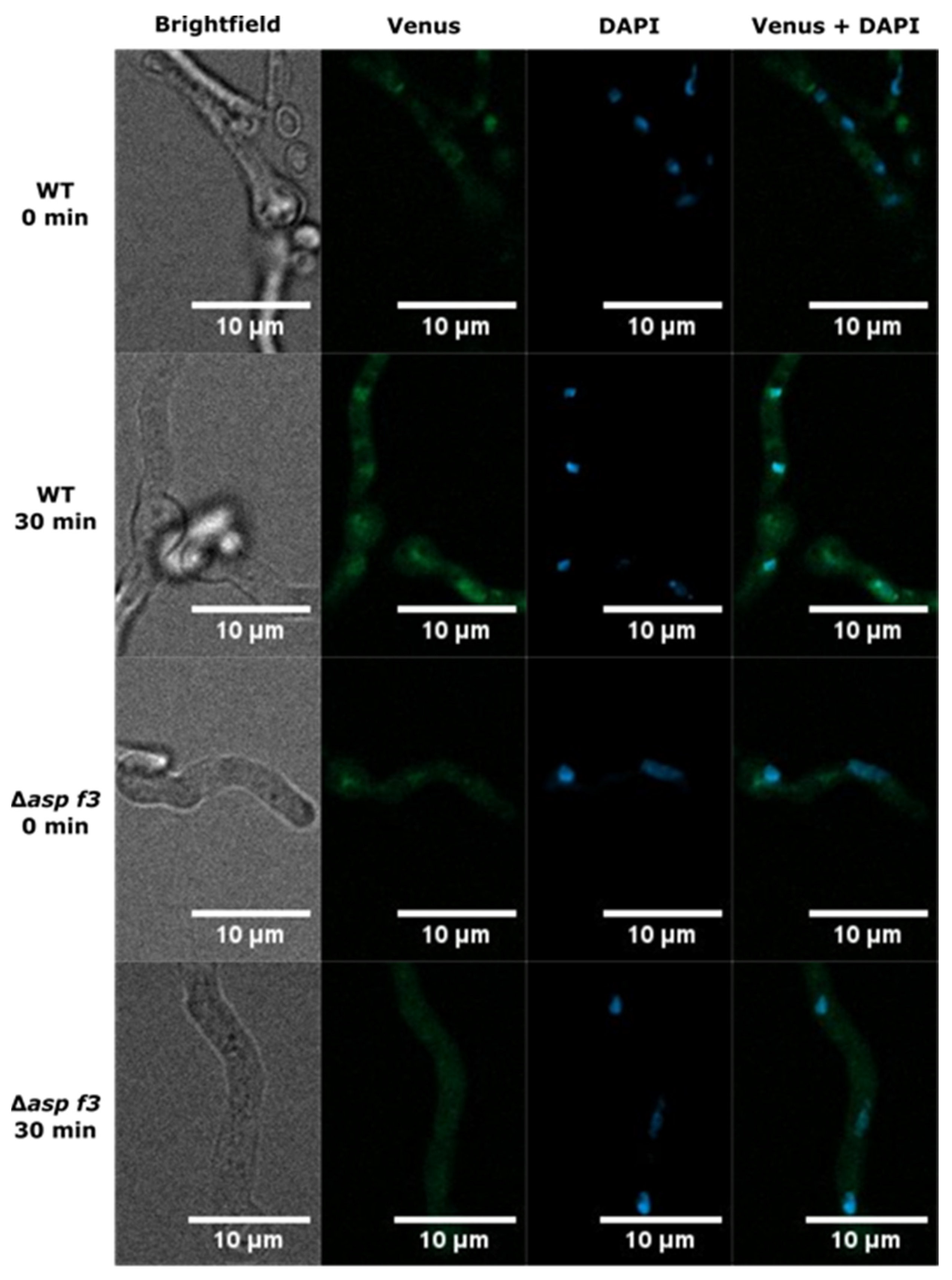
3.4. Asp f3 Is Required for Afyap1 Activation and Nuclear Localization

The oxidative stress regulator Afyap1 localizes to the nucleus in response to ROS and activates the transcription of target genes, such as cat2 [29]. The reduced expression levels of cat2 in Δasp f3, as well as the lower catalase activity in the mutant, prompted us to evaluate whether Afyap1 activation depends on this peroxiredoxin. Thus, we monitored the subcellular localization of the AfYap1-Venus fusion protein in germlings of the wild type and Δasp f3 (Figure 6). In the absence of H2O2, AfYap1VENUS displayed diffused cytosolic localization in both, wild type and Δasp f3 (0 min). An addition of 2 mM H2O2, induced nuclear localization of AfYap1VENUS in wild type germlings within 30 min. Conversely, in the Δasp f3 background, a larger proportion of the AfYap1VENUS remained diffused in the cytoplasm with only a minor nuclear concentration of the activator. To overcome heterogeneity in the microscopic data, we quantified the fluorescence intensity signals for AfYap1VENUS and DAPI as a nucleus specific signal. Co-localization of the two different fluorescent intensities was determined as Pearson’s correlation coefficient (PCC, Figure 7). In the absence of H2O2, PCC values were 0.48 and 0.46 for wild type and Δasp f3, respectively. Whereas, after 30 min of H2O2 exposure PCC values significantly increased for the wild type (0.80). However, a significantly lower PCC value was observed for Δasp f3 (0.61) when compared with the treated wild type indicating that efficient nuclear localization of Afyap1 depends on the peroxiredoxin Asp f3.

4. Discussion

The peroxiredoxin Asp f3 is a major allergen and an abundant protein of A. fumigatus, as an allergen on the conidial surface, but also within growing hyphae. It was shown to function in the defense against ROS and was essential during invasive aspergillosis in a mouse infection model [11]. However, it remained unclear how this confirmed biochemical function as a peroxiredoxin would serve precisely during infection in the host. In the absence of oxidative stress, the absence of Δasp f3 yields an inconspicuous phenotype, nearly indistinguishable from the wild type. Our transcriptome analysis confirmed, under in vitro conditions without ROS exposure, only minor transcriptional changes were detected and the Asp f3 protein appears to be dispensable for growth. This observation is in line with earlier results for Saccharomyces cerevisiae which demonstrated that the yeast was still viable despite a deletion of all five peroxiredoxin genes and that single mutants grew like the wild type in aerobic conditions [36]. Interestingly, the authors also showed that the Asp f3 orthologue in yeast, Tsa1p, secured long-term genomic stability by preventing mutations [37,38]. Such a protective function may well be conserved in A. fumigatus but would most likely not explain the avirulent phenotype of the Δasp f3 strain in the aspergillosis animal model.
While Asp f3 was dispensable in hyphae during the absence of ROS, confrontation with ROS induced major changes in the transcriptome. Resulting from its hypersensitive phenotype, one may have expected a slightly higher expression level of oxidative stress genes to compensate the phenotype, but indeed the opposite was observed. Several ROS defense genes were slightly downregulated under ambient growth conditions, and for others no induction was seen in response to ROS. Amongst the unaffected or even downregulated genes are several which are pivotal to the oxidative stress response or involved in virulence. Several of the genes, including trxR, ccp1 and gpx3, also coincide with genes upregulated in A. fumigatus conidia when exposed to neutrophils, indicating their relevance during virulence [35]. Although not all of these gene products may be crucial to defend against innate immune cells, it confirms the presence of a perceptible exposure to ROS. Furthermore, TrxR was recently described as an essential gene which not only affects oxidative stress resistance but is needed for full virulence in animal models of both Galleria melonella and immunosuppressed mice [39]. Seven genes of the gliotoxin gene cluster are downregulated in Δasp f3, including gliP, which encodes the nonribosomal peptide synthase catalysing the first step in the biosynthesis of gliotoxin. This mycotoxin is produced in vivo during infections and is known to mediate immunosuppressive effects on human cells [40,41,42,43]. When interacting with human neutrophils, gliotoxin was shown to re-organize the actin-skeleton, thus inhibiting phagocytosis and further inhibiting the respiratory burst and other neutrophil functions such as superoxide production [44,45]. Additionally, deletion of gliP was shown to attenuate virulence in mice immunosuppressed with hydrocortisone [42,46]. With regard to its reversible dithiol linkage, a regulatory link between gliotoxin biosynthesis and the fungal redox state seems likely and has previously been observed [47]. We saw a mild H2O2 dependent upregulation in the wild type and the opposite tendency in the absence of Asp f3. Such downregulation of the gliotoxin gene cluster in the Δasp f3 strain may lead to lower levels of the mycotoxin during infection which might thus attenuate its virulence potential in the lung environment in the host.
Some of the downregulated genes in Δasp f3 were confirmed regulatory targets of Afyap1. Furthermore, Afyap1VENUS overexpression in a Δasp f3 background could not compensate for the absence of Asp f3. These results suggested that Afyap1 and Asp f3 could be functionally interconnected, especially as a reversible disulphide bond formation is known to regulate Yap1 localization and activity. In baker’s yeast, activation of Yap1 was first reported to occur by the glutathione peroxidase (Gpx3), acting as the hydroperoxide receptor and redox transducer [48]. In our transcriptome the Gpx3 orthologue of A. fumigatus was clearly upregulated in the wild type in response to ROS but transcription remained unchanged in Δasp f3 after ROS treatment. It should not be excluded that lower levels of Gpx3 in the mutant may attenuate Afyap1 activation, either via direct interaction or as a member in a redox relay system.
Peroxiredoxin dependent activation of the Yap1 regulator has also been proposed for filamentous fungi previously [49]. In Aspergillus nidulans, the Yap1 orthologue is coined NapA and regulates a wide set of genes far beyond ROS defense. Neither GpxA (Gpx3 in A. fumigatus) or two other peroxiredoxins, TpxA (AFUA_4g08580, Prx1 in A. fumigatus) and TpxB (AFUA_8G07130 in A. fumigatus), were found to be involved in NapA activation [50], making it seem unlikely that their orthologous proteins would fulfil this role in A. fumigatus. Both peroxiredoxins were slightly downregulated under oxidative stress, independent of Asp f3. Another peroxiredoxin, Tsa1p was shown to activate Yap1 in specific yeast strains [51]. Asp f3 is most likely not a true homologue of Tsa1p, as the amino acid identity between the two proteins is lower when comparing Asp f3 to Ahp1 (18% and 37%, respectively). In contrast to Tsa1p, Ahp1p is specific for alkylhydroperoxides [52].
We found Asp f3 to be the peroxiredoxin that mediates nuclear retention of Afyap1 under ROS exposure in A. fumigatus, indicating that this function may well be conserved for its homologue in A. nidulans-PrxA, which was found to be involved in oxidative stress defense and suspected to be the regulatory peroxiredoxin for NapA [49,53]. We speculate that this occurs via direct interaction of the two proteins and that rather its cellular abundance rather than specific biochemical properties of Asp f3 determine this interaction. As Afyap1 was previously found to be dispensable for virulence in a mouse model of aspergillosis our results also suggest that another, Afyap1 independent function of Asp f3 must be essential during infection conditions. A previous study has identified that iron availability may be compromised in response to oxidative stress [54] and Asp f3 may represent a regulatory hub between these interconnected stress responses.

5. Conclusions

Aspergillus fumigatus puts immunocompromised patients at a high risk of severe and often fatal infections. It is thus imperative to find not only more reliable diagnostic tools but also research a more targeted approach for antimycotic treatment. In this study we investigated the in vivo function of Asp f3, a protein that plays an essential in virulence. A transcriptomic approach showed clear differences between the wild type and the highly ROS-sensitive Δasp f3 mutants. Further investigations led to the conclusion that Asp f3 deficient mutants suffer from a deregulation of the oxidative stress response due to lacking nuclear retention of the regulator Afyap1. However, loss of Afyap1 does not lead to avirulence of A. fumigatus, strongly suggesting additional cellular effects upon challenge with ROS.

Supplementary Materials

The following are available online at https://www.mdpi.com/article/10.3390/genes12050668/s1, Figure S1: Overexpression construct for Afyap1VENUS. A Vector Map of gpdA-Afyap1-VENUS including the pyrithiamine resistance gene thiA (ptrA) gene of Aspergillus oryzae as a selectable marker for successful transformation in A. fumigatus. Figure S2: Growth of Aspergillus fumigatus on minimal media (AMM) with various carbon sources. 2 × 10⁴ conidia of the wild type (D141) and the asp f3 deletion mutant (Δasp f3) were point-inoculated on AMM with indicated supplements (% w/v) as carbon/nutrient sources and incubated at 37 °C for 48 h. The Afyap1 deletion strain (ΔAfyap1) and its wild type like parent CEA17ΔakuBKU80 (CEA17) are shown for comparison. Table S1: Primer list for the generation of Afyap1VENUS. Table S2: Results of the gene enrichment analysis (GO-term search, molecular function). Table S3: Results of the gene enrichment analysis (GO-term search, biological process).

Author Contributions

Conceptualization, F.H.; methodology, J.M.B., N.S., T.W., software, T.W., G.P.; formal analysis, J.M.B., N.S., T.W., F.H.; resources, G.P., F.H.; data curation, J.M.B., T.W., G.P.; writing—original draft preparation, J.M.B., N.S., T.W.; writing—review and editing J.M.B., N.S., T.W., G.P., F.H.; visualization, J.M.B., N.S.; supervision, G.P., F.H.; funding acquisition, G.P., F.H.; All authors have read and agreed to the published version of the manuscript.

Funding

This study was supported by the Leibniz Institute for Natural Product Research and Infection Biology–Hans Knöll Institute, Jena and funded by a grant of the Deutsche Forschungsgemeinschaft (DFG, German Research Foundation DFG, HI 1574-2/1).

Institutional Review Board Statement

Not applicable.

Informed Consent Statement

Not applicable.

Data Availability Statement

The sequencing data have been deposited in NCBI’s Gene Expression Omnibus database and are accessible through GEO Series accession number GSE173349 (https://www.ncbi.nlm.nih.gov/geo/query/acc.cgi?acc=GSE173349, accessed on 28 February 2021).

Conflicts of Interest

The authors declare no competing interests.

References

  1. Brakhage, A.A.; Langfelder, K. Menacing mold: The molecular biology of Aspergillus fumigatus. Annu. Rev. Microbiol. 2002, 56, 433–455. [Google Scholar] [CrossRef]
  2. Latgé, J.-P. Aspergillus fumigatus and aspergillosis. Clin. Microbiol. Rev. 1999, 12, 310–350. [Google Scholar] [CrossRef] [Green Version]
  3. Tekaia, F.; Latgé, J.-P. Aspergillus fumigatus: Saprophyte or pathogen? Curr. Opin. Microbiol. 2005, 8, 385–392. [Google Scholar] [CrossRef]
  4. Rhodes, J.C. Aspergillus fumigatus: Growth and virulence. Med. Mycol. 2006, 44, S77–S81. [Google Scholar] [CrossRef] [PubMed] [Green Version]
  5. Schrettl, M.; Bignell, E.; Kragl, C.; Joechl, C.; Rogers, T.; Arst, H.N., Jr.; Haynes, K.; Haas, H. Siderophore biosynthesis but not reductive iron assimilation is essential for Aspergillus fumigatus virulence. J. Exp. Med. 2004, 200, 1213–1219. [Google Scholar] [CrossRef] [Green Version]
  6. Willger, S.D.; Grahl, N.; Willger, S.D.; Grahl, N.; Cramer, R.A., Jr. Aspergillus fumigatus metabolism: Clues to mechanisms of in vivo fungal growth and virulence. Med. Mycol. 2009, 47, S72–S79. [Google Scholar] [CrossRef] [Green Version]
  7. Brown, G.D.; Denning, D.W.; Gow, N.A.; Levitz, S.M.; Netea, M.G.; White, T.C. Hidden killers: Human fungal infections. Sci. Transl. Med. 2012, 4, rv113–rv165. [Google Scholar] [CrossRef] [Green Version]
  8. Asif, A.R.; Oellerich, M.; Amstrong, V.W.; Riemenschneider, B.; Monod, M.; Reichard, U. Proteome of Conidial Surface Associated Proteins of Aspergillus fumigatus Reflecting Potential Vaccine Candidates and Allergens. J. Proteome Res. 2006, 5, 954–962. [Google Scholar] [CrossRef] [PubMed]
  9. Ito, J.I.; Lyons, J.M.; Hong, T.B.; Tamae, D.; Liu, Y.-K.; Wilczynski, S.P.; Kalkum, M. Vaccinations with recombinant variants of Aspergillus fumigatus allergen Asp f 3 protect mice against invasive aspergillosis. Infect. Immun. 2006, 74, 5075–5084. [Google Scholar] [CrossRef] [PubMed] [Green Version]
  10. Diaz-Arevalo, D.; Bagramyan, K.; Hong, T.B.; Ito, J.I.; Kalkum, M. CD4+ T cells mediate the protective effect of the recombinant Asp f3-based anti-aspergillosis vaccine. Infect. Immun. 2011, 79, 2257–2266. [Google Scholar] [CrossRef] [PubMed] [Green Version]
  11. Hillmann, F.; Bagramyan, K.; Straßburger, M.; Heinekamp, T.; Hong, T.B.; Bzymek, K.P.; Williams, J.C.; Brakhage, A.A.; Kalkum, M. The crystal structure of peroxiredoxin Asp f3 provides mechanistic insight into oxidative stress resistance and virulence of Aspergillus fumigatus. Sci. Rep. 2016, 6, 33396. [Google Scholar] [CrossRef]
  12. Shlezinger, N.; Irmer, H.; Dhingra, S.; Beattie, S.R.; Cramer, R.A.; Braus, G.H.; Sharon, A.; Hohl, T.M. Sterilizing immunity in the lung relies on targeting fungal apoptosis-like programmed cell death. Science 2017, 357, 1037. [Google Scholar] [CrossRef] [Green Version]
  13. Brakhage, A.A.; Van den Brulle, J. Use of reporter genes to identify recessive trans-acting mutations specifically involved in the regulation of Aspergillus nidulans penicillin biosynthesis genes. J. Bacteriol. 1995, 177, 2781–2788. [Google Scholar] [CrossRef] [PubMed] [Green Version]
  14. Weidner, G.; d’Enfert, C.; Koch, A.; Mol, P.C.; Brakhage, A.A. Development of a homologous transformation system for the human pathogenic fungus Aspergillus fumigatus based on the pyrG gene encoding orotidine 5″-monophosphate decarboxylase. Curr. Genet. 1998, 33, 378–385. [Google Scholar] [CrossRef]
  15. Reichard, U.; Büttner, S.; Eiffert, H.; Staib, F.; Rüchel, R. Purification and characterisation of an extracellular serine proteinase from Aspergillus fumigatus and its detection in tissue. J. Med. Microbiol. 1990, 33, 243–251. [Google Scholar] [CrossRef] [Green Version]
  16. Quan, J.; Tian, J. Circular polymerase extension cloning of complex gene libraries and pathways. PLoS ONE 2009, 4, e6441. [Google Scholar] [CrossRef]
  17. Ballance, D.; Turner, G. Development of a high-frequency transforming vector for Aspergillus nidulans. Gene 1985, 36, 321–331. [Google Scholar] [CrossRef]
  18. Tilburn, J.; Sarkar, S.; Widdick, D.; Espeso, E.; Orejas, M.; Mungroo, J.; Penalva, M.; Arst, H., Jr. The Aspergillus PacC zinc finger transcription factor mediates regulation of both acid-and alkaline-expressed genes by ambient pH. Embo J. 1995, 14, 779–790. [Google Scholar] [CrossRef] [PubMed]
  19. Cenis, J. Rapid extraction of fungal DNA for PCR amplification. Nucleic Acids Res. 1992, 20, 2380. [Google Scholar] [CrossRef] [PubMed]
  20. Andrews, S. FastQC: A Quality Control Tool for High Throughput Sequence Data. 2010. Available online: http://www.bioinformatics.babraham.ac.uk/projects/fastqc (accessed on 29 April 2021).
  21. Bolger, A.M.; Lohse, M.; Usadel, B. Trimmomatic: A flexible trimmer for Illumina sequence data. Bioinformatics 2014, 30, 2114–2120. [Google Scholar] [CrossRef] [PubMed] [Green Version]
  22. Kim, D.; Paggi, J.M.; Park, C.; Bennett, C.; Salzberg, S.L. Graph-based genome alignment and genotyping with HISAT2 and HISAT-genotype. Nat. Biotechnol. 2019, 37, 907–915. [Google Scholar] [CrossRef] [PubMed]
  23. Ritchie, M.E.; Phipson, B.; Wu, D.; Hu, Y.; Law, C.W.; Shi, W.; Smyth, G.K. limma powers differential expression analyzes for RNA-sequencing and microarray studies. Nucleic Acids Res. 2015, 43, e47. [Google Scholar] [CrossRef] [PubMed]
  24. Robinson, M.D.; McCarthy, D.J.; Smyth, G.K. edgeR: A Bioconductor package for differential expression analysis of digital gene expression data. Bioinformatics 2010, 26, 139–140. [Google Scholar] [CrossRef] [PubMed] [Green Version]
  25. Anders, S.; Huber, W. Differential expression analysis for sequence count data. Nat. Preced. 2010. [Google Scholar] [CrossRef]
  26. Love, M.I.; Huber, W.; Anders, S. Moderated estimation of fold change and dispersion for RNA-seq data with DESeq2. Genome Biol. 2014, 15, 1–21. [Google Scholar] [CrossRef] [Green Version]
  27. Abràmoff, M.D.; Magalhães, P.J.; Ram, S.J. Image processing with ImageJ. Biophotonics Int. 2004, 11, 36–42. [Google Scholar]
  28. Bradford, M.M. A rapid and sensitive method for the quantitation of microgram quantities of protein utilizing the principle of protein-dye binding. Anal. Biochem. 1976, 72, 248–254. [Google Scholar] [CrossRef]
  29. Lessing, F.; Kniemeyer, O.; Wozniok, I.; Loeffler, J.; Kurzai, O.; Haertl, A.; Brakhage, A.A. The Aspergillus fumigatus transcriptional regulator AfYap1 represents the major regulator for defense against reactive oxygen intermediates but is dispensable for pathogenicity in an intranasal mouse infection model. Eukaryot. Cell 2007, 6, 2290–2302. [Google Scholar] [CrossRef] [Green Version]
  30. Goldberg, I.; Hochman, A. Purification and characterization of a novel type of catalase from the bacterium Klebsiella pneumoniae. Biochim. Biophys. Acta (BBA) Gen. Subj. 1989, 991, 330–336. [Google Scholar] [CrossRef]
  31. Priebe, S.; Kreisel, C.; Horn, F.; Guthke, R.; Linde, J. FungiFun2: A comprehensive online resource for systematic analysis of gene lists from fungal species. Bioinformatics 2015, 31, 445–446. [Google Scholar] [CrossRef] [PubMed] [Green Version]
  32. Cerqueira, G.C.; Arnaud, M.B.; Inglis, D.O.; Skrzypek, M.S.; Binkley, G.; Simison, M.; Miyasato, S.R.; Binkley, J.; Orvis, J.; Shah, P. The Aspergillus Genome Database: Multispecies curation and incorporation of RNA-Seq data to improve structural gene annotations. Nucleic Acids Res. 2014, 42, D705–D710. [Google Scholar] [CrossRef] [PubMed] [Green Version]
  33. Paris, S.; Wysong, D.; Debeaupuis, J.-P.; Shibuya, K.; Philippe, B.; Diamond, R.D.; Latgé, J.-P. Catalases of Aspergillus fumigatus. Infect. Immun. 2003, 71, 3551–3562. [Google Scholar] [CrossRef] [PubMed] [Green Version]
  34. Takahashi, H.; Kusuya, Y.; Hagiwara, D.; Takahashi-Nakaguchi, A.; Sakai, K.; Gonoi, T. Global gene expression reveals stress-responsive genes in Aspergillus fumigatus mycelia. BMC Genom. 2017, 18, 1–15. [Google Scholar] [CrossRef] [PubMed]
  35. Sugui, J.A.; Kim, H.S.; Zarember, K.A.; Chang, Y.C.; Gallin, J.I.; Nierman, W.C.; Kwon-Chung, K.J. Genes differentially expressed in conidia and hyphae of Aspergillus fumigatus upon exposure to human neutrophils. PLoS ONE 2008, 3, e2655. [Google Scholar] [CrossRef] [PubMed]
  36. Wong, C.M.; Siu, K.L.; Jin, D.Y. Peroxiredoxin-null yeast cells are hypersensitive to oxidative stress and are genomically unstable. J. Biol. Chem. 2004, 279, 23207–23213. [Google Scholar] [CrossRef] [Green Version]
  37. Tang, H.-M.V.; Siu, K.-L.; Wong, C.-M.; Jin, D.-Y. Loss of yeast peroxiredoxin Tsa1p induces genome instability through activation of the DNA damage checkpoint and elevation of dNTP levels. PLoS Genet. 2009, 5, e1000697. [Google Scholar] [CrossRef] [Green Version]
  38. West, J.D.; Roston, T.J.; David, J.B.; Allan, K.M.; Loberg, M.A. Piecing together how peroxiredoxins maintain genomic stability. Antioxidants 2018, 7, 177. [Google Scholar] [CrossRef] [Green Version]
  39. Binder, J.; Shadkchan, Y.; Osherov, N.; Krappmann, S. The essential thioredoxin reductase of the human pathogenic mold Aspergillus fumigatus is a promising antifungal target. Front. Microbiol. 2020, 11, 1383. [Google Scholar] [CrossRef] [PubMed]
  40. Kupfahl, C.; Heinekamp, T.; Geginat, G.; Ruppert, T.; Hartl, A.; Hof, H.; Brakhage, A.A. Deletion of the gliP gene of Aspergillus fumigatus results in loss of gliotoxin production but has no effect on virulence of the fungus in a low-dose mouse infection model. Mol. Microbiol. 2006, 62, 292–302. [Google Scholar] [CrossRef]
  41. Sugui, J.A.; Rose, S.R.; Nardone, G.; Swamydas, M.; Lee, C.-C.R.; Kwon-Chung, K.J.; Lionakis, M.S. Host immune status-specific production of gliotoxin and bis-methyl-gliotoxin during invasive aspergillosis in mice. Sci. Rep. 2017, 7, 10977. [Google Scholar] [CrossRef] [Green Version]
  42. Sugui, J.A.; Pardo, J.; Chang, Y.C.; Zarember, K.A.; Nardone, G.; Galvez, E.M.; Müllbacher, A.; Gallin, J.I.; Simon, M.M.; Kwon-Chung, K.J. Gliotoxin is a virulence factor of Aspergillus fumigatus: gliP deletion attenuates virulence in mice immunosuppressed with hydrocortisone. Eukaryot. Cell 2007, 6, 1562–1569. [Google Scholar] [CrossRef] [Green Version]
  43. Lewis, R.E.; Wiederhold, N.P.; Chi, J.; Han, X.Y.; Komanduri, K.V.; Kontoyiannis, D.P.; Prince, R.A. Detection of gliotoxin in experimental and human aspergillosis. Infect. Immun. 2005, 73, 635–637. [Google Scholar] [CrossRef] [PubMed] [Green Version]
  44. Coméra, C.; André, K.; Laffitte, J.; Collet, X.; Galtier, P.; Maridonneau-Parini, I. Gliotoxin from Aspergillus fumigatus affects phagocytosis and the organization of the actin cytoskeleton by distinct signalling pathways in human neutrophils. Microbes Infect. 2007, 9, 47–54. [Google Scholar] [CrossRef] [PubMed] [Green Version]
  45. Cramer, R.A.; Gamcsik, M.P.; Brooking, R.M.; Najvar, L.K.; Kirkpatrick, W.R.; Patterson, T.F.; Balibar, C.J.; Graybill, J.R.; Perfect, J.R.; Abraham, S.N. Disruption of a nonribosomal peptide synthetase in Aspergillus fumigatus eliminates gliotoxin production. Eukaryot. Cell 2006, 5, 972–980. [Google Scholar] [CrossRef] [Green Version]
  46. Hof, H.; Kupfahl, C. Gliotoxin in Aspergillus fumigatus: An example that mycotoxins are potential virulence factors. Mycotoxin Res. 2009, 25, 123. [Google Scholar] [CrossRef] [PubMed]
  47. Gallagher, L.; Owens, R.A.; Dolan, S.K.; O’Keeffe, G.; Schrettl, M.; Kavanagh, K.; Jones, G.W.; Doyle, S. The Aspergillus fumigatus protein GliK protects against oxidative stress and is essential for gliotoxin biosynthesis. Eukaryot. Cell 2012, 11, 1226–1238. [Google Scholar] [CrossRef] [Green Version]
  48. Delaunay, A.; Pflieger, D.; Barrault, M.-B.; Vinh, J.; Toledano, M.B. A thiol peroxidase is an H2O2 receptor and redox-transducer in gene activation. Cell 2002, 111, 471–481. [Google Scholar] [CrossRef] [Green Version]
  49. Mendoza-Martínez, A.E.; Cano-Domínguez, N.; Aguirre, J. Yap1 homologs mediate more than the redox regulation of the antioxidant response in filamentous fungi. Fungal Biol. 2020, 124, 253–262. [Google Scholar] [CrossRef]
  50. Mendoza-Martínez, A.E.; Lara-Rojas, F.; Sánchez, O.; Aguirre, J. NapA Mediates a Redox Regulation of the Antioxidant Response, Carbon Utilization and Development in Aspergillus nidulans. Front. Microbiol. 2017, 8, 516. [Google Scholar] [CrossRef]
  51. Okazaki, S.; Naganuma, A.; Kuge, S. Peroxiredoxin-mediated redox regulation of the nuclear localization of Yap1, a transcription factor in budding yeast. Antioxid Redox Signal. 2005, 7, 327–334. [Google Scholar] [CrossRef]
  52. Park, S.G.; Cha, M.K.; Jeong, W.; Kim, I.H. Distinct physiological functions of thiol peroxidase isoenzymes in Saccharomyces cerevisiae. J. Biol. Chem. 2000, 275, 5723–5732. [Google Scholar] [CrossRef] [PubMed] [Green Version]
  53. Xia, Y.; Yu, H.; Zhou, Z.; Takaya, N.; Zhou, S.; Wang, P. Peroxiredoxin System of Aspergillus nidulans Resists Inactivation by High Concentration of Hydrogen Peroxide-Mediated Oxidative Stress. J. Microbiol. Biotechnol. 2018, 28, 145–156. [Google Scholar] [CrossRef] [PubMed]
  54. Kurucz, V.; Krüger, T.; Antal, K.; Dietl, A.-M.; Haas, H.; Pócsi, I.; Kniemeyer, O.; Emri, T. Additional oxidative stress reroutes the global response of Aspergillus fumigatus to iron depletion. BMC Genom. 2018, 19, 357. [Google Scholar] [CrossRef] [PubMed]
Figure 1. Principal component analysis of transcriptome data from wild type Aspergillus fumigatus (WT) and a strain lacking the asp f3 gene (Δasp f3) grown with (+ROS) or without (−ROS) reactive oxygen species (ROS). The strains, treatments and biological replicates are represented by different colours and symbols, respectively.
Genes 12 00668 g001
Figure 2. Reactive oxygen species dependent gene expression in Aspergillus fumigatus wild type (WT) and hyphae lacking Asp f3 (Δasp f3). (A): Comparison of the total number of differentially expressed genes (DEGs) and the direction of their regulation between strains and conditions. (B): Venn-diagram of DEGs in the transcriptomes of the WT and Δasp f3 during ROS exposure. Given numbers represent the total number of DEGs. Arrows pointing up- or downwards indicate the direction of regulation.
Genes 12 00668 g002
Figure 3. Global transcription in wild type and an asp f3 deletion mutant of Aspergillus fumigatus after treatment with ROS. (A): Differential gene expression displayed as RPKM values of single genes in the asp f3 deletion mutant (Δasp f3) and the wild type (WT) during oxidative stress. Differences in gene expression were defined as either insignificant (bright blue) or significant (blue) according to EdgeR. Genes with log2 fold differences >1 and <−1 are highlighted in dark blue. (B,C): Gene ontology enrichment according to Molecular function (B) and Biological process (C) of genes specifically down-regulated in Δasp f3 in response to ROS.
Genes 12 00668 g003
Figure 4. Catalase activity in hyphae of the wild type and the asp f3 deletion mutant of Aspergillus fumigatus. (A): Catalase activity was measured in total protein isolated from swollen spores 30 min after stress treatment with either 0 mM (−H2O2) or 2 mM (+H2O2) H2O2. (B): Catalase activity staining was performed according to the method of Goldberg and Hochman [30]. Cultures of wild type and Δasp f3 were grown for 20 h and treated with 5 mM H2O2. Negative staining shows catalase activity of Cat1 and Cat2 as described previously [29,33] (C): Loading control stained with Coomassie. *: p < 0.05
Genes 12 00668 g004
Figure 5. Gene expression of ROS defense genes in wild type (WT) and aspf3-deleted (Δasp f3) hyphae of Aspergillus fumigatus in Czapek Dox (A) and Aspergillus minimal medium (B) in the absence (−ROS) or presence (+ROS) oxidative stress. Data from qRT-PCR are displayed as logFC normalized to the housekeeping gene tubA and represent the mean and SD of three biological replicates. For statistical analysis, Student’s t-test with *: p < 0.1; **: p < 0.05; ***: p < 0.01 was used.
Genes 12 00668 g005
Figure 6. Subcellular localization of AfYap1VENUS. Aspergillus fumigatus conidia were incubated in Czapec Dox for 10 h until germination. Both strains were challenged with 2 mM H2O2 for 30 min before microscopy. The VENUS-tag shows a green fluorescent signal for the target protein AfYap1, nuclei were stained with NucBlue™ Live ReadyProbes™.
Genes 12 00668 g006
Figure 7. Quantification of co-localization of DAPI and AfYap1VENUS dependent fluorescence signals. Pearson’s correlation coefficient (PCC) for DAPI and AfYap1VENUS co-localization calculation in the presence and absence of H2O2 in the wild type and Δasp f3 background strains. The data represents the mean and standard deviation from at least three independent experiments (n ≥ 3). Significant differences calculated by Student’s t-test are shown as *: p < 0.05; **: p < 0.01; ***: p < 0.001.
Genes 12 00668 g007
Table 1. Strains of Aspergillus fumigatus.
StrainGenotypeReferences
A. fumigatus D141 WT[15]
A. fumigatus Δasp f3Asp f3::hph; HygR[11]
A. fumigatus Δasp f3CAsp f3::hph; HygR
Δasp f3::Asp f3; PTR
[11]
A. fumigatus
OE::Afyap1VENUS
PGpdA-Afyap1Venus-Tnos::ptrA; PTRThis study
A. fumigatus
Δasp f3 OE::Afyap1VENUS
Asp f3::hph; HygR
PGpdA-Afyap1Venus-Tnos::ptrA; PTR
This study
Table 2. Expression of oxidative stress genes in the Aspergillus fumigatus wild type and Δasp f3 in response to reactive oxygen species (ROS).
Gene IDAfyap1
Target *
WT + ROS vs.
WT − ROS
Δasp f3 + ROS vs. Δasp f3 − ROS Δasp f3 + ROS vs. WT + ROS
1Putative NADH flavin oxidoreductase (AFUA_2g04060)4.161.93−2.65
2bifunctional catalase-peroxidase (cat2, AFUA_8g01670)+2.850.31−2.64
3p-Nitroreductase family protein (pnr1, AFUA_5g09910)+4.052.4−2.15
4Oxidoreductase, putative (AFUA_5G01250)2.120.39−1.87
5Thioredoxin reductase
(trxR, AFUA_4g12990)
2.751.79−1.74
6Glutathione transferase, putative (AFUA_2g15770)2.530.95−1.66
7NADH-dependent flavin oxidoreductase, putative (AFUA_7G06420)1.810.88−1.47
8Glutathione peroxidase
(gpx3, AFUA_3g12270)
1.310.35−1.34
9Cytochrome c peroxidase
(ccp1, AFUA_4G09110)
+1.360.21−1.3
10Glutathione S-transferase, putative (AFUA_2G00590)0.850.22−1.18
11Ferric-chelate reductase, putative (AFUA_6G13750)0.86−0.26−1.14
12Gliotoxin Cluster e.G. gliM (AFUA_6G09680)−0.30.62−1.12
13Metalloreductase, putative (AFUA_6g02820)1.210.27−1.1
14Thioredoxin (Asp29/Trx1) (AFUA_5g11320)1.630.7−1.1
15Mitochondrial peroxiredoxin Prx1 (AFUA_4G08580)+−0.75−0.660.13
16Methionine synthase MetH/D (AFUA_4G07360)+−0.11−0.020.07
* Putative members of the Yap1 regulon according to Lessing et al., 2007 [29].
Publisher’s Note: MDPI stays neutral with regard to jurisdictional claims in published maps and institutional affiliations.

