Review

DNA Polymerase θ: A Cancer Drug Target with Reverse Transcriptase Activity

by
Xiaojiang S. Chen
1 and
Richard T. Pomerantz
2,*
1
Molecular and Computational Biology, USC Dornsife Department of Biological Sciences, University of Southern California, Los Angeles, CA 90089, USA
2
Department of Biochemistry and Molecular Biology, Sidney Kimmel Cancer Center, Thomas Jefferson University, Philadelphia, PA 19107, USA
*
Author to whom correspondence should be addressed.
Genes 2021, 12(8), 1146; https://doi.org/10.3390/genes12081146
Submission received: 16 June 2021 / Revised: 16 July 2021 / Accepted: 20 July 2021 / Published: 27 July 2021
(This article belongs to the Special Issue BRCA1 and BRCA2: Genome Instability and Tumorigenesis)

Abstract

:
The emergence of precision medicine from the development of Poly (ADP-ribose) polymerase (PARP) inhibitors that preferentially kill cells defective in homologous recombination has sparked wide interest in identifying and characterizing additional DNA repair enzymes that are synthetic lethal with HR factors. DNA polymerase theta (Polθ) is a validated anti-cancer drug target that is synthetic lethal with HR factors and other DNA repair proteins and confers cellular resistance to various genotoxic cancer therapies. Since its initial characterization as a helicase-polymerase fusion protein in 2003, many exciting and unexpected activities of Polθ in microhomology-mediated end-joining (MMEJ) and translesion synthesis (TLS) have been discovered. Here, we provide a short review of Polθ‘s DNA repair activities and its potential as a drug target and highlight a recent report that reveals Polθ as a naturally occurring reverse transcriptase (RT) in mammalian cells.

1. Introduction

Mutations in homologous recombination (HR) genes BRCA1 and BRCA2 are strongly predisposed to breast and ovarian cancer [1,2,3,4,5,6]. Since BRCA deficient cancer cells are impaired in HR, they are highly susceptible to DNA damage compared to normal cells [4,5]. Drugs that cause DNA damage or inhibit DNA repair, such as Poly (ADP-ribose) polymerase 1 (PARP1) inhibitors, can therefore cause synthetic lethality in BRCA deficient cells while sparing normal cells [4,7,8,9]. Highly anticipated PARP inhibitors (PARPi), however, lead to drug resistance, which often causes patient mortality [7,10,11,12]. Thus, it remains important to identify and develop alternative drug targets involved in DNA repair for BRCA deficient cancers that reduce drug resistance and potential side effects.
Studies performed in 2015 identified the multi-functional DNA repair protein DNA polymerase θ (Polθ) as a promising drug target in HR-deficient cancers [13,14]. Polθ is upregulated in the majority (70%) of breast tumors and epithelial ovarian cancers [14,15,16,17,18], and its overexpression correlates with HR defects and a poor clinical outcome [14,15,16,19]. Polθ also confers resistance to ionizing radiation, genotoxic chemotherapy drugs (e.g., topoisomerase inhibitors, cisplatin), and PARPi [14,20,21,22,23]. Thus, in addition to promoting the proliferation of HR deficient cells, Polθ’s DNA repair activities, such as microhomology-mediated end-joining (MMEJ) of double-strand breaks (DSBs), contribute to cellular resistance of a variety of genotoxic anti-cancer agents.

2. Overview of Polθ DNA Repair Activities

Polθ is a large multi-functional protein containing an N-terminal superfamily 2 (SF2) helicase (Polθ-hel) [24], an unstructured central domain, and a C-terminal A-family polymerase domain (Polθ-pol) that is structurally similar to bacterial Pol I enzymes, such as Klenow fragment and Thermus aquaticus (Taq) Pol (Figure 1A) [25,26,27]. However, in contrast to related Pol I enzymes, Polθ-pol is highly error-prone and is promiscuous in regards to its use of nucleic acid and nucleotide substrates [17,28,29,30,31]. For example, despite being an A-family polymerase that typically possess relatively high fidelity DNA synthesis, Polθ-pol possesses a deficient proofreading domain due to acquired mutations, and carries out translesion synthesis (TLS) opposite various DNA lesions in vitro and in cells, and therefore is involved in DNA damage tolerance (Figure 1B) [17,31,32]. Recent studies demonstrate that Polθ confers resistance to ultraviolet (UV) light-induced intrastrand DNA crosslinks via its error-prone TLS activity [33]. The only other A-family mammalian Pol known to exhibit error-prone DNA synthesis and accommodate template lesions is Polν, which also lacks proofreading activity [34]. The majority of TLS Pols belong to the Y-family of Pols such as Polκ, Polη, and Polι, which possess more solvent-exposed active sites and are also deficient in proofreading [35].
Polθ additionally facilitates DSB repair via MMEJ—also referred to as alternative end-joining (alt-EJ) and polymerase theta mediated end-joining (TMEJ) (Figure 1C) [13,21,25,36,37,38]. For example, Polθ-pol specifically facilitates MMEJ of DNA with 3′ overhangs containing short tracts (2–6 bp) of microhomology in vitro and is essential for MMEJ in cells [13,21,25,36,38]. Recent studies additionally demonstrate that Polθ dependent MMEJ promotes the repair of 5′ DNA-protein crosslinks, which can form as a result of etoposide-induced covalent trapping of topoisomerase 2 onto DNA [20]. These studies explain how Polθ and collaborating MMEJ factors, such as Mre11-Rad50-Nbs1 (MRN), confer resistance to etoposide and potentially other DNA-protein crosslinking agents, such as topoisomerase 1 inhibitors. Another recent report found that Polθ-pol unexpectedly exhibits DNA endonuclease activity, which is implicated in end-trimming during MMEJ [39]. Prior studies also discovered 5′-deoxyribose phosphate lyase activity exhibited by the polymerase domain, suggesting a possible function in base excision repair [40].
Polθ has also been implicated in the repair of replication-associated DNA breaks, which is supported by its semi-synthetic lethal interaction with ATR [41]. Prior studies demonstrated a semi-synthetic lethal interaction between Polθ and ATM, which provided early evidence in support of Polθ activity in DSB repair [42]. Lastly, recent studies reveal a new function for Polθ in suppressing mitotic crossovers following DSBs, which preserves genome integrity by suppressing the loss of heterozygosity [43,44]. Carvajal-Garcia et al. additionally found that Polθ is synthetic lethal with Holliday junction resolvases (SLX4, GEN1), which supports a model whereby Polθ acts on HR intermediates [43]. Although more mechanistic studies are needed to fully elucidate how Polθ suppresses mitotic crossovers, Carvajal-Garcia, J. et al. suggest that Polθ acts upstream from Holliday junction resolvases on HR intermediates where it can potentially perform MMEJ [43].

3. Polθ Enzymatic Domains as Drug Targets

Although MMEJ is active during S-phase, DSB repair is primarily performed by the more accurate HR DSB repair pathway that relies on BRCA1, BRCA2, and associated proteins (i.e., PALB2, RAD51, RAD51 paralogs) (Figure 2A) [45,46,47,48]. The DNA nuclease complex MRN along with CTBP-interacting protein CtIP is involved in the initial DSB 5′-3′ resection process that is required for the generation of 3′ ssDNA overhangs that support HR and MMEJ (Figure 2A) [48,49]. This suggests competition exists between MMEJ and HR pathways for acting on 3′ ssDNA overhangs [48]. Since MMEJ can serve as a backup repair pathway in HR-deficient cells (Figure 2B), suppression of Polθ in BRCA deficient cells causes synthetic lethality but has little or no effect in BRCA proficient cells (Figure 2C) [13,50]. For example, suppression of POLQ gene expression in BRCA1 mutant (BRCA1-mut) and BRCA2 mutant (BRCA2-mut) cells resulted in a severe reduction in cell survival [13,14,51] (https://depmap.org/portal/ccle/ accessed on 1 May 2021), whereas suppression of POLQ in BRCA wild-type (BRCA-WT) cells had little or no effect [13,14,51]. Consistent with this, BRCA-deficient cancer cells were shown to be more dependent on Polθ expression for their survival in the presence of genotoxic agents and PARPi [14,23]. This synthetic lethal relationship between Polθ and HR was also demonstrated in mouse models [14].
Intriguingly, the DNA synthesis and ATPase activities of Polθ-pol and Polθ-hel, respectively, were shown to promote the survival of BRCA1-mut cells [14,52]. For example, respective inactivation of these domains via site-specific CRISPR/Cas9 mutagenesis within the endogenous Polq gene significantly reduced colony formation of BRCA1-deficient cells versus BRCA1-proficient cells [52]. This indicates that pharmacological inhibition of either Polθ enzymatic domain will kill BRCA1 deficient cancer cells while having little or no effect on normal cells. CRISPR/Cas9 knock-out studies also showed that Polθ is synthetic lethal with BRCA2 and other DNA repair factors (i.e., RAD54, Ku70/80, FANCJ) [38,51,53]. Lastly, POLQ gene suppression appears to be additive or synergistic with PARPi and DNA damaging agents in killing HR defective cells [14]. Consistent with this, a recently reported potent and selective Polθ polymerase (Polθ-pol) inhibitor developed by Artios Pharma acts additively with PARPi in HR-deficient cells and selectively kills HR-deficient cells as a single agent at low micromolar concentrations [54]. Because Polθ confers resistance to IR and topoisomerase inhibitors (i.e., etoposide, camptothecin) [20,22], Polθ inhibitors are also expected to reduce cellular resistance to these widely used cancer therapies that also cause DNA breaks and replicative stress. Indeed, Artios Pharma’s Polθ-pol inhibitor significantly reduced cellular resistance to IR [54]. Polθ inhibitors are also likely to act synergistically with platinum-based chemotherapy agents in HR-deficient cells based on genetic studies [23].
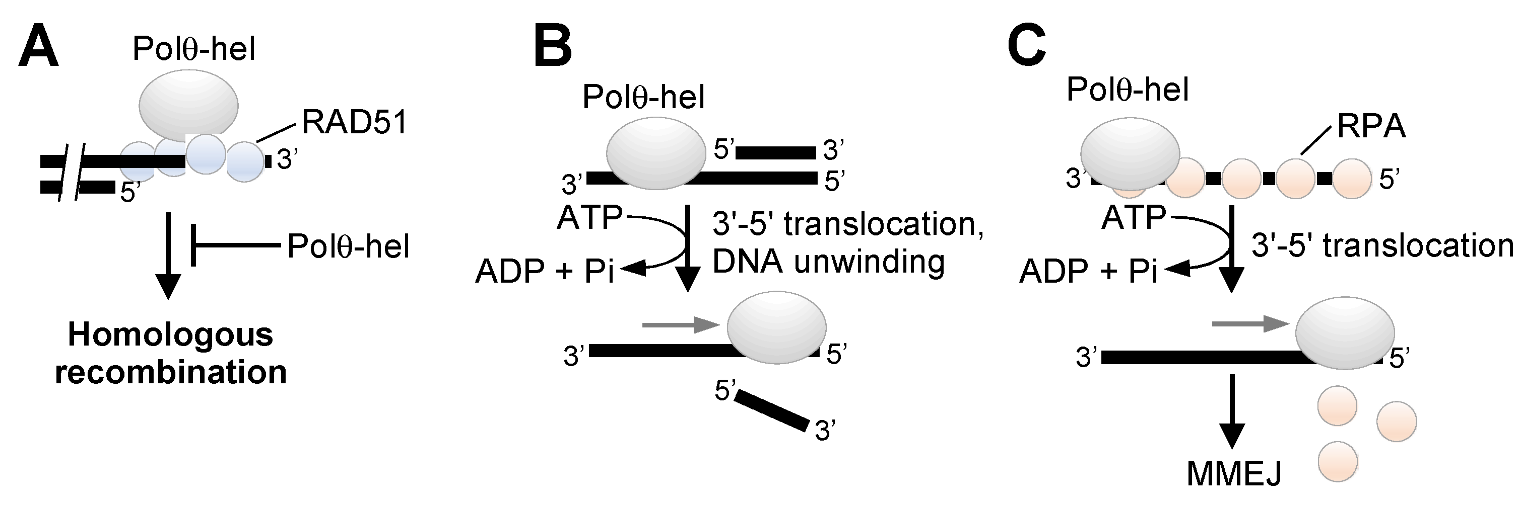
A more in-depth understanding of the respective enzymatic functions of Polθ-pol and Polθ helicase (Polθ-hel) in MMEJ and potentially other DNA repair mechanisms will better inform strategies for successfully developing this multi-functional protein as a therapeutic target. For example, although Polθ-pol likely has a single function in MMEJ (extension of minimally paired ssDNA overhangs) (Figure 1C), the helicase appears to promote MMEJ dependent and independent DNA repair activities. Ceccaldi et al. reported that Polθ-hel interacts with RAD51 and suppresses toxic RAD51 intermediates to confer a survival advantage in BRCA-deficient cells (Figure 3A) [14]. However, it was subsequently found that genetic mutation of the previously characterized Polθ-hel RAD51 binding site had no effect on the survival of BRCA1-deficient cells [52]. Thus, further research is needed to confirm the functional relevance of reported Polθ-hel:RAD51 interactions. We recently demonstrated that Polθ-hel exhibits ATP-dependent DNA unwinding activity with 3′–5′ polarity, similar to RECQ type SF2 helicases (Figure 3B) [55]. However, whether this activity contributes to MMEJ or other DNA repair activities remains unclear. We previously found that Polθ-hel utilizes the energy of ATP hydrolysis to dissociate RPA from ssDNA overhangs that enables ssDNA annealing and stimulation of MMEJ (Figure 3C) [52]. The ability of RPA:ssDNA complexes to suppress MMEJ in mammalian cells is consistent with a previously reported role for RPA in suppressing a different form of microhomology-mediated DSB repair in yeast [56].
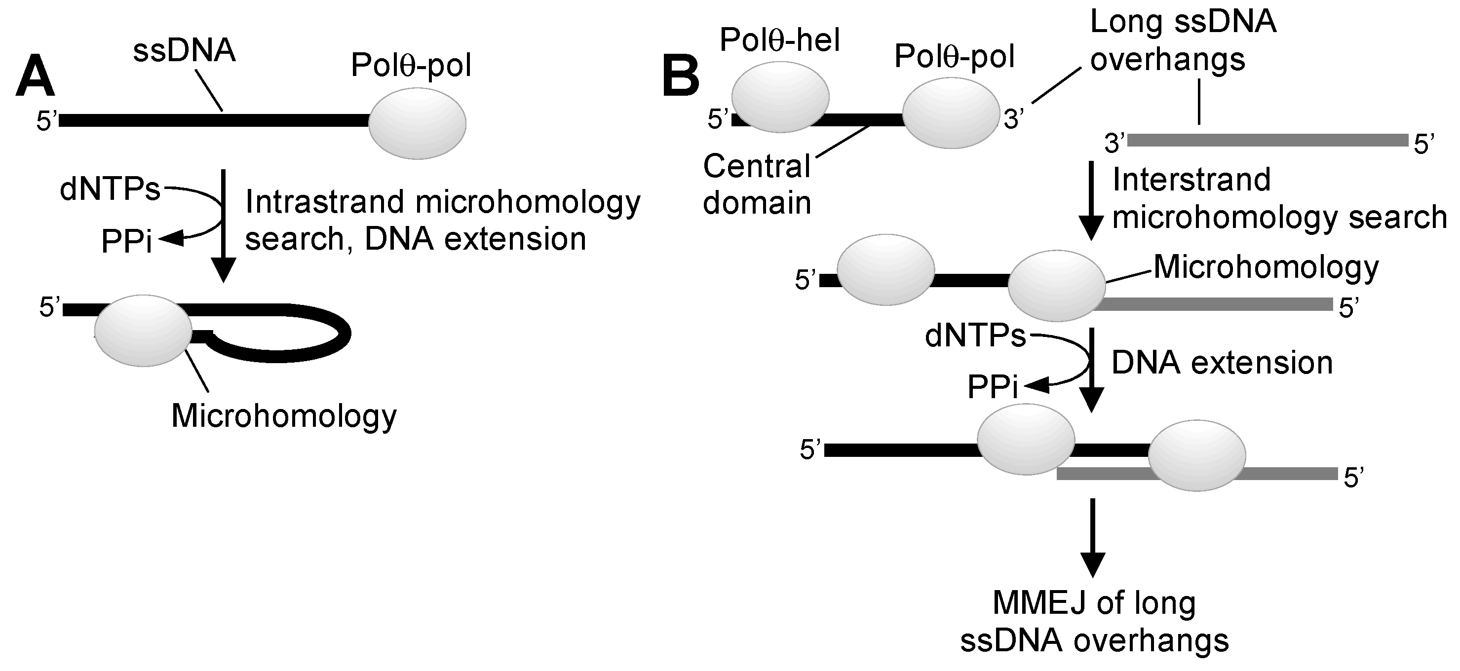
More recently, we found that Polθ-hel strongly contributes to MMEJ in a purified system by cooperating with Polθ-pol on ssDNA overhangs, which was independent of its ATPase function [25]. For example, although Polθ-pol can perform MMEJ of DNA substrates with short 3′ ssDNA overhangs, it is deficient in MMEJ of substrates with long 3′ ssDNA overhangs as a result of its prominent snap-back replication activity on ssDNA (Figure 4A) [25]. So-called snap-back replication results from the enzyme’s unique ability to search for microhomology upstream from the 3′ terminus of ssDNA in cis [25,36]. This enables Polθ-pol to form a stem-loop structure resembling a minimally base-paired primer-template that is efficiently extended by the enzyme (Figure 4A) [25]. Full-length Polθ, on the other hand, exhibited minimal snap-back replication activity on long 3′ ssDNA overhangs and instead primarily performed MMEJ of long ssDNA substrates with 3′ terminal microhomology (Figure 4B) [25]. Moreover, an ATPase mutant version of full-length Polθ, and a Polθ-hel-Polθ-pol fusion protein, which includes a short flexible linker instead of the long central domain, also performed efficient MMEJ of substrates with long 3′ ssDNA overhangs [25]. Combining Polθ-pol and Polθ-hel as separate domains in trans, however, failed to promote MMEJ of long 3′ ssDNA overhangs and instead primarily resulted in Polθ-pol snap-back replication [25]. These biochemical structure function studies indicate that close cooperation between physically linked Polθ-pol and Polθ-hel is required for MMEJ of DNA with long 3′ ssDNA overhangs, and that Polθ-hel ATPase activity is not essential for MMEJ, at least in this minimal purified system that lacks RPA and other auxiliary DNA repair factors. Overall, these studies indicate the Polθ-hel domain binds ssDNA upstream from Polθ-pol, which effectively blocks the polymerase’s prominent snap-back replication activity, and as a result, enables Polθ-pol intermolecular microhomology search as a major mode of action (Figure 4B).
Taken together, multiple lines of evidence demonstrate that both Polθ enzymatic domains contribute to MMEJ and the survival of HR-deficient cells. Consistent with this, small-molecule inhibition of either domain causes the preferential killing of BRCA-deficient cells [54,57]. For example, in addition to the synthetic lethal effects observed for Artios Pharma’s Polθ-pol inhibitor, a recent paper indicates that small-molecule inhibition of Polθ-hel also induces synthetic lethality in BRCA-deficient cells [57]. On the other hand, simultaneous inactivation of both domains may have the largest therapeutic index in BRCA-deficient cells and potentially other DNA repair defective cells, such as those deficient in non-homologous end-joining (NHEJ). In support of this, all of the reported synthetic lethal studies on Polθ used either CRISPR/Cas9 or shRNA to respectively knock-out or strongly suppress the expression of Polθ, which is equivalent to simultaneous inactivation of both enzymatic domains (https://depmap.org/portal/ccle/ accessed on 1 May 2021) [13,14,51,58]. The development of a proteolysis targeting chimera (PROTAC) [59,60,61] degrader of Polθ therefore, represents a plausible therapeutic option since this would fully abolish all of Polθ activities.

4. Polθ as a Reverse Transcriptase

Considering that Polθ definitively functions in MMEJ and TLS pathways and suppressing mitotic crossovers and has been implicated in anti-recombination activity and base excision repair, the question arises whether it exhibits additional DNA repair activities that contribute to cancer cell proliferation. A curious characteristic of Polθ-pol is its deficient proofreading function due to acquired mutations (Figure 1A). Pols lacking exonuclease activity typically exhibit low-fidelity DNA synthesis, and in some cases, this enables TLS activity. Interestingly, inactivating the 3′–5′ exonuclease activity of some related A-family bacterial Pol I enzymes allows these polymerases to reverse transcribe RNA like retroviral reverse transcriptases (RTs), which lack proofreading activity [62,63]. Because Polθ is a highly error-prone enzyme that can utilize various double-strand and ssDNA substrates and is void of exonuclease activity such as retroviral RTs, we examined in recent studies whether Polθ-pol can additionally utilize RNA as a template and thus act as an RT [64]. To determine whether Polθ-pol performs RNA-dependent DNA synthesis activity, we chose a biochemical approach. Our studies utilizing radio-labeled DNA/RNA primer templates found that Polθ-pol exhibits robust RT activity, similar to retroviral RTs (Figure 5A) [64]. Notably, Polθ-pol did not require any specific reaction or buffer conditions and performed RNA-dependent DNA synthesis activity on various template constructs and sequences, and its RT activity mimicked retroviral RTs, such as those expressed by Human Immunodeficiency Virus (HIV), Avian Myeoloblastosis Virus (AMV), and Moloney Murine Leukemia Virus (M-MulV). Most remarkably, our recent report found that Polθ-pol exhibits a higher fidelity and velocity of 2′-deoxyribonucleotide incorporation on RNA versus DNA [64]. This suggests the enzyme was selected to be more accurate and efficient on RNA. In contrast to Polθ-pol, all other recombinant human Pols from the A, X, and B families failed to show any RT activity. Y-family Pols κ and η, however, showed minimal RT activity, resulting in the addition of only a few nucleotides on RNA under identical conditions as Polθ-pol and HIV RT, which fully extended DNA/RNA primer-templates.
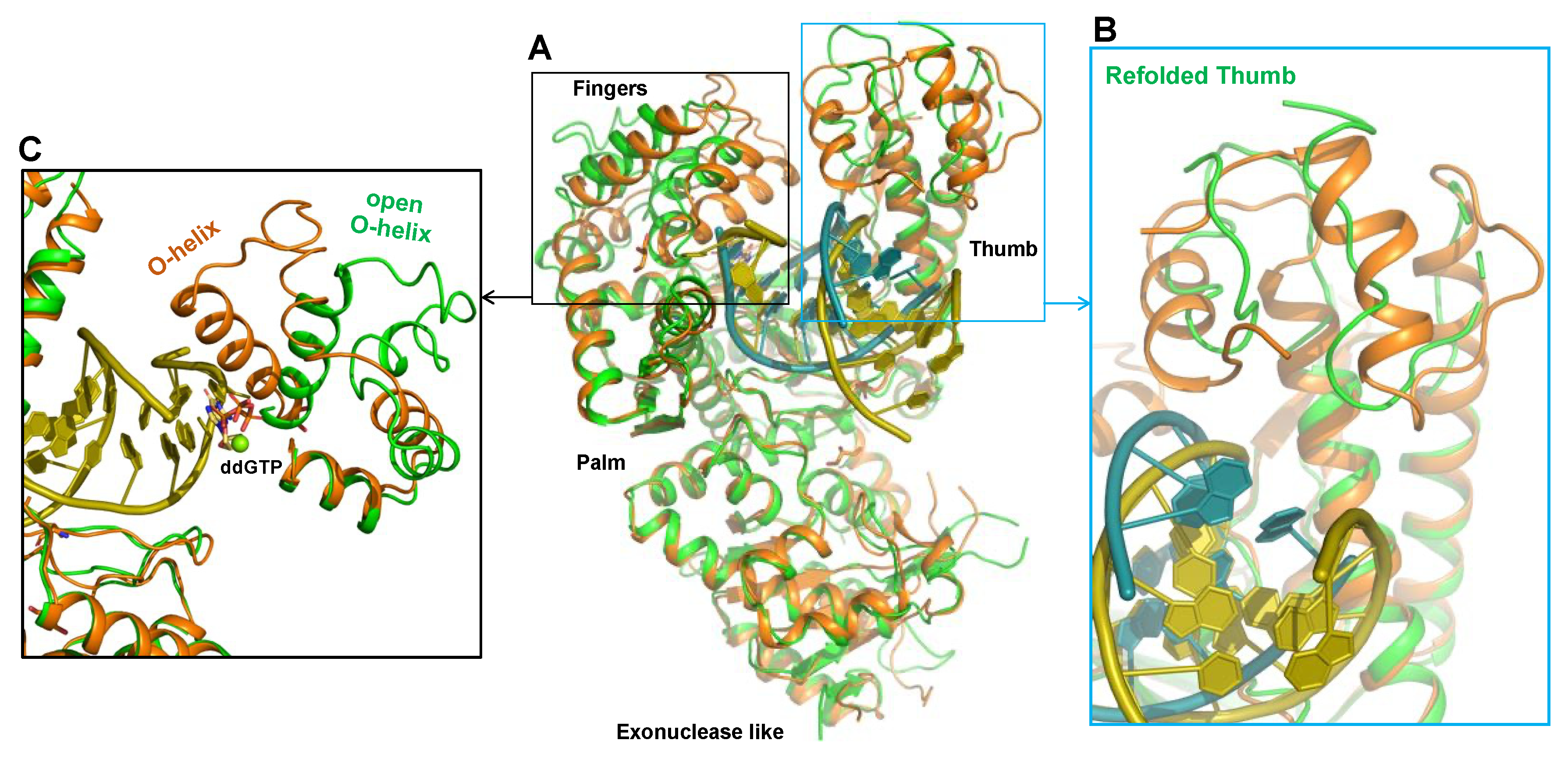
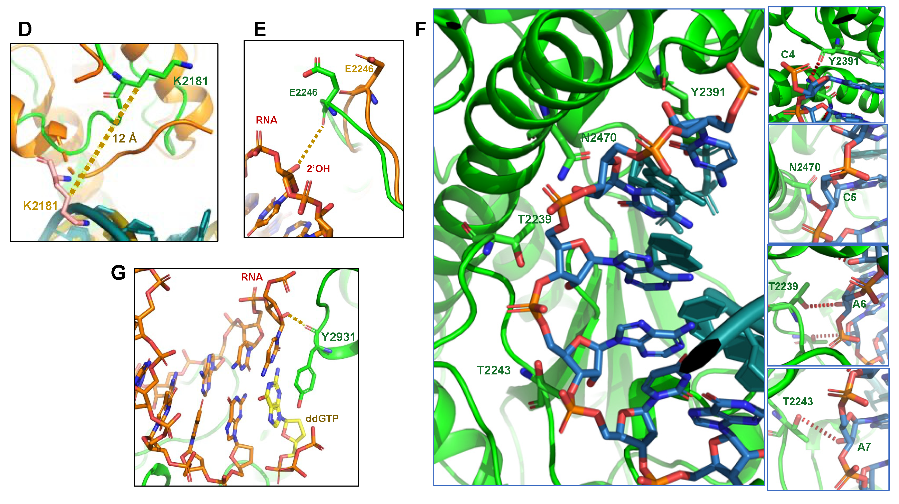
To probe the mechanism by which Polθ-pol uniquely accommodates DNA/RNA hybrids, we utilized X-ray crystallography to solve the ternary structure of Polθ-pol on a DNA/RNA primer-template with incoming 2′,3′-dideoxyguanosine triphosphate (ddGTP). Remarkably, the 3.2 Å structure of the Polθ-pol:DNA/RNA:ddGTP complex revealed that the thumb domain undergoes a major structural rearrangement to accommodate the DNA/RNA hybrid when compared to the previously solved structures of Polθ-pol binding to a DNA/DNA primer-template (Figure 6A–C) [26]. For example, 57% of residues within this subdomain, which interacts with the hybrid double-helix region five base-pairs upstream from the 3′ primer terminus, undergo conformational changes from helices to loops (Figure 6B). This partial refolding of the thumb domain may be needed to provide the necessary interactions with the DNA/RNA hybrid that is wider than B-form DNA/DNA and takes a slightly different orientation in its interaction with the polymerase (Figure 6B). The 12 Å shift observed for residue K2181 highlights the dramatic reconfiguration of the thumb subdomain (Figure 6D). In contrast to the two previously solved structures of Polθ-pol:DNA/DNA:ddGTP complexes, which were in the closed configuration, our Polθ-pol:DNA/RNA:ddGTP complex was captured in the open configuration. Thus, the O-helix in the fingers subdomain is rotated outward, and the incoming ddGTP substrate is partially solvent-exposed (Figure 6C). The open to closed conformational change upon dNTP binding by A-family Pols on DNA/DNA has been extensively characterized by many laboratories and continues to provide intriguing mechanistic insight into these highly conserved enzymes [65,66,67,68,69]. Another notable shift observed in the Polθ-pol:DNA/RNA:ddGTP complex includes residue E2246 (palm subdomain), which appears to form a specific ribose 2′-hydroxyl interaction with the RNA template (Figure 6E). Additional multiple hydrogen bonds are observed between the 2′-hydroxyl group of the RNA template and Thr/Gln residues of Polθ-pol (Figure 6F). Polθ residue Y2391 also forms a hydrogen bond with the template ribonucleotide that pairs with the incoming ddGTP (Figure 6G). Similar hydrogen bonds are observed in structures of retroviral RTs, indicating a similar binding mechanism on RNA [70,71]. Overall, the major conformational changes observed by Polθ-pol on the DNA/RNA relative to prior structures of the enzyme captured on DNA/DNA are unprecedented and reveal that Polθ-pol possesses extraordinary plasticity, which likely explains its unusual promiscuity relative to other Pols.
Our studies also confirmed the ability of Polθ to utilize ribonucleotide template bases during its DNA repair activities in cells. For instance, we utilized a newly developed MMEJ GFP reporter assay in which MMEJ activity results in activation of a linear GFP expression vector [20]. To probe Polθ RNA-dependent DNA synthesis activity in cells, the GFP reporter was modified to include multiple ribonucleotides adjacent to the microhomology tract (Figure 5B). Here, activation of the GFP expression vector required RNA-dependent DNA repair synthesis activity during MMEJ. Our studies clearly showed that the inactivation of Polθ via CRISPR-Cas9 genetic engineering resulted in a significant reduction in MMEJ, which is consistent with the ability of Polθ to utilize template ribonucleotides during its DNA repair synthesis activity during MMEJ (Figure 5B). Intriguingly, recent studies reveal RNA polymerase II synthesis of RNA at DSBs, which depends on the MRN complex [72,73,74]. Whether Polθ acts on these DSB associated RNA substrates that are involved in the DNA damage response remains unknown.
Being that multiple studies suggest RNA-mediated DNA repair as a potential mechanism of DNA repair in eukaryotic cells [75,76,77], our findings suggest a plausible function for Polθ in tolerating ribonucleotides during DNA repair. In one scenario, unrepaired ribonucleotides at or near DSB ends can serve as template bases during DNA repair synthesis by Polθ during MMEJ (Figure 5B). The possibility also exists that Polθ utilizes ribonucleotides during TLS (Figure 5C). In support of this idea, ribonucleotides are the most frequently occurring lesion in eukaryotic genomes as a result of their misincorporation by replicative Pols [78]. Thus, in the event that genome embedded ribonucleotides are not efficiently repaired, such as in RNASEH2 deficient cells, replication forks are likely to become arrested. This is owing to the fact that replicative Pols are unable to tolerate template ribonucleotides and stall upon encountering these lesions [78,79]. In this scenario, TLS would be a desirable outcome, especially if the ribonucleotide lesions can be replicated accurately. Although future studies will be needed to test Polθ TLS activity opposite genome-embedded ribonucleotides in cells, the enzyme’s ability to easily accommodate A-form DNA/RNA hybrids relative to other Pols reveals its unique and extraordinary structural plasticity. Hence, it will be interesting to determine whether similar structural rearrangements within the enzyme’s thumb subdomain occur during MMEJ in order to accommodate minimally paired ssDNA overhangs in an active configuration within the enzyme’s active site.

5. Conclusions and Perspectives

The continued and growing interest in Polθ biology and drug development is expected to lead to many exciting discoveries regarding the respective activities of Polθ helicase and polymerase, as well as inform the development of improved therapeutics targeting this multi-functional enzyme. The recent report on Artios Pharma’s allosteric Polθ-pol inhibitor class demonstrates for the first time that this specific domain of Polθ is indeed druggable. Another recent study indicates that the helicase domain is also druggable. Here, the authors re-purposed the antibiotic Novabiocin as a Polθ-hel inhibitor that exhibited high micromolar IC50. Further development of potent and selective Polθ-hel inhibitors will be needed to fully assess the potential of this domain as a drug target in HR-deficient cells. As with all early-stage drugs, it remains to be seen whether Polθ-pol or Polθ-hel inhibitors will lead to clinically effective therapeutics. Notably, decades of drug development research targeting HIV RT and other viral polymerases demonstrate the successful development of selective and potent inhibitors against these polymerases, including both competitive nucleoside prodrugs and allosteric non-nucleoside drug inhibitors [80,81,82,83,84]. For example, the prodrug nucleotide analog remdesivir was recently approved for emergency use against Sars-Cov-2 RNA polymerase [85,86]. Clinical grade drug development against human DNA helicases, however, has yet to be achieved. However, considering that the ATP binding pocket within Polθ-hel is well defined and solvent-exposed like kinases [24], this domain will likely be a viable drug target for clinical-grade candidates specifically designed as Polθ-hel inhibitors.
A major question that arises from the recent research published by Chandramouly et al. is whether Polθ’s RT activity specifically contributes to particular DNA repair functions, such as cellular tolerance to genome embedded ribonucleotides. Separation of function mutations may be needed to pursue such questions. Considering that Polθ’s RT activity appears to be similar to HIV and other retroviral RTs, a question that arises is whether HIV RT nucleoside inhibitors may be useful for targeting Polθ DNA synthesis activity in cells, and thus, can potentially be re-purposed as anti-cancer agents. Indeed, Polθ and other A-family Pols with a conserved tyrosine residue within the fingers subdomain are known to be inhibited by 2′,3′,-dideoxyribonucleoside triphosphates (ddNTPs) [27]. Thus, it would be interesting to determine the effects of nucleoside prodrug inhibitors of HIV RT on Polθ cellular activities and the survival of HR-deficient cells. For instance, prodrugs such as zalcitabine and didanosine are converted by nucleoside/nucleotide kinases into their respective active metabolites, ddCTP and ddATP, which inhibit Polθ. The clinical impact of such nucleoside analogs, however, may be limited due to their modest potency and cross-reactivity against mitochondrial Polγ and possibly other Pols. Nevertheless, based on the successful history of competitive nucleotide inhibitors of viral polymerases, the possibility exists that potent and selective nucleotide analog inhibitors of Polθ-pol can be developed in addition to allosteric inhibitors. Regardless of their mechanism of action, the continued development of Polθ-pol and Polθ-hel drug-like inhibitors is expected to generate a lot of excitement for both the translational and basic research communities.

Author Contributions

R.T.P. wrote the review. X.S.C. provided figures and editorial input. All authors have read and agreed to the published version of the manuscript.

Funding

This article was supported by NIH grants 1R01GM130889-01 and 1R01GM137124-01 to R.T.P. and in part by a Tower Cancer Research Foundation grant to X.S.C.

Institutional Review Board Statement

Not applicable.

Informed Consent Statement

Not applicable.

Data Availability Statement

Not applicable.

Conflicts of Interest

R.T.P. and X.S.C. are co-founders of Recombination Therapeutics, LLC.

References

  1. Krais, J.J.; Johnson, N. BRCA1 Mutations in Cancer: Coordinating Deficiencies in Homologous Recombination with Tumorigenesis. Cancer Res. 2020, 80, 4601–4609. [Google Scholar] [CrossRef]
  2. Pan, Z.; Xie, X. BRCA mutations in the manifestation and treatment of ovarian cancer. Oncotarget 2017, 8, 97657–97670. [Google Scholar] [CrossRef] [Green Version]
  3. Couch, F.J.; Nathanson, K.L.; Offit, K. Two decades after BRCA: Setting paradigms in personalized cancer care and prevention. Science 2014, 343, 1466–1470. [Google Scholar] [CrossRef] [Green Version]
  4. Tutt, A.N.; Lord, C.J.; McCabe, N.; Farmer, H.; Turner, N.; Martin, N.M.; Jackson, S.P.; Smith, G.C.; Ashworth, A. Exploiting the DNA repair defect in BRCA mutant cells in the design of new therapeutic strategies for cancer. Cold Spring Harb. Symp. Quant. Biol. 2005, 70, 139–148. [Google Scholar] [CrossRef]
  5. Konstantinopoulos, P.A.; Ceccaldi, R.; Shapiro, G.I.; D’Andrea, A.D. Homologous Recombination Deficiency: Exploiting the Fundamental Vulnerability of Ovarian Cancer. Cancer Discov. 2015, 5, 1137–1154. [Google Scholar] [CrossRef] [Green Version]
  6. Patel, P.S.; Algouneh, A.; Hakem, R. Exploiting synthetic lethality to target BRCA1/2-deficient tumors: Where we stand. Oncogene 2021, 40, 3001–3014. [Google Scholar] [CrossRef]
  7. Sonnenblick, A.; de Azambuja, E.; Azim, H.A.; Piccart, M. An update on PARP inhibitors--moving to the adjuvant setting. Nat. Rev. Clin. Oncol. 2015, 12, 27–41. [Google Scholar] [CrossRef]
  8. Farmer, H.; McCabe, N.; Lord, C.J.; Tutt, A.N.; Johnson, D.A.; Richardson, T.B.; Santarosa, M.; Dillon, K.J.; Hickson, I.; Knights, C.; et al. Targeting the DNA repair defect in BRCA mutant cells as a therapeutic strategy. Nature 2005, 434, 917–921. [Google Scholar] [CrossRef]
  9. Bryant, H.E.; Schultz, N.; Thomas, H.D.; Parker, K.M.; Flower, D.; Lopez, E.; Kyle, S.; Meuth, M.; Curtin, N.J.; Helleday, T. Specific killing of BRCA2-deficient tumours with inhibitors of poly(ADP-ribose) polymerase. Nature 2005, 434, 913–917. [Google Scholar] [CrossRef]
  10. Lord, C.J.; Ashworth, A. PARP inhibitors: Synthetic lethality in the clinic. Science 2017, 355, 1152–1158. [Google Scholar] [CrossRef]
  11. Noordermeer, S.M.; van Attikum, H. PARP Inhibitor Resistance: A Tug-of-War in BRCA-Mutated Cells. Trends Cell Biol. 2019, 29, 820–834. [Google Scholar] [CrossRef] [Green Version]
  12. Li, H.; Liu, Z.Y.; Wu, N.; Chen, Y.C.; Cheng, Q.; Wang, J. PARP inhibitor resistance: The underlying mechanisms and clinical implications. Mol. Cancer 2020, 19, 107. [Google Scholar] [CrossRef] [PubMed]
  13. Mateos-Gomez, P.A.; Gong, F.; Nair, N.; Miller, K.M.; Lazzerini-Denchi, E.; Sfeir, A. Mammalian polymerase theta promotes alternative NHEJ and suppresses recombination. Nature 2015, 518, 254–257. [Google Scholar] [CrossRef] [PubMed] [Green Version]
  14. Ceccaldi, R.; Liu, J.C.; Amunugama, R.; Hajdu, I.; Primack, B.; Petalcorin, M.I.R.; O’Connor, K.W.; Konstantinopoulos, P.A.; Elledge, S.J.; Boulton, S.J.; et al. Homologous-recombination-deficient tumours are dependent on Polθ-mediated repair. Nature 2015, 518, 258–262. [Google Scholar] [CrossRef] [Green Version]
  15. Lemee, F.; Bergoglio, V.; Fernandez-Vidal, A.; Machado-Silva, A.; Pillaire, M.J.; Bieth, A.; Gentil, C.; Baker, L.; Martin, A.L.; Leduc, C.; et al. DNA polymerase theta up-regulation is associated with poor survival in breast cancer, perturbs DNA replication, and promotes genetic instability. Proc. Natl. Acad. Sci. USA 2010, 107, 13390–13395. [Google Scholar] [CrossRef] [PubMed] [Green Version]
  16. Higgins, G.S.; Harris, A.L.; Prevo, R.; Helleday, T.; McKenna, W.G.; Buffa, F.M. Overexpression of POLQ confers a poor prognosis in early breast cancer patients. Oncotarget 2010, 1, 175–184. [Google Scholar] [CrossRef]
  17. Arana, M.E.; Seki, M.; Wood, R.D.; Rogozin, I.B.; Kunkel, T.A. Low-fidelity DNA synthesis by human DNA polymerase theta. Nucleic Acids Res. 2008, 36, 3847–3856. [Google Scholar] [CrossRef] [Green Version]
  18. Seki, M.; Masutani, C.; Yang, L.W.; Schuffert, A.; Iwai, S.; Bahar, I.; Wood, R.D. High-efficiency bypass of DNA damage by human DNA polymerase Q. EMBO J. 2004, 23, 4484–4494. [Google Scholar] [CrossRef] [Green Version]
  19. Begg, A. POLQ in breast cancer. Oncotarget 2010, 1, 161–162. [Google Scholar] [CrossRef]
  20. Chandramouly, G.; Liao, S.; Rusanov, T.; Borisonnik, N.; Calbert, M.L.; Kent, T.; Sullivan-Reed, K.; Vekariya, U.; Kashkina, E.; Skorski, T.; et al. Poltheta promotes the repair of 5′-DNA-protein crosslinks by microhomology-mediated end-joining. Cell Rep. 2021, 34, 108820. [Google Scholar] [CrossRef]
  21. Yousefzadeh, M.J.; Wyatt, D.W.; Takata, K.; Mu, Y.; Hensley, S.C.; Tomida, J.; Bylund, G.O.; Doublie, S.; Johansson, E.; Ramsden, D.A.; et al. Mechanism of suppression of chromosomal instability by DNA polymerase POLQ. PLoS Genet. 2014, 10, e1004654. [Google Scholar] [CrossRef] [PubMed] [Green Version]
  22. Higgins, G.S.; Prevo, R.; Lee, Y.F.; Helleday, T.; Muschel, R.J.; Taylor, S.; Yoshimura, M.; Hickson, I.D.; Bernhard, E.J.; McKenna, W.G. A small interfering RNA screen of genes involved in DNA repair identifies tumor-specific radiosensitization by POLQ knockdown. Cancer Res. 2010, 70, 2984–2993. [Google Scholar] [CrossRef] [Green Version]
  23. Dai, C.H.; Chen, P.; Li, J.; Lan, T.; Chen, Y.C.; Qian, H.; Chen, K.; Li, M.Y. Co-inhibition of pol theta and HR genes efficiently synergize with cisplatin to suppress cisplatin-resistant lung cancer cells survival. Oncotarget 2016, 7, 65157–65170. [Google Scholar] [CrossRef] [Green Version]
  24. Newman, J.A.; Cooper, C.D.O.; Aitkenhead, H.; Gileadi, O. Structure of the Helicase Domain of DNA Polymerase Theta Reveals a Possible Role in the Microhomology-Mediated End-Joining Pathway. Structure 2015, 23, 2319–2330. [Google Scholar] [CrossRef] [Green Version]
  25. Black, S.J.; Ozdemir, A.Y.; Kashkina, E.; Kent, T.; Rusanov, T.; Ristic, D.; Shin, Y.; Suma, A.; Hoang, T.; Chandramouly, G.; et al. Molecular basis of microhomology-mediated end-joining by purified full-length Poltheta. Nat. Commun. 2019, 10, 4423. [Google Scholar] [CrossRef] [PubMed] [Green Version]
  26. Zahn, K.E.; Averill, A.M.; Aller, P.; Wood, R.D.; Doublie, S. Human DNA polymerase theta grasps the primer terminus to mediate DNA repair. Nat. Struct. Mol. Biol. 2015, 22, 304–311. [Google Scholar] [CrossRef] [PubMed] [Green Version]
  27. Seki, M.; Marini, F.; Wood, R.D. POLQ (Pol theta), a DNA polymerase and DNA-dependent ATPase in human cells. Nucleic Acids Res. 2003, 31, 6117–6126. [Google Scholar] [CrossRef] [Green Version]
  28. Kent, T.; Rusanov, T.D.; Hoang, T.M.; Velema, W.A.; Krueger, A.T.; Copeland, W.C.; Kool, E.T.; Pomerantz, R.T. DNA polymerase theta specializes in incorporating synthetic expanded-size (xDNA) nucleotides. Nucleic Acids Res. 2016, 44, 9381–9392. [Google Scholar] [CrossRef] [Green Version]
  29. Kent, T.; Mateos-Gomez, P.A.; Sfeir, A.; Pomerantz, R.T. Polymerase theta is a robust terminal transferase that oscillates between three different mechanisms during end-joining. eLife 2016, 5, e13740. [Google Scholar] [CrossRef] [PubMed] [Green Version]
  30. Seki, M.; Wood, R.D. DNA polymerase theta (POLQ) can extend from mismatches and from bases opposite a (6-4) photoproduct. DNA Repair 2008, 7, 119–127. [Google Scholar] [CrossRef] [Green Version]
  31. Hogg, M.; Seki, M.; Wood, R.D.; Doublie, S.; Wallace, S.S. Lesion bypass activity of DNA polymerase theta (POLQ) is an intrinsic property of the pol domain and depends on unique sequence inserts. J. Mol. Biol. 2011, 405, 642–652. [Google Scholar] [CrossRef] [PubMed] [Green Version]
  32. Yoon, J.H.; Roy Choudhury, J.; Park, J.; Prakash, S.; Prakash, L. A Role for DNA Polymerase theta in Promoting Replication through Oxidative DNA Lesion, Thymine Glycol, in Human Cells. J. Biol. Chem. 2014, 289, 13177–13185. [Google Scholar] [CrossRef] [PubMed] [Green Version]
  33. Yoon, J.H.; McArthur, M.J.; Park, J.; Basu, D.; Wakamiya, M.; Prakash, L.; Prakash, S. Error-Prone Replication through UV Lesions by DNA Polymerase theta Protects against Skin Cancers. Cell 2019, 176, 1295–1309. [Google Scholar] [CrossRef] [Green Version]
  34. Arana, M.E.; Takata, K.; Garcia-Diaz, M.; Wood, R.D.; Kunkel, T.A. A unique error signature for human DNA polymerase nu. DNA Repair 2007, 6, 213–223. [Google Scholar] [CrossRef] [Green Version]
  35. Jain, R.; Aggarwal, A.K.; Rechkoblit, O. Eukaryotic DNA polymerases. Curr. Opin. Struct. Biol. 2018, 53, 77–87. [Google Scholar] [CrossRef]
  36. Kent, T.; Chandramouly, G.; McDevitt, S.M.; Ozdemir, A.Y.; Pomerantz, R.T. Mechanism of microhomology-mediated end-joining promoted by human DNA polymerase theta. Nat. Struct. Mol. Biol. 2015, 22, 230–237. [Google Scholar] [CrossRef] [Green Version]
  37. Koole, W.; van Schendel, R.; Karambelas, A.E.; van Heteren, J.T.; Okihara, K.L.; Tijsterman, M. A Polymerase Theta-dependent repair pathway suppresses extensive genomic instability at endogenous G4 DNA sites. Nat. Commun. 2014, 5, 3216. [Google Scholar] [CrossRef] [Green Version]
  38. Wyatt, D.W.; Feng, W.; Conlin, M.P.; Yousefzadeh, M.J.; Roberts, S.A.; Mieczkowski, P.; Wood, R.D.; Gupta, G.P.; Ramsden, D.A. Essential Roles for Polymerase theta-Mediated End Joining in the Repair of Chromosome Breaks. Mol. Cell 2016, 63, 662–673. [Google Scholar] [CrossRef] [Green Version]
  39. Zahn, K.E.; Jensen, R.B.; Wood, R.D.; Doublie, S. Human DNA polymerase theta harbors DNA end-trimming activity critical for DNA repair. Mol. Cell 2021, 81, 1534–1547.e4. [Google Scholar] [CrossRef] [PubMed]
  40. Prasad, R.; Longley, M.J.; Sharief, F.S.; Hou, E.W.; Copeland, W.C.; Wilson, S.H. Human DNA polymerase theta possesses 5’-dRP lyase activity and functions in single-nucleotide base excision repair in vitro. Nucleic Acids Res. 2009, 37, 1868–1877. [Google Scholar] [CrossRef] [Green Version]
  41. Wang, Z.; Song, Y.; Li, S.; Kurian, S.; Xiang, R.; Chiba, T.; Wu, X. DNA polymerase theta (POLQ) is important for repair of DNA double-strand breaks caused by fork collapse. J. Biol. Chem. 2019, 294, 3909–3919. [Google Scholar] [CrossRef]
  42. Shima, N.; Munroe, R.J.; Schimenti, J.C. The mouse genomic instability mutation chaos1 is an allele of Polq that exhibits genetic interaction with Atm. Mol. Cell Biol. 2004, 24, 10381–10389. [Google Scholar] [CrossRef] [Green Version]
  43. Carvajal-Garcia, J.; Crown, K.N.; Ramsden, D.A.; Sekelsky, J. DNA polymerase theta suppresses mitotic crossing over. PLoS Genet. 2021, 17, e1009267. [Google Scholar] [CrossRef] [PubMed]
  44. Davis, L.; Khoo, K.J.; Zhang, Y.; Maizels, N. POLQ suppresses interhomolog recombination and loss of heterozygosity at targeted DNA breaks. Proc. Natl. Acad. Sci. USA 2020, 117, 22900–22909. [Google Scholar] [CrossRef]
  45. Moynahan, M.E.; Jasin, M. Mitotic homologous recombination maintains genomic stability and suppresses tumorigenesis. Nat. Rev. Mol. Cell Biol. 2010, 11, 196–207. [Google Scholar] [CrossRef] [PubMed] [Green Version]
  46. San Filippo, J.; Sung, P.; Klein, H. Mechanism of eukaryotic homologous recombination. Annu. Rev. Biochem. 2008, 77, 229–257. [Google Scholar] [CrossRef] [PubMed] [Green Version]
  47. Li, X.; Heyer, W.D. Homologous recombination in DNA repair and DNA damage tolerance. Cell Res. 2008, 18, 99–113. [Google Scholar] [CrossRef] [Green Version]
  48. Truong, L.N.; Li, Y.; Shi, L.Z.; Hwang, P.Y.; He, J.; Wang, H.; Razavian, N.; Berns, M.W.; Wu, X. Microhomology-mediated End Joining and Homologous Recombination share the initial end resection step to repair DNA double-strand breaks in mammalian cells. Proc. Natl. Acad. Sci. USA 2013, 110, 7720–7725. [Google Scholar] [CrossRef] [PubMed] [Green Version]
  49. Zhang, Y.; Jasin, M. An essential role for CtIP in chromosomal translocation formation through an alternative end-joining pathway. Nat. Struct. Mol. Biol. 2011, 18, 80–84. [Google Scholar] [CrossRef]
  50. Cho, N.W.; Greenberg, R.A. DNA repair: Familiar ends with alternative endings. Nature 2015, 518, 174–176. [Google Scholar] [CrossRef]
  51. Mengwasser, K.E.; Adeyemi, R.O.; Leng, Y.; Choi, M.Y.; Clairmont, C.; D’Andrea, A.D.; Elledge, S.J. Genetic Screens Reveal FEN1 and APEX2 as BRCA2 Synthetic Lethal Targets. Mol. Cell 2019, 73, 885–899.e886. [Google Scholar] [CrossRef] [Green Version]
  52. Mateos-Gomez, P.A.; Kent, T.; Deng, S.K.; McDevitt, S.; Kashkina, E.; Hoang, T.M.; Pomerantz, R.T.; Sfeir, A. The helicase domain of Poltheta counteracts RPA to promote alt-NHEJ. Nat. Struct. Mol. Biol. 2017, 24, 1116–1123. [Google Scholar] [CrossRef]
  53. Feng, W.; Simpson, D.A.; Carvajal-Garcia, J.; Price, B.A.; Kumar, R.J.; Mose, L.E.; Wood, R.D.; Rashid, N.; Purvis, J.E.; Parker, J.S.; et al. Genetic determinants of cellular addiction to DNA polymerase theta. Nat. Commun. 2019, 10, 4286. [Google Scholar] [CrossRef] [Green Version]
  54. Zatreanu, D.; Robinson, H.M.R.; Alkhatib, O.; Boursier, M.; Finch, H.; Geo, L.; Grande, D.; Grinkevich, V.; Heald, R.A.; Langdon, S.; et al. Poltheta inhibitors elicit BRCA-gene synthetic lethality and target PARP inhibitor resistance. Nat. Commun. 2021, 12, 3636. [Google Scholar] [CrossRef]
  55. Ozdemir, A.Y.; Rusanov, T.; Kent, T.; Siddique, L.A.; Pomerantz, R.T. Polymerase theta-helicase efficiently unwinds DNA and RNA-DNA hybrids. J. Biol. Chem. 2018, 293, 5259–5269. [Google Scholar] [CrossRef] [Green Version]
  56. Deng, S.K.; Gibb, B.; de Almeida, M.J.; Greene, E.C.; Symington, L.S. RPA antagonizes microhomology-mediated repair of DNA double-strand breaks. Nat. Struct. Mol. Biol. 2014, 21, 405–412. [Google Scholar] [CrossRef] [Green Version]
  57. Zhou, J.; Gelot, C.; Pantelidou, C.; Li, A.; Yucel, H.; Davis, R.E.; Farkkila, A.; Kochupurakkal, B.; Syed, A.; Shapiro, G.I.; et al. A first-in-class Polymerase Theta Inhibitor selectively targets Homologous-Recombination-Deficient Tumors. Nat. Cancer 2021, 2, 598–610. [Google Scholar] [CrossRef] [PubMed]
  58. Schrempf, A.; Slyskova, J.; Loizou, J.I. Targeting the DNA Repair Enzyme Polymerase θ in Cancer Therapy. Trends Cancer 2020, 7, 98–111. [Google Scholar] [CrossRef]
  59. Zeng, S.; Huang, W.; Zheng, X.; Liyan, C.; Zhang, Z.; Wang, J.; Shen, Z. Proteolysis targeting chimera (PROTAC) in drug discovery paradigm: Recent progress and future challenges. Eur. J. Med. Chem. 2021, 210, 112981. [Google Scholar] [CrossRef] [PubMed]
  60. Paiva, S.L.; Crews, C.M. Targeted protein degradation: Elements of PROTAC design. Curr. Opin. Chem. Biol. 2019, 50, 111–119. [Google Scholar] [CrossRef] [PubMed]
  61. Sun, X.; Gao, H.; Yang, Y.; He, M.; Wu, Y.; Song, Y.; Tong, Y.; Rao, Y. PROTACs: Great opportunities for academia and industry. Signal Transduct. Target. Ther. 2019, 4, 64. [Google Scholar] [CrossRef] [PubMed] [Green Version]
  62. Shi, C.; Shen, X.; Niu, S.; Ma, C. Innate Reverse Transcriptase Activity of DNA Polymerase for Isothermal RNA Direct Detection. J. Am. Chem. Soc. 2015, 137, 13804–13806. [Google Scholar] [CrossRef] [PubMed]
  63. Menendez-Arias, L.; Sebastian-Martin, A.; Alvarez, M. Viral reverse transcriptases. Virus Res. 2017, 234, 153–176. [Google Scholar] [CrossRef] [PubMed]
  64. Chandramouly, G.; Zhao, J.; McDevitt, S.; Rusanov, T.; Hoang, T.; Borisonnik, N.; Treddinick, T.; Lopezcolorado, F.W.; Kent, T.; Siddique, L.A.; et al. Poltheta reverse transcribes RNA and promotes RNA-templated DNA repair. Sci. Adv. 2021, 7. [Google Scholar] [CrossRef] [PubMed]
  65. Li, Y.; Korolev, S.; Waksman, G. Crystal structures of open and closed forms of binary and ternary complexes of the large fragment of Thermus aquaticus DNA polymerase I: Structural basis for nucleotide incorporation. EMBO J. 1998, 17, 7514–7525. [Google Scholar] [CrossRef] [Green Version]
  66. Doublie, S.; Sawaya, M.R.; Ellenberger, T. An open and closed case for all polymerases. Structure 1999, 7, R31–R35. [Google Scholar] [CrossRef] [Green Version]
  67. Miller III, B.R.; Beese, L.S.; Parish, C.A.; Wu, E.Y. The Closing Mechanism of DNA Polymerase I at Atomic Resolution. Structure 2015, 23, 1609–1620. [Google Scholar] [CrossRef] [Green Version]
  68. Kiefer, J.R.; Mao, C.; Braman, J.C.; Beese, L.S. Visualizing DNA replication in a catalytically active Bacillus DNA polymerase crystal. Nature 1998, 391, 304–307. [Google Scholar] [CrossRef]
  69. Chim, N.; Meza, R.A.; Trinh, A.M.; Yang, K.; Chaput, J.C. Following replicative DNA synthesis by time-resolved X-ray crystallography. Nat. Commun. 2021, 12, 2641. [Google Scholar] [CrossRef]
  70. Nowak, E.; Potrzebowski, W.; Konarev, P.V.; Rausch, J.W.; Bona, M.K.; Svergun, D.I.; Bujnicki, J.M.; Le Grice, S.F.; Nowotny, M. Structural analysis of monomeric retroviral reverse transcriptase in complex with an RNA/DNA hybrid. Nucleic Acids Res. 2013, 41, 3874–3887. [Google Scholar] [CrossRef] [Green Version]
  71. Sarafianos, S.G.; Das, K.; Tantillo, C.; Clark, A.D., Jr.; Ding, J.; Whitcomb, J.M.; Boyer, P.L.; Hughes, S.H.; Arnold, E. Crystal structure of HIV-1 reverse transcriptase in complex with a polypurine tract RNA:DNA. EMBO J. 2001, 20, 1449–1461. [Google Scholar] [CrossRef] [PubMed] [Green Version]
  72. Michelini, F.; Pitchiaya, S.; Vitelli, V.; Sharma, S.; Gioia, U.; Pessina, F.; Cabrini, M.; Wang, Y.; Capozzo, I.; Iannelli, F.; et al. Damage-induced lncRNAs control the DNA damage response through interaction with DDRNAs at individual double-strand breaks. Nat. Cell Biol. 2017, 19, 1400–1411. [Google Scholar] [CrossRef] [Green Version]
  73. Wei, W.; Ba, Z.; Gao, M.; Wu, Y.; Ma, Y.; Amiard, S.; White, C.I.; Rendtlew Danielsen, J.M.; Yang, Y.G.; Qi, Y. A role for small RNAs in DNA double-strand break repair. Cell 2012, 149, 101–112. [Google Scholar] [CrossRef] [PubMed] [Green Version]
  74. Sharma, S.; Anand, R.; Zhang, X.; Francia, S.; Michelini, F.; Galbiati, A.; Williams, H.; Ronato, D.A.; Masson, J.Y.; Rothenberg, E.; et al. MRE11-RAD50-NBS1 Complex Is Sufficient to Promote Transcription by RNA Polymerase II at Double-Strand Breaks by Melting DNA Ends. Cell Rep. 2021, 34, 108565. [Google Scholar] [CrossRef]
  75. Mazina, O.M.; Keskin, H.; Hanamshet, K.; Storici, F.; Mazin, A.V. Rad52 Inverse Strand Exchange Drives RNA-Templated DNA Double-Strand Break Repair. Mol. Cell 2017, 67, 19–29. [Google Scholar] [CrossRef] [PubMed] [Green Version]
  76. Meers, C.; Keskin, H.; Banyai, G.; Mazina, O.; Yang, T.; Gombolay, A.L.; Mukherjee, K.; Kaparos, E.I.; Newnam, G.; Mazin, A.; et al. Genetic Characterization of Three Distinct Mechanisms Supporting RNA-Driven DNA Repair and Modification Reveals Major Role of DNA Polymerase zeta. Mol. Cell 2020, 79, 1037–1050. [Google Scholar] [CrossRef] [PubMed]
  77. Lazzaro, F.; Novarina, D.; Amara, F.; Watt, D.L.; Stone, J.E.; Costanzo, V.; Burgers, P.M.; Kunkel, T.A.; Plevani, P.; Muzi-Falconi, M. RNase H and postreplication repair protect cells from ribonucleotides incorporated in DNA. Mol. Cell 2012, 45, 99–110. [Google Scholar] [CrossRef] [PubMed] [Green Version]
  78. Williams, J.S.; Kunkel, T.A. Ribonucleotides in DNA: Origins, repair and consequences. DNA Repair 2014, 19, 27–37. [Google Scholar] [CrossRef] [PubMed] [Green Version]
  79. Williams, J.S.; Lujan, S.A.; Kunkel, T.A. Processing ribonucleotides incorporated during eukaryotic DNA replication. Nat. Rev. Mol. Cell Biol. 2016, 17, 350–363. [Google Scholar] [CrossRef] [Green Version]
  80. Cihlar, T.; Ray, A.S. Nucleoside and nucleotide HIV reverse transcriptase inhibitors: 25 years after zidovudine. Antivir. Res. 2010, 85, 39–58. [Google Scholar] [CrossRef]
  81. Mehellou, Y.; Rattan, H.S.; Balzarini, J. The ProTide Prodrug Technology: From the Concept to the Clinic. J. Med. Chem. 2018, 61, 2211–2226. [Google Scholar] [CrossRef] [PubMed]
  82. Jordheim, L.P.; Durantel, D.; Zoulim, F.; Dumontet, C. Advances in the development of nucleoside and nucleotide analogues for cancer and viral diseases. Nat. Rev. Drug Discov. 2013, 12, 447–464. [Google Scholar] [CrossRef] [PubMed]
  83. Das, K.; Martinez, S.E.; Bauman, J.D.; Arnold, E. HIV-1 reverse transcriptase complex with DNA and nevirapine reveals non-nucleoside inhibition mechanism. Nat. Struct. Mol. Biol. 2012, 19, 253–259. [Google Scholar] [CrossRef] [Green Version]
  84. Namasivayam, V.; Vanangamudi, M.; Kramer, V.G.; Kurup, S.; Zhan, P.; Liu, X.; Kongsted, J.; Byrareddy, S.N. The Journey of HIV-1 Non-Nucleoside Reverse Transcriptase Inhibitors (NNRTIs) from Lab to Clinic. J. Med. Chem. 2019, 62, 4851–4883. [Google Scholar] [CrossRef] [PubMed]
  85. Kokic, G.; Hillen, H.S.; Tegunov, D.; Dienemann, C.; Seitz, F.; Schmitzova, J.; Farnung, L.; Siewert, A.; Hobartner, C.; Cramer, P. Mechanism of SARS-CoV-2 polymerase stalling by remdesivir. Nat. Commun. 2021, 12, 279. [Google Scholar] [CrossRef]
  86. Grein, J.; Ohmagari, N.; Shin, D.; Diaz, G.; Asperges, E.; Castagna, A.; Feldt, T.; Green, G.; Green, M.L.; Lescure, F.X.; et al. Compassionate Use of Remdesivir for Patients with Severe Covid-19. N. Engl. J. Med. 2020, 382, 2327–2336. [Google Scholar] [CrossRef]
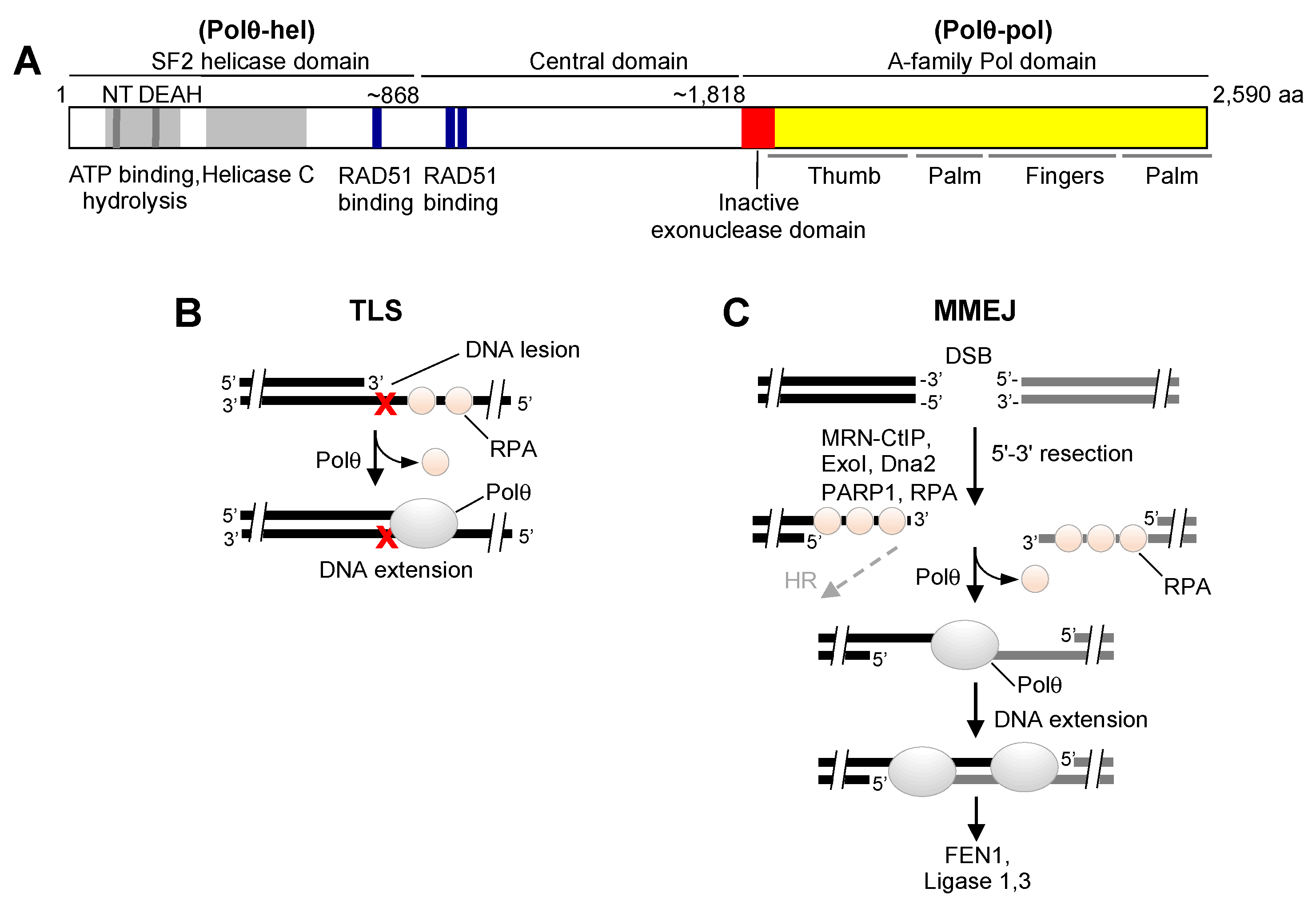
Figure 1. Overview of Polθ structure and function in DNA repair. (A) Schematic of full-length Polθ. (B) Polθ performs TLS enabling cellular tolerance to DNA damaging agents, such as ultraviolet light. (C) Polθ promotes MMEJ, which results in the error-prone repair of DSBs and resistance to ionizing radiation and topoisomerase inhibitors.
Genes 12 01146 g001
Figure 2. Model of Polθ dependency in HR-deficient cells. (A) DSBs are predominantly repaired in S/G2 cell cycle phases by accurate HR, which is promoted by BRCA1/2, RAD51, and associated proteins. Polθ-dependent MMEJ serves as a backup DSB repair pathway that acts on 3′ ssDNA overhangs and causes large indels. (B) HR-deficient cells rely on Polθ-dependent MMEJ for DSB repair and cell survival. (C) Inactivation or suppression of Polθ causes synthetic lethality in HR-deficient cells as a result of insufficient DNA repair.
Genes 12 01146 g002
Figure 3. Biochemical mechanisms of Polθ helicase. (A) Polθ-hel was reported to interact with RAD51 and suppress HR and toxic RAD51:DNA intermediates via its ATPase activity. (B) Polθ-hel was shown to unwind short double-strand DNA with 3′–5′ polarity in an ATP hydrolysis-dependent manner. (C) Polθ-hel was shown to dissociate RPA from ssDNA in an ATP hydrolysis-dependent manner, and evidence indicates that this activity stimulates MMEJ.
Genes 12 01146 g003
Figure 4. Full-length Polθ promotes MMEJ of long 3′ ssDNA overhangs. (A) Polθ-pol promotes snap-back replication on ssDNA by intramolecular microhomology search, transient microhomology annealing, and subsequent extension of the minimally paired 3′ terminal end of ssDNA. This activity suppresses MMEJ of long ssDNA overhangs by Polθ-pol. (B) Full-length Polθ performs MMEJ of long ssDNA. Covalent attachment of Polθ-hel to Polθ-pol through the Polθ central domain or a short peptide linker suppresses intramolecular microhomology search and snap-back replication, which results in promoting intermolecular microhomology search and subsequent MMEJ.
Genes 12 01146 g004
Figure 5. Polθ-pol RNA-dependent DNA synthesis activities. (A) Polθ-pol reverse transcribes RNA, similar to retroviral reverse transcriptases. (B) Schematic of an MMEJ GFP DNA reporter construct used to detect Polθ RNA-templated DNA repair synthesis in cells. (C) Model of Polθ TLS opposite a ribonucleotide lesion.
Genes 12 01146 g005
Figure 6. Structure of the Polθ-pol:DNA/RNA:ddGTP ternary complex. (A) Overlap of the structures of Polθ-pol:DNA/RNA:ddGTP (green) and Polθ-pol:DNA/DNA:ddGTP (orange, PDB: 4 × 0 q). The fingers and thumb domains (boxed in black and blue) differ in the two structures. (B) The thumb domain of Polθ-pol in the DNA/DNA has typical α-helix folding (orange), but the helices of the thumb of Polθ-pol in complex with DNA/RNA is partially re-folded to loops (green). (C) The O-helix of the fingers of Polθ-pol in complex with DNA/DNA has a closed state (orange), but the fingers of Polθ-pol in complex with DNA/RNA have an open state (in green). (D) The conformational shift of K2181 of the thumb domain between the closed Polθ-pol:DNA/DNA and the open Polθ-pol:DNA/RNA structures. (E) In the Polθ-pol:DNA/RNA structure, E2246 forms a hydrogen bond with the 2′-OH of the RNA template. (F) Additional hydrogen bond interactions between 2′-OH groups of the RNA template and Polθ-pol residues. Zoomed-in images of these bonding interactions are shown on the left. (G) The main-chain carbonyl of Y2931 in Polθ-pol forms a hydrogen bond with the 2′-OH of the template ribonucleotide that pairs with the incoming ddGTP site in the active.
Genes 12 01146 g006aGenes 12 01146 g006b
Publisher’s Note: MDPI stays neutral with regard to jurisdictional claims in published maps and institutional affiliations.