Article

A Rearrangement of the Mitochondrial Genes of Centipedes (Arthropoda, Myriapoda) with a Phylogenetic Analysis

College of Biology and Food Engineering, Chuzhou University, Chuzhou 239000, China
*
Author to whom correspondence should be addressed.
Genes 2022, 13(10), 1787; https://doi.org/10.3390/genes13101787
Submission received: 8 September 2022 / Revised: 29 September 2022 / Accepted: 30 September 2022 / Published: 3 October 2022

Abstract

:
Due to the limitations of taxon sampling and differences in results from the available data, the phylogenetic relationships of the Myriapoda remain contentious. Therefore, we try to reconstruct and analyze the phylogenetic relationships within the Myriapoda by examining mitochondrial genomes (the mitogenome). In this study, typical circular mitogenomes of Mecistocephalus marmoratus and Scolopendra subspinipes were sequenced by Sanger sequencing; they were 15,279 bp and 14,637 bp in length, respectively, and a control region and 37 typical mitochondrial genes were annotated in the sequences. The results showed that all 13 PCGs started with ATN codons and ended with TAR codons or a single T; what is interesting is that the gene orders of M. marmoratus have been extensively rearranged compared with most Myriapoda. Thus, we propose a simple duplication/loss model to explain the extensively rearranged genes of M. marmoratus, hoping to provide insights into mitogenome rearrangement events in Myriapoda. In addition, our mitogenomic phylogenetic analyses showed that the main myriapod groups are monophyletic and supported the combination of the Pauropoda and Diplopoda to form the Dignatha. Within the Chilopoda, we suggest that Scutigeromorpha is a sister group to the Lithobiomorpha, Geophilomorpha, and Scolopendromorpha. We also identified a close relationship between the Lithobiomorpha and Geophilomorpha. The results also indicate that the mitogenome can be used as an effective mechanism to understand the phylogenetic relationships within Myriapoda.

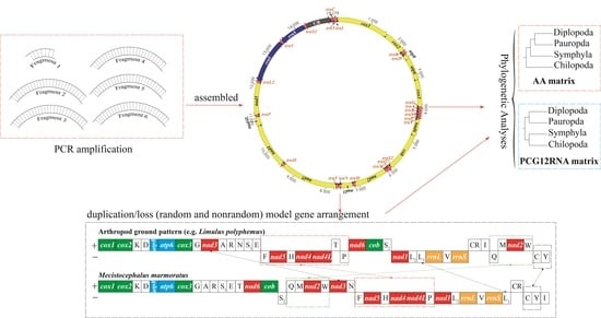
Graphical Abstract

1. Introduction

Centipedes, also known as the Chilopoda (CHI), and their related groups (Diplopoda (DIP), Symphyla (SYM), and Pauropda (PAU)) comprise the subphylum Myriapoda. Most centipedes are fast-moving, have a predatory lifestyle in terrestrial habitats, and possess poisonous modified maxillipeds [1]. Centipedes comprise more than 3000 species in five extant orders: Scutigeromorpha, Lithobiomorpha, Craterostigmomorpha, Scolopendromorpha, and Geophilomorpha [2,3]. In some studies, Strigamia maritima is treated as an ideal model species for ecological and developmental research [4,5]. Recent studies using either comparative morphological or molecular evidence have found that myriapods and all extant myriapod classes are monophyletic [6,7,8,9,10,11,12,13,14,15,16,17,18,19,20]. Most of these analyses support the contention that the Chilopoda represent the basal lineage of the Myriapoda, with the remaining three classes united as the Progoneata.
However, the phylogenetic relationships among the major extant groups of myriapods remain uncertain, and recent debates have focused on the Edafopoda hypothesis (PAU + SYM group) vs. the Dignatha hypothesis (PAU + DIP group). The Dignatha hypothesis has been universally accepted for more than a century [8,10,20,21,22,23,24,25,26], whereas that of Edafopoda is corroborated by nuclear ribosomal genes and some mitochondrial genomes (mitogenomes) [12,14,17]. However, recent phylogenomic analyses based on transcriptomic data showed slightly different results. The results obtained by Szucsich et al., Benavides et al., and Wang et al. are basically the same, demonstrating strong evidence for the clade Pauropoda + Symphyla (=Edafopoda) as well as for Chilopoda + Diplopoda (=Pectinopoda) [19,21,22]. The difference is that Fernández et al. identified two alternative phylogenetic relationships for Symphyla: one that classifies it as a sister group to the Diplopoda + Chilopoda, and one that places Symphyla closer to Dignatha [27].
The relationships between Chilopoda clades have been elucidated using morphological characteristics and molecular analyses, as follows: (1) the five centipede orders are all monophyletic; (2) the basal division, Scutigeromorpha, is a sister group to the four other centipede lineages; and (3) the Scolopendromorpha + Geophilomorpha comprise the clade Epimorpha [11,27,28,29,30]. However, some transcriptome-based phylogenetic analyses did not reveal a sister group relationship between Geophilomorpha and Scolopendromorpha [24], whereas others did [31]. Thus, centipede phylogeny remains a topic that needs further investigation in terms of the position of different centipede lineages within the chilopod orders and the earliest evolutionary splitting events within the centipede lineages.
Arthropod mitogenomes encode 13 protein-coding genes (PCGs), 22 transfer RNA genes (tRNAs), two ribosomal RNA genes (rRNAs), and a long noncoding region (control region, CR), which have been extensively used to study genetics and evolution at multiple hierarchical levels [31,32,33,34,35]. The order of mitochondrial genes can provide additional phylogenetic information because mitochondrial gene rearrangements are generally rare events. Moreover, most mitochondrial gene arrangements are generally stable in arthropods over a long evolutionary period. Wang et al. found two types of gene orders in the Neuropterida [35]. One was the same as the ancestral mitochondrial gene orders of most insects, and the other was the result of a shuffle of trnC to upstream of trnW (trnC-trnW-trnY), an arrangement that is present in all remaining families of Neuroptera. Song et al. reported that various rearrangements of the hotspot region between the CR and cox1 are found in several insects; all species of the Lepidoptera suborder Ditrysia had the arrangement trnM-trnI-trnQ, and most species of Neuroptera had the transposition of trnW and trnC [34]. Most hymenopterans have the trnI, trnQ, and/or trnM genes in different positions; for example, the trnM-trnI-trnQ order is the most common in Formicidae [36,37].
Although taxon sampling has been limited, the mitogenome has provided evolutionary evidence related to the phylogenetic and evolutionary histories of the Myriapoda. At the same time, a variety of gene rearrangement events have been found. The gene orders in the mitogenomes of Cermatobius longicornis (centipede) and Prionobelum sp. (millipede) are identical to those in Limulus polyphemus (Arthropoda: Xiphosura) [38,39,40,41]. All the sequenced mitogenomes of millipedes have a nad6 + cob placement that differs from that of L. polyphemus, except Sphaerotheriida; the pattern of nad6 + cob is believed to be reliable molecular evidence supporting the Helminthomorpha clade, and the inversion of the entire side of a genome (the trnF-nad5-trnH-nad4-nad4L cluster, trnP, the nad1-trnL2-trnL1-rrnL-trnV-rrnS cluster, trnQ, trnC, and trnY) could represent a synapomorphy of a subgroup within Polydesmida [13]. Nine myriapod mitogenomes were compared by Dong et al., who posited that a translocation of trnT from the 5′ end of nad4L was a common event in derived progoneate lineages [12]. Xu et al. sequenced the first Spirobolus mitogenomes and analyzed the phylogenetic relationships within Diplopoda based on 9 orders and 27 species [42].
In previous studies, the mitochondrial gene orders of Scolopendra dehaani, Scolopendra mutilans, S. maritima, Scutigera coleoptrata, and Spirobolus bungii were distinctly different from those of any other myriapod species [42,43,44,45]. A high rate of rearrangement makes the Myriapoda an ideal class group for exploring the interactions between gene rearrangements and phylogenetic relationships. Further sequencing of mitogenomes from additional members of the Chilopoda can demonstrate whether such an extensive rearrangement is unique. Common models that attempt to explain gene rearrangement events and investigate the evolutionary implications of these events involve duplication–random loss (TDRL) and duplication–nonrandom loss (TDNL) as the molecular drivers of gene rearrangement [32].
In this study, complete mitogenomes of M. marmoratus and S. subspinipes were sequenced and annotated, and we used mitogenomes to investigate the gene rearrangement model and the phylogenetic relationships within centipedes, hoping to provide more molecular evidence to explore the relationships within the Myriapoda.

2. Materials and Methods

2.1. Taxon Sampling and Mitochondrial DNA Sequencing

Specimens of M. marmoratus and S. subspinipes were collected from the Langya mountains, Chuzhou, Anhui province, China (32°16′ N, 118°16′ E), in July 2014. They were initially preserved in 100% ethanol in the field and were then transferred to −20 °C conditions for long-term storage at the Molecular Biology Laboratory of Chuzhou University (Chuzhou, China). Genomic DNA was extracted from the dehydrated muscle tissues using DNeasy Blood and Tissue kits (Qiagen, Hilden, Germany). The entire mitogenome was amplified using six primer pairs (Table 1), and all the primers were valid reference primers that had been used for the published species. After using and screening, the six most suitable primer pairs were obtained. Short polymerase chain reaction (PCR) assays (<1.5 kb) were performed using KOD Dash DNA polymerase (Toyobo). The cycling conditions were 94 °C for 5 min; followed by 35 cycles of 30 s at 94 °C, 50 s at 49–52 °C, and 1–2 min at 72 °C (depending on the amplicon size); with a final elongation step at 72 °C for 10 min. Long PCR assays (>1.5 kb) were performed using LA Taq DNA polymerase (TaKaRa). The two-step conditions were as follows: 35 cycles at 96 °C for 2 min and 68 °C for 10 min, followed by incubation at 68 °C for 10 min. The amplified PCR products were electrophoresed on 2% agarose gel, excised, purified, and then analyzed by primer walking on an ABI-PRISM 3730 Automated DNA Sequencer (Applied Biosystems, Waltham, MA, USA).

2.2. Gene Annotation and Secondary Structure Prediction

PCR product sequences were assembled using SeqMan II (DNASTAR Inc., Madison, WI, USA) after checking. Preliminary annotation using the MITOS web server (http://mitos.bioinf.uni-leipzig.de/index.py (accessed on: 10 October 2021)) provided overall information on the mitogenomes [38]. Further annotation of 13 PCGs was performed by identifying their open reading frames and aligning them with homologous genes from other reported myriapod mitogenomes from the NCBI database (https://blast.ncbi.nlm.nih.gov/Blast.cgi (accessed on: 11 October 2021)). tRNA genes were identified by comparing the results predicted using the software programs tRNAscan-SE Search Server v2.0 and ARWEN [48,49]. Based on known gene order information, the boundaries of rrnL (16S rRNA) were assumed to be delimited by the ends of the trnV and trnL2 pair. Further, rrnS (12S rRNA) was assumed to start from the end of trnV, and its end was roughly identified by alignment with the other published millipede sequences. Nucleotide frequencies and codon usage were determined using MEGA X software [50].

2.3. Sequence Alignment and Phylogenetic Analyses

Phylogenetic trees were constructed based on 32 ingroups and 2 outgroups (Table S1), and Priapulus caudatus and Epiperipatus biolleyi (GenBank accession numbers NC_008557 and NC_009082, respectively) were selected as the outgroups in our analyses. Datasets of 2 rRNA sequences and 13 PCG sequences were selected to analyze the phylogenetic relationships within Myriapoda. The sequences of PCG genes were initially aligned using MASCE v2, and rRNA genes were initially aligned using MAFFT with the E-INS-I strategy [51,52]. Poorly aligned positions were subsequently eliminated using Gblock 9.1b, with default settings [53]. Finally, we used MEGA to concatenate all genes and selected their positions to form two datasets: (1) the PCG12RNA matrix and the first and second codon positions of the 13 PCGs and 2 rRNAs, for a total of 7954 bp; and (2) the AA matrix and the amino acid sequences of 13 PCGs, for a total of 3451 aa.
The optimal partition scheme for each dataset and the best model for each partition were determined using Partition Finder 2 (Table S2), with the Akaike information criterion model and a greedy search algorithm with unlinked branch lengths [54]. We analyzed the phylogenetic relationships using the BI and ML methods using the IQ-TREE and MrBayes v3.2.6 under models, respectively [55,56]. For ML analyses, we used an ultrafast bootstrap approximation approach with 1 × 10⁴ replicates, whereas for BI analyses, we used the default settings by simulating four independent runs for 1 × 10⁷–5 × 10⁷ generations and sampling every 100 generations after the average standard deviation of split frequencies fell below 0.001. The first 2000 trees were discarded as burn-in. Three replicates of these BI runs were conducted, all of which produced the same topology.

3. Results and Discussion

3.1. Organization of the Mitogenome

As shown in Figure 1 and Tables S3 and S4, the complete mitogenomes of M. marmoratus (KX774322) and S. subspinipes (MN642577) were sequenced and annotated. The length of the M. marmoratus mitogenome was 15,279 bp, and that of S. subspinipes was 14,637 bp. Both complete mitogenomes included 37 typical mitochondrial genes—two rRNA genes (rrnS (16S rRNA) and rrnL (12S rRNA)), 13 PCGs (cox1-3, cob, nad1-6, nad4L, atp6, and atp8), 22 tRNA genes, and a control region (CR) (Tables S3 and S4). The sizes of these two mitogenomes were within the range reported for known myriapod mitogenomes, from 14,487 bp (Pauropus longiramus) to 16,833 bp (C. longicornis) [13]. The A + T content in the mitogenomes of Chilopoda ranged from 63.4% (C. longicornis) to 78.8% (S. mutilans). The A + T contents in the mitogenomes of M. marmoratus and S. subspinipes were 69.5% and 72.7%, respectively. Additionally, both genomes showed an obvious A + T and C + G bias (Table 2).
The length variation was minimal in the PCGs, with greater variation in the putative CR, intergenic overlaps, and tRNAs. Frequent intergenic overlaps (17/37 = 46%) occurred in the mitogenome of S. subspinipes. The two newly sequenced mitogenomes contained the 37 genes commonly found in metazoan mitogenomes as well as a putative CR, including the presumed origin of replication and promoters for transcription initiation. All PCGs started with ATN. The AT contents of the PCGs of Chilopoda ranged from 59.6% (S. dehaani) [43] to 77.2% (S. mutilans) [41]. The A + T contents of the PCGs in the mitogenomes of M. marmoratus and S. subspinipes were 67.1% and 71.6%, respectively. Additionally, both genomes showed an obvious T + A bias and a slight C + G bias, except S. dehaani, which has an obvious A + T bias, and S. mutilans, which has a slight G + C bias (Table 2).

3.2. Transfer RNAs

The secondary structures of the 22 potential tRNA genes in M. marmoratus and S. subspinipes were predicted and are shown in Figures S1 and S2, respectively. The newly sequenced mitogenome of M. marmoratus revealed the loss of the dihydrouridine arm in trnC and trnS1. Among the 22 tRNAs in the mitogenome of S. subspinipes, trnT and trnP lacked a TΨC loop, and trnS1 lacked the DHU arm. Many tRNA genes in the newly sequenced mitogenomes were shortened, with the shortest tRNAs having only 52 nucleotides. A total of 22 tRNAs ranged in length from 52 bp (trnS1) to 79 bp (trnW) in S. subspinipes, and from 52 bp (trnT) to 73 bp (trnN) in M. marmoratus. This difference was mainly caused by the loop region, particularly the variable loop.

3.3. Phylogenetic Analyses

As in the previous study, the monophyly of the Myriapoda and the three classes (CHI, DIP, and SYM) were verified using phylogenomic analyses [12,13,19,21,22]. This is considered to be uncontroversial; the current controversy is the phylogenetic relationships among these groups, including PAU. However, the phylogenetic relationships among centipedes reported in previous studies differ due to differences in the molecular markers and analysis methods used [16,27,31,43,44]. Our study provided compelling support for Dignatha (DIP + PAU) being the closest relatives, which supported the relationships among the four groups that were suggested based on morphological and some molecular evidence [24], but was in conflict with the results of phylogenomic analyses with transcriptomic data published by Szucsich et al. and Benavides et al. [19,21]. Regrettably, the position of SYM in the phylogenetic tree may be unstable due to the limited number of SYM samples used in this study. In our study, we also observed that different results were obtained from different datasets. In the trees produced using the AA matrix (Figure 2a), Chilopoda was identified as the basal lineage of the remaining myriapods and as a sister group with the Progoneata, in both the BI and ML analyses. The relationship within Progoneata was established as (DIP + PAU) + SYM; this result is consistent with earlier results published by Fernández, Edgecombe, and Giribet and is further consistent with the morphological evidence [24]. The results produced using the PCG12RNA matrix were slightly different (Figure 2b). The topology DIP + PAU, with a sister group of SYM + CHI, was identified with high support based on the BI and ML analyses. Thus, in order to clarify the phylogenetic relationships among the major groups of Myriapoda, we need to develop new methods and increase the sampling richness.
Focusing on the Chilopoda, M. marmoratus and S. maritima grouped into one branch, which corresponds to the Geophilomorpha in all trees. In the Scolopendromorpha, the relationships between S. mutilans, S. dehaani, and S. morsitans were closer than the relationship of any of them to S. subspinipes. Contrary to the phylogenetic relationship, the gene orders of the mitogenomes of S. dehaani and S. subspinipes were closer; trnE is missing in S. dehaani. Thus, to verify the relationship between evolution and gene order within the Scolopendromorpha, we need to systematically add more samples to identify the rules of gene rearrangement and clarify the relationships between genera. In previous studies, Scutigeromorpha was identified as a sister group to three other groups, namely Lithobiomorpha, Geophilomorpha, and Scolopendromorpha [19,27,31]. We found that the relationship between the Lithobiomorpha and Geophilomorpha was closer, although traditional morphological evidence indicates that Geophilomorpha and Scolopendromorpha are more closely related. These relationships identified in the present study are consistent with the conclusions obtained from a previous study [43].
Taxon sampling is essential for the accuracy of phylogenetic inference, and it is important to understand the effect of taxon sampling in a whole-genome or multi-locus phylogenetic study [57]. More Pauropoda should be included, and the selection of different markers will be necessary in future studies to reconstruct a stable phylogenetic topology.

3.4. Evolution of Gene Rearrangements in the Mitochondrial Genome of Centipedes

Previous studies have shown that the mitogenomes of members of the class Chilopoda are characterized by extensive mitochondrial gene rearrangements [40,41,43,44,45,58]. In the newly sequenced mitogenomes, the gene arrangement in S. subspinipes was identical to that in L. polyphemus. Within the Scolopendra, S. dehaani and S. subspinipes have the same gene order, except that S. dehaani does not possess trnE and trnL2. Compared with these two species, five genes or gene blocks in the mitogenome of S. mutilans (trnF-nad4L, trnP-trnS2, nad1-CR, trnV, and trnQ) were rearranged. Compared with ancestral gene orders of myriapods (L. polyphemus), at least eight genes and gene clusters (nad3, nad6-cob, trnM-nad2-trnW, trnT, trnN, trnY, trnL1, and trnI) in the mitogenome of M. marmoratus were rearranged. The overall arrangement of genes in the mitogenome of M. marmoratus was unique compared with that of the mitogenomes of other myriapod species or any other arthropods that have been studied. The same transcriptional polarity genes were clustered together, except trnS2, which overlapped with the genes on the opposite strand.
In order to better understand the evolution of the extensive mitochondrial gene rearrangements of centipedes, the gene orders of published centipede mitogenomes were summarized and mapped on the estimated phylogenetic tree (Figure 3). The gene arrangement of the Lithobiomorpha is relatively conservative, and other classes have been extensively rearranged. At present, the most widely accepted model involves duplication–random loss (TDRL) and duplication–nonrandom loss (TDNL) to explain the molecular drivers of gene rearrangement [59]. A recent study on the pattern of gene rearrangements in Polydesmus sp. GZCS-2019 suggested a new rearrangement model based on three factors: genome-scale duplication, loss, and recombination (TD(N/R) L + C) [21]. This model provides a reference for improving the understanding of the mechanism of gene rearrangement in Myriapoda. However, several unique rearrangement units of M. marmoratus prevent the application of these models to the species.
In this analysis, we assumed that the original mitogenomes of L. polyphemus had an identical gene arrangement, which appears to be ancestral for myriapods and arthropods (Figure 4A). We propose an immature duplication/loss (random and nonrandom) model that resulted in the generation of the mitochondrial gene arrangement of M. marmoratus (Figure 4).
Our hypothesis involves three steps. First, two derivative monomers are arranged in a circular dimer, similar to that found in some myriapods, for example, Narceus annularus, Thyropygus sp., Antrokoreana gracilipes, Trigoniulus corallines, Abacion magnum, Brachycybe lecontii, and Symphylella sp. Subsequently, nonrandom loss occurred according to the transcriptional orientation of each gene. Second, duplication and nonrandom loss events are necessary to explain the translocation of nad3, trnF-nad5-trnH-nad4-nad4L-trnT-trnP, and trnQ-trnM-nad2-trnW (Figure 4B1). In this study, the model of duplication and random loss of genes was used to explain the translocation of the mitochondrial genes trnN, trnL1, and trnI (Figure 4B2) [39]. Thus, the mitochondrial gene arrangement in M. marmoratus was deduced from the original gene order (Figure 4C). Gene density in a duplicated region can be determined via biological constraints rather than by chance. Because mitochondrial gene rearrangements are rare events in animal evolution, they appear to be well-suited for deriving phylogenetic inferences from ancient relationships.
The highly unusual organization of the mitogenome of S. maritima is possibly due to the stem and loop structures. The rare gene cluster with opposite transcriptional polarity in the mitogenome of M. marmoratus suggests that a nonrandom mechanism is involved in generating this gene order. Based on these two sequenced mitogenomes (S. maritima and M. marmoratus) within Geophilomorpha, gene orders are derived using different mechanisms. In order to explain the reason for the unusual organization of each mitogenome within Geophilomorpha, more mitogenomes need to be sequenced in the future.
The gene transfer and gene block arrangements may represent a synapomorphy in the related lineage, resolving the phylogenetic controversy at multiple hierarchical levels [13]. We believe that meaningful evolutionary information can be obtained by comparing the gene order of myriapod species, provided that data on broader taxon sampling are available. Ultimately, the use of a large number of samples would help elucidate the evolutionary details.

4. Conclusions

At present, we report the complete mitogenomes of M. marmoratus (15,279 bp) and S. maritime (14,637 bp) (Myriapoda: Chilopoda). Both mitogenomes contain 37 typical genes, and the gene order of S. subspinipes was the same as that of the original arthropod and myriapod mitogenome (L. polyphemus), whereas M. marmoratus changed considerably. A simple duplication/loss (random and nonrandom) model was proposed to explain the mitochondrial gene arrangement in M. marmoratus; we hope that this model can also reveal mitochondrial gene rearrangement events in other species. Further, we explored the phylogeny of Myriapoda based on mitogenomes. The results suggested the monophyly of the Myriapoda and its main groups and supported Pauropoda and Diplopoda forming the Dignatha, although the relationship between Dignatha and Symphyla needs to be explored further with more systematic evidence. Within the Chilopoda, Scutigeromorpha was a sister group to three other groups: Lithobiomorpha, Geophilomorpha, and Scolopendromorpha. Our study identified a close relationship between Lithobiomorpha and Geophilomorpha. Overall, our results help to better understand the phylogeny of the Myriapoda and explain gene rearrangement events.

Supplementary Materials

The following supporting information can be downloaded at: https://www.mdpi.com/article/10.3390/genes13101787/s1: Figure S1: Predicted secondary structure of the 22 tRNAs in the S. subspinipes mitogenome (dashes (−) indicate Watson–Crick base pairing); Figure S2: Predicted secondary structure of the 22 tRNAs in the M. marmoratus mitogenome (dashes (−) indicate Watson–Crick base pairing); Table S1: GenBank accession numbers for taxa used in this study [12,13,40,40,41,43,44,45,47,58,59,60,61,62,63,64,65,66,67,68,69,70,71,72,73,74,75,76]; Table S2: Optimal partition strategy and evolutionary models used in phylogenetic analyses; Table S3: Mitochondrial genome organization of S. subspinipes; Table S4: Mitochondrial genome organization of M. marmoratus.

Author Contributions

Methodology, Y.B.; software, J.-J.W.; validation, J.-J.W., Y.B. and Y.D.; formal analysis, J.-J.W.; investigation, J.-J.W.; resources, Y.D.; data curation, Y.B.; writing—original draft preparation, J.-J.W.; writing—review and editing, Y.D.; project administration, Y.D. All authors have read and agreed to the published version of the manuscript.

Funding

This research was funded by the Natural Science Foundation of the Higher Education Institutions of Anhui Province, China, grant number KJ2018ZD041; and the Key Program in the Youth Elite Support Plan in Universities of Anhui Province, China, grant number gxyqZD2020045; the National Science and Technology Fundamental Resources Investigation Program of China, grant number 2019FY101806.

Institutional Review Board Statement

Not applicable.

Informed Consent Statement

Not applicable.

Data Availability Statement

The newly sequenced mitogenomes were submitted to the GenBank database under the accession numbers of M. marmoratus (KX774322) and S. subspinipes (MN642577).

Conflicts of Interest

The authors declare no conflict of interest.

References

  1. Minelli, A. The Myriapoda, Volume 1, Chapter: Chilopoda. In The Myriapoda; Minelli, A., Ed.; Brill: Leiden, The Netherlands, 2011; pp. 1–20. [Google Scholar]
  2. Edgecombe, G.D.; Giribet, G. Evolutionary biology of centipedes (Myriapoda: Chilopoda). Annu. Rev. Entomol. 2007, 52, 151–170. [Google Scholar] [CrossRef] [PubMed] [Green Version]
  3. So, W.L.; Nong, W.Y.; Xie, Y.C.; Baril, T.; Ma, H.Y.; Qu, Z.; Haimovitz, J.; Swale, T.; Gaitan-Espitia, J.D.; Lau, K.F.; et al. Myriapod genomes reveal ancestral horizontal gene transfer and hormonal gene loss in millipedes. Nat. Commun. 2022, 13, 3010. [Google Scholar] [CrossRef]
  4. Arthur, W.; Chipman, A.D. The centipede Strigamia maritima: What it can tell us about the development and evolution of segmentation. Bioessays 2005, 27, 653–660. [Google Scholar] [CrossRef] [PubMed]
  5. Brena, C.; Akam, M. The embryonic development of the centipede Strigamia maritima. Dev. Biol. 2012, 363, 290–307. [Google Scholar] [CrossRef] [Green Version]
  6. Green, J.; Akam, M. Evolution of the pair rule gene network: Insights from a centipede. Dev. Biol. 2013, 382, 235–245. [Google Scholar] [CrossRef] [PubMed] [Green Version]
  7. Ax, P. Ein Lehrbuch der Phylogenetischen Systematik; Gustav Fischer Verlag: Stuttgart, Germany, 1999; p. 384. [Google Scholar]
  8. Bäcker, H.; Fanenbruck, M.; Wägele, J.W. A forgotten homology supporting the monophyly of Tracheata: The subcoxa of insects and myriapods revisited. Zool. Anz. J. Comp. Zool. 2008, 247, 185–207. [Google Scholar] [CrossRef]
  9. Bitsch, C.; Bitsch, J. Phylogenetic relationships of basal hexapods among the mandibulate arthropods: A cladistic analysis based on comparative morphological characters. Zool. Scr. 2004, 33, 511–550. [Google Scholar] [CrossRef]
  10. Boudreaux, H. Significance of intersegmental tendon system in arthropod phylogeny and a monophyletic classification of Arthropoda. In Arthropod Phylogeny; Van Nostrand Reinhold: New York, NY, USA, 1979; pp. 551–586. [Google Scholar]
  11. Chipman, A.D.; Ferrier, D.E.; Brena, C.; Qu, J.; Hughes, D.S.; Schröder, R.; Torres-Oliva, M.; Znassi, N.; Jiang, H.; Almeida, F.C.; et al. The first myriapod genome sequence reveals conservative arthropod gene content and genome organisation in the centipede Strigamia maritima. PLoS Biol. 2014, 12, e1002005. [Google Scholar] [CrossRef] [Green Version]
  12. Dong, Y.; Sun, H.; Guo, H.; Pan, D.; Qian, C.; Hao, S.; Zhou, K. The complete mitochondrial genome of Pauropus longiramus (Myriapoda: Pauropoda): Implications on early diversification of the myriapods revealed from comparative analysis. Gene 2012, 505, 57–65. [Google Scholar] [CrossRef]
  13. Dong, Y.; Zhu, L.; Bai, Y.; Ou, Y.; Wang, C. Complete mitochondrial genomes of two flat-backed millipedes by next-generation sequencing (Diplopoda, Polydesmida). ZooKeys 2016, 637, 1–20. [Google Scholar] [CrossRef]
  14. Gai, Y.H.; Song, D.X.; Sun, H.Y.; Zhou, K.Y. Myriapod monophyly and relationships among myriapod classes based on nearly complete 28S and 18S rDNA sequences. Zool. Sci. 2006, 23, 1101–1108. [Google Scholar] [CrossRef] [PubMed]
  15. Jamieson, B.G.; Jamieson, J.B.G. The Ultrastructure and Phylogeny of Insect Spermatozoa; Cambridge University Press: New York, NY, USA, 1987. [Google Scholar]
  16. Regier, J.C.; Shultz, J.W.; Ganley, A.R.; Hussey, A.; Shi, D.; Ball, B.; Zwick, A.; Stajich, J.E.; Cummings, M.P.; Martin, J.W.; et al. Resolving arthropod phylogeny: Exploring phylogenetic signal within 41 kb of protein-coding nuclear gene sequence. Syst. Biol. 2008, 57, 920–938. [Google Scholar] [CrossRef] [PubMed]
  17. Regier, J.C.; Shultz, J.W.; Zwick, A.; Hussey, A.; Ball, B.; Wetzer, R.; Martin, J.W.; Cunningham, C.W. Arthropod relationships revealed by phylogenomic analysis of nuclear protein-coding sequences. Nature 2010, 463, 1079–1083. [Google Scholar] [CrossRef] [PubMed]
  18. Regier, J.C.; Wilson, H.M.; Shultz, J.W. Phylogenetic analysis of Myriapoda using three nuclear protein-coding genes. Mol. Phylogenet. Evol. 2005, 34, 147–158. [Google Scholar] [CrossRef]
  19. Szucsich, N.U.; Bartel, D.; Blanke, A.; Böhm, A.; Donath, A.; Fukui, M.; Grove, S.; Liu, S.; Macek, O.; Machida, R.; et al. Four myriapod relatives—But who are sisters? No end to debates on relationships among the four major myriapod subgroups. BMC Evol. Biol. 2020, 20, 144. [Google Scholar] [CrossRef]
  20. Zuo, Q.; Zhang, Z.; Shen, Y. Novel mitochondrial gene rearrangements pattern in the millipede Polydesmus sp. GZCS-2019 and phylogenetic analysis of the Myriapoda. Ecol. Evol. 2022, 12, e8764. [Google Scholar] [CrossRef]
  21. Benavides, L.R.; Edgecombe, G.D.; Giribet, G. Re-evaluating and dating myriapod diversification with phylotranscriptomics under a regime of dense taxon sampling. Mol. Phylogenet. Evol. 2022, 178, 107621. [Google Scholar] [CrossRef]
  22. Wang, J.J.; Bai, Y.; Zhao, H.; Mu, R.; Dong, Y. Reinvestigating the phylogeny of Myriapoda with more extensive taxon sampling and novel genetic perspective. PeerJ 2021, 9, e12691. [Google Scholar] [CrossRef]
  23. Edgecombe, G.D.; Wilson, G.D.; Colgan, D.J.; Gray, M.R.; Cassis, G. Arthropod cladistics: Combined analysis of histone H3 and U2 snRNA sequences and morphology. Cladistics 2000, 16, 155–203. [Google Scholar] [CrossRef]
  24. Fernández, R.; Edgecombe, G.D.; Giribet, G. Phylogenomics illuminates the backbone of the Myriapoda Tree of Life and reconciles morphological and molecular phylogenies. Sci. Rep. 2018, 8, 83. [Google Scholar] [CrossRef]
  25. Pocock, R.I. Contributions to our knowledge of the arthropod fauna of the West Indies. Part III. Diplopoda and Malacopoda, with a supplement on the Arachnida of the class Pedipalpi. Zool. J. Linn. Soc. 1894, 24, 473–544. [Google Scholar] [CrossRef]
  26. Tiegs, O.W. The development and affinities of the Pauropoda, based on a study of Pauropus silvaticus. J. Cell Sci. 1947, 3, 275–336. [Google Scholar] [CrossRef]
  27. Fernández, R.; Edgecombe, G.D.; Giribet, G. Exploring phylogenetic relationships within Myriapoda and the effects of matrix composition and occupancy on phylogenomic reconstruction. Syst. Biol. 2016, 65, 871–889. [Google Scholar] [CrossRef]
  28. Edgecombe, G.D. Morphological data, extant Myriapoda, and the myriapod stem-group. Contrib. Zool. 2004, 73, 207–252. [Google Scholar] [CrossRef] [Green Version]
  29. Edgecombe, G.D.; Giribet, G.; Wheeler, W.C. Phylogeny of Henicopidae (Chilopoda: Lithobiomorpha): A combined analysis of morphology and five molecular loci. Syst. Entomol. 2002, 27, 31–64. [Google Scholar] [CrossRef] [Green Version]
  30. Giribet, G.; Carranza, S.; Riutort, M.; Baguna, J.; Ribera, C. Internal phylogeny of the Chilopoda (Myriapoda, Arthropoda) using complete 18S rDNA and partial 28S rDNA sequences. Philos. Trans. R. Soc. Lond. B Biol. Sci. 1999, 354, 215–222. [Google Scholar] [CrossRef] [PubMed]
  31. Benavides, L.R.; Jiang, C.; Giribet, G. Mimopidae is the sister group to all other scolopendromorph centipedes (Chilopoda, Scolopendromorpha): A phylotranscriptomic approach. Org. Divers. Evol. 2021, 21, 591–598. [Google Scholar] [CrossRef]
  32. Boore, J.L. Animal mitochondrial genomes. Nucleic Acids Res. 1999, 27, 1767–1780. [Google Scholar] [CrossRef] [Green Version]
  33. Cameron, S.L. Insect mitochondrial genomics: Implications for evolution and phylogeny. Annu. Rev. Entomol. 2014, 59, 95–117. [Google Scholar] [CrossRef] [Green Version]
  34. Song, F.; Li, H.; Shao, R.; Shi, A.; Bai, X.; Zheng, X.; Heiss, E.; Cai, W. Rearrangement of mitochondrial tRNA genes in flat bugs (Hemiptera: Aradidae). Sci. Rep. 2016, 6, 25725. [Google Scholar] [CrossRef]
  35. Wang, Y.; Liu, X.; Garzón-Orduña, I.J.; Winterton, S.L.; Yan, Y.; Aspöck, U.; Aspöck, H.; Yang, D. Mitochondrial phylogenomics illuminates the evolutionary history of Neuropterida. Cladistics 2017, 33, 617–636. [Google Scholar] [CrossRef] [PubMed]
  36. Dowton, M.; Cameron, S.L.; Dowavic, J.I.; Austin, A.D.; Whiting, M.F. Characterization of 67 mitochondrial tRNA gene rearrangements in the Hymenoptera suggests that mitochondrial tRNA gene position is selectively neutral. Mol. Biol. Evol. 2009, 26, 1607–1617. [Google Scholar] [CrossRef] [PubMed] [Green Version]
  37. Ruiz-Mena, A.; Mora, P.; Montiel, E.E.; Palomeque, T.; Lorite, P. Complete Nucleotide Sequence of the Mitogenome of Tapinoma ibericum (Hymenoptera: Formicidae: Dolichoderinae), Gene Organization and Phylogenetics Implications for the Dolichoderinae Subfamily. Genes 2022, 13, 1325. [Google Scholar] [CrossRef]
  38. Bernt, M.; Donath, A.; Jühling, F.; Externbrink, F.; Florentz, C.; Fritzsch, G.; Pütz, J.; Middendorf, M.; Stadler, P.F. MITOS: Improved de novo metazoan mitochondrial genome annotation. Mol. Phylogenet. Evol. 2013, 69, 313–319. [Google Scholar] [CrossRef] [PubMed]
  39. Dong, Y.; Xu, J.J.; Hao, S.J.; Sun, H.Y. The complete mitochondrial genome of the giant pill millipede, Sphaerotheriidae sp. (Myriapoda: Diplopoda: Sphaerotheriida). Mitochondrial DNA 2012, 23, 333–335. [Google Scholar] [CrossRef] [PubMed]
  40. Gai, Y.; Ma, H.; Ma, J.; Li, C.; Yang, Q. The complete mitochondrial genome of Scolopocryptops sp. (Chilopoda: Scolopendromorpha: Scolopocryptopidae). Mitochondrial DNA 2014, 25, 192–193. [Google Scholar] [CrossRef]
  41. Lavrov, D.V.; Brown, W.M.; Boore, J.L. A novel type of RNA editing occurs in the mitochondrial tRNAs of the centipede Lithobius forficatus. Proc. Natl. Acad. Sci. USA 2000, 97, 13738–13742. [Google Scholar] [CrossRef] [Green Version]
  42. Xu, H.; Fang, Y.; Cao, G.; Shen, C.; Liu, H.; Ruan, H. The Complete Mitochondrial Genome of Spirobolus bungii (Diplopoda, Spirobolidae): The First Sequence for the Genus Spirobolus. Genes 2022, 13, 1587. [Google Scholar] [CrossRef]
  43. Hu, C.; Wang, S.; Huang, B.; Liu, H.; Xu, L.; Hu, Z.; Liu, Y. The complete mitochondrial genome sequence of Scolopendra mutilans L. Koch, 1878 (Scolopendromorpha, Scolopendridae), with a comparative analysis of other centipede genomes. ZooKeys 2020, 925, 73–88. [Google Scholar] [CrossRef] [Green Version]
  44. Sun, L.; Qi, Y.; Tian, X. Analysis of mitochondrial genome of Scolopendra subspinipes dehaani. Tianjin J. Tradit. Chin. Med. 2018, 35, 225–229. [Google Scholar]
  45. Robertson, H.E.; Lapraz, F.; Rhodes, A.C.; Telford, M.J. The complete mitochondrial genome of the geophilomorph centipede Strigamia maritima. PLoS ONE 2015, 10, e0121369. [Google Scholar] [CrossRef] [PubMed]
  46. Folmer, O.; Black, M.; Hoeh, W.; Lutz, R.; Vrijenhoek, R. DNA primers for amplification of mitochondrial cytochrome c oxidase subunit I from diverse metazoan invertebrates. Mol. Mar. Biol. Biotechnol. 1994, 3, 294–299. [Google Scholar] [PubMed]
  47. Gai, Y.; Song, D.; Sun, H.; Yang, Q.; Zhou, K. The complete mitochondrial genome of Symphylella sp. (Myriapoda: Symphyla): Extensive gene order rearrangement and evidence in favor of Progoneata. Mol. Phylogenet. Evol. 2008, 49, 574–585. [Google Scholar] [CrossRef] [PubMed]
  48. Chan, P.P.; Lin, B.Y.; Mak, A.J.; Lowe, T.M. tRNAscan-SE 2.0: Improved detection and functional classification of transfer RNA genes. Nucleic Acids Res. 2021, 49, 9077–9096. [Google Scholar] [CrossRef] [PubMed]
  49. Laslett, D.; Canbäck, B. ARWEN: A program to detect tRNA genes in metazoan mitochondrial nucleotide sequences. Bioinformatics 2008, 24, 172–175. [Google Scholar] [CrossRef] [PubMed] [Green Version]
  50. Kumar, S.; Stecher, G.; Li, M.; Knyaz, C.; Tamura, K. MEGA X: Molecular evolutionary genetics analysis across computing platforms. Mol. Biol. Evol. 2018, 35, 1547–1549. [Google Scholar] [CrossRef]
  51. Katoh, K.; Rozewicki, J.; Yamada, K.D. MAFFT online service: Multiple sequence alignment, interactive sequence choice and visualization. Brief. Bioinform. 2019, 20, 1160–1166. [Google Scholar] [CrossRef] [Green Version]
  52. Ranwez, V.; Douzery, E.J.; Cambon, C.; Chantret, N.; Delsuc, F. MACSE v2: Toolkit for the alignment of coding sequences accounting for frameshifts and stop codons. Mol. Biol. Evol. 2018, 35, 2582–2584. [Google Scholar] [CrossRef]
  53. Talavera, G.; Castresana, J. Improvement of phylogenies after removing divergent and ambiguously aligned blocks from protein sequence alignments. Syst. Biol. 2007, 56, 564–577. [Google Scholar] [CrossRef] [Green Version]
  54. Lanfear, R.; Frandsen, P.B.; Wright, A.M.; Senfeld, T.; Calcott, B. PartitionFinder 2: New methods for selecting partitioned models of evolution for molecular and morphological phylogenetic analyses. Mol. Biol. Evol. 2017, 34, 772–773. [Google Scholar] [CrossRef] [Green Version]
  55. Huelsenbeck, J.P.; Ronquist, F. MrBayes: Bayesian inference of phylogenetic trees. Bioinformatics 2001, 17, 754–755. [Google Scholar] [CrossRef] [PubMed]
  56. Nguyen, L.T.; Schmidt, H.A.; Von Haeseler, A.; Minh, B.Q. IQ-TREE: A fast and effective stochastic algorithm for estimating maximum-likelihood phylogenies. Mol. Biol. Evol. 2015, 32, 268–274. [Google Scholar] [CrossRef] [PubMed]
  57. Nabhan, A.R.; Sarkar, I.N. The impact of taxon sampling on phylogenetic inference: A review of two decades of controversy. Brief. Bioinform. 2012, 13, 122–134. [Google Scholar] [CrossRef] [Green Version]
  58. Negrisolo, E.; Minelli, A.; Valle, G. The mitochondrial genome of the house centipede Scutigera and the monophyly versus paraphyly of myriapods. Mol. Biol. Evol. 2004, 21, 770–780. [Google Scholar] [CrossRef] [PubMed]
  59. Lavrov, D.V.; Boore, J.L.; Brown, W.M. Complete mtDNA sequences of two millipedes suggest a new model for mitochondrial gene rearrangements: Duplication and nonrandom loss. Mol. Biol. Evol. 2002, 19, 163–169. [Google Scholar] [CrossRef] [Green Version]
  60. Park, S.J.; Choi, E.H.; Hwang, J.S.; Hwang, U.W. The complete mitochondrial genome of a centipede Bothropolys sp. (Chilopoda, Lithobiomorpha, Lithobiidae). Mitochondrial DNA Part A 2016, 27, 2268–2269. [Google Scholar]
  61. Gai, Y.; Ma, H.; Sun, X.; Ma, J.; Li, C.; Yang, Q. The complete mitochondrial genome of Cermatobius longicornis (Chilopoda: Lithobiomorpha: Henicopidae). Mitochondrial DNA 2013, 24, 331–332. [Google Scholar] [CrossRef]
  62. Woo, H.J.; Lee, Y.S.; Park, S.J.; Lim, J.T.; Jang, K.H.; Choi, E.H.; Choi, Y.G.; Hwang, U.W. Complete mitochondrial genome of a troglobite millipede Antrokoreana gracilipes (Diplopoda, Juliformia, Julida), and juliformian phylogeny. Mol. Cells 2007, 23, 182–191. [Google Scholar]
  63. Brewer, M.S.; Swafford, L.; Spruill, C.L.; Bond, J.E. Arthropod phylogenetics in light of three novel millipede (Myriapoda: Diplopoda) mitochondrial genomes with comments on the appropriateness of mitochondrial genome sequence data for inferring deep level relationships. PLoS ONE 2013, 8, e68005. [Google Scholar] [CrossRef]
  64. Podsiadlowski, L.; Kohlhagen, H.; Koch, M. The complete mitochondrial genome of Scutigerella causeyae (Myriapoda: Symphyla) and the phylogenetic position of Symphyla. Mol. Phylogenet. Evol. 2007, 45, 251–260. [Google Scholar] [CrossRef]
  65. Park, S.J.; Lee, Y.S.; Hwang, U.W. The complete mitochondrial genome of the sea spider Achelia bituberculata (Pycnogonida, Ammotheidae): Arthropod ground pattern of gene arrangement. BMC Genom. 2007, 8, 343. [Google Scholar] [CrossRef] [PubMed] [Green Version]
  66. Masta, S.E.; Boore, J.L. Parallel evolution of truncated transfer RNA genes in arachnid mitochondrial genomes. Mol. Biol. Evol. 2008, 25, 949–959. [Google Scholar] [CrossRef] [PubMed] [Green Version]
  67. Lavrov, D.V.; Boore, J.L.; Brown, W.M. The complete mitochondrial DNA sequence of the horseshoe crab Limulus polyphemus. Mol. Biol. Evol. 2000, 17, 813–824. [Google Scholar] [CrossRef] [PubMed]
  68. Shingate, P.; Ravi, V.; Prasad, A.; Tay, B.H.; Venkatesh, B. Chromosome-level genome assembly of the coastal horseshoe crab (Tachypleus gigas). Mol. Ecol. Resour. 2020, 20, 1748–1760. [Google Scholar] [CrossRef]
  69. Wilson, K.; Cahill, V.; Ballment, E.; Benzie, J. The complete sequence of the mitochondrial genome of the crustacean Penaeus monodon: Are malacostracan crustaceans more closely related to insects than to branchiopods? Mol. Biol. Evol. 2000, 17, 863–874. [Google Scholar] [CrossRef] [Green Version]
  70. Lavrov, D.V.; Brown, W.M.; Boore, J.L. Phylogenetic position of the Pentastomida and (pan) crustacean relationships. Proc. R. Soc. Lond. Ser. B Biol. Sci. 2004, 271, 537–544. [Google Scholar] [CrossRef]
  71. Podsiadlowski, L. The mitochondrial genome of the bristletail Petrobius brevistylis (Archaeognatha: Machilidae). Insect Mol. Biol. 2006, 15, 253–258. [Google Scholar] [CrossRef]
  72. Cook, C.E.; Yue, Q.; Akam, M. Mitochondrial genomes suggest that hexapods and crustaceans are mutually paraphyletic. Proc. R. Soc. B Biol. Sci. 2005, 272, 1295–1304. [Google Scholar] [CrossRef] [Green Version]
  73. Clary, D.O.; Wolstenholme, D.R. The ribosomal RNA genes of Drosophila mitochondrial DNA. Nucleic Acids Res. 1985, 13, 4029–4045. [Google Scholar] [CrossRef] [Green Version]
  74. Carapelli, A.; Nardi, F.; Dallai, R.; Boore, J.; Lio, P.; Frati, F. Relationships between hexapods and crustaceans based on four mitochondrial genes. Crustacean Issues 2005, 16, 295. [Google Scholar]
  75. Webster, B.L.; Copley, R.R.; Jenner, R.A.; Mackenzie-Dodds, J.A.; Bourlat, S.J.; Rota-Stabelli, O.; Littlewood, D.; Telford, M.J. Mitogenomics and phylogenomics reveal priapulid worms as extant models of the ancestral Ecdysozoan. Evol. Dev. 2006, 8, 502–510. [Google Scholar] [CrossRef] [PubMed]
  76. Ding, J.; Lan, H.; Xu, W.; Chen, Y.N.; Wu, H.; Jiang, H.M.; Wang, J.C.; Wu, Y.B.; Liu, H. Two complete mitochondrial genomes in Scolopendra and a comparative analysis of tRNA rearrangements in centipedes. Mol. Biol. Rep. 2022, 49, 6173–6180. [Google Scholar] [CrossRef] [PubMed]
Figure 1. Circular map of the mitogenomes of S. subspinipes and M. marmoratus.
Genes 13 01787 g001
Figure 2. (a) Phylogenetic trees of myriapods inferred using Bayesian inference (BI) and the maximum likelihood method (ML) based on the AA matrix. Numbers on the branches indicate the posterior probability (BI) (left) and bootstrap values (ML) (right). Newly sequenced taxa are indicated using red stars. (b) Phylogenetic trees of myriapods inferred using the MrBayes program with the maximum likelihood method based on the PCG12RNA matrix. Numbers on the branches indicate the posterior probability (BI) (left) and bootstrap values (ML) (right). Newly sequenced taxa are indicated using red stars.
Genes 13 01787 g002aGenes 13 01787 g002b
Figure 3. Gene arrangements of the Chilopoda mitochondrial genomes. The mitogenomes have been linearized for ease of comparison and arbitrarily begin with cox1 when possible. Different genes are shown in different colors. Underlined labels indicate that the gene was transcribed from the minority strand.
Genes 13 01787 g003
Figure 4. Inferred intermediate steps between Limulus polyphemus and M. marmoratus. The lost genes are labeled in gray. (A) The ancestral gene arrangement of the myriapod. (B1) Two monomers derived from the duplication of the ancestor arranged in a circular dimer. Subsequently, nonrandom loss occurred according to the orientation of transcription for each gene. (B2) Tandem duplication followed by the random loss of genes and the translocation of tRNAs. (C) Final gene orders of the M. marmoratus mitogenome.
Genes 13 01787 g004
Table 1. PCR primers used in this study.
Primer NameNucleotide Sequence (5′-3′)PCR Amplification Product LengthReference
CO1CFGCACGTCTACAAATCATAAAGATATTGG0.7 kb[46]
CO1CRTAAACTTCAGGGTGACCGAAAAATCA
Lco1TTATAATTTTTTTTATAGTGATACC3.7 kb[12]
CO3RACATCTACAAAATGTCAGTATCA[47]
Dco3FTATCATCCTATCAATGATGACGAGA3.7 kb[12]
Dn4RATTTATGATTACCTAAGGCTCATG
Dn4FATGAACAACAGAAGAATAAGC2.9 kb
HcobGCAAATAAAAAATATCATTCTGGTTG
DcobFATAATAACCGCCTTCTTGGGAT3.4 kb
D12SRCTGTTTCTGAATCGATATTCCACGTTT
D12SFATAATAGGGTATCTAATCCTAGTCT2.7 kb
Dco1RATGGGGGATATACGGTCCATCCGG
Table 2. Nucleotide composition of the mitogenomes and protein-coding genes of Chilopoda.
SpeciesMitochondrial GenomePCGs
A + TAT SkewGC SkewA + TAT SkewGC Skew
Bothropolys sp.70.60.07−0.3168.6−0.13−0.06
C.s longicornis63.40.09−0.3260.5−0.17−0.05
Lithobius forficatus67.90.09−0.2765.7−0.14−0.03
M. marmoratus69.50.12−0.3367.1−0.11−0.02
S. maritima64.10.22−0.3362.1−0.12−0.02
S. dehaani74.10.13−0.3459.6−0.12−0.01
S. mutilans78.80.05−0.2677.2−0.12−0.04
S. subspinipes72.70.06−0.3371.6−0.140
Scolopendra morsitans72.80.02−0.3871.6−0.14−0.02
Scolopocryptops sp.71.60.03−0.3170.4−0.14−0.01
S. coleoptrata69.40.04−0.3168.3−0.14−0.05
Publisher’s Note: MDPI stays neutral with regard to jurisdictional claims in published maps and institutional affiliations.