Article

Responses of Humoral and Cellular Immune Mediators in BALB/c Mice to LipX (PE11) as Seed Tuberculosis Vaccine Candidates

by
Andriansjah Rukmana
1,*,
Lulut Azmi Supardi
2,
Fithriyah Sjatha
1 and
Mifa Nurfadilah
2
1
Department of Microbiology, Faculty of Medicine, Universitas Indonesia, Jakarta 10320, Indonesia
2
Masters’s Programme in Biomedical Sciences, Faculty of Medicine, Universitas Indonesia, Jakarta 10430, Indonesia
*
Author to whom correspondence should be addressed.
Genes 2022, 13(11), 1954; https://doi.org/10.3390/genes13111954
Submission received: 9 September 2022 / Revised: 17 October 2022 / Accepted: 24 October 2022 / Published: 26 October 2022
(This article belongs to the Special Issue Genetics and Genomics of Human Pathogens)

Abstract

:
A member of the pe/ppe gene family, lipX (pe11), is capable of directing persistent Mycobacterium tuberculosis and avoiding host immune responses. Some studies have indicated that LipX (PE11) can detect humoral antibodies in tuberculosis patients. Hence, information on immune mediators’ responses to this protein is essential to understand its protective efficacy against M. tuberculosis infections. This study aimed to examine the response of immune mediators to pCDNA3.1-lipX expression in vivo. In the experiment, pCDNA3.1-lipX was injected into BALB/c strain male mice aged between 6 and 8 weeks, and they were compared to groups injected with pCDNA3.1 and without injection. The injection was carried out three times intramuscularly every two weeks. Blood was taken retro-orbitally and used for humoral response analysis by Western blotting against LipX-His protein. Simultaneously, the splenocytes were cultured and induced with LipX-His protein for cellular immunity analyses. Our study showed that the recombinant DNA of pCDNA3.1-lipX induced a humoral and cellular immune response, especially in IL-4, IL-12, and IFN-γ, which are the primary cellular responses to M. tuberculosis infections. However, additional studies, such as a challenge study, are needed to strengthen the argument that this plasmid construction is feasible as a tuberculosis seed vaccine candidate.

1. Introduction

Tuberculosis (TB) is an infectious disease that has become a persistent global health problem, especially in developing countries. The World Health Organization (WHO) reports 10.4 million new TB cases annually, with a mortality rate of 1.8 million worldwide [1,2,3]. M. tuberculosis infection is the tenth-highest cause of death in the world. In Indonesia, it is estimated that one million new TB cases occur every year [2,4].
The most effective intervention for suppressing TB cases is vaccination, rather than drugs. The anti-TB vaccine currently being used is Bacillus Calmette–Guerin (BCG) [5,6,7]. However, the BCG vaccine is less able to protect adults and immunocompromised or HIV-positive individuals [5]. HIV-positive cases often result in coinfection with TB [4,5,6,8]. The BCG vaccine has varying efficacy against pulmonary tuberculosis infection, especially in developing countries and populations infected by the M. tuberculosis Beijing strain [1,6,9]. Thus, a study of new TB vaccine candidates is needed [8,10].
DNA vaccines consist of vectors that contain specific antigen-encoding proteins from specific microbes [8]. DNA vaccines are a safer form of vaccine to give to various people. This kind of vaccine can stimulate the host’s humoral and cellular immune responses to various diseases [11,12,13,14]. The pcDNA3.1 vector is a plasmid commonly used in recombinant protein expression in mammalian cells. This plasmid is also widely used to develop recombinant DNA-based vaccines for infectious diseases and other purposes, such as cancer gene therapy [15,16,17,18]. To construct a DNA vaccine, specific antigens that can induce an immune response are needed [19].
The family of pe/ppe genes occupies 8–10% of the M. tuberculosis DNA genome. This family is predicted to contribute to the level of M. tuberculosis virulence [20,21,22,23]. One member of this family, lipX (pe11), is known to have a significant role in directing the occurrence of infection, and it is considered one of M. tuberculosis’s main factors in hindering host immune evasion, especially by macrophage cells [24,25]. LipX is present in some pathogenic types of Mycobacterium, like M. tuberculosis, M. bovis, and CDC1551, a clinical isolate, but is absent in nonpathogenic strains of M. smegmatis [25]. In silico analysis has shown that LipX can induce the adaptive host immune response, which enables its other potential function as a biomarker in extrapulmonary TB in children [26].
M. tuberculosis infection control depends on the development of an adaptive immune response in the humoral and cellular stages at the site of infection in the host [27]. Immunity to TB infection is predominantly mediated by cellular immune responses and interactions between CD4+ lymphocytes and macrophage cells. These cells interact with each other via a complex network of cytokines. T helper 1 (Th1) cells are considered necessary because they produce cytokines IL-2 and IFN-γ, thus enabling the host to fight Mycobacterium infection by activating macrophages. IL-12 is a cytokine that is important in the maturation and development of lymphocytes. In contrast, Th2 secretes IL-4 to regulate lesions. The IL-10 from regulated cells inhibits the activity of CD4+, IL-12, and macrophages and then induces the host to fail against infection [28,29].
In this study, we analyzed the cellular immune response of IFN-γ, IL-4, IL-10, and IL-12 to LipX protein in a BALB/c mice strain. Mice were used to evaluate the efficacy of this new vaccine candidate because of their low cost, feasibility of use in the laboratory, very well-characterized immune system, and abundant available commercial reagents [30]. Following BCG immunization, as a resistant mouse strain, BALB/c showed protective effects induced by the vaccine, which also occur in humans [31]. Mouse models can tell us about the immune mechanisms associated with the effectiveness and application of vaccines in humans.

2. Materials and Methods

2.1. Strains

M. tuberculosis Beijing and the top 10 Escherichia coli strains were used in this study. Both strains are culture stocks of the Microbiology Department, Faculty of Medicine, Universitas Indonesia.

2.2. Plasmid and Protein Recombinants

We used recombinant plasmids pET28a-lipX (unpublished data) and pCDNA3.1-lipX, which were constructed in our previous work [32]. All plasmids are stocks of the Department of Microbiology, Faculty of Medicine, Universitas Indonesia (Appendix A).

2.3. LipX-His Protein Isolation

The E. coli BL21 strain harboring pET28a-lipX was grown in Luria Bertani (LB) medium (merck) and induced by 1 mM of IPTG for 4 h in a shaker incubator at 37 °C at 200 rpm, which was followed by protein isolation using an Ni-NTA affinity column (Qiagen). Isolated LipX-His protein was then confirmed by Western blot using mouse antibody monoclonal anti-His (Thermo Fisher Scientific, Waltham, MA, USA). The recombinant LipX-His protein was stored for further use in the analysis of the humoral response and as an inducer of a cellular immune response from mice immunized with pcDNA3.1-lipX recombinant DNA.

2.4. Sample Size and Animal Care

This experiment used the BALB/c mouse strain from AniLab Bogor. The mice were male and aged 6–8 weeks. The number of test animals used for each test group was determined using the Federer formula. Federer’s formula is (n − 1) (t − 1) > 15, where the t value is the number of treatment groups and the n value is the number of animals per treatment group. Based on this formula, three groups were formed, and each group consisted of eight mice. However, according to the ethics committee’s advice, the number of mice was reduced to six in each group. The mice were grouped without randomization due to having the same characteristics. The three groups included the pcDNA3.1-lipX group, the pcDNA3.1 immunization group, and the without immunization group. All test animals underwent a process of acclimatization for two weeks in a cage regulated for temperature and air circulation. The maintenance of the cage was carried out twice a week, and food and drink were provided ad libitum. Retro-orbital blood collection, spleen isolation, and cage cleaning were carried out sequentially and carried out one by one between groups to avoid mixing mice and causing data collection errors from each group.

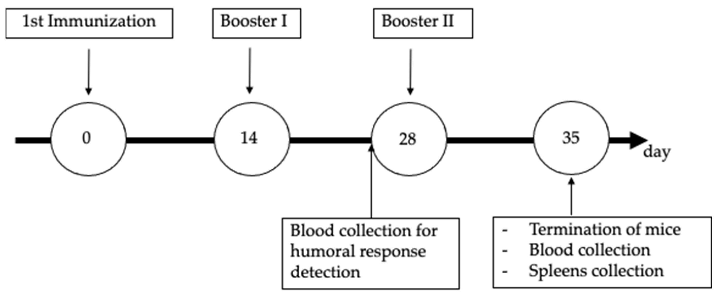
2.5. Procedure for Mouse Immunization and Blood Collection

Six experimental mice in each group were immunized with 100 μg of pcDNA3.1-lipX or 100 μg of pcDNA3.1 in 100 μL of saline per injection, respectively, and one group was not immunized. Immunization was carried out intramuscularly using a 27G syringe three times within two weeks, and the mice were terminated one week after the last immunization (Appendix B). The protein concentration and duration of immunization until termination were based on a previous study [33,34]. The mice were anesthetized with ketamine-xylazine collection before every retro-orbital blood collection and immunization. Sera from blood collected before the third immunization (booster II) were analyzed for their humoral immune response against LipX-His protein through Western blot, and splenocytes from terminated mice were analyzed for their cellular immune response against LipX-His through enzyme-linked immunosorbent assay (ELISA).

2.6. Examination of Humoral Immune Response Using Western Blotting

LipX-His protein was obtained from a DNA recombinant of pET28a-lipX in the E. coli BL21 strain (unpublished data). This protein was expressed by the induction of 1 mM of isopropyl-β-d-thiogalactopyranoside (IPTG) (Thermo Fisher Scientific, Waltham, MA, USA) and isolated using an Ni-NTA column. LipX-His protein was run through sodium dodecyl sulfate-polyacrylamide gel electrophoresis (SDS-PAGE) and transferred into a PVDF membrane, which was followed by 2 h of incubation with 5% skim milk at room temperature (RT) in a 50 rpm shaker. The membrane was then incubated with 1:1000 diluted pooled mice sera, which was followed by incubation with 1:5000 diluted goat anti-mouse IgG-labeled peroxidase (Invitrogen) for 1 h each at RT in a 50 rpm shaker. As a positive control, an antibody anti-His protein conjugate with peroxidase (Thermo Fisher Scientific, Waltham, MA, USA) was also added to other similar membranes. TBS-tween 0.1% was added to each incubation step for 5 min, and this step was repeated three times. The protein band was visualized by the addition of a TMB substrate (KPL).

2.7. Cellular Immune Mediator Response Assay

One week after the last immunization, the mice were terminated by cervical dislocation; the spleens were then isolated aseptically. Spleens were homogenized in RPMI medium containing 1% antibiotics (Gibco) and 10% FBS (Gibco) to obtain 2 × 10⁵ cells/mL of splenocyte suspension. Splenocytes were then plated in 96-well culture plates with an RPMI-1640 media (Gibco) and divided into three groups: induced by LipX-His protein 507.28 ng/µL, induced by PHA, and noninduced. Splenocytes were incubated at 37 °C with 5% CO2 for 72 h. After incubation, the supernatant was collected for IFN-γ, IL-12, IL-4, and IL-10 analysis by ELISA (Thermo Fisher Scientific, Waltham, MA, USA).

2.8. Statistical Analysis

Data from the measurement of cytokines IL-4, IL-10, IL-12, and IFN-γ were statistically analyzed using the two-way analysis of variance (ANOVA) test for normally distributed data and the Kruskal–Wallis test for nonnormally distributed data. Significant differences were determined at p < 0.05.

2.9. Limitations of the Study

This study was conducted only on the BALB/C mice strain. Therefore, the resulting data may be different when applied to other test animals or humans.

3. Results

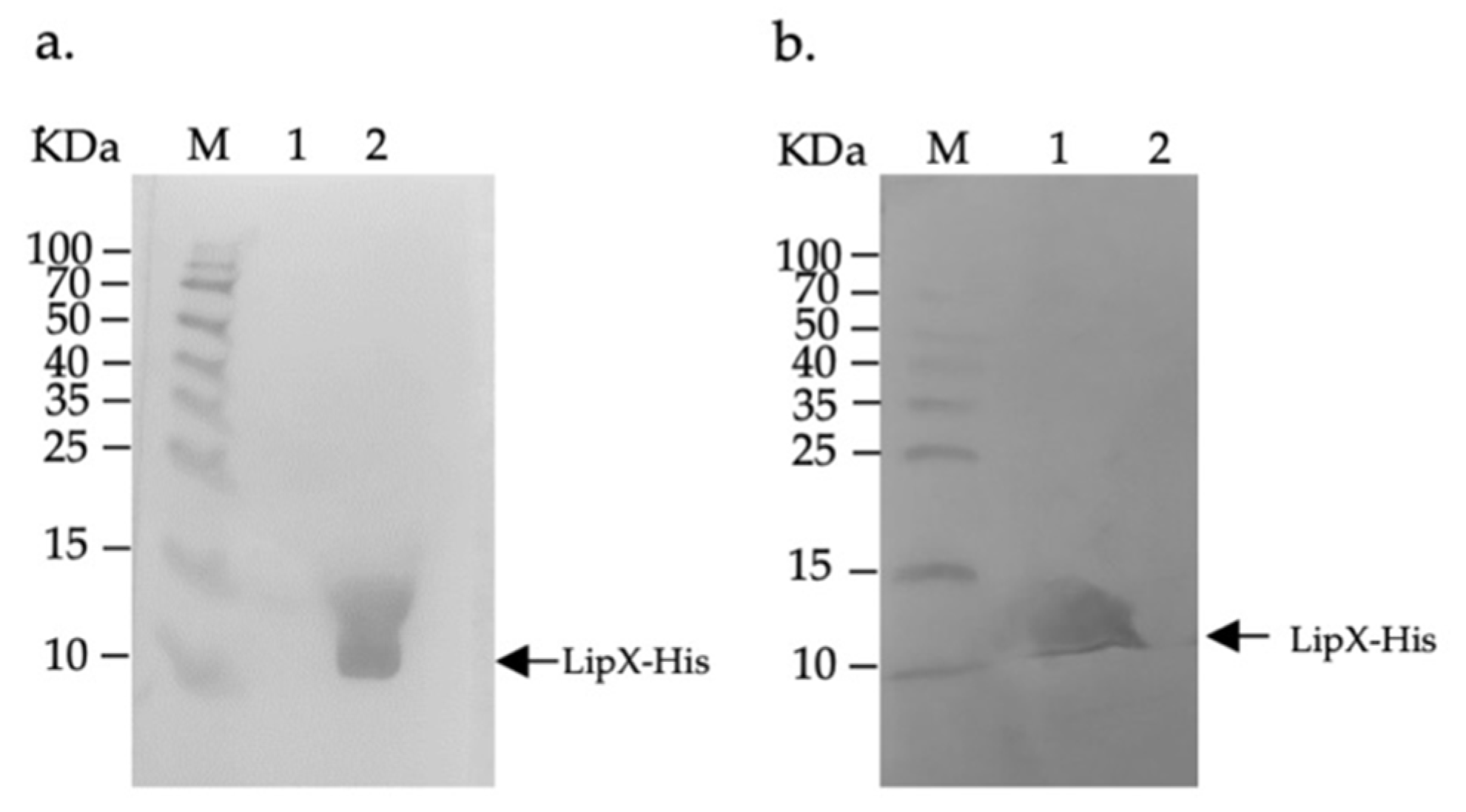
3.1. LipX Isolation and Observation of Humoral Immune Response by Western Blotting

Before observing LipX expression in the group of immunized mice, we isolated the LipX protein for use in the analysis of the humoral response. The isolation results using a Ni-NTA column showed that we successfully isolated the protein with the appropriate size for the length. The protein bands produced followed the expected protein length of about 13.34 KDa, which was similar to LipX-His protein’s length (Figure 1).
To determine whether there was an adaptive humoral response in the serum of mice injected with recombinant DNA pCDNA3.1-lipX as an indicator of LipX expression in vivo, we used Western blotting of LipX-His against immunized sera. Observation of the humoral response in the serum of mice in this step showed anti-LipX antibody binding marked by a protein band measuring about 13.4 KDa, which was equivalent to the size of the LipX-His protein detected by the anti-His monoclonal antibody. The presence of this band indicated that pCDNA3.1-lipX was successful in expressing LipX in vivo (Figure 2). These results also prove that LipX can stimulate an adaptive humoral immune response in test animals.

3.2. Analysis of the Cellular Immune Mediator Response Assay

Identification of the cellular immune mediator to LipX was carried out using ELISA. The analysis was undertaken by measuring the concentration of selected cytokines, specifically IL-4, IL-10, IL-12, and IFN-γ from the splenocyte cultures of mice immunized with pcDNA3.1-lipX, pcDNA3.1, and without immunization (Figure 2 and Supplementary Data). Based on the results of statistical analysis, it was shown that there was a significant difference between the splenocytes of mice in the pcDNA3.1-lipX immunization group induced by LipX-His and the splenocytes induced by PHA and without any induction.
The LipX-His protein induced the splenocyte culture from pcDNA3.1-lipX immunization group to have the highest levels of IFN-γ cytokines (mean + SD: 441.222 + 54,548) compared to the splenocyte culture induced by PHA (mean + SD: 307.222 + 51,768) and without any induction (mean + SD: 307.222 + 51,768) in the immunization group (mean + SD: 258,555 + 16.431). Based on statistical analysis, a significant difference in the IFNγ titer was obtained between the splenocytes from the pcDNA3.1-lipX immunization group which was induced by LipX-His and the splenocytes induced by PHA and without any induction, at a p value of 0.000.
In the measurement of IL-4 titers in all groups of mice, it was found that the PCDNA3.1-lipX immunization group induced by LipX-His had the highest levels of the IL-4 cytokines titer (mean + SD: 59.881 + 8.32) compared to the PHA-induced splenocyte culture supernatant (mean + SD: 39.441 + 7.89) and splenocyte culture supernatant without induction (mean + SD: 32.017 + 2.50). Statistical data showed a significant difference between the pCDNA3.1-lipX immunization group, the pCDNA3.1 immunization group, and the nonimmunized group (p = 0.000).
A significant difference was also found in the production of IL-12 cytokines in the PCDNA3.1-lipX immunization group which was induced by LipX-His, where the levels of the IL-12 cytokines titer (mean + SD: 221.142 + 46.235) were higher than those of the splenocyte culture induced by PHA (mean + SD: 54,857 + 13.982) and without any induction (mean + SD: 52,285 + 11,926). Statistical analysis showed a p value of 0.000 for this cytokine titer comparison.
No statistically significant difference was found between groups of mice in the IL-10 titer measurement; the statistical analysis showed a p value of 0.232 (Figure 3).

4. Discussion

As the only available vaccine against M. tuberculosis infection, BCG has several drawbacks, from varied efficacy in healthy adults to safety concerns in immunosuppressed individuals. Thus, many researchers have sought to find an alternative tuberculosis vaccine to be used together with or to replace the BCG vaccine. LipX is a member of the PE protein family, which is rich in proline amino acids and responsible for lipid remodeling in the M. tuberculosis cell wall in terms of hindering immune evasion [25]. Furthermore, in silico analysis of this protein has proven that LipX tends to have a recognition domain for B cells and major histocompatibility complex (MHC) class I [26]. In this study, we evaluated LipX immunogenicity in the form of a DNA vaccine.
In detecting anti-LipX IgG in the serum of mice, an IgG-specific response against LipX was detected by HRP-labeled horse anti-mouse IgG. The interaction between the LipX-His protein, mouse IgG, and HRP-labeled anti-mouse IgG on the PVDF membrane was indicated by the presence of a specific band that appeared after the TMB substrate was added. This band’s appearance indicated that our recombinant pcDNA3.1-lipX plasmid could express recombinant LipX protein in vivo. Although the presence of the LipX antigen in mice was not directly analyzed, a specific IgG response against LipX was successfully proven using Western blot, indicating the antigenicity of the expressed LipX protein. This specific humoral immune response was formed due to the presence of antigen presentation by APC cells or infected somatic cells [13].
In terms of the DNA vaccine, our pcDNA3.1-lipX would be taken up by muscle cells and neighboring dendritic cells before immunization. In theory, LipX will be expressed and presented to the cell’s surface through MHC or secreted outside the cells. Alongside the in silico result, we assumed that our LipX protein would be presented by MHC class II to further activate the T cell response and initiate cellular immunity [27]. However, secreted LipX can also be recognized by antibodies or B cells. Generally, in M. tuberculosis infection, the activation of B cells through Th1 or Th2 cells in the presence of IL-4 signals is received by B cells. The formation of plasma cells leads to secretion of specific IgG antibodies against antigens [13,33]. Moreover, even though we did not perform an analysis of activated T cells in the serum of immunized mice, the Western blot analysis results might indicate T cell activation. Based on these data, we conclude that the duration from immunization until termination of the mice was sufficient to detect a cellular immunity response.
The cellular immune response is the most crucial line of defense against M. tuberculosis [35,36]. This response is characterized by the host’s ability to act to develop a subset of Th1 and Th2 cells, thereby leading to the continuous activity of macrophages and dendritic cells. In evaluating cellular immunity against the DNA vaccine seed, pcDNA3.1-lipX, ELISA was carried out to determine the cytokine levels significant in adaptive immunity during infection. The activation and performance of macrophages are essential in fighting infection from M. tuberculosis, an intracellular microbe [27].
There are two types of cellular immunity reactions that arise when dealing with microbes. Specifically, these include CD4+ T helper cells that secrete mediators that recruit cytokines, thus activating other leukocytes in the phagocytes of pathogenic microbes. For example, they are activated to defend against intracellular microbes, such as M. tuberculosis. CD8+ cytotoxic T lymphocyte (CTL) cells can also be activated. CTL cells kill infected cells or contain pathogenic proteins, such as viruses, that infect cells in the cytosol [27]. In the case of M. tuberculosis infection, the dominant type of cellular immune reaction is the type that leads to macrophage activity [37,38]. The cytokines that mediate macrophage activation are the IFN-γ and TNF-α cytokines secreted by the Th1 cell subset [39].
The cellular immunity results obtained through ELISA in this study showed a significant increase in cytokine levels in the splenocyte culture samples of mice immunized with pcDNA3.1-lipX and induced by recombinant protein LipX-His compared to those that were induced by PHA or not induced. The cytokines that showed increasing levels were IL-4, IL-12, and IFN-γ. This is consistent with the host’s defense mechanism against M. tuberculosis infection [27,39]. These three cytokines play a synergistic role in fighting infection. Meanwhile, the titers of IL-10 did not show a significant increase in the splenocyte cultures of mice immunized with pcDNA3.1-lipX versus those induced with LipX-His protein and not induced. IL-10 cytokines are known to have antagonistic functions with the three cytokines described above [27,40]; IL-10 cytokines inhibit the production of cytokines and chemokines and reduce the expression of stimulators and MHC-II molecules. The main targets of IL-10 cytokines are macrophage and dendritic cells; in other words, IL-10 directs the failure of the host defense process against M. tuberculosis infection [27,41]. Although IL-10 will naturally indeed be produced in instances of prolonged inflammation, it acts as a form of physiological balance for the body in promoting anti-inflammation [42].
Cellular immune responses occur in several stages, starting with antigen-stimulated T cells presented by APC cells through MHC class I or class II [24]. In peripheral lymph nodes, the introduction of antigens through MHC-II activates CD4+ T cells to secrete IL-12, which causes the development of subsets of Th1 that will produce IL-2, IFN-γ, and TNF-α, which will send signals to macrophages and dendritic cells to be more active in the phagocytosis of pathogens. Additionally, CD4+ T cells can secrete IL-4 cytokines, which can activate the development of the Th2 subset, thus producing IL-4, which targets B cell activation to activate or mediate adaptive humoral immunity [27,28].
As mentioned in the introduction, LipX plays a role in hindering macrophages. Research conducted on M. smegmatis showed that several genes in the pe family, including lipX, play a role in macrophage intracellular survival [25,43]. Overexpression of LipX in experimental M. smegmatis (Msmeg-PE11) causes changes in colony morphology and lipid composition of cell walls, and envelopes of this strain have a higher number of glycolipids and polar lipids; the bacteria become more difficult to kill by macrophages thereby increasing survival [25]. Mice immunized with M. smegmatis expressing LipX also had high bacterial counts, organ damage, and death [25]. LipX expression also decreased interleukin 6 levels [25,44]. These data indicate that LipX protein plays a role as a virulence factor. We assume that, based on the status of the analyzed cytokines and the results of humoral analysis, the pcDNA3.1-lipX DNA vaccine seed has the potential to be developed as a TB vaccine candidate. However, vaccination may be a mediation for bacteria in avoiding macrophages and increasing the number of bacteria, especially in hosts that already contain M. tuberculosis, for example, in latent tuberculosis. This assumption may indicate the pcDNA3.1-lipX vaccine only can be administered as the first vaccination in newborns and not as a booster for the BCG vaccine or for the vaccination to adults who might have been exposed to M. tuberculosis previously. In addition, due to LipX only being conserved in the M. tuberculosis complex and some clinical isolates, it might be that pcDNA3.1-lipX is only effective against infection of those species.
In addition, since we have not carried out the challenge study, some information, such as the effectiveness of the recombinant DNA of pcDNA3.1-lipX in inhibiting the growth of M. tuberculosis in the host, has not been confirmed. This study is limited to information on the ability of our constructed pcDNA3.1-lipX to stimulate humoral and cellular immune responses in the splenocyte culture of BALB/c mice. We also assume different strains of mice in the experiment may also reflect different humoral and cellular responses.

5. Conclusions

Our seed vaccine construction, pcDNA3.1-lipX, has potential as a candidate tuberculosis vaccine. This argument is based on its ability to induce cellular immune reactions, which are the primary protection against M. tuberculosis infection. However, further studies, such as challenge testing and the effect of LipX on M. tuberculosis survival in macrophage, are needed to strengthen information on this seed vaccine’s power to protect against disease in the host.

Supplementary Materials

The following supporting information can be downloaded at: https://www.mdpi.com/article/10.3390/genes13111954/s1, Table S1: Comparison of cytokine production induced by LipX-His or PHA or without induction in the pcDNA3.1-lipX-immunized group.

Author Contributions

Conceptualization, A.R. and F.S.; methodology, A.R. and F.S.; formal analysis, A.R., F.S., L.A.S. and M.N.; writing—original draft preparation, A.R. and L.A.S.; writing—review and editing, A.R.; funding acquisition, A.R. and L.A.S. All authors have read and agreed to the published version of the manuscript.

Funding

This research was funded by Hibah PUTI 2020 Universitas Indonesia, contract number NKB-2283/UN2.RST/HKP.05.00/2020 and addendum number NKB 4778/UN2.RST/HKP.05.00/2020.

Institutional Review Board Statement

Research on experimental animals was carried out according to a protocol approved by the Ethics Committee of the Medical Faculty, Universitas Indonesia, no. KET-128/UN2.F1/ETIK/PPM00.02/2019.

Data Availability Statement

Not applicable.

Acknowledgments

We would like to thank the technicians of the Animal Facility and Tuberculosis Laboratory Department of Microbiology Medical Faculty Universitas Indonesia who helped us care for the experimental animals.

Conflicts of Interest

The authors declare no conflict of interest.

Appendix A

Table A1. Recombinant plasmids used in this research.
Plasmid NameVectorInsertFunctionsSourceReference
pET28a-lipXpET28aFull length of lipX geneExpression of LipX protein in E.coli cellsDepartment of Microbiology, Medical Faculty Universitas IndonesiaThis work (unpublished data).
pCDNA3.1-lipXpCDNA3.1Full length of lipX geneExpression of LipX protein in eukaryotic cellsDepartment of Microbiology, Medical Faculty Universitas Indonesia[32]

Appendix B

Figure A1. Immunization scheme of pCDNA3.1-lipX for the mouse immunization group as well as the blood collection, termination, and spleen collection for all groups.
Genes 13 01954 g0a1

References

  1. Marimani, M.; Ahmad, A.; Duse, A. The role of epigenetics, bacterial and host factors in progression of Mycobacterium tuberculosis infection. Tuberculosis 2018, 113, 200–214. [Google Scholar] [CrossRef] [PubMed]
  2. Amin, M.; Yanti, B.; Harapan, H.; Mertaniasih, N.M. The role of Mycobacterium tuberculosis lineages on lung tissue damage and TNF-α level among tuberculosis patients Indonesia. Clin. Epidemiol. Glob. Health 2019, 7, 263–267. [Google Scholar] [CrossRef] [Green Version]
  3. Rosser, A.; Stover, C.; Pareek, M.; Mukamolova, G.V.; Rosser, A.; Stover, C.; Mukamolova, G.V. Resuscitation-promoting factors are important determinants of the pathophysiology in Mycobacterium tuberculosis infection. Crit. Rev. Microbiol. 2017, 43, 621–630. [Google Scholar] [CrossRef] [Green Version]
  4. WHO. Global Tuberculosis Report 2019; WHO: Geneva, Switzerland, 2019. [Google Scholar]
  5. Fine, P.E.M. Variation in protection by BCG: Implications of and for heterologous immunity. Lancet 1995, 349, 1339–1345. [Google Scholar] [CrossRef]
  6. Colditz, G.A.; Brewer, T.F.; Angeles, L.; Berkey, C.S.; Wilson, M.E.; Francisco, S. Efficacy of BCG vaccine in the prevention of tuberculosis. Meta-analysis of the published literature. JAMA 1994, 271, 698–702. [Google Scholar] [CrossRef] [PubMed]
  7. Tran, V.; Ahn, S.K.; Ng, M.; Li, M.; Liu, J. Loss of lipid virulence factors reduces the efficacy of the BCG vaccine. Sci. Rep. 2016, 6, 29076. [Google Scholar] [CrossRef] [Green Version]
  8. Moreno, S.; Tim, M. DNA vaccination: An immunological perspective. Immunologia 2004, 23, 41–55. [Google Scholar]
  9. Abebe, F.; Bjune, G. The emergence of Beijing family genotypes of Mycobacterium tuberculosis and low-level protection by bacille Calmette-Guérin (BCG) vaccines: Is there a link? Clin. Exp. Immunol. 2006, 145, 389–397. [Google Scholar] [CrossRef]
  10. Madacki, J.; Fiol, G.M.; Brosch, R. Update on the virulence factors of the obligate pathogen Mycobacterium tuberculosis and related tuberculosis-causing mycobacteria. Infect. Genet. Evol. 2019, 72, 67–77. [Google Scholar] [CrossRef]
  11. Lowrie, D.B. DNA vaccines for therapy of tuberculosis: Where are we now? Vaccine 2006, 24, 1983–1989. [Google Scholar] [CrossRef]
  12. Morris, S.; Kelley, C.; Howard, A.; Li, Z.; Collins, F. The immunogenicity of single and combination DNA vaccines against tuberculosis. Vaccine 2000, 18, 2155–2163. [Google Scholar] [CrossRef]
  13. Lee, J.; Kumar, S.A.; Jhan, Y.Y.; Bishop, C.J. Engineering DNA vaccines against infectious diseases. Acta Biomater. 2018, 80, 31–47. [Google Scholar] [CrossRef] [PubMed]
  14. Rashidian, S.; Teimourpour, R.; Meshkat, Z. Designing and construction of a DNA vaccine encoding Tb10.4 gene of Mycobacterium tuberculosis. Irian J. Pathol. 2016, 11, 112–119. [Google Scholar]
  15. Khoshnood, S.; Heidary, M.; Haeili, M.; Drancourt, M.; Darban-sarokhalil, D.; Javad, M.; Lohrasbi, V. Novel vaccine candidates against Mycobacterium tuberculosis. Int. J. Biol. Macromol. 2018, 120, 180–188. [Google Scholar] [CrossRef] [PubMed]
  16. Ye, Z.; Peng, H.; Fang, Y.; Feng, J.; Yang, D.S. The construction of the eukaryotic plasmid pcDNA3.1/Azurin and increase apoptosis of U2OS cells transfected with it. Cell. Mol. Biol. Lett. 2007, 12, 407–421. [Google Scholar] [CrossRef] [Green Version]
  17. Gao, Y.; Pei, C.; Sun, X.; Zhang, C.; Li, L.; Kong, X. Plasmid pcDNA3.1-s11 constructed based on the S11 segment of grass carp reovirus as DNA vaccine provides immune protection. Vaccine 2018, 36, 3613–3621. [Google Scholar] [CrossRef]
  18. Liu, S.; Shi, L.; Cheng, Y.; Fan, G.; Ren, H.; Yuan, Y. Evaluation of protective effect of multi-epitope DNA vaccine encoding six antigen segments of Toxoplasma gondii in mice. Parasitol. Res. 2009, 105, 267. [Google Scholar] [CrossRef]
  19. Naaz, S.; Kazim, S.N. Designing and reconstruction of pcDNA3.0 mammalian expression vector with its multiple cloning sites by directional cloning method. Cloning Transgenes 2018, 7, 1–5. [Google Scholar] [CrossRef] [Green Version]
  20. Brennan, M.J. The enigmatic PE/PPE multigene family of Mycobacteria and tuberculosis vaccination. Infect. Immun. 2017, 85, e00969-16. [Google Scholar] [CrossRef] [Green Version]
  21. Delogu, G.; Cole, S.T.; Brosch, R. The PE and PPE protein families of Mycobacterium tuberculosis; Wiley Online Library: Hoboken, NJ, USA, 2008; pp. 133–144. [Google Scholar]
  22. Hasnain, S.E.; Ehtesham, N.Z.; Grover, S. Mycobacterium tuberculosis Molecular Infection Biology, Pathogenesis, Diagnostics and New Interventions; Springer: Singapore, 2019; pp. 123–167. [Google Scholar]
  23. Phelan, J.E.; Coll, F.; Bergval, I.; Anthony, R.M.; Warren, R.; Sampson, S.L.; Clark, T.G. Recombination in pe/ppe genes contributes to genetic variation in Mycobacterium tuberculosis lineages. Genomics 2016, 17, 151. [Google Scholar] [CrossRef] [Green Version]
  24. Rastogi, S.; Singh, A.K.; Pant, G.; Mitra, K.; Sashidhara, K.V.; Krishnan, M.Y. Down-regulation of PE11, a cell wall associated esterase, enhances the biofilm growth of Mycobacterium tuberculosis and reduces cell wall virulence lipid levels. Microbiology 2017, 163, 52–61. [Google Scholar] [CrossRef] [PubMed]
  25. Singh, P.; Rao, R.N.; Ram, J.; Reddy, C.; Prasad, R.B.N. PE11, a PE/PPE family protein of Mycobacterium tuberculosis is involved in cell wall remodeling and virulence. Sci. Rep. 2016, 6, 21624. [Google Scholar] [CrossRef]
  26. Narayana, Y.; Joshi, B.; Katoch, V.M.; Mishra, K.C.; Balaji, K.N. Differential B-cell responses are induced by Mycobacterium tuberculosis PE antigens Rv1169c, Rv0978c, and Rv1818c. Clin. Vaccine Immunol. 2007, 14, 1334–1341. [Google Scholar] [CrossRef] [PubMed] [Green Version]
  27. Abbas, A.K.; Lichtman, A.H.; Pillai, S. Basic Immunology, 5th ed.; Elsevier Inc.: North York, ON, Canada, 2015; pp. 69–73. [Google Scholar]
  28. Ismail, N.; Olano, J.P.; Feng, H.; Walker, D.H. Current status of immune mechanisms of killing of intracellular microorganism. FEMS Microbiol. Lett. 2002, 207, 111–120. [Google Scholar] [CrossRef] [PubMed]
  29. Sakamoto, K. The pathology of Mycobacterium tuberculosis infection. Vet. Pathol. 2012, 42, 423–439. [Google Scholar] [CrossRef] [Green Version]
  30. Kiros, T.G.; Levast, B.; Auray, G.; Strom, S.; van Kessel, J.; Gerdts, V. The importance of animal models in the development of vaccines. Innov. Vaccinol. 2012, 29, 251–264. [Google Scholar]
  31. Garcia-Pelayo, M.C.; Bachy, V.S.; Kaveh, D.A.; Hogarth, P.J. BALB/c mice display more enhanced BCG vaccine induced Th1 and Th17 response than C57BL/6 mice but have equivalent protection. Tuberculosis 2015, 95, 48–53. [Google Scholar] [CrossRef] [Green Version]
  32. Azmi, L.; Rukmana, A.; Sjatha, F. Cloning of the pe11 (LipX, Rv1169c) gene of a Mycobacterium tuberculosis Beijing Strain into the pcDNA3.1 Plasmid Vector. Makara J. Sci. 2021, 25, 35–42. [Google Scholar]
  33. Zhang, W.; Jiang, H.; Bai, Y.-L.; Kang, J.; Xu, Z.-K.; Wang, L.-M. Construction and immunogenicity of the DNA vaccine of Mycobacterium Tuberculosis dormancy antigen rv1733c. Scand. J. Immunol. 2014, 79, 292–298. [Google Scholar] [CrossRef]
  34. Fan, X.; Gao, Q.; Fu, R. Differential immunogenicity and protective efficacy of DNA vaccines expressing proteins of Mycobacterium tuberculosis in a mouse model. Microbiol. Res. 2009, 164, 374–382. [Google Scholar] [CrossRef]
  35. Ahmad, S. Pathogenesis, immunology, and diagnosis of latent Mycobacterium tuberculosis infection. Clin. Dev. Immunol. 2011, 2011, 814943. [Google Scholar] [CrossRef] [PubMed] [Green Version]
  36. Cooper, A.M.; Roberts, A.D.; Rhoades, E.R.; Callahan, J.E.; Getzy, D.M.; Orme, I.M. The role of interleukin-12 in acquired immunity to Mycobacterium tuberculosis infection. Immunology 1995, 84, 423–432. [Google Scholar] [PubMed]
  37. Garra, A.O.; Redford, P.S.; Mcnab, F.W.; Bloom, C.I.; Wilkinson, R.J.; Berry, M.P.R. The Immune Response in Tuberculosis. Annu. Rev. Immunol. 2013, 31, 475–527. [Google Scholar] [CrossRef]
  38. Sharma, D.; Sarkar, D. Pathophysiology of tuberculosis: An update review. Pharma Tutor 2018, 6, 15–21. [Google Scholar] [CrossRef]
  39. Salim, T.; Sershen, C.L.; May, E.E. Investigating the Role of TNF-α and IFN-γ Activation on the Dynamics of iNOS Gene Expression in LPS Stimulated Macrophages. PLoS ONE 2016, 11, e0153289. [Google Scholar] [CrossRef] [PubMed] [Green Version]
  40. Fulton, S.A.; Cross, J.V.; Toossi, Z.T.; Boom, W.H. Regulation of interleukin-12 by interleukin-10, transforming growth factor-β, tumor necrosis factor-α, and interferon-γ in human monocytes infected with Mycobacterium tuberculosis H37Ra. J. Infect. Dis. 1998, 178, 1105–1114. [Google Scholar] [CrossRef] [Green Version]
  41. Murray, P.J.; Young, R.A. Increased antimycobacterial immunity in interleukin-10-deficient mice. Infect. Immun. 1999, 67, 3087–3095. [Google Scholar] [CrossRef] [Green Version]
  42. D’Andrea, A.; Aste-Amezaga, M.; Valiante, N.M.; Ma, X.; Kubin, M.; Trinchieri, G. Interleukin 10 (IL-10) inhibits human lymphocyte interferon γ-production by suppressing natural killer cell stimulatory factor/IL-12 synthesis in accessory cells. J. Exp. Med. 1993, 178, 1041–1048. [Google Scholar] [CrossRef] [Green Version]
  43. Tiwari, B.M.; Kannan, N.; Vemu, L.; Raghunand, T.R. The Mycobacterium tuberculosis PE Proteins Rv0285 and Rv1386 Modulate Innate Immunity and Mediate Bacillary Survival in Macrophages. PLoS ONE 2012, 7, e51686. [Google Scholar] [CrossRef] [Green Version]
  44. Deng, W.; Zeng, J.; Xiang, X.; Li, P.; Xie, J. PE11 (Rv1169c) selectively alters fatty acid components of Mycobacterium smegmatis and host cell interleukin-6 level accompanied with cell death. Front Microbiol. 2015, 23, 613. [Google Scholar] [CrossRef]
Figure 1. LipX-His protein isolation. Lane 1, cell lysates from the 1 mM IPTG induction culture of E. coli BL21 containing the pET28-lipX plasmid; Lane 2, Protein isolated using the Ni-NTA affinity column from the culture of E. coli BL21 containing the pET28-lipX plasmid inducted by 1 mM of IPTG. KDa—kilo Dalton; M—marker.
Genes 13 01954 g001
Figure 2. Analysis of the humoral immune response using Western blotting. (a) Confirmation of LipX-His protein expression using the HRP-labeled anti-His antibody detector: Lane 1, cell lysate from the culture of E. coli BL21 containing the pET28a-lipX plasmid without IPTG induction; Lane 2, cell lysates from the IPTG induction culture of E. coli BL21 containing the pET28a-lipX plasmid; (b) Proving the humoral immune response using serum pcDNA3.1-lipX-immunized mice to determine the expression of the LipX-His protein: Lane 1, cell lysates from the IPTG induction culture; Lane 2, cell lysates from the culture without IPTG induction. KDa—kilo Dalton; M—marker.
Genes 13 01954 g002
Figure 3. Concentration of IFN-γ, IL-4, IL-10, and IL-12 between immunization groups following the stimulation of splenocytes with LipX-His or PHA, as well as without induction. Black, splenocytes from mice immunized with pcDNA3.1-lipX. Gray, splenocytes from mice immunized with pcDNA3.1. White, splenocytes from mice without immunization. Asterix label (*) indicates a statistically significant difference.
Genes 13 01954 g003
Publisher’s Note: MDPI stays neutral with regard to jurisdictional claims in published maps and institutional affiliations.

