Article

Novel Insights into MEG3/miR664a-3p/ADH4 Axis and Its Possible Role in Hepatocellular Carcinoma from an in Silico Perspective

by
Shreyas H. Karunakara
,
Lakshana D. Puttahanumantharayappa
,
Nirmala G. Sannappa Gowda
,
Varsha D. Shiragannavar
and
Prasanna K. Santhekadur
*
Department of Biochemistry, Center of Excellence in Molecular Biology & Regenerative Medicine, JSS Medical College, JSS Academy of Higher Education and Research, Mysore 570015, India
*
Author to whom correspondence should be addressed.
Genes 2022, 13(12), 2254; https://doi.org/10.3390/genes13122254
Submission received: 21 October 2022 / Revised: 19 November 2022 / Accepted: 28 November 2022 / Published: 30 November 2022
(This article belongs to the Special Issue Liver Disease: Genetic Research and Clinical Diagnosis)

Abstract

:
Hepatocellular carcinoma (HCC) is a complex disease involving altered interactomes of transcripts and proteins. MicroRNAs (miRNAs) are small-noncoding RNAs that can interact with specific gene transcripts and an array of other vital endogenous non-coding RNAs (lncRNAs) that can influence gene expression. Maternally Expressed Gene 3 (MEG3) is an imprinted lncRNA that is reported to be downregulated in HCC (in both cell lines and tumors). Alcohol Dehydrogenase 4 (ADH4) is a well-known prognostic protein biomarker for predicting the survival outcomes of patients with hepatocellular carcinoma whose expression is regulated by miR-664a-3p, which is upregulated in HCC. In this study, we performed a battery of robust and systematic in silico analyses to predicate the possible lncRNA–miRNA interactions between MEG3, miR-664a-3p, and ADH4. miRNA–mRNA and lncRNA–miRNA hybrid structures were primarily obtained, and the minimum free energies (MFEs) for the 3′UTR (Untranslated Regions) of ADH4-miR-664a-3p and the 3′UTR of MEG3-miR-664a-3p interactions were assessed to predict the stability of the obtained RNA heteroduplex hybrids. The hybrid with the least minimum free energy (MFE) was considered to be the most favorable. The MFEs were around −28.1 kcal/mol and −31.3 kCal/mol for the ADH4-miR-664a-3p and MEG3-miR-66a-3p RNA hybrids, respectively. This demonstrated that lncRNA-MEG3 might be a competitive endogenous RNA that acts as a molecular sponge for miR-664a-3p. In summary, our interaction analyses results predict the significance of the MEG3/miR-664a-3p/ADH4 axis, where MEG3 downregulation results in miR-664a-3p overexpression and the subsequential underexpression of ADH4 in HCC, as a novel axis of interest that demands further validation.

Graphical Abstract

1. Introduction

Hepatocellular carcinoma (HCC) refers to the primary carcinoma of the liver and ranks as the sixth most common cancer affecting the human population [1]. According to the Global Cancer Observatory (https://gco.iarc.fr/ (accessed on 5 October 2022)), the national mortality burden in India for HCC stands at 2.4%, accounting for about 33,793 deaths. Preliminary causes include chronic liver conditions, such as Hepatitis B Virus (HBV) [2,3,4,5] and Hepatitis C Virus (HCV) [6,7] infections, cirrhosis, non-alcoholic fatty liver disease (NAFLD) [8,9,10,11], and non-alcoholic steatohepatitis (NASH) [11,12,13]. Additionally, several other epigenetic mechanisms involving long non-coding RNAs (lncRNAs), microRNAs (miRNAs), histone modifications, and DNA methylation are gaining importance in regulating various target genes vital to the development and progression of HCC [14].
Maternally Expressed Gene 3 (MEG3) is located on Chromosome 14q32.3. In humans, it is located on the imprinted DLK1-MEG3 locus. The mouse ortholog commonly referred to as Meg3 or Gene trap locus 2 (Glt2) is positioned on Ch 12 [15,16]. MEG3 encodes for an lncRNA, which is commonly expressed in normal tissues [17,18,19]. However, MEG3 expression is lost in cancers of the brain [20], breast [21], lung [21], cervix [21], and colon [22]. In HCC, MEG3 is underexpressed when compared to in normal liver tissues [23]. MEG3 underexpression has been validated in various HCC cell line models, such as PLC/PRF/5 [23], HepG2 [24], and Huh7 [25]. Overall, these experimental pieces of evidence suggest that MEG3 is downregulated and acts as a bona fide tumor suppressor in HCC.
MicroRNAs (miRNAs) play a crucial role in regulating the gene expression of target genes by binding to the 3′UTRs of target transcripts and degrading mRNA through RISC-mediated endonuclease activity [26,27]. Several studies have reported miR-664a-3p expression to be overexpressed in HCC [28]. A study by Wang in 2019 reported miR-664 to be overexpressed in HCC cell lines, as well as in HCC patients, adding to the progression and spread of HCC [29].
The alcohol dehydrogenase (ADH) superfamily spans five classes of dehydrogenases: class I (ADHIA, ADHIB, and ADHIC), class II (ADH4), class III (ADH5), class IV (ADH6), and class V (ADH7) [30]. ADH4 is primarily involved in the metabolism of aliphatic alcohols, including retinol, ethanol, and hydroxysteroid, and it occupies center stage in the hepatic regulation of the metabolism of various lipid peroxidation processes [31]. Previous studies have shown that ADH4 is significantly downregulated, and they have predicted its suitability as a prognostic marker in HCC [32,33,34].
In the present study, we employed a range of robust algorithms and bioinformatics tools to identify a novel axis that might be central to HCC development and progression. Based on RNA–RNA interactions, we predicted that MEG3 interacts with miR-664a-3p, resulting in the overexpression of ADH4 in normal liver, while the reverse effect occurs due to the underexpression of MEG3 in HCC.

2. Materials and Methods

2.1. Retrieval of lncRNA, miR-664a, and ADH4 Sequences

Reference nucleotide sequences were obtained from standard databases. The gene coordinates for human lncRNA MEG3 were found to be Chr14: 100,826,108–100,861,026. The full-length nucleotide sequence of MEG3 (NC_000014.9: https://www.ncbi.nlm.nih.gov/nuccore/NC_000014.9?rport=fasta&from=100826108&to=100861026 (accessed 5 October 2022)) and the transcript of ADH4 (M15943.1: https://www.ncbi.nlm.nih.gov/nuccore/M15943.1?report=fasta (accessed 5 October 2022)) were obtained from the National Center for Biotechnology Information Nucleotide (Internet). Bethesda (MD): National Library of Medicine (US), National Center for Biotechnology Information; (1988)—Accession No. NC_000014.9, Homo sapiens Maternally Expressed Gene 3; (cited 5 October 2022). The mature sequence of miR-664a-3p (UAUUCAUUUAUCCCCAGCCUACA) was obtained from miRbase (https://www.mirbase.org/ (accessed 5 October 2022)) [35].

2.2. Expression of ADH4 in HCC

The next-generation sequencing (RNA-Seq) gene expression quantification data derived from the complete transcriptome profiling of the genes expressed in normal liver tissues (n = 70) and in HCC (n = 352) were downloaded from the TCGA-LIHC dataset from The Genomic Data Commons (GDC: https://gdc.cancer.gov/about-gdc) (data accessed on 5 October 2022) of the National Cancer Institute. The expression values of ADH4 in terms of transcripts per million from the whole transcriptome profile were obtained for which a differential expression analysis was performed on normal and HCC datasets (Supplementary Data 1: TCGA miR664a and ADH4 values.xlsx). The differential expression values were used to obtain box plots using Microsoft Excel. Further, the protein expression of ADH4 in the normal, as well as the TCGA-LIHC dataset, was obtained and analyzed using the Human Protein Atlas (HPA: https://www.proteinatlas.org/ (accessed on 5 October 2022)) [36]. The association of the differential expression of ADH4 in the normal and HCC datasets was determined in terms of overall survival (OS) and disease-free survival (DFS) using Gene Expression Profiling Interactive Analysis 2 (GEPIA 2.0: http://gepia2.cancer-pku.cn/ (accessed on 5 October 2022)) [37]. KM survival plots were plotted such that the true median expression values occurred with a confidence interval of 95%.

2.3. Differential Expression of miR-664a-3p and MEG3 in HCC

The miR-Seq quantification data were downloaded from the TCGA-LIHC dataset from the Genomic Data Commons portal (accessed on 5 October 2022). The expression values of miR-664a-3p in terms of transcripts per million in normal liver and HCC datasets were obtained from the whole transcriptome profile (Supplementary Data 2: TCGA miR664a and ADH4 values.xlsx). A differential expression analysis of miR-664a-3p in normal liver and HCC datasets were performed for which box plots were plotted. Further, mRNA expression was analyzed using The University of ALabama at Birmingham CANcer data analysis portal tool (UALCAN: http://ualcan.path.uab.edu/index.html (accessed on 5 October 2022)) [38]. For analyzing MEG3 expression in HCC, the TCGA-LIHC datasets were analyzed using UALCAN. The datasets for the differential expression of miR-664a-3p and lncRNA-MEG3 for normal and primary HCC were obtained from TCGA and analyzed using the UALCAN tool. The differential expression between the normal and tumor datasets was considered statistically significant if the means of differential expression for miRNA and lncRNA was p < 0.05 (95% CI).

2.4. Construction of miRNA–mRNA RNA Heteroduplex Hybrids

The interaction of miR-664a-3p and the 3′UTR of ADH4 mRNA was constructed using the BiBiserve2-RNAhybrid tool (https://bibiserv.cebitec.uni-bielefeld.de/rnahybrid/ (accessed on 5 October 2022)) [39]. The RNA heteroduplex hybrids were scored in terms of mean free energy (MFE). The top five RNA–RNA hybrids were considered for further analysis, and the hybrid with the least MFE was considered the best possible hybrid predicted.

2.5. Construction of lncRNA–miRNA RNA Heteroduplex Hybrids

The interaction of lncRNA MEG3 and miR-664a-3p was constructed based on sequence complementarity using the BiBiserve2-RNAhybrid tool. The top five hybrids were considered for further analysis, and the hybrid with the least MFE was considered the best possible hybrid predicted based on sequence complementarity.

2.6. Predicting the Accessibility of Binding Regions of MEG3-miRNA RNA Heteroduplex

Once the complementary sequences involved in the hybrid were identified, the actual binding of the lncRNA and miRNA was computed considering intramolecular interactions to provide a biophysically realistic model with accurate MFE using Freiburg RNA Tools: IntaRNA (http://rna.informatik.uni-freiburg.de/IntaRNA/Input.jsp (accessed on 5 October 2022)) [40,41].

3. Results

3.1. ADH4 Is Downregulated in HCC

The differential expression analysis of the expression values derived from the GDC portal for ADH4 in TCGA-LIHC (Supplementary Data 1) pointed towards a significant downregulation in HCC (Figure 1A). Moreover, the immunohistochemistry (IHC) details of ADH4 from the HPA analysis of normal and HCC tissue samples clearly showed the protein to be downregulated in HCC as compared to its higher expression in normal liver tissues (Supplementary Figure S1: HPA.TIF). It was observed that, when compared to the normal datasets, the HCC tissue datasets showed lower or no expression for ADH4. The individual patient details and IHC characteristics can be seen in Table 1. The significance of ADH4 expression in HCC was scored in terms of OS and DFS. It was observed that the differential expression of ADH4 is significant both in terms of OS (Figure 1B) and DFS (Figure 1C). The KM plots for OS showed that higher ADH4 expression corresponded to better survival as compared to decreased ADH4 expression. This clearly shows that ADH4 is downregulated in terms of differential expression in HCC.

3.2. LncRNA-MEG3 Is Downregulated and miR-664a-3p Is Upregulated in HCC

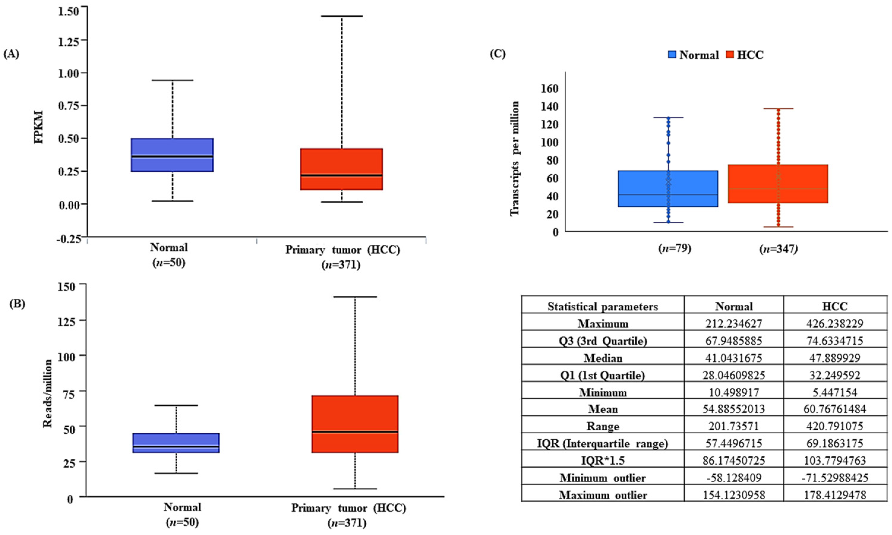
The differential expression of lncRNA-MEG3 was obtained from TCGA-LIHC. The UALCAN analysis showed that MEG3 was downregulated in HCC tissues (n = 371) as compared to normal liver tissues (n = 50), with a significance of p = 7.0333E-06 (Figure 2A). Likewise, the UALCAN analysis of the differential expression of miR-664a-3p showed a significant upregulation of miRNA in HCC (p = 1.2608 × 10⁻¹¹ (Figure 2B) (a value of p < 0.05 was considered statistically significant). Further, the expression values derived from the GDC Data Portal (Supplementary Data 2: TCGA miR664a and ADH4 values.xlsx) validated the upregulation of miR-664a-3p expression in HCC (Figure 2C).

3.3. ADH4 Is Targeted by miR-664a-3p in HCC

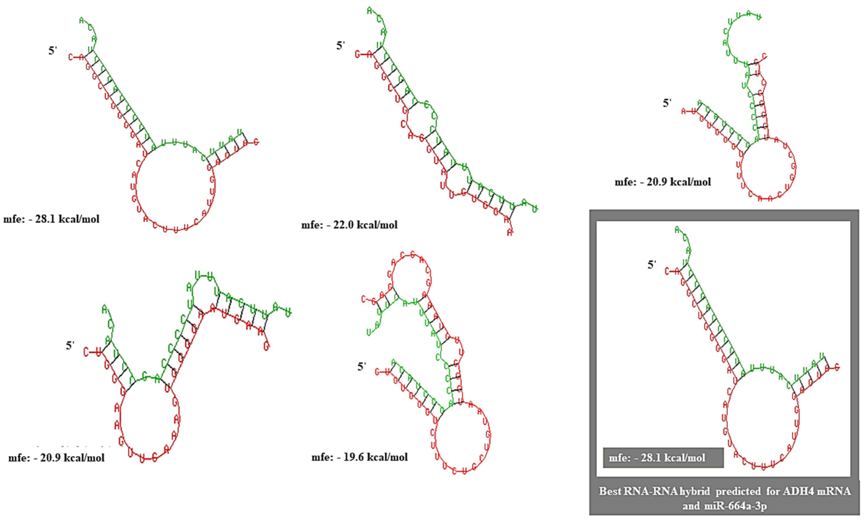
The probability of RNA–RNA interactions between miR-664a-3p and the 3′UTR of ADH4 was identified and scored using the RNA Interactome Database (RNAInter: http://www.rnainter.org/ (accessed on 5 October 2022)) on a scale of 0.0–1.0. The interaction had a score of 0.4571. The miRNA–mRNA interaction predicted was supplemented by strong published experimental evidence (PUBMED ID: 28374914). miRNA–mRNA heteroduplex hybrid structures were constructed using RNAhybrid, and the best-predicted structure had an MFE of −28.1 kcal/mol (Figure 3).

3.4. MEG3 Acts as a Competitive Endogenous Sponge for miR-664a-3p

The RNAInter analysis of lncRNA–miRNA predicted the interaction between MEG3-miR-664a-3p with a score of 0.528 on a confidence scale of 0.0–1.0. The miRNA–mRNA interaction predicted was supplemented by strong published experimental evidence (PUBMED ID: 28374914). Further, the RNA heteroduplex hybrid structures constructed using RNAhybrid predicted the best hybrid to have an MFE of −31.3 kcal/mol (Figure 4). IntaRNA predicted realistic model for the mRNA–miRNA interaction between the 10th–20th base positions of miR-664a-3p and the 931st–941st base positions of ADH4, with an overall MFE of −16.64 kcal/mol (Figure 5A–C). Moreover, a more realistic hybrid model based on the accessibility of nucleotides for base pairing predicted binding between the 3rd–22nd bases of miR-664a-3p and the 2nd–21st bases of MEG3, with an overall MFE of −24.75 kcal/mol (Figure 5D–F). The individual energy values for the interaction can be obtained from Supplementary Tables S1–S3: List of Supplementary Tables MEG3.pdf.

4. Discussion

RNA–RNA interactions involving lncRNAs and miRNAs occupy center stage in the post-transcriptional regulation of gene expression [42]. Various studies have revealed the significance of the tumor-suppressive effect of lncRNA MEG3 in HCC [42,43,44,45,46,47,48,49]. In our study, the datasets obtained from TCGA-LIHC showed a unanimous underexpression of MEG3 in HCC as compared to in normal liver, pointing out that MEG3 acts as a bona fide tumor suppressor in HCC.
miR-644 overexpression is also found to be associated with the downregulation of methionine adenosyl transferase and global hypomethylation in HCC [28], pointing towards the intricate effect and regulation of other epigenetic processes, such as DNA methylation by miRNAs. miR-664a-3p is reported to be upregulated in HepG2, Huh7, and MHCC97 HCC cell lines and is predicted to be associated with poor overall survival in HCC [29]. The analysis of the TCGA-LIHC dataset pointed towards a similar upregulation of miR-664a-3p in HCC.
ADH4 is a well-known prognostic biomarker for HCC [32]. Various studies have provided evidence that the downregulation of ADH4 is linked to the poor overall survival of HCC [50,51]. We mined the HPA-IHC data of ADH4, and they clearly show that ADH4 is underexpressed in HCC tissues. The Kaplan–Meier survival analysis inferred overall survival to be higher under the positive expression of ADH4, while the survival outcomes dropped under decreased ADH4 expression. Further, studies involving the validation of ADH4 in HCC using various methods, such as Western blot [32], RTqPCR [52], and DNA microarrays [53], have accurately predicted ADH4 to be downregulated. Additionally, ADH4 was predicted to be a prominent prognostic biomarker for the evaluation of immunotherapy efficiency in HCC patients [52].
Through our in silico analysis, we showed ADH4 to be a direct target for miR-664a-3p, wherein the structures of the miRNA–mRNA interaction between miR-664a-3p and the 3′UTR of ADH4 were identified and scored in terms of sequence complementarity and MFE. Likewise, similar models of interactions between miR-664a-3p and lncRNA-MEG3 were constructed based on sequence complementarity and MFE. We found that the mRNA–miRNA heteroduplex hybrid yielded a higher MFE (−28.1 kcal/mol) than a stable heteroduplex between miRNA and lncRNA, which yielded a lower MFE (−31.3 kCal/mol). Further, a more realistic model for mRNA–miRNA and lncRNA–miRNA interactions was predicted based on the accessibility of binding regions, which assigned an MFE of −16.64 kcal/mol for the miR-664a-3p-ADH4 interaction and an MFE of −24.75 kcal/mol for the MEG3-miR-664a-3p interaction. Based on the overall MFE predicted by three separate parameters (sequence complementarity, accessibility of binding regions, and MFE), it is inferred that MEG3 acts as a competitive sponge for miR-664a-3p.
Based on the previous discussion, it can be concluded that the MEG3/miR-664a-3p/ADH4 axis is vital in the process of HCC carcinogenesis. In normal liver, MEG3 is not repressed and can act as a competitive sponge for miR-664a-3p, and MEG3 also acts as a molecular checker of miRNA expression and, in turn, prevents its binding to the 3′UTR of ADH4 mRNA (Figure 6A). However, in HCC, due to MEG3 downregulation, miR-664a-3p is upregulated and is free to bind to the 3′UTR of ADH4 mRNA, resulting in RNA-induced silencing of ADH4 (Figure 6B). Interestingly, we did find a study validating the existence of such an axis in HCC [54]. However, our study predicts miRNA–mRNA and miRNA–lncRNA hybrids based on MFE, which is novel and can serve as a template for in silico predictions of other RNA–RNA interactions that can form the basis for further in vitro validations in HCC.

Supplementary Materials

The following supporting information can be downloaded at: https://www.mdpi.com/article/10.3390/genes13122254/s1, Figure S1: HPA-Immunohistochemistry data for ADH4 expression in HCC; Supplementary Data 1: TCGA miR-664a expression in terms of transcripts per million; Supplementary Data 2: TCGA ADH4 expression values in terms of Transcripts per million Supplementary Table S1: Energies of individual nucleotides of MEG3 involved in miRNA-lncRNA interaction; Supplementary Table S2: Energies of individual nucleotides of miR-664a-3p involved in miRNA-lncRNA interaction; Supplementary Table S3: Final MFEs of nucleotides involved in miR-664a-3p-MEG3 interaction.

Author Contributions

Conceptualization, S.H.K. and P.K.S.; methodology, S.H.K. and P.K.S.; data curation, S.H.K., V.D.S., N.G.S.G. and L.D.P.; validation, S.H.K. and P.K.S., writing—original draft preparation, S.H.K.; writing—review and editing, S.H.K., V.D.S., N.G.S.G., L.D.P. and P.K.S.; supervision, P.K.S. All authors have read and agreed to the published version of the manuscript.

Funding

This research received no external funding.

Institutional Review Board Statement

Ethical review and approval were waived for this study as the study mainly involved secondary data analysis from publicly available datasets such as the TCGA for validation purposes.

Informed Consent Statement

Not applicable.

Data Availability Statement

Data available in a publicly accessible repository that does not issue DOIs. Publicly available datasets were analyzed in this study. This data can be found here: [https://portal.gdc.cancer.gov/] (accessed on 5 October 2022).

Acknowledgments

All the authors acknowledge the support extended by JSS Medical College and JSS AHER, Mysore, India.

Conflicts of Interest

The authors declare no conflict of interest.

References

  1. Konyn, P.; Ahmed, A.; Kim, D. Current epidemiology in hepatocellular carcinoma. Expert Rev. Gastroenterol. Hepatol. 2021, 15, 1295–1307. [Google Scholar] [CrossRef] [PubMed]
  2. Chen, Y.; Tian, Z. HBV-Induced Immune Imbalance in the Development of HCC. Front. Immunol. 2019, 10, 2048. [Google Scholar] [CrossRef] [PubMed] [Green Version]
  3. Jia, L.; Gao, Y.; He, Y.; Hooper, J.D.; Yang, P. HBV induced hepatocellular carcinoma and related potential immunotherapy. Pharmacol. Res. 2020, 159, 104992. [Google Scholar] [CrossRef]
  4. Jiang, Y.; Han, Q.; Zhao, H.; Zhang, J. The Mechanisms of HBV-Induced Hepatocellular Carcinoma. J. Hepatocell. Carcinoma 2021, 8, 435–450. [Google Scholar] [CrossRef] [PubMed]
  5. Rana, M.A.; Ijaz, B.; Daud, M.; Tariq, S.; Nadeem, T.; Husnain, T. Interplay of Wnt β-catenin pathway and miRNAs in HBV pathogenesis leading to HCC. Clin. Res. Hepatol. Gastroenterol. 2019, 43, 373–386. [Google Scholar] [CrossRef] [PubMed]
  6. De Battista, D.; Zamboni, F.; Gerstein, H.; Sato, S.; Markowitz, T.E.; Lack, J.; Engle, R.E.; Farci, P. Molecular Signature and Immune Landscape of HCV-Associated Hepatocellular Carcinoma (HCC): Differences and Similarities with HBV-HCC. J. Hepatocell. Carcinoma 2021, 8, 1399–1413. [Google Scholar] [CrossRef]
  7. Hu, J.; Liu, K.; Luo, J. HIV-HBV and HIV-HCV Coinfection and Liver Cancer Development. Cancer Treat. Res. 2019, 177, 231–250. [Google Scholar] [CrossRef]
  8. Foerster, F.; Gairing, S.J.; Müller, L.; Galle, P.R. NAFLD-driven HCC: Safety and efficacy of current and emerging treatment options. J. Hepatol. 2022, 76, 446–457. [Google Scholar] [CrossRef]
  9. Huang, D.Q.; El-Serag, H.B.; Loomba, R. Global epidemiology of NAFLD-related HCC: Trends, predictions, risk factors and prevention. Nat. Rev. Gastroenterol. Hepatol. 2021, 18, 223–238. [Google Scholar] [CrossRef]
  10. Ioannou, G.N. Epidemiology and risk-stratification of NAFLD-associated HCC. J. Hepatol. 2021, 75, 1476–1484. [Google Scholar] [CrossRef]
  11. Michelotti, G.A.; Machado, M.V.; Diehl, A.M. NAFLD, NASH and liver cancer. Nat. Rev. Gastroenterol. Hepatol. 2013, 10, 656–665. [Google Scholar] [CrossRef] [PubMed]
  12. Anstee, Q.M.; Reeves, H.L.; Kotsiliti, E.; Govaere, O.; Heikenwalder, M. From NASH to HCC: Current concepts and future challenges. Nat. Rev. Gastroenterol. Hepatol. 2019, 16, 411–428. [Google Scholar] [CrossRef] [PubMed]
  13. Kim, H.; Lee, D.S.; An, T.H.; Park, H.-J.; Kim, W.K.; Bae, K.-H.; Oh, K.-J. Metabolic Spectrum of Liver Failure in Type 2 Diabetes and Obesity: From NAFLD to NASH to HCC. Int. J. Mol. Sci. 2021, 22, 4495. [Google Scholar] [CrossRef] [PubMed]
  14. Han, T.-S.; Ban, H.S.; Hur, K.; Cho, H.-S. The Epigenetic Regulation of HCC Metastasis. Int. J. Mol. Sci. 2018, 19, 3978. [Google Scholar] [CrossRef] [PubMed] [Green Version]
  15. Ghafouri-Fard, S.; Taheri, M. Maternally expressed gene 3 (MEG3): A tumor suppressor long non coding RNA. Biomed. Pharmacother. 2019, 118, 109129. [Google Scholar] [CrossRef]
  16. Al-Rugeebah, A.; Alanazi, M.; Parine, N.R. MEG3: An Oncogenic Long Non-coding RNA in Different Cancers. Pathol. Oncol. Res. 2019, 25, 859–874. [Google Scholar] [CrossRef]
  17. Mahpour, A.; Mullen, A.C. Our emerging understanding of the roles of long non-coding RNAs in normal liver function, disease, and malignancy. JHEP Rep. Innov. Hepatol. 2021, 3, 100177. [Google Scholar] [CrossRef]
  18. He, Y.; Luo, Y.; Liang, B.; Ye, L.; Lu, G.; He, W. Potential applications of MEG3 in cancer diagnosis and prognosis. Oncotarget 2017, 8, 73282–73295. [Google Scholar] [CrossRef] [Green Version]
  19. Xu, X.; Zhong, Z.; Shao, Y.; Yi, Y. Prognostic Value of MEG3 and Its Correlation With Immune Infiltrates in Gliomas. Front. Genet. 2021, 12, 679097. [Google Scholar] [CrossRef]
  20. Astuti, D.; Latif, F.; Wagner, K.; Gentle, D.; Cooper, W.N.; Catchpoole, D.; Grundy, R.; Ferguson-Smith, A.C.; Maher, E.R. Epigenetic alteration at the DLK1-GTL2 imprinted domain in human neoplasia: Analysis of neuroblastoma, phaeochromocytoma and Wilms’ tumour. Br. J. Cancer 2005, 92, 1574–1580. [Google Scholar] [CrossRef]
  21. Zhang, X.; Zhou, Y.; Mehta, K.R.; Danila, D.C.; Scolavino, S.; Johnson, S.R.; Klibanski, A. A Pituitary-Derived MEG3 Isoform Functions as a Growth Suppressor in Tumor Cells. J. Clin. Endocrinol. Metab. 2003, 88, 5119–5126. [Google Scholar] [CrossRef] [PubMed] [Green Version]
  22. Zhou, Y.; Zhong, Y.; Wang, Y.; Zhang, X.; Batista, D.L.; Gejman, R.; Ansell, P.J.; Zhao, J.; Weng, C.; Klibanski, A. Activation of p53 by MEG3 Non-coding RNA. J. Biol. Chem. 2007, 282, 24731–24742. [Google Scholar] [CrossRef] [Green Version]
  23. Braconi, C.; Kogure, T.; Valeri, N.; Huang, N.; Nuovo, G.; Costinean, S.; Negrini, M.; Miotto, E.; Croce, C.M.; Patel, T. microRNA-29 can regulate expression of the long non-coding RNA gene MEG3 in hepatocellular cancer. Oncogene 2011, 30, 4750–4756. [Google Scholar] [CrossRef] [PubMed] [Green Version]
  24. Pu, Z.; Wu, L.; Guo, Y.; Li, G.; Xiang, M.; Liu, L.; Zhan, H.; Zhou, X.; Tan, H. LncRNA MEG3 contributes to adenosine-induced cytotoxicity in hepatoma HepG2 cells by downregulated ILF3 and autophagy inhibition via regulation PI3K-AKT-mTOR and beclin-1 signaling pathway. J. Cell. Biochem. 2019, 120, 18172–18185. [Google Scholar] [CrossRef] [PubMed]
  25. Liu, L.-X.; Deng, W.; Zhou, X.-T.; Chen, R.-P.; Xiang, M.-Q.; Guo, Y.-T.; Pu, Z.-J.; Li, R.; Wang, G.-F.; Wu, L.-F. The mechanism of adenosine-mediated activation of lncRNA MEG3 and its antitumor effects in human hepatoma cells. Int. J. Oncol. 2016, 48, 421–429. [Google Scholar] [CrossRef] [PubMed] [Green Version]
  26. Santhekadur, P.K.; Kumar, D.P. RISC assembly and post-transcriptional gene regulation in Hepatocellular Carcinoma. Genes Dis. 2019, 7, 199–204. [Google Scholar] [CrossRef]
  27. Yoo, B.K.; Santhekadur, P.K.; Gredler, R.; Chen, D.; Emdad, L.; Bhutia, S.; Pannell, L.; Fisher, P.B.; Sarkar, D. Increased RNA-Induced Silencing Complex (RISC) Activity Contributes to Hepatocellular Carcinoma. Hepatology 2011, 53, 1538–1548. [Google Scholar] [CrossRef] [PubMed] [Green Version]
  28. Yang, H.; Cho, M.E.; Li, T.W.H.; Peng, H.; Ko, K.S.; Mato, J.M.; Lu, S.C. MicroRNAs regulate methionine adenosyltransferase 1A expression in hepatocellular carcinoma. J. Clin. Investig. 2013, 123, 285–298. [Google Scholar] [CrossRef]
  29. Wang, X.; Zhou, Z.; Zhang, T.; Wang, M.; Xu, R.; Qin, S.; Zhang, S. Overexpression of miR-664 is associated with poor overall survival and accelerates cell proliferation, migration and invasion in hepatocellular carcinoma. OncoTargets Ther. 2019, 12, 2373–2381. [Google Scholar] [CrossRef] [Green Version]
  30. Edenberg, H.J. The genetics of alcohol metabolism: Role of alcohol dehydrogenase and aldehyde dehydrogenase variants. Alcohol Res. Health J. Natl. Inst. Alcohol Abus. Alcohol. 2007, 30, 5–13. [Google Scholar]
  31. Li, H.; Toth, E.; Cherrington, N.J. Alcohol Metabolism in the Progression of Human Nonalcoholic Steatohepatitis. Toxicol. Sci. 2018, 164, 428–438. [Google Scholar] [CrossRef] [PubMed]
  32. Wei, R.-R.; Zhang, M.-Y.; Rao, H.-L.; Pu, H.-Y.; Zhang, H.-Z.; Wang, H.-Y. Identification of ADH4 as a novel and potential prognostic marker in hepatocellular carcinoma. Med. Oncol. 2012, 29, 2737–2743. [Google Scholar] [CrossRef]
  33. Hong, L.; Zhou, Y.; Xie, X.; Wu, W.; Shi, C.; Lin, H.; Shi, Z. A stemness-based eleven-gene signature correlates with the clinical outcome of hepatocellular carcinoma. BMC Cancer 2021, 21, 716. [Google Scholar] [CrossRef] [PubMed]
  34. Jelski, W.; Zalewski, B.; Szmitkowski, M. Alcohol dehydrogenase (ADH) isoenzymes and aldehyde dehydrogenase (ALDH) activity in the sera of patients with liver cancer. J. Clin. Lab. Anal. 2008, 22, 204–209. [Google Scholar] [CrossRef] [PubMed]
  35. Kozomara, A.; Birgaoanu, M.; Griffiths-Jones, S. miRBase: From microRNA sequences to function. Nucleic Acids Res. 2019, 47, D155–D162. [Google Scholar] [CrossRef]
  36. Thul, P.J.; Lindskog, C. The human protein atlas: A spatial map of the human proteome. Protein Sci. Publ. Protein Soc. 2018, 27, 233–244. [Google Scholar] [CrossRef] [Green Version]
  37. GEPIA: A web Server for Cancer and Normal Gene Expression Profiling and interactive Analyses. Available online: https://academic.oup.com/nar/article/45/W1/W98/3605636 (accessed on 22 June 2021).
  38. Chandrashekar, D.S.; Bashel, B.; Balasubramanya, S.A.H.; Creighton, C.J.; Ponce-Rodriguez, I.; Chakravarthi, B.V.S.K.; Varambally, S. UALCAN: A Portal for Facilitating Tumor Subgroup Gene Expression and Survival Analyses. Neoplasia 2017, 19, 649–658. [Google Scholar] [CrossRef]
  39. Fast and Effective Prediction of microRNA/Target Duplexes. Available online: https://rnajournal.cshlp.org/content/10/10/1507.abstract (accessed on 17 October 2022).
  40. Freiburg RNA Tools: A Central Online Resource for RNA-Focused Research and Teaching. Available online: https://pubmed.ncbi.nlm.nih.gov/29788132/ (accessed on 17 October 2022).
  41. Mann, M.; Wright, P.R.; Backofen, R. IntaRNA 2.0: Enhanced and customizable prediction of RNA-RNA interactions. Nucleic Acids Res. 2017, 45, W435–W439. [Google Scholar] [CrossRef]
  42. Chan, J.J.; Tay, Y. Noncoding RNA:RNA Regulatory Networks in Cancer. Int. J. Mol. Sci. 2018, 19, 1310. [Google Scholar] [CrossRef] [Green Version]
  43. Zhang, Y.; Liu, J.; Lv, Y.; Zhang, C.; Guo, S. LncRNA meg3 suppresses hepatocellular carcinoma in vitro and vivo studies. Am. J. Transl. Res. 2019, 11, 4089–4099. [Google Scholar]
  44. Mohammed, S.R.; Shaker, O.G.; Mohamed, M.M.; Abdelhafez Mostafa, M.N.; Gaber, S.N.; Ali, D.Y.; Hussein, H.A.; Elebiary, A.M.; Abonar, A.A.; Eid, H.M.; et al. The emerging role of lncRNA MEG3 and MEG3 rs7158663 in hepatocellular carcinoma. Eur. Rev. Med. Pharmacol. Sci. 2022, 26, 11–21. [Google Scholar] [CrossRef] [PubMed]
  45. Ghafouri-Fard, S.; Gholipour, M.; Hussen, B.M.; Taheri, M. The Impact of Long Non-Coding RNAs in the Pathogenesis of Hepatocellular Carcinoma. Front. Oncol. 2021, 11, 649107. [Google Scholar] [CrossRef]
  46. Gailhouste, L.; Liew, L.C.; Yasukawa, K.; Hatada, I.; Tanaka, Y.; Kato, T.; Nakagama, H.; Ochiya, T. MEG3-derived miR-493-5p overcomes the oncogenic feature of IGF2-miR-483 loss of imprinting in hepatic cancer cells. Cell Death Dis. 2019, 10, 553. [Google Scholar] [CrossRef] [PubMed] [Green Version]
  47. Zhu, J.; Liu, S.; Ye, F.; Shen, Y.; Tie, Y.; Zhu, J.; Wei, L.; Jin, Y.; Fu, H.; Wu, Y.; et al. Long Noncoding RNA MEG3 Interacts with p53 Protein and Regulates Partial p53 Target Genes in Hepatoma Cells. PLoS ONE 2015, 10, e0139790. [Google Scholar] [CrossRef] [PubMed]
  48. Liu, Z.; Chen, J.Y.; Zhong, Y.; Xie, L.; Li, J.S. lncRNA MEG3 inhibits the growth of hepatocellular carcinoma cells by sponging miR-9-5p to upregulate SOX11. Braz. J. Med. Biol. Res. 2019, 52, e8631. [Google Scholar] [CrossRef] [Green Version]
  49. Li, M.; Liao, H.; Wu, J.; Chen, B.; Pang, R.; Huang, J.; Zhu, Y. Long noncoding RNA matrilineal expression gene 3 inhibits hepatocellular carcinoma progression by targeting microRNA-5195-3p and regulating the expression of forkhead box O1. Bioengineered 2021, 12, 12880–12890. [Google Scholar] [CrossRef]
  50. Liu, X.; Li, T.; Kong, D.; You, H.; Kong, F.; Tang, R. Prognostic implications of alcohol dehydrogenases in hepatocellular carcinoma. BMC Cancer 2020, 20, 1204. [Google Scholar] [CrossRef]
  51. Sun, Y.; Chen, Z.-Y.; Gan, X.; Dai, H.; Cai, D.; Liu, R.-H.; Zhou, J.-M.; Zhang, H.-L.; Li, Z.-H.; Luo, Q.-Q.; et al. A novel four-gene signature for predicting the prognosis of hepatocellular carcinoma. Scand. J. Gastroenterol. 2022, 57, 1227–1237. [Google Scholar] [CrossRef]
  52. Xiao, J.; Liu, T.; Liu, Z.; Xiao, C.; Du, J.; Zuo, S.; Li, H.; Gu, H. A Differentiation-Related Gene Prognostic Index Contributes to Prognosis and Immunotherapy Evaluation in Patients with Hepatocellular Carcinoma. Cells 2022, 11, 2302. [Google Scholar] [CrossRef]
  53. Wang, P.; Ouyang, L.; Zheng, L.; Wang, Z. Identifying hepatocellular carcinoma-related genes and pathways by system biology analysis. Ir. J. Med. Sci. 2015, 184, 357–364. [Google Scholar] [CrossRef]
  54. He, J.; Han, Z.; Liu, J.; Zhou, J.; Zou, M.; Lv, Y.; Li, Y.; Cao, M. Overexpression of Long Non-Coding RNA MEG3 Inhibits Proliferation of Hepatocellular Carcinoma Huh7 Cells via Negative Modulation of miRNA-664. J. Cell. Biochem. 2017, 118, 3713–3721. [Google Scholar] [CrossRef] [PubMed]
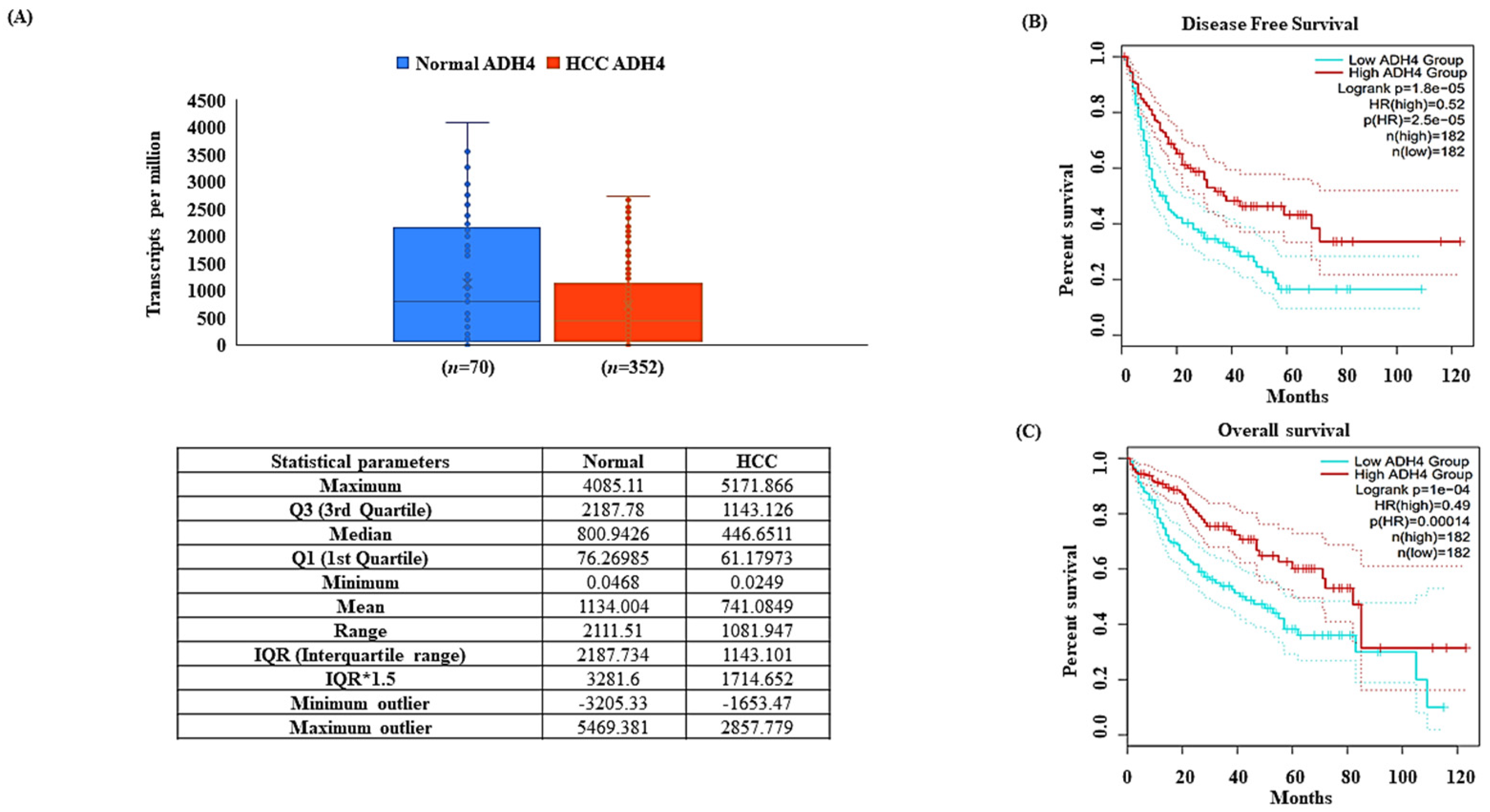
Figure 1. ADH4 is downregulated in hepatocellular carcinoma. ADH4 expression profiles (A). ADH4 mRNA expression box plot (statistical parameters included) (B). Overall survival of ADH4 in TCGA-LIHC datasets (C). Disease-free survival of ADH4 in TCGA-LIHC datasets.
Genes 13 02254 g001
Figure 2. Differential expression of MEG3 and miR-664a-3p in TCGA-LIHC. (A) MEG3 is downregulated in HCC as compared to normal liver tissues in TCGA-LIHC. (B) miR-664a-3p is upregulated in HCC as compared to normal liver tissues in TCGA-LIHC. (C) Validation of transcriptome profile of miR-664a-3p in HCC (statistical parameters included).
Genes 13 02254 g002
Figure 3. RNA heteroduplexes constructed for ADH4 mRNA and miR-664a-3p with the top five predicted miRNA–mRNA interactions. The best possible interaction had an MFE of −28.1 kcal/mol.
Genes 13 02254 g003
Figure 4. RNA heteroduplexes constructed for lncRNA-MEG3 and miR-664a-3p with the top five predicted lncRNA–miRNA interactions. The best possible model with an MFE of −31.3 kcal/mol is highlighted.
Genes 13 02254 g004
Figure 5. Prediction of accessible regions in RNA heteroduplexes. (A) Graph showing minimum free energy prediction for stable ADH4 mRNA. (B) Graph showing minimum free energy prediction for stable miR-664a-3p. (C) Minimum free energy prediction for the miRNA–mRNA hybrid. (D) Graph showing minimum free energy prediction for stable lncRNA MEG3 structure. (E) Graph showing minimum free energy graph for the stable miR-664a-3p structure. (F) Minimum free energy prediction for the lncRNA–miRNA hybrid.
Genes 13 02254 g005
Figure 6. The overall mechanism of MEG3/miR-664a-3p/ADH4 axis. (A) In normal liver, MEG3 acts as a competitive molecular sponge for miR-664a-3p and prevents miR-664a-3p-mediated RISC targeting and degradation of ADH4 mRNA. (B) In HCC, MEG3 is downregulated resulting in miR-664a-3p expression and binding to ADH4 mRNA, causing subsequent downregulation of ADH4 mRNA through RISC mediated cleavage of the mRNA.
Genes 13 02254 g006
Table 1. Patient characteristics for HPA ADH4 expression in HCC and normal liver tissues.
Patient ID SexAgeStateStainingIntensityQuantityMore Details
3402Female54NormalMediumModerate>75%https://www.proteinatlas.org/ENSG00000198099-ADH4/tissue/liver#img (accessed on 5 October 2022)
3222Female63NormalMediumModerate>75%
2429Male55NormalMediumModerate>75%
3215Female61HCCLowWeak25–75%https://www.proteinatlas.org/ENSG00000198099-ADH4/pathology/liver+cancer#img (accessed on 5 October 2022)
2766Female73HCCNot detectedNegativeNone
2177Female58HCCLowWeak25–75%
3196Male65HCCNot detectedNegativeNone
2280Male80HCCMediumModerate25–75%
2556Male72HCCLowWeak25–75%
3346Female73HCCMediumModerate25–75%
3477Male67HCCLowWeak25–75%
Publisher’s Note: MDPI stays neutral with regard to jurisdictional claims in published maps and institutional affiliations.