Article

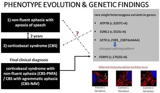
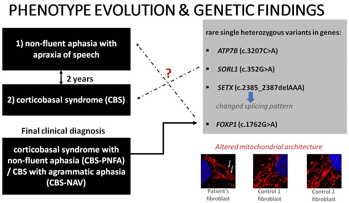
A Patient with Corticobasal Syndrome and Progressive Non-Fluent Aphasia (CBS-PNFA), with Variants in ATP7B, SETX, SORL1, and FOXP1 Genes

by
Katarzyna Gaweda-Walerych
1,*,
Emilia J. Sitek
2,3,*,
Małgorzata Borczyk
4,
Ewa Narożańska
3,
Bogna Brockhuis
5,
Michał Korostyński
4,
Michał Schinwelski
3,6,7,
Mariusz Siemiński
8,
Jarosław Sławek
3,7 and
Cezary Zekanowski
1
1
Department of Neurogenetics and Functional Genomics, Mossakowski Medical Research Institute, Polish Academy of Sciences, 02-106 Warsaw, Poland
2
Laboratory of Clinical Neuropsychology, Neurolinguistics and Neuropsychotherapy, Division of Neurological and Psychiatric Nursing, Faculty of Health Sciences, Medical University of Gdansk, 80-211 Gdansk, Poland
3
Neurology Department, St. Adalbert Hospital, Copernicus PL, 80-462 Gdansk, Poland
4
Laboratory of Pharmacogenomics, Department of Molecular Pharmacology, Maj Institute of Pharmacology Polish Academy of Sciences, 31-343 Krakow, Poland
5
Department of Nuclear Medicine, Faculty of Health Sciences, Medical University of Gdansk, 80-214 Gdansk, Poland
6
Neurocentrum Miwomed, 80-207 Gdansk, Poland
7
Division of Neurological and Psychiatric Nursing, Faculty of Health Sciences, Medical University of Gdansk, 80-211 Gdansk, Poland
8
Department of Emergency Medicine, Faculty of Health Sciences, Medical University of Gdansk, 80-210 Gdansk, Poland
*
Authors to whom correspondence should be addressed.
Genes 2022, 13(12), 2361; https://doi.org/10.3390/genes13122361
Submission received: 11 November 2022 / Revised: 8 December 2022 / Accepted: 9 December 2022 / Published: 14 December 2022
(This article belongs to the Special Issue Study on Genotypes and Phenotypes of Neurodegenerative Diseases)

Abstract

:
Our aim was to analyze the phenotypic-genetic correlations in a patient diagnosed with early onset corticobasal syndrome with progressive non-fluent aphasia (CBS-PNFA), characterized by predominant apraxia of speech, accompanied by prominent right-sided upper-limb limb-kinetic apraxia, alien limb phenomenon, synkinesis, myoclonus, mild cortical sensory loss, and right-sided hemispatial neglect. Whole-exome sequencing (WES) identified rare single heterozygous variants in ATP7B (c.3207C>A), SORL1 (c.352G>A), SETX (c.2385_2387delAAA), and FOXP1 (c.1762G>A) genes. The functional analysis revealed that the deletion in the SETX gene changed the splicing pattern, which was accompanied by lower SETX mRNA levels in the patient’s fibroblasts, suggesting loss-of-function as the underlying mechanism. In addition, the patient’s fibroblasts demonstrated altered mitochondrial architecture with decreased connectivity, compared to the control individuals. This is the first association of the CBS-PNFA phenotype with the most common ATP7B pathogenic variant p.H1069Q, previously linked to Wilson’s disease, and early onset Parkinson’s disease. This study expands the complex clinical spectrum related to variants in well-known disease genes, such as ATP7B, SORL1, SETX, and FOXP1, corroborating the hypothesis of oligogenic inheritance. To date, the FOXP1 gene has been linked exclusively to neurodevelopmental speech disorders, while our study highlights its possible relevance for adult-onset progressive apraxia of speech, which guarantees further study.

Graphical Abstract

1. Introduction

Corticobasal syndrome (CBS) and progressive non-fluent aphasia (PNFA) are predominantly sporadic syndromes [1,2]. Only c.a. one-third of CBS cases are caused by a genetic mutation in MAPT, PGRN, LRKK2, PSEN1, or C9orf72 genes [3,4,5,6], reviewed in [2,7,8], with a few predisposing factors, including MAPT H1 haplotype [9,10].
PNFA is usually associated with frontotemporal lobar degeneration (FTLD), where monogenic mutations account for about one-third of cases [11]. The PNFA phenotype has shown the strongest association with PGRN mutations [12,13,14]. Patients diagnosed clinically with distinct syndromes from the FTLD spectrum typically show symptoms of other syndromes later in the disease course. PNFA patients frequently develop CBS while individuals diagnosed initially with CBS are likely to present with non-fluent aphasia later in the disease course [15,16]. Apraxia of speech and non-fluent aphasia have (relatively recently) been acknowledged as parts of CBS [17]. CBS is characterized by strongly asymmetric presentation, comprising of at least 2 among the following motor features: (a) limb rigidity or akinesia, (b) limb dystonia, (c) limb myoclonus. Additionally, at least 2 other features should be present: (a) orobuccal or limb apraxia, (b) cortical sensory deficit, (c) alien limb phenomena [18,19]. Current criteria of corticobasal degeneration (CBD) specify the variant with non-fluent/agrammatic progressive aphasia (CBD-NAV) as one of the possible disease phenotypes [18]. This variant is associated with predominant dysfunction of the left-hemisphere and predominantly right-sided motor symptoms [18].
Of note, there have been very few genetic reports regarding the overlapping CBS-PNFA phenotype. To date, one case of symmetrical CBS with non-fluent aphasia caused by a PGRN mutation [20], two cases with CBS associated with speech apraxia caused by a MAPT mutation [21,22] and two cases sharing CBS and PPA features with a LRRK2 mutation [6] were described. However, genetic screening for PGRN and MAPT mutations was negative in a group of nine patients with PNFA associated with CBD pathology [23], suggesting that most of these overlapping phenotypes may not be monogenic, which highlights the need to look for oligogenic etiology. The implementation of whole exome/whole genome sequencing (WES/WGS) technology highlighted a complex genomic architecture of disease phenotypes, inciting researchers to revisit the classical concepts of genetic causality and monogenic disorders [24]. The evidence that oligogenic inheritance may account for a substantial fraction of atypical and unresolved cases of neurodegenerative diseases is mounting [25,26,27]. Indeed, it is estimated that c.a. 50% of sporadic patients from the FTLD spectrum carry at least one rare missense variant in neurodegenerative disease-associated genes [28]. Although there are detailed clinical and research criteria for FTLD syndromes, the clinical phenotype frequently evolves in the disease course often comprising cross-disease symptoms of variable intensity. Thus, from the point of view of genetic testing, next-generation sequencing is more adequate for the detection of the genetic variants contributing to overlapping phenotypes.
Our aim was to analyze the phenotypic-genetic correlations in a patient diagnosed with early onset CBS-NAV with predominant apraxia of speech. As polygenic etiology may be of particular relevance in overlapping dementia syndromes with early onset [29], we conducted whole exome sequencing (WES).

2. Materials and Methods

2.1. Whole Exome Sequencing (WES) Data Preprocessing

WES was performed commercially by CeGaTGmbH|Paul-Ehrlich-Str. 23|D-72076 Tübingen|Germany. The following library preparation kit was used: Twist Human Core + RefSeq + Mitochondrial Panel (Twist Bioscience); 92.37% percent of sequenced bases had a predicted quality score of 30 or more (Q30 value). Sequencing parameters were NovaSeq 6000; 2 × 100 bp. Demultiplexing of the sequencing reads was performed with Illumina bcl2fastq (2.20). Statistics of mapped reads are provided in Table 1.
Raw read files were then processed with Intelliseq Flow Annotation Pipeline (https://intelliseq.com/ accessed on 25 August 2022) built in Cromwell (https://cromwell.readthedocs.io/en/stable/ accessed on 25 August 2022). Within the pipeline, fastq file quality was assessed with FastQC. Fastq files were then aligned to the Broad Institute Hg38 Human Reference Genome with GATK 4.0.3. Duplicate reads were removed with Picard and base quality Phred scores were recalibrated using GATK’s covariance recalibration. Variants were called using GATK best practices. Identified variants were assessed using Intelliseq Flow annotation pipeline that implements the American College of Medical Genetics and Genomics (ACMG) recommendations.
In short variant annotation and annotation-based filtering, 3182 genes were included in the screening based on Human Phenotype Ontology (HPO) terms (Bioinformatics, Supplementary Table S1). Databases used for annotation included gnomAD v2.1 and v3 (frequencies, coverage, constraint), 1000Genomes (frequencies), MITOMAP (frequencies, contributed diseases), ClinVar (contributed diseases, pathogenicity), Human Phenotype Ontology (HPO) terms (inheritance mode, contributed phenotypes and diseases), UCSC (repeats, PHAST conservation scores), SIFT4G (constraint), SnpEff (predicted impact on gene product), dbSNP (rsID), Ensembl (gene and transcript information), and COSMIC (somatic mutations data). Common and low-impact variants (with a max frequency threshold of 0.05 and minimal SnpEff predicted impact on gene product set as MODERATE) were then removed. Variants with the following impacts on protein and mRNA were retained: missense, nonsense, frameshift, and splice site variants. At this step, 711 variants remained (Bioinformatics, Supplementary Table S2). Annotated variants were then classified according to the ACMG criteria and prioritized, yielding 164 variants (Bioinformatics, Supplementary Table S3). Selected variants were manually evaluated for quality in IGV and confirmed in Sanger sequencing (Genetics Supplementary Figure S1 ATP7B, Figure S5 SETX).
The C9orf72 expansion analysis was performed using previously described protocols [30] using Genetic Analyzer 3130 and SeqScape v2.5 software (Applied Biosystems, Foster City, CA, USA).

2.2. Fibroblast Cultures and Inhibition of Nonsense-Mediated Decay (NMD)

Primary skin fibroblasts (obtained from the patient and age-matched, unrelated, neurologically healthy subjects) were collected and cultured as previously described [31]. For inhibition of nonsense-mediated mRNA decay (NMD), primary fibroblast cultures were treated with an NMD inhibitor, cycloheximide (500 µg/mL), or DMSO for 10 h (Sigma Aldrich, Saint Louis, MO, USA), as previously described [31].

2.3. Reverse Transcription, mRNA Isoform Analysis, and Real-Time PCR (RT-PCR)

RNA was extracted according to standard protocol with QIAzol Lysis Reagent (Qiagen, Manchester, UK) and reverse transcribed with cDNA synthesis kit, NG dART RT (EURx Molecular Biology Products, Gdańsk, Poland; cat. no. E0801), using random hexamers primers [32]. For the PCR, AllTaq Master Mix Kit was used (Qiagen, Manchester, UK). The real-time PCR analysis was conducted with RT HS-PCR Mix SYBR (A&A BIOTECHNOLOGY, Gdańsk, Poland) using a StepOne Plus system (Applied Biosystems, Foster City, CA, USA). Changes in gene expressions were determined with the DeltaCt method using GAPDH levels for normalization. Similar results were obtained with PPIB as a housekeeping gene. Primers are listed in Supplementary Table S4. PCR products were run on 2% agarose gel, selected bands were excised from the gel and extracted with QIAquick Gel Extraction Kit (Qiagen, Manchester, UK). Sanger sequencing was performed by (https://oligo.ibb.waw.pl accessed on 2 September 2022).

2.4. Mitochondrial Network Staining and Analysis

Patient fibroblasts (P) and control fibroblasts (C1 and C2) were grown on glass coverslips in standard conditions for 24 h [31]. For mitochondrial staining, cells were washed twice with Phosphate Buffered Saline, fixed with 4% paraformaldehyde (PFA) for 10 min, blocked with Normal Goat Serum (Thermo Fisher Scientific Inc., Waltham, MA, USA; cat no. 50197Z) for one hour, incubated overnight with rabbit anti-Hsp60 (HSPD1, Heat Shock Protein Family D (Hsp60) Member 1) antibody (1:500; Abcam, Cat No. ab53109) to visualize mitochondria [33], followed by one hour of incubation with secondary anti-rabbit antibody Alexa Fluor−555 (Thermo Fisher Scientific Inc., USA; Cat No. A-31570) and 10-min incubation with DAPI to visualize cell nuclei. Images were obtained by Zeiss LSM 510 confocal microscopy under 63x objective. Mitochondrial morphology parameters, including Form Factor (reflecting mitochondrial length) and Aspect Ratio (reflecting both the length and the extent of mitochondrial branching), were assessed with ImageJ as described in [34,35,36].

2.5. Statistical Analysis

For each experiment, the relative values obtained from different biological replicates. (n = 3) were used to calculate means, medians, standard deviations (SD), and statistical significance. The Kruskal–Wallis test and two-tailed unpaired t-test with Welch’s correction were used (GraphPad Prism 6.0). p < 0.05 was considered significant.

3. Results

3.1. Case Description

The patient was followed clinically by our team from the age of 66 until her death at the age of 72. Her medical history included type 2 diabetes, hypertension, and gout. At the age of 63, she experienced minor problems when parking her car (distance judgment) (Clinical Supplementary Figure S2. Case timeline). Later, at the age of 64, she noticed speech problems (possibly apraxia of speech) and fell from a chair on wheels. Soon afterward she left her job due to speech problems. PNFA was diagnosed at the age of 66. Her speech was very effortful and non-fluent. The pattern of speech impairment indicated the predominance of speech apraxia, but mild agrammatism was also present and more evident in writing. Spontaneous falls became more frequent. She dropped objects and presented with a strong left-sided preference when navigating the environment, suggestive of right-sided unilateral neglect. She was unable to tie shoelaces, zip her jacket, and struggled to use cutlery, which corresponded to apraxia. Her poor decision-making was evidenced by her succumbing to advertising. She had preserved insight into communication problems. Apathy, mild impulsivity, and irritability were also present. Her handwriting with her right hand was extremely effortful so she switched to writing on a keyboard using her left hand. Of note, she used to be left-handed and had been forced to write with her right hand. Moreover, she used to play the accordion and her left finger dexterity could have been above average. The patient was diagnosed with possible CBD with a non-fluent/agrammatic aphasia variant (NAV) phenotype [18].
At the age of 67, she became depressed. When she was 69, her gait deteriorated further and she demonstrated apraxia in her right lower limb. At the age of 70, she walked only with assistance, her speech was limited mainly to yes/no answers, she could not initiate movement with her right upper limb, and swallowing problems were evident.
When she was 72, she was unable to maintain a sitting position and dysphagia was severe. She died a few months later at a nursing home. Proband’s father presented symptoms similar to those observed in our patient (tremors, loss of speech), at the age of 68, four years before his death.

3.1.1. Clinical Assessment

The patient underwent her first neurological examination at the age of 67. Non-fluent aphasia was the predominant syndrome with the right upper limb apraxia, involuntary mirror right limb movement, parkinsonian tremor, rigidity, and dystonia. The patient had postural instability with a tendency to fall forward. The symptoms were not levodopa responsive. With disease progression, predominantly right-sided spastic dystonia, facial dystonia, and spastic dysarthria appeared. The patient was assessed neuropsychologically four times (see: Clinical Supplementary Tables S5–S8, see also Figure 1C,D and Figure S3). At the age of 67, the patient presented with apraxia of speech, non-fluent aphasia, aprosody, right-sided unilateral neglect, ideomotor apraxia, mirror movements, synkinesis (e.g., when the patient tried to write with her right hand, lip and right leg movements were observed), alien limb phenomenon, and mild cortical sensory loss, were accompanied by executive deficits. At this time, motor hemispatial neglect was predominant. During alternate bilateral hand movements, there was reduced amplitude, and then extinction in the right hand. The severity of deficits corresponded to mild cognitive impairment as the patient’s disability was mainly due to motor impairment and aphasia. A year later, apraxia and alien limb phenomenon extended to the right lower limb and hemispatial neglect was more pronounced in visual search tasks. At the age of 68, there was a marked deterioration of language; agrammatism was present both in speech and writing. The patient was unable to perform a pegboard task with the right hand. Moreover, her tactile object recognition and finger recognition deteriorated, and cortical sensory loss was more prominent, especially when the right hand was tested. Her cognitive status progressed to mild dementia. At the last examination, when she was 69, she could hardly repeat two-syllable words and perform a simple pegboard task with her left hand due to marked hypokinesia and difficulty in moving pegs. Her visual episodic memory was still quite well preserved at this time.

3.1.2. Laboratory Testing

Repeated standard laboratory blood tests, including alanine and aspartate transferase, and an ultrasonography of the abdomen with the liver assessment were normal.
The evoked autonomic sympathetic skin potentials were within normal range, but the heart rate variability was high. Electroencephalography was normal. Electroneurography revealed no clinically significant changes.

3.1.3. Neuroimaging

Magnetic resonance imaging (MRI) of the head performed at the age of 67 demonstrated mild cortical atrophy of both frontal lobes (Figure 1A). The subtentorial atrophy did not exceed the normal range. Single photon emission computed tomography (SPECT) revealed bilateral frontal and left parietal hypoperfusion (Figure 1B).

3.1.4. Genetic Assessment

Whole exome sequencing (WES) of the patient’s DNA revealed 164 rare variants classified according to the American College of Medical Genetics and Genomics (ACMG) criteria for pathogenicity (Table S3) [37].
We focused on the rare variants with the highest putative impact on the disease phenotype (Table 2). The presence of C9orf72 expansion was excluded [30]. Among common risk variants, the patient had the APOE ε2, ε2 genotype.
According to the Human Splicing Finder prediction, the SETX variant c.2385_2387delAAA in exon 10 activates a cryptic donor site, which may lead to splicing alteration (Supplementary Material Genetics, Table S9, Scheme S1). cDNA from the patient’s fibroblasts (Figure 2A, lane 6, P) had an altered pattern of exon 10 splicing with an additional aberrant isoform (band *A), visible only upon inhibition of nonsense-mediated decay mechanism with cycloheximide (CHX) treatment. Sequencing of the band *A, excised from agarose gel (Figure 2A, line 6, band *A) confirmed that band A corresponds to splicing isoform with 358 bp of exon 10 skipped (Figure 2C, Scheme S1, Supplementary Material Genetics Figure S4). The skipping of the 358 bp fragment led to the frameshift and introduced a premature termination codon (PTC). The skipped fragment had canonical splicing consensus sequences GT-AG (donor and acceptor, respectively). However, the new alternative donor site (used during the splicing event) was located downstream of the sites predicted by HSF, which highlighted the importance of performing a functional analysis since in silico predictions may not be precise.
As a positive control, to confirm correct NMD inhibition, we used mRNA transcribed from the DSCR1 gene (the Down syndrome critical region 1), a known NMD target [39,40]. As expected, DSCR1 levels increased upon cycloheximide treatment, showing robust NMD inhibition (Figure 2B).
Sequencing of the excised band *B (Figure 2A, lane 6) revealed that it is an artifact. The sequencing of the main band (Figure 2A, line 6) confirmed 100% identity with the expected PCR product—645 bp fragment of SETX exon 10 with c.2385_2387delAAA variant (Supplementary Material Genetics Figure S5, Scheme S1). In parallel, a real-time analysis showed lower levels of SETX mRNA (Figure 2D, see also Table S4, Scheme S1).

3.1.5. Mitochondrial Network Assessment

Since SORL1, ATP7B, SETX, and FOXP1 genes have been previously linked to mitochondrial dysfunction [41,42,43,44], we performed mitochondrial staining in the patient’s fibroblasts with Hsp60, a mitochondrial chaperonin essential for protein folding and assembly in the mitochondrial matrix [33]. We observed a particular network shape in the patient with significantly altered mitochondrial parameters compared to neurologically healthy controls (Figure 3A–C). The lower aspect ratio in the patient indicated decreased mitochondrial connectivity (Figure 3B), while a lower form factor pointed to shorter (and less branched) mitochondria, compared to CTRLs (Figure 3C).

4. Discussion

The variability of CBS-NAV clinical phenotype suggests the important contribution of oligogenic background in disease development [24]. Detection of rare, putatively functional genetic variants linked to similar clinical phenotypes would corroborate this hypothesis.
We discuss below how the rare genetic variants detected in the patient (Table 2) could have contributed to the observed phenotype in light of the up-to-date literature and our clinical, and genetic analyses.

4.1. Complex Genetic Landscape

The ATP7B gene encodes a Cu-transporting P-type ATPase protein responsible for incorporating copper into apo-ceruloplasmin transporting copper in the serum. Homozygous or compound heterozygous mutations in the ATP7B gene caused Wilson disease (WD), characterized by excessive copper accumulation in various organs, such as the brain, liver, and cornea, leading to a wide spectrum of symptoms from hepatic to neuro-psychiatric [45,46,47,48]. The biochemical hallmarks of WD are decreased ceruloplasmin and elevated copper levels in blood serum.
In most WD patients, mutations are clustered in exon 14 of the ATP7B gene [49,50] that encodes part of the ATPase nucleotide-binding domain, responsible for ATP binding and enzyme activity [51]. The most common ATP7B pathogenic variant H1069Q in exon 14 accounts for 72% of WD cases in the Polish population [52,53]. Very rarely, carriers of single heterozygous ATP7B mutations (Hzgs) demonstrate clinical symptoms, and biochemical and/or brain abnormalities [54,55] (Table S10). However, recently, more disease phenotypes have been described in ATP7B Hzgs, such as early or late-onset Parkinson’s disease (EOPD or LOPD) (summarized in Table S10). Given the mounting evidence of the possible involvement of single heterozygous ATP7B mutations in phenotypes with parkinsonism (Table S10), it can be speculated that p.H1069Q mutation in our CBS-NAV patient has led to the subclinical dysregulation of copper metabolism. Motor symptoms that were present in our patient included dystonia, rigidity, and rest tremor, and they initially impaired the function of the right limbs only. The marked asymmetry observed in our case was atypical for WD. No hyper- and hypointensities in the basal ganglia in T1- and T2-weighted MRI images typical for WD were seen in our patient. However, with disease progression, our patient did develop facial dystonia and dysarthria, which are common symptoms in WD.
High heart rate variability observed in our patient might be a sign of autonomic dysfunction observed in WD patients [56]. However, a comprehensive evaluation of autonomic function in WD is lacking.
Cognitive abnormalities in WD occur mainly with regard to attention, executive functions, and memory, while in our patient, despite the presence of mild executive deficits, language impairment and apraxia were predominant. Attention was not tested in detail as speech and limb apraxia made the assessment of processing speed unfeasible.
The SETX gene codes for senataxin, a ubiquitously expressed protein that functions as a DNA/RNA helicase, that is crucial for RNA processing, DNA damage response, neurogenesis, and autophagy [57,58]. The SETX gene may be considered a cross-disease gene as its mutations cause distinct neurodegenerative phenotypes. Dominant mutations in SETX cause juvenile-onset amyotrophic lateral sclerosis type 4 (ALS4), while recessive mutations are associated with ataxia-oculomotor apraxia 2 (AOA2) characterized by cerebellar ataxia, oculomotor apraxia, and axonal sensorimotor neuropathy [57,59]. SETX mutations have been detected also in Charcot Marie Tooth (CMT), distal hereditary motor neuropathy (dHMN) [60,61], childhood apraxia of speech [62], and Alzheimer’s disease [63]. None of the symptoms inherent to ALS4 or AOA2 have been observed in our patient.
We observed decreased SETX mRNA levels in the patient’s fibroblasts suggesting loss-of-function as the underlying mechanism (Figure 2D). The SETX variant c.2385_2387delAAA detected in our patient (Table 2) altered the splicing pattern of exon 10 (Figure 2), generating the aberrant isoform with the 358 bp fragment skipped form exon 10, which was further degraded by the nonsense-mediated decay (Figure 2A, Figure S4). However, as evidenced by sequencing of the main band (Figure 2A, line 6; Figure S5), mRNA carrying the original 3 bp deletion was also generated. It cannot be excluded that the isoform with the original 3 bp deletion also contributes to the decreased levels of SETX mRNA possibly affecting the efficiency of downstream processes such as mRNA stability, maturation, transport, or translation. The production of two abnormal transcripts from the mutated allele was described previously [64,65]. For example, neurofibromatosis type 1 (NF1) mutations generated different proportions of mutated transcripts i.e., transcripts containing the exon with the original mutation and those with skipped exon [64,66]. It is worth noting that splicing of exceptionally long exons such as SETX exon 10 (4176 bp, Figure 2C, Figure S2), requires precise exon definition to ensure its proper inclusion [67]. Functional variants, such as c.2385_2387delAAA may deregulate this process.
The SORL1 (sortilin-related receptor 1) gene encodes a transmembrane protein, named sortilin-related receptor (SORLA), involved in endo-lysosomal processes, amyloid precursor protein (APP) sorting and in the degradation of amyloid-beta (Ab) peptide, responsible for Alzheimer’s disease (AD) pathology [68,69]. Initially, both common and rare SORL1 variants have been associated with AD [70,71,72], while recently, deleterious variants have been also detected in frontotemporal lobar degeneration (behavioral variant and primary progressive aphasia), and dementia with Lewy bodies (DLB) [28,63,73,74,75]. For this reason, SORL1 is considered a cross-disease gene.
A recent functional study revealed that many SORL1 rare variants resulted in altered maturation and cellular trafficking of the SORLA protein [76]. The V118M variant detected in our patient localized between two previously characterized mutations S114R and S124R in SORLA protein [76]. The S124R mutation showed a maturation profile similar to the wild-type protein, and the S114R mutation led to the decreased production of a mature, glycosylated form of the protein, which resulted in inefficient transport to the cell membrane [76]. It is thus plausible that also V118M variant may affect maturation defects and cellular localization of SORLA since position 118 in the protein is strongly conserved (phyloP100way).
The FOXP1 (Forehead Box P1) gene encodes a transcription factor regulating tissue and cell-type-specific gene transcription during development and adulthood [77]. Pathogenic variants in this gene cause neurodevelopmental speech disorders with overlapping apraxic and dysarthric features, predominant motor programming problems, and poor speech intelligibility [78]. Children and adolescents with FOXP1 syndrome present with considerable delays in language milestones [79]. Expressive deficits may be accompanied by comparable receptive problems in patients with missense variants [78] (expressive language > receptive language). Delays may also encompass motor and intellectual functions [80].
Given the frequency of the FOXP1 c.1762G>A variant identified in our proband and low scores of various predictive algorithms, ACMG = 0.0188, SIFT = 0.142; PhastCons = 1.0; GERP = 6.17; MCAP = 0.0582, it seems to be benign. However, because our patient had predominant and pronounced speech impairment, characterized by both speech apraxia and—later in the disease course—also spastic dysarthria—it cannot be excluded that FOXP1 rare variants, such as c.1762G>A, in a particular genetic or epigenetic context, can lead to progressive speech apraxia in adulthood. Such a hypothesis requires further studies. Of note, genetic variations in the FOXP1 homolog and FOXP2 gene have been previously found to modulate language performance in FTLD [81].

4.2. The significance of Motor Speech Disorders for Delineating the Patient’s Complex Clinical Phenotype

Apraxia of speech (AOS), a motor speech disorder with effortful speech, impaired sequencing of articulatory gestures, and sound-based errors, distinct from both dysarthria and aphasia, is very likely to co-occur with non-fluent aphasia. It may be both neurodevelopmental (childhood-onset) or progressive. In most cases, progressive AOS accompanies PNFA or CBS.
Heterogenous criteria are used in clinical practice to diagnose AOS [82]. In the older literature, apraxia of speech may be misnamed as dysarthria since isolated primary progressive apraxia of speech (PPAOS) was recognized in the context of neurodegenerative diseases, especially CBS, relatively recently [17].
In WD, mixed dysarthria is often observed [83], but dystonic dysarthria associated with orofacial dystonia was also reported [84]. In ATP7B heterozygous mutation carriers dysarthria has been reported [85]. In our case, orofacial dystonia appeared late in the disease course. Initially, the patient struggled to perform vertical tongue movements which corresponded to mild oral apraxia. Progressive AOS in our patient was present in the context of full-blown progressive-non fluent aphasia, accompanied by aprosody and later in the disease course, also by spastic dysarthria. Ruggeri et al. reported the presence of dysarthria (not further specified) in 6 out of 11 cases with progressive aphasia and speech apraxia in CBS [86]. Only in 1 out of 12 cases was dysarthria unaccompanied by speech apraxia.
Detailed clinical case descriptions with very long follow-ups and neuropathology data are unfortunately rarely reported. Of note, Tetzloff et al. provided a 10-year-long observation of the PPAOS case with autopsy-confirmed CBD [87]. In this case, spastic dysarthria was added to the AOS in the 8th year of the observation period, about 1–2 years prior to the patient’s death. At the same time, CBS symptoms became prominent.
In the context of FOXP1 findings, it remains to be established if FOXP1 abnormalities may influence the onset or pattern of progression of progressive AOS in adults.

4.3. Altered Mitochondrial Architecture

Finally, we provide evidence of an altered mitochondrial network in the patient (less branched mitochondria with decreased connectivity) compared to control individuals. As mentioned before, all four genes, ATP7B, SETX, SORL1, and FOXP1, have been associated with mitochondrial dysfunction [41,42,43,44], a common hallmark of many neurodegenerative conditions including PD and FTLD. Thus, we can hypothesize that the interaction among the four genes may contribute to altered mitochondria architecture in the patient.

4.4. Limitations of the Study

The patient’s clinical diagnosis was not supported by the analysis of WD metabolic markers, cerebrospinal fluid (CSF) biomarkers, dopamine transporter SPECT [88], or amyloid/tau PET imaging, which could have helped to further elucidate the etiology [89]. Moreover, the autopsy was not performed.
As DNA from the patient’s family members was not available, the co-segregation of the identified variants with the disease phenotype in the family has not been established.

5. Conclusions

The large-scale sequencing techniques have the potential to facilitate a diagnosis for intractable dementia cases with heterogenous clinical manifestations. Our results are in line with those of Ciani et al., who found that 50% of sporadic patients from the FTLD spectrum showed at least one rare missense variant in AD, PD, ALS, and LBD-associated genes [28]. Our results spoke in favor of the oligogenic inheritance in complex neurodegenerative phenotypes, such as CBS-NAV.
To our knowledge, this is the first reported case of the CBS-NAV phenotype with a single heterozygous ATP7B mutation. While parkinsonian phenotypes have been previously linked to ATP7B heterozygotes, we also described other variants of potential functional impacts in well-known disease-related genes, such as SETX, SORL1, and FOXP1. Of note, these three latter genes have been associated with speech and/or language deficits. The dissection of the molecular mechanisms underlying possible complex interactions among these genes would be a great challenge for the future.

Supplementary Materials

The following supporting information can be downloaded at: https://www.mdpi.com/article/10.3390/genes13122361/s1, Bioinformatics Supplementary: Table S1. Genes included in the screening; Table S2. WES Annotated; Table S3. WES ACMG classified variants; Genetics Supplementary: Figure S1. ATP7B Sanger sequencing; Table S4. Primers used in the study; Table S9. Human Splicing Finder analysis; Scheme S1. The scheme of aberrant splicing; Figure S4. Sequencing result of band *A; Figure S5. Sequencing result of the main band; Clinical Supplementary: Figure S2 Case Timeline; Table S5. Cognitive and behavioral screening; Figure S3. Addenbrooke’s Cognitive Examination-III; Table S6. Speech and language evaluation; Table S7. Visuospatial, calculation, memory, and executive function; Table S8. Praxis, finger identification, orientation in the body schema, cortical sensory loss and tactile recognition; Table S10. Clinical phenotypes, biochemical, and brain abnormalities observed in carriers of single heterozygous ATP7B mutation. References [90,91,92,93,94,95] are cite in the Supplementary Materials.

Author Contributions

Conceptualization, K.G.-W., E.J.S., E.N., M.B. and M.K.; methodology, K.G.-W. and M.B.; clinical diagnosis, E.N., E.J.S., B.B., M.S. (Michał Schinwelski) and M.S. (Mariusz Siemiński); software, M.B. and M.K.; validation, K.G.-W. and M.B.; formal analysis K.G.-W. and M.B.; investigation, K.G.-W., E.J.S., M.B., E.N., B.B., M.S. (Michał Schinwelski) and M.S. (Mariusz Siemiński); resources, K.G.-W., C.Z., M.K. and J.S.; data curation, M.B.; writing—original draft preparation, K.G.-W., E.J.S., E.N. and M.B.; writing—review and editing, K.G.-W., E.J.S., M.B., E.N., B.B., M.K., M.S. (Michał Schinwelski), M.S. (Mariusz Siemiński), J.S. and C.Z.; supervision, K.G.-W., E.J.S., E.N. and M.K.; project administration, K.G.-W.; funding acquisition, K.G.-W., C.Z. and J.S. All authors have read and agreed to the published version of the manuscript.

Funding

This research received no external funding. K. Gaweda-Walerych received internal Mossakowski Medical Research Institute, Polish Academy of Sciences funding (project FBW016).

Institutional Review Board Statement

The study was conducted according to the guidelines of the Declaration of Helsinki, and approved by the Bioethics Committee of the Central Clinical Hospital of the Ministry of Interior Affairs and Administration in Warsaw (protocol code 203/2020, date of approval 2 December 2020) and the Bioethical Committee of the Medical University of Gdansk (protocol code NKB.B.N/244/2017, date of approval 29 June 2017).

Informed Consent Statement

Written informed consent was obtained from all subjects to publish this paper.

Data Availability Statement

The data presented in this study are sequencing data of human samples thus cannot be openly shared due to personal data protection.

Acknowledgments

We thank the patient and his family and other participants of the study (control subjects). We would also like to thank Emanuele Buratti (ICGEB, Italy) for the critical reading of the manuscript and providing consultation on functional analysis of the splicing. We thank Magdalena Lachwa-From for the technical assistance in confocal microscopy, which was done in the Laboratory of Advanced Microscopy Techniques at MMRI PAS, Warsaw. We thank Artur Wolny (Laboratory of Imaging Tissue Structure and Function, Nencki Institute of Experimental Biology PAS, Warsaw) for the technical assistance with the analysis of mitochondrial morphology with ImageJ.

Conflicts of Interest

The authors declare no conflict of interest.

References

  1. Ramos, E.M.; Dokuru, D.R.; Van Berlo, V.; Wojta, K.; Wang, Q.; Huang, A.Y.; Miller, Z.A.; Karydas, A.M.; Bigio, E.H.; Rogalski, E.; et al. Genetic screen in a large series of patients with primary progressive aphasia. Alzheimer’s Dement. 2019, 15, 553–560. [Google Scholar] [CrossRef] [PubMed]
  2. Arienti, F.; Lazzeri, G.; Vizziello, M.; Monfrini, E.; Bresolin, N.; Saetti, M.C.; Picillo, M.; Franco, G.; Di Fonzo, A. Unravelling Genetic Factors Underlying Corticobasal Syndrome: A Systematic Review. Cells 2021, 10, 171. [Google Scholar] [CrossRef] [PubMed]
  3. Bugiani, O.; Murrell, J.R.; Giaccone, G.; Hasegawa, M.; Ghigo, G.; Tabaton, M.; Morbin, M.; Primavera, A.; Carella, F.; Solaro, C.; et al. Frontotemporal dementia and corticobasal degeneration in a family with a P301S mutation in tau. J. Neuropathol. Exp. Neurol. 1999, 58, 667–677. [Google Scholar] [CrossRef] [PubMed] [Green Version]
  4. Benussi, L.; Binetti, G.; Sina, E.; Gigola, L.; Bettecken, T.; Meitinger, T.; Ghidoni, R. A novel deletion in progranulin gene is associated with FTDP-17 and CBS. Neurobiol. Aging 2008, 29, 427–435. [Google Scholar] [CrossRef]
  5. Lindquist, S.G.; Duno, M.; Batbayli, M.; Puschmann, A.; Braendgaard, H.; Mardosiene, S.; Svenstrup, K.; Pinborg, L.H.; Vestergaard, K.; Hjermind, L.E.; et al. Corticobasal and ataxia syndromes widen the spectrum of C9ORF72 hexanucleotide expansion disease. Clin. Genet. 2013, 83, 279–283. [Google Scholar] [CrossRef] [PubMed]
  6. Chen-Plotkin, A.S.; Yuan, W.; Anderson, C.; McCarty Wood, E.; Hurtig, H.I.; Clark, C.M.; Miller, B.L.; Lee, V.M.; Trojanowski, J.Q.; Grossman, M.; et al. Corticobasal syndrome and primary progressive aphasia as manifestations of LRRK2 gene mutations. Neurology 2008, 70, 521–527. [Google Scholar] [CrossRef] [Green Version]
  7. Constantinides, V.C.; Paraskevas, G.P.; Paraskevas, P.G.; Stefanis, L.; Kapaki, E. Corticobasal degeneration and corticobasal syndrome: A review. Clin. Park. Relat. Disord. 2019, 1, 66–71. [Google Scholar] [CrossRef]
  8. Koga, S.; Josephs, K.A.; Aiba, I.; Yoshida, M.; Dickson, D.W. Neuropathology and emerging biomarkers in corticobasal syndrome. J. Neurol. Neurosurg. Psychiatry 2022, 93, 919–929. [Google Scholar] [CrossRef]
  9. Houlden, H.; Baker, M.; Morris, H.R.; MacDonald, N.; Pickering-Brown, S.; Adamson, J.; Lees, A.J.; Rossor, M.N.; Quinn, N.P.; Kertesz, A.; et al. Corticobasal degeneration and progressive supranuclear palsy share a common tau haplotype. Neurology 2001, 56, 1702–1706. [Google Scholar] [CrossRef]
  10. Kouri, N.; Ross, O.A.; Dombroski, B.; Younkin, C.S.; Serie, D.J.; Soto-Ortolaza, A.; Baker, M.; Finch, N.C.A.; Yoon, H.; Kim, J.; et al. Genome-wide association study of corticobasal degeneration identifies risk variants shared with progressive supranuclear palsy. Nat. Commun. 2015, 6, 7247. [Google Scholar] [CrossRef]
  11. Borroni, B.; Pilotto, A.; Bianchi, M.; Gilberti, N.; Padovani, A. Genetic contributors to frontotemporal lobar degeneration: Beyond monogenic disease. Mini Rev. Med. Chem. 2011, 11, 988–1001. [Google Scholar] [CrossRef] [PubMed]
  12. Snowden, J.S.; Adams, J.; Harris, J.; Thompson, J.C.; Rollinson, S.; Richardson, A.; Jones, M.; Neary, D.; Mann, D.M.; Pickering-Brown, S. Distinct clinical and pathological phenotypes in frontotemporal dementia associated with MAPT, PGRN and C9orf72 mutations. Amyotroph. Lateral Scler. Front. Degener. 2015, 16, 497–505. [Google Scholar] [CrossRef] [PubMed]
  13. Gossye, H.; Van Broeckhoven, C.; Engelborghs, S. The Use of Biomarkers and Genetic Screening to Diagnose Frontotemporal Dementia: Evidence and Clinical Implications. Front. Neurosci. 2019, 13, 757. [Google Scholar] [CrossRef] [PubMed] [Green Version]
  14. Snowden, J.S.; Pickering-Brown, S.M.; Mackenzie, I.R.; Richardson, A.M.; Varma, A.; Neary, D.; Mann, D.M. Progranulin gene mutations associated with frontotemporal dementia and progressive non-fluent aphasia. Brain 2006, 129, 3091–3102. [Google Scholar] [CrossRef] [PubMed] [Green Version]
  15. Harciarek, M.; Sitek, E.J.; Kertesz, A. The patterns of progression in primary progressive aphasia—Implications for assessment and management. Aphasiology 2014, 28, 964–980. [Google Scholar] [CrossRef]
  16. Kertesz, A.; McMonagle, P.; Blair, M.; Davidson, W.; Munoz, D.G. The evolution and pathology of frontotemporal dementia. Brain 2005, 128, 1996–2005. [Google Scholar] [CrossRef] [Green Version]
  17. Josephs, K.A.; Duffy, J.R. Apraxia of speech and nonfluent aphasia: A new clinical marker for corticobasal degeneration and progressive supranuclear palsy. Curr. Opin. Neurol. 2008, 21, 688–692. [Google Scholar] [CrossRef]
  18. Armstrong, M.J.; Litvan, I.; Lang, A.E.; Bak, T.H.; Bhatia, K.P.; Borroni, B.; Boxer, A.L.; Dickson, D.W.; Grossman, M.; Hallett, M.; et al. Criteria for the diagnosis of corticobasal degeneration. Neurology 2013, 80, 496–503. [Google Scholar] [CrossRef] [Green Version]
  19. Wilson, D.; Le Heron, C.; Anderson, T. Corticobasal syndrome: A practical guide. Pract. Neurol. 2021, 21, 276–285. [Google Scholar] [CrossRef]
  20. Dopper, E.G.; Seelaar, H.; Chiu, W.Z.; de Koning, I.; van Minkelen, R.; Baker, M.C.; Rozemuller, A.J.; Rademakers, R.; van Swieten, J.C. Symmetrical corticobasal syndrome caused by a novel C.314dup progranulin mutation. J. Mol. Neurosci. 2011, 45, 354–358. [Google Scholar] [CrossRef]
  21. Marshall, C.R.; Guerreiro, R.; Thust, S.; Fletcher, P.; Rohrer, J.D.; Fox, N.C. A Novel MAPT Mutation Causing Corticobasal Syndrome Led by Progressive Apraxia of Speech. J. Alzheimer’s Dis. 2015, 48, 923–926. [Google Scholar] [CrossRef] [PubMed]
  22. Mazzon, G.; Menichelli, A.; Fabretto, A.; Cattaruzza, T.; Manganotti, P. A new MAPT deletion in a case of speech apraxia leading to corticobasal syndrome. Neurocase 2018, 24, 140–144. [Google Scholar] [CrossRef]
  23. Santos-Santos, M.A.; Mandelli, M.L.; Binney, R.J.; Ogar, J.; Wilson, S.M.; Henry, M.L.; Hubbard, H.I.; Meese, M.; Attygalle, S.; Rosenberg, L.; et al. Features of Patients With Nonfluent/Agrammatic Primary Progressive Aphasia With Underlying Progressive Supranuclear Palsy Pathology or Corticobasal Degeneration. JAMA Neurol. 2016, 73, 733–742. [Google Scholar] [CrossRef] [PubMed]
  24. Katsanis, N. The continuum of causality in human genetic disorders. Genome Biol. 2016, 17, 233. [Google Scholar] [CrossRef] [PubMed] [Green Version]
  25. Tabuas-Pereira, M.; Guerreiro, R.; Kun-Rodrigues, C.; Almeida, M.R.; Bras, J.; Santana, I. Whole-exome sequencing reveals PSEN1 and ATP7B combined variants as a possible cause of early-onset Lewy body dementia: A case study of genotype-phenotype correlation. Neurogenetics 2022, 23, 279–283. [Google Scholar] [CrossRef]
  26. Gaweda-Walerych, K.; Sitek, E.J.; Borczyk, M.; Berdynski, M.; Narozanska, E.; Brockhuis, B.; Korostynski, M.; Slawek, J.; Zekanowski, C. Two Rare Variants in PLAU and BACE1 Genes-Do They Contribute to Semantic Dementia Clinical Phenotype? Genes 2021, 12, 1806. [Google Scholar] [CrossRef]
  27. Cochran, J.N.; McKinley, E.C.; Cochran, M.; Amaral, M.D.; Moyers, B.A.; Lasseigne, B.N.; Gray, D.E.; Lawlor, J.M.J.; Prokop, J.W.; Geier, E.G.; et al. Genome sequencing for early-onset or atypical dementia: High diagnostic yield and frequent observation of multiple contributory alleles. Mol. Case Stud. 2019, 5, a003491. [Google Scholar] [CrossRef] [Green Version]
  28. Ciani, M.; Bonvicini, C.; Scassellati, C.; Carrara, M.; Maj, C.; Fostinelli, S.; Binetti, G.; Ghidoni, R.; Benussi, L. The Missing Heritability of Sporadic Frontotemporal Dementia: New Insights from Rare Variants in Neurodegenerative Candidate Genes. Int. J. Mol. Sci. 2019, 20, 3903. [Google Scholar] [CrossRef] [Green Version]
  29. Svenningsson, P. Corticobasal degeneration: Advances in clinicopathology and biomarkers. Curr. Opin. Neurol. 2019, 32, 597–603. [Google Scholar] [CrossRef]
  30. DeJesus-Hernandez, M.; Mackenzie, I.R.; Boeve, B.F.; Boxer, A.L.; Baker, M.; Rutherford, N.J.; Nicholson, A.M.; Finch, N.A.; Flynn, H.; Adamson, J.; et al. Expanded GGGGCC hexanucleotide repeat in noncoding region of C9ORF72 causes chromosome 9p-linked FTD and ALS. Neuron 2011, 72, 245–256. [Google Scholar] [CrossRef]
  31. Gaweda-Walerych, K.; Sitek, E.J.; Narozanska, E.; Wezyk, M.; Brockhuis, B.; Zekanowski, C.; Slawek, J. Functional characterization of a novel progranulin mutation in a patient with progressive nonfluent aphasia. Neurobiol. Aging 2018, 72, 186-e9. [Google Scholar] [CrossRef] [PubMed]
  32. Leparc, G.G.; Mitra, R.D. A sensitive procedure to detect alternatively spliced mRNA in pooled-tissue samples. Nucleic Acids Res. 2007, 35, e146. [Google Scholar] [CrossRef] [PubMed]
  33. Wauters, F.; Cornelissen, T.; Imberechts, D.; Martin, S.; Koentjoro, B.; Sue, C.; Vangheluwe, P.; Vandenberghe, W. LRRK2 mutations impair depolarization-induced mitophagy through inhibition of mitochondrial accumulation of RAB10. Autophagy 2020, 16, 203–222. [Google Scholar] [CrossRef] [PubMed]
  34. Merrill, K.H.F.; Strack, S. Measuring Mitochondrial Shape with ImageJ. In Techniques to Investigate Mitochondrial Function in Neurons; Strack, S., Usachev, Y.M., Eds.; Springer: Berlin/Heidelberg, Germany, 2017. [Google Scholar]
  35. Durand, M.J.; Ait-Aissa, K.; Levchenko, V.; Staruschenko, A.; Gutterman, D.D.; Beyer, A.M. Visualization and quantification of mitochondrial structure in the endothelium of intact arteries. Cardiovasc. Res. 2019, 115, 1546–1556. [Google Scholar] [CrossRef]
  36. Onesto, E.; Colombrita, C.; Gumina, V.; Borghi, M.O.; Dusi, S.; Doretti, A.; Fagiolari, G.; Invernizzi, F.; Moggio, M.; Tiranti, V.; et al. Gene-specific mitochondria dysfunctions in human TARDBP and C9ORF72 fibroblasts. Acta Neuropathol. Commun. 2016, 4, 47. [Google Scholar] [CrossRef] [Green Version]
  37. Richards, S.; Aziz, N.; Bale, S.; Bick, D.; Das, S.; Gastier-Foster, J.; Grody, W.W.; Hegde, M.; Lyon, E.; Spector, E.; et al. Standards and guidelines for the interpretation of sequence variants: A joint consensus recommendation of the American College of Medical Genetics and Genomics and the Association for Molecular Pathology. Genet. Med. 2015, 17, 405–424. [Google Scholar] [CrossRef] [Green Version]
  38. Rentzsch, P.; Witten, D.; Cooper, G.M.; Shendure, J.; Kircher, M. CADD: Predicting the deleteriousness of variants throughout the human genome. Nucleic Acids Res. 2019, 47, D886–D894. [Google Scholar] [CrossRef]
  39. Wittmann, J.; Hol, E.M.; Jack, H.M. hUPF2 silencing identifies physiologic substrates of mammalian nonsense-mediated mRNA decay. Mol. Cell Biol. 2006, 26, 1272–1287. [Google Scholar] [CrossRef] [Green Version]
  40. Gong, C.; Kim, Y.K.; Woeller, C.F.; Tang, Y.; Maquat, L.E. SMD and NMD are competitive pathways that contribute to myogenesis: Effects on PAX3 and myogenin mRNAs. Genes Dev. 2009, 23, 54–66. [Google Scholar] [CrossRef] [Green Version]
  41. Barthelson, K.; Pederson, S.M.; Newman, M.; Lardelli, M. Brain transcriptome analysis reveals subtle effects on mitochondrial function and iron homeostasis of mutations in the SORL1 gene implicated in early onset familial Alzheimer’s disease. Mol. Brain 2020, 13, 142. [Google Scholar] [CrossRef]
  42. Renaudin, X.; Venkitaraman, A.R. A mitochondrial response to oxidative stress mediated by unscheduled RNA-DNA hybrids (R-loops). Mol. Cell Oncol. 2021, 8, 2007028. [Google Scholar] [CrossRef] [PubMed]
  43. Dev, S.; Kruse, R.L.; Hamilton, J.P.; Lutsenko, S. Wilson Disease: Update on Pathophysiology and Treatment. Front. Cell Dev. Biol. 2022, 10, 871877. [Google Scholar] [CrossRef] [PubMed]
  44. Wang, J.; Frohlich, H.; Torres, F.B.; Silva, R.L.; Poschet, G.; Agarwal, A.; Rappold, G.A. Mitochondrial dysfunction and oxidative stress contribute to cognitive and motor impairment in FOXP1 syndrome. Proc. Natl. Acad. Sci. USA 2022, 119, e2112852119. [Google Scholar] [CrossRef] [PubMed]
  45. Roberts, E.A.; Schilsky, M.L. A practice guideline on Wilson disease. Hepatology 2003, 37, 1475–1492. [Google Scholar] [CrossRef] [Green Version]
  46. Lutsenko, S. Modifying factors and phenotypic diversity in Wilson’s disease. Ann. N. Y. Acad. Sci. 2014, 1315, 56–63. [Google Scholar] [CrossRef] [Green Version]
  47. Bandmann, O.; Weiss, K.H.; Kaler, S.G. Wilson’s disease and other neurological copper disorders. Lancet Neurol. 2015, 14, 103–113. [Google Scholar] [CrossRef] [Green Version]
  48. Pfeiffer, R.F. Wilson Disease. Contin. Lifelong Learn. Neurol. 2016, 22, 1246–1261. [Google Scholar] [CrossRef]
  49. Shah, A.B.; Chernov, I.; Zhang, H.T.; Ross, B.M.; Das, K.; Lutsenko, S.; Parano, E.; Pavone, L.; Evgrafov, O.; Ivanova-Smolenskaya, I.A.; et al. Identification and analysis of mutations in the Wilson disease gene (ATP7B): Population frequencies, genotype-phenotype correlation, and functional analyses. Am. J. Hum. Genet. 1997, 61, 317–328. [Google Scholar] [CrossRef] [Green Version]
  50. Ivanova-Smolenskaya, I.A.; Ovchinnikov, I.V.; Karabanov, A.V.; Deineko, N.L.; Poleshchuk, V.V.; Markova, E.D.; Illarioshkin, S.N. The His1069Gln mutation in the ATP7B gene in Russian patients with Wilson disease. J. Med. Genet. 1999, 36, 174. [Google Scholar]
  51. Rodriguez-Granillo, A.; Sedlak, E.; Wittung-Stafshede, P. Stability and ATP binding of the nucleotide-binding domain of the Wilson disease protein: Effect of the common H1069Q mutation. J. Mol. Biol. 2008, 383, 1097–1111. [Google Scholar] [CrossRef]
  52. Gromadzka, G.; Schmidt, H.H.; Genschel, J.; Bochow, B.; Rodo, M.; Tarnacka, B.; Litwin, T.; Chabik, G.; Czlonkowska, A. Frameshift and nonsense mutations in the gene for ATPase7B are associated with severe impairment of copper metabolism and with an early clinical manifestation of Wilson’s disease. Clin. Genet. 2005, 68, 524–532. [Google Scholar] [CrossRef] [PubMed]
  53. Kucinskas, L.; Jeroch, J.; Vitkauskiene, A.; Sakalauskas, R.; Petrenkiene, V.; Kucinskas, V.; Naginiene, R.; Schmidt, H.; Kupcinskas, L. High frequency of the c.3207C>A (p.H1069Q) mutation in ATP7B gene of Lithuanian patients with hepatic presentation of Wilson’s disease. World J. Gastroenterol. 2008, 14, 5876–5879. [Google Scholar] [CrossRef] [PubMed]
  54. Tarnacka, B.; Szeszkowski, W.; Buettner, J.; Golebiowski, M.; Gromadzka, G.; Czlonkowska, A. Heterozygous carriers for Wilson’s disease--magnetic spectroscopy changes in the brain. Metab. Brain Dis. 2009, 24, 463–468. [Google Scholar] [CrossRef] [PubMed]
  55. Skowronska, M.; Litwin, T.; Kurkowska-Jastrzebska, I.; Czlonkowska, A. Transcranial sonography changes in heterozygotic carriers of the ATP7B gene. Neurol. Sci. 2020, 41, 2605–2612. [Google Scholar] [CrossRef] [PubMed]
  56. Li, K.; Lindauer, C.; Haase, R.; Rudiger, H.; Reichmann, H.; Reuner, U.; Ziemssen, T. Autonomic Dysfunction in Wilson’s Disease: A Comprehensive Evaluation during a 3-Year Follow Up. Front. Physiol. 2017, 8, 778. [Google Scholar] [CrossRef] [PubMed]
  57. Groh, M.; Albulescu, L.O.; Cristini, A.; Gromak, N. Senataxin: Genome Guardian at the Interface of Transcription and Neurodegeneration. J. Mol. Biol. 2017, 429, 3181–3195. [Google Scholar] [CrossRef] [PubMed]
  58. Richard, P.; Feng, S.; Tsai, Y.L.; Li, W.; Rinchetti, P.; Muhith, U.; Irizarry-Cole, J.; Stolz, K.; Sanz, L.A.; Hartono, S.; et al. SETX (senataxin), the helicase mutated in AOA2 and ALS4, functions in autophagy regulation. Autophagy 2021, 17, 1889–1906. [Google Scholar] [CrossRef]
  59. Grunseich, C.; Patankar, A.; Amaya, J.; Watts, J.A.; Li, D.; Ramirez, P.; Schindler, A.B.; Fischbeck, K.H.; Cheung, V.G. Clinical and Molecular Aspects of Senataxin Mutations in Amyotrophic Lateral Sclerosis 4. Ann. Neurol. 2020, 87, 547–555. [Google Scholar] [CrossRef] [Green Version]
  60. Hoyer, H.; Braathen, G.J.; Busk, O.L.; Holla, O.L.; Svendsen, M.; Hilmarsen, H.T.; Strand, L.; Skjelbred, C.F.; Russell, M.B. Genetic diagnosis of Charcot-Marie-Tooth disease in a population by next-generation sequencing. Biomed Res. Int. 2014, 2014, 210401. [Google Scholar] [CrossRef] [Green Version]
  61. Dierick, I.; Baets, J.; Irobi, J.; Jacobs, A.; De Vriendt, E.; Deconinck, T.; Merlini, L.; Van den Bergh, P.; Rasic, V.M.; Robberecht, W.; et al. Relative contribution of mutations in genes for autosomal dominant distal hereditary motor neuropathies: A genotype-phenotype correlation study. Brain 2008, 131, 1217–1227. [Google Scholar] [CrossRef] [Green Version]
  62. Worthey, E.A.; Raca, G.; Laffin, J.J.; Wilk, B.M.; Harris, J.M.; Jakielski, K.J.; Dimmock, D.P.; Strand, E.A.; Shriberg, L.D. Whole-exome sequencing supports genetic heterogeneity in childhood apraxia of speech. J. Neurodev. Disord. 2013, 5, 29. [Google Scholar] [CrossRef] [PubMed]
  63. Sun, L.; Zhang, J.; Su, N.; Zhang, S.; Yan, F.; Lin, X.; Yu, J.; Li, W.; Li, X.; Xiao, S. Analysis of Genotype-Phenotype Correlations in Patients With Degenerative Dementia Through the Whole Exome Sequencing. Front. Aging Neurosci. 2021, 13, 745407. [Google Scholar] [CrossRef] [PubMed]
  64. Pros, E.; Larriba, S.; Lopez, E.; Ravella, A.; Gili, M.L.; Kruyer, H.; Valls, J.; Serra, E.; Lazaro, C. NF1 mutation rather than individual genetic variability is the main determinant of the NF1-transcriptional profile of mutations affecting splicing. Hum. Mutat. 2006, 27, 1104–1114. [Google Scholar] [CrossRef] [PubMed]
  65. Baralle, D.; Buratti, E. RNA splicing in human disease and in the clinic. Clin. Sci. 2017, 131, 355–368. [Google Scholar] [CrossRef]
  66. Pros, E.; Gomez, C.; Martin, T.; Fabregas, P.; Serra, E.; Lazaro, C. Nature and mRNA effect of 282 different NF1 point mutations: Focus on splicing alterations. Hum. Mutat. 2008, 29, E173–E193. [Google Scholar] [CrossRef]
  67. Raponi, M.; Smith, L.D.; Silipo, M.; Stuani, C.; Buratti, E.; Baralle, D. BRCA1 exon 11 a model of long exon splicing regulation. RNA Biol. 2014, 11, 351–359. [Google Scholar] [CrossRef] [Green Version]
  68. Willnow, T.E.; Andersen, O.M. Sorting receptor SORLA--a trafficking path to avoid Alzheimer disease. J. Cell Sci. 2013, 126, 2751–2760. [Google Scholar] [CrossRef] [PubMed] [Green Version]
  69. Schmidt, V.; Subkhangulova, A.; Willnow, T.E. Sorting receptor SORLA: Cellular mechanisms and implications for disease. Cell Mol. Life Sci. 2017, 74, 1475–1483. [Google Scholar] [CrossRef] [Green Version]
  70. Rogaeva, E.; Meng, Y.; Lee, J.H.; Gu, Y.; Kawarai, T.; Zou, F.; Katayama, T.; Baldwin, C.T.; Cheng, R.; Hasegawa, H.; et al. The neuronal sortilin-related receptor SORL1 is genetically associated with Alzheimer disease. Nat. Genet. 2007, 39, 168–177. [Google Scholar] [CrossRef]
  71. Bellenguez, C.; Charbonnier, C.; Grenier-Boley, B.; Quenez, O.; Le Guennec, K.; Nicolas, G.; Chauhan, G.; Wallon, D.; Rousseau, S.; Richard, A.C.; et al. Contribution to Alzheimer’s disease risk of rare variants in TREM2, SORL1, and ABCA7 in 1779 cases and 1273 controls. Neurobiol. Aging 2017, 59, 220.e1–220.e9. [Google Scholar] [CrossRef]
  72. Campion, D.; Charbonnier, C.; Nicolas, G. SORL1 genetic variants and Alzheimer disease risk: A literature review and meta-analysis of sequencing data. Acta Neuropathol. 2019, 138, 173–186. [Google Scholar] [CrossRef] [PubMed]
  73. Ramos-Campoy, O.; Antonell, A.; Falgas, N.; Balasa, M.; Borrego-Ecija, S.; Rodriguez-Santiago, B.; Datta, D.; Armengol, L.; Fernandez-Villullas, G.; Bosch, B.; et al. Screening of dementia genes by whole-exome sequencing in Spanish patients with early-onset dementia: Likely pathogenic, uncertain significance and risk variants. Neurobiol. Aging 2020, 93, e1–e9. [Google Scholar] [CrossRef] [PubMed]
  74. Sassi, C.; Capozzo, R.; Hammer, M.; Zecca, C.; Federoff, M.; Blauwendraat, C.; Bernstein, N.; Ding, J.; Gibbs, J.R.; Price, T.; et al. Exploring dementia and neuronal ceroid lipofuscinosis genes in 100 FTD-like patients from 6 towns and rural villages on the Adriatic Sea cost of Apulia. Sci. Rep. 2021, 11, 6353. [Google Scholar] [CrossRef] [PubMed]
  75. Csaban, D.; Illes, A.; Renata, T.B.; Balicza, P.; Pentelenyi, K.; Molnar, V.; Gezsi, A.; Grosz, Z.; Gal, A.; Kovacs, T.; et al. Genetic landscape of early-onset dementia in Hungary. Neurol. Sci. 2022, 43, 5289–5300. [Google Scholar] [CrossRef]
  76. Rovelet-Lecrux, A.; Feuillette, S.; Miguel, L.; Schramm, C.; Pernet, S.; Quenez, O.; Segalas-Milazzo, I.; Guilhaudis, L.; Rousseau, S.; Riou, G.; et al. Impaired SorLA maturation and trafficking as a new mechanism for SORL1 missense variants in Alzheimer disease. Acta Neuropathol. Commun. 2021, 9, 196. [Google Scholar] [CrossRef]
  77. Co, M.; Anderson, A.G.; Konopka, G. FOXP transcription factors in vertebrate brain development, function, and disorders. Wiley Interdiscip. Rev. Dev. Biol. 2020, 9, e375. [Google Scholar] [CrossRef]
  78. Braden, R.O.; Amor, D.J.; Fisher, S.E.; Mei, C.; Myers, C.T.; Mefford, H.; Gill, D.; Srivastava, S.; Swanson, L.C.; Goel, H.; et al. Severe speech impairment is a distinguishing feature of FOXP1-related disorder. Dev. Med. Child Neurol. 2021, 63, 1417–1426. [Google Scholar] [CrossRef]
  79. Siper, P.M.; De Rubeis, S.; Trelles, M.D.P.; Durkin, A.; Di Marino, D.; Muratet, F.; Frank, Y.; Lozano, R.; Eichler, E.E.; Kelly, M.; et al. Prospective investigation of FOXP1 syndrome. Mol. Autism 2017, 8, 57. [Google Scholar] [CrossRef] [Green Version]
  80. Bacon, C.; Rappold, G.A. The distinct and overlapping phenotypic spectra of FOXP1 and FOXP2 in cognitive disorders. Hum. Genet. 2012, 131, 1687–1698. [Google Scholar] [CrossRef] [Green Version]
  81. Padovani, A.; Cosseddu, M.; Premi, E.; Archetti, S.; Papetti, A.; Agosti, C.; Bigni, B.; Cerini, C.; Paghera, B.; Bellelli, G.; et al. The speech and language FOXP2 gene modulates the phenotype of frontotemporal lobar degeneration. J. Alzheimer’s Dis. 2010, 22, 923–931. [Google Scholar] [CrossRef]
  82. Molloy, J.; Jagoe, C. Use of diverse diagnostic criteria for acquired apraxia of speech: A scoping review. Int. J. Lang. Commun. Disord. 2019, 54, 875–893. [Google Scholar] [CrossRef] [PubMed]
  83. Jauer-Niworowska. A specific picture of speech disturbances in polish speaking patients with mixed dysarthria in multiple sclerosis (MS) and in Wilson’s disease (WD). Acta Neuropsychol. 2014, 12, 155–166. [Google Scholar]
  84. Poujois, A.; Pernon, M.; Trocello, J.M.; Woimant, F. Dystonic Dysarthria in Wilson Disease: Efficacy of Zolpidem. Front. Neurol. 2017, 8, 559. [Google Scholar] [CrossRef] [PubMed] [Green Version]
  85. Ilyechova, E.Y.; Miliukhina, I.V.; Karpenko, M.N.; Orlov, I.A.; Puchkova, L.V.; Samsonov, S.A. Case of Early-Onset Parkinson’s Disease in a Heterozygous Mutation Carrier of the ATP7B Gene. J. Pers. Med. 2019, 9, 41. [Google Scholar] [CrossRef] [PubMed] [Green Version]
  86. Ruggeri, M.; Biagioli, C.; Ricci, M.; Gerace, C.; Blundo, C. Progressive aphasia, apraxia of speech and agraphia in corticobasal degeneration: A 12-case series clinical and neuropsychological descriptive study. Int. J. Lang. Commun. Disord. 2020, 55, 867–874. [Google Scholar] [CrossRef] [PubMed]
  87. Tetzloff, K.A.; Duffy, J.R.; Strand, E.A.; Machulda, M.M.; Boland, S.M.; Utianski, R.L.; Botha, H.; Senjem, M.L.; Schwarz, C.G.; Josephs, K.A.; et al. Clinical and imaging progression over 10 years in a patient with primary progressive apraxia of speech and autopsy-confirmed corticobasal degeneration. Neurocase 2018, 24, 111–120. [Google Scholar] [CrossRef]
  88. Constantinides, V.C.; Souvatzoglou, M.; Paraskevas, G.P.; Chalioti, M.; Boufidou, F.; Stefanis, L.; Kapaki, E. Dopamine transporter SPECT imaging in corticobasal syndrome: A peak into the underlying pathology? Acta Neurol. Scand. 2022, 145, 762–769. [Google Scholar] [CrossRef]
  89. Nakano, Y.; Shimada, H.; Shinotoh, H.; Hirano, S.; Tagai, K.; Sano, Y.; Yamamoto, Y.; Endo, H.; Matsuoka, K.; Takahata, K.; et al. PET-based classification of corticobasal syndrome. Park. Relat. Disord. 2022, 98, 92–98. [Google Scholar] [CrossRef]
  90. Chung, S.W.; Choi, B.M.; Kim, J.Y.; Lee, Y.S.; Yoon, J.P.; Oh, K.S.; Park, K.S. Altered Gene and Protein Expressions in Torn Rotator Cuff Tendon Tissues in Diabetic Patients. Arthroscopy 2017, 33, 518–526.e1. [Google Scholar] [CrossRef]
  91. Lefever, S.; Vandesompele, J.; Speleman, F.; Pattyn, F. RTPrimerDB: The portal for real-time PCR primers and probes. Nucleic Acids Res. 2009, 37, D942–D945. [Google Scholar] [CrossRef] [Green Version]
  92. Manoochehri, J.; Masoumi Dehshiri, R.; Faraji, H.; Mohammadi, S.; Dastsooz, H.; Moradi, T.; Rezaei, E.; Sadeghi, K.; Fardaei, M. Family screening for a novel ATP7B gene mutation, c.2335T>G, in the South of Iran. Iran. J. Ped. Hematol. Oncol. 2014, 4, 26–31. [Google Scholar] [PubMed]
  93. Sechi, G.; Antonio Cocco, G.; Errigo, A.; Deiana, L.; Rosati, G.; Agnetti, V.; Stephen Paulus, K.; Mario Pes, G. Three sisters with very-late-onset major depression and parkinsonism. Park. Relat. Disord. 2007, 13, 122–125. [Google Scholar] [CrossRef] [PubMed]
  94. Moller, J.C.; Leinweber, B.; Rissling, I.; Oertel, W.H.; Bandmann, O.; Schmidt, H.H. Prevalence of the H1069Q mutation in ATP7B in discordant pairs with early-onset Parkinson’s disease. Mov. Disord. 2006, 21, 1789–1790. [Google Scholar] [CrossRef] [PubMed]
  95. Zhang, Z.; Liu, J.; Zheng, W.; Hou, Q.; Zhang, L. A novel heterozygous carrier of ATP7B mutation with muscle weakness and tremor: A Chinese Case Report. J. Musculoskelet. Neuronal Interact. 2020, 20, 614–618. [Google Scholar]
Figure 1. Neuroimaging and neuropsychology of the patient. (A). MRI at the age of 67 y.o.—bilateral, moderate frontal lobe atrophy; (B) SPECT- bilateral frontal and left parietal hypoperfusion; (C) cube copying task performed at 3 neuropsychological assessments. Even at the last assessment, when the patient drew with her left hand, the attempt to depict three-dimensionality is evident; (D) preserved sentence copying. Despite marked right upper limb rigidity, the patient was initially able to write.
Genes 13 02361 g001
Figure 2. (A). Visualization of cDNA analysis on agarose gel. Fibroblasts from the patient (P) and controls (C1, C2) were treated with DMSO (lines 1, 3, 5) or cycloheximide (CHX) (line 2, 4, 6) for 10 h to block nonsense-mediated decay. White asterisk indicates aberrant isoform (*A) with partial skipping (358 bp) of SETX exon 10 in the patient’s fibroblasts, arising due to the presence of variant c.2385_2387delAAA, as evidenced by sequencing of band A (see Figure S4). Sequencing of the excised band *B (Figure 2A, lane 6) revealed that it is an artifact; (B) mRNA transcribed from DSCR1 gene (the Down syndrome critical region 1), a known NMD target, was used as a positive control to confirm NMD inhibition; (C) schematic representation of SETX exon 10, black empty square denotes 358 bp sequence skipped from exon 10 carrying c.2385_2387delAAA variant. Black arrows correspond to primers used for isoform analysis; (D) real-time PCR demonstrated lower levels of SETX mRNA in the patient compared to healthy controls (Table S4, Scheme S1). P was calculated in two-tailed t-test, * p < 0.05, ** p < 0.01, *** p < 0.001.
Genes 13 02361 g002
Figure 3. (A) The mitochondrial network in the patient’s fibroblasts (P) compared to neurologically healthy, age-matched controls (C1, C2). Area delineated with the white square in the upper panel is enlarged in the lower panel. White arrows point to round, fragmented mitochondria. Red: Hsp60—mitochondria, blue: DAPI—nucleus; representative images are shown; (B) decreased form factor and (C) decreased Aspect Ratio in the patient’s mitochondria compared to controls. Aspect Ratio < 0.0001, Kruskal–Wallis test; Form Factor < 0.0001, Kruskal–Wallis test; P for differences between individual cell lines: P vs. C1, P vs. C2 were calculated with unpaired t test with Welch’s correction, **** < 0.0001, ** < 0.01. All images were taken at 63× magnification. Each dot corresponds to a measurement from one cell. Upper panel scale bar = 10 µm.
Genes 13 02361 g003
Table 1. Statistics of mapped reads (DNA).
Number of
Mapped Reads
(in Millions)
Proportion of
Sequenced Reads
(in %)
PCR
Duplicates
(in %) *
Median
Insert Size
(in bp)
Average
Coverage
198.43586.712.44206213.5
(*) PCR duplicates are duplicated reads that are removed during mapping. In general, a high PCR duplicate rate correlates with low sample quality.
Table 2. The rare variants of.the highest impact on the disease phenotype according to bioinformatic priotitization.
GeneHGVS 1
DNA/Exon/Protein/rs
Predicted EffectMAF
gnomAD
CADD *
Phred
ACMG
Criteria
ATP7Bc.3207C>A
exon 14
p.His1069Gln
rs76151636
heterozygous
missense0.00101924PS1, BP4, BP5, PP3
likely pathogenic
SETXc.2385_2387delAAA
exon 10
p.Ile795_Lys796delinsMet
rs755971927
heterozygous
in frame
deletion
0.0000329NAPM2, PM4, PP4
uncertain significance
SORL1c.352G>A
p.Val118Met
exon 2
rs749389644
heterozygous
missense0.0000106028.1PM2, PP3
uncertain significance
FOXP1c.1762G>A
p.Ala588Thr
exon 20
rs202173892
heterozygous
missense0.00031821.7BP5
pathogenic/likely pathogenic or of uncertain impact
1 Variant nomenclature according to Human Genome Variation Society. *—Combined Annotation-Dependent Depletion score Phred-scaled [38].
Publisher’s Note: MDPI stays neutral with regard to jurisdictional claims in published maps and institutional affiliations.

