Article

Identification and Functional Analysis of the Regulatory Elements in the pHSPA6 Promoter

1
College of Animal Science, Jilin University, Changchun 130062, China
2
Key Lab for Zoonoses Research, Ministry of Education, Animal Genome Editing Technology Innovation Center, Jilin University, Changchun 130062, China
3
Chongqing Research Institute, Jilin University, Chongqing 401123, China
4
Chongqing Jitang Biotechnology Research Institute Co., Ltd., Chongqing 401123, China
*
Authors to whom correspondence should be addressed.
These authors contributed equally to this work.
Genes 2022, 13(2), 189; https://doi.org/10.3390/genes13020189
Submission received: 7 December 2021 / Revised: 14 January 2022 / Accepted: 19 January 2022 / Published: 21 January 2022
(This article belongs to the Section Molecular Genetics and Genomics)

Abstract

:
Functional and expressional research of heat shock protein A6 (HSPA6) suggests that the gene is of great value for neurodegenerative diseases, biosensors, cancer, etc. Based on the important value of pigs in agriculture and biomedicine and to advance knowledge of this little-studied HSPA member, the stress-sensitive sites in porcine HSPA6 (pHSPA6) were investigated following different stresses. Here, two heat shock elements (HSEs) and a conserved region (CR) were identified in the pHSPA6 promoter by a CRISPR/Cas9-mediated precise gene editing strategy. Gene expression data showed that sequence disruption of these regions could significantly reduce the expression of pHSPA6 under heat stress. Stimulation studies indicated that these regions responded not only to heat stress but also to copper sulfate, MG132, and curcumin. Further mechanism studies showed that downregulated pHSPA6 could significantly affect some important members of the HSP family that are involved in HSP40, HSP70, and HSP90. Overall, our results provide a new approach for investigating gene expression and regulation that may contribute to gene regulatory mechanisms, drug target selection, and breeding stock selection.

1. Introduction

The heat shock protein 70 (HSP70) family is a set of highly conserved proteins that are known for increased synthesis, exposing organisms to a mass of cellular stresses. More recently, a vast array of post-translational modifications (PTMs) of HSP70 family proteins has been uncovered, involving altering chaperone activity, localization, and selectivity [1]. HSPA6 is a poorly characterized member of the HSP70 family and is induced after severe cellular stress [2,3]. Heat stress is not only detrimental to animal production and reproduction but also seriously damages the health of breeding animals [4]. HSPA6 was identified to have higher expression in cattle and goats under heat stress [5,6,7]. This may be because severe stress conditions cause HSPA6 to evolve into a gene that maintains basic biological functions [8]. Therefore, HSPA6 can be used as a candidate gene for breeding heat-resistant herds [7,9]. In addition, a previous study indicated that Parkinson’s disease and Alzheimer’s disease (AD) led to 30.4-fold and 6.3-fold increases, respectively, in HSPA6 expression [10,11,12]. HSPA6 was strongly induced by MG132 treatment of human nerve cells [13,14]. Knockdown experiments showed that HSPA6 could protect human nerve cells from stress [15]. Therefore, the results implied that human nerve cells treated with MG132 could be used as cell models for studying neurodegenerative diseases. Furthermore, HSPA6 was induced and functioned as a positive regulator for Enterovirus A71 infection, indicating that HSPA6 may assist the function of a cellular protein generally required for viral IRES activities [16]. In fact, recent studies have shown that HSPA6 is also involved in cancer progression, including that of lung cancer, triple-negative breast cancer (TNBC), and bladder cancer [17,18,19]. Interestingly, the HSPA6 promoter is also widely used in the biomedical field because of its ability to control cell activity at specific sites. For example, engineered T cells can control gene expression remotely [20]. Moreover, recent findings used the HSPA6 promoter to construct a cell line that can evaluate cell sensors in response to a stimulus based on EGFP expression [21].
HSPA6 was cloned as a novel HSP70 gene that is different from others and expressed in many organisms [22]. There is no HSPA6 homolog in rodents, but HSPA6 exists in higher mammals, such as humans, goats, and swine [5,23,24]. In addition to expression among limited species, HSPA6 is barely expressed in many cells under normal culture conditions. The mRNA expression level of HSPA6 has a large increase under various cellular stresses, such as mechanical [3], copper and zinc [25], nitric oxide [26,27], heavy metal [28], and antibiotic [29] stresses. Additionally, constitutive expression of HSPA6 has been explained through the proximal promoter [30]. Deletion of the DNA fragment (−346 to −217 bp) resulted in the loss of most promoter basal activation. Furthermore, a previous assay using the DNA fragment from −287 to +110 of the HSPA6 gene (accession no. AL590385) revealed cadmium chloride-induced cytotoxicity with fourfold higher sensitivity than a cell viability test. Then, tandem repeats of the cytotoxic responding element (CRE) in the HSPA6 promoter were used to construct highly sensitive cytotoxicity sensor cells [31]. Meanwhile, some transcriptional elements were predicted, and their effect on the expression of HSPA6 was tested. AP1 plays more roles in the constitutive and inducible expression of HSPA6, whereas HSF regulates HSPA6 transcription under stress conditions [32].
After briefly summarizing the HSPA6 context, these studies mainly focused on HSPA6 promoter characterization in humans rather than pigs, which can serve as biomedical models. To further explore the pHSPA6 promoter, we identified the regulatory elements in the pHSPA6 promoter and validated their functions based on our previously established pHSPA6-based EGFP fluorescent reporter cell line using precise gene editing strategies mediated by CRISPR/Cas9 [21].

2. Materials and Methods

2.1. Cell Culture and Treatment

Porcine primary kidney cells (PK), which were isolated from 2-day-old fetuses, porcine kidney cell lines (PK-15) (Lot Number: 58808810 ATCC Number: CCL-33), and H2E were cultured in Dulbecco’s modified Eagle’s medium (DMEM) supplemented with 5% fetal bovine serum (FBS) (Gibco, Grand Island, NY, USA). Porcine fetal fibroblasts (PFFs) were isolated from 33-day-old fetuses, and porcine alveolar macrophage cell lines (3D4/2) were obtained from Prof. Li Feng of Harbin Veterinary Research Institute, and the cells were grown in DMEM supplemented with 15% FBS (Gibco, Grand Island, NY, USA). All cells were incubated at 37 °C in an atmosphere of 5% CO2, and the medium was changed every 2 days.
Cells were exposed to 42 °C for 100 min and recovered at 37 °C with 5% CO2. Concentrations of 500 μM copper sulfate, 30 μM MG-132, and 40 μM curcumin were used to stimulate cells for 100 min and cultured under standard conditions (DMEM supplemented with 5% FBS) after washing with PBS buffer solution. Finally, cells were collected at 0, 1, 2, 4, 8, 12, and 24 h post-heat/treatment for follow-up experiments.

2.2. Plasmids

sgRNAs (listed in Table 1) targeting the three regions of the pHSPA6 promoter were designed by online software (http://crispr.mit.edu/, accessed on 10 December 2020) and synthesized by Comate Bioscience Co., Ltd. (Changchun, China). Targeting sgRNAs were ligated to the PX330 vector (42230, Addgene) using the method [33] described by Zhang at the Broad Institute of MIT.
Four complementary sgRNA oligo DNAs were synthesized and annealed to double-stranded DNA in the presence of 10 × NEB standard Taq buffer, and the whole targeting plasmids formed using this product were ligated into the BbsI sites of the vector backbone (PX330).

2.3. Electroporation of H2E Cells

For electroporation, approximately 30 µg of plasmids and 3 × 10⁶ H2E cells were suspended in 300 μL of Opti-MEM (Gibco, Grand Island, NY, USA) in 2 mm gap cuvettes and electroporated using specified parameters with a BTX-ECM 2001. The electroporation parameters for H2E cells were as follows: 280 volts (V), 1 millisecond (ms), and 3 pulses for 1 repeat.

2.4. Selection of Cell Clones

At 48 h posttransfection, H2E cells were inoculated into ten 100 mm dishes at an appropriate density (1000–2000 cells/dish). Single-cell clones were picked and inoculated in 24-well plates. When the confluence reached 80% or more, 15% of each clone was digested and lysed with 10 μL NP40 lysis buffer (0.45% NP40 plus 0.6% proteinase K) for 1 h at 56 °C and 10 min at 95 °C. The lysate was used as the PCR template and was subjected to 1% agarose gel electrophoresis. To confirm the genotype of cell clones, pLB vectors (Tiangen, Beijing, China) were combined with some PCR products, and constructors were sequenced to determine the exact sequences. Finally, the cell clones were frozen in liquid nitrogen, and the locations were recorded.

2.5. PCR Detection

We performed PCR to test cell clones for site-specific knockout of CR. The primers are listed in Table 2. The PCR conditions were 95 °C for 4 min; 94 °C for 30 s, 58 °C for 30 s, 72 °C for 40 s, for 32 cycles; 72 °C for 5 min; hold at 16 °C; and the polymerase was 2× Taq PCR Mastermix (Tiangen, Beijing, China).

2.6. Quantitative Real-Time PCR Analysis

RZ reagent (Tiangen, Beijing, China) was used to extract total RNA. First-strand cDNAs were generated through reverse transcription using total RNA and oligo-dT primers. Porcine GAPDH served as the reference gene for the relative expression of pHSPA6. The 2−ΔΔCt method was performed for mRNA expression, which was standardized to the expression of endogenous U6. Primers are listed in Table 2.

2.7. Fluorescence Microscopy and Flow Cytometric Analysis

These positive clones were treated with stressors at 100 min. After 1 h incubation at 37 °C, cells were washed three times with PBS and then incubated with Hoechest 33342 diluted at 1 to 1000 for 6 min at 37 °C. After three washes with PBS, the cells were examined under a fluorescence microscope (Olympus BX51, Tokyo, Japan). The harvested cells were washed twice, resuspended in 300 µL of DPBS, and analysed using a BD Accuri C6 flow cytometer.

2.8. Statistical Analysis

The data were statistically analyzed with GraphPad Prism software (t test), and all values were considered statistically significant at p < 0.05.

3. Results

3.1. Expression of HSPA6 in Porcine Cells

To measure the expression of pHSPA6, we studied the effects on pHSPA6 expression in different types of porcine cells. Constitutive and inducible expression of pHSPA6 was examined in PK-15, PFF, and 3D4/2 cells at 37 °C and 42 °C. Constitutive expression of pHSPA6 was detected with 2977.42-, 20,190-, and 48.87-fold increases in mRNA levels post heat stress, respectively (Figure 1a). At the same time, we analysed fluorescence intensity in EGFP-KI reporter PK-15 and PFF cells to further confirm the induction of HSPA6 in the cells (Figure 1e). Then, we detected that the expression of pHSPA6 in PK cells was significantly higher than that in PK-15 cells (Figure 1b). Next, we used PK cells for passage experiments and observed a significant reduction in F3 cells compared to the expression of pHSPA6 in F0 cells (Figure 1c). Finally, a cell density assay was performed using PK cells at F0 and three cell culture densities (30%, 80%, and 130%), which showed the highest expression of pHSPA6 in low-density cell culture (30%) and significantly reduced expression of pHSPA6 when the cell density reached 80%, which may have been due to inhibition of pHSPA6 expression when the cell density exceeded 68%. However, when the cell density reached 130%, that is, the cells showed lamination, the expression of pHSPA6 did not change significantly (Figure 1d).
We concluded that cell type, cell line, generation, and cell density could impact the expression of pHSPA6. The results indicated that pHSPA6 was characterized by unnoticeable constitutive expression but was greatly induced following stress. We thus sought to identify key regulatory elements affecting pHSPA6 expression.

3.2. Effect of HSE-1 on the Expression of pHSPA6

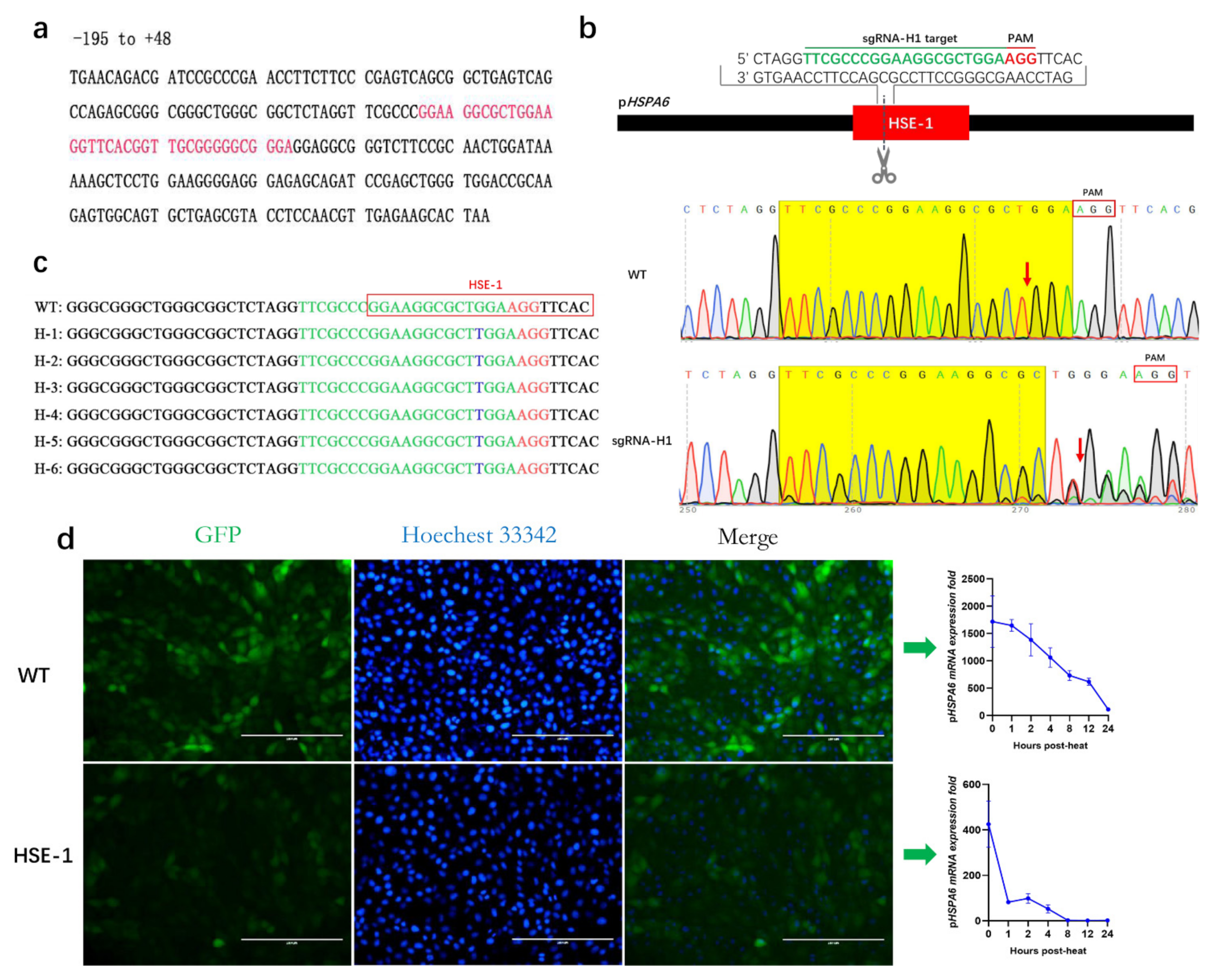
Previous analysis for heat shock element (HSE) detection revealed a site at −100 to −60 bp within the human HSPA6 promoter [22,30,32]. To validate whether the site has an impact on pHSPA6 expression, we blasted its sequence against the pHSPA6 promoter and named it HSE-1; it is located at −73~−107 bp (Figure 2a, Supplementary Figure S1a). Then, we designed a guide RNA (sgRNA-H1) to target HSE-1. Vectors combining sgRNA-H1 with Cas9 were transfected into PFFs using electroporation to assess the efficiency of sgRNA-H1. After eighty-four hours, genomic DNA was isolated from these cells, and Sanger sequencing was performed using PCR products containing the target site. Chromatograms showed obvious multipeaks around the cleavage site. The results suggested that sgRNA-H1 could be used to effectively target HSE-1 in subsequent experiments (Figure 2b).
Previous studies used the HSPA6 promoter to construct a cell line (H2E) that can evaluate cell sensors in response to a stimulus based on EGFP expression [21]. Next, sgRNA-H1-specific PX330 plasmids were injected into H2E cells through electroporation. After forty-eight hours, we used a limiting dilution method for injecting the transfected H2E cells into 100 mm dishes (1500 cells per plate), and then clones were selected. The TA cloning and Sanger sequencing results were analyzed for indels, which suggested that the genotype of the HSE-1 clone was homozygous −89 T ins (an insertion of ‘T’ at −89 bp) (Figure 2c; Supplementary Figure S1b).
Therefore, we speculated that the pHSPA6-89 T ins variant, which destroys an HSF binding site, determines the molecular phenotype. Furthermore, we also examined an association between the variant genotype and the fold change of pHSPA6 mRNA expression under heat stress, and the results suggested that the mutation could significantly reduce the mRNA expression level of pHSPA6 (by 75.23%) (Figure 2d). In addition, to explore whether the pHSPA6-89 T ins variant would influence the response of pHSPA6 to other stressors, we treated wild-type and HSE-1 clone cells with copper sulfate, MG132, and curcumin. Our results indicated that the mutation resulted in 99.54%, 99.11%, and 96.95% decreases, respectively (Supplementary Figure S1c–e). EGFP fluorescence was further confirmed via FACS analysis (Supplementary Figure S2).

3.3. Effect of HSE-2 on the Expression of pHSPA6

A previous study predicted an HSE at –284 to –265 bp within the human HSPA6 promoter but did not explain its function [32]. To identify whether the element has an impact on the expression of pHSPA6, we blasted its sequence against the pHSPA6 promoter and named it HSE-2, which is located at –254~–235 bp (Figure 3a, Supplementary Figure S3a). Then, we designed a guide RNA (sgRNA-H2) to target HSE-2 and used the same method we used for HSE-1 to evaluate the targeting efficiency of sgRNA-H2. Sanger sequencing showed obvious multipeaks near the cleavage site of Cas9. Hence, sgRNA-H2 could be used to effectively target HSE-2 in subsequent experiments (Figure 3b).
Furthermore, sgRNA-H2-specific PX330 plasmids were injected into H2E cells through electroporation, and clones were selected. The TA cloning and Sanger sequencing results were analyzed for indels, which suggested that the genotype of the HSE-2 clone was homozygous –230 10n del (10 nt deletion at –230 bp) (Figure 3c; Supplementary Figure S3b).
We thus speculated that the pHSPA6 –230 10n del variant, which destroys an HSF binding site, determines the molecular phenotype. Then, we also examined the association between the variant genotype and the fold change in pHSPA6 mRNA expression under stressors including heat, copper sulfate, MG132, and curcumin, and the results suggested that the mutation could significantly reduce the mRNA expression level of pHSPA6 (by 45.00%, 87.19%, 36.27%, and 72.37%, respectively) (Figure 3d, Supplementary Figure S3c–e). EGFP fluorescence was further confirmed via FACS analysis (Supplementary Figure S4).

3.4. Effect of the CR on the Expression of pHSPA6

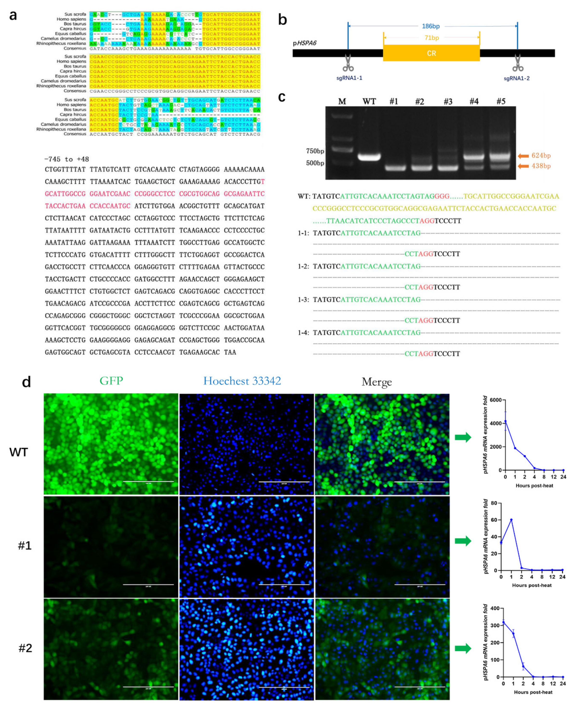
Based on homology analysis of seven species, we first found a highly conserved region (CR) located at −646 bp ~ −575 bp within the pHSPA6 promoter (Figure 4a). To detect whether CR contributes to pHSPA6 expression, we designed a pair of sgRNAs to knock out the region. Then, we used the same method we used for HSE-1 to evaluate the targeting efficiency of the pair of sgRNAs, and Sanger sequencing revealed obvious deletions between the Cas9 cleavage sites of sgRNA1-1 and sgRNA1-2. Hence, the pair of sgRNAs could be used to effectively target the CR in subsequent experiments (Figure 4b).
Next, the pair of sgRNA-specific PX330 plasmids was injected into H2E cells through electroporation, and clones were selected. According to the results of TA cloning and Sanger sequencing, we obtained two genotypes: homozygous −525 183n del (183 nt deletion at –525 bp) (#1) and heterozygous –525 183n del (#2) (Figure 4c; Supplementary Figure S5b–d). In addition, we examined the insertion of EGFP at a specific site by PCR (Supplementary Figure S5a).
Then, we also detected the fold change in pHSPA6 mRNA expression under stressors including heat, copper sulfate, MG132, and curcumin, and the results suggested that the #1 clone could significantly reduce the mRNA expression level of pHSPA6 (by 99.21%, 94.85%, 98.68%, and 94.51%, respectively) and that the #2 clone could significantly reduce the mRNA expression level of pHSPA6 (by 92.40%, 94.18%, 91.08%, and 88.66%, respectively) (Figure 4d; Supplementary Figure S6a–c). EGFP fluorescence was further confirmed via FACS analysis (Supplementary Figure S7).

3.5. Effects of the Three Regulatory Regions on the Expression of Other Genes

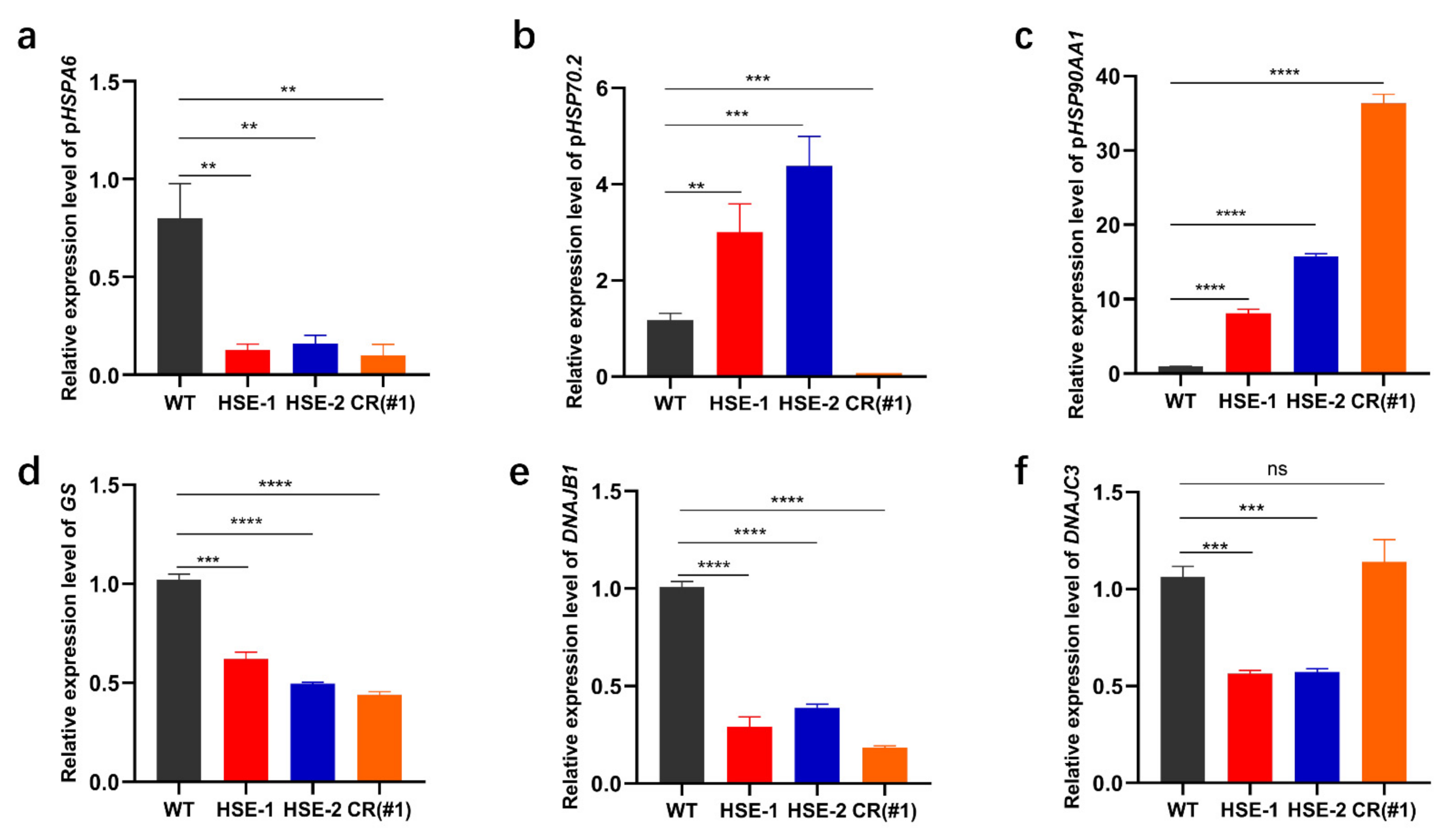
A previous study confirmed that HSPA6 induction is likely to link with some members of the HSP40 family [17]. To further determine whether decreased pHSPA6 expression could alter the expression of HSP40 family members, we measured the expression of some genes related to HSPA6 in WT and clone cells. The results indicated that GS and DNAJB1 had a positive correlation with downregulated pHSPA6 in the three clones, and DNAJC3 had a positive correlation with downregulated pHSPA6 in clones HSE-1 and HSE-2, whereas DNAJC3 was uncorrelated with CR (Figure 5a,d–f).
Since HSP70 and HSP90 play an important role in cancer and neurodegenerative diseases, we wondered whether CRISPR/Cas9 editing of HSE-1, HSE-2, and CR affected the expression of pHSP70.2 and pHSP90AA1. Therefore, the expression levels of pHSPA6, pHSP70.2, and pHSP90AA1 in the four cell lines were detected. qPCR results determined that the expression of pHSP70.2 was negatively correlated with changes in pHSPA6 in clones HSE-1 and HSE-2 but positively correlated with CR, and the expression of pHSP90AA1 was negatively correlated with changes in pHSPA6 in clones HSE-1, HSE-2, and CR (Figure 5a–c). Therefore, the expression of pHSPA6 can affect the expression of pHSP70.2 and pHSP90AA1.

4. Discussion

HSPA6 plays an important role in research on neurodegenerative diseases, tumors, biosensors, and other topics. Pigs have important research value in agriculture, biomedicine, and so on. Currently, most promoter studies of HSPA6 focus on humans, whereas pigs are studied less frequently. Here, we identified and verified the regulatory sequence of the pHSPA6 promoter based on H2E, which can evaluate cell sensors in response to a stimulus based on EGFP expression. HSPA6 is basically not expressed in different human cells without stress, but the expression level increases rapidly after stress [34]. Consistent with the results of this study, pHSPA6 also increased significantly in different porcine cells after stress, while the increases were not consistent among cell types. This may be due to differences in the sensitivity of different types of porcine cells to the same stressor (Figure 1a). Then, we found that compared with that in porcine kidney cell lines, the expression of pHSPA6 was higher in porcine primary kidney cells after heating. The results of the passage test proved that the subgenerations were related to the expression of pHSPA6. Previous research has shown that HSPA6 expression depends on the number of cells; therefore, we also performed a cell density test. The highest expression of pHSPA6 was detected in low-density cell culture (30%), and the expression of pHSPA6 was significantly reduced when the cell density reached 80%, which may have been due to pHSPA6 expression inhibition when the cell density exceeded 68%. However, when the cell density reached 130%, that is, the cells showed lamination, the expression of pHSPA6 did not change significantly. Hence, the results were consistent with those of a previous study [35].
The expression of HSPA6 is strictly induced, which is necessarily related to the regulatory elements in the promoter of HSPA6. Previous studies have reported that compared with other HSP70 promoters with basic expression, the upstream promoter elements, TATA box and CAAT box, of HSPA6 show nucleotide variation, and mutation analysis of these two elements indicated that they are necessary for basic expression of the human HSP70 gene [36]. In addition, it has been determined that RNA polymerase II is suspended on the HSP70 promoter under normal conditions but ceases ahead of activation with stress-induced HSF-1, leading to initiation of transcription and rapid induction [37,38]. When cells or bodies are subjected to physiological stress (such as heat), HSF1 oligomerizes to convert monomers to homotrimers, which translocate to the nucleus and bind to the HSE, thus inducing transcription of heat shock genes [39]. Two HSEs were edited by CRISPR/Cas9 technology, and our findings suggested that nucleotide variation in the two HSEs significantly changed the expression of pHSPA6. This may be because the mutation destroys the HSE sequence and prevents HSF from binding to it; thus, it would reduce the promoter activity of HSPA6 and the expression of the gene. Interestingly, compared with the deletion of 10 bases in the HSE sequence, the insertion of a base (T) at a specific location could reduce pHSPA6 expression more. Furthermore, CR inhibited most of the promoter activity of pHSPA6, suggesting that this region holds a crucial position in the induction of pHSPA6 expression.
The promoter of HSPA6 can be used to construct biosensors based on the characteristics of strict induction. The sensitivity of the biosensor can be improved by modifying the regulatory element of the HSPA6 promoter. A previous study placed CRE and mutated AP-1 in front of the HSPA6 promoter to improve sensor sensitivity for cadmium chloride [31]. HSE-1, HSE-2, and CR significantly reduced the expression of pHSPA6. We might well use these regions to improve the sensitivity of biosensors by replicating them in tandem to overexpress pHSPA6.
Various cellular stresses can greatly induce the expression of HSPA6, such as mechanical [3], copper and zinc [25], nitric oxide [26,27], heavy metal [28], and antibiotic [29] stress. MG132, as a proteasome inhibitor, can cause protein misfolding, inducing HSPA6 expression to promote protein refolding [40]. Curcumin, a root extract, has antitumor effects and can also induce HSPA6 expression. Here, cell lines were treated by heating, copper sulfate, MG132, and curcumin. The results showed that all four treatments could induce pHSPA6 expression in WT cells, which was considered the same as our previous report [21]. However, after the three regulatory sequences were edited, the expression of pHSPA6 decreased significantly, indicating that the three regulatory sequences responded not only to heat stress but also to copper sulfate, MG132, and curcumin.
HSP40, HSP70, and HSP90 have been well studied. Hsp40/DnaJ, as a co-partner, helps to achieve the complex functions of HSP70 members. The editing of three regulatory sequences in the pHSPA6 promoter can reduce the expression levels of GS, DNAJB1, and DNAJC3, but the expression of DNAJC3 was uncorrelated with the changes in pHSPA6 in clone CR. These data showed that the downregulation of pHSPA6 could affect the expression of the genes. This may be because induced HSPA6 interacts with some members of the HSP40 family [17]. HSP70 inhibitors would be a new strategy for enhancing cancer treatment due to the antiapoptotic activity of the HSP70 gene [41]. It is worth noting that these inhibitors have been hampered when used in patients due to toxicity [42]. DNAJA1 acts as a co-partner to regulate the expression of HSP70, and the study suggests that inhibition of DNAJA1 may be a novel anticancer strategy [43]. In addition, targeting the HSP40/HSP70 partner axis may also be feasible for the treatment of castration-resistant prostate cancer [44]. Interestingly, HSP90 is also associated with the development of a variety of cancers, but current HSP90 inhibitors do not have ideal clinical effects [45]. Therefore, HSP40s, HSP70, and HSP90 can be considered potential therapeutic targets for cancers. HSP70 and HSP90 also play an important role in neurodegenerative diseases [46,47]. Previous studies have shown that inhibition of HSP90 can promote the expression of HSAP6 [48], but the effect of HSPA6 expression on HSP70 and HSP90 has not been studied, and the results of our study help fill this gap. Interestingly, although the editing of all three regulatory sequences reduces the expression of pHSPA6, different regulatory regions have different effects on the expression of pHSP70.2 and pHSP90AA1. The expression of pHSP70.2 was negatively correlated with downregulated pHSPA6 in clones HSE-1 and HSE-2 but positively correlated with CR, and the expression of pHSP90AA1 was negatively correlated with downregulated pHSPA6 in clones HSE-1, HSE-2, and CR (Figure 5a–c). Therefore, three regulatory regions in the pHSPA6 promoter are potential drug targets for the study of neurodegenerative diseases and cancer. Meanwhile, HSP70 and HSP90 family members have a significant role in animal thermotolerance [49,50]. Thus, these regions contribute to breeding heat-resistant herds.

5. Conclusions

In summary, our study reveals two HSEs and a newly discovered CR in the pHSPA6 promoter by a CRISPR/Cas9-mediated precise gene editing strategy. Gene expression data showed that sequence disruption of these regions could significantly reduce the expression of pHSPA6 under heat stress. Stimulation studies indicated that these regions responded not only to heat stress but also to copper sulfate, MG132, and curcumin. Among these regions, the knockout of CR could more strongly reduce the expression of pHSPA6 under stressors. Further mechanistic studies showed that downregulated pHSPA6 could significantly affect some important members of the HSP family that are involved in heat resistance and many diseases. Therefore, our novel results may provide a potential therapeutic strategy for neurodegenerative diseases and cancers, as well as beneficial strategies for breeding heat-resistant herds and research on biosensors.

Supplementary Materials

The following are available online at https://www.mdpi.com/article/10.3390/genes13020189/s1, Figure S1: Identification of HSE-1 and its effect on pHSPA6 expression. Figure S2: EGFP fluorescence in WT and HSE-1 cells was confirmed via FACS. Figure S3: Identification of HSE-2 and its effect on pHSPA6 expression. Figure S4. EGFP fluorescence in WT and HSE-2 cells was confirmed via FACS. Figure S5: Selection of CR knockout cell clones. Figure S6: pHSPA6 expression analysis on CR under stressors. Figure S7. EGFP fluorescence in WT, #1 and #2 cells was confirmed via FACS.

Author Contributions

Conceptualization, Data curation, Methodology, Writing-original draft: S.J. and C.B.; Investigation, Methodology: C.Q., H.W. and L.H.; Formal Analysis: F.L., K.Y. and C.Z.; Methodology, Writing-review & editing: H.O. and D.P.; Investigation, Writing-review & editing, Formal analysis, Writing-original draft: X.T. and Z.X. All authors have read and agreed to the published version of the manuscript.

Funding

This work was financially supported by the National Key Research and Development Program of China Stem Cell and Translational Research (2019YFA0110702), Youth Program of the National Natural Science Foundation of China (31902229), and the Program for JLU Science and Technology Innovative Research Team (2017TD-28).

Institutional Review Board Statement

Not applicable.

Informed Consent Statement

Not applicable.

Data Availability Statement

Data are contained within the article or Supplementary Materials.

Conflicts of Interest

The authors declare no conflict of interest.

References

  1. Nitika; Porter, C.M.; Truman, A.W.; Truttmann, M.C. Post-translational modifications of Hsp70 family proteins: Expanding the chaperone code. J. Biol. Chem. 2020, 295, 10689–10708. [Google Scholar] [CrossRef]
  2. Noonan, E.J.; Place, R.F.; Giardina, C.; Hightower, L.E. Hsp70B′ regulation and function. Cell Stress Chaperon. 2007, 12, 219–229. [Google Scholar] [CrossRef] [Green Version]
  3. Xu, Q.; Schett, G.; Li, C.; Hu, Y.; Wick, G. Mechanical stress-induced heat shock protein 70 expression in vascular smooth muscle cells is regulated by Rac and Ras small G proteins but not mitogen-activated protein kinases. Circ. Res. 2000, 86, 1122–1128. [Google Scholar] [CrossRef] [Green Version]
  4. Hyder, I.; Sejian, V.; Bhatta, R.; Gaughan, J.B. Biological role of melatonin during summer season related heat stress in livestock. Biol. Rhythm. Res. 2017, 48, 297–314. [Google Scholar] [CrossRef]
  5. Banerjee, D.; Upadhyay, R.C.; Chaudhary, U.B.; Kumar, R.; Singh, S.; Ashutosh, G.J.M.; Polley, S.; Mukherjee, A.; Das, T.K.; De, S. Seasonal variation in expression pattern of genes under HSP70. Cell Stress Chaperon. 2014, 19, 401–408. [Google Scholar] [CrossRef] [PubMed] [Green Version]
  6. Mohanarao, G.J.; Mukherjee, A.; Banerjee, D.; Gohain, M.; Dass, G.; Brahma, B.; Datta, T.K.; Upadhyay, R.C.; De, S. HSP70 family genes and HSP27 expression in response to heat and cold stress in vitro in peripheral blood mononuclear cells of goat (Capra hircus). Small Rumin. Res. 2014, 116, 94–99. [Google Scholar] [CrossRef]
  7. Rocha, R.; Baena, M.M.; Estopa, A.D.C.; Gervásio, I.C.; Ibelli, A.M.G.; Gionbelli, T.R.S.; Gionbelli, M.P.; De Freitas, R.T.F.; Meirelles, S.L.C. Differential expression of HSF1 and HSPA6 genes and physiological responses in Angus and Simmental cattle breeds. J. Therm. Biol. 2019, 84, 92–98. [Google Scholar] [CrossRef]
  8. Hageman, J.; Van Waarde-Verhagen, M.; Zylicz, A.; Walerych, D.; Kampinga, H.H. The diverse members of the mammalian HSP70 machine show distinct chaperone-like activities. Biochem. J. 2011, 435, 127–142. [Google Scholar] [CrossRef] [PubMed] [Green Version]
  9. Hyder, I.; Pasumarti, M.; Reddy, P.R.; Prasad, C.S.; Kumar, K.A.; Sejian, V. Thermotolerance in Domestic Ruminants: A HSP70 Perspective. In Heat Shock Proteins and Stress; Springer: Singapore, 2017; Volume 12, pp. 3–35. [Google Scholar]
  10. Santpere, G.; Esparcia, P.G.; Benito, P.A.; Lorente-Galdos, B.; Navarro, A.; Ferrer, I. Transcriptional network analysis in frontal cortex in Lewy body diseases with focus on dementia with Lewy bodies. Brain Pathol. 2018, 28, 315–333. [Google Scholar] [CrossRef] [Green Version]
  11. Henderson-Smith, A.; Corneveaux, J.J.; De Both, M.; Cuyugan, L.; Liang, W.S.; Huentelman, M.; Adler, C.; Driver-Dunckley, E.; Beach, T.G.; Dunckley, T.L. Next-generation profiling to identify the molecular etiology of Parkinson dementia. Neurol. Genet. 2016, 2, e75. [Google Scholar] [CrossRef] [PubMed] [Green Version]
  12. Annese, A.; Manzari, C.; Lionetti, C.; Picardi, E.; Horner, D.S.; Chiara, M.; Caratozzolo, M.F.; Tullo, A.; Fosso, B.; Pesole, G.; et al. Whole transcriptome profiling of Late-Onset Alzheimer’s Disease patients provides insights into the molecular changes involved in the disease. Sci. Rep. 2018, 8, 4282. [Google Scholar] [CrossRef] [Green Version]
  13. Becirovic, L.; Brown, I.R. Targeting of Heat Shock Protein HSPA6 (HSP70B′) to the Periphery of Nuclear Speckles is Disrupted by a Transcription Inhibitor Following Thermal Stress in Human Neuronal Cells. Neurochem. Res. 2016, 42, 406–414. [Google Scholar] [CrossRef]
  14. Deane, C.; Brown, I.R. Differential Targeting of Hsp70 Heat Shock Proteins HSPA6 and HSPA1A with Components of a Protein Disaggregation/Refolding Machine in Differentiated Human Neuronal Cells following Thermal Stress. Front. Neurosci. 2017, 11, 227. [Google Scholar] [CrossRef] [PubMed]
  15. Deane, C.; Brown, I.R. Knockdown of Heat Shock Proteins HSPA6 (Hsp70B’) and HSPA1A (Hsp70-1) Sensitizes Differentiated Human Neuronal Cells to Cellular Stress. Neurochem. Res. 2017, 43, 340–350. [Google Scholar] [CrossRef]
  16. Su, Y.-S.; Hwang, L.-H.; Chen, C.-J. Heat Shock Protein A6, a Novel HSP70, Is Induced During Enterovirus A71 Infection to Facilitate Internal Ribosomal Entry Site-Mediated Translation. Front. Microbiol. 2021, 12, 664955. [Google Scholar] [CrossRef]
  17. Wang, L.; Hou, J.; Wang, J.; Zhu, Z.; Zhang, W.; Zhang, X.; Shen, H.; Wang, X. Regulatory roles of HSPA6 in Actinidia chinensis Planch. root extract (acRoots)-inhibited lung cancer proliferation. Clin. Transl. Med. 2020, 10, e46. [Google Scholar] [CrossRef] [Green Version]
  18. Shen, S.; Wei, C.; Fu, J. RNA-Sequencing Reveals Heat Shock 70-kDa Protein 6 (HSPA6) as a Novel Thymoquinone-Upregulated Gene That Inhibits Growth, Migration, and Invasion of Triple-Negative Breast Cancer Cells. Front. Oncol. 2021, 11, 667995. [Google Scholar] [CrossRef]
  19. Shin, S.-S.; Song, J.-H.; Hwang, B.; Noh, D.-H.; Park, S.L.; Kim, W.T.; Kim, W.-J.; Moon, S.-K. HSPA6 augments garlic extract-induced inhibition of proliferation, migration, and invasion of bladder cancer EJ cells; Implication for cell cycle dysregulation, signaling pathway alteration, and transcription factor-associated MMP-9 regulation. PLoS ONE 2017, 12, e0171860. [Google Scholar] [CrossRef] [PubMed] [Green Version]
  20. Miller, I.C.; Castro, M.G.; Maenza, J.; Weis, J.P.; Kwong, G.A. Remote Control of Mammalian Cells with Heat-Triggered Gene Switches and Photothermal Pulse Trains. ACS Synth. Biol. 2018, 7, 1167–1173. [Google Scholar] [CrossRef] [PubMed] [Green Version]
  21. Xie, Z.; Sun, R.; Qi, C.; Jiao, S.; Jiang, Y.; Liu, Z.; Zhao, D.; Liu, R.; Li, Q.; Yang, K.; et al. Generation of a pHSPA6 gene-based multifunctional live cell sensor. Biochim. Biophys. Acta (BBA) Bioenerg. 2021, 1868, 118919. [Google Scholar] [CrossRef]
  22. Leung, T.K.; Rajendran, M.Y.; Monfries, C.; Hall, C.; Lim, L. The human heat-shock protein family. Expression of a novel heat-inducible HSP70 (HSP70B′) and isolation of its cDNA and genomic DNA. Biochem. J. 1990, 267, 125–132. [Google Scholar] [CrossRef] [Green Version]
  23. Dezeure, F.; Vaiman, M.; Chardon, P. Characterization of a polymorphic heat shock protein 70 gene in swine outside the SLA major histocompatibility complex. Biochim. Biophys. Acta (BBA) Gene Struct. Expr. 1993, 1174, 17–26. [Google Scholar] [CrossRef]
  24. Parsian, A.J.; Sheren, J.E.; Tao, T.Y.; Goswami, P.; Malyapa, R.; Van Rheeden, R.; Watson, M.S.; Hunt, C.R. The human Hsp70B gene at the HSPA7 locus of chromosome 1 is transcribed but non-functional. Biochim. Biophys. Acta (BBA) Gene Struct. Expr. 2000, 1494, 201–205. [Google Scholar] [CrossRef]
  25. Hatayama, T.; Asai, Y.; Wakatsuki, T.; Kitamura, T.; Imahara, H. Regulation of hsp70 Synthesis Induced by Cupric Sulfate and Zinc Sulfate in Thermotolerant HeLa Cells. J. Biochem. 1993, 114, 592–597. [Google Scholar] [CrossRef] [PubMed]
  26. Byrne, C.R.; Hanson, P.J. Induction of heat shock protein 72 by a nitric oxide donor in guinea-pig gastric mucosal cells. Eur. J. Pharmacol. 1998, 353, 117–122. [Google Scholar] [CrossRef]
  27. Xu, Q.; Hu, Y.; Kleindienst, R.; Wick, G. Nitric oxide induces heat-shock protein 70 expression in vascular smooth muscle cells via activation of heat shock factor 1. J. Clin. Investig. 1997, 100, 1089–1097. [Google Scholar] [CrossRef] [PubMed] [Green Version]
  28. Levinson, W.; Oppermann, H.; Jackson, J. Transition series metals and sulfhydryl reagents induce the synthesis of four proteins in eukaryotic cells. Biochim. Biophys. Acta (BBA) Nucleic Acids Protein Synth. 1980, 606, 170–180. [Google Scholar] [CrossRef]
  29. Romano, C.; Benedetto, N.; Catania, M.; Rizzo, A.; Gallè, F.; Losi, E.; Hasty, D.; Rossano, F. Commonly used antibiotics induce expression of Hsp 27 and Hsp 60 and protect human lymphocytes from apoptosis. Int. Immunopharmacol. 2004, 4, 1067–1073. [Google Scholar] [CrossRef]
  30. Rohmer, S.; Mainka, A.; Knippertz, I.; Hesse, A.; Nettelbeck, D.M. Insulated hsp70B′ promoter: Stringent heat-inducible activity in replication-deficient, but not replication-competent adenoviruses. J. Gene Med. 2008, 10, 340–354. [Google Scholar] [CrossRef]
  31. Wada, K.-I.; Taniguchi, A.; Okano, T. Highly sensitive detection of cytotoxicity using a modified HSP70B′ promoter. Biotechnol. Bioeng. 2007, 97, 871–876. [Google Scholar] [CrossRef]
  32. Ramirez, V.P.; Stamatis, M.; Shmukler, A.; Aneskievich, B.J. Basal and stress-inducible expression of HSPA6 in human keratinocytes is regulated by negative and positive promoter regions. Cell Stress Chaperon. 2014, 20, 95–107. [Google Scholar] [CrossRef] [PubMed] [Green Version]
  33. Ran, F.A.; Hsu, P.D.; Lin, C.-Y.; Gootenberg, J.S.; Konermann, S.; Trevino, A.E.; Scott, D.A.; Inoue, A.; Matoba, S.; Zhang, Y.; et al. Double Nicking by RNA-Guided CRISPR Cas9 for Enhanced Genome Editing Specificity. Cell 2013, 154, 1380–1389. [Google Scholar] [CrossRef] [Green Version]
  34. Chow, A.M.; Mok, P.; Xiao, D.; Khalouei, S.; Brown, I.R. Heteromeric complexes of heat shock protein 70 (HSP70) family members, including Hsp70B′, in differentiated human neuronal cells. Cell Stress Chaperon. 2010, 15, 545–553. [Google Scholar] [CrossRef] [PubMed] [Green Version]
  35. Noonan, E.J.; Place, R.F.; Rasoulpour, R.J.; Giardina, C.; Hightower, L.E. Cell number-dependent regulation of Hsp70B′ expression: Evidence of an extracellular regulator. J. Cell. Physiol. 2007, 210, 201–211. [Google Scholar] [CrossRef] [PubMed]
  36. Greene, J.M.; Larin, Z.; Taylor, I.C.; Prentice, H.; A Gwinn, K.; E Kingston, R. Multiple basal elements of a human hsp70 promoter function differently in human and rodent cell lines. Mol. Cell. Biol. 1987, 7, 3646–3655. [Google Scholar] [CrossRef]
  37. Giardina, C.; Pérez-Riba, M.; Lis, J.T. Promoter melting and TFIID complexes on Drosophila genes in vivo. Genes Dev. 1992, 6, 2190–2200. [Google Scholar] [CrossRef] [Green Version]
  38. Lis, J.; Wu, C. Protein traffic on the heat shock promoter: Parking, stalling, and trucking along. Cell 1993, 74, 1–4. [Google Scholar] [CrossRef]
  39. Tang, H.; Liu, Y.; Madabusi, L.; Gilmour, D.S. Promoter-Proximal Pausing on the hsp70 Promoter in Drosophila melanogaster Depends on the Upstream Regulator. Mol. Cell. Biol. 2000, 20, 2569–2580. [Google Scholar] [CrossRef] [Green Version]
  40. Deane, C.A.; Brown, I.R. Intracellular Targeting of Heat Shock Proteins in Differentiated Human Neuronal Cells Following Proteotoxic Stress. J. Alzheimer’s Dis. 2018, 66, 1295–1308. [Google Scholar] [CrossRef]
  41. Tukaj, S. Heat Shock Protein 70 as a Double Agent Acting Inside and Outside the Cell: Insights into Autoimmunity. Int. J. Mol. Sci. 2020, 21, 5298. [Google Scholar] [CrossRef]
  42. Evans, C.G.; Chang, C.Y.; Gestwicki, J.E. Heat Shock Protein 70 (Hsp70) as an Emerging Drug Target. J. Med. Chem. 2010, 53, 4585–4602. [Google Scholar] [CrossRef] [PubMed] [Green Version]
  43. Nitika; Blackman, J.S.; Knighton, L.E.; Takakuwa, J.E.; Calderwood, S.K.; Truman, A.W. Chemogenomic screening identifies the Hsp70 co-chaperone DNAJA1 as a hub for anticancer drug resistance. Sci. Rep. 2020, 10, 13831. [Google Scholar] [CrossRef] [PubMed]
  44. Moses, M.A.; Kim, Y.S.; Rivera-Marquez, G.M.; Oshima, N.; Watson, M.J.; Beebe, K.E.; Wells, C.; Lee, S.; Zuehlke, A.D.; Shao, H.; et al. Targeting the Hsp40/Hsp70 Chaperone Axis as a Novel Strategy to Treat Castration-Resistant Prostate Cancer. Cancer Res. 2018, 78, 4022–4035. [Google Scholar] [CrossRef] [Green Version]
  45. Tukaj, S.; Węgrzyn, G. Anti-Hsp90 therapy in autoimmune and inflammatory diseases: A review of preclinical studies. Cell Stress Chaperon. 2016, 21, 213–218. [Google Scholar] [CrossRef] [PubMed] [Green Version]
  46. Hu, F.; Zhou, J.; Lu, Y.; Guan, L.; Wei, N.-N.; Tang, Y.-Q.; Wang, K. Inhibition of Hsp70 Suppresses Neuronal Hyperexcitability and Attenuates Epilepsy by Enhancing A-Type Potassium Current. Cell Rep. 2019, 26, 168–181.e4. [Google Scholar] [CrossRef] [Green Version]
  47. Luo, W.; Sun, W.; Taldone, T.; Rodina, A.; Chiosis, G. Heat shock protein 90 in neurodegenerative diseases. Mol. Neurodegener. 2010, 5, 24. [Google Scholar] [CrossRef] [Green Version]
  48. Kuballa, P.; Baumann, A.-L.; Mayer, K.; Bär, U.; Burtscher, H.; Brinkmann, U. Induction of heat shock protein HSPA6 (HSP70B′) upon HSP90 inhibition in cancer cell lines. FEBS Lett. 2015, 589, 1450–1458. [Google Scholar] [CrossRef] [Green Version]
  49. Badri, T.M.; Chen, K.L.; Alsiddig, M.A.; Li, L.; Cai, Y.; Wang, G.L. Genetic polymorphism in Hsp90AA1 gene is associated with the thermotolerance in Chinese Holstein cows. Cell Stress Chaperon. 2018, 23, 639–651. [Google Scholar] [CrossRef]
  50. Hassan, F.; Nawaz, A.; Rehman, M.S.; Ali, M.A.; Dilshad, S.M.; Yang, C. Prospects of HSP70 as a genetic marker for thermo-tolerance and immuno-modulation in animals under climate change scenario. Anim. Nutr. 2019, 5, 340–350. [Google Scholar] [CrossRef]
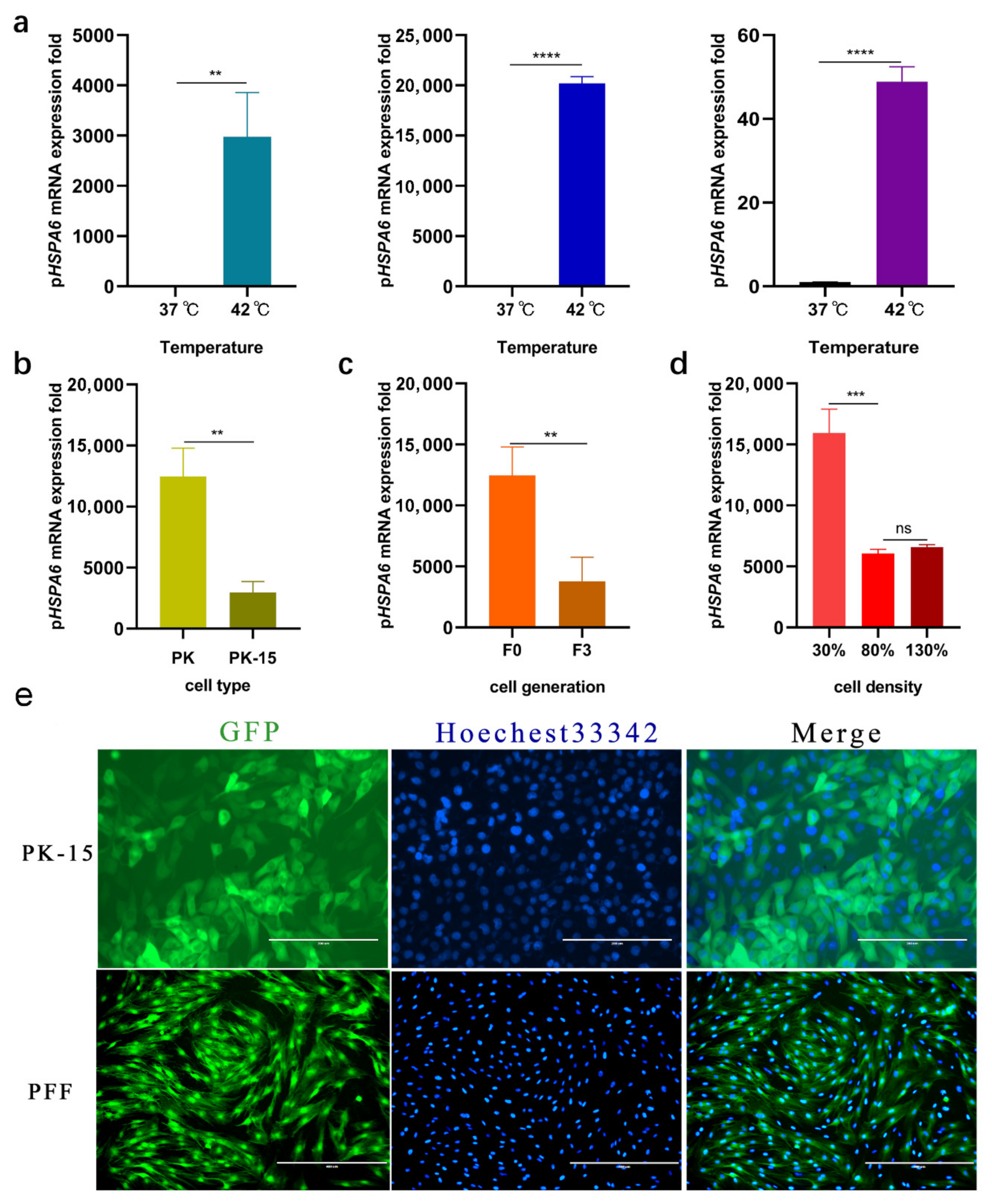
Figure 1. Analysis of the HSPA6 gene in porcine cells. (a) The mRNA expression of HSPA6 in PK-15, PFF, and 3D4/2 cells was detected by qPCR under heat. ** p < 0.01, **** p < 0.0001. (b) qPCR analysis of inducible pHSPA6 expression in PK and PK-15 cells. ** p < 0.01. (c) qPCR analysis of inducible pHSPA6 expression in F0 and F3 cells. ** p < 0.01. (d) qPCR analysis of the cell density of inducible pHSPA6 expression in PK cells. *** p < 0.001, ns = not significant. (e) Fluorescence microscopy analysis of the eGFP expression in PK-15 cell line and PFF cells under heat.
Genes 13 00189 g001
Figure 2. Effect of HSE-1 on the expression of pHSPA6. (a) The position of HSE-1 was located in the pHSPA6 promoter. (b) Strategy of sgRNA-H1 knock-in into pHSPA6; the cutting efficiency of sgRNA-H1 was evaluated using chromatograms. Red blocks and red arrows represent PAMs and cleavage sites, respectively. (c) Diagram of positive clone HSE-1 after pLB vector cloning. (d) Fluorescence microscopy and qPCR analysis of pHSPA6 expression in WT and positive clone cells (data show the mean ± S.E.M. in triplicate assay).
Genes 13 00189 g002
Figure 3. Effect of HSE-2 on the expression of pHSPA6. (a) The position of HSE-2 was located in the pHSPA6 promoter. (b) Strategy of sgRNA-H2 knock-in into pHSPA6; and the cutting efficiency of sgRNA-H2 was evaluated using chromatograms. Red blocks and red arrows represent PAMs and cleavage sites, respectively. (c) Diagram of positive clone HSE-2 after pLB vector cloning. (d) Fluorescence microscopy and qPCR analysis of pHSPA6 expression in WT and positive clone cells (data show the mean ± S.E.M. in triplicate assay).
Genes 13 00189 g003
Figure 4. Effect of CR on the expression of pHSPA6. (a) CR was found by blasting the HSPA6 promoter of seven species, and the position of CR was located in the pHSPA6 promoter. (b) Strategy of sgRNA1-1 and sgRNA1-2 knock-in into pHSPA6. (c) Selecting of positive clones using PCR. Primers are shown in Table 2; diagram of positive clone #1 after pLB vector cloning. (d) Fluorescence microscopy and qPCR analysis of pHSPA6 expression in WT and positive clone cells (data show the mean ± S.E.M. in triplicate assay).
Genes 13 00189 g004
Figure 5. Effects of the three regulatory regions on the expression of other genes after thermal treatment. (a) HSE-1, HSE-2, and CR significantly reduced the expression of pHSPA6. ** p < 0.01. (b) The expression of pHSP70.2 was negatively correlated with changes in pHSPA6 in clones HSE-1 and HSE-2 but positively correlated with CR. ** p < 0.01, *** p < 0.001. (c) The expression of pHSP90AA1 was negatively correlated with changes in pHSPA6 in clones HSE-1, HSE-2, and CR. **** p < 0.0001. (d) The expression of GS was positively correlated with changes in pHSPA6 in clones HSE-1, HSE-2, and CR. *** p < 0.001, **** p < 0.0001. (e) The expression of DNAJB1 was positively correlated with changes in pHSPA6 in clones HSE-1, HSE-2, and CR. **** p < 0.0001. (f) The expression of DNAJC3 was positively correlated with changes in pHSPA6 in clones HSE-1 and HSE-2 but uncorrelated with CR. *** p < 0.001, ns = not significant.
Genes 13 00189 g005
Table 1. Sequences of sgRNAs.
NamePrimerSequence (5′–3′)
sgRNA-H1sgRNA-H1-FCACCGTTCGCCCGGAAGGCGCTGGA
sgRNA-H1-RAAACTCCAGCGCCTTCCGGGCGAAC
sgRNA-H2sgRNA-H2-FCACCGTAGAACCAGCTGGGAGAAGC
sgRNA-H2-RAAACGCTTCTCCCAGCTGGTTCTAC
sgRNA1-1sgRNA1-1-FCACCGATTGTCACAAATCCTAGTAG
sgRNA1-1-RAAACCTACTAGGATTTGTGACAATC
sgRNA1-2sgRNA1-2-FCACCGTTAACATCATCCCTAGCCCT
sgRNA1-2-RAAACAGGGCTAGGGATGATGTTAAC
Table 2. Primers used in this study.
NamePrimerSequence (5′–3′)Usage
HSE-1-KOH1-FCTCTCTTCCCATGGTGAPCR
H1-RGCTGGTGCATCTGACTTCAT
HSE-2-KOH2-FCTCTCTTCCCATGGTGAPCR
H2-RGCTGGTGCATCTGACTTCAT
CR-KOF1-FCCTTTCTGGGCTGCGACTTGATPCR
R1-RGGGCGGATCGTCTGTTCAAGGA
EGFP-KI [21]EGFP-KI -FGAGGCGCATGTTCTCCAAAAACCPCR
EGFP-KI -RAGCCACACTTGTAGTTGCACTGG
pHSPA6 [21]pHSPA6 q-FATCCATGATATTGTCCTAqPCR
pHSPA6 q-RTTATGCTCTTGTTCAGTT
GS [21]GS q-FCTTGCATCGTGTGTGCGAAGqPCR
GS q-RGCTTAGCTTCTCGATGGCCT
DNAJB1 [21]B1 q-FTGACCATCGAAGTGAAGCGGqPCR
B1 q-RTCGGCTGGAATGTTGTTGGA
DNAJC3 [21]C3 q-FGGAGCCTGACAATGTGAATGCqPCR
C3 q-RGACCTTCTCGAATCTGCTGGT
pHSP70.270 q-FGAGCAAGGAGGAGATCGAGCqPCR
70 q-RGTTGAAGGCGTACGACTCCA
pHSP90AA190 q-FTCGAAGGGCAGTTGGAGTTCqPCR
90 q-RATGAGCTCCTCGCAGTTGTC
Publisher’s Note: MDPI stays neutral with regard to jurisdictional claims in published maps and institutional affiliations.