Article

Association of LBX1 Gene Methylation Level with Disease Severity in Patients with Idiopathic Scoliosis: Study on Deep Paravertebral Muscles

by
Piotr Janusz
1,†,
Małgorzata Tokłowicz
2,*,†,
Mirosław Andrusiewicz
2,
Małgorzata Kotwicka
2 and
Tomasz Kotwicki
1
1
Department of Spine Disorders and Pediatric Orthopedics, Poznan University of Medical Sciences, 28 Czerwca 1956 r. Street 135/147, 61-545 Poznań, Poland
2
Chair and Department of Cell Biology, Poznan University of Medical Sciences, Rokietnicka 5D, 60-806 Poznań, Poland
*
Author to whom correspondence should be addressed.
These authors contributed equally to this work.
Genes 2022, 13(9), 1556; https://doi.org/10.3390/genes13091556
Submission received: 12 July 2022 / Revised: 18 August 2022 / Accepted: 27 August 2022 / Published: 29 August 2022
(This article belongs to the Section Epigenomics)

Abstract

:
Idiopathic scoliosis (IS) is a multifactorial disease with a genetic background. The association of Ladybird Homeobox 1 (LBX1) polymorphisms with IS has been proven in multiple studies. However, the epigenetic mechanisms have not been evaluated. This study aimed to evaluate the LBX1 methylation level in deep paravertebral muscles in order to analyze its association with IS occurrence and/or IS severity. Fifty-seven IS patients and twenty non-IS patients were examined for the paravertebral muscles’ methylation level of the LBX1 promoter region. There was no significant difference in methylation level within paravertebral muscles between patients vs. controls, except for one CpG site. The comparison of the paravertebral muscles’ LBX1 promoter region methylation level between patients with a major curve angle of ≤70° vs. >70° revealed significantly higher methylation levels in 17 of 23 analyzed CpG sequences at the convex side of the curvature in patients with a major curve angle of >70° for the reverse strand promoter region. The association between LBX1 promoter methylation and IS severity was demonstrated. In patients with severe IS, the deep paravertebral muscles show an asymmetric LBX1 promoter region methylation level, higher at the convex scoliosis side, which reveals the role of locally acting factors in IS progression.

1. Introduction

Idiopathic scoliosis (IS) is the most common structural deformity of the spine in adolescents. The curvature may remain stable or progress to severe deformation [1]. It is associated with back pain and cosmetic and psychological burdens [2]. Severe IS can lead to pulmonary impairment and functional disability [3]. IS is a multifactorial disease with an important genetic background, probably modulated by environmental factors, which are claimed to impact IS occurrence or progression [4]. Although many genetic studies concerning the IS genetic background have been conducted and several target genes suggested, most of them have not been confirmed in replication studies [5,6,7,8].
Although the LBX1 (Ladybird Homeobox 1) association with IS has been established using genome-wide association studies [9,10,11], little is known in general about LBX1 function in humans and specifically about its mechanism in IS onset and progression [12,13]. It is known that in vertebrates, the LBX1 gene plays an essential role in regulating muscle precursor cell migration, maintaining their migratory potential, and promoting muscle precursor cell proliferation [14]. It is also important in specifying dorsal spinal cord interneurons and neural tube closure [15,16]. The molecular mechanisms by which LBX1 contributes to the development and propagation of neurons need to be explored further in muscle and other tissues [13]. Although LBX1 expression in developing skeletal muscles and the nervous system during embryogenesis has been described [12,13], the epigenetic impact on paravertebral muscle tissue and its role in IS development is unknown.
According to Grauers et al., the risk of developing scoliosis in a particular subject depends on an additive effect of genetic factors and the impact of environmental factors [17]. Many environmental factors have been evaluated in search of an association with IS. Among the best proven can be listed: low D vitamin level [18], high selenium level [19], and low BMI [20]. It is difficult to indicate a prevalent theory. Thus, IS is considered a multifactorial disease. The higher prevalence of scoliosis in families with an affected member compared to the general population supports the importance of the idea of genetic background. It is a well-described phenomenon associated with the degree of relationship [20,21,22]. However, twin studies show that genetic impact is limited, and other factors are very important [17]. Crosstalk between already described factors and searching for new ones seems to be essential for understanding the reasons for the occurrence and progression of IS [23,24].
It is suggested that epigenetic factors may represent a linkage between the genome and the environment for IS [25,26]. DNA methylation is an epigenetic DNA modification associated with the regulatory regions of many genes. It may alter genes’ expression but does not change the DNA sequence. Genomic DNA methylation occurs mostly on cytosines that precede a guanine nucleotide called CpG sites. When located in a gene promoter, DNA methylation typically acts to repress gene expression [27]. There have been described thousands of genes that exhibit DNA methylation differences between, e.g., males and females in human skeletal muscle. It may modulate mechanisms controlling muscle metabolism and health [28], as well as muscle development [29].
Few studies describing the role of DNA methylation in IS have been published [26,30,31,32,33]. However, epigenetic mechanisms have not been evaluated for the LBX1 gene yet. This study aims to evaluate the LBX1 methylation level in deep paravertebral muscles in order to analyze its association with IS occurrence and/or IS severity.

2. Materials and Methods

2.1. Patients

The study group consisted of 57 patients (50 girls, 7 boys) with IS. All patients underwent posterior spinal surgery correction for IS. The control group consisted of 20 patients (11 girls, 9 boys) with nonidiopathic spine deformities: spondylolisthesis, n = 2; congenital spine deformation, n = 14 (congenital scoliosis, congenital kyphosis); scoliosis secondary to prior thoracic surgery (lateral thoracotomy), n = 2; or Scheuermann’s disease, n = 2. All controls underwent posterior spinal surgery due to spine disorders (other than IS). All surgeries were performed in one hospital in a European country (Poland) from January 2017 until December 2019. All patients (both from the study group and control group) were subjected to a clinical, radiological, and molecular examination. The patients underwent standing anteroposterior radiographs before surgery. The curve pattern (number and localization of the curvatures), major curve angle (measured according to Cobb’s method) [34], and Risser sign (the radiological sign of skeletal maturity) [35] were measured in all patients by an experienced spine surgeon. The patients without coronal deformity (Scheuermann’s disease and spondylolisthesis) were not included in the major curve angle calculation.
The inclusion criteria for the study group were as follows: (1) clinically and radiologically confirmed IS diagnosis, (2) no coexisting orthopedic, genetic, or neurological disorders, (3) primary thoracic spinal curvature, and (4) surgical treatment due to IS. The study group was divided into two subgroups according to disease severity. The first subgroup consisted of 28 patients with a moderate form of IS, with curvatures ranging from 50° to 70° and a Risser sign of ≥4 and age of ≥15 years old. The second subgroup consisted of 29 patients with a very progressive form of IS, with larger curvatures exceeding 70° regardless of Risser’s sign of age.
The inclusion criteria for the control group were as follows: (1) clinically and radiologically confirmed diagnosis of congenital spine deformation with (defects of formation or segmentation), spondylolisthesis, scoliosis secondary to prior thoracic surgery, or Scheuermann’s disease, (2) no coexisting orthopedic, genetic, or neurological disorders, (3) no diagnosis of IS, and (4) surgical treatment due to spine pathology.

2.2. Tissue Samples

During the surgery, three muscle tissue samples (1 cm3 each) were obtained from each patient via the surgical approach used to correct the deformity. The first sample was obtained from the deep paravertebral muscles (M. longissimus) on the (1) convex side at the apex of the major curvature. The second sample was obtained from the deep paravertebral muscles (M. longissimus) on the (2) concave side at the apex of the major curvature. The third sample was obtained from the (3) superficial muscle layer (M. deltoideus).
During the surgery, three muscle tissue samples (1 cm3 each) were obtained from each patient from the control group. Two were taken from the deep paravertebral muscles (M. longissimus). When curvature was present (congenital scoliosis), the samples were taken as follows: (1) on the convex and (2) concave sides at the apex of the major curvature. For patients with kyphosis or spondylolisthesis, the samples were taken as follows: (1) on the right side of the spine and (2) on the left side of the spine. The third sample was obtained in all patients from the (3) superficial muscle layer (M. deltoideus).
All samples were stored in sterile tubes containing 5 mL nucleic acid stabilizing solution (Novazym, cat no. ST01; Poznan, Poland).

2.3. Genomic DNA Methylation Analysis

Total genomic DNA was extracted and processed as described before [36]. The DNA was bisulfite converted and used as a template for polymerase chain reaction (PCR) followed by pyrosequencing (PSQ). The DNA sequences analyzed corresponded to the forward and reverse DNA strands of the LBX1 promoter region (https://www.ncbi.nlm.nih.gov (accessed on 31 March 2018); GenBank N°: NG_009236). The PCR primers used are shown in Table 1.
PCR reactions were performed using conditions validated for ZymoTaqTM PreMix (Zymo Research; cat no. E2004; Irvine, CA, USA) [36]. Reaction mixture components, concentrations, and thermal profiles are presented in Table 2.
PSQ analysis was performed using the PyroMark Q48 instrument (Qiagen; Hilden, Germany) for complementary CpG dinucleotides (located in opposite strands of DNA). Assays were designed with Pyromark Q48 Autoprep 2.4.2 software (Qiagen; Hilden, Germany). For each strand, 23 CpG sites were analyzed. In each reaction, internal sodium bisulfite treatment quality control was included. The methylation level was quantified using Pyromark Q48 Autoprep 2.4.2 software and expressed as a percentage ratio of methylated to nonmethylated dinucleotides.

2.4. Statistical Analysis

Data analyses were performed using Statistica 13.3 software (TIBCO Software Inc.; Palo Alto, CA, USA) and PQStat 1.8.0.414 software (PQStat software; Poznan, Poland). The methylation level was analyzed in both strands separately for each CpG site and compared between selected subgroups. The Shapiro–Wilk test was used for the normality of continuous variable distribution assessment. The differences in methylation levels between concave, convex, and superficial muscles were evaluated using Friedman ANOVA with Dunn’s Bonferroni post hoc test. Methylation between patient subgroups with a major curve angle of ≤70° or >70° was compared using the Mann–Whitney U test. Data were considered statistically significant when p < 0.05.

3. Results

3.1. Patients and Controls

The patient group consisted of 57 IS patients (7 boys and 50 girls), and the control group consisted of 20 individuals (9 boys and 11 girls). The mean age at surgery for patients was 14.1 ± 1.6 years (ranging from 11 to 18 years), and for controls was 13.6 ± 3.2 years (ranging from 7 to 18 years), and there were no significant differences between groups (p > 0.05). The major curve angle for patients ranged from 50° to 115°, with a mean of 76 ± 17°, and for controls, the major curve angle value ranged from 30° to 105°, with a mean of 64 ± 24°.

3.2. DNA Methylation at the LBX1 Promoter Regions—A Case-Control Study

The methylation level within the LBX1 DNA forward strand promoter region differed slightly between patients and controls for the superficial muscles used as our internal control but not for the deep muscles. In particular, a significantly lower methylation level was observed in the superficial muscles of the control group at one site, CpG-20 (p < 0.05, Figure 1; Additional file 1: Table S1; Additional file 2: Figure S1). There were no significant differences observed in the DNA reverse strand promoter region (p > 0.05, Figure 1; Additional file 1: Table S2; Additional file 2: Figure S2).

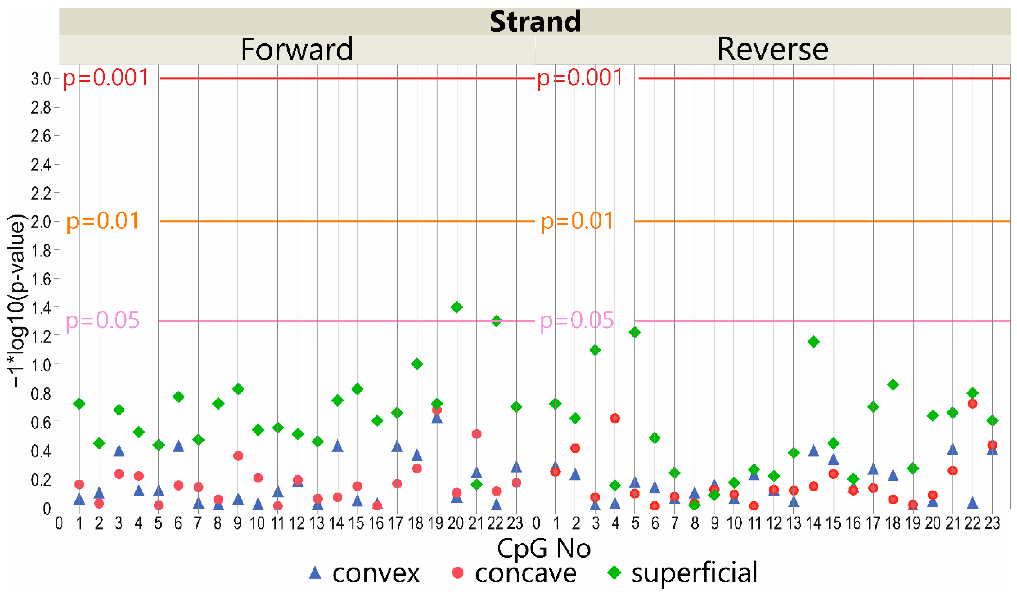
3.3. DNA Methylation at the LBX1 Promoter Regions—Deep Paravertebral Muscles vs. Superficial Muscles

3.3.1. DNA Forward Strand Promoter Region

Considering the IS patients’ group, the methylation level within the LBX1 DNA forward strand promoter region differed significantly between the deep paravertebral muscles (on the convex and concave side of the curvature) and the superficial muscles (p < 0.05; Figure 2). The methylation level was significantly higher in the superficial muscle compared with the convex side of the curvature in 13 CpG sequences (p < 0.05; Additional file 1: Table S1; Additional file 2: Figure S1). A significantly higher methylation level was also observed in the superficial muscle compared to the concave side of the curvature at CpG-3 and CpG-14 sites (p < 0.05; Additional file 1: Table S1; Additional file 2: Figure S1). However, there was no difference in the methylation level of the DNA from the deep paravertebral muscles between the convex or concave side of the curvature (p > 0.05; Additional file 1: Table S1; Additional file 2: Figure S1).
In the control group, a significant difference in methylation level was observed between the superficial muscles compared with the convex side deep muscles of the curvature at CpG-14 (p < 0.05, Figure 2; Additional file 1: Table S1; Additional file 2: Figure S1). In the deep paravertebral muscles, the methylation level differed significantly between the convex and concave sides of the curvature at CpG-14 (p < 0.05, Figure 2; Additional file 1: Table S1; Additional file 2: Figure S1).

3.3.2. DNA Reverse Strand Promoter Region

Considering the patients’ group, the methylation level within the LBX1 DNA reverse strand promoter region was significantly different between deep paravertebral muscles (on the convex and concave sides of the curvature) and superficial muscles (p < 0.05, Figure 2). The methylation level was higher in the superficial muscle tissue compared with the deep convex muscles of the curvature in four CpG sequences (p < 0.05; Additional file 1: Table S2; Additional file 2: Figure S2). In the control group, no difference was observed (p > 0.05; Additional file 1: Table S2; Additional file 2: Figure S2). The methylation level differed significantly at CpG-18 between the superficial muscles and the concave side of scoliosis relative to the control group (p > 0.05; Additional file 1: Table S2; Additional file 2: Figure S2). A significantly higher methylation level was observed in the convex side of the curvature compared with the concave one at CpG-1 and CpG-2 within the patients’ group (p < 0.05; Additional file 1: Table S2; Additional file 2: Figure S2). In the control subgroup, the methylation level did not differ significantly between these muscles (Additional file 1: Table S2; Additional file 2: Figure S2).

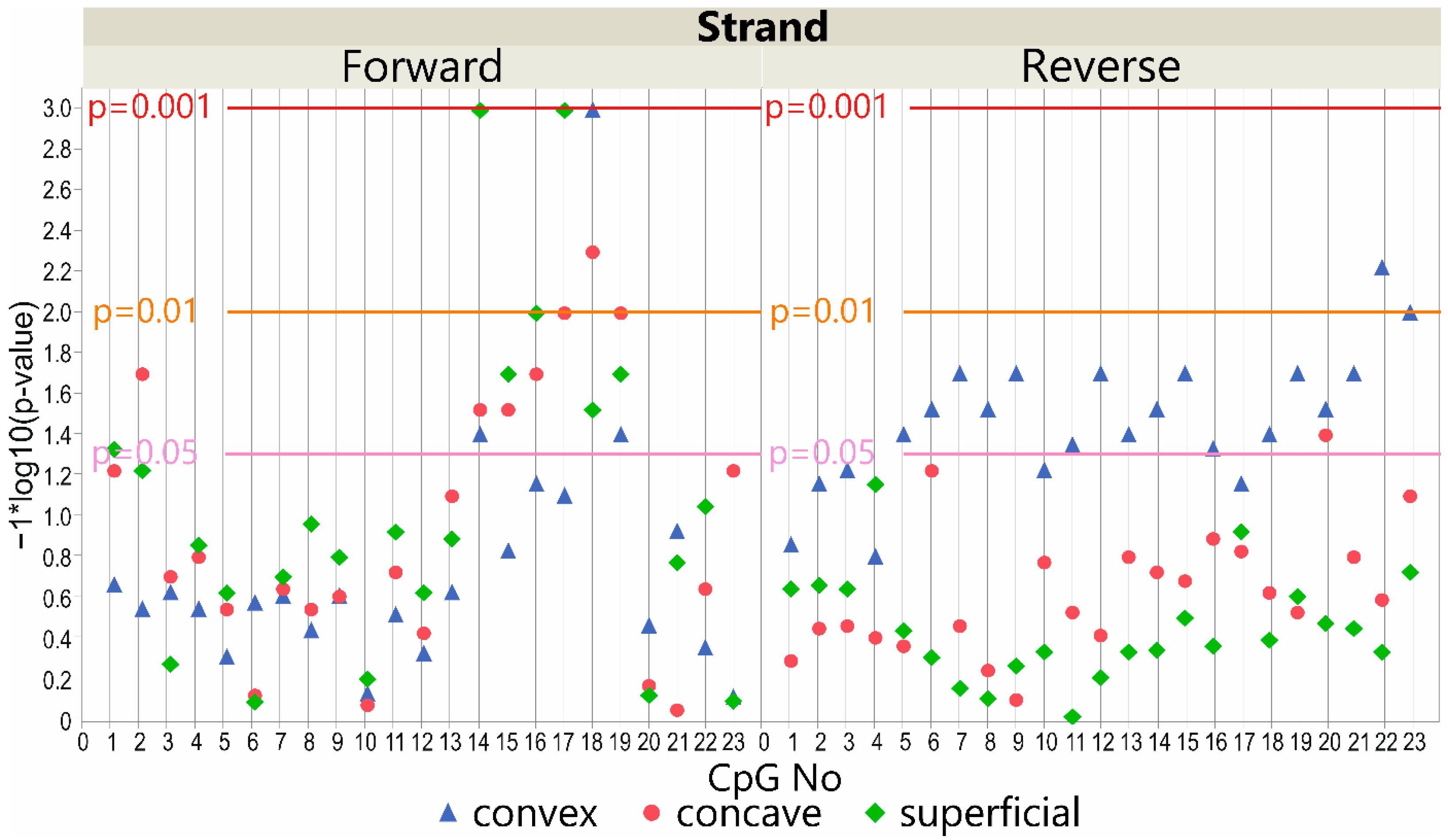
3.4. LBX1 Methylation Status and Major Curve Angle—Case-Only Study

3.4.1. DNA Forward Strand Promoter Region

In the study group, we observed significant differences between case subgroups with a major curve angle of ≤70° vs. >70°. The methylation level was higher in patients with a major curve angle of >70° (p < 0.05; Additional file 1: Table S3; Additional file 2: Figure S3). The methylation from DNA isolated at the convex side of the curvature differed at three CpG sites on the forward strand (p < 0.05, Figure 3). In seven CpG sequences, differences were also detected on the forward strand of the concavity of thoracic scoliosis and the superficial muscles (p < 0.05, Figure 3; Additional file 1: Table S3; Additional file 2: Figure S3).

3.4.2. DNA Reverse Strand Promoter Region

For the study group, we mainly observed significant differences between cases with a major curve angle of ≤70° vs. >70° on the convex side of the curvature. The methylation level was higher in patients with a major curve angle of >70° in 17 of 23 analyzed CpG sequences (p < 0.05, Figure 3; Additional file 1: Table S4; Additional file 2: Figure S4). The methylation at the concave side of the curvature differed only in one CpG and was lower in patients with a major curve angle of ≤70° (p < 0.05, Figure 3; Additional file 1: Table S4; Additional file 2: Figure S4). No differences were observed in the superficial muscles (p > 0.05, Figure 3; Additional file 1: Table S4; Additional file 2: Figure S4).

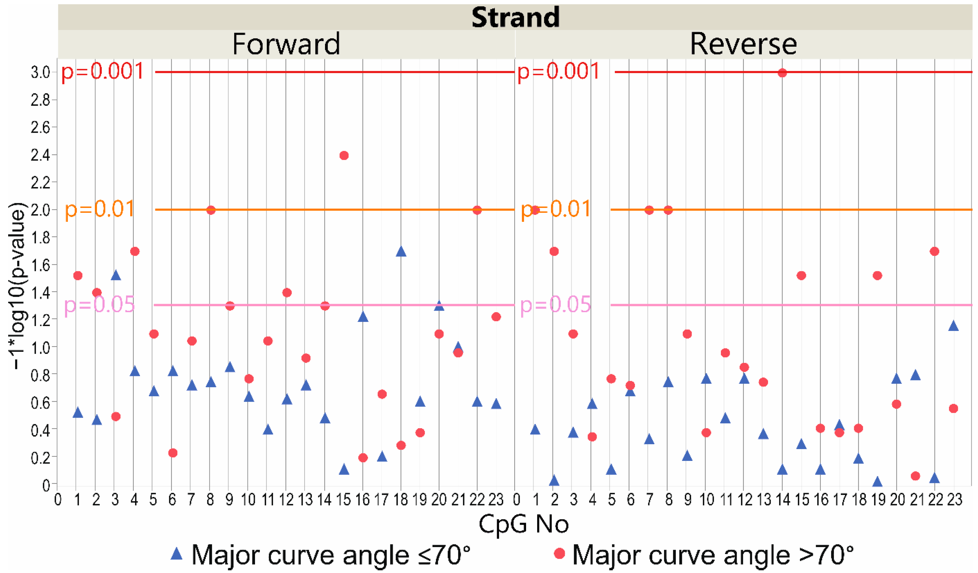
3.5. LBX1 Methylation Status and Major Curve Angle—Deep Paravertebral Muscles vs. Superficial Muscles

3.5.1. DNA Forward Strand Promoter Region

Considering the patients’ subgroup with a major curve angle of ≤70°, the methylation level within the LBX1 DNA forward strand promoter region differed significantly between deep paravertebral muscles (on the convex and concave sides of the curvature) and superficial muscles in two CpG sites (p < 0.05, Figure 4). The methylation level was significantly higher in the superficial muscle compared with the concave side of the curvature at CpG-3 and CpG-18 (p < 0.05; Additional file 1: Table S3; Additional file 2: Figure S3). A significantly higher methylation level was also observed in the superficial muscle compared with the convex side of the curvature at the same CpG sites (p < 0.05; Additional file 1: Table S3; Additional file 2: Figure S3).
There was a difference in the patients’ subgroup with a major curve angle of >70° regarding the methylation level in deep paravertebral muscles on the convex and concave sides of the curvature and the superficial muscles in seven CpG sequences (p < 0.05, Figure 4). A higher methylation level was observed in the superficial muscle compared with the convex side of the curvature at CpG sites: 1, 2, 4, 8, 12, 15, and 22 (p < 0.05; Additional file 1: Table S3; Additional file 2: Figure S3).

3.5.2. DNA Reverse Strand Promoter Region

Considering the patients’ subgroup with a major curve angle of ≤70°, we observed no differences in the methylation level within the LBX1 DNA reverse strand promoter region (p > 0.05, Figure 4).
There was, however, a difference in the patients’ subgroup with a major curve angle of >70° in regard to the methylation level in deep paravertebral muscles on the convex and concave sides of the curvature and the superficial muscles in eight CpG sequences (p < 0.05, Figure 4). A higher level of methylation was observed in the convexity of scoliosis compared with the concave side of the curvature at CpG sites: 1, 2, 7, and 8 (p < 0.05; Additional file 1: Table S4; Additional file 2: Figure S4). A higher level of methylation was also observed in the convex side of the curvature compared with the superficial muscle at CpG sites: 1, 7, 8, 14, 15, 19, and 22 (p < 0.05; Additional file 1: Table S4; Additional file 2: Figure S4). A higher level of methylation was observed in the concave side of the curvature compared to the superficial muscle at CpG-14 (p < 0.05; Additional file 1: Table S4; Additional file 2: Figure S4).

4. Discussion

Although many studies concerning the etiology of IS have been conducted, the IS background remains unsolved [4,37,38]. Multiple theories concerning IS etiology have been suggested. These concepts cover, e.g., aberrations in hormonal level, metabolic factors, connective tissue, skeletal and muscle structure, biomechanical features, neurological mechanisms, molecular and genetic factors, biochemistry, environment, lifestyle, or possible interrelationships among them [4,37,38,39].
In this study, we focused on muscle tissue as a scoliosis-inducing factor. However, we cannot exclude another important background, such as a relative anterior spinal overgrowth. It may induce rotational instability and functional tethering of the spinal cord [40,41,42].
This study revealed a new factor associated with a tendency to scoliosis progression. We found an association between LBX1 methylation level and disease severity. Understanding the background of the disease can contribute to the prediction of the IS course in patients and may create the possibility of prevention or moderate treatment.
Although we assumed that LBX1 promoter methylation might be associated with a predisposition to IS, our results did not confirm this concept. No difference in the methylation level between IS patients versus controls at evaluated CpG sequences in deep paravertebral muscles was found. What is more, we found one significant difference in CpG methylation on the forward strand in superficial muscles (Figure 1). We included the evaluation of the superficial muscles in our study as an internal control. We believe that the superficial muscles are not associated with IS onset due to their function and the anatomic borderlines between them and the deep paravertebral muscles. Thus, we do not consider them an important causative factor.
The most important finding of our study is a higher methylation level in the LBX1 promoter region on the reverse strand at the deep convex muscles in patients with more severe IS. We found significant differences in 17 out of 23 analyzed CpG sequences and a tendency in almost all CpG sequences (Figure 3). The methylation was also higher in the subgroup of patients with greater curvatures on the forward strand. However, this difference was present in all muscle layers in several CpG sequences and seemed to be more diffused. Interestingly, results for CpG-14, CpG-18, and CpG-19 were analogous, regardless of the tissue location (Figure 3).
The analysis of disease severity is essential for IS treatment. It is unknown why certain IS patients progress more than others. Therefore, identifying the patients at risk of scoliosis occurrence or curve progression is crucial to providing early treatment [1,43].
To evaluate the association of the LBX1 gene methylation level with IS progression, we divided the study group into two subgroups according to disease severity. Unfortunately, there is no defined major curve angle threshold when severe curvature significantly impacts patients’ health. It is established that severe curvatures affect patients’ health, such as decreased lung function, cardiac function, back pains, and degenerative spine disease [44,45,46]. A correlation between the degree of patients’ impairment with the severity of the spinal deformity was revealed [3,44,45]. The group of skeletally mature patients with a major curve angle moderately exceeding 50° needs a surgical scoliosis correction to avoid further curvature deterioration in adulthood, while in patients with a bigger curve angle, direct impairment can be found [2,46,47]. Studies concerning surgical scoliosis treatment classify severe curvature as a major curve angle exceeding 70° [48,49,50]. Thus, we used this value to categorize study subgroups. The inclusion criteria of a Risser sign of ≥4 and age of ≥15 years old were used for the subgroup of patients with a moderate form of IS—having the curvature range from 50° to 70°. The rationale was to ensure that these patients developed the final major curve angle during the natural history of the curvature.
A difference in the LBX1 methylation level between the muscle layers is another interesting finding in the context of IS background. Within the patients’ group, we found a tendency for higher DNA methylation levels in superficial muscles compared with deep muscles. This difference was significant in 13 and 4 CpG sequences on the forward and reverse DNA strands, respectively. This issue was less pronounced in the control group and was present in one CpG on both forward and reverse strands. Thus, we found a different LBX1 methylation level between the muscle tissue harvested from different localizations (M. longissimus vs. M. deltoideus). It raises doubt about the possibility of using LBX1 methylation for prediction in IS patients based on samples from different tissues, such as blood. It is difficult to distinguish the causative factor from the effect of the disease. The difference in methylation could be the cause of the asymmetry in muscles, which may contribute to the etiology of IS. Conversely, differences in methylation levels could be a consequence of the muscles being exposed to different conditions on either side of the curvature due to asymmetric loading. The difference in methylation level between the groups is found mostly in deep paravertebral muscles on the convex side at the reverse DNA strand LBX1 gene promoter region. The difference is less demonstrated on the forward strand and is more equally spread in all muscles. In case of secondary changes, we could expect more similar distribution of methylation at both DNA strands. Thus, we can assume that it is rather a causative factor.
A theory of IS background connecting the impact of dysfunctional paravertebral muscles on the development of spine curvature has been proposed [51,52]. Differences in the proportion of muscle fiber types between convex and concave sides of IS were described: a lower percentage of slow fibers type I in IS patients than in controls was found [53]. Other studies described greater fibrosis and fatty involution on the deep concave paravertebral muscles than on the convex side [51,54]. What is more, asymmetry of muscle activation patterns in IS patients between the concave and convex sides has also been described [55,56]. We found differences in the LBX1 gene DNA methylation level in deep paravertebral muscles between the analyzed groups. DNA methylation does not directly affect muscles but impacts LBX1 gene expression. LBX1 acts primarily during embryogenesis, and its asymmetric expression in relation to biological action is difficult to establish [12,13,14]. We do not know if the difference between both sides of the curvature could modulate muscle strength or elasticity. Nevertheless, it might be of important clinical relevance; unfortunately, this study did not evaluate muscle tissue properties.
To our knowledge, one study concerning LBX1 gene expression in paravertebral muscles has been published [13]. Jennings et al. evaluated deep paravertebral muscles from 25 IS patients. The patients evaluated by Jennings et al. were comparable to ours by age and gender, while our group presented a slightly higher major curve angle value. They found no difference in mRNA and protein expression between the concave and convex sides of the curvature. What is more, the results did not correlate with the major curve angle. However, no subgroup analysis according to curve severity was conducted. Because LBX1 expression was not evaluated in this study, direct comparison is difficult. When all patients were pooled together, the paired analysis did not find a convex–concave difference in the LBX1 methylation level on the forward strand. A difference was only observed in two CpG sequences on the reverse strand. It is comparable with Jennings et al.’s description of the expression pattern. However, we found a significant difference associated with curve severity. Jennings et al. theorized that LBX1 expression occurs in the time of ontogeny and IS-associated LBX1 genetic variants modify gene expression unequally on both sides of the scoliotic curve during the embryogenesis of muscle cells. Still, this expression aligns after birth [13]. It could result from the different methylation patterns on the forward and reverse DNA strands revealed in our study. It is possible that at distinct stages of development, the forward or reverse strand is more relevant to gene expression.
Previous work has shown an association between DNA methylation and IS. However, all these studies used peripheral blood samples. Liu and colleagues, using whole-genome methylation evaluation in a twin pair, selected DNA regions potentially associated with adolescent IS and described a significantly higher methylated region in chromosome 15 [30]. Meng et al. described an association between lower methylation levels at site cg01374129 and curve severity [26]. According to Shi et al., susceptibility to IS and severity of the curvature are associated with the level of methylation of the PITX1 and PCDH10 genes [32,33]. Mao et al. evaluated the methylation level of the COMP gene. They found that hypermethylation of the gene promoter correlated with curve severity [31].
Taking into consideration that our study was focused on a local evaluation in search of a causative factor in muscle tissue, it is difficult to compare it with the results obtained using blood samples, especially as we found a difference in methylation levels between the deep and superficial groups of muscles in IS patients. Thus, the level of LBX1 methylation in blood samples may be vastly different from that revealed in muscles.
One of the biggest challenges of this study was to find a suitable control group. This study was focused on paraspinal muscle tissues, and it was unreasonable, because of ethical issues, to obtain paraspinal muscle samples from healthy individuals. We decided not to include elderly patients who underwent surgery due to degenerative spine changes, which may cause muscle atrophy or unknown methylation changes. What is more, there would be an important difference in age. Thus, we decided to include into the control group patients of similar age, including children operated on due to spine abnormalities for known reasons and excluding patients with scoliosis due to a neuromuscular or genetic background. Spinal pathologies requiring surgical treatment in children meeting the inclusion criteria are much rarer than IS. Thus, the control group is smaller than the IS patients’ group. Patients with congenital scoliosis and scoliosis after surgical thoracic surgery had a mean Cobb 12° smaller than IS patients. Two patients with Scheuermann’s disease and two patients with spondylolisthesis were not included in the major curve angle calculation due to deformation in the sagittal plane, and they did not present scoliosis. The most significant differences between the groups were found in the gender distribution. However, it is a known phenomenon that severe IS is much more common in females, and the prevalence of congenital scoliosis did not differ between boys and girls [57]. What is more, we did not find an association between LBX1 and gender. Thus, we decided to accept this difference.
Patients with congenital scoliosis were the largest part of the control group. While evaluating our results, it is important to notice that there are studies describing genetic predisposition in congenital scoliosis [58,59,60]. Several studies have reported several genes associated with congenital scoliosis, including TBX6 [58,59], FBN1 [61], PAX1 [62], DLL1 [63], and other genes. However, to our knowledge, the association between congenital scoliosis and the LBX1 gene has never been found. However, to our knowledge, the association between congenital scoliosis and the LBX1 gene has never been found. What is more, in genetic studies evaluating tissue samples of patients with idiopathic scoliosis, the patients with congenital scoliosis have been used as a control group [64,65,66].
The strength of this study is that it links known facts about the genetic background of IS concerning LBX1 with possible modification of its expression or function in the area of deformation. This brings a new insight into IS pathology and evaluates a probable cause of curvature progression.

5. Conclusions

The association between LBX1 promoter methylation and IS severity was demonstrated. In patients with severe IS, the deep paravertebral muscles demonstrate an asymmetric LBX1 promoter region methylation level, higher at the convex scoliosis side, which reveals the role of locally acting factors in IS progression.

Supplementary Materials

The following supporting information can be downloaded at: https://www.mdpi.com/article/10.3390/genes13091556/s1, Additional file 1: Table S1: methylation level at LBX1 DNA forward strand promoter region in control and patient subgroups in deep paravertebral muscles and superficial muscles, Table S2: methylation level at LBX1 DNA reverse strand promoter region in control and patient subgroups in deep paravertebral muscles and superficial muscles, Table S3: methylation level at LBX1 DNA forward strand promoter region in patient subgroups with major curve angle ≤70° and >70° in deep paravertebral muscles and superficial muscles, Table S4: methylation level at LBX1 DNA reverse strand promoter region in patients subgroup with major curve angle ≤70° and >70° in deep paravertebral muscles and superficial muscles, Additional file 2: Figure S1: DNA methylation level within LBX1 forward strand promoter region in deep paravertebral muscles and superficial muscles. Upper solid horizontal lines represent significant differences between muscles, and lower lines show significant differences in case-control study, Figure S2: DNA methylation level within LBX1 reverse strand promoter region in deep paravertebral muscles and superficial muscles. Upper solid horizontal lines represent significant differences between muscles, and lower lines show significant differences in case-control study, Figure S3: DNA methylation level within LBX1 forward strand promoter region in deep paravertebral muscles and superficial muscles. Upper solid horizontal lines represent significant differences between muscles in case of patients, and lower lines show significant differences in subgroups divided according to major curve angle values, Figure S4: DNA methylation level within LBX1 forward strand promoter region in deep paravertebral muscles and superficial muscles. Upper solid horizontal lines represent significant differences between muscles in case of patients, and lower lines show significant differences in subgroups divided according to major curve angle values.

Author Contributions

Conceptualization, P.J., M.T., M.A., M.K. and T.K.; methodology, P.J., M.T. and M.A.; validation, M.T. and M.A.; formal analysis, P.J., M.T., M.A. and M.K.; investigation, P.J., M.T. and M.A.; resources, P.J.; data curation, P.J., M.T. and M.A.; writing—original draft preparation, P.J., M.T., M.A. and M.K.; writing—review and editing, M.T., M.A. and T.K.; visualization, M.T. and M.A.; supervision, P.J. and T.K.; project administration, P.J.; funding acquisition, P.J. All authors have read and agreed to the published version of the manuscript.

Funding

This research was funded by a National Science Centre grant (2016/23/D/NZ5/02606), and the APC was funded by the Poznan University of Medical Sciences.

Institutional Review Board Statement

The study was conducted according to the guidelines of the Declaration of Helsinki and approved by the Institutional Review Board of Poznan University of Medical Sciences (protocol code no. 546/17 and 741/19, and date of approval 11 May 2017 and 19 June 2019, respectively).

Informed Consent Statement

Informed consent was obtained from all the patients or their parents/legal guardians in the case of underage participants.

Data Availability Statement

The datasets used and analyzed during the current study are available from the corresponding author on reasonable request.

Acknowledgments

Not applicable.

Conflicts of Interest

The authors declare no conflict of interest. The funders had no role in the design of the study; in the collection, analyses, or interpretation of data; in the writing of the manuscript; or in the decision to publish the results.

References

  1. Weinstein, S.L.; Dolan, L.A.; Cheng, J.C.; Danielsson, A.; Morcuende, J.A. Adolescent idiopathic scoliosis. Lancet 2008, 371, 1527–1537. [Google Scholar] [CrossRef]
  2. Weinstein, S.L. The Natural History of Adolescent Idiopathic Scoliosis. J. Pediatr. Orthop. 2019, 39, S44–S46. [Google Scholar] [CrossRef]
  3. Huh, S.; Eun, L.Y.; Kim, N.K.; Jung, J.W.; Choi, J.Y.; Kim, H.S. Cardiopulmonary function and scoliosis severity in idiopathic scoliosis children. Korean J. Pediatr. 2015, 58, 218–223. [Google Scholar] [CrossRef]
  4. Lowe, T.G.; Edgar, M.; Margulies, J.Y.; Miller, N.H.; Raso, V.J.; Reinker, K.A.; Rivard, C.H. Etiology of idiopathic scoliosis: Current trends in research. J. Bone Jt. Surg.-Ser. A 2000, 82, 1157–1168. [Google Scholar] [CrossRef]
  5. De Salvatore, S.; Ruzzini, L.; Longo, U.G.; Marino, M.; Greco, A.; Piergentili, I.; Costici, P.F.; Denaro, V. Exploring the association between specific genes and the onset of idiopathic scoliosis: A systematic review. BMC Med. Genom. 2022, 15, 115. [Google Scholar] [CrossRef]
  6. Khanshour, A.M.; Kou, I.; Fan, Y.; Einarsdottir, E.; Makki, N.; Kidane, Y.H.; Kere, J.; Grauers, A.; Johnson, T.A.; Paria, N.; et al. Genome-wide meta-analysis and replication studies in multiple ethnicities identify novel adolescent idiopathic scoliosis susceptibility loci. Hum. Mol. Genet. 2018, 27, 3986–3998. [Google Scholar] [CrossRef]
  7. Grauers, A.; Einarsdottir, E.; Gerdhem, P. Genetics and pathogenesis of idiopathic scoliosis. Scoliosis Spinal Disord. 2016, 11, 45. [Google Scholar] [CrossRef]
  8. Gorman, K.F.; Julien, C.; Moreau, A. The genetic epidemiology of idiopathic scoliosis. Eur. Spine J. 2012, 21, 1905–1919. [Google Scholar] [CrossRef]
  9. Takahashi, Y.; Kou, I.; Takahashi, A.; Johnson, T.A.; Kono, K.; Kawakami, N.; Uno, K.; Ito, M.; Minami, S.; Yanagida, H.; et al. A genome-wide association study identifies common variants near LBX1 associated with adolescent idiopathic scoliosis. Nat. Genet. 2011, 43, 1237–1240. [Google Scholar] [CrossRef] [PubMed]
  10. Li, Y.-L.; Gao, S.-J.; Xu, H.; Liu, Y.; Li, H.-L.; Chen, X.-Y.; Ning, G.-Z.; Feng, S.-Q. The association of rs11190870 near LBX1 with the susceptibility and severity of AIS, a meta-analysis. Int. J. Surg. 2018, 54, 193–200. [Google Scholar] [CrossRef] [PubMed]
  11. Jiang, H.; Yang, Q.; Liu, Y.; Guan, Y.; Zhan, X.; Xiao, Z.; Wei, Q. Association between ladybird homeobox 1 gene polymorphisms and adolescent idiopathic scoliosis: A MOOSE-compliant meta-analysis. Medicine 2019, 98, e16314. [Google Scholar] [CrossRef] [PubMed]
  12. Luo, M.; Zhang, Y.; Huang, S.; Song, Y. The Susceptibility and Potential Functions of the LBX1 Gene in Adolescent Idiopathic Scoliosis. Front. Genet. 2021, 11, 1802. [Google Scholar] [CrossRef] [PubMed]
  13. Jennings, W.; Hou, M.; Perterson, D.; Missiuna, P.; Thabane, L.; Tarnopolsky, M.; Samaan, M.C. Paraspinal muscle ladybird homeobox 1 (LBX1) in adolescent idiopathic scoliosis: A cross-sectional study. Spine J. 2019, 19, 1911–1916. [Google Scholar] [CrossRef]
  14. Brohmann, H.; Jagla, K.; Birchmeier, C. The role of Lbx1 in migration of muscle precursor cells. Development 2000, 127, 437–445. [Google Scholar] [CrossRef]
  15. Sieber, M.A.; Storm, R.; Martinez-de-la-Torre, M.; Müller, T.; Wende, H.; Reuter, K.; Vasyutina, E.; Birchmeier, C. Lbx1 Acts as a Selector Gene in the Fate Determination of Somatosensory and Viscerosensory Relay Neurons in the Hindbrain. J. Neurosci. 2007, 27, 4902–4909. [Google Scholar] [CrossRef] [PubMed]
  16. Krüger, M.; Schäfer, K.; Braun, T. The homeobox containing gene Lbx1 is required for correct dorsal-ventral patterning of the neural tube. J. Neurochem. 2002, 82, 774–782. [Google Scholar] [CrossRef]
  17. Grauers, A.; Rahman, I.; Gerdhem, P. Heritability of scoliosis. Eur. Spine J. 2012, 21, 1069–1074. [Google Scholar] [CrossRef]
  18. Balioglu, M.B.; Aydin, C.; Kargin, D.; Albayrak, A.; Atici, Y.; Tas, S.K.; Kaygusuz, M.A. Vitamin-D measurement in patients with adolescent idiopathic scoliosis. J. Pediatr. Orthop. B 2017, 26, 48–52. [Google Scholar] [CrossRef]
  19. Yang, Z.; Xie, Y.; Chen, J.; Zhang, D.; Yang, C.; Li, M. High selenium may be a risk factor of adolescent idiopathic scoliosis. Med. Hypotheses 2010, 75, 126–127. [Google Scholar] [CrossRef]
  20. Watanabe, K.; Michikawa, T.; Yonezawa, I.; Takaso, M.; Minami, S.; Soshi, S.; Tsuji, T.; Okada, E.; Abe, K.; Takahashi, M.; et al. Physical Activities and Lifestyle Factors Related to Adolescent Idiopathic Scoliosis. J. Bone Jt. Surg. Am. 2017, 99, 284–294. [Google Scholar] [CrossRef]
  21. Grauers, A.; Danielsson, A.; Karlsson, M.; Ohlin, A.; Gerdhem, P. Family history and its association to curve size and treatment in 1,463 patients with idiopathic scoliosis. Eur. Spine J. 2013, 22, 2421–2426. [Google Scholar] [CrossRef] [PubMed]
  22. Tang, N.L.S.; Yeung, H.Y.; Hung, V.W.Y.; Di Liao, C.; Lam, T.P.; Yeung, H.M.; Lee, K.M.; Ng, B.K.W.; Cheng, J.C.Y. Genetic epidemiology and heritability of AIS: A study of 415 Chinese female patients. J. Orthop. Res. 2012, 30, 1464–1469. [Google Scholar] [CrossRef] [PubMed]
  23. Maqsood, A.; Frome, D.K.; Gibly, R.F.; Larson, J.E.; Patel, N.M.; Sarwark, J.F. IS (Idiopathic Scoliosis) etiology: Multifactorial genetic research continues. A systematic review 1950 to 2017. J. Orthop. 2020, 21, 421–426. [Google Scholar] [CrossRef] [PubMed]
  24. Faldini, C.; Manzetti, M.; Neri, S.; Barile, F.; Viroli, G.; Geraci, G.; Ursini, F.; Ruffilli, A. Epigenetic and Genetic Factors Related to Curve Progression in Adolescent Idiopathic Scoliosis: A Systematic Scoping Review of the Current Literature. Int. J. Mol. Sci. 2022, 23, 5914. [Google Scholar] [CrossRef]
  25. Burwell, R.G.; Dangerfield, P.H.; Moulton, A.; Grivas, T.B.; Meredith, D.; Wägele, B.; Altmaier, E.; Deloukas, P.; Erdmann, J.; Grundberg, E.; et al. Adolescent idiopathic scoliosis (AIS), environment, exposome and epigenetics: A molecular perspective of postnatal normal spinal growth and the etiopathogenesis of AIS with consideration of a network approach and possible implications for medical therapy. Scoliosis 2011, 6, 26. [Google Scholar] [CrossRef]
  26. Meng, Y.; Lin, T.; Liang, S.; Gao, R.; Jiang, H.; Shao, W.; Yang, F.; Zhou, X. Value of DNA methylation in predicting curve progression in patients with adolescent idiopathic scoliosis. EBioMedicine 2018, 36, 489–496. [Google Scholar] [CrossRef]
  27. Moore, L.D.; Le, T.; Fan, G. DNA methylation and its basic function. Neuropsychopharmacology 2013, 38, 23–38. [Google Scholar] [CrossRef]
  28. Landen, S.; Jacques, M.; Hiam, D.; Alvarez-Romero, J.; Harvey, N.R.; Haupt, L.M.; Griffiths, L.R.; Ashton, K.J.; Lamon, S.; Voisin, S.; et al. Skeletal muscle methylome and transcriptome integration reveals profound sex differences related to muscle function and substrate metabolism. Clin. Epigenet. 2021, 13, 202. [Google Scholar] [CrossRef]
  29. Yang, Y.; Fan, X.; Yan, J.; Chen, M.; Zhu, M.; Tang, Y.; Liu, S.; Tang, Z. A comprehensive epigenome atlas reveals DNA methylation regulating skeletal muscle development. Nucleic Acids Res. 2021, 49, 1313–1329. [Google Scholar] [CrossRef]
  30. Liu, G.; Wang, L.; Wang, X.; Yan, Z.; Yang, X.; Lin, M.; Liu, S.; Zuo, Y.; Niu, Y.; Zhao, S.; et al. Whole-Genome Methylation Analysis of Phenotype Discordant Monozygotic Twins Reveals Novel Epigenetic Perturbation Contributing to the Pathogenesis of Adolescent Idiopathic Scoliosis. Front. Bioeng. Biotechnol. 2019, 7, 364. [Google Scholar] [CrossRef]
  31. Mao, S.-H.; Qian, B.-P.; Shi, B.; Zhu, Z.-Z.; Qiu, Y. Quantitative evaluation of the relationship between COMP promoter methylation and the susceptibility and curve progression of adolescent idiopathic scoliosis. Eur. Spine J. 2018, 27, 272–277. [Google Scholar] [CrossRef] [PubMed]
  32. Shi, B.; Xu, L.; Mao, S.; Xu, L.; Liu, Z.; Sun, X.; Zhu, Z.; Qiu, Y. Abnormal PITX1 gene methylation in adolescent idiopathic scoliosis: A pilot study. BMC Musculoskelet. Disord. 2018, 19, 138. [Google Scholar] [CrossRef]
  33. Shi, B.; Mao, S.; Xu, L.; Li, Y.; Sun, X.; Liu, Z.; Zhu, Z.; Qiu, Y. Quantitation Analysis of PCDH10 Methylation In Adolescent Idiopathic Scoliosis Using Pyrosequencing Study. Spine 2020, 45, E373–E378. [Google Scholar] [CrossRef]
  34. Cobb, J.R. Outline for the Study of Scoliosis. In Instructional Course Lectures; Edwards: Ann Arbor, MI, USA, 1948; Volume 5, pp. 261–275. [Google Scholar]
  35. Risser, J.C.; Brand, R.A. The iliac apophysis: An invaluable sign in the management of scoliosis. Clin. Orthop. Relat. Res. 2010, 468, 646–653. [Google Scholar] [CrossRef]
  36. Janusz, P.; Chmielewska, M.; Andrusiewicz, M.; Kotwicka, M.; Kotwicki, T. Methylation of Estrogen Receptor 1 Gene in the Paraspinal Muscles of Girls with Idiopathic Scoliosis and Its Association with Disease Severity. Genes 2021, 12, 790. [Google Scholar] [CrossRef]
  37. Cheung, K.M.C.; Wang, T.; Qiu, G.X.; Luk, K.D.K. Recent advances in the aetiology of adolescent idiopathic scoliosis. Int. Orthop. 2008, 32, 729–734. [Google Scholar] [CrossRef]
  38. Peng, Y.; Wang, S.R.; Qiu, G.X.; Zhang, J.G.; Zhuang, Q.Y.; Wang, N.N. Research progress on the etiology and pathogenesis of adolescent idiopathic scoliosis. Chin. Med. J. 2020, 133, 483–493. [Google Scholar] [CrossRef] [PubMed]
  39. Fadzan, M.; Bettany-Saltikov, J. Etiological Theories of Adolescent Idiopathic Scoliosis: Past and Present. Open Orthop. J. 2017, 11, 1466–1489. [Google Scholar] [CrossRef]
  40. Roaf, R. The basic anatomy of scoliosis. J. Bone Jt. Surg. 1966, 48, 786–792. [Google Scholar] [CrossRef]
  41. Guo, X.; Chau, W.W.; Chan, Y.L.; Cheng, J.C.Y. Relative anterior spinal overgrowth in adolescent idiopathic scoliosis. Results of disproportionate endochondral-membranous bone growth. J. Bone Jt. Surg. Br. 2003, 85, 1026–1031. [Google Scholar] [CrossRef]
  42. Chu, W.C.W.; Lam, W.W.M.; Chan, Y.L.; Ng, B.K.W.; Lam, T.P.; Lee, K.M.; Guo, X.; Cheng, J.C.Y. Relative shortening and functional tethering of spinal cord in adolescent idiopathic scoliosis? Study with multiplanar reformat magnetic resonance imaging and somatosensory evoked potential. Spine 2006, 31, E19–E25. [Google Scholar] [CrossRef] [PubMed]
  43. Roye, B.D.; Wright, M.L.; Matsumoto, H.; Yorgova, P.; McCalla, D.; Hyman, J.E.; Roye, D.P.; Shah, S.A.; Vitale, M.G. An Independent Evaluation of the Validity of a DNA-Based Prognostic Test for Adolescent Idiopathic Scoliosis. J. Bone Jt. Surg. Am. 2015, 97, 1994–1998. [Google Scholar] [CrossRef] [PubMed]
  44. Weinstein, S.L.; Zavala, D.C.; Ponseti, I.V. Idiopathic scoliosis. Long-term follow-up and prognosis in untreated patients. J. Bone Jt. Surg.-Ser. A 1981, 63, 702–712. [Google Scholar] [CrossRef]
  45. Nepple, J.J.; Lenke, L.G. Severe idiopathic scoliosis with respiratory insufficiency treated with preoperative traction and staged anteroposterior spinal fusion with a 2-level apical vertebrectomy. Spine J. 2009, 9, e9–e13. [Google Scholar] [CrossRef]
  46. Asher, M.A.; Burton, D.C. Adolescent idiopathic scoliosis: Natural history and long term treatment effects. Scoliosis 2006, 1, 2. [Google Scholar] [CrossRef]
  47. Weinstein, S.L.; Ponseti, I. V Curve progression in idiopathic scoliosis. J. Bone Jt. Surg. Am. 1983, 65, 447–455. [Google Scholar] [CrossRef]
  48. Luhmann, S.J.; Lenke, L.G.; Kim, Y.J.; Bridwell, K.H.; Schootman, M. Thoracic adolescent idiopathic scoliosis curves between 70° and 100°: Is anterior release necessary? Spine 2005, 30, 2061–2067. [Google Scholar] [CrossRef]
  49. Suk, S.I.; Kim, J.H.; Cho, K.J.; Kim, S.S.; Lee, J.J.; Han, Y.T. Is anterior release necessary in severe scoliosis treated by posterior segmental pedicle screw fixation? Eur. Spine J. 2007, 16, 1359–1365. [Google Scholar] [CrossRef]
  50. Solla, F.; Clement, J.L.; Doria, C.; Bertoncelli, C.; Rosello, O.; Rampal, V. Adolescent idiopathic scoliosis exceeding 70°: A single unit surgical experience. Minerva Ortop. Traumatol. 2018, 69, 69–77. [Google Scholar] [CrossRef]
  51. Wajchenberg, M.; Martins, D.E.; De Paiva Luciano, R.; Puertas, E.B.; Del Curto, D.; Schmidt, B.; De Souza Oliveira, A.B.; Faloppa, F. Histochemical analysis of paraspinal rotator muscles from patients with adolescent idiopathic scoliosis. Medicine 2015, 94, e598. [Google Scholar] [CrossRef] [Green Version]
  52. Newton Ede, M.M.P.; Jones, S.W. Adolescent idiopathic scoliosis: Evidence for intrinsic factors driving aetiology and progression. Int. Orthop. 2016, 40, 2075–2080. [Google Scholar] [CrossRef] [PubMed]
  53. Mannion, A.F.; Meier, M.; Grob, D.; Muntener, M. Paraspinal muscle fibre type alterations associated with scoliosis: An old problem revisited with new evidence. Eur. Spine J. 1998, 7, 289–293. [Google Scholar] [CrossRef] [PubMed]
  54. Jiang, H.; Meng, Y.; Jin, X.; Zhang, C.; Zhao, J.; Wang, C.; Gao, R.; Zhou, X. Volumetric and fatty infiltration imbalance of deep paravertebral muscles in adolescent idiopathic scoliosis. Med. Sci. Monit. 2017, 23, 2089–2095. [Google Scholar] [CrossRef]
  55. Avikainen, V.; Rezasoltani, A.; Kauhanen, H. Asymmetry of paraspinal EMG-time characteristics in idiopathic scoliosis. J. Spinal Disord. 1999, 12, 61–67. [Google Scholar] [CrossRef] [PubMed]
  56. Cheung, J.; Veldhuizen, A.G.; Halbertsma, J.P.K.; Maurits, N.M.; Sluiter, W.J.; Cool, J.C.; Van Horn, J.R. The Relation Between Electromyography and Growth Velocity of the Spine in the Evaluation of Curve Progression in Idiopathic Scoliosis. Spine 2004, 29, 1011–1016. [Google Scholar] [CrossRef]
  57. Konieczny, M.R.; Senyurt, H.; Krauspe, R. Epidemiology of adolescent idiopathic scoliosis. J. Child. Orthop. 2013, 7, 3–9. [Google Scholar] [CrossRef]
  58. Wu, N.; Ming, X.; Xiao, J.; Wu, Z.; Chen, X.; Shinawi, M.; Shen, Y.; Yu, G.; Liu, J.; Xie, H.; et al. TBX6 null variants and a common hypomorphic allele in congenital scoliosis. N. Engl. J. Med. 2015, 372, 341–350. [Google Scholar] [CrossRef]
  59. Takeda, K.; Kou, I.; Kawakami, N.; Iida, A.; Nakajima, M.; Ogura, Y.; Imagawa, E.; Miyake, N.; Matsumoto, N.; Yasuhiko, Y.; et al. Compound Heterozygosity for Null Mutations and a Common Hypomorphic Risk Haplotype in TBX6 Causes Congenital Scoliosis. Hum. Mutat. 2017, 38, 317–323. [Google Scholar] [CrossRef]
  60. Sparrow, D.B.; Chapman, G.; Smith, A.J.; Mattar, M.Z.; Major, J.A.; O’Reilly, V.C.; Saga, Y.; Zackai, E.H.; Dormans, J.P.; Alman, B.A.; et al. A mechanism for gene-environment interaction in the etiology of congenital scoliosis. Cell 2012, 149, 295–306. [Google Scholar] [CrossRef]
  61. Lin, M.; Zhao, S.; Liu, G.; Huang, Y.; Yu, C.; Zhao, Y.; Wang, L.; Zhang, Y.; Yan, Z.; Wang, S.; et al. Identification of novel FBN1 variations implicated in congenital scoliosis. J. Hum. Genet. 2020, 65, 221–230. [Google Scholar] [CrossRef] [Green Version]
  62. Giampietro, P.F.; Raggio, C.L.; Reynolds, C.E.; Shukla, S.K.; McPherson, E.; Ghebranious, N.; Jacobsen, F.S.; Kumar, V.; Faciszewski, T.; Pauli, R.M.; et al. An analysis of PAX1 in the development of vertebral malformations. Clin. Genet. 2005, 68, 448–453. [Google Scholar] [CrossRef] [PubMed]
  63. Barhoumi, T.; Nashabat, M.; Alghanem, B.; Alhallaj, A.S.; Boudjelal, M.; Umair, M.; Alarifi, S.; Alfares, A.; Al Mohrij, S.A.; Alfadhel, M. Delta Like-1 Gene Mutation: A Novel Cause of Congenital Vertebral Malformation. Front. Genet. 2019, 10, 534. [Google Scholar] [CrossRef] [PubMed]
  64. Xu, L.; Feng, Z.; Dai, Z.; Lee, W.Y.W.; Wu, Z.; Liu, Z.; Sun, X.; Tang, N.; Cheng, J.C.Y.; Qiu, Y.; et al. A Functional SNP in the Promoter of LBX1 Is Associated With the Development of Adolescent Idiopathic Scoliosis Through Involvement in the Myogenesis of Paraspinal Muscles. Front. Cell Dev. Biol. 2021, 9, 777890. [Google Scholar]
  65. Xu, L.; Dai, Z.; Xia, C.; Wu, Z.; Feng, Z.; Sun, X.; Liu, Z.; Qiu, Y.; Cheng, J.C.Y.; Zhu, Z. Asymmetric Expression of Wnt/B-catenin Pathway in AIS: Primary or Secondary to the Curve? Spine 2020, 45, E677–E683. [Google Scholar] [CrossRef] [PubMed]
  66. Wang, Y.; Feng, Z.; Cheng, K.L.; Zhang, J.; Xu, L.; Lam, T.P.; Hung, A.L.H.; Cheng, J.C.Y.; Qiu, Y.; Lee, W.Y.W. Role of differentially expressed LBX1 in Adolescent Idiopathic Scoliosis (AIS) paraspinal muscle phenotypes and muscle-bone crosstalk through modulating myoblasts. Stud. Health Technol. Inform. 2021, 280, 14–17. [Google Scholar]
Figure 1. Scatter plot of −log10 p-values showing the difference between controls and patients in methylation level at LBX1 DNA forward strand (left) and DNA reverse strand (right) promoter CpG sites in deep convex (blue triangles), deep concave (red triangles), and superficial (green diamonds) muscles. Reference horizontal lines represent the p-values.
Genes 13 01556 g001
Figure 2. Scatter plot of −log10 p-values showing the difference between methylation level at LBX1 DNA forward strand (left) and DNA reverse strand (right) promoter CpG sites in cases and controls (convex vs. concave vs. superficial muscles). Reference horizontal lines represent the p-values.
Genes 13 01556 g002
Figure 3. Scatter plot of −log10 p-values showing the difference between patients with major curve angle ≤70° and >70° in methylation level at LBX1 DNA forward strand (left) and DNA reverse strand (right) promoter CpG sites in deep convex (blue triangles), deep concave (red triangles), and superficial (green diamonds) muscles. Reference horizontal lines represent the p-values.
Genes 13 01556 g003
Figure 4. Scatter plot of −log10 p-values showing the difference between methylation level at LBX1 DNA forward strand and DNA reverse strand promoter CpG sites in patients with major curve angle ≤70° and >70° (convex vs. concave vs. superficial muscles). Reference horizontal lines represent the p-values.
Genes 13 01556 g004
Table 1. Primer sequences designed using PyroMark Assay Design software (version 2.0.1.15; Qiagen; Hilden, Germany).
PrimerSequenceLength (nt)Tm (°C)GC (%)PCR
Product
Size
LBX1
Forward
B→ PCRTTTAGGTAGTGGGGTGAG1855.850.0256 bp
← PCRCCCCAACTATTTATAAATTACATTAACTAC3051.926.7
← SEQATAAATTACATTAACTACTCCTT2344.021.7-
LBX1
Reverse
→ PCRGTAGTGGGGTGAGGGGTAA1960.357.9333 bp
B PCRACATTAACTACTCCTTTATTACACC2557.232.0
→ SEQGAGGGGTAAGAGGGT1550.860.0-
→ PCR, forward primer; ← PCR, reverse primer; B, biotinylated primer; Tm, melting temperature; GC, guanine–cytosine content; bp, base pairs; → SEQ, forward sequencing primer; ← SEQ, reverse sequencing primer.
Table 2. PCR mixture content and thermal profile of the reactions (data adapted from previously published paper [36]).
ComponentInitial ConcentrationVolume AddedFinal ConcentrationMixture Volume
ZymoTaqTM Premix5 µL10 µL
→ PCR10 µM1 µL1 µM
← PCR10 µM1 µL1 µM
DNA100 ng/µL0.2 µL2 ng/µL
Nuclease-free water 2.8 µL
Thermal profile of the reactions
Number of cyclesStepDuration, temperature
1Initial denaturation10 min, 95 °C
37Denaturation30 s, 95 °C
Annealing30 s, 54 °C A, 58 °C B
Extension60 s, 72 °C
1Final extension7 min, 72 °C
1Hold∞, 4 °C
→ PCR, forward primer; ← PCR, reverse primer; min, minutes; s, seconds; A, for forward strand; B, for reverse strand.
Publisher’s Note: MDPI stays neutral with regard to jurisdictional claims in published maps and institutional affiliations.

