Article

The Genome of the Korean Island-Originated Perilla citriodora ‘Jeju17’ Sheds Light on Its Environmental Adaptation and Fatty Acid and Lipid Production Pathways

1
Genomics Division, Department of Agricultural Biotechnology, National Institute of Agricultural Sciences, Rural Development Administration, Jeonju 54874, Republic of Korea
2
Upland Crop Breeding Research Division, Department of Southern Area Crop Science, Rural Development Administration (RDA), Miryang 50424, Republic of Korea
3
SEEDERS Inc., 118, Jungang-ro, Jung-gu, Daejeon 34912, Republic of Korea
4
DNACARE Co., Ltd., 48, Teheran-ro 25-gil, Gangnam-gu, Seoul 06126, Republic of Korea
5
Department of Horticulture, College of Agriculture and Life Sciences, Jeonbuk National University, Jeonju 54896, Republic of Korea
*
Authors to whom correspondence should be addressed.
Genes 2023, 14(10), 1898; https://doi.org/10.3390/genes14101898
Submission received: 29 August 2023 / Revised: 27 September 2023 / Accepted: 28 September 2023 / Published: 30 September 2023
(This article belongs to the Section Genes & Environments)

Abstract

:
Perilla is a key component of Korean food. It contains several plant-specialized metabolites that provide medical benefits. In response to an increased interest in healthy supplement food from the public, people are focusing on the properties of Perilla. Nevertheless, unlike rice and soybeans, there are few studies based on molecular genetics on Perilla, so it is difficult to systematically study the molecular breed. The wild Perilla, Perilla citriodora ‘Jeju17’, was identified a decade ago on the Korean island of Jeju. Using short-reads, long-reads, and Hi-C, a chromosome-scale genome spanning 676 Mbp, with high contiguity, was assembled. Aligning the ‘Jeju17’ genome to the ‘PC002’ Chinese species revealed significant collinearity with respect to the total length. A total of 31,769 coding sequences were predicted, among which 3331 were ‘Jeju17’-specific. Gene enrichment of the species-specific gene repertoire highlighted environment adaptation, fatty acid metabolism, and plant-specialized metabolite biosynthesis. Using a homology-based approach, genes involved in fatty acid and lipid triacylglycerol biosynthesis were identified. A total of 22 fatty acid desaturases were found and comprehensively characterized. Expression of the FAD genes in ‘Jeju17’ was examined at the seed level, and hormone signaling factors were identified. The results showed that the expression of FAD genes in ‘Jeju17’ at the seed level was high 25 days after flowering, and their responses of hormones and stress were mainly associated with hormone signal transduction and abiotic stress via cis-elements patterns. This study presents a chromosome-level genome assembly of P. citriodora ‘Jeju17’, the first wild Perilla to be sequenced from the Korean island of Jeju. The analyses provided can be useful in designing ALA-enhanced Perilla genotypes in the future.

1. Introduction

Perilla is a member of the mint family and is distributed mainly in East Asia, including China, Japan, and South Korea [1]. The plant is traditionally used as a fragrance and herbal medicine [2]. It is also employed in traditional food as a leafy vegetable for wrapping boiled rice or meat [2]. Perilla is also employed as a complement for poultry [3] and livestock feed [4,5,6].
To the best of our knowledge, the Perilla genus encompasses one tetraploid species (cultivated) named Perilla frutescens and two diploid species (wild), Perilla citriodora and Perilla setoyensis [1,7]. Although Perilla is well distributed in East Asia, China is assumed to be the primary center of diversity [2,8].
In the last decade, Perilla has captured the attention of scientists due to its rich diversity of phytochemical compounds, including volatile oils, flavonoids, triterpenes, tocopherols, phytosterols, polycosanols, quinines, steroids, alkaloids, fatty acids, and others [8,9]. Therefore, Perilla presents diverse health-promoting benefits such as anti-cancer [10,11,12], anti-inflammatory [13,14], anti-cough [15], antioxidant [16,17,18], anti-depressive [19,20,21,22], and anti-allergic [23,24] properties. Perilla oil has a unique fatty acid composition that is high (50–60%) in vegetable omega-3 polyunsaturated fatty acid (PUFA, about 70%) and a-linolenic acid (ALA, C18:3, ω−3, about 60–68%) [25]. The major fatty acids of Perilla oil are linolenic acid (LA, C18:3), linoleic acid (C18:2, ω−6), and oleic acids (OA, C18:1, ω−9) [26]. Perilla is an important oilseed crop with the highest ALA level [27]. It has also been reported that neutral lipids (more than 90%) are a major lipid class in Perilla oil, of which about 90% are triacylglycerols [26]. Moreover, neutral lipids (more than 90%) are a major lipid class in Perilla oil, of which about 90% are triacylglycerols [26].
The fatty acid desaturases (FAD) mainly responsible for the biosynthesis from ALA in seed storage lipids are FAD2 and f FAD3. The plastidial delta-12 (∆12), FAD6, plays a key role in linoleic acid and α-linolenic acid biosynthesis [27]. FAD6 genes have been known to be characterized in a variety of plant species, including oil palm [28]. PfFAD6 was expressed in various organs at different levels and responded to multiple biotic and abiotic stresses [27]. As the benefits of Perilla oil for human health have become obvious, there has been an increase in the production of Perilla [29].
Recently, the genome of the tetraploid P. frutescens isolate PF40 and the diploid progenitor P. citriodora have been released [7]. Both sequenced genomes originated from China and enabled the identification of genes involved in fatty acid biosynthesis and leaf coloring [7]. So far, several fatty acid desaturases have been cloned, and transcriptome investigation has helped to identify key enzymes in the Perilla fatty acid and lipid production pathways [30].
In 2002, P. citriodora was reported for the first time on a Korean island called Jeju [31]. Jeju Island is a detached territory of Korea, characterized by a humid subtropical climate. The island arose from a volcanic eruption that occurred about 2 million years ago (MYA) [32]. Although the genome of P. citriodora has been sequenced, generating high-quality genomic data using diverse genetic resources will help to capture the genetic diversity among Perilla species. Therefore, the investigation of particular isolates, especially from the islands, offers an opportunity to explore their species-specific gene repertoire.
The present study aimed to provide high-quality genomic data from P. citriodora ‘Jeju17’ in order to determine the genes related to island adaptation as well as the identification of fatty acid and lipid biosynthesis genes.

2. Materials and Methods

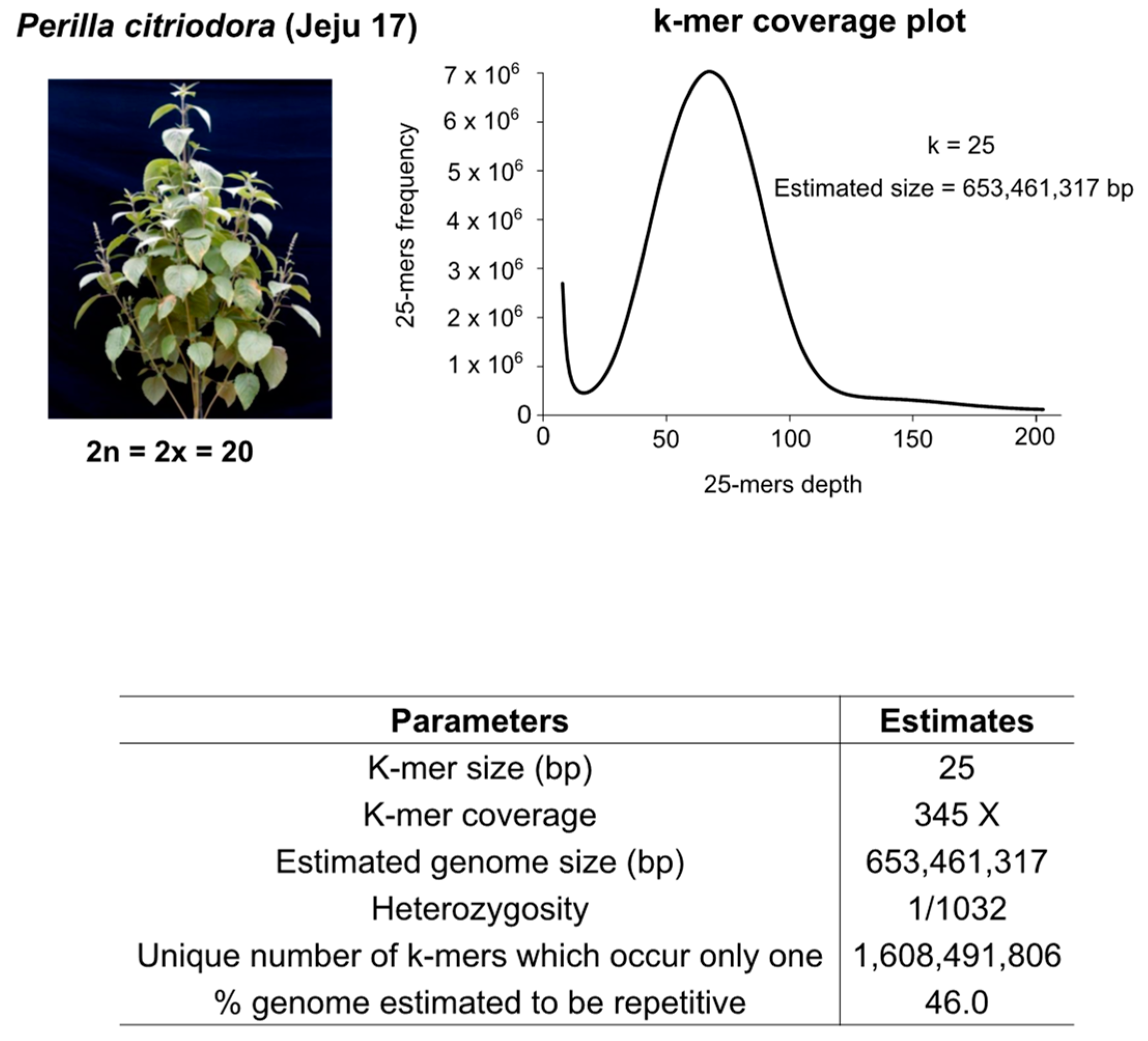
2.1. Whole-Genome Sequencing, Genome Size Estimation, and Genome Assembly

P. citriodora ‘Jeju17’ (Figure 1) was discovered and collected on Jeju island (33°57′24.2424″ N 84°7′52.77″ W) in 2002 and preserved at the National Institute of Crops Science of Korea [31]. It was used as a material by advancing more than 10 generations advancement. Two-week-old leaves were sampled for whole-genome sequencing using Illumina, PacBioRSII, and Hi-C platforms (Figure S1). A genome survey was implemented to estimate the plant genome size using the Jellyfish v2.2.7 tool [33]. For annotation purposes, short-read RNA sequencing was performed on seed, bud, and leaf tissues, following the manufacturer’s protocols. The sequence reads archive (SRA) for the genome and transcriptome data are provided in Table S1.
A hybrid assembly strategy (Figure S1) combining short and long reads was conducted to produce a chromosome-scale assembly. Briefly, after an initial assembly with Platanus v1.2.1 [34], gaps were filled using PBJELLY v2.0 [35], and the redundant sequences were filtered out with CD-HIT [36]. Meanwhile, long-read assemblies were executed with Falcon v0.7.0 [37]. Both assemblies were merged using HaploMerger v2 [38]. The hybrid assembly was shipped to Phase Genomics® for the Hi-C scaffolding step.

2.2. Genome Assembly Quality Assessment

The assembly was assessed for contiguity, gene completeness, and structural variation by comparison with the reference genome P. citriodora PC002 [7]. The chromosomes were arranged from 1 to 10 in order of length (bp). BUSCO analysis [39] was completed using the Embryophyta odb_10 database. Whole-genome alignment with PC002 was performed using MashMap v2.0 [40] and visualized with D-Genies v1.4.0 [41]. In addition, MUMmer v4.0.0 [42] and Assemblytics [43] assessed the structural variants between ‘PC002’ and ‘Jeju17’.
In order to validate the assembled genome, bacterial artificial chromosome (BAC) libraries were produced. High-molecular-weight DNA was extracted from young leaves of ‘Jeju17’, as described by Kang et al. [44]. After a digestion step with HindIII- and BamHI-restriction enzymes, insert size selection was performed, followed by ligation using a pSMART BAC vector. The ligated substrate was then transformed into DH10B-competent cells. A total of 10 BAC clones were subsequently sequenced using an ABI 3730 × l DNA Analyzer and 454 Life Sciences GS FLX System (GS FLX). BAC clone sequencing was used to select 10 genes (Tables S3 and S4) related to triacylglycerol (TAG) biosynthetic pathways [45]. Assembly was carried out with Newbler v2.8 (https://www.ncbi.nlm.nih.gov/assembly/GCF_000588835.1/, accessed on 24 September 2013). Gap filling was conducted following the primers walking method [46]. The assembly was error-corrected with short-read data. The generated BAC-based assembly was aligned onto the chromosome-scale assembled genome using MUMmer v4.0.0 [42].

2.3. Repeat Identification and Genome Annotation

For repeat identification, RepeatModeler v. 1.0.8 (http://www.repeatmasker.org/RepeatModeler/, accessed on 5 February 2014) and RepeatMasker v. 4.0.5 (http://www.repeatmasker.org, accessed on 5 February 2014) packages were employed. Non-coding RNA (rRNA, tRNA, miRNA, and snRNA) was detected using Infernal v1.1.4 [47] with the Rfam database [48] (https://rfam.xfam.org/, accessed on 14 March 2022).
Prior to structural annotation, a genome-based transcript assembly was generated. Raw RNA-seq data were checked and trimmed with FastQC v0.11.2 [49] and Trimmomatic v0.36 [50]. The reads were mapped onto the genome using HISAT v2.2.1 [51]. Then, StrigTie v2.2.1 [52] was used to construct the transcript assembly. Gene prediction was carried out with MAKER2 pipeline [53], while functional annotation was executed by BLASTp [54], HMMER [55], and BLAST2GO [56] searches against plant reference sequences and NCBI non-redundant (nr), UniProt, Pfam, GO, KEGG, and InterProScan databases.

2.4. Gene Family Analysis, Time Divergence Estimation, and Phylogenetic Placement of Jeju 17

This study used OrthoFinder v.2.3.12 [57] to determine the conserved and species-specific gene families. Gene sets from Lamiales, including P. citriodora ‘Jeju17’, P. citriodora ‘PC002’, P. citriodora ‘PC099’, P. frutescens ‘PF40’, Salvia splendens, Salvia miltiorrhiza, Mimulus guttatus, Sesamum indicum var. Zhongzhi13, Sesamum indicum var. Goenbaek, and Olea europeae were used with Solanum lycopersicum and Arabidopsis thaliana as the outgroup species (Table S2).
KEGG and GO enrichment analyses of species-specific gene families were assessed with the KOBAS-I online server [58] using the default settings (statistical test: “Fisher’s exact test”; FDR correction method: “Benjamini and Hochberg”).
Using the single-copy orthologues, a multiple-sequence alignment (MSA) was performed by MAFFT v.7.464-0 [59]. After trimming the MSA file with trimAl v.1.4.1 [60], a maximum likelihood phylogenetic construction with 1000 replicates was conducted following the JTT+F+R3 model, using IQ-TREE v1.6.12 [61].
Time divergence was estimated using the Reltime method [62,63] and the JTT matrix-based model [64]. The calibrations were set to 25.0–57.1 MYA (Mimulus versus Sesamum genera) and 112.4–125.0 MYA (Arabidopsis vs. Lycopersicum genera) following the TimeTree database (www.timetree.org, accessed on 6 April 2017).

2.5. Identification of Putative Orthologs

Orthologs among the 12 species were identified by conducting an OthoFinder analysis [52]. Orthologous gene pairs were retrieved from otho groups containing one gene per species. The analysis was conducted as previously described [65,66]. For each ortholog or paralog, the protein sequences were aligned using ClustalW [67], and the corresponding codons were aligned using PAL2NAL [68] (Suyama et al. 2006) with the guidance of coding sequences. The Ka and Ks values were calculated using the Nei Gojobori method [69] implemented in the PAML package [70].

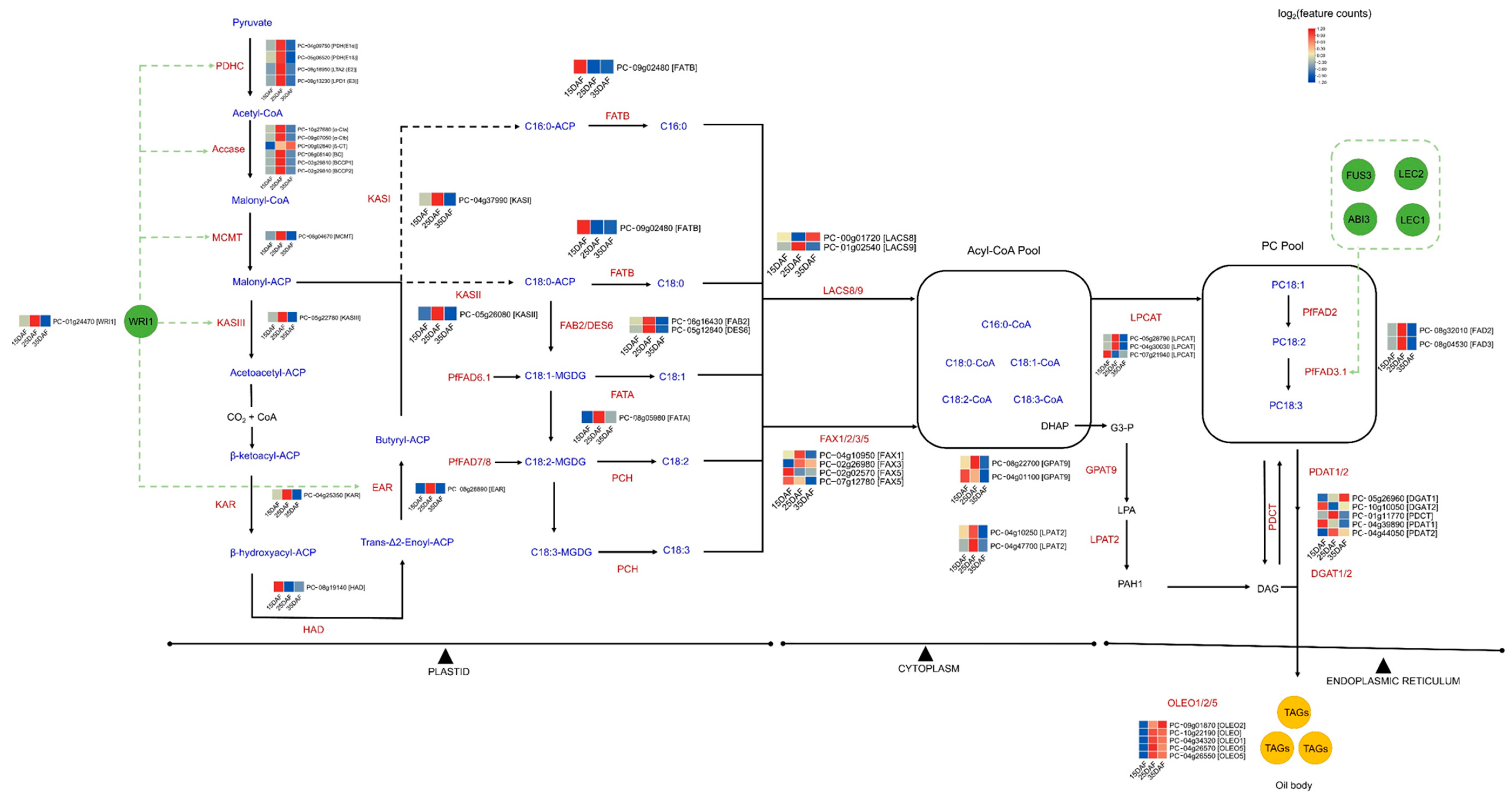
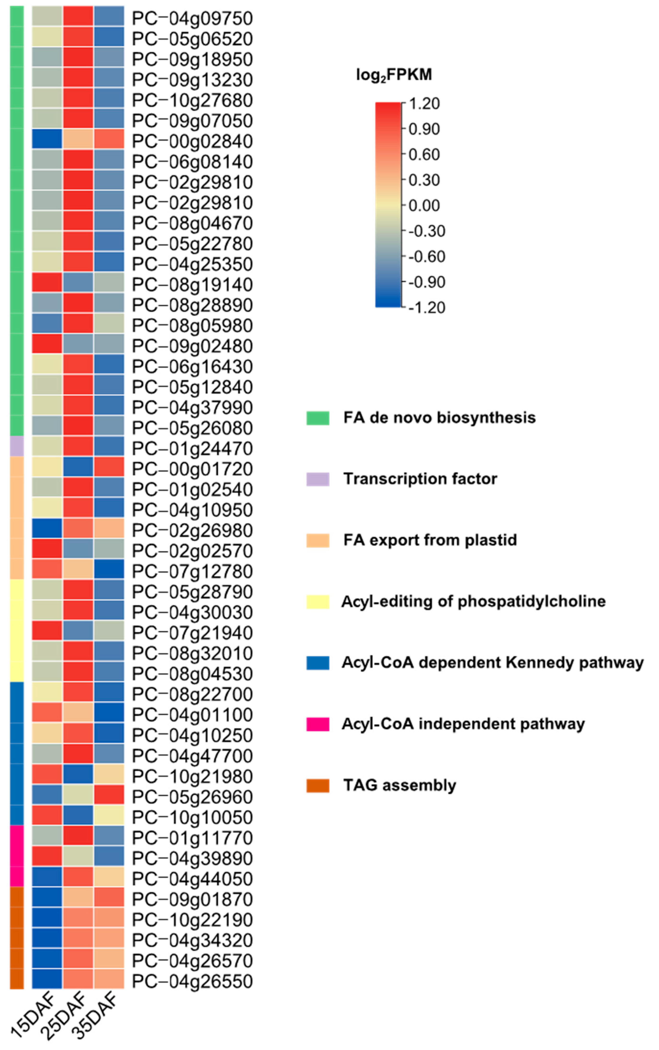
2.6. Identification of Genes Involved in Fatty Acid and Lipid Triacylglycerol Biosynthesis

Using the A. thaliana acyl-lipid metabolism database [71], we inferred genes involved in fatty acid and triacylglycerol biosynthesis in P. citriodora ‘Jeju17’ by performing a protein BLASTp search (E = 1× 10⁻¹⁰). A cross-check validation was also performed using the reference genome PC002 from Zhang et al. [7].
Furthermore, using the curated fatty acid desaturase hmm profiles PF00487 and PF03405 (from http://pfam.xfam.org/, accessed on 1 January 2004) in addition to previously identified desaturases from P. frutescens ‘PF40’ [72], this study identified and classified fatty acid desaturases that were potentially involved in fatty acid and lipid triacylglycerol biosynthesis in the wild species P. citriodora ‘Jeju17’. The study employed both HMMER and BLASTp searches for the identification of candidate genes. The protein domain was checked using PfamScan v1.6 [73].
For phylogenetic tree construction, fatty acid desaturases from P. frutescens and S. indicum served as baits. The study involved a blast search against the P. citriodora ‘PC002’ genome and retrieved corresponding desaturase genes that were also included as baits. The sequences were aligned using MAFFT v.7.464-0 [59]. The resulting MSA was trimmed with trimAl v.1.4.1 [60], and the tree was produced with IQ-TREE v1.6.12 [61] using 1000 replicates.
The subcellular localization and chemical properties of the proteins were assessed using ExPASy (http://web.expasy.org/protparam/, accessed on 5 February 2014) and Cell-PLoc (http://www.csbio.sjtu.edu.cn/bioinf/plant/, accessed on 5 February 2014) [74,75].
A 2Kbp upstream promoter region was extracted to investigate cis-acting regulatory elements using the PlantCARE webserver (http://bioinformatics.psb.ugent.be/webtools/plantcare/html/, accessed on 5 February 2014) [76].

2.7. Synteny Analysis

Gene-to-gene synteny analysis was executed with MCScanX [77] embedded in TBTools [78] using the default settings.

3. Results and Discussion

3.1. Genome Survey, Sequencing, and Assembly Quality

With initial short-read sequencing data representing 345X coverage, the k-mer-based genome size was estimated as 653 Mbp (Figure 1). By using short- and long-read sequencing platforms, the study generated 339X (Illumina), 1195X (Illumina), 72X (PacBio), and 26X (Hi-C) fold on MiSeq, HiSeq, PacBioRSII, and Hi-C systems, respectively (Figure S1, Table S1). Due to the low quality of the long-read data, the long-read assembly resulted in an N50 of 0.6 Mbp. To address this issue, we opted for hybrid assembly combining both short- and long reads. Therefore, an initial assembly base consisting of diverse insert sizes (Table S1) produced an assembly of 718 Mbp with an N50 of 7.5 Mbp. Redundant sequences were removed, leading to an assembly spanning 644 Mbp with an N50 value of 9.6 Mbp. Furthermore, HaploMerger helped to combine both long- and short-read assemblies to reach an assembly size of 678 Mbp with an N50 of 12.3 Mbp (Figure S1). By adding Hi-C data, the assembly contiguity was drastically improved up to 68 Mbp (N50) for an assembly size of 676 Mbp (Figure S1).
To validate our assembly, BAC alignment identity > 99% was retained (Tables S3 and S4). In addition, BUSCO analysis showed similar single-copy orthologue coverage for assembly (~93%). However, the BUSCO analysis on annotation data revealed higher coverage (84.1%) of ‘Jeju17’ compared to ‘PC002’ (79.8%) (Table S5).
The assembly results were compared with the already published PC002, which is karyotypic, and ‘Jeju17’, like ‘PC002’, consists of a diploid species (2n  =  2x  =  20). The alignment of ‘Jeju17’ onto ‘PC002’ revealed extensive exact matches (73.78%) (Figure S2). However, inverted regions were noted within chromosomes 1, 3, 4, 6, 7, and 10. In-depth analysis revealed that the structural variants represented 19.1 Mbp of encompassing insertion (3.36 Mbp), deletion (2.57 Mbp), tandem expansion (2.42 Mbp), tandem contraction (0.59 Mbp), repeat expansion (4.59 Mbp), and repeat contraction (5.57 Mbp) (Table S6).

3.2. Genome Annotation Features

Prior to gene prediction, repeats and non-coding RNA were assessed. The transposable elements (TE) content of Jeju17 was around 63.37% and mainly dominated by long terminal repeat (LTR) retrotransposon elements (27.39%) (Table S7). In PC002, the TE content was lower (56.7%), with LTR elements estimated at around 22% [7].
The non-coding RNA spanned 416,914 bp, representing 0.001% of the genome. The tRNA (n = 933), rRNA (n = 434), miRNA (n = 127), and snRNA (n = 190) occupied 68 Kbp, 313 Kbp, 16 Kbp, and 19 Kbp, respectively (Table S8).
A total number of 32,769 coding-protein sequences were predicted (Table S9). Compared to ‘PC002’, ‘Jeju17’ exhibited a higher number of genes (31,273) with a mean mRNA length of 4140 bp (Table S9).
Figure 2 presents the genome map of ‘Jeju17’.

3.3. Genome Evolution

Timetree calculations were retrieved from the TimeTree database and are as follows: S. lycopersicum and O. europaea, ranging 75 MYA~88 MYA; O. europaea and U. gibba, ranging 60 MYA~77 MYA; U. gibba and A. majus, ranging 48 MYA~88 MYA; and A. majus and S. indicum, ranging 52 MYA~67 MYA. The minimum and maximum branch time estimated for 52 MYA to 67 MYA were applied [79]. In the literature, the estimated time of divergence is 12 MYA for Oleaceae [80] and 62 MYA for Plantaginaceae [81], and the branching time is considered to be consistent with the estimation of the branching time from 11 species. Thus, diploid Perilla was estimated to branch at about 30 MYA. For branch analysis, the Ks value of each gene pair between ‘Jeju17’ and each species was measured and plotted (Figure S3).

3.4. Phylogenetic Placement of ‘Jeju17’-Specific Gene Family Assessment

Using the whole-genome gene sets of 12 species, we inferred the phylogenetic placement of ‘Jeju17’ in the Lamiales order (Figure 3a). As expected, ‘Jeju17’ falls into Perilla’s clade and is closely related to P. citriodora ‘PC002’. The time-divergence analysis revealed that Perilla split from the common ancestor, a member of the Salvia taxa, 1.17 MYA. A comparative analysis of gene orthology within Perilla representatives (Figure 3b) showed a set of 123, 163, 1250, and 3331 species-specific gene families for ‘PC099’, ‘PC002’, ‘PF40’, and ‘Jeju17’, respectively.
To understand the biological implication of species-specific genes, KEGG and GO enrichment analyses were performed (Figure 4, Figures S3, and S4, and Table S10).
The top 100 significantly differential pathways were shown independently in the histogram (Figure S4) and bubble plot (Figure 4).
A KEGG enrichment of ‘Jeju17’ species-specific genes showed diverse biological functions that are mainly related to the biosynthesis of secondary metabolites (ko01100, ko01110) such as sesquiterpenoid and triterpenoid biosynthesis (ko00909), diterpenoid biosynthesis (ko00904), and zeatin biosynthesis (ko00908) (Figure 4, Table S10). As expected, fatty-acid-metabolism-related genes were also identified (ko01230, ko01212, ko00592, ko00062, ko00061, ko00460, ko01210, ko00071). Similar findings were also found in ‘PC002’, ‘PC099’, and ‘PF40’ (Figure S4, Tables S11–S13).
Interestingly, and probably due to the Jeju island subtropical climate, genes related to photosynthesis (ko00195) and adverse environmental responses such as oxidative phosphorylation (ko00190) were detected in the ‘Jeju17’-specific gene set. In addition, genes involved in the plant abiotic stress response, including the MAPK signaling pathway (ko04016), ubiquitin–proteasome system (ko03050), and nucleotide excision repair (ko03420), were identified.
The MAPK machinery pathway is well known for its role in the transfer of information from sensors to initiate cellular responses in environmental stress conditions [82,83,84]. To protect the plant against DNA damage caused by stresses such as exposure to ultraviolet light, nucleotide excision repair can be adopted by the plant to mitigate the damage caused by DNA lesions and therefore maintain genome integrity [85]. Furthermore, the ubiquitin–proteasome system is also deployed in adverse growth conditions to facilitate cellular changes by regulating the abundance of regulatory and structural proteins and enzymes [86,87]. Overall, ‘Jeju17’ harbored not only fatty acid and specialized metabolite genes but also the genetic resources to tolerate environmental stress such as salt stress, high light, temperature, and heat. These results suggest that the lipid biosynthetic pathway is related to the physiological and environmental conditions of plants.
To compare ‘Jeju17’ and ‘PC002’, KEGG analysis was performed using the Perilla sequence reported in China (Figure 4 and Figure S4A). As a result, the percentage of gene annotations related to metabolic pathways was high in ‘PC002’, and the analysis result was different from ‘Jeju17’ (Figure S4A).

3.5. Identification of Genes Involved in Fatty Acid and Lipid Triacylglycerol Biosynthesis

A comparative analysis of genes involved in fatty acid and lipid triacylglycerol biosynthesis revealed consistent gene conservation between ‘PC002’ and ‘Jeju17’ (Figure 3c). Based on the identified genes, we reconstructed the fatty acid and lipid triacylglycerol biosynthesis pathways (Figure 5). As reviewed by Bae et al. [25], palmitic acid (C16:0), stearic acid (C18:0), and oleic acid (C18:1) were produced in plastids and transported into the cytoplasm, where they entered an acyl-CoA pool for esterification at the sn-2 position, generating phosphatidylcholine by the acyl-CoA:lysophosphatidylcholine acyltransferase (LPCAT) enzyme (PC_05g28790, PC_04g30030, and PC_07g21940) (Figure 5). After desaturation in the endoplasmic reticulum, oleic acid is transformed into linoleic acid and alpha-linolenic acid by FAD2 (PC_08g32010) and FAD3 (PC_08g04530) catalysts. The derived fatty acids were then transacylated or sent to the acyl-CoA pool by LPCAT for insertion into triacylglycerol production through the Kennedy pathway (Figure 5). The transcription factor WR1, known to actively regulate fatty acid biosynthesis [88,89], was also identified in the ‘Jeju17’ genome, paving the way for functional validation and potential fatty acid and/or lipid bioengineering from wild Perilla. The mechanism of the regulation of FA synthesis by TFs in Perilla is still unknown. Meanwhile, Moreno-Perez et al. suggested the implications of histone methylation (H3K4me3) for fatty acid biosynthesis and interaction with TFs in sunflowers. Moreover, acetyl-CoA, an organelle agent involved in fatty acid synthesis in plants, has been found to correlate with histone acetylation and DNA methylation in A. thaliana through a beta-oxidation process [90]. Therefore, the investigation of the epigenome of Perilla and identified TFs, such as ABI3, FUS3, LEC1, and LEC2, will open up new avenues to decipher the entire landscape of fatty acid biosynthesis in Perilla.
This study also explored the identification of fatty acid desaturases, which play key roles in fatty acid production. A total of 22 fatty acid desaturases (Figure 6, Table S14) were identified and grouped into 5 families, including delta-12 desaturase (n = 4), front-end desaturase (n = 5), delta-7 desaturase (n = 1), delta-9 desaturase (n = 9), and omega-3 desaturase (n = 3).
Fatty acid desaturases were identified in all chromosomes, except for chromosome 10 (Table S14, Figure 6). The highest protein length was noted for PC_05g17080 (PcDES.1), a sphingolipid delta (4)-desaturase DES1-like gene with a molecular weight of 58.59 kDa.
Regarding the expression of genes involved in fatty acid and lipid triacylglycerol biosynthesis, prominent expression was noted 25 days after flowering (Figure 7). Interestingly, TAG-assembly-related genes were highly expressed later on (after 35 days of flowering), which is similar to the lipid triacylglycerol biosynthesis described by Bates et al. [91].
Cis-acting element analysis in a 2 Kb promoter region of fatty acid desaturase genes was revealed. Some diverse distribution patterns of cis-acting elements were observed in the promoter region of fatty acid desaturase genes, indicating that fatty acid desaturase is important in various biological processes. This study identified twenty-five cis-acting elements according to their functional annotations, which were related to hormone responses and abiotic stress. Notably, the genes contained cis-acting elements related to hormone regulation, such as auxin, gibberellin, methyl jasmonate, abscisic acid, and salicylic acid responsiveness elements. In addition, there is potential implication for the involvement of the genes in diverse biological processes, including light, physiological development, and abiotic stress response (Figure 8). The Perilla are closely correlated with lipid metabolism and several metabolic pathways [92].
Fatty acids play important roles in lipid supply in plants and have valuable medicinal properties for humans [93]. This study made a breakthrough in revealing the genetic and molecular determinants of FAs and TAG synthesis in Perilla. Transcriptomics and genomics studies have also disclosed the key enzymes responsible for FAs synthesis in Perilla, including polyunsaturated fatty acid desaturases, acyl-related enzymes, and transcription factors.

4. Conclusions

This study provides a high-quality genome resource for the wild P. citriodora ‘Jeju17’ that originated from the Korean island of Jeju. Although a genome from a Chinese species was recently released, the genetic specificity of ‘Jeju17’ needs to be explored. Thus, the assembly results were compared with the already published PC002, which is karyotypically P. citriodora ‘Jeju17’ and, like ‘PC002’, consists of a diploid species (2n  =  2x  =  20). Alignment of ‘Jeju17’ to the Chinese species ‘PC002’ showed significant collinearity, but sequence inversions, mainly on chromosomes 1, 4, 6, 7, and 10, were also detected. The genomic data enabled the identification of species-specific genes, mainly involved in response to abiotic stress, environment adaptation, metabolite biosynthesis, fatty acids, and lipid biosynthesis. These results suggest that the expression of fatty acid desaturase genes in P. citriodora is related to cis-elements associated with hormone signal transduction and abiotic stress tolerance. In addition, using a homology-based approach, this study identified a set of 22 fatty acid desaturases potentially involved in the fatty acid production pathway. The present genomic analyses constitute a valuable resource for comparative genomics and Perilla breeding for fatty acids and oil-related trait improvement.

Supplementary Materials

The following supporting information can be downloaded at: https://www.mdpi.com/article/10.3390/genes14101898/s1, Figure S1. Hybrid assembly strategy using Illumina, PacBio, and Hi-C sequences. #represents the number of scaffold/contig; Figure S2: Whole-genome alignment view of ‘Jeju17’ onto ‘PC002’; Figure S3. Genome evolutionary history. a Divergence times with the node age confidence intervals labeled. b The upper right area (insert) shows the Ks distribution from orthologs between Perilla citriodora and each of the eleven species; Figure S4. KEGG enrichment bubble plot depicting the functional attributes of ‘PC002’, ‘PC09’, and ‘PF40’ species-specific gene sets; Table S1: SRA details of the genome and transcriptome data employed in the current study; Table S2: List of species used in the comparative genomics study; Table S3: Alignment results for the BAC clone onto the ‘Jeju17’ genome; Table S4. Conservation of ten BAC-end sequences in scaffolds; Table S5. Summary statistics of genome assemblies of ‘Jeju17’ and ‘PC002’; Table S6: Assembly-based structural variations detected by aligning ‘Jeju17’ onto ‘PC002’; Table S7. ‘Jeju17’ repeats content statistics; Table S8. Predicted non-coding RNA statistics; Table S9. Comparative annotation statistics of ‘Jeju17’ and ‘PC002’; Table S10: KEGG and GO analyses results of the ‘Jeju17’ species-specific genes set; Table S11: KEGG and GO analyses results of the ‘PC002’ species-specific genes set; Table S12: KEGG and GO analyses results of the ‘PC09’ species-specific genes set; Table S13: KEGG and GO analyses results of the ‘PF40’ species-specific genes set; Table S14: Information on the identified fatty acid desaturase genes in ‘Jeju17’.

Author Contributions

Conceptualization, T.-H.K.; methodology, S.-H.B., M.H.L., and T.-H.K.; software, S.-H.B., J.-H.L., and Y.Y.; validation, S.-H.B.; formal analysis, S.-H.B., M.H.L., J.-H.L., and Y.Y.; Resources, M.H.L.; data curation, S.-H.B., J.-H.L., and Y.Y.; writing—original draft preparation, S.-H.B.; writing—review and editing, S.-H.B., M.H.L., J.-H.L., Y.Y., J.L., and T.-H.K.; visualization, S.-H.B.; supervision, J.L. and T.-H.K.; project administration and funding acquisition, T.-H.K. All authors have read and agreed to the published version of the manuscript.

Funding

This work was carried out with the support of the National Institute of Agricultural Sciences (Project no. PJ010408 and PJ013355), the Rural Development Administration, the Republic of Korea.

Institutional Review Board Statement

Not applicable.

Informed Consent Statement

Not applicable.

Data Availability Statement

Data are contained within the article or Supplementary Material.

Acknowledgments

We thank DNACARE for assistance in the acquisition and interpretation of data according to the Perilla genome analysis. We would also like to thank SEEDERS for assistance in obtaining and interpreting sequencing data for fatty acid-related analysis. We thank all the participants in this study.

Conflicts of Interest

The authors declare no conflict of interest.

References

  1. Nitta, M.; Lee, J.K.; Kang, C.W.; Katsuta, M.; Yasumoto, S.; Liu, D.; Nagamine, T.; Ohnishi, O. The Distribution of Perilla Species. Genet. Resour. Crop Evol. 2005, 52, 797–804. [Google Scholar] [CrossRef]
  2. Nitta, M.; Lee, J.K.; Ohnishi, O. Asian Perilla crops and their weedy forms: Their cultivation, utilization and genetic relationships. Econ. Bot. 2003, 57, 245–253. [Google Scholar] [CrossRef]
  3. Cui, X.; Gou, Z.; Fan, Q.; Li, L.; Lin, X.; Wang, Y.; Jiang, S.; Jiang, Z. Effects of dietary perilla seed oil supplementation on lipid metabolism, meat quality, and fatty acid profiles in Yellow-feathered chickens. Poult. Sci. 2019, 98, 5714–5723. [Google Scholar] [CrossRef] [PubMed]
  4. Arjin, C.; Souphannavong, C.; Norkeaw, R.; Chaiwang, N.; Mekchay, S.; Sartsook, A.; Thongkham, M.; Yosen, T.; Ruksiriwanich, W.; Sommano, S.R.; et al. Effects of Dietary Perilla Cake Supplementation in Growing Pig on Productive Performance, Meat Quality, and Fatty Acid Profiles. Animals 2021, 11, 3213. [Google Scholar] [CrossRef]
  5. Peiretti, P.G.; Gasco, L.; Brugiapaglia, A.; Gai, F. Effects of perilla (Perilla frutescens L.) seeds supplementation on performance, carcass characteristics, meat quality and fatty acid composition of rabbits. Livest. Sci. 2011, 138, 118–124. [Google Scholar] [CrossRef]
  6. Xia, J.Q.; He, X.; Wang, L.; Wang, L.; Zhang, D.J.; Wang, J.F.; Liu, D. Evaluation of dietary Perilla frutescens seed on performance and carcass quality in finishing castrated male Songliao black pigs. Vet. Med. Sci. 2022, 8, 598–606. [Google Scholar] [CrossRef] [PubMed]
  7. Zhang, Y.; Shen, Q.; Leng, L.; Zhang, D.; Chen, S.; Shi, Y.; Ning, Z.; Chen, S. Incipient diploidization of the medicinal plant Perilla within 10,000 years. Nat. Commun. 2021, 12, 5508. [Google Scholar] [CrossRef] [PubMed]
  8. Yu, H.; Qiu, J.-F.; Ma, L.-J.; Hu, Y.-J.; Li, P.; Wan, J.-B. Phytochemical and phytopharmacological review of Perilla frutescens L. (Labiatae), a traditional edible-medicinal herb in China. Food Chem. Toxicol. 2017, 108, 375–391. [Google Scholar] [CrossRef] [PubMed]
  9. Hou, T.; Netala, V.R.; Zhang, H.; Xing, Y.; Li, H.; Zhang, Z. Perilla frutescens: A Rich Source of Pharmacological Active Compounds. Molecules 2022, 27, 3578. [Google Scholar] [CrossRef] [PubMed]
  10. Lin, C.-S.; Kuo, C.-L.; Wang, J.-P.; Cheng, J.-S.; Huang, Z.-W.; Chen, C.-F. Growth inhibitory and apoptosis inducing effect of Perilla frutescens extract on human hepatoma HepG2 cells. J. Ethnopharmacol. 2007, 112, 557–567. [Google Scholar] [CrossRef]
  11. Narisawa, T.; Takahashi, M.; Kotanagi, H.; Kusaka, H.; Yamazaki, Y.; Koyama, H.; Fukaura, Y.; Nishizawa, Y.; Kotsugai, M.; Isoda, Y.; et al. Inhibitory Effect of Dietary Perilla Oil Rich in the n-3 Polyunsaturated Fatty Acid α-Linolenic Acid on Colon Carcinogenesis in Rats. Jpn. J. Cancer Res. 1991, 82, 1089–1096. [Google Scholar] [CrossRef]
  12. Banno, N.; Akihisa, T.; Tokuda, H.; Yasukawa, K.; Higashihara, H.; Ukiya, M.; Watanabe, K.; Kimura, Y.; Hasegawa, J.I.; Nishino, H. Triterpene acids from the leaves of Perilla frutescens and their anti-inflammatory and antitumor-promoting effects. Biosci. Biotechnol. Biochem. 2004, 68, 85–90. [Google Scholar] [CrossRef] [PubMed]
  13. Chen, C.-Y.; Leu, Y.-L.; Fang, Y.; Lin, C.-F.; Kuo, L.-M.; Sung, W.-C.; Tsai, Y.-F.; Chung, P.-J.; Lee, M.-C.; Kuo, Y.-T.; et al. Anti-inflammatory effects of Perilla frutescens in activated human neutrophils through two independent pathways: Src family kinases and Calcium. Sci. Rep. 2016, 5, 18204. [Google Scholar] [CrossRef]
  14. Jeon, I.; Kim, H.; Kang, H.; Lee, H.-S.; Jeong, S.; Kim, S.; Jang, S. Anti-Inflammatory and Antipruritic Effects of Luteolin from Perilla (P. frutescens L.) Leaves. Molecules 2014, 19, 6941–6951. [Google Scholar] [CrossRef] [PubMed]
  15. Zhang, H.; Tian, Y.; Guan, J.; Xie, Q.; Zhao, Y. The anti-tussive, anti-inflammatory effects and sub-chronic toxicological evaluation of perilla seed oil. J. Sci. Food Agric. 2021, 101, 1419–1427. [Google Scholar] [CrossRef] [PubMed]
  16. Wijendran, V.; Hayes, K.C. Dietary n-6 and n-3 fatty acid balance and cardiovascular health. Annu. Rev. Nutr. 2004, 24, 597–615. [Google Scholar] [CrossRef]
  17. Wang, F.; Zhu, H.; Hu, M.; Wang, J.; Xia, H.; Yang, X.; Yang, L.; Sun, G. Perilla Oil Supplementation Improves Hypertriglyceridemia and Gut Dysbiosis in Diabetic KKAy Mice. Mol. Nutr. Food Res. 2018, 62, 1800299. [Google Scholar] [CrossRef]
  18. Zhang, T.; Zhao, S.; Li, W.; Ma, L.; Ding, M.; Li, R.; Liu, Y. High-fat diet from perilla oil induces insulin resistance despite lower serum lipids and increases hepatic fatty acid oxidation in rats. Lipids Health Dis. 2014, 13, 15. [Google Scholar] [CrossRef]
  19. Song, Y.; Sun, R.; Ji, Z.; Li, X.; Fu, Q.; Ma, S. Perilla aldehyde attenuates CUMS-induced depressive-like behaviors via regulating TXNIP/TRX/NLRP3 pathway in rats. Life Sci. 2018, 206, 117–124. [Google Scholar] [CrossRef]
  20. Nakazawa, T.; Yasuda, T.; Ueda, J.; Ohsawa, K. Antidepressant-like effects of apigenin and 2,4,5-trimethoxycinnamic acid from Perilla frutescens in the forced swimming test. Biol. Pharm. Bull. 2003, 26, 474–480. [Google Scholar] [CrossRef]
  21. Takeda, H.; Tsuji, M.; Inazu, M.; Egashira, T.; Matsumiya, T. Rosmarinic acid and caffeic acid produce antidepressive-like effect in the forced swimming test in mice. Eur. J. Pharmacol. 2002, 449, 261–267. [Google Scholar] [CrossRef] [PubMed]
  22. Takeda, H.; Tsuji, M.; Miyamoto, J.; Matsumiya, T. Rosmarinic acid and caffeic acid reduce the defensive freezing behavior of mice exposed to conditioned fear stress. Psychopharmacology 2002, 164, 233–235. [Google Scholar] [CrossRef]
  23. Makino, T.; Furuta, Y.; Wakushima, H.; Fujii, H.; Saito, K.; Kano, Y. Anti-allergic effect of Perilla frutescens and its active constituents. Phyther. Res. 2003, 17, 240–243. [Google Scholar] [CrossRef] [PubMed]
  24. Liang, K.-L.; Yu, S.-J.; Huang, W.-C.; Yen, H.-R. Luteolin Attenuates Allergic Nasal Inflammation via Inhibition of Interleukin-4 in an Allergic Rhinitis Mouse Model and Peripheral Blood from Human Subjects with Allergic Rhinitis. Front. Pharmacol. 2020, 11, 291. [Google Scholar] [CrossRef] [PubMed]
  25. Narisawa, T.; Fukaura, Y.; Ishikawa, C.; Isoda, Y.; Nishizawa, N. Colon cancer prevention with a small amount of dietary perilla oil high in alpha-linolenic acid in an animal model. Cancer 1994, 73, 2069–2075. [Google Scholar] [CrossRef] [PubMed]
  26. Yoon, S.-H.; Noh, S. Positional distribution of fatty acids in perilla (Perilla frutescens L.) oil. J. Am. Oil Chem. Soc. 2011, 88, 157–158. [Google Scholar] [CrossRef]
  27. Xue, Y.; Chen, B.; Win, A.; Fu, C.; Lian, J.; Liu, X.; Wang, R.; Zhang, X.; Chai, Y. Omega-3 fatty acid desaturase gene family from two ω-3 sources, Salvia hispanica and Perilla frutescens: Cloning, characterization and expression. PLoS ONE 2018, 13, e0191432. [Google Scholar] [CrossRef]
  28. Zhu, S.; Zhu, Z.; Wang, H.; Wang, L.; Cheng, L.; Yuan, Y.; Li, D. Characterization and functional analysis of a plastidial FAD6 gene and its promoter in the mesocarp of oil palm (Elaeis guineensis). Sci. Hortic. 2018, 239, 163–170. [Google Scholar] [CrossRef]
  29. Lee, K.-R.; Lee, Y.; Kim, E.-H.; Lee, S.-B.; Roh, K.-H.; Kim, J.-B.; Kim, H.-U. Functional identification of oleate 12-desaturase and ω-3 fatty acid desaturase genes from Perilla frutescens var. frutescens. Plant Cell Rep. 2016, 35, 2523–2537. [Google Scholar] [CrossRef]
  30. Bae, S.-H.; Zoclanclounon, Y.A.B.; Kumar, T.S.; Oh, J.-H.; Lee, J.; Kim, T.-H.; Park, K.Y. Advances in Understanding the Genetic Basis of Fatty Acids Biosynthesis in Perilla: An Update. Plants 2022, 11, 1207. [Google Scholar] [CrossRef]
  31. Jung, C.; Lee, M.; Oh, K.; Kim, H.; Park, C.; Sung, J.; Suh, D. Discovery of New Diploid Perilla Species in Korea. Korean J. Breed 2005, 37, 2003–2005. [Google Scholar]
  32. Sohn, Y.K.; Park, K.H. Early-stage volcanism and sedimentation of Jeju Island revealed by the Sagye borehole, SW Jeju Island, Korea. Geosci. J. 2004, 8, 73–84. [Google Scholar] [CrossRef]
  33. Marçais, G.; Kingsford, C. A fast, lock-free approach for efficient parallel counting of occurrences of k-mers. Bioinformatics 2011, 27, 764–770. [Google Scholar] [CrossRef]
  34. Kajitani, R.; Toshimoto, K.; Noguchi, H.; Toyoda, A.; Ogura, Y.; Okuno, M.; Yabana, M.; Harada, M.; Nagayasu, E.; Maruyama, H.; et al. Efficient de novo assembly of highly heterozygous genomes from whole-genome shotgun short reads. Genome Res. 2014, 24, 1384–1395. [Google Scholar] [CrossRef] [PubMed]
  35. English, A.C.; Richards, S.; Han, Y.; Wang, M.; Vee, V.; Qu, J.; Qin, X.; Muzny, D.M.; Reid, J.G.; Worley, K.C.; et al. Mind the Gap: Upgrading Genomes with Pacific Biosciences RS Long-Read Sequencing Technology. PLoS ONE 2012, 7, e47768. [Google Scholar] [CrossRef]
  36. Li, W.; Godzik, A. Cd-hit: A fast program for clustering and comparing large sets of protein or nucleotide sequences. Bioinformatics 2006, 22, 1658–1659. [Google Scholar] [CrossRef] [PubMed]
  37. Chin, C.-S.; Peluso, P.; Sedlazeck, F.J.; Nattestad, M.; Concepcion, G.T.; Clum, A.; Dunn, C.; O’Malley, R.; Figueroa-Balderas, R.; Morales-Cruz, A.; et al. Phased diploid genome assembly with single-molecule real-time sequencing. Nat. Methods 2016, 13, 1050–1054. [Google Scholar] [CrossRef]
  38. Huang, S.; Kang, M.; Xu, A. HaploMerger2: Rebuilding both haploid sub-assemblies from high-heterozygosity diploid genome assembly. Bioinformatics 2017, 33, 2577–2579. [Google Scholar] [CrossRef]
  39. Simão, F.A.; Waterhouse, R.M.; Ioannidis, P.; Kriventseva, E.V.; Zdobnov, E.M. BUSCO: Assessing genome assembly and annotation completeness with single-copy orthologs. Bioinformatics 2015, 31, 3210–3212. [Google Scholar] [CrossRef]
  40. Jain, C.; Koren, S.; Dilthey, A.; Phillippy, A.M.; Aluru, S. A fast adaptive algorithm for computing whole-genome homology maps. Bioinformatics 2018, 34, i748–i756. [Google Scholar] [CrossRef] [PubMed]
  41. Cabanettes, F.; Klopp, C. D-GENIES: Dot plot large genomes in an interactive, efficient and simple way. PeerJ 2018, 6, e4958. [Google Scholar] [CrossRef] [PubMed]
  42. Marçais, G.; Delcher, A.L.; Phillippy, A.M.; Coston, R.; Salzberg, S.L.; Zimin, A. MUMmer4: A fast and versatile genome alignment system. PLoS Comput. Biol. 2018, 14, e1005944. [Google Scholar] [CrossRef] [PubMed]
  43. Nattestad, M.; Schatz, M.C. Assemblytics: A web analytics tool for the detection of variants from an assembly. Bioinformatics 2016, 32, 3021–3023. [Google Scholar] [CrossRef] [PubMed]
  44. Kang, S.-H.; Pandey, R.P.; Lee, C.-M.; Sim, J.-S.; Jeong, J.-T.; Choi, B.-S.; Jung, M.; Ginzburg, D.; Zhao, K.; Won, S.Y.; et al. Genome-enabled discovery of anthraquinone biosynthesis in Senna tora. Nat. Commun. 2020, 11, 5875. [Google Scholar] [CrossRef] [PubMed]
  45. Kim, H.U.; Chen, G.Q. Identification of hydroxy fatty acid and triacylglycerol metabolism-related genes in lesquerella through seed transcriptome analysis. BMC Genom. 2015, 16, 230. [Google Scholar] [CrossRef]
  46. Kaczorowski, T.; Szybalski, W. Genomic DNA sequencing by SPEL-6 primer walking using hexamer ligation. Gene 1998, 223, 83–91. [Google Scholar] [CrossRef]
  47. Nawrocki, E.P.; Kolbe, D.L.; Eddy, S.R. Infernal 1.0: Inference of RNA alignments. Bioinformatics 2009, 25, 1335–1337. [Google Scholar] [CrossRef]
  48. Kalvari, I.; Nawrocki, E.P.; Ontiveros-Palacios, N.; Argasinska, J.; Lamkiewicz, K.; Marz, M.; Griffiths-Jones, S.; Toffano-Nioche, C.; Gautheret, D.; Weinberg, Z.; et al. Rfam 14: Expanded coverage of metagenomic, viral and microRNA families. Nucleic Acids Res. 2021, 49, D192–D200. [Google Scholar] [CrossRef]
  49. Andrews, S. FastQC: A Quality Control Tool for High Throughput Sequence Data. Available online: http://www.bioinformatics.babraham.ac.uk/projects/fastqc/2010 (accessed on 7 December 2016).
  50. Bolger, A.M.; Lohse, M.; Usadel, B. Trimmomatic: A flexible trimmer for Illumina sequence data. Bioinformatics 2014, 30, 2114–2120. [Google Scholar] [CrossRef]
  51. Kim, D.; Langmead, B.; Salzberg, S.L. HISAT: A fast spliced aligner with low memory requirements. Nat. Methods 2015, 12, 357–360. [Google Scholar] [CrossRef]
  52. Pertea, M.; Pertea, G.M.; Antonescu, C.M.; Chang, T.-C.; Mendell, J.T.; Salzberg, S.L. StringTie enables improved reconstruction of a transcriptome from RNA-seq reads. Nat. Biotechnol. 2015, 33, 290–295. [Google Scholar] [CrossRef]
  53. Holt, C.; Yandell, M. MAKER2: An annotation pipeline and genome-database management tool for second-generation genome projects. BMC Bioinform. 2011, 12, 491. [Google Scholar] [CrossRef]
  54. Altschul, S.F.; Gish, W.; Miller, W.; Myers, E.W.; Lipman, D.J. Basic local alignment search tool. J. Mol. Biol. 1990, 215, 403–410. [Google Scholar] [CrossRef] [PubMed]
  55. Johnson, L.S.; Eddy, S.R.; Portugaly, E. Hidden Markov model speed heuristic and iterative HMM search procedure. BMC Bioinform. 2010, 11, 431. [Google Scholar] [CrossRef] [PubMed]
  56. Götz, S.; García-Gómez, J.M.; Terol, J.; Williams, T.D.; Nagaraj, S.H.; Nueda, M.J.; Robles, M.; Talón, M.; Dopazo, J.; Conesa, A. High-throughput functional annotation and data mining with the Blast2GO suite. Nucleic Acids Res. 2008, 36, 3420–3435. [Google Scholar] [CrossRef]
  57. Emms, D.M.; Kelly, S. OrthoFinder: Phylogenetic orthology inference for comparative genomics. Genome Biol. 2019, 20, 238. [Google Scholar] [CrossRef]
  58. Bu, D.; Luo, H.; Huo, P.; Wang, Z.; Zhang, S.; He, Z.; Wu, Y.; Zhao, L.; Liu, J.; Guo, J.; et al. KOBAS-i: Intelligent prioritization and exploratory visualization of biological functions for gene enrichment analysis. Nucleic Acids Res. 2021, 49, W317–W325. [Google Scholar] [CrossRef] [PubMed]
  59. Katoh, K.; Standley, D.M. MAFFT multiple sequence alignment software version 7: Improvements in performance and usability. Mol. Biol. Evol. 2013, 30, 772–780. [Google Scholar] [CrossRef]
  60. Capella-Gutierrez, S.; Silla-Martinez, J.M.; Gabaldon, T. trimAl: A tool for automated alignment trimming in large-scale phylogenetic analyses. Bioinformatics 2009, 25, 1972–1973. [Google Scholar] [CrossRef]
  61. Nguyen, L.-T.; Schmidt, H.A.; von Haeseler, A.; Minh, B.Q. IQ-TREE: A Fast and Effective Stochastic Algorithm for Estimating Maximum-Likelihood Phylogenies. Mol. Biol. Evol. 2015, 32, 268–274. [Google Scholar] [CrossRef]
  62. Mello, B. Estimating TimeTrees with MEGA and the TimeTree Resource. Mol. Biol. Evol. 2018, 35, 2334–2342. [Google Scholar] [CrossRef] [PubMed]
  63. Tamura, K.; Tao, Q.; Kumar, S. Theoretical Foundation of the RelTime Method for Estimating Divergence Times from Variable Evolutionary Rates. Mol. Biol. Evol. 2018, 35, 1770–1782. [Google Scholar] [CrossRef] [PubMed]
  64. Jones, D.T.; Taylor, W.R.; Thornton, J.M. The rapid generation of mutation data matrices from protein sequences. Comput. Appl. Biosci. 1992, 8, 275–282. [Google Scholar] [CrossRef] [PubMed]
  65. Blanc, G.; Wolfe, K.H. Widespread paleopolyploidy in model plant species inferred from age distributions of duplicate genes. Plant Cell 2004, 16, 1667–1678. [Google Scholar] [CrossRef] [PubMed]
  66. Kim, C.; Wang, X.; Lee, T.-H.; Jakob, K.; Lee, G.-J.; Paterson, A.H. Comparative analysis of Miscanthus and Saccharum reveals a shared whole genome duplication but different evolutionary fates. Plant Cell 2014, 26, 2420–2429. [Google Scholar] [CrossRef]
  67. Larkin, M.A.; Blackshields, G.; Brown, N.P.; Chenna, R.; McGettigan, P.A.; McWilliam, H.; Higgins, D.G. Clustal W and Clustal X version 2.0. Bioinformatics 2007, 23, 2947–2948. [Google Scholar] [CrossRef]
  68. Suyama, M.; Torrents, D.; Bork, P. PAL2NAL: Robust conversion of protein sequence alignments into the corresponding codon align ments. Nucleic Acids Res. 2006, 34, W609–W612. [Google Scholar] [CrossRef] [PubMed]
  69. Nei, M.; Gojobori, T. Simple methods for estimating the numbers of synonymous and nonsynonymous nucleotide substitutions. Mol. Biol. Evol. 1986, 3, 418–426. [Google Scholar]
  70. Yang, Z. PAML 4: Phylogenetic analysis by maximum likelihood. Mol. Biol. Evol. 2007, 24, 1586–1591. [Google Scholar] [CrossRef]
  71. Koo, A.J.K.; Ruuska, S.; Pollard, M.; Thelen, J.J.; Paddock, T.; Salas, J.; Savage, L.; Milcamps, A.; Mhaske, V.B.; Cho, Y.; et al. Arabidopsis Genes Involved in Acyl Lipid Metabolism. A 2003 Census of the Candidates, a Study of the Distribution of Expressed Sequence Tags in Organs, and a Web-based database. Plant Physiol. 2003, 132, 681–697. [Google Scholar] [CrossRef]
  72. Duan, W.; Shi-Mei, Y.; Zhi-Wei, S.; Jing, X.; De-Gang, Z.; Hong-Bin, W.; Qi, S. Genome-Wide Analysis of the Fatty Acid Desaturase Gene Family Reveals the Key Role of PfFAD3 in α-Linolenic Acid Biosynthesis in Perilla Seeds. Front. Genet. 2021, 12, 735862. [Google Scholar] [CrossRef] [PubMed]
  73. Madeira, F.; Park, Y.M.; Lee, J.; Buso, N.; Gur, T.; Madhusoodanan, N.; Basutkar, P.; Tivey, A.R.N.; Potter, S.C.; Finn, R.D.; et al. The EMBL-EBI search and sequence analysis tools APIs in 2019. Nucleic Acids Res. 2019, 47, W636–W641. [Google Scholar] [CrossRef]
  74. Artimo, P.; Jonnalagedda, M.; Arnold, K.; Baratin, D.; Csardi, G.; de Castro, E.; Duvaud, S.; Flegel, V.; Fortier, A.; Gasteiger, E.; et al. ExPASy: SIB bioinformatics resource portal. Nucleic Acids Res. 2012, 40, W597–W603. [Google Scholar] [CrossRef] [PubMed]
  75. Chou, K.-C.; Shen, H.-B. Cell-PLoc: A package of Web servers for predicting subcellular localization of proteins in various organisms. Nat. Protoc. 2008, 3, 153–162. [Google Scholar] [CrossRef] [PubMed]
  76. Lescot, M.; Déhais, P.; Thijs, G.; Marchal, K.; Moreau, Y.; Van De Peer, Y.; Rouzé, P.; Rombauts, S. PlantCARE, a database of plant cis-acting regulatory elements and a portal to tools for in silico analysis of promoter sequences. Nucleic Acids Res. 2002, 30, 325–327. [Google Scholar] [CrossRef] [PubMed]
  77. Wang, Y.; Tang, H.; Debarry, J.D.; Tan, X.; Li, J.; Wang, X.; Lee, T.H.; Jin, H.; Marler, B.; Guo, H.; et al. MCScanX: A toolkit for detection and evolutionary analysis of gene synteny and collinearity. Nucleic Acids Res. 2012, 40, e49. [Google Scholar] [CrossRef]
  78. Chen, C.; Chen, H.; Zhang, Y.; Thomas, H.R.; Frank, M.H.; He, Y.; Xia, R. TBtools: An Integrative Toolkit Developed for Interactive Analyses of Big Biological Data. Mol. Plant 2020, 13, 1194–1202. [Google Scholar] [CrossRef]
  79. Kumar, S.; Stecher, G.; Suleski, M.; Hedges, S.-B. TimeTree: A Resource for Timelines, Timetrees, and Divergence Times. Mol. Biol. Evol. 2017, 34, 1812–1819. [Google Scholar] [CrossRef]
  80. Hinsinger, D.; Basak, D.; Gaudeul, J.; Cruaud, M.; Bertolino, C.; Bousquet, J. The phylogeny and biogeographic history of ashes (Fraxinus, Oleaceae) highlight the roles of migration and vicariance in the diversification of temperate trees. PLoS ONE 2013, 8, e80431. [Google Scholar] [CrossRef]
  81. Li, M.; Zhang, D.; Gao, Q.; Luo, Y.; Zhang, H.; Ma, B.; Chen, C.; Whibley, A.; Zhang, Y.; Cao, Y.; et al. Genome structure and evolution of Antirrhinum majus L. Nat. Plants 2019, 5, 174–183. [Google Scholar] [CrossRef]
  82. Melvin, P.; Prabhu, S.A.; Anup, C.P.; Shailasree, S.; Shetty, H.S.; Kini, K.R. Involvement of mitogen-activated protein kinase signalling in pearl millet-downy mildew interaction. Plant Sci. 2014, 214, 29–37. [Google Scholar] [CrossRef] [PubMed]
  83. Jagodzik, P.; Tajdel-Zielinska, M.; Ciesla, A.; Marczak, M.; Ludwikow, A. Mitogen-Activated Protein Kinase Cascades in Plant Hormone Signaling. Front. Plant Sci. 2018, 9, 1387. [Google Scholar] [CrossRef] [PubMed]
  84. Bigeard, J.; Hirt, H. Nuclear Signaling of Plant MAPKs. Front. Plant Sci. 2018, 9, 469. [Google Scholar] [CrossRef] [PubMed]
  85. Oztas, O.; Selby, C.P.; Sancar, A.; Adebali, O. Genome-wide excision repair in Arabidopsis is coupled to transcription and reflects circadian gene expression patterns. Nat. Commun. 2018, 9, 1503. [Google Scholar] [CrossRef]
  86. Sharma, B.; Joshi, D.; Yadav, P.K.; Gupta, A.K.; Bhatt, T.K. Role of Ubiquitin-Mediated Degradation System in Plant Biology. Front. Plant Sci. 2016, 7, 806. [Google Scholar] [CrossRef] [PubMed]
  87. Kurepa, J.; Wang, S.; Li, Y.; Smalle, J. Proteasome regulation, plant growth and stress tolerance. Plant Signal. Behav. 2009, 4, 924–927. [Google Scholar] [CrossRef]
  88. Huang, R.; Liu, M.; Gong, G.; Wu, P.; Bai, M.; Qin, H.; Wang, G.; Liao, H.; Wang, X.; Li, Y.; et al. BLISTER promotes seed maturation and fatty acid biosynthesis by interacting with WRINKLED1 to regulate chromatin dynamics in Arabidopsis. Plant Cell 2022, 34, 2242–2265. [Google Scholar] [CrossRef]
  89. Sánchez, R.; González-Thuillier, I.; Venegas-Calerón, M.; Garcés, R.; Salas, J.J.; Martínez-Force, E. The Sunflower WRINKLED1 Transcription Factor Regulates Fatty Acid Biosynthesis Genes through an AW Box Binding Sequence with a Particular Base Bias. Plants 2022, 11, 972. [Google Scholar] [CrossRef]
  90. Wang, L.; Wang, C.; Liu, X.; Cheng, J.; Li, S.; Zhu, J.-K.; Gong, Z. Peroxisomal β-oxidation regulates histone acetylation and DNA methylation in Arabidopsis. Proc. Natl. Acad Sci. USA 2019, 116, 10576–10585. [Google Scholar] [CrossRef]
  91. Bates, P.D. The plant lipid metabolic network for assembly of diverse triacylglycerol molecular species. Adv. Bot. Res. 2022, 101, 225–252. [Google Scholar] [CrossRef]
  92. Yu, W.L.; Ansari, W.; Schoepp, N.G.; Hannon, M.J.; Mayfield, S.P.; Burkart, M.D. Modifications of the metabolic pathways of lipid and triacylglycerol production in microalgae. Microb. Cell Factories 2011, 10, 91. [Google Scholar] [CrossRef] [PubMed]
  93. Silva, A.P.d.S.e.; da Costa, W.A.; Salazar, M.d.L.A.R.; Bezerra, P.D.N.; Pires, F.C.S.; Ferreira, M.C.R.; Menezes, E.G.O.; Urbina, G.R.O.; Barbosa, J.R.; Nunes, d.C.R. Commercial and therapeutic potential of plant-based fatty acids. In Biochemistry and Health Benefits of Fatty Acids; IntechOpen: London, UK, 2018; Volume 5, pp. 73–90. [Google Scholar] [CrossRef]
Figure 1. Genome characteristics for Perilla citriodora ‘Jeju17’.
Genes 14 01898 g001
Figure 2. Circos plot of the ‘Jeju17’ genome showing the chromosome (a), genes density (b), repeat density (c), SNPs density (d), structural variant density (e), and GC content (f). The chromosomes are arranged in order of length.
Genes 14 01898 g002
Figure 3. Comparative genomics showing the phylogenetic placement of ‘Jeju17’. (a) The phylogenetic tree and divergence times for the other species. The age of divergence is indicated for selected nodes. (b) A Venn diagram showing the unique and overlapping gene families in P. citriodora ‘Jeju17’, P. citriodora ‘PC002’, and P. citriodora ‘PF40’.
Genes 14 01898 g003
Figure 4. KEGG enrichment bubble plot depicting the functional attributes of the ‘Jeju17’ species-specific gene sets. These differentially expressed genes were grouped into gene pathways using pathway enrichment analysis with the KEGG database. Low q-values are in purple, and high q-values are in blue. The size of the circle is proportional to the number of enriched genes.
Genes 14 01898 g004
Figure 5. A simplified overview of the fatty acid and lipid triacylglycerol biosynthesis pathways in Perilla. The heat map shows transcript changes in triacylglycerol biosynthesis. Upregulated genes are in red, and downregulated genes are in green.
Genes 14 01898 g005
Figure 6. Phylogenetic tree of fatty acid desaturases (FAD) genes with ‘Jeju17’, ‘PC002’, Perilla frutescens ‘PF40’, and Sesamum indicum genes as baits. Each subfamily group of FAD gene family members has a differently colored label; there are five FAD subfamilies.
Genes 14 01898 g006
Figure 7. Seed expression of fatty acid and lipid triacylglycerol-related enzymes. A heatmap of the expression dynamics of gene members in seven clusters (FA de novo biosynyhesis, transcription factor, FA export from plastid, acyl editing of phospatidylcholine, acyl-CoA dependent Kennedy pathway, acyl-CoA independent pathway, and TAG assembly) representing specifically expressed genes is shown.
Genes 14 01898 g007
Figure 8. Cis-acting element analysis results from a 2 Kbp promoter region of fatty acid desaturases identified in the ‘Jeju17’ genome. The distribution of cis-elements in the 2000 bp promoter region related to abiotic stress responses is depicted. Each cis-element is represented by a specific color.
Genes 14 01898 g008
Disclaimer/Publisher’s Note: The statements, opinions and data contained in all publications are solely those of the individual author(s) and contributor(s) and not of MDPI and/or the editor(s). MDPI and/or the editor(s) disclaim responsibility for any injury to people or property resulting from any ideas, methods, instructions or products referred to in the content.

