Article

Characterization and Expression Analysis of Genes from Megalobrama amblycephala Encoding Hemoglobins with Extracellular Microbicidal Activity

1
Shaanxi Key Laboratory of Qinling Ecological Security, Shaanxi Institute of Zoology, Xi’an 710032, China
2
Co-Innovation Center of Jiangsu Marine Bio-Industry Technology, Jiangsu Key Laboratory of Marine Bioresources and Environment, Jiangsu Ocean University, Lianyungang 222005, China
3
Jiangsu Key Laboratory of Marine Biotechnology, School of Marine Science and Fisheries, Jiangsu Ocean University, Lianyungang 222005, China
4
Shaanxi Environmental Survey and Evaluation Center, Xi’an 710054, China
5
College of Fisheries, Huazhong Agricultural University, Wuhan 430070, China
*
Author to whom correspondence should be addressed.
Genes 2023, 14(10), 1972; https://doi.org/10.3390/genes14101972
Submission received: 2 October 2023 / Revised: 18 October 2023 / Accepted: 20 October 2023 / Published: 22 October 2023
(This article belongs to the Special Issue Genetics and Genomics Applied to Aquatic Animal Science)

Abstract

:
Hemoglobin (Hb) usually comprises two α and two β subunits, forming a tetramer responsible for oxygen transportation and storage. Few studies have elucidated fish hemoglobin immune functions. Megalobrama amblycephala is a freshwater-cultured fish prevalent in China. We identified two M. amblycephala hemoglobin subunits and analyzed their expression patterns and antibacterial activities. The respective full-length cDNA sequences of the M. amblycephala Hb α (MaHbα) and β (MaHbβ) subunits were 588 and 603 bp, encoding 143 and 148 amino acids. MaHbα and MaHbβ were highly homologous to hemoglobins from other fish, displaying typical globin-like domains, most heme-binding sites, and tetramer interface regions highly conserved in teleosts. In phylogenetic analyses, the hemoglobin genes from M. amblycephala and other cypriniformes clustered into one branch, and those from other fishes and mammals clustered into other branches, revealing fish hemoglobin conservation. These M. amblycephala Hb subunits exhibit different expression patterns in various tissues and during development. MaHbα is mainly expressed in the blood and brain, while MaHbβ gene expression is highest in the muscle. MaHbα expression was detectable and abundant post-fertilization, with levels fluctuating during the developmental stages. MaHbβ expression began at 3 dph and gradually increased. Expression of both M. amblycephala Hb subunits was down-regulated in most examined tissues and time points post-Aeromonas hydrophila infection, which might be due to red blood cell (RBC) and hematopoietic organ damage. Synthetic MaHbα and MaHbβ peptides showed excellent antimicrobial activities, which could inhibit survival and growth in five aquatic pathogens. Two M. amblycephala hemoglobin subunits were identified, and their expression patterns and antibacterial activities were analyzed, thereby providing a basis for the understanding of evolution and functions of fish hemoglobins.

1. Introduction

Hemoglobin (Hb) is predominantly expressed in red blood cells (RBCs). Vertebrate hemoglobin is a globular protein with a tetrahedral structure composed of four globin protein subunits (two α and two β), each containing a prosthetic group called heme, allowing the binding of four oxygen (O2) molecules. Hemoglobin is primarily responsible for oxygen transport, and plays a particularly important role in facilitating the adaptation of fish to changing external environments and dissolved oxygen conditions. The oxygen affinity of hemoglobin is affected by the structural state of its two subunits. In oxygen-rich tissues, the subunit structure is relaxed, and oxygen binds to heme to form Hb-O2, allowing oxygen transport to tissues that require it for oxidative respiration. In hypoxic tissues, the subunits are in tense conformation, weakening their affinity for oxygen, resulting in the dissociation of Hb-O2 and the release of oxygen in tissues that require it.
While hemoglobin is a well-known oxygen-carrying protein in the blood, other functions of hemoglobins have not been thoroughly described. Recent studies revealed that hemoglobin can exhibit considerable antibacterial effects [1,2,3]. Assays of a purified Hb fragment found that it could inhibit the proliferation of Escherichia coli, Micrococcus luteus, and Listeria monocytogenes [4]. When Hb is oxidized to methemoglobin, superoxide anions are produced by peroxide-like enzymes, and other toxic substances, such as hydroxyl radicals, which have sterilizing effects, are derived from these superoxide anions [5]. In addition, in response to bacterial invasion and the release of virulence factors, animals can oxidize host Hb, thereby producing a large number of reactive oxygen species and effectively killing pathogens [6]. The antimicrobial properties of mammalian hemoglobins have been well characterized; however, few studies have been conducted on fish [7].
The molecular organization of α- and β-globin genes has been studied in several teleost species, including Atlantic salmon [8], rainbow trout [9], zebrafish [10], carp [11], yellowtail [12], puffer fish [13], and medaka [14]. Characterization and functional analyses of hemoglobins from other aquatic animals, including blood clams [15,16] and ark shells [17], have also been performed. At present, limited study has been devoted to hemoglobin genes in aquatic animals. Thus, its gene characterization, roles in oxygen transport, iron ion metabolism, and immune function need to be further studied to better understand the regulation of hypoxia and disease resistance in aquatic animals.
Blunt snout bream (Megalobrama amblycephala) is an economically important fish species in China’s freshwater aquaculture system that is susceptible to bacterial septicemia caused by Aeromonas hydrophila infection. A. hydrophila is a Gram-negative bacterium that causes widespread infection in cultured fish, and a large-scale epidemic of septicemia caused by A. hydrophila-infection has led to significant economic losses. Although most studies have shown that blood cells in teleosts exert significant antibacterial effects, few studies have directly demonstrated that hemoglobin exerts an antibacterial immune function. Studies of fish hemoglobin participating in other vital physiological processes in vivo, such as innate immune homeostasis and signal transduction, should thus be conducted. In the present study, we identified and characterized two M. amblycephala hemoglobin subunits (MaHbs), an α subunit (MaHbα) and a β subunit (MaHbβ). We characterized these genes’ expression patterns and the antimicrobial activity of natural and synthesized MaHbs against the fish pathogen A. hydrophila, thereby laying a foundation for revealing the potential immune roles and mechanisms of MaHbs against A. hydrophila infection.

2. Materials and Methods

2.1. Ethics Statement

This study was approved by the Animal Care and Use Committee of Jiangsu Ocean University (protocol no. 2020-37, approval date: 1 September 2019). All animal procedures were performed according to the Guidelines for the Care and Use of Laboratory Animals in China.

2.2. Sample Collection

Adult M. amblycephala (400 ± 30 g), obtained from the Ezhou breeding base of Huazhong Agricultural University, were cultured in a recirculating aquaculture system for two weeks. To investigate the expression patterns of target genes in various healthy tissues, nine tissues were collected from six adult M. amblycephala, including liver, spleen, kidney, intestine, gill, brain, heart, muscle, and blood. Experimental fish were anesthetized using 100 mg/L MS-222 before dissection, and all samples were flash-frozen in liquid nitrogen for 24 h and then used for RNA extraction.
Fertilized eggs were cultured in a recirculating aquaculture system at 25 ± 1 °C. To explore target gene expression patterns during early developmental stages, embryos at 0, 2, 6, 12, 19, 26, and 32 h post-fertilization (hpf), and fish larvae at 3, 5, and 15 days post-hatching (dph) were collected.
Bacterial challenge was conducted as previously described [18]. Briefly, 600 juvenile M. amblycephala (14.6 ± 0.6 g) were sorted into the control and challenge groups, which were intraperitoneally injected with 100 μL 0.6% saline solution or pathogenic A. hydrophila (1 × 10⁷ CFU/mL, half lethal dosage), respectively. Then, thirty experimental fish from each group were randomly dissected to collect the gill, spleen, kidney, and liver at 0, 4, 24, 72, and 120 h post-injection (hpi).

2.3. RNA Isolation and cDNA Synthesis

The above collected samples were homogenized in liquid nitrogen, and the total RNA was isolated by using TRIzol Reagent (TaKaRa, Dalian, China). The quality and concentration of the total RNA were detected using agarose gel electrophoresis and a NanoDrop 2000 (Thermo Scientific, Waltham, MA, USA), respectively. Then, 1 μg of the total RNA was used for first-strand cDNA synthesis using PrimeScript® RT reagent Kit with gDNA Eraser (TaKaRa), which was further used for RT-qPCR analysis.

2.4. Identification of M. amblycephala Hemoglobin Genes

The coding regions of the M. amblycephala Hbα and Hbβ genes were obtained from the M. amblycephala genome database [19], which were verified using PCR amplification. To obtain the full-length cDNA sequences of the MaHbα and MaHbβ genes, 3′- and 5′- rapid-amplification of cDNA ends polymerase chain reactions (RACE-PCR) were conducted using a SMART™ RACE cDNA Amplification Kit (TaKaRa), and the primers are presented in Table 1. The RACE-PCR products were ligated with a pGEM®-T Easy Vector (Promega, Fitchburg, WI, USA) and transformed into trans5α competent cells, which were then sequenced in Beijing Genomics Institute (Wuhan, China). The full-length cDNA sequences of the M. amblycephala Hbα and Hbβ genes were assembled using the SeqMan version 7.1 software in the DNASTAR package.

2.5. Sequence and Phylogenetic Analysis

The amino acid sequence of MaHbs was predicted using Open Reading Frame Finder on the NCBI website (http://www.ncbi.nlm.nih.gov/gorf/orfig.cgi; accessed on 20 August 2023). The amino acid composition, theoretical molecular weight, and isoelectric points of the protein-coding regions were analyzed using the ExPASy ProtParam website (https://web.expasy.org/protparam/; accessed on 20 August 2023). Conserved domains and heme-oxygen binding sites were predicted using the ExPASy Prosite database (http://prosite.expasy.org; accessed on 20 August 2023) and SMART program (http://smart.embl-heidelberg.de/; accessed on 20 August 2023). Predicted 3D protein structural models were established using the SWISSMODEL prediction algorithm (http://swissmodel.expasy.org; accessed on 20 August 2023). Multiple sequence alignments were performed using CLUSTAL (accession numbers are listed in Supplementary Table S1). The nucleotide sequences of various vertebrate hemoglobins (accession numbers are listed in Supplementary Tables S2 and S3) retrieved from GenBank were used for phylogenetic tree construction, which was performed using MEGA 11.0 software, using the neighbor-joining method [20].

2.6. Quantitative Real-Time PCR Analysis

The expression patterns of the M. amblycephala Hbα and Hbβ genes were detected using quantitative real-time PCR (RT-qPCR) as previously described [21]. Briefly, RT-qPCR was conducted in triplicate on a real-time PCR detection system (QIAGEN, Dusseldorf, Germany) using a SYBR® Premix Ex Taq™ kit (TaKaRa). 18S rRNA was selected as the internal reference gene using geNorm version 3.5 [22], with the primers presented in Table 1. The RT-qPCR specificity was assessed using melting curve analysis and direct sequencing of the RT-qPCR products. The amplification efficiency, threshold, and formula for the relative copies of the target genes were analyzed by using constructed double standard curves. Then, the relative expression levels of target genes were evaluated using the ratios of the relative copies of the target genes to 18S rRNA.

2.7. Antimicrobial Activities of Synthesized MaHbα and MaHbβ Peptides

The MaHbα peptide (107–136, IIVVIGMLFPADFTPEVHVSVDKFFQCLAL) and MaHbβ peptide (116–146, AMKFGPSGFNADVQEAWQKFLSVVVSALCRQ) were synthesized by ChinaPeptides (QYAOBIO) co. Ltd. (Shanghai, China). The underlined sites are phosphorylation sites. These peptides incorporate N-terminal acetylation and C-terminal amidation modifications, and exhibit over 95% purity.
To analyze the effects of the synthesized MaHbα and MaHbβ peptides on the growth of aquatic pathogens, including A. hydrophila, Aeromonas sobria, Vibrio harveyi, Edwardsiella tarda, and Escherichia coli, their antimicrobial activities were determined using liquid growth inhibition assay in a microtiter plate assay system as previously described with some modifications [23]. Briefly, the bacteria were cultured to an exponential phase in a liquid LB medium at 28 °C, and then diluted (1:100) with fresh LB medium. The diluted cultures were then supplemented with synthetic MaHbα and MaHbβ peptides to a final concentration of 30 μg/mL, with the normal LB medium as the control. These bacteria were incubated at 28 °C for 24 h. The microbial growth and bacterial density in the liquid LB medium were assessed by measuring the absorbance at 600 nm at different time points (3, 6, 9, 12, 18, and 24 h) post-incubation with synthetic MaHbα and MaHbβ peptides. These assays were conducted in triplicate.

2.8. Statistical Analyses

All data are presented as means ± SEM and statistical significance was assessed using one-way analysis of variance (ANOVA) using SPSS (version 17.0, Chicago, IL, USA), with p < 0.05 considered as a statistically significant difference.

3. Results

3.1. Identification and Characterization of MaHb Genes

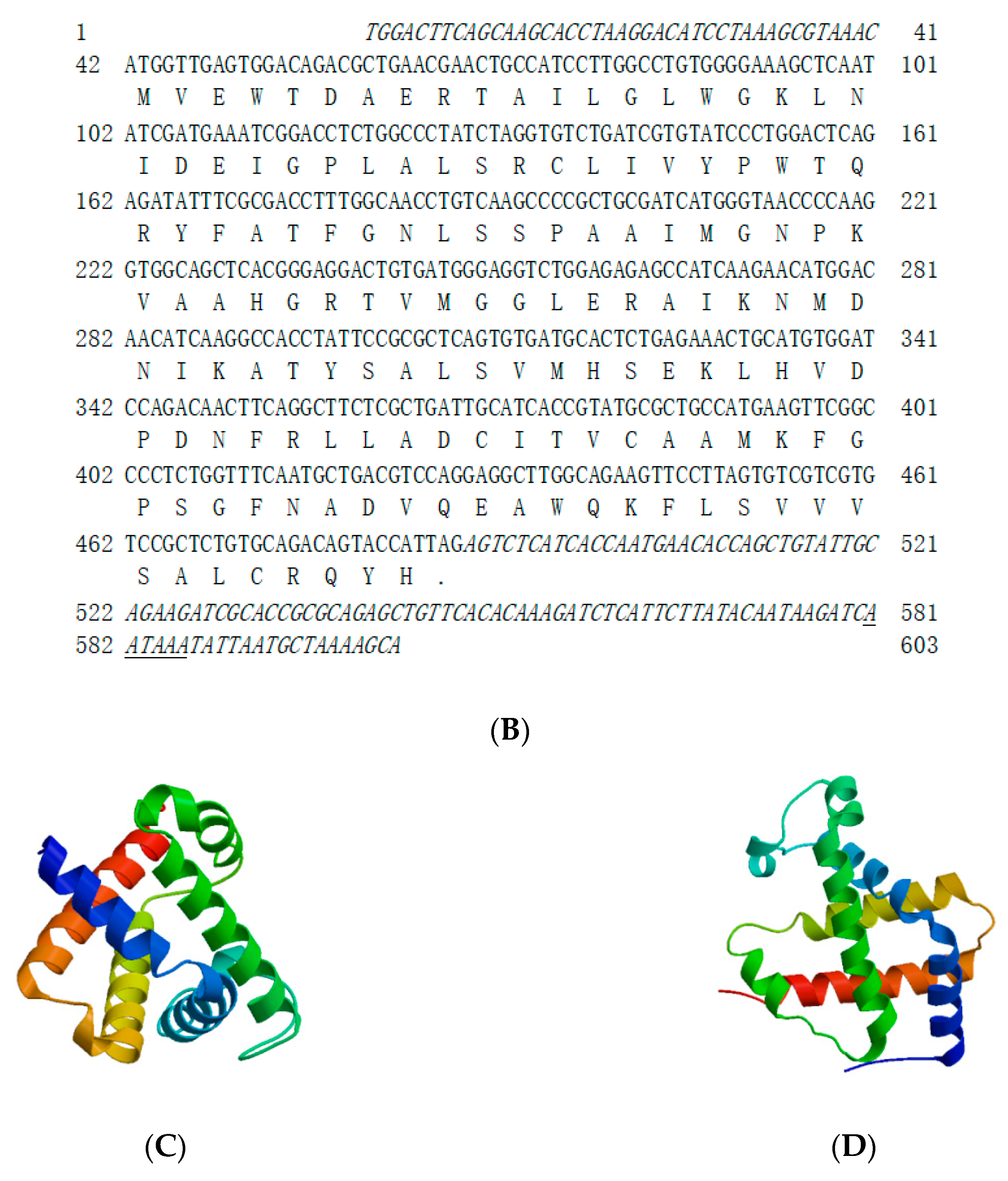
In the present study, the complete cDNA sequences for the MaHbα and MaHbβ genes were identified and characterized, and have been deposited in GenBank with the respective accession numbers KP288030 and OR594269. The full-length cDNA sequences of the MaHbα and MaHbβ genes were 588 and 603 bp in length, and encoded 143 and 148 amino acids, with predicted molecular weights of 15.5 and 16.4 kDa, and theoretical isoelectric points of 9.02 and 7.70, respectively. MaHbα and MaHbβ possessed no signal peptide or transmembrane domain, and were consistent with sequences from other fish species (Figure 1). The respective aliphatic amino acid indices of MaHbα and MaHbβ were 100.28 and 91.01, and their respective grand averages of hydropathicity (GRAVY) were 0.182 and 0.095. The total numbers of negatively charged (Asp+Glu) and positively charged (Arg+Lys) residues in MaHbα were 14 and 17, and those in MaHbβ were 13 and 14, respectively. Both MaHbα and MaHbβ showed the typical structural characteristics of hemoglobins with a globin-like fold and plenty of α-helices in the predicted tertiary structures (Figure 1).

3.2. Multiple Sequence Alignment and Phylogenetic Analysis of MaHb Genes

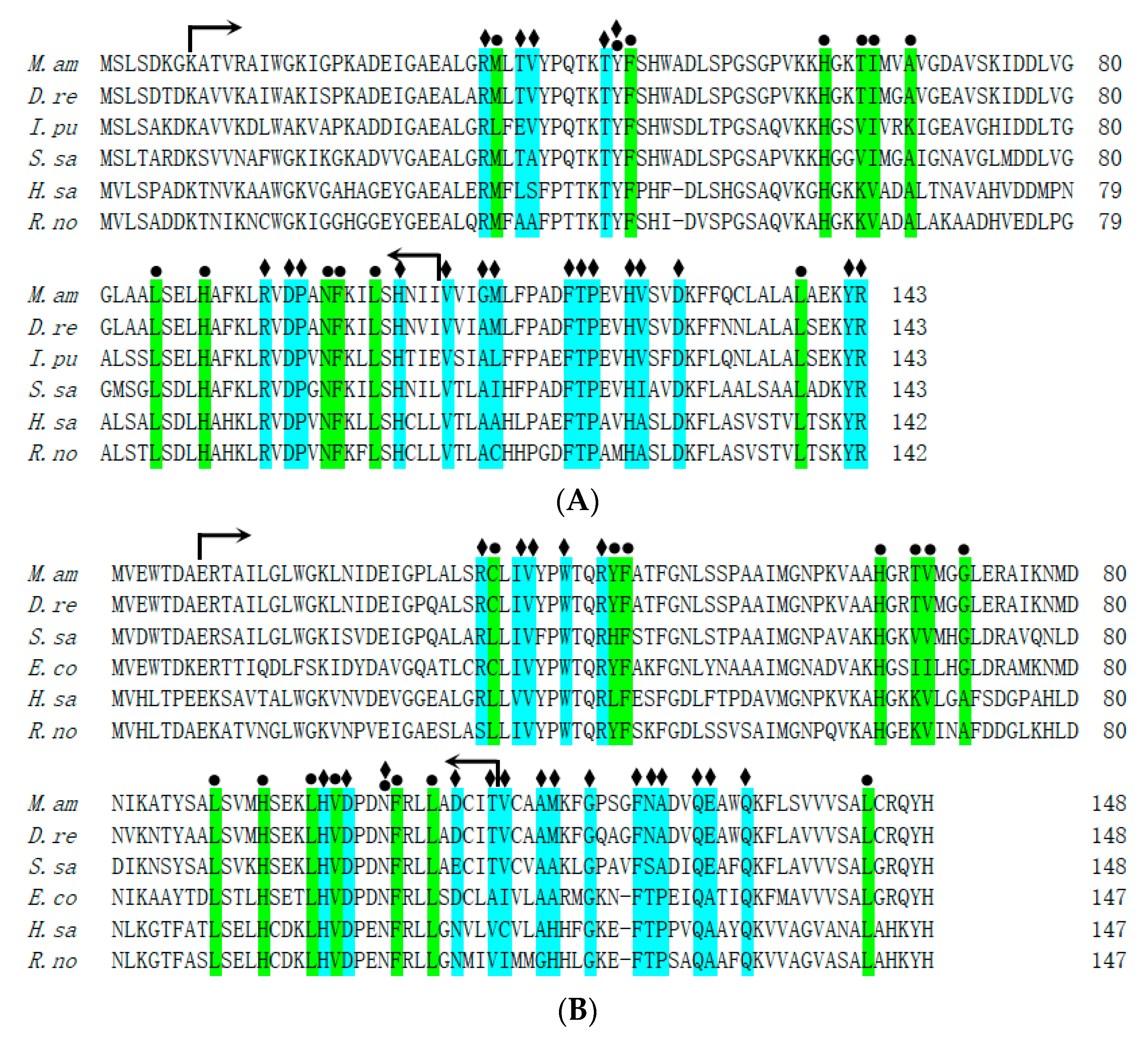
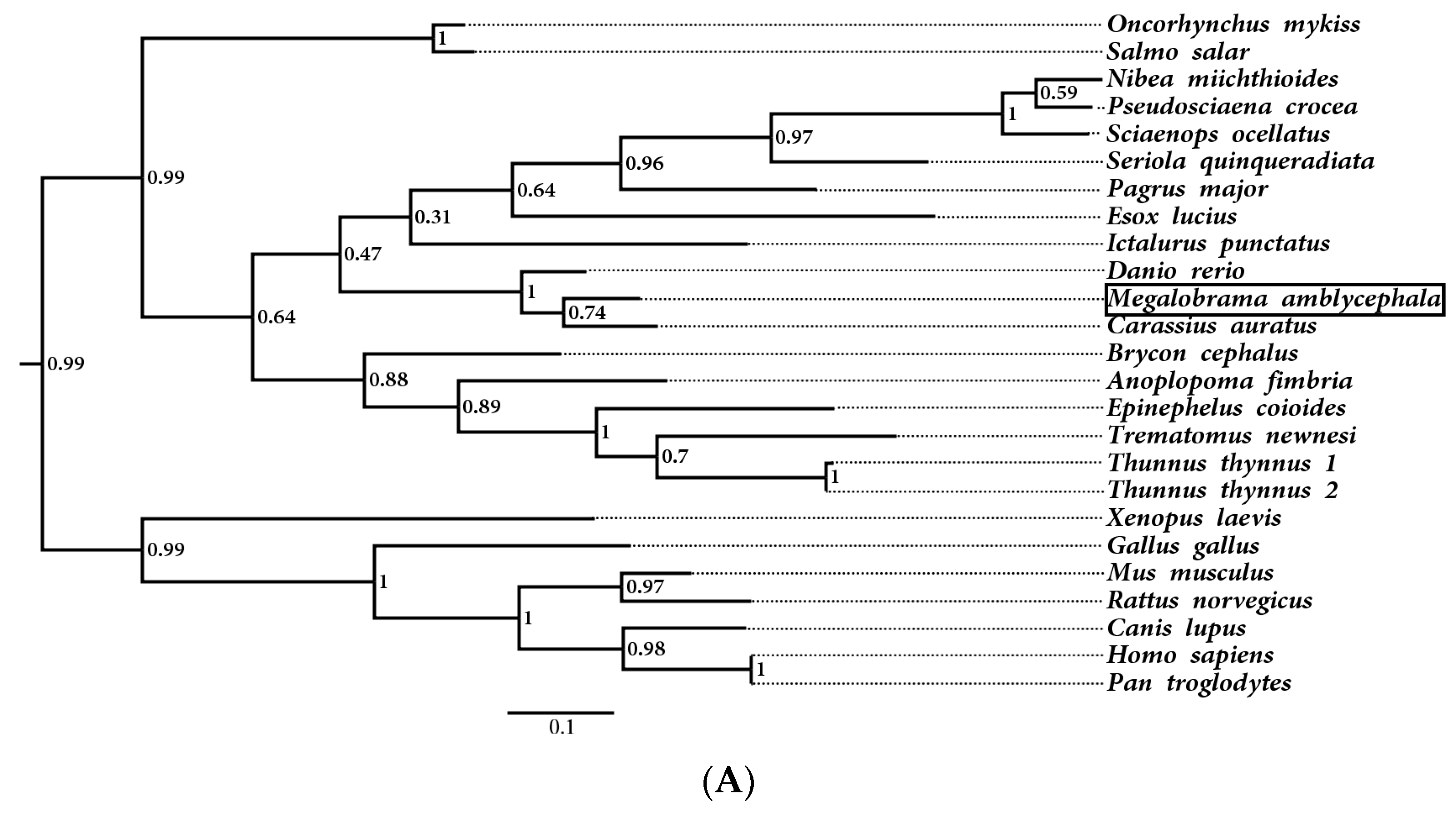
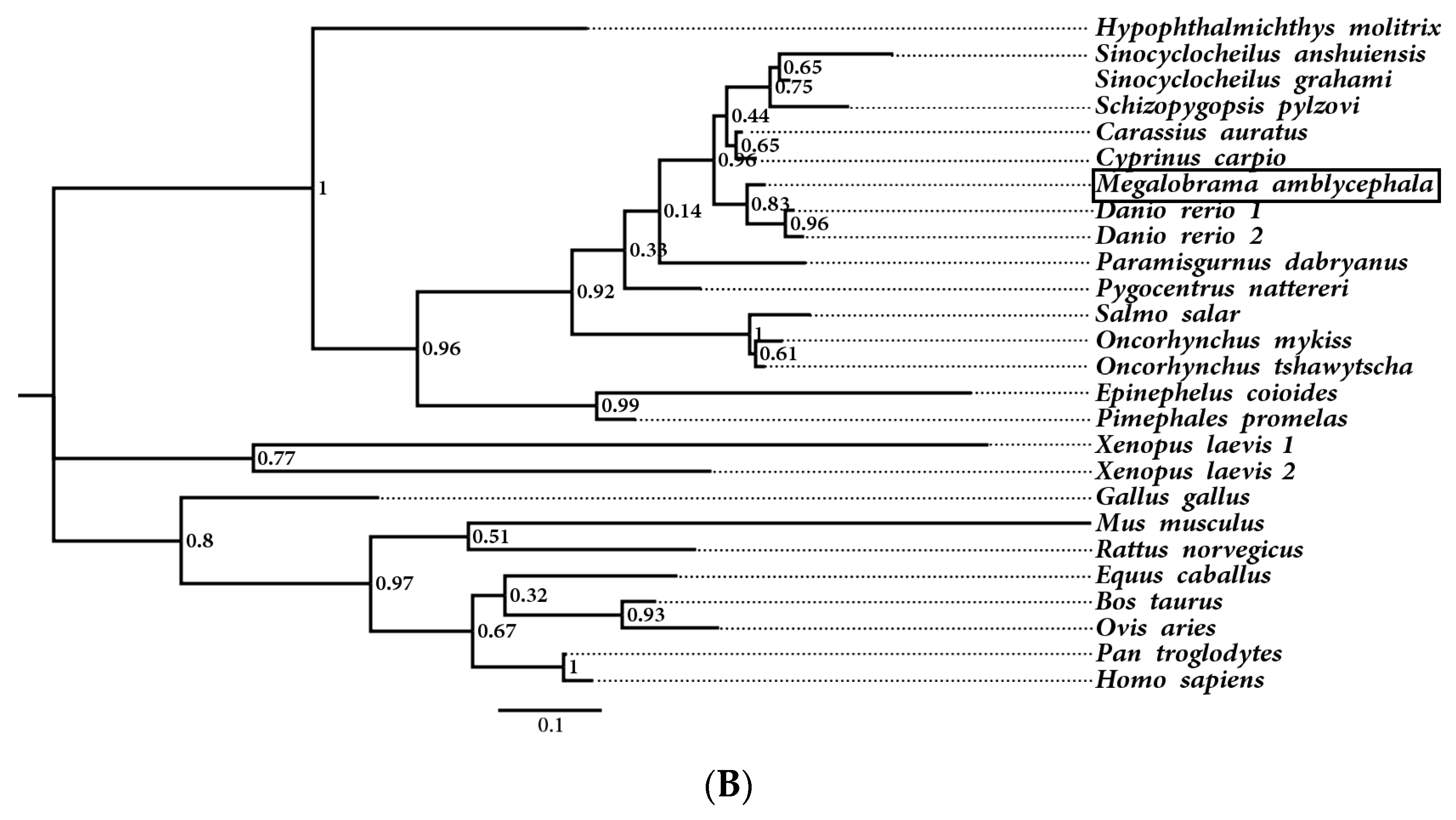
Multiple sequence alignments of the hemoglobin homologs from M. amblycephala, Danio rerio, Ictalurus punctatus, Salmo salar, Homo sapiens, and Rattus norvegicus were performed. The MaHb proteins were highly homologous to hemoglobins from other fish, with a typical globin-like domain, and most of the heme-binding sites and tetramer interfaces (polypeptide-binding sites) were highly conserved among teleosts (Figure 2), indicating fish hemoglobins have maintained their biological functions during evolution. Phylogenetic analysis showed that the hemoglobin genes from M. amblycephala and other cypriniformes clustered into one branch, and those from other fish and mammals clustered into other branches (Figure 3), which also revealed the evolutionary conservation of fish hemoglobins.

3.3. Expression Patterns of MaHbα and MaHbβ mRNA in Different Tissues and Various Developmental Stages

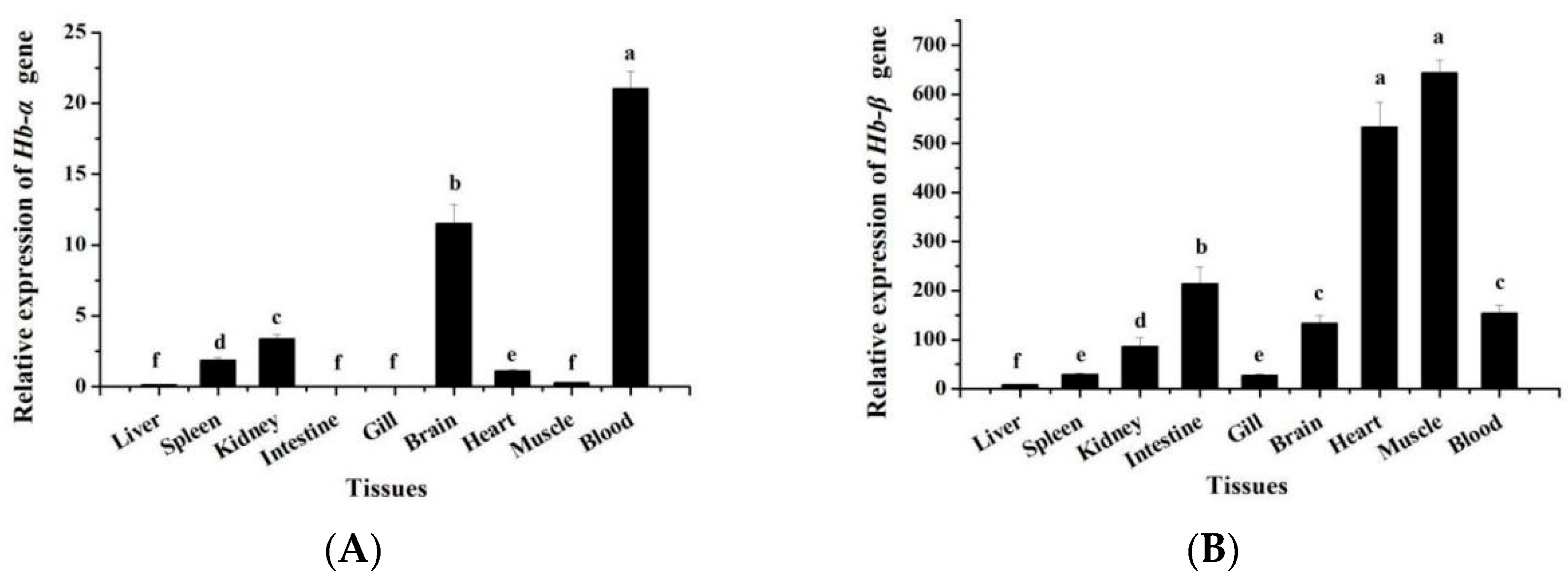
The expression patterns of MaHbα and MaHbβ mRNA in various tissues were elucidated, revealing that MaHbα and MaHbβ exhibit different tissue distribution patterns. Unlike MaHbβ, MaHbα is expressed in few tissues, primarily the blood, brain, kidneys, and spleen, followed by the heart and muscle, with marginal expression in the liver, intestine, and gills (Figure 4A). As shown in Figure 4B, MaHbβ is widely expressed in M. amblycephala tissues. It is strongly expressed in the muscle and heart, followed by the intestine, blood, brain, and kidneys, with low expression in the spleen, gills, and liver (Figure 4B).
The MaHbα and MaHbβ mRNA also exhibited different expression patterns during various developmental stages. As shown in Figure 5A, MaHbα expression gradually decreased from 0 hpf to 5 dph with some fluctuation, and then reached its peak level at 15 dph. However, MaHbβ has a different mode of expression during early development, showing almost no expression before 26 hpf, then increasing in expression, beginning at 3 dph and reaching extremely high expression at 15 dph (Figure 5B).

3.4. MaHbα and MaHbβ Expression in Response to Bacterial Infection

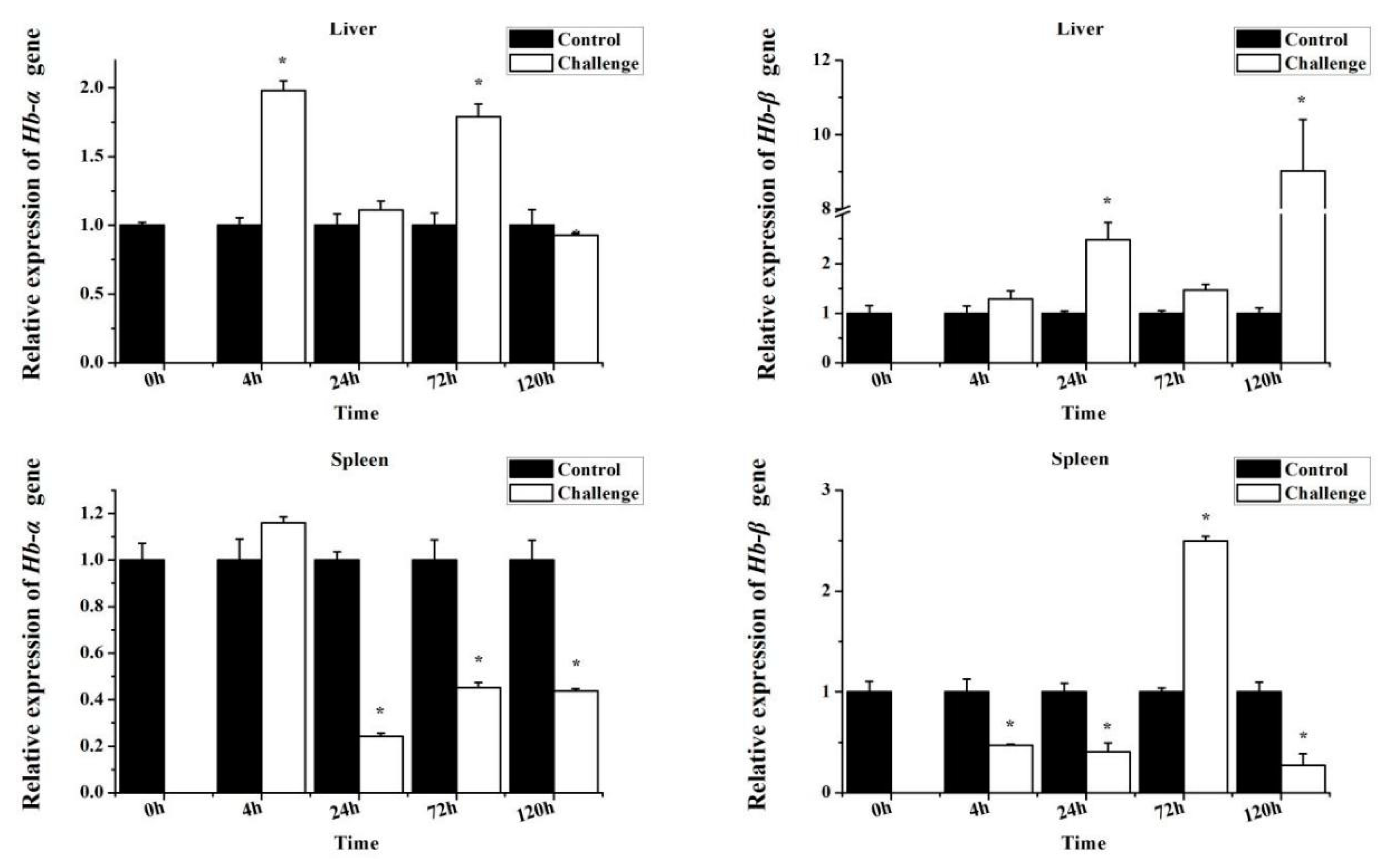
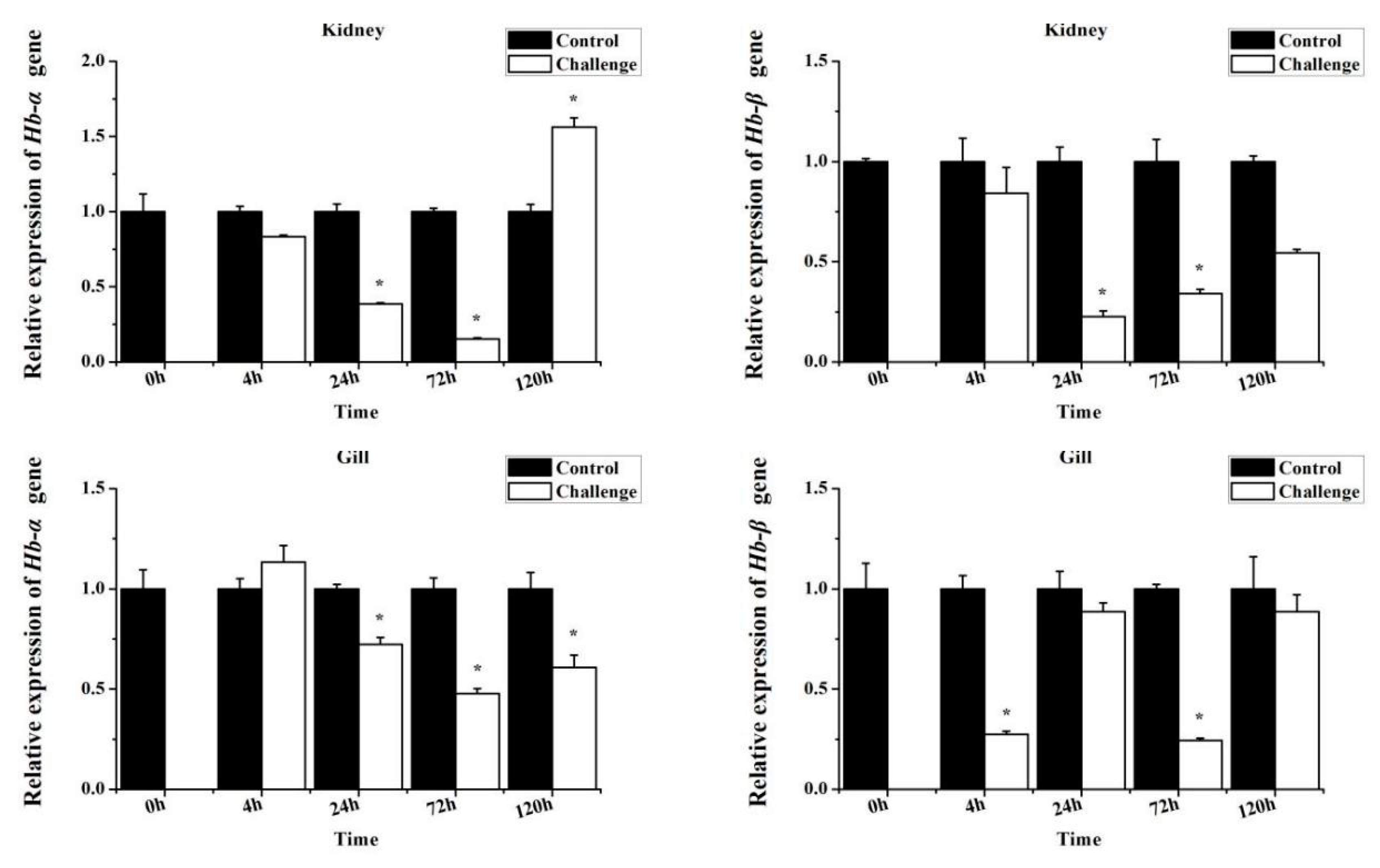
To explore the immune response of MaHbs to bacterial infection, juvenile M. amblycephala fish were challenged with A. hydrophila, and the MaHbα and MaHbβ mRNA expression levels in the liver, spleen, kidney, and gill tissues were detected using RT-qPCR. In the spleen, kidney, and gills, both MaHbα and MaHbβ mRNA were significantly down-regulated at most time points within 120 hpi (Figure 6). In the liver, the expression patterns of both MaHbα and MaHbβ dramatically increased at some time points, unlike the expression patterns in the other tissues (Figure 6).

3.5. Antimicrobial Activities of Synthetic MaHbα and MaHbβ Peptides

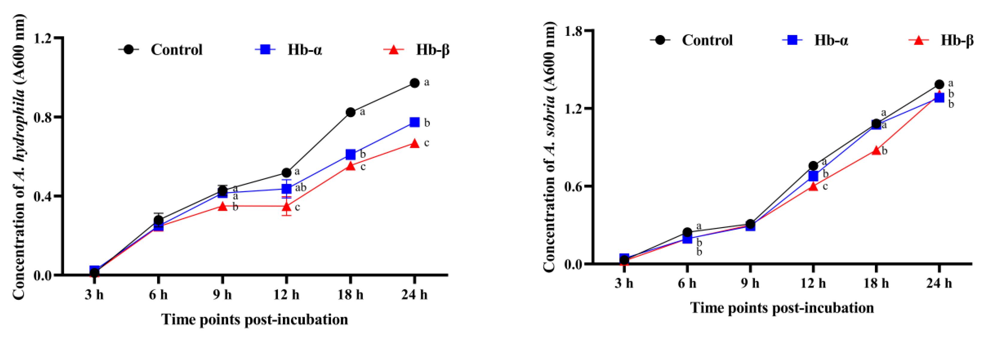
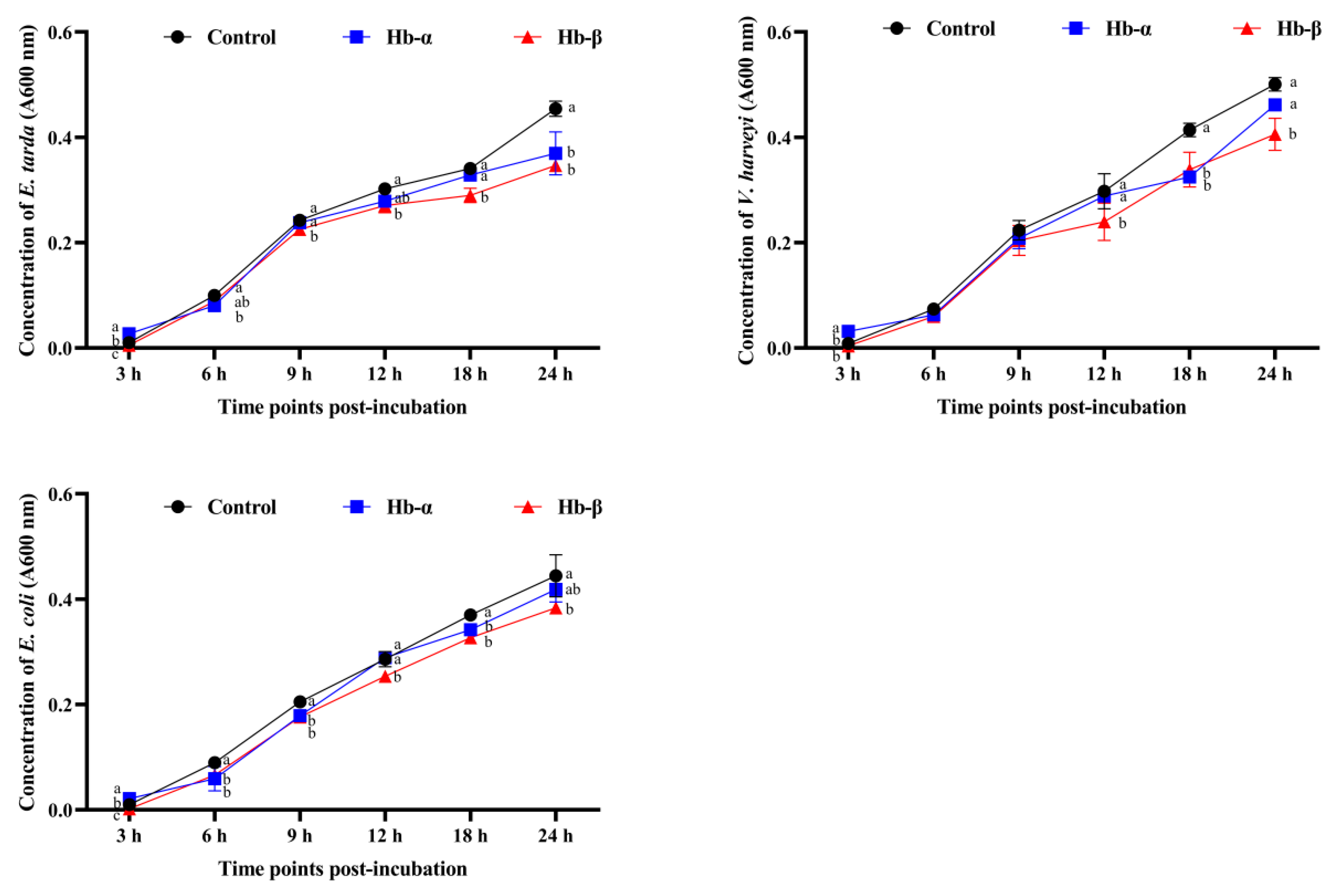
The core functional peptides of the MaHbα and MaHbβ proteins, as defined in previous studies, were synthesized. The antimicrobial activities of the synthetic MaHbα and MaHbβ peptides were characterized by assessing their growth inhibition and cytotoxicity toward five aquatic pathogens: A. hydrophila, A. sobria, V. harveyi, E. tarda, and E. coli. As shown in Figure 7, supplementation of media with the MaHbα and MaHbβ peptides exerted significant effects on bacterial abundance (absorbance at 600 nm), and the bacteriostatic effects displayed exposure time dependence, revealing the efficient antimicrobial activities of the synthetic MaHbα and MaHbβ peptides.

4. Discussion

Hemoglobin is an iron-containing allosteric protein found in vertebrate red blood cells. Its main functions including oxygen and carbon dioxide transportation, as well as the maintenance of acid–base balance in the blood. In the present study, we identified and characterized two hemoglobin genes from M. amblycephala, termed MaHbα and MaHbβ. Several hemoglobin subunits from other teleosts, including Atlantic salmon [8], rainbow trout [9], zebrafish [10], carp [11], yellowtail [12], puffer fish [13], medaka [14], and channel catfish [24,25] have been previously characterized. Two hemoglobin gene clusters in the fish genome have been demonstrated to constitute hemoglobin tetramers in several fish species. To date, the exact composition of the M. amblycephala hemoglobin tetramers remains unknown.
Multiple alignments show that the deduced amino acid sequences of fish hemoglobins are highly conserved, especially vital residues and functional domains. For instance, the Root effect is a specific functional property of fish hemoglobins [26], and M. amblycephala hemoglobins retain most of the pivotal residues for the Root effect as its structural basis. Phylogenetic analysis shows that hemoglobin genes from M. amblycephala and other cypriniformes cluster into one branch, indicating fish hemoglobin gene conservation during the evolutionary process.
Previous studies have reported that initial embryonic hemoglobin is involved in primitive hematopoiesis, which occurs in an intermediate cell mass, whereas adult hemoglobin participates in definitive hematopoiesis, mainly in the spleen and kidneys after hatching [14]. Thus, all hemoglobin genes from channel catfish are highly expressed in the spleen and kidneys. In the present study, we found that MaHbα is mainly expressed in the blood, brain, kidneys, and spleen in M. amblycephala, while MaHbβ is highly expressed in the muscle, heart, and intestine. The differential expression patterns of these two M. amblycephala hemoglobin subunits should reflect their biological functions and suggest that the MaHbα subunit is responsible for oxygen storage in the blood vessels and oxygen transport to brain tissue and other oxygen-requiring tissues, but MaHbβ may play the oxygen storage and transportation role in the muscle, heart, and intestine. More hemoglobin subunits from M. amblycephala need to be identified and genomically characterized to systematically elucidate the composition of gene clusters and expression patterns.
The expression of zebrafish hemoglobin genes exhibits developmental-stage-specific patterns, that is, different subunits can be activated at the embryonic, fetal, and adult stages, respectively [27,28]. For instance, some zebrafish hemoglobin subunits are exclusively expressed in mature and adult stages, while others may be exclusively expressed in the embryonic stage or through the embryonic and larval stages [28]. In the present study, MaHbβ expression began at 3 dph during development and gradually increased, and was widely expressed in most adult tissues, indicating that MaHbβ is synthesized during development and acts as a constitutively expressed subunit. Unlike MaHbβ, MaHbα expression is detectable and abundant post-fertilization with fluctuating levels during developmental stages, and was detectable in a restricted number of adult tissues, revealing that it likely performs specific functions in the blood and brain.
The expression patterns of hemoglobin genes from different aquatic animals upon pathogenic infection are inconsistent. Transcription of two Scapharca kagoshimensis hemoglobins (skHbI and skHbII) is significantly up-regulated after Bacillus subtilis infection [16], whereas the hemoglobin concentrations of Cyprinus carpio and Carassius auratus decrease significantly upon A. hydrophila infection [29]. In this study, the expression of both M. amblycephala hemoglobin subunits was down-regulated in most tissues and time points. This might be due to the invading pathogenic strain of A. hydrophila producing virulence factors, such as hemolysin, cytotoxins, or enterotoxins, thereby dissolving red blood cells, damaging capillaries, causing bleeding and sepsis, and reducing the number of host red blood cells [30]. Thereafter, A. hydrophila proliferation in the blood to a certain level and circulation of the blood to all parts of the host increase its toxicity and destructiveness, causing damage and dysfunction of the organs, including the kidneys, spleen, and heart [31]. Fish spleen and kidneys are hematopoietic organs; thus, their dysfunction may further reduce the number of red blood cells [32]. Hemoglobin is the main component of red blood cells, and its abundance is positively correlated with the number of red blood cells. Therefore, a reduction in red blood cell number invariably reduces hemoglobin concentration [33]. Thus, reduced M. amblycephala hemoglobin genes expression may result from damage to the red blood cells and hematopoietic organs by A. hydrophila virulence factors. In addition, the different expression patterns of fish hemoglobin genes upon infection might be related to host species, developmental stages, and pathogenic challenge concentrations.
Antimicrobial and bactericidal assays have both shown that chicken hemoglobin antimicrobial peptides (CHAP) exhibit strong and rapid bacteriostatic activities against various bacteria, and electron microscopy (EM) analyses further revealed that CHAP could accumulate many pathogens nearby and quickly penetrate their cell surfaces [34]. Hemoglobin from both horseshoe crabs and humans possesses pseudoperoxidase (POX) activity, which can be synergistically triggered by microbial proteases and pathogen-associated molecular patterns (PAMPs) to produce superoxide anions [35]. Similarly, activation of the POX cycle by bacterial components and stimulated Hb could spontaneously release superoxide radicals in Ctenopharyngodon idella, thereby eliciting significant antibacterial properties [7]. Purified S. kagoshimensis hemoglobins (skHbs) possess excellent antimicrobial activities against Gram-positive bacteria, which might be attributed to their phenoloxidase (PO)-like activities that can detect and kill invading pathogens and synthesize melanin for pathogen encapsulation, thereby involving them in host innate immune responses [15,36].
A previous study provides strong evidence that human hemoglobin proteolysis leads to the formation of peptides (β-hemoglobin (111-146) and γ-hemoglobin (130–146)), which effectively inhibit the growth of microorganisms [2]. Minimal inhibitory concentrations of purified bovine α-hemoglobin 107–141, 133–141, and 137–141 peptides clearly reveal the minimal peptide sequence necessary for antibacterial activity to be KYR [37]. Similarly, a bacteriostatic assay detected the minimum inhibitory concentration of β-hemoglobin 114-145 peptide, which showed the minimum antimicrobial peptide to be RYH [38]. In this study, the M. amblycephala hemoglobin MaHbα (107–136) and MaHbβ (116–146) peptides were synthesized because they should also be the direct precursors of core antimicrobial peptides [37], which possessed some characteristics of the reported antibacterial peptides [39], containing more than 20 amino acid residues with positive charge, a large number of hydrophobic residues, and a high proportion of α-helical structures. These results show that both synthesized MaHbs peptides exhibited efficient antimicrobial activities against various pathogens, confirming the core antimicrobial peptides were involved in the synthesized regions, while the specific mechanism needs to be further investigated.

5. Conclusions

In the present study, M. amblycephala’s hemoglobin α and β subunits were identified and characterized, and their expression patterns and antibacterial activities were elucidated. Multiple sequence alignments and phylogenetic analyses have revealed the evolutionary conservation of fish hemoglobins. The two identified M. amblycephala Hb subunits exhibit different expression patterns in various tissues and during development, indicating that they have different physiological functions. In addition, down-regulation of the expression of both subunits after A. hydrophila infection may be attributed to damage to the red blood cells and hematopoietic organs. Moreover, the efficient antimicrobial activities of synthesized MaHbα and MaHbβ peptides indicated their immunoprotective effects.

Supplementary Materials

The following supporting information can be downloaded at: https://www.mdpi.com/article/10.3390/genes14101972/s1, Table S1: Hemoglobin alpha and beta amino acid sequences used for multiple alignment; Table S2: Hemoglobin alpha subunits cDNA sequences used for phylogenetic analysis; Table S3: Hemoglobin beta subunits cDNA sequences used for phylogenetic analysis.

Author Contributions

Conceptualization, Z.D. and X.Z.; methodology, H.W.; software, J.Z. and H.D.; validation, J.Z.; formal analysis, Y.L.; investigation, Z.D., Y.L. and Q.W.; resources, H.L. and Z.D.; data curation, H.C.; writing—original draft preparation, Q.W.; writing—review and editing, Z.D. and X.Z.; visualization, Q.W. and Y.L.; supervision, Z.D. and X.Z.; project administration, H.L. and X.Z.; funding acquisition, Z.D. and X.Z. All authors have read and agreed to the published version of the manuscript.

Funding

This work was supported by the Chinese Postdoctoral Science Foundation (No. 2020M671386), the Jiangsu Agriculture Science and Technology Innovation Fund (No. CX(20)3110), the Postdoctoral Science Foundation of Jiangsu Province (2020Z157), Lianyungang city (China), and the Priority Academic Program Development of Jiangsu Higher Education Institutions (Innovation Project No. 2022JSPAPD017).

Institutional Review Board Statement

This study was approved by the Animal Care and Use Committee of Jiangsu Ocean University (protocol no. 2020-37, approval date: 1 September 2019). All animal procedures were performed according to the Guidelines for the Care and Use of Laboratory Animals in China.

Data Availability Statement

The complete cDNA sequences of MaHbα and MaHbβ genes have been deposited in NCBI database (accession no. KP288030 and OR594269), and other supporting information can be found in Supplementary Materials.

Conflicts of Interest

The authors declare no conflict of interest.

References

  1. Daoud, R.; Dubois, V.; Bors-Dodita, L.; Nedjar-Arroume, N.; Krier, F.; Chihib, N.-E.; Mary, P.; Kouach, M.; Briand, G.; Guillochon, D. New antibacterial peptide derived from bovine hemoglobin. Peptides 2005, 26, 713–719. [Google Scholar] [CrossRef] [PubMed]
  2. Liepke, C.; Baxmann, S.; Heine, C.; Breithaupt, N.; Ständker, L.; Forssmann, W.-G. Human hemoglobin-derived peptides exhibit antimicrobial activity: A class of host defense peptides. J. Chromatogr. B 2003, 791, 345–356. [Google Scholar] [CrossRef] [PubMed]
  3. Parish, C.A.; Jiang, H.; Tokiwa, Y.; Berova, N.; Nakanishi, K.; McCabe, D.; Zuckerman, W.; Xia, M.M.; Gabay, J.E. Broad-spectrum antimicrobial activity of hemoglobin. Bioorganic Med. Chem. 2001, 9, 377–382. [Google Scholar] [CrossRef] [PubMed]
  4. Nedjar-Arroume, N.; Dubois-Delval, V.; Adje, E.Y.; Traisnel, J.; Krier, F.; Mary, P.; Kouach, M.; Briand, G.; Guillochon, D. Bovine hemoglobin: An attractive source of antibacterial peptides. Peptides 2008, 29, 969–977. [Google Scholar] [CrossRef] [PubMed]
  5. Kawano, T.; Pinontoan, R.; Hosoya, H.; Muto, S. Monoamine-dependent production of reactive oxygen species catalyzed by pseudoperoxidase activity of human hemoglobin. Biosci. Biotechnol. Biochem. 2002, 66, 1224–1232. [Google Scholar] [CrossRef] [PubMed]
  6. Decker, H.; Jaenicke, E. Recent findings on phenoloxidase activity and antimicrobial activity of hemocyanins. Dev. Comp. Immunol. 2004, 28, 673–687. [Google Scholar] [CrossRef]
  7. Qin, Z.; Vijayaraman, S.B.; Lin, H.; Dai, Y.; Zhao, L.; Xie, J.; Lin, W.; Wu, Z.; Li, J.; Lin, L. Antibacterial activity of erythrocyte from grass carp (Ctenopharyngodon idella) is associated with phagocytosis and reactive oxygen species generation. Fish Shellfish. Immunol. 2019, 92, 331–340. [Google Scholar] [CrossRef]
  8. Wagner, A.; Deryckere, F.; McMorrow, T.; Gannon, F. Tail-to-tail orientation of the Atlantic salmon alpha- and beta-globin genes. J. Mol. Evol. 1994, 38, 28–35. [Google Scholar] [CrossRef]
  9. Yoshizaki, G.; Takano, A.; Aoki, T.; Takashima, F. Rapid communication: Nucleotide sequence of rainbow trout alpha-globin I and IV cDNA. J. Anim. Sci. 1997, 75, 1426. [Google Scholar] [CrossRef]
  10. Chan, F.-Y.; Robinson, J.; Brownlie, A.; Shivdasani, R.A.; Donovan, A.; Brugnara, C.; Kim, J.; Lau, B.-C.; Witkowska, H.E.; Zon, L.I. Characterization of adult alpha- and beta-globin genes in the zebrafish. Blood 1997, 89, 688–700. [Google Scholar] [CrossRef]
  11. Miyata, M.; Aoki, T. Head-to-head linkage of carp alpha- and beta-globin genes. Biochim. et Biophys. Acta 1997, 1354, 127–133. [Google Scholar] [CrossRef] [PubMed]
  12. Okamoto, K.; Sakai, M.; Miyata, M. Molecular cloning and sequence analysis of alpha- and beta-globin cDNAs from yellowtail, Seriola quinqueradiata. Comp. Biochem. Physiol. Part B Biochem. Mol. Biol. 2001, 130, 207–216. [Google Scholar] [CrossRef] [PubMed]
  13. Gillemans, N.; McMorrow, T.; Tewari, R.; Wai, A.W.K.; Burgtorf, C.; Drabek, D.; Ventress, N.; Langeveld, A.; Higgs, D.; Tan-Un, K.; et al. Functional and comparative analysis of globin loci in pufferfish and humans. Blood 2003, 101, 2842–2849. [Google Scholar] [CrossRef] [PubMed]
  14. Maruyama, K.; Yasumasu, S.; Iuchi, I. Characterization and expression of embryonic and adult globins of the teleost Oryzias latipes (medaka). J. Biochem. 2002, 132, 581–589. [Google Scholar] [CrossRef] [PubMed]
  15. Xu, B.; Zhao, J.; Jing, Z.; Zhang, Y.; Shi, Y.; Fan, T. Role of hemoglobin from blood clam Scapharca kagoshimensis beyond oxygen transport. Fish Shellfish. Immunol. 2015, 44, 248–256. [Google Scholar] [CrossRef] [PubMed]
  16. Xu, B.; Zhang, Y.; Jing, Z.; Fan, T. Molecular characteristics of hemoglobins in blood clam and their immune responses to bacterial infection. Int. J. Biol. Macromol. 2017, 99, 375–383. [Google Scholar] [CrossRef]
  17. Zhao, Q.; Wu, B.; Liu, Z.; Sun, X.; Zhou, L.; Yang, A.; Zhang, G. Molecular cloning, expression and biochemical characterization of hemoglobin gene from ark shell Scapharca broughtonii. Fish Shellfish. Immunol. 2018, 78, 60–68. [Google Scholar] [CrossRef]
  18. Cui, H.; Shen, X.; Zheng, Y.; Guo, P.; Gu, Z.; Gao, Y.; Zhao, X.; Cheng, H.; Xu, J.; Chen, X.; et al. Identification, expression patterns, evolutionary characteristics and recombinant protein activities analysis of CD209 gene from Megalobrama amblycephala. Fish Shellfish. Immunol. 2022, 126, 47–56. [Google Scholar] [CrossRef]
  19. Liu, H.; Chen, C.H.; Gao, Z.X.; Min, J.M.; Gu, Y.M.; Jian, J.B.; Jiang, X.W.; Cai, H.M.; Ebersberger, I.; Xu, M.; et al. The draft genome of blunt snout bream (Megalobrama amblycephala) reveals the development of intermuscular bone and adaptation to herbivorous diet. GigaScience 2017, 6, 1–13. [Google Scholar] [CrossRef]
  20. Tamura, K.; Peterson, D.; Peterson, N.; Stecher, G.; Nei, M.; Kumar, S. MEGA5: Molecular evolutionary genetics analysis using maximum likelihood, evolutionary distance, and maximum parsimony methods. Mol. Biol. Evol. 2011, 28, 2731–2739. [Google Scholar] [CrossRef]
  21. Liu, Y.; Yan, Y.; Han, Z.; Zheng, Y.; Wang, X.; Zhang, M.; Li, H.; Xu, J.; Chen, X.; Ding, Z.; et al. Comparative effects of dietary soybean oil and fish oil on the growth performance, fatty acid composition and lipid metabolic signaling of grass carp, Ctenopharyngodon idella. Aquac. Rep. 2022, 22, 101002. [Google Scholar] [CrossRef]
  22. Vandesompele, J.; De Preter, K.; Pattyn, F.; Poppe, B.; Van Roy, N.; De Paepe, A.; Speleman, F. Accurate normalization of real-time quantitative RT-PCR data by geometric averaging of multiple internal control genes. Genome Biol. 2002, 3, 467–470. [Google Scholar] [CrossRef] [PubMed]
  23. Cui, H.; Li, H.; Zhang, M.; Li, H.; Wang, X.; Wang, Z.; Zhai, W.; Chen, X.; Cheng, H.; Xu, J.; et al. Molecular characterization, expression, evolutionary selection, and biological activity analysis of CD68 gene from Megalobrama amblycephala. Int. J. Mol. Sci. 2022, 23, 13133. [Google Scholar] [CrossRef] [PubMed]
  24. Ullal, A.J.; Litaker, R.W.; Noga, E.J. Antimicrobial peptides derived from hemoglobin are expressed in epithelium of channel catfish (Ictalurus punctatus, Rafinesque). Dev. Comp. Immunol. 2008, 32, 1301–1312. [Google Scholar] [CrossRef]
  25. Feng, J.; Liu, S.; Wang, X.; Wang, R.; Zhang, J.; Jiang, Y.; Li, C.; Kaltenboeck, L.; Li, J.; Liu, Z. Channel catfish hemoglobin genes: Identification, phylogenetic and syntenic analysis, and specific induction in response to heat stress. Comp. Biochem. Physiol. Part D Genom. Proteom. 2014, 9, 11–22. [Google Scholar] [CrossRef]
  26. Vergara, A.; Vitagliano, L.; Merlino, A.; Sica, F.; Marino, K.; Verde, C.; di Prisco, G.; Mazzarella, L. An order-disorder transition plays a role in switching off the root effect in fish hemoglobins. J. Biol. Chem. 2010, 285, 32568–32575. [Google Scholar] [CrossRef]
  27. Maruyama, K.; Yasumasu, S.; Naruse, K.; Mitani, H.; Shima, A.; Iuchi, I. Genomic organization and developmental expression of globin genes in the teleost Oryzias latipes. Gene 2004, 335, 89–100. [Google Scholar] [CrossRef]
  28. Ganis, J.J.; Hsia, N.; Trompouki, E.; de Jong, J.L.; DiBiase, A.; Lambert, J.S.; Jia, Z.; Sabo, P.J.; Weaver, M.; Sandstrom, R.; et al. Zebrafish globin switching occurs in two developmental stages and is controlled by the LCR. Dev. Biol. 2012, 366, 185–194. [Google Scholar] [CrossRef]
  29. Zhang, W.; Cai, X.; Ge, M.; Jin, X.; Wang, Z. Effects of Aeromonas hydrophila On haemoglobin of several kinds of fish. Hebei Fisheries. 2009, 11, 7–10. [Google Scholar] [CrossRef]
  30. Xiong, N.X.; Ou, J.; Fan, L.F.; Kuang, X.Y.; Fang, Z.X.; Luo, S.W.; Mao, Z.W.; Liu, S.J.; Wang, S.; Wen, M.; et al. Blood cell characterization and transcriptome analysis reveal distinct immune response and host resistance of different ploidy cyprinid fish following Aeromonas hydrophila infection. Fish Shellfish. Immunol. 2021, 120, 547–559. [Google Scholar] [CrossRef]
  31. Chen, N.; Jiang, J.; Gao, X.; Li, X.; Zhang, Y.; Liu, X.; Yang, H.; Bing, X.; Zhang, X. Histopathological analysis and the immune related gene expression profiles of mandarin fish (Siniperca chuatsi) infected with Aeromonas hydrophila. Fish Shellfish. Immunol. 2018, 83, 410–415. [Google Scholar] [CrossRef] [PubMed]
  32. De Souza, P.; Bonilla-Rodriguez, G. Fish hemoglobins. Braz. J. Med. Biol. Res. 2007, 40, 769–778. [Google Scholar] [CrossRef]
  33. Chiesa, M.E.; Rosenberg, C.E.; Fink, N.E.; Salibián, A. Serum protein profile and blood cell counts in adult toads Bufo arenarum (Amphibia: Anura: Bufonidae): Effects of sublethal lead acetate. Arch. Environ. Contam. Toxicol. 2006, 50, 384–391. [Google Scholar] [CrossRef] [PubMed]
  34. Hu, F.; Wu, Q.; Song, S.; She, R.; Zhao, Y.; Yang, Y.; Zhang, M.; Du, F.; Soomro, M.H.; Shi, R. Antimicrobial activity and safety evaluation of peptides isolated from the hemoglobin of chickens. BMC Microbiol. 2016, 16, 287. [Google Scholar] [CrossRef] [PubMed]
  35. Jiang, N.; Tan, N.S.; Ho, B.; Ding, J.L. Respiratory protein–generated reactive oxygen species as an antimicrobial strategy. Nat. Immunol. 2007, 8, 1114–1122. [Google Scholar] [CrossRef]
  36. Fan, T.; Zhang, Y.; Yang, L.; Yang, X.; Jiang, G.; Yu, M.; Cong, R. Identification and characterization of a hemocyanin-derived phenoloxidase from the crab Charybdis japonica. Comp. Biochem. Physiol. B Biochem. Mol. Biol. 2008, 152, 144–149. [Google Scholar] [CrossRef]
  37. Catiau, L.; Traisnel, J.; Delval-Dubois, V.; Chihib, N.-E.; Guillochon, D.; Nedjar-Arroume, N. Minimal antimicrobial peptidic sequence from hemoglobin alpha-chain: KYR. Peptides 2010, 32, 633–638. [Google Scholar] [CrossRef]
  38. Catiau, L.; Traisnel, J.; Chihib, N.-E.; Le Flem, G.; Blanpain, A.; Melnyk, O.; Guillochon, D.; Nedjar-Arroume, N. RYH: A minimal peptidic sequence obtained from beta-chain hemoglobin exhibiting an antimicrobial activity. Peptides 2011, 32, 1463–1468. [Google Scholar] [CrossRef]
  39. Powers, J.-P.S.; Hancock, R.E. The relationship between peptide structure and antibacterial activity. Peptides 2003, 24, 1681–1691. [Google Scholar] [CrossRef] [PubMed]
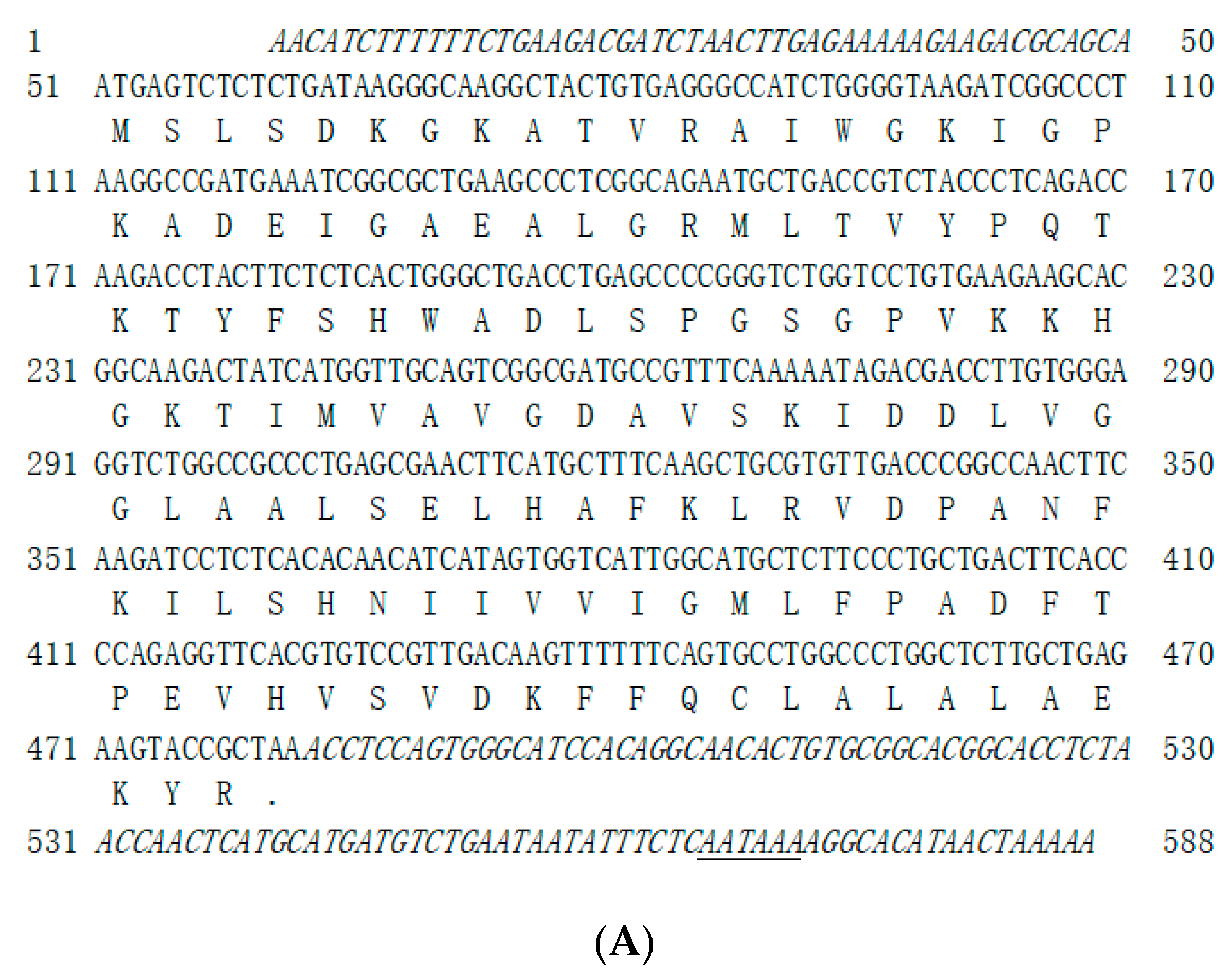
Figure 1. Nucleotide/amino acid sequence diagram and predicted tertiary structures of Megalobrama amblycephala hemoglobins α (A,C) and β (B,D) subunits.
Genes 14 01972 g001aGenes 14 01972 g001b
Figure 2. Multiple alignments of the amino acid sequences of hemoglobin α (A) and β (B) subunits. Notes: M. am, M. amblycephala; D. re, Danio rerio; I. pu, Ictalurus punctatus; S. sa, Salmo salar; H. sa, Homo sapiens; R. no, Rattus norvegicus; E. co, Epinephelus coioides. The green columns under filled circles represent heme-binding sites, the blue columns under filled diamonds represent the tetramer interface (polypeptide-binding sites), and the spans in between arrows represent globin-like domains. The accession numbers for these amino acid sequences are listed in Supplementary Table S1.
Genes 14 01972 g002
Figure 3. Phylogenetic analysis of vertebrate hemoglobin α (A) and β (B) subunits. The accession numbers for these cDNA sequences are listed in Supplementary Tables S2 and S3.
Genes 14 01972 g003aGenes 14 01972 g003b
Figure 4. MaHbα (A) and MaHbβ (B) expression in various tissues of adult M. amblycephala. 18S rRNA is selected as the reference gene. Data are shown as means ± SE. Different letters above columns indicate statistical significance (p < 0.05).
Genes 14 01972 g004
Figure 5. MaHbα (A) and MaHbβ (B) expression at different stages of M. amblycephala development. 18S rRNA is selected as the reference gene. Data are shown as means ± SE. Different letters above columns indicate statistical significance (p < 0.05).
Genes 14 01972 g005
Figure 6. Effect of A. hydrophila infection on MaHbα and MaHbβ mRNA expression. 18S rRNA is selected as the reference gene. Data are shown as means ± SE. Asterisks (*) above columns indicate statistical significance (p < 0.05).
Genes 14 01972 g006aGenes 14 01972 g006b
Figure 7. Antimicrobial activities of synthetic MaHbα and MaHbβ peptides against A. hydrophila, A. sobria, V. harveyi, E. tarda, and E. coli. Data are shown as means ± SE. Different letters above points indicate statistical significance (p < 0.05).
Genes 14 01972 g007aGenes 14 01972 g007b
Table 1. Primers used in this study.
PrimersPrimer Sequences (5′-3′)Purpose
MaHbα-5′-1ATGCCCACTGGAGGTTTAGCGG5′-RACE
MaHbα-5′-2GCCGTGCTTCTTCACAGGACCAG
MaHbα-3′-1 CCCTCGGCAGAATGCTGACCGTCTACCCT3′-RACE
MaHbα-3′-2TCACACAACATCATAGTGGTCATTGGCAT
MaHbα-FCGGCAGAATGCTGACCGTCORF amplification
MaHbα-R GCCCACTGGAGGTTTAGCG
qMaHbα-FATGCTCTTCCCTGCTGACTTC qRT-PCR
qMaHbα-RGGATGCCCACTGGAGGTTTAG
MaHbβ-5′-1AGCACAACTTTACCGTGAGCAGCAACC5′-RACE
MaHbβ-5′-2TTGCCAGGGCTTGAGGACCAACGACT
MaHbβ-3′-1GCAGAAGTTCCTTAGTGTCGTCGTGTCC3′-RACE
MaHbβ-3′-2CAATGAACACCAGCTGTATTGCAGAAG
MaHbβ-FCATGGTTGAGTGGACAGACGCORF amplification
MaHbβ-R GCGCGGTGCGATCTTCTGC
qMaHbβ-FGAAACCTCTACAACGCCGC qRT-PCR
qMaHbβ-RCTTTACCGTGAGCAGCAACC
q18S rRNA-FCGGAGGTTCGAAGACGATCAqRT-PCR [18]
q18S rRNA-RGGGTCGGCATCGTTTACG
qβ-actin-FGCTCTTACAGGAAACGGGTCqRT-PCR [18]
qβ-actin-RGCAGCAGCTCTGTAGGTCAT
qEF1α-FCTTCTCAGGCTGACTGTGCqRT-PCR [18]
qEF1α-RCCGCTAGCATTACCCTCC
qGAPDH-FTGCCGGCATCTCCCTCAAqRT-PCR [18]
qGAPDH-RTCAGCAACACGGTGGCTGTAG
Disclaimer/Publisher’s Note: The statements, opinions and data contained in all publications are solely those of the individual author(s) and contributor(s) and not of MDPI and/or the editor(s). MDPI and/or the editor(s) disclaim responsibility for any injury to people or property resulting from any ideas, methods, instructions or products referred to in the content.