Article

Expression of Long Noncoding RNAs in Fibroblasts from Mucopolysaccharidosis Patients

Department of Molecular Biology, Faculty of Biology, University of Gdansk, Wita Stwosza 59, 80-308 Gdansk, Poland
*
Author to whom correspondence should be addressed.
Genes 2023, 14(2), 271; https://doi.org/10.3390/genes14020271
Submission received: 15 December 2022 / Revised: 24 December 2022 / Accepted: 18 January 2023 / Published: 20 January 2023
(This article belongs to the Section RNA)

Abstract

:
In this report, changes in the levels of various long non-coding RNAs (lncRNAs) were demonstrated for the first time in fibroblasts derived from patients suffering from 11 types/subtypes of mucopolysaccharidosis (MPS). Some kinds of lncRNA (SNHG5, LINC01705, LINC00856, CYTOR, MEG3, and GAS5) were present at especially elevated levels (an over six-fold change relative to the control cells) in several types of MPS. Some potential target genes for these lncRNAs were identified, and correlations between changed levels of specific lncRNAs and modulations in the abundance of mRNA transcripts of these genes (HNRNPC, FXR1, TP53, TARDBP, and MATR3) were found. Interestingly, the affected genes code for proteins involved in various regulatory processes, especially gene expression control through interactions with DNA or RNA regions. In conclusion, the results presented in this report suggest that changes in the levels of lncRNAs can considerably influence the pathomechanism of MPS through the dysregulation of the expression of certain genes, especially those involved in the control of the activities of other genes.

1. Introduction

Lysosomal storage disease (LSD) constitutes a group of disorders caused by a lack of the activity of specific lysosomal enzymes (such as acid hydrolases), membrane proteins, and deficiencies in lysosomal transporters or signals required for delivery of an enzyme to the lysosome. This leads to a deficiency in the lysosomal degradation of various macromolecules, and, in turn, to their accumulation in lysosomes [1,2].
Mucopolysaccharidoses (MPS) are a group of LSDs caused by a significant deficiency in the activity of lysosomal enzymes involved in the degradation of glycosaminoglycans (GAGs) [3]. GAGs are long, non-branched polysaccharides that are degraded due to their cleavage by endohexosidases or exoglucuronidases, followed by the removal of their shorter fragments by the subsequent actions of other enzymes [3]. The actions of all GAG-degrading enzymes are correlated and sequential, i.e., the next enzyme can only act after the action of the previous one. Thus, if only one enzyme is deficient, the whole degradation pathway is highly impaired.
There are 13 types/subtypes of MPS occurring in humans (plus one that, to date, has only been identified in an animal model), which are classified according to a lack or deficiency of a specific enzyme and the kind of stored GAG(s) [4,5]. These diseases are inherited in an autosomal recessive manner, except MPS II, which is a sex-linked disease. MPS is a rare disease, but its specific prevalence depends on the particular type/subtype. For example, MPS I occurs at a frequency of 1 per 88,000 live births, while there are only 4 cases of MPS IX described in the literature [6]. There are some MPS symptoms occurring in all or most types, such as coarse facial features, organomegaly, and changes in the cardio-vascular system and bones. However, there are also symptoms specific for particular types, such as mental deterioration in some clinical forms of MPS I and II and in all patients with MPS III [4,5,7].
Every MPS type is a severe disease, and each significantly shortens life spans (up to 2 decades on average) [6,7,8]. Although some therapeutic options are available, such as enzyme replacement therapy (ERT) or hematopoietic stem cell transplantation, and even though other therapeutic approaches are being investigated [9], the treatment of all the symptoms of patients cannot be achieved despite reductions in GAG levels [10,11]. This suggests that there are other—perhaps secondary and tertiary—causes of cellular dysfunctions in MPS cells. Recent reports have indicated that not only the functions of lysosomes, but also other cellular processes are disturbed in patients with this disease. These include dysfunctions of the cytoskeleton and mitochondria [12], the disturbed regulation of apoptosis, and impaired autophagy [13,14]. However, studies on the secondary and tertiary effects of MPS were not extensive, and our knowledge in this field is limited [15]. Nevertheless, the results of such investigations indicated that there is a need for research into additional pathomechanisms of MPS that may indicate novel therapeutic targets.
One of the secondary pathomechanisms of MPS constitutes changes in the expression of certain genes [16]. The products of such genes can include regulatory proteins, transcription factors, and other molecules involved in various biochemical reactions. Changes in their levels may negatively influence cellular functions and thus cause perturbations at the level of the organism. Such phenomena have been demonstrated recently and include gene expression dysregulation-related disturbances of cell activation [17], changes in proteasome composition and activity [18], alterations in the modulation of apoptosis [19], abnormalities in the morphologies of organelles and cellular components’ organization [20], impaired ion homeostasis [21], disturbances in signal transduction [22], and the dysregulation of the cell cycle [23].
Among the various possible causes of the disturbed regulation of gene expression, there are the actions of non-coding RNA (ncRNA) molecules. Such RNAs include small interfering RNAs (siRNAs), piwi-interacting RNAs (piRNAs) [24], micro RNAs (miRNAs) [25], long non-coding RNAs (lncRNAs) [26], circular RNAs (circRNAs) [27], and others. The ncRNA species do not encode proteins but influence gene expression and may affect modifications of DNA and histones [28,29].
The group of lncRNAs consists of differentiated transcripts of lengths exceeding 200 nucleotides [26]. Various lncRNAs were identified and classified into sub-classes, depending on their lengths and functions [30]. Some lncRNAs are encoded by DNA fragments located between protein-coding genes; thus, they are called long intergenic non-coding RNAs (lincRNAs) [31]. Other lncRNAs, which are particularly long (over 50 kb), are called very-long intergenic non-coding RNAs [32]. Generally, lncRNAs influence gene expression by two different pathways. First, they bind to miRNAs to sponge them and prevent their interactions with target mRNAs. This causes enhanced synthesis of the proteins encoded by the target genes. Second, lncRNAs can interact directly with target mRNAs, thus modulating their translation. Therefore, lncRNAs can significantly down- or up-regulate the expression of target genes [33,34].
Recently, there has been an increasing number reports on the roles of ncRNAs in the pathogenesis of many diseases, including neurodegenerative diseases (such as Alzheimer’s disease [35], Parkinson’s disease [36], Huntington’s disease [37], and amyotrophic lateral sclerosis [38]), cancer (such as lung cancer, gastric tumors, colon cancer, melanoma, ovarian cancer, Burkitt’s lymphoma [39,40], and others [41]), and cardiovascular diseases (e.g., myocardial infarction, cardiomyopathies and heart failure [42], Alport syndrome [43], Loeys–Dietz syndrome [44], and others). Changed levels of ncRNAs were found also in some LSDs, namely, in Niemmann–Pick disease type C (NPC) and Gaucher disease [2]. In NPC, where cholesterol is a stored material, miRNA can be involved in the regulation of the expression of genes coding for enzymes necessary for the metabolism of cholesterol and other lipids. Studies on fibroblasts derived from patients suffering from Gaucher disease indicated that some miRNAs can either enhance or inhibit the activity of glucocerebrosidase, an enzyme that is deficient in patients suffering from this disease. Thus, miRNAs can act as modulators of metabolism in these diseases [45,46,47,48]. This finding was the basis for the hypothesis that ncRNAs may be potential targets for novel therapeutics to be used against inherited diseases [49].
There is only very limited knowledge regarding the role of ncRNAs in MPS. The only studies conducted in this field and published recently concerned a mouse model of MPS I in which only one miRNA was investigated. It was demonstrated that the modulation of the level of miR-17 is responsible for the impaired expression of the Neu1 gene [50]. This gene codes for neuraminidase 1, which is involved in the degradation of gangliosides [51], which are compounds that act as secondary storage material in MPS [6].
It is worth mentioning that studies on ncRNA in the pathogenesis of LSD—published to date—have focused solely on miRNA molecules. The role of lcnRNAs remains unknown, and it is definitely worth investigation as the influence of such RNAs on pathomechanisms has been demonstrated for various neurodegenerative diseases (see above). Thus, the aim of this work was to assess the potential role of lncRNAs in MPS. In this first (to our knowledge) study on lncRNAs in MPS, we employed a transcriptomic approach that should facilitate the demonstration of a global picture of the changes in the levels of these RNA species as well as the estimation of their potential roles in the pathomechanisms of these complex diseases. Moreover, it was assumed that studies on lncRNAs in MPS might help to explain the incomplete efficiency of therapies based solely on reducing GAG levels, and perhaps indicate new methods for the development of combined therapies that could involve the modulation of the levels of lncRNAs and/or miRNA, thus improving the control of the expression of specific genes, which is crucial for the effective abolition of all cellular defects occurring in MPS.

2. Materials and Methods

2.1. Lines of Human Fibroblasts and Cell Cultures

Patient-derived fibroblasts were used in this study. The cell lines were purchased from the NIGMS Human Genetic Cell Repository at the Coriell Institute for Medical Research (this Institute incorporates all required forms of bio-ethical approval). MPS cell lines were characterized by the presence of specific mutations or dysfunction of the corresponding enzyme (when mutations were not determined). Their features are presented in Table 1. HDFa cell line (healthy fibroblasts) was used as a control. Cells were cultured in the DMEM medium, which was supplemented with 10% fetal bovine serum. Standard mixture of antibiotics was added to this medium. Cultures of fibroblasts were incubated at 37 °C, with 5% CO2 saturation, and at 95% humidity.

2.2. Transcriptomic Analyses

Transcriptomic analyses were performed according to the previously described procedure (for statistical analyses, results from 4 independent biological experiments were used) [52]. Briefly, 5 × 10⁵ cells were withdrawn from cell cultures (passages between 4 and 15) and homogenized using the QIAshredder columns in the presence of guanidine isothiocyanate and β-mercaptoethanol. RNA was extracted with the RNeasy Mini kit (Qiagen, Hilden, Germany) and treated with Turbo DNase (Life Technologies, Life Technologies, Carlsbad, CA, USA). RNA quality was tested using RNA Nano Chips in the Agilent 2100 Bioanalyzer System (Agilent Technologies, Santa Clara, CA, USA). cDNA libraries were constructed based on mRNA libraries using the Illumina TruSeq Stranded mRNA Library Prep Kit (Illumina, San Diego, CA, USA). Sequencing was performed by employing HiSeq4000 (Illumina, San Diego, CA, USA). Raw reads (4 × 10⁷ from every single experiment) were analyzed, yielding over 12 Gb of raw data per sample. In order to map the raw readings, the GRCh38 human reference genome, taken from the Ensembl database (https://www.ensembl.org/, last accessed on 1 December 2022), was employed. For annotation and classification of transcripts, the BioMart interface was used. Raw data of the RNA-seq analysis have been deposited in the NCBI Sequence Read Archive (SRA) (accession no. PRJNA562649).

2.3. Statistical Analyses
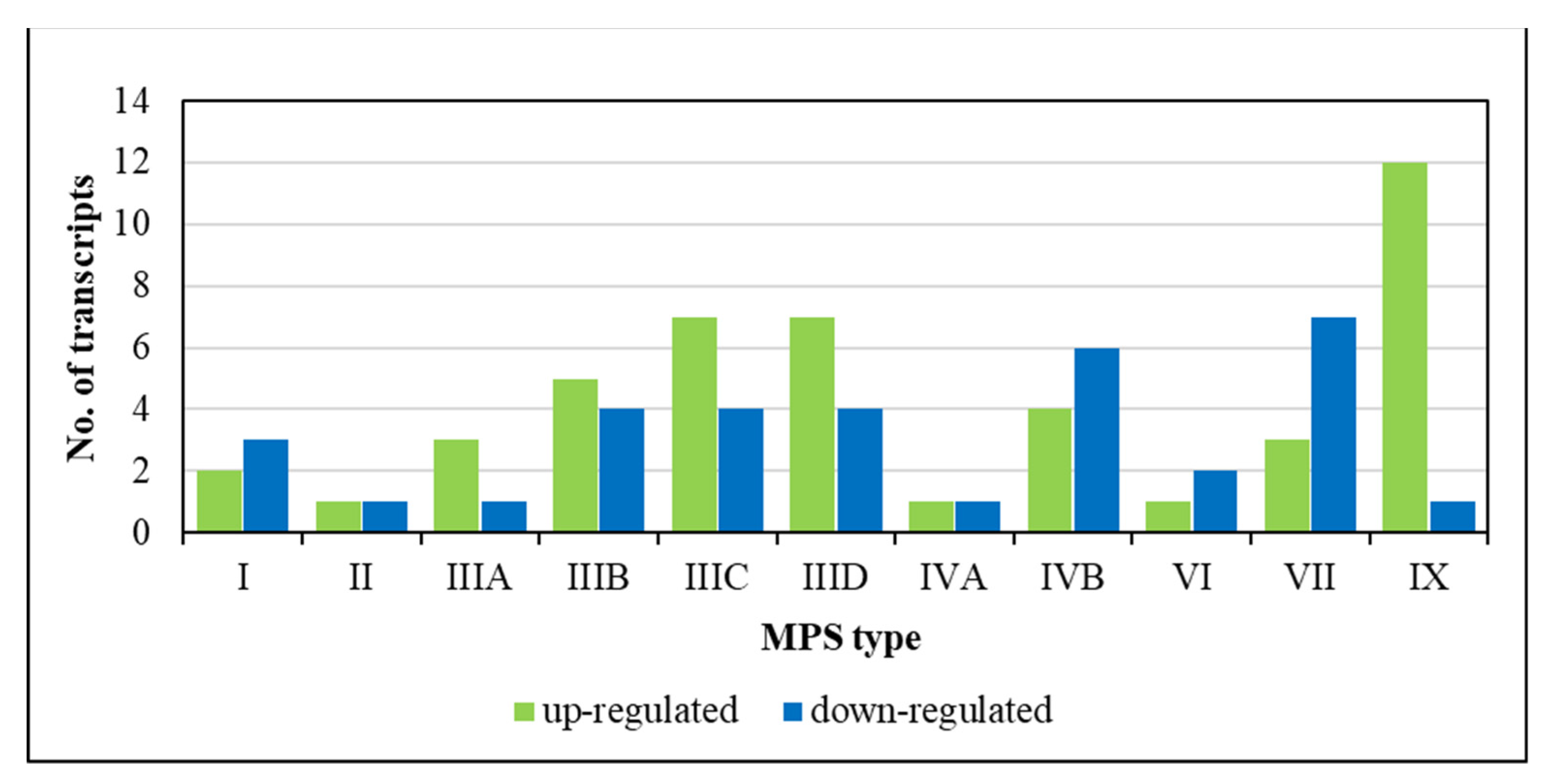
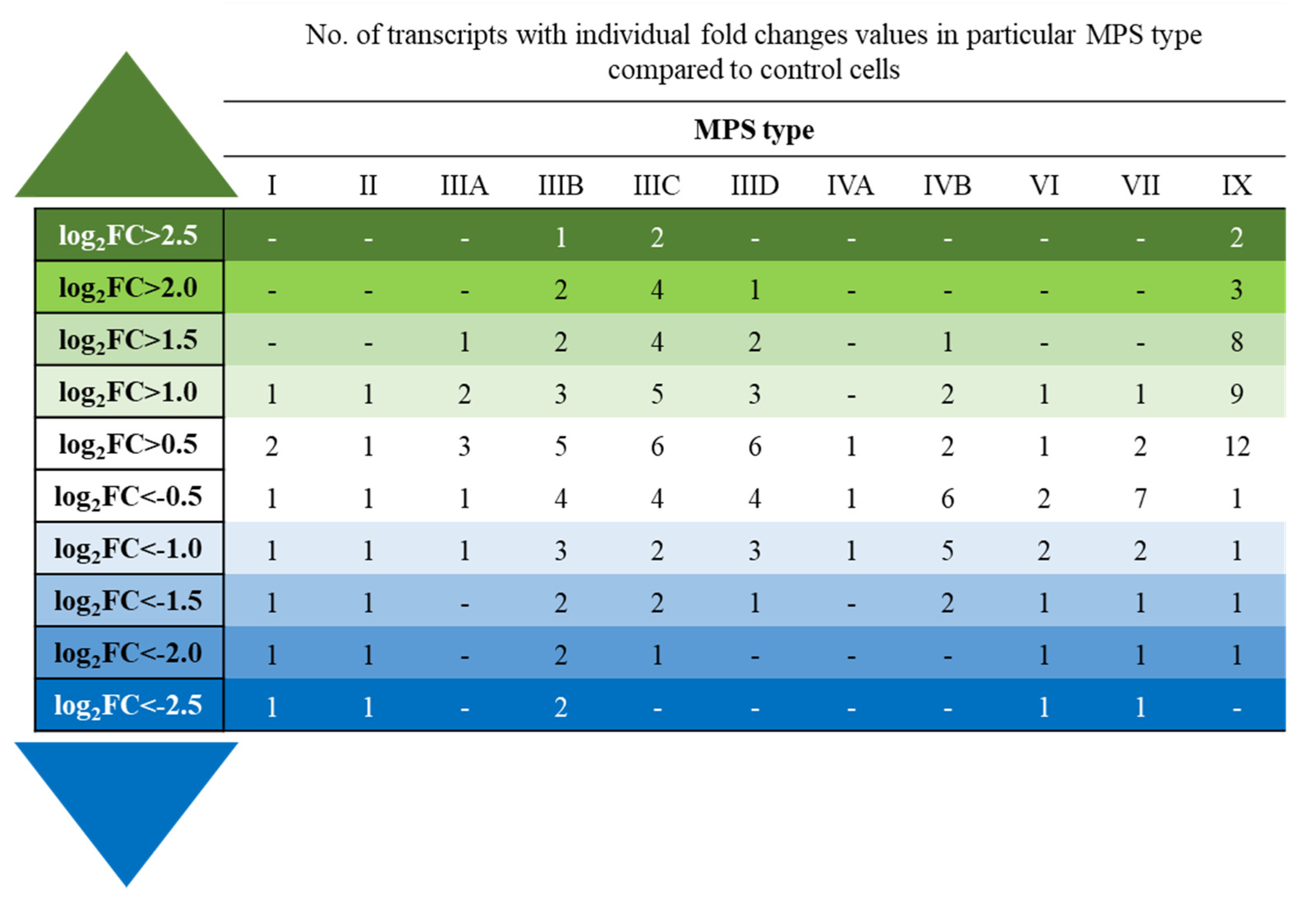
In the transcriptomic analyses, statistical significance was calculated based on results obtained from 4 independent biological experiments (n = 4). For the primary assessment of values with normal continuous distribution, one-way ANOVA was used with log2(1 + x). To calculate the false discovery rate (FDR), the Benjamini–Hochberg method was used. To compare two groups, post hoc Student’s t-test was used with Bonferroni correction. These calculations were performed using the R software v3.4.3 (https://cran.r-project.org/bin/windows/base/old/3.4.3/, last accessed on 1 December 2022 last accessed on 5 December 2022). The differences were considered significant when p < 0.1, which is used as a standard in transcriptomic analyses [15,16,17,18,19,20,21,22,23].

3. Results

To investigate changes in the levels of lncRNAs in the MPS cells relative to the control fibroblasts, we conducted transcriptomic analyses using cell lines derived from patients suffering from most MPS types (I, II, IIIA, IIIB, IIIC, IIID, IVA, IVB, VI, VII, and IX). The RNA-seq studies were performed as described in Section 2. Although only one cell line per MPS type was used, every experiment was performed independently four times; thus, there were four biological repeats (n = 4). Since our goal was to investigate potential changes in the levels of lncRNAs in MPS patients for the first time, we also considered all MPS types as a group of diseases and assessed whether any changes were common for most or all types. This made our transcriptomic analyses reliable, as indicated in previously published articles [15,16,17,18,19,20,21,22,23]. For analyses concerning long RNA molecules, the standard RNA isolation procedure allowed for the purification of both mRNA and lncRNA species, which enabled us to compare the abundance of specific kinds of these nucleic acids in a single experiment.
Our global analysis of the abundance of RNA molecules indicated that there are changes in the levels of various lncRNAs in every MPS type. Between 2 (in MPS II) and 13 (in MPS IX) lncRNA transcripts were significantly changed (either up- or down-regulated) relative to the control fibroblasts (Figure 1). These results indicated that there can be potential changes in the lncRNA-mediated regulation of the expression of various genes in MPS cells.
To assess which kinds of lncRNA are commonly up- or down-regulated in MPS cells, we assessed specific long non-coding transcripts that revealed either increased or decreased abundance relative to the control cells in fibroblasts of at least four MPS types. Five of such lncRNAs were identified, and are specified in Table 2. Importantly, in every case, a specific lncRNA was either up- or down-regulated in all MPS types wherein significant differences relative to the control cells occurred (no cases of up-regulation in some and down-regulation in other MPS types were identified). Therefore, it appears that similar changes in the levels of specific lncRNA occur in all/most of the MPS types.
In the next step, we analyzed how many and which lncRNAs occurred at especially increased or decreased levels in MPS cells. For this kind of analysis, we grouped lncRNAs into cohorts, revealing specific log2FC values (where FC states for fold change). These calculations indicated that the levels of most affected lncRNAs changed a few times; however, there were long non-coding transcripts that had been up- or down-regulated over ~6 times (log2FC > 2.5 or <−2.5) (Figure 2).
Specific genes encoding lncRNAs occurring at especially elevated or decreased levels (over ~6-fold, i.e., log2FC > 2.5 or <−2.5) in the MPS cells have been identified, and they are presented in Table 3. Again, the direction of the changes was always the same for each specific lncRNA type, i.e., every lncRNA was either up- or down-regulated in all MPS types. This confirms the uniform character of the changes in the levels of lncRNAs in MPS, irrespective of the disease type.
To identify potential target genes for specific lncRNA-mediated regulations, we employed the NPInter v4.0 data base (http://bigdata.ibp.ac.cn/npinter4/#, last accessed on 5 December 2022). Then, among all the genes potentially regulated by a given lncRNA, we selected those which revealed significant changes in at least one MPS type, as indicated by the results of our transcriptomic (RNA-seq) analyses for mRNAs (NCBI Sequence Read Archive (SRA) accession no. PRJNA562649). The results of these analyses are presented in Figure 3. They revealed that increased levels of the GAS5 lncRNA correlate with the up-regulation of the HNRNPC gene in MPS IIIC, the down-regulation of the FXR1 gene in MPS IIIC, and the down-regulation of the MATR3 gene in MPS IIIB (Figure 3A). Higher abundance of the MEG3 lncRNA occurred simultaneously with increased levels of TP53 mRNA in MPS IIIC (Figure 3B) and decreased levels of TARDBP mRNA in MPS IIIB (Figure 3C). The down-regulation of LINC00856 lncRNA correlated with the down-regulation of the MATR3 gene in MPS IIIB (Figure 3D).

4. Discussion

Although MPS is a group of monogenic diseases characterized by the accumulation of GAGs in lysosomes, recent studies clearly indicated that primary storage is not the only cause of significant changes in cellular physiology. Among various secondary and tertiary defects that significantly contribute to the development of specific MPS symptoms in patients, the dysregulation of the expression of a battery of genes has been demonstrated to significantly influence the structures and functions of cellular organelles as well as different cellular processes [15,16,17,18,19,20,21,22,23]. Recent findings have suggested that changes in the expression of genes coding for regulators of activities of other genes might be responsible for triggering chains of processes that considerably contribute to dysfunctions of cells, tissues, organs, and entire organisms [16]. Since non-coding RNA molecules can modulate the ex-pression of hundreds or thousands of genes [24,25,26,27], we investigated whether the levels of these kinds of regulatory RNAs are changed in MPS cells. In this study, we have focused on lncRNAs, as this kind of non-coding RNAs was not investigated previously in MPS cells.
In this report, RNA-seq analyses of the biological material of fibroblasts derived from patients suffering from most MPS types indicated, for the first time, that the levels of various lncRNAs are significantly changed relative to control cells (Figure 1, Table 2). Some lncRNAs were up- or down-regulated especially significantly (over 6-times; Figure 2, Table 3), and using the NPInter v4.0 data base, we identified potential target genes whose expression could be regulated by the actions of these non-coding transcripts. Then, by analyzing the levels of mRNAs derived from the same cells, genes that could be affected by specific lncRNAs and whose expression was up- or down-regulated were indicated. Therefore, we assume that the expression of these genes is likely modulated by changes in the levels of specific lncRNAs in MPS cells. The putative pairs of such lncRNAs and protein-encoding genes are as follows: GAS5 lncRNA—HNRNPC, FXR1, and MATR3; MEG3 lncRNA—TP53 and TARDBP; and LINC00856 lncRNA —MATR3 (Figure 3). Interestingly, this putative lncRNA-mediated regulation was especially pronounced in different subtypes of Sanfilippo disease (MPS III).
One may ask: what are the roles of the genetic elements involved in the above-described regulations? It has been demonstrated that the GAS5 lncRNA controls cancer development [53,54]. Its potential target genes, HNRNPC and FXR1, whose expression is changed in MPS cells, encode heterogeneous nuclear ribonucleoprotein C (a protein involved in the control of splicing and translation) [55] and RNA-binding protein fragile-X mental retardation autosomal 1 (a factor that participates in the mRNA transport from the nucleus and is bound to polysomes) [56,57], respectively. The MATR3 gene product, matrin 3, is a protein that interacts with nucleic acids and is able to bind to both DNA and RNA fragments, though its exact functions are still poorly understood [58,59]. Like GAS5, the MEG3 lncRNA is a tumor-growth inhibitor [60,61]. Potential MEG3-regulated genes, whose changed expression correlated with increased levels of this lncRNA in MPS cells, include TP53 and TARDBP. These genes encode the major tumor-suppressor protein p53 [62] and TAR DNA-binding protein 43 (or TDP-43) (an RNA/DNA-binding protein that is involved in RNA transactions) [63], respectively. Previously, the LINC00856 lncRNA was found to be involved in the response to acute lymphoblastic leukemia [64], and the down-regulation of MATR3 correlated with decreased levels of this lncRNA in MPS cells.
Interestingly, as indicated in the preceding paragraph, genes strongly affected by lncRNAs in MPS cells are involved in the regulation of the expression of other genes, which is mainly performed through interactions with nucleic acids. It is, therefore, tempting to speculate that the dysregulation of the expression of regulatory genes might significantly contribute to the development of cellular dysfunctions in MPS cells. If such a dysregulation is strong enough, the cellular changes might become irreversible or hardly reversible. In such a case, the pathogenic cascade of cellular misfunctions would not be corrected by the simple removal of the primary storage material (GAGs). This might explain why various therapies for MPS fail to treat all the symptoms of patients, despite the normalization of GAG levels in body fluids [9,10,11]. In this light, specific lncRNAs might be considered as potential targets for novel auxiliary drugs against MPS. Namely, the primary therapeutic intervention (ERT, hematopoietic stem cell transplantation, gene therapy, or others) would cause the normalization of GAG levels, while the accompanying, lcnRNA-specific drug might modulate the expression of specific genes, leading to the corrections of otherwise hardly improved cellular processes.
The observed correlations between the changes in the levels of lncRNAs in MPS cells and the expression efficiency of the target genes were either direct or reverse (Figure 3). Various lncRNAs can regulate gene expression by either sponging miRNAs (alleviating negative regulation) or interacting directly with mRNA molecules (modulating their abundance and translational efficiency) [33,34,65]. Therefore, when the directions of the changes in the levels of specific lncRNA and an mRNA of its target gene are the same, the lncRNA acts, most probably, through the interaction with miRNA(s) and the prevention of mRNA degradation. On the other hand, when the directions of changes are opposite, direct lncRNA-mRNA interactions are more likely.
This study, which demonstrates changes in levels of lncRNAs in MPS cells for the first time, also has some limitations. One of these was the use of only one cell line per MPS type. However, the biological experiments were repeated four times, which made the results of the RNA-seq analyses reliable [15,16,17,18,19,20,21,22,23]. The presented conclusions can also be corroborated by the fact that the directions of the changes in the levels of specific lncRNAs were always the same in all MPS types. Another limitation was that our study only focused on transcriptomic analyses. Indeed, further studies with different investigatory methods of lncRNA functions are necessary to understand the details of their roles in MPS. Nevertheless, this is the first report signaling the importance of changes in the levels of lncRNAs in MPS; thus, it can open a new field of research into understanding the molecular mechanisms of the pathogenesis of this disease.

5. Conclusions

For the first time, changed levels of various lncRNAs were demonstrated in 11 types/subtypes of MPS. Increased or decreased levels of specific lncRNAs correlated with the up- or down-regulation of the expression of genes that are potential targets for lncRNA-mediated control, suggesting functional relationships. These analyses suggest that changes in the abundance of lncRNAs can considerably influence the pathomechanism of MPS through the dysregulation of the expression of certain genes, especially those involved in the control of the activities of other genes.

Author Contributions

Conceptualization, Z.C., K.P. and G.W.; methodology, L.G. and K.P.; validation, K.P.; investigation, Z.C, L.G. and K.P.; data curation, K.P; writing—original draft preparation, Z.C., K.P. and G.W.; writing—review and editing, Z.C., L.G., K.P. and G.W.; visualization, K.P.; supervision, K.P. and G.W.; project administration, Z.C. and G.W.; funding acquisition, Z.C. and G.W. All authors have read and agreed to the published version of the manuscript.

Funding

This research was funded by Fundacja Sanfilippo (Poland) (project 15/02/2022 to G.W.), and University of Gdansk (project no. 1220/158/2022 to Z.C.).

Institutional Review Board Statement

Not applicable.

Informed Consent Statement

Not applicable.

Data Availability Statement

Raw data of the RNA-seq analysis are deposited in the NCBI Sequence Read Archive (SRA), under accession no. PRJNA562649.

Conflicts of Interest

The authors declare no conflict of interest.

References

  1. Parenti, G.; Andria, G.; Ballabio, A. Lysosomal storage diseases: From pathophysiology to therapy. Annu. Rev. Med. 2015, 66, 471–486. [Google Scholar] [CrossRef] [PubMed]
  2. de Queiroz, M.T.; Gonçalves Pereira, V.; do Nascimento, C.C.; D’Almeida, V. The Underexploited Role of Non-Coding RNAs in Lysosomal Storage Diseases. Front. Endocrinol. 2016, 7, 133. [Google Scholar] [CrossRef] [PubMed] [Green Version]
  3. Freeze, H.; Eklund, E.; Ng, B.; Patterson, M. Neurological aspects of human glycosylation disorders. Annu. Rev. Neurosci. 2015, 38, 105–125. [Google Scholar] [CrossRef] [PubMed] [Green Version]
  4. Węgrzyn, G.; Pierzynowska, K.; Pavone, L.M. Editorial: Molecular Aspects of Mucopolysaccharidoses. Front. Mol. Biosci. 2022, 9, 874267. [Google Scholar] [CrossRef] [PubMed]
  5. Wiśniewska, K.; Wolski, J.; Gaffke, L.; Cyske, Z.; Pierzynowska, K.; Węgrzyn, G. Misdiagnosis in mucopolysaccharidoses. J. Appl. Genet. 2022, 63, 475–495. [Google Scholar] [CrossRef] [PubMed]
  6. Tomatsu, S.; Lavery, C.; Giugliani, R.; Harmatz, P.; Scarpa, M.; Węgrzyn, G.; Orii, T. Mucopolysaccharidoses Update; Nova Science Publisher: New York, NY, USA, 2018; ISBN 978-1-53613-986-0. [Google Scholar]
  7. Cyske, Z.; Anikiej-Wiczenbach, P.; Wisniewska, K.; Gaffke, L.; Pierzynowska, K.; Mański, A.; Wegrzyn, G. Sanfilippo Syndrome: Optimizing Care with a Multidisciplinary Approach. J. Multidiscip. Healthc. 2022, 15, 2097–2110. [Google Scholar] [CrossRef] [PubMed]
  8. McBride, K.L.; Flanigan, K.M. Update in the Mucopolysaccharidoses. Semin. Pediatr. Neurol. 2021, 37, 100874. [Google Scholar] [CrossRef]
  9. Penon-Portmann, M.; Blair, D.R.; Harmatz, P. Current and new therapies for mucopolysaccharidoses. Pediatr. Neonatol. 2022. [Google Scholar] [CrossRef]
  10. Dornelles, A.; Artigalás, O.; da Silva, A.; Ardila, D.; Alegra, T.; Pereira, T.; Vairo, F.; Schwartz, I. Efficacy and safety of intravenous laronidase for mucopolysaccharidosis type I: A systematic review and meta-analysis. PLoS ONE 2017, 12, e0184065. [Google Scholar] [CrossRef] [Green Version]
  11. Aldenhoven, M.; van den Broek, B.; Wynn, R.; O’Meara, A.; Veys, P.; Rovelli, A.; Jones, S.; Parini, R.; van Hasselt, P.; Renard, M.; et al. Quality of life of Hurler syndrome patients after successful hematopoietic stem cell transplantation. Blood Adv. 2017, 1, 2236–2242. [Google Scholar] [CrossRef]
  12. Parente, M.; Rozen, R.; Seeholzer, S.; Wolfe, J. Integrated analysis of proteome and transcriptome changes in the mucopolysaccharidosis type VII mouse hippocampus. Mol. Genet. Metab. 2016, 118, 41–54. [Google Scholar] [CrossRef] [PubMed] [Green Version]
  13. Tessitore, A.; Pirozzim, M.; Auricchio, A. Abnormal autophagy, ubiquitination, inflammation and apoptosis are dependent upon lysosomal storage and are useful biomarkers of mucopolysaccharidosis VI. Pathogenetics 2009, 2, 4. [Google Scholar] [CrossRef] [PubMed] [Green Version]
  14. Pierzynowska, K.; Gaffke, L.; Podlacha, M.; Brokowska, J.; Węgrzyn, G. Mucopolysaccharidosis and Autophagy: Controversies on the Contribution of the Process to the Pathogenesis and Possible Therapeutic Applications. Neuromol. Med. 2020, 22, 25–30. [Google Scholar] [CrossRef] [PubMed] [Green Version]
  15. Gaffke, L.; Pierzynowska, K.; Podlacha, M.; Brokowska, J.; Węgrzyn, G. Changes in cellular processes occurring in mucopolysaccharidoses as underestimated pathomechanisms of these diseases. Cell Biol. Int. 2021, 45, 498–506. [Google Scholar] [CrossRef] [PubMed]
  16. Cyske, Z.; Gaffke, L.; Pierzynowska, K.; Węgrzyn, G. Complex Changes in the Efficiency of the Expression of Many Genes in Monogenic Diseases, Mucopolysaccharidoses, May Arise from Significant Disturbances in the Levels of Factors Involved in the Gene Expression Regulation Processes. Genes 2022, 13, 593. [Google Scholar] [CrossRef] [PubMed]
  17. Rintz, E.; Gaffke, L.; Podlacha, M.; Brokowska, J.; Cyske, Z.; Węgrzyn, G.; Pierzynowska, K. Transcriptomic Changes Related to Cellular Processes with Particular Emphasis on Cell Activation in Lysosomal Storage Diseases from the Group of Mucopolysaccharidoses. Int. J. Mol. Sci. 2020, 21, 3194. [Google Scholar] [CrossRef]
  18. Pierzynowska, K.; Gaffke, L.; Jankowska, E.; Rintz, E.; Witkowska, J.; Wieczerzak, E.; Podlacha, M.; Węgrzyn, G. Proteasome Composition and Activity Changes in Cultured Fibroblasts Derived from Mucopolysaccharidoses Patients and Their Modulation by Genistein. Front. Cell Dev. Biol. 2020, 8, 540726. [Google Scholar] [CrossRef]
  19. Brokowska, J.; Pierzynowska, K.; Gaffke, L.; Rintz, E.; Węgrzyn, G. Expression of genes involved in apoptosis is dysregulated in mucopolysaccharidoses as revealed by pilot transcriptomic analyses. Cell Biol. Int. 2021, 45, 549–557. [Google Scholar] [CrossRef]
  20. Gaffke, L.; Pierzynowska, K.; Rintz, E.; Cyske, Z.; Giecewicz, I.; Węgrzyn, G. Gene Expression-Related Changes in Morphologies of Organelles and Cellular Component Organization in Mucopolysaccharidoses. Int. J. Mol. Sci. 2021, 22, 2766. [Google Scholar] [CrossRef]
  21. Gaffke, L.; Szczudło, Z.; Podlacha, M.; Cyske, Z.; Rintz, E.; Mantej, J.; Krzelowska, K.; Węgrzyn, G.; Pierzynowska, K. Impaired ion homeostasis as a possible associate factor in mucopolysaccharidosis pathogenesis: Transcriptomic, cellular and animal studies. Metab. Brain Dis. 2022, 37, 299–310. [Google Scholar] [CrossRef]
  22. Pierzynowska, K.; Żabińska, M.; Gaffke, L.; Cyske, Z.; Węgrzyn, G. Changes in expression of signal transduction-related genes, and formation of aggregates of GPER1 and OXTR receptors in mucopolysaccharidosis cells. Eur. J. Cell Biol. 2022, 101, 151232. [Google Scholar] [CrossRef] [PubMed]
  23. Brokowska, J.; Gaffke, L.; Pierzynowska, K.; Cyske, Z.; Węgrzyn, G. Cell cycle disturbances in mucopolysaccharidoses: Transcriptomic and experimental studies on cellular models. Exp. Biol. Med. 2022, 247, 1639–1649. [Google Scholar] [CrossRef] [PubMed]
  24. Lekka, E.; Hall, J. Noncoding RNAs in disease. FEBS Lett. 2018, 592, 2884–2900. [Google Scholar] [CrossRef] [PubMed]
  25. Ling, H.; Fabbri, M.; Calin, G. MicroRNAs and other non-coding RNAs as targets for anticancer drug development. Nat. Rev. Drug Discov. 2013, 12, 847–865. [Google Scholar] [CrossRef] [PubMed] [Green Version]
  26. St Laurent, G.; Wahlestedt, C.; Kapranov, P. The Landscape of long noncoding RNA classification. Trends Genet. 2015, 31, 239–251. [Google Scholar] [CrossRef] [Green Version]
  27. Ebbesen, K.; Kjems, J.; Hansen, T. Circular RNAs: Identification, biogenesis and function. Biochim. Biophys. Acta 2016, 1859, 163–168. [Google Scholar] [CrossRef]
  28. Yang, J.; Rastetter, R.; Wilhelm, D. Non-coding RNAs: An Introduction. Adv. Exp. Med. Biol. 2016, 886, 13–32. [Google Scholar]
  29. van Rooij, E.; Kauppinen, S. Development of microRNA therapeutics is coming of age. EMBO Mol. Med. 2014, 6, 851–864. [Google Scholar] [CrossRef]
  30. Djebali, S.; Davis, C.A.; Merkel, A.; Dobin, A.; Lassmann, T.; Mortazavi, A.; Tanzer, A.; Lagarde, J.; Lin, W.; Schlesinger, F.; et al. Landscape of transcription in human cells. Nature 2012, 489, 101–108. [Google Scholar] [CrossRef] [Green Version]
  31. Guttman, M.; Amit, I.; Garber, M.; French, C.; Lin, M.F.; Feldser, D.; Huarte, M.; Zuk, O.; Carey, B.W.; Cassady, J.P.; et al. Chromatin signature reveals over a thousand highly conserved large non-coding RNAs in mammals. Nature 2009, 458, 223–227. [Google Scholar] [CrossRef]
  32. St Laurent, G.; Vyatkin, Y.; Antonets, D.; Ri, M.; Qi, Y.; Saik, O.; Shtokalo, D.; De Hoon, M.J.; Kawaji, H.; Itoh, M.; et al. Functional annotation of the vlinc class of non-coding RNAs using systems biology approach. Nucleic Acids Res. 2016, 44, 3233–3252. [Google Scholar] [CrossRef] [PubMed]
  33. Sauvageau, M. Diverging RNPs: Toward Understanding lncRNA-Protein Interactions and Functions. Adv. Exp. Med. Biol. 2019, 1203, 285–312. [Google Scholar] [PubMed]
  34. Chattopadhyay, P.; Mishra, P.; Mehta, P.; Soni, J.; Gupta, R.; Tarai, B.; Pandey, R. Transcriptomic study reveals lncRNA-mediated downregulation of innate immune and inflammatory response in the SARS-CoV-2 vaccination breakthrough infections. Front. Immunol. 2022, 13, 1035111. [Google Scholar] [CrossRef] [PubMed]
  35. Cortini, F.; Chiara, F.; Villa, C. Emerging roles of long non-coding RNAs in the pathogenesis of Alzheimer’s disease. Aging Res. Rev. 2019, 50, 19–26. [Google Scholar] [CrossRef]
  36. Elkouris, M.; Kouroupi, G.; Vourvoukelis, A.; Papagiannakis, N.; Kaltezioti, V.; Matsas, R.; Stefanis, L.; Xilouri, S.; Politis, P. Long Non-coding RNAs Associated with Neurodegeneration-Linked Genes Are Reduced in Parkinson’s Disease Patients. Front. Cell Neurosci. 2019, 13, 58. [Google Scholar] [CrossRef] [Green Version]
  37. Johnson, R. Long non-coding RNAs in Huntington’s disease neurodegeneration. Neurobiol. Dis. 2012, 46, 245–254. [Google Scholar] [CrossRef]
  38. Riva, P.; Ratti, A.; Venturin, M. The Long Non-Coding RNAs in Neurodegenerative Diseases: Novel Mechanisms of Pathogenesis. Curr. Alzheimer Res. 2016, 13, 1219–1231. [Google Scholar] [CrossRef] [Green Version]
  39. He, J.; Wu, W. Comprehensive landscape and future perspectives of long noncoding RNAs (lncRNAs) in colorectal cancer (CRC): Based on a bibliometric analysis. Noncoding RNA Res. 2022, 8, 33–52. [Google Scholar] [CrossRef]
  40. Wang, X.; Cao, L.; Wang, Y.; Wang, X.; Liu, N.; You, Y. Regulation of let-7 and its target oncogenes. Oncol. Lett. 2012, 3, 955–960. [Google Scholar] [CrossRef]
  41. Thakral, S.; Ghoshal, K. miR-122 is a unique molecule with great potential in diagnosis, prognosis of liver disease, and therapy both as miRNA mimic and antimir. Curr. Gene Ther. 2015, 15, 142–150. [Google Scholar] [CrossRef] [Green Version]
  42. Samra, M.; Srivastavam, K. Non-coding RNA and their potential role in cardiovascular diseases. Gene 2023, 851, 147011. [Google Scholar] [CrossRef] [PubMed]
  43. Gomez, I.G.; MacKenna, D.A.; Johnson, B.G.; Kaimal, V.; Roach, A.M.; Ren, S.; Duffield, J.S. Anti-microRNA-21 oligonucleotides prevent Alport nephropathy progression by stimulating metabolic pathways. J. Clin. Investig. 2015, 125, 141–156. [Google Scholar] [CrossRef] [PubMed]
  44. Sparber, P.; Filatova, A.; Khantemirova, M.; Skoblov, M. The role of long non-coding RNAs in the pathogenesis of hereditary diseases. BMC Med. Genom. 2019, 12 (Suppl. 2), 42. [Google Scholar] [CrossRef] [PubMed] [Green Version]
  45. Ozsait, B.; Komurcu-Bayrak, E.; Levula, M.; Erginel-Unaltuna, N.; Kähönen, M.; Rai, M.; Lehtimäki, T.; Laaksonen, R. Niemann-Pick type C fibroblasts have a distinct microRNA profile related to lipid metabolism and certain cellular components. Biochem. Biophys. Res. Commun. 2010, 403, 316–321. [Google Scholar] [CrossRef]
  46. Arora, S.; Beaudry, C.; Bisanz, K.; Sima, C.; Kiefer, J.; Azorsa, D. A high-content RNAi-screening assay to identify modulators of cholesterol accumulation in Niemann-Pick type C cells. Assay Drug Dev. Technol. 2010, 8, 295–320. [Google Scholar] [CrossRef]
  47. Siebert, M.; Westbroek, W.; Chen, Y.; Moaven, N.; Li, Y.; Velayati, A.; Saraiva-Pereira, M.; Martin, S.; Sidransky, E. Identification of miRNAs that modulate glucocerebrosidase activity in Gaucher disease cells. RNA Biol. 2014, 11, 1291–1300. [Google Scholar] [CrossRef] [Green Version]
  48. Ginns, E.; Mak, S.; Ko, N.; Karlgren, J.; Akbarian, S.; Chou, V.; Guo, Y.; Lim, A.; Samuelsson, S.; LaMarca, M.; et al. Neuroinflammation and α-synuclein accumulation in response to glucocerebrosidase deficiency are accompanied by synaptic dysfunction. Mol. Genet. Metab. 2014, 111, 152–162. [Google Scholar] [CrossRef]
  49. Matsui, M.; Corey, D. Non-coding RNAs as drug targets. Nat. Rev. Drug Discov. 2017, 16, 167–179. [Google Scholar] [CrossRef] [Green Version]
  50. Pereira, V.; Queiroz, M.; D’Almeida, V. Differential expression of microRNAs from miR-17 family in the cerebellum of mucopolysaccharidosis type I mice. Gene 2016, 595, 207–211. [Google Scholar] [CrossRef]
  51. Kreutz, F.; dos Santos Petry, F.; Camassola, M.; Schein, V.; Guma, F.; Nardi, N.; Trindade, V. Alterations of membrane lipids and in gene expression of ganglioside metabolism in different brain structures in a mouse model of mucopolysaccharidosis type I (MPS I). Gene 2013, 527, 109–114. [Google Scholar] [CrossRef] [Green Version]
  52. Gaffke, L.; Pierzynowska, K.; Podlacha, M.; Hoinkis, D.; Rintz, E.; Brokowska, J.; Wegrzyn, G. Underestimated Aspect of Mucopolysaccharidosis Pathogenesis: Global Changes in Cellular Processes Revealed by Transcriptomic Studies. Int. J. Mol. Sci. 2020, 21, 1204. [Google Scholar] [CrossRef] [PubMed]
  53. Wang, H.; Wang, D.; Shen, Y.; Sun, C.; Hu, Q.; Jiang, L.; Du, Q. GAS5 attenuates the malignant progression of glioma stem-like cells by promoting E-cadherin. Cancer Gene Ther. 2022. [Google Scholar] [CrossRef] [PubMed]
  54. Zhang, Z.; Liu, T.; Cheng, C.; Wang, J.; Wang, C.; Huang, H.; Li, Y. LncRNA GAS5 regulates the Wnt/β-catenin pathway through the miR-18a-5p/AXIN2/GSK3β axis to inhibit the proliferation and migration of bladder cancer cells. Carcinogenesis 2022. [Google Scholar] [CrossRef] [PubMed]
  55. Zhou, R.; Chun, R.F.; Lisse, T.S.; Garcia, A.J.; Xu, J.; Adams, J.S.; Hewison, M. Vitamin D and alternative splicing of RNA. J. Steroid Biochem. Mol. Biol. 2015, 148, 310–317. [Google Scholar] [CrossRef] [Green Version]
  56. McClure, J.J.; Palanisamy, V. Muscle-Specific FXR1 Isoforms in Squamous Cell Cancer. Trends Cancer 2019, 5, 82–84. [Google Scholar] [CrossRef] [PubMed]
  57. Datta, C.; Truesdell, S.S.; Wu, K.Q.; Bukhari, S.I.A.; Ngue, H.; Buchanan, B.; Le Tonqueze, O.; Lee, S.; Kollu, S.; Granovetter, M.A.; et al. Ribosome changes reprogram translation for chemosurvival in G0 leukemic cells. Sci. Adv. 2022, 8, eabo1304. [Google Scholar] [CrossRef]
  58. Malik, A.M.; Barmada, S.J. Matrin 3 in neuromuscular disease: Physiology and pathophysiology. JCI Insight 2021, 6, e143948. [Google Scholar] [CrossRef]
  59. Salem, A.; Wilson, C.J.; Rutledge, B.S.; Dilliott, A.; Farhan, S.; Choy, W.Y.; Duennwald, M.L. Matrin3: Disorder and ALS Pathogenesis. Front. Mol. Biosci. 2022, 8, 794646. [Google Scholar] [CrossRef]
  60. Hu, Y.; Lv, F.; Li, N.; Yuan, X.; Zhang, L.; Zhao, S.; Jin, L.; Qiu, Y. The long non-coding RNA MEG3 inhibits oral squamous cell carcinoma progression via GATA3. FEBS Open Bio 2023, 13, 195–208. [Google Scholar] [CrossRef]
  61. Yan, X.; Jia, H.; Zhao, J. LncRNA MEG3 attenuates the malignancy of retinoblastoma cells through inactivating PI3K /Akt/mTOR signaling pathway. Exp. Eye Res. 2022, 226, 109340. [Google Scholar] [CrossRef]
  62. Chen, X.; Zhang, T.; Su, W.; Dou, Z.; Zhao, D.; Jin, X.; Lei, H.; Wang, J.; Xie, X.; Cheng, B.; et al. Mutant p53 in cancer: From molecular mechanism to therapeutic modulation. Cell Death Dis. 2022, 13, 974. [Google Scholar] [CrossRef] [PubMed]
  63. Lye, Y.S.; Chen, Y.R. TAR DNA-binding protein 43 oligomers in physiology and pathology. IUBMB Life 2022, 74, 794–811. [Google Scholar] [CrossRef] [PubMed]
  64. Richard, M.A.; Brown, A.L.; Belmont, J.W.; Scheurer, M.E.; Arroyo, V.M.; Foster, K.L.; Kern, K.D.; Hudson, M.M.; Leisenring, W.M.; Okcu, M.F.; et al. Genetic variation in the body mass index of adult survivors of childhood acute lymphoblastic leukemia: A report from the Childhood Cancer Survivor Study and the St. Jude Lifetime Cohort. Cancer 2021, 127, 310–318. [Google Scholar] [CrossRef]
  65. Rajakumar, S.; Jamespaulraj, S.; Shah, Y.; Kejamurthy, P.; Jaganathan, M.K.; Mahalingam, G.; Devi, K.T.R. Long non-coding RNAs: An overview on miRNA sponging and its co-regulation in lung cancer. Mol. Biol. Rep. 2022. [Google Scholar] [CrossRef] [PubMed]
Figure 1. Number of up- and down-regulated transcripts of genes coding for lncRNAs (Gene Ontology term GO:0140742 (lncRNA transcription) in the QuickGO database (https://www.ebi.ac.uk/QuickGO/, last accessed on 5 December 2022)) in different types of MPS relative to control cells (HDFa).
Genes 14 00271 g001
Figure 2. Number of transcripts included in the ‘lncRNA transcription’ term (GO:0140742) in the QuickGO database with altered expression depending on the level of fold-change (log2FC) in different types of MPS relative to control cells (HDFa).
Genes 14 00271 g002
Figure 3. Correlations of increased (red, upward-pointing arrows) and decreased (red, downward-pointing arrows) levels of GAS5 (A), MEG3 (B,C), and LINC00856 (D) lncRNAs with significant changes (either increased or decreased abundance of mRNAs, marked by upward- or downward-pointing red arrows, respectively) in expression of specific genes in MPS cells.
Genes 14 00271 g003
Table 1. Fibroblast lines derived from MPS patients and used in this study.
MPS TypeCoriell Institute
Catalogue Number
Sex of Patient Age (Number of Years at the Time of Sample Collection)Mutated GeneMutation(s)
MPS IGM00798F1IDUAp.Trp402Ter/p.Trp402Ter
MPS IIGM13203M3IDSp.His70ProfsTer29/-
MPS IIIAGM00879F3SGSHp.Glu447Lys/p.Arg245His
MPS IIIBGM00156M7NAGLUp.Arg626Ter/p.Arg626Ter
MPS IIICGM05157M8HGSNATp.Gly262Arg/p.Arg509Asp
MPS IIIDGM05093M7GNSp.Arg355Ter/p.Arg355Ter
MPS IVAGM00593F7GALNSp.Arg386Cys/p.Phe285Ter
MPS IVBGM03251F4GLB1p.Trp273Leu/p.Trp509Cys
MPS VIGM03722F3ARSBNot determined
MPS VIIGM00121M3GUSBp.Trp627Cys/p.Arg356Ter
MPS IXGM17494F14HYAL1Not determined
Table 2. Genes encoding lncRNAs whose expression is significantly changed in at least four MPS types relative to the control cells. Changes in expression (transcripts’ levels) of particular genes in MPS types are depicted, indicating log2 fold change (FC) relative to control cells; up-regulation is marked in green while down-regulation in marked in blue; non-statistically significant differences are not colored.
Transcriptlog2FC of Selected Transcripts’ Levels in at Least 4 MPS Types vs. HDFa Line
IIIIIIAIIIBIIICIIIDIVAIVBVIVIIIX
GAS50.831.161.260.932.980.110.360.691.35−0.020.96
ILF3-DT0.630.550.33−0.340.790.680.770.310.810.010.82
SNHG80.76−0.111.68−0.621.061.250.33−1.29−0.46−0.581.54
LINC006671.100.730.730.80−0.08−0.40−0.491.10−0.10−1.141.23
LINC02381−0.10−0.57−0.10−1.39−2.14−1.58−1.48−1.39−1.060.16−1.27
Table 3. Genes included in the ‘lncRNA transcription’ term (GO:0140742) with log2FC > 2.5 in specific MPS types relative to control cells (HDFa). Upward- and downward-pointing arrows indicate up- and down-regulation, respectively, while minus symbols indicate lower levels of changes. For MEG3, three transcripts were identified, which were marked as t.1, t.2, and t.3.
Transcriptlog2FC > 2.5 or <−2.5 of Selected Transcripts’ Levels in Particular MPS Type vs. HDFa Line
IIIIIIAIIIBIIICIIIDIVAIVBVIVIIIX
SNHG5
LINC01705
LINC00856
CYTOR
MEG3 (t.1)
MEG3 (t.2)
MEG3 (t.3)
GAS5
Disclaimer/Publisher’s Note: The statements, opinions and data contained in all publications are solely those of the individual author(s) and contributor(s) and not of MDPI and/or the editor(s). MDPI and/or the editor(s) disclaim responsibility for any injury to people or property resulting from any ideas, methods, instructions or products referred to in the content.

