Article

Identification of ZmBK2 Gene Variation Involved in Regulating Maize Brittleness

1
Agronomy/State Key Laboratory of Crop Biology, Shandong Agricultural University, Tai’an 271000, China
2
Qingdao Academy of Agricultural Sciences, Qingdao 266100, China
3
Taian Denghai Wuyue Taishan Seed Industry Co., Ltd., Tai’an 271000, China
*
Author to whom correspondence should be addressed.
Genes 2023, 14(6), 1126; https://doi.org/10.3390/genes14061126
Submission received: 2 May 2023 / Revised: 18 May 2023 / Accepted: 19 May 2023 / Published: 23 May 2023
(This article belongs to the Section Plant Genetics and Genomics)

Abstract

:
Maize stalk strength is a crucial agronomic trait that affects lodging resistance. We used map-based cloning and allelic tests to identify a maize mutant associated with decreased stalk strength and confirmed that the mutated gene, ZmBK2, is a homolog of Arabidopsis AtCOBL4, which encodes a COBRA-like glycosylphosphatidylinositol (GPI)-anchored protein. The bk2 mutant exhibited lower cellulose content and whole-plant brittleness. Microscopic observations showed that sclerenchymatous cells were reduced in number and had thinner cell walls, suggesting that ZmBK2 affects the development of cell walls. Transcriptome sequencing of differentially expressed genes in the leaves and stalks revealed substantial changes in the genes associated with cell wall development. We constructed a cell wall regulatory network using these differentially expressed genes, which revealed that abnormal cellulose synthesis may be a reason for brittleness. These results reinforce our understanding of cell wall development and provide a foundation for studying the mechanisms underlying maize lodging resistance.

1. Introduction

Lodging threatens maize harvests and reduces the total annual yield by 5–20% worldwide, increasing the risk of food insecurity [1]. Lodging resistance is closely related to plant brittleness, which involves complex physicochemical processes associated with cell wall development, specifically cellulose and lignin biosynthesis [2]. Research on brittle mutants is important for understanding the regulatory mechanisms of plant mechanical strength and has major implications for crop lodging resistance breeding [3].
The plant cell wall acts as a rigid and dynamic network that primarily consists of polysaccharides and supports plant cell morphogenesis. Cell walls are mainly composed of cellulose, hemicellulose, pectin, and lignin, with cellulose being the main component in most plants [4]. Cellulose consists of unbranched β-(1,4)-linked glucan chains and provides support for cell walls. According to their structural and compositional characteristics, cell walls are classified into primary and secondary cell walls [4]. Primary cell walls form during cytokinesis and modify and expand with cell growth, providing strength, resilience, and plasticity to the cell [5]. Secondary cell walls develop between the primary cell wall and protoplast after the cell stops growing; they have a cellulose content of 40–90% and also contain other principal components such as lignin. Primary and secondary cell walls contribute greatly to the mechanical strength of a plant [6]. Rice mutants Tos17, bc7, and bc11 have defects in cellulose synthase catalytic subunit (CESA) genes, causing reduced cellulose content in secondary cell walls and decreased mechanical strength [7]. Arabidopsis irregular xylem mutants irx1 and irx3 exhibit a disordered secondary cell wall structure [8], suggesting that cellulose deposition plays a key role in the integrity and assembly of cell wall polymers.
Studies on brittle mutants have identified some genes involved in cellulose biosynthesis [9,10]. In higher plants, cellulose synthesis is generally catalyzed by cellulose synthase complexes that form a rosette structure and synthesize at the plasma membrane (PM) using uridine diphosphate (UDP)-glucose as substrates [11]. The plant CESA gene was first isolated from cotton fiber based on its high similarity to bacterial CESA sequences. Most CESAs are highly conserved and co-expressed to regulate primary or secondary cell wall synthesis with multispan membrane domains [12,13]. Twelve CESAs have been cloned in Arabidopsis; of these, seven are required for primary cell wall synthesis, five are involved in secondary cell wall metabolism, and the functions of the remaining two are unknown [8,10]. Rice contains 11 CESAs that play key roles in secondary cell wall synthesis, with most of them being homologs of AtCESAs [14]. Of the twelve CESAs that were separated from maize, ZmCESA1 to ZmCESA9 are involved in primary cell wall formation, and ZmCESA10 to ZmCESA12 interact to form enzyme complexes that participate in secondary cell wall synthesis [15].
Additional proteins, such as COBRA, are necessary for the synthesis and orderly deposition of cellulose. First identified in Arabidopsis, the COBRA mutant had decreased cellulose content and exhibited plant brittleness [16]. The COBRA family genes encode glycosylphosphatidylinositol (GPI)-anchored plant-specific proteins with four conserved structural domains: a carbohydrate-binding motif, N-terminal protein targeting sites, a CCVS (Cys-rich) motif, and a hydrophobic C-terminus with an attachment site [17]. When the GPI-anchor modification site (ω-site) is cleaved, this structure combines with the C-terminus of the COBRA protein via an amide bond [18]. Research on Arabidopsis cob-1, cob-2, and cob-3 revealed that the COBRA protein affected the direction of cell expansion and decreased crystalline cellulose content [17]. The COBRA proteins play extremely important roles in anisotropic cell expansion and influence the morphogenesis of the whole plant by directing cellulose microfibril formation [19]. Rice bc1 reduces glucose and xylose and alters cellulose and lignin contents, resulting in defective development of secondary cell walls, which suggests that it may act as an auxiliary protein in regulating secondary cell wall biosynthesis [20]. Maize ZmBK1ZmBK6 were identified to be associated with defective cell wall development, and ZmBK2 was first identified in maize as a member of the COBRA family [21,22,23]. The ZmBK2 gene greatly reduces tissue mechanical strength, along with a substantial reduction in cellulose content and impaired deposition of secondary cell wall components [22,24].
A maize brittle stalk mutant (bk2) was identified and exhibited reduced mechanical strength, decreased cellulose content, and abnormal cell wall morphology in the whole plant. According to homology analyses, ZmBK2 encodes a GPI-anchored plant-specific protein that participates in cellular metabolic processes. We identified a series of genes concerned with cell wall development using transcriptome analysis and constructed a putative regulatory network for cell wall development. This study illuminates the role of ZmBK2 in cell wall biosynthesis and provides new insights into the mechanisms of maize brittleness.

2. Materials and Methods

2.1. Plant Materials

In this study, we analyzed three brittle mutants (mu-J1862, mu-J184, and 916C) and their wild-type counterparts (J1862, J184, and W22). The spontaneous mutant mu-J1862 was crossed with B73 to obtain F1 and backcrossed with mu-J1862 to obtain BC1F1. Thereafter, BC1F1 was self-crossed three times to obtain a homozygous non-brittle plant, J1862. mu-J184 was crossed with B73 to generate F1 and F2 for further research on homozygous brittle and non-brittle plants. To generate F1 and F2, mu-J1862 was crossed with B73 and Huangzaosi, with B73 and Huangzaosi being chosen as the two inbred lines for the mapping population due to their significant genetic differences from the mutant. Brittle plants from the F2 population were used for genetic mapping. Maize stock 916C containing the reference allele of bk2 (bk2-ref) was obtained from the Maize Genetics COOP Stock Center (https://www.maizegdb.org/data_center/stock, accessed on 21 May 2021) and backcrossed three times with W22, with W22 serving as the background material. All materials were grown in Shandong and Hainan Provinces in China.

2.2. Map-Based Cloning of the Brittle Gene

We used 3107 plants with a brittle phenotype from the F2 population for fine mapping by the BSA method. Simple sequence repeat (SSR) primers were obtained from the Gramene database (http://archive.gramene.org/markers/microsat/, accessed on 10 February 2022). Sequence polymorphisms were screened between mu-J1862 and B73 for the development of SSR and indel markers. Primer pairs were automatically designed in Primer3 (https://bioinfo.ut.ee/primer3-0.4.0/, accessed on 21 May 2022) and dCAPS Finder 2.0 (http://helix.wustl.edu/dcaps/dcaps.html, accessed on 15 July 2022). The Maize Genome Annotation Project (https://www.maizegdb.org/, accessed on 7 October 2022) was used for annotating putative genes.

2.3. Allelic Tests of bk2

Two allelic tests were performed by crossing mu-J1862 with mu-J184 and mu-J1862 with 916C.

2.4. Stalk Stiffness Test and Measurement of Stalk and Leaf Composition

Stalk stiffness was measured with a digital force gauge that recorded the maximum tensile force by pulling the specimen until it snapped.
To determine cellulose and lignin compositions, HPLC and UV spectrophotometry were performed based on the National Renewable Energy Laboratory (NREL/TP-510-42618) procedures [25]. Cellulose content was measured by HPLC based on degraded glucose and xylose sugar units and the amount of acid-soluble lignin by a UV spectrophotometer. Hemicellulose was measured according to Jin et al. [26]. Maize stalks and leaves were collected at the 12-leaf stage.

2.5. Subcellular Localization

The full-length CDS of ZmBK2 was amplified from B73 and cloned into the transient expression vector Pzp211-GFP-ZmBK2, driven by a constitutive CaMV 35S promoter. Clones were introduced into tobacco leaves to generate fusion proteins. Green fluorescent signals were detected using a TCS SP5 II laser-scanning confocal microscope (Leica, Wetzlar, Germany).

2.6. Phylogenetic Analysis and Protein Modeling

Homologous ZmBK2 amino acid sequences were searched through the NCBI database (https://blast.ncbi.nlm.nih.gov/Blast.cgi, accessed on 14 November 2022). Clustal Omega (http://www.clustal.org/, accessed on 14 November 2022) was used to perform sequence alignments. We employed MEGAX (https://www.megasoftware.net/, accessed on 14 November 2022) with default settings to reconstruct a phylogenetic tree, using 1000 bootstrap iterations to estimate the tree’s dependability. SWISS-MODEL (https://swissmodel.expasy.org/, accessed on 14 November 2022) was used to predict the protein structure of ZmBK2.

2.7. Transcriptome Analysis

Total RNA from the V5 stalks and leaves of mu-J1862 and wild-type plants was extracted using the TRIzol method. Three biological replicates were performed, each consisting of mixed stalks or leaves from 10 plants. The cDNA library was constructed from 12 samples and sequenced on an Illumina HiSeq 4000 platform at Novogene Bioinformatics Technology (Beijing, China), The electrophoresis patterns of RNA in the 12 samples are presented in Supplementary Figure S1, while the RIN values of the 12 RNA samples are shown in Table S1. Clean reads were mapped to the maize reference genome (B73 Ref-Gen v5.0.54) in HISAT2 [27]. Differentially expressed genes (DEGs) were screened using DEseq2 [28], based on the criteria of fold-change ≥ 2 and FDR < 0.01. We used the GOseq package in R to assess the enrichment of Gene Ontology (GO) terms among DEGs [29]. A pathway-based analysis was performed using KEGG to further investigate genes’ biological functions and interactions. RNA-seq data were deposited in the NCBI Sequence Read Archive (SRA) under accession number SRP12182200 (BioProject ID: PRJNA896366).

2.8. RNA Extraction and RT-qPCR

Total RNA was extracted from tissue samples using 1 mL of TRIzol reagent (Thermo Fisher Scientific, Waltham, MA, USA), according to the manufacturer’s guidelines. After isopropanol precipitation, RNA was resuspended in 50 µL RNase-free water and treated with RNase-free DNase I. First-strand cDNA was reverse-transcribed from RNA (1 μg/20 μL) using the HiScript II 1st Strand cDNA Synthesis Kit (Vazyme, Nanjing, China). SYBR Green (Bio-Rad, Hercules, CA, USA) was added to PCR reactions, and three independent RNA samples were analyzed by RT-qPCR using the Bio-Rad CFX96 Touch Real-Time PCR detection system with ZmActin1 as an internal control. The primers for RT-qPCR are listed in Supplemental Table S2, and relative transcript levels were calculated using the 2−ΔΔCt method [30]. Melting Curve Analysis of the primers used are shown in Figure S2. To examine the expression pattern of ZmBK2, three-leaf stage leaf, three-leaf stage root, three-leaf stage stalk, thirteen-leaf stalk, above-ear leaf, lower-ear leaf, functional leaf, anther, filament, female spikelet, tassel, 18-day embryo, and 18-day endosperm were analyzed.

2.9. Putative Cell Wall Regulatory Network

Results from GO enrichment analyses were used to predict the ZmBK2 regulatory network with STRING V11 (https://cn.string-db.org/, accessed on 14 January 2022) [31].

2.10. Phylogenetic Analysis of ZmBK2

To determine the evolutionary relationships of ZmBK2 based on the protein sequences, a phylogenetic tree was generated using the Neighbour-Joining (NJ) algorithm.

3. Results

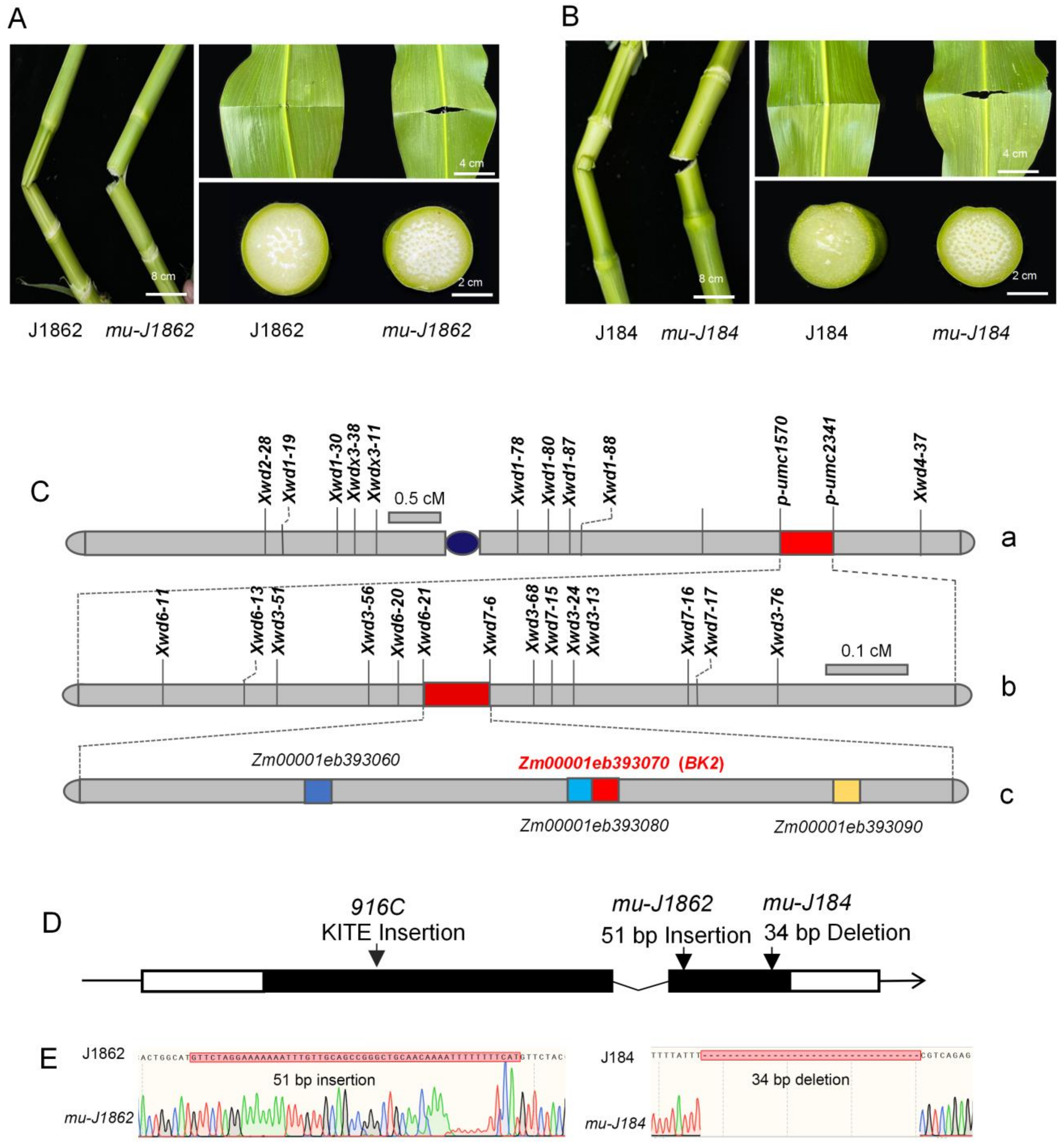
3.1. Brittle Mutant Decreased Maize Tissue Strength

Spontaneous maize mutants mu-J1862 and mu-J184 exhibited whole-plant brittleness, including the leaves, stalks, male spikes, ears, and roots. Field-grown mutants were susceptible to breakage even under mildly windy conditions. After V5, brittle mutant veins and leaves snapped and broke away cleanly, with a crisp sound and a clear cut after bending. As more stress was applied, the stalk lost its toughness and snapped as the internodes continued to bend. The wild-type stalk remained in a bent position but did not snap under constant stress (Figure 1A,B). After 24 days of pollination, we measured the residue rates of wild-type and mutant kernels, yielding 14.4% and 12.6%, respectively (Figure S3).

3.2. Genetic Analysis and Map-Based Cloning of the Brittle Gene
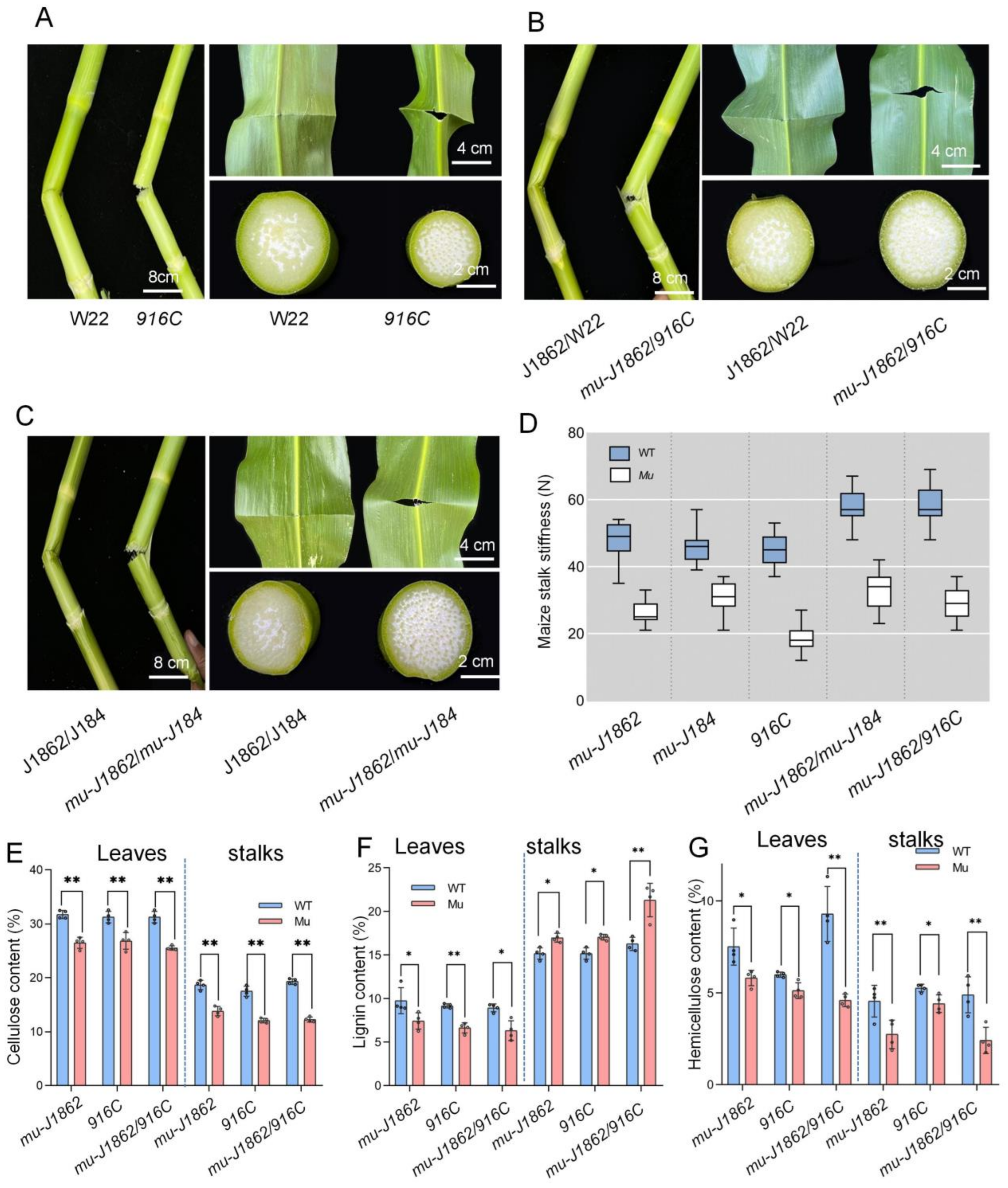
Two F2 populations derived from mu-J1862 and wild-types (B73, Huang Zaosi) were used for genetic analyses. The segregation ratio of wild-type to mutant was approximately 3:1 (Table 1), indicating that a single recessive nuclear gene controlled the brittle phenotype. Preliminary linkage analysis suggested that the brittle gene was located between SSR markers Xwd2-28 and Xwd4-37 on the long arm of chromosome 9 (Figure 1C-a). After developing 10 SSR markers located in this interval, the brittle gene was further delimited to 31.1 Mb bracketed by p-umc1570 and p-umc2341 (Figure 1C-a). Fourteen SSR markers listed in Table S3 were designed between p-umc1570 and p-umc2341 for fine mapping. Using 3607 brittle plants from the two F2 populations of 11,850 plants, the brittle gene was finally narrowed down to a 147-kb region between markers Xwd6-21 and Xwd7-6 (Figure 1C-b). Four putative genes in the 147-kb region were annotated (Figure 1C-c). Zm00001eb393060 encodes a PLC-like phosphodiesterase, Zm00001eb393070 is the ZmBK2 that has been reported to be associated with the brittleness of maize plants, and the function of Zm00001eb393080 and Zm00001eb393090 is unknown.
Previous studies on bk2 showed that the abnormal cellulose deposits in the secondary cell wall dramatically reduced tissue mechanical strength [22,24]. Our results indicate that ZmBK2 encodes a COBRA-like protein and contains two exons and one intron (Figure 1D). The primers were designed based on ZmBK2 to amplify 2.31-kb fragments from the two mutants, with a 51-bp insertion at 1357 bp in mu-J1862 and a 34-bp deletion at 1727 bp in mu-J184 (Figure 1E). Both insertions and deletions altered the amino acid sequence and protein structure of BK2. Primers BK2-J1862F/R and BK2-J184F/R were then designed based on the deletion and insertion sequences (also presented in Table S3). The 51-bp insertion and 34-bp deletion were not detected in 12 and 10 different inbred lines, respectively, indicating that the mutation sites were unique and could be used as markers to detect the brittle phenotype (Figure S4A,B). The BK2-J1862F/R marker was verified in the backcross population (mu-J1862/B73//mu-J1862). Ten mutant plants exhibited a 252-bp band consistent with mu-J1862, while the 10 wild-type plants exhibited two bands consistent with B73 and mu-J1862 (Figure S4C).

3.3. Allelic Tests

To verify whether Zm00001eb393070 was responsible for brittleness, 916C with a transposon in the first exon was used (Figure 2A). The two F1 hybrids of mu-J1862 × mu-J184 and mu-J1862 × 916C exhibited brittleness (Figure 2B,C). Based on these results, mu-J1862, mu-J184, and 916C were identified as brittle alleles.

3.4. Stalk Stiffness Test and Composition Measurements of Stalks and Leaves

Mutants (mu-J1862, mu-J184) and wild-types (J1862, J184) did not differ significantly in stalk thickness or 100-grain weight (Figure S5). Stiffness testing of the third above-ground internode in mutants was half of that of the wild-types (Figure 2D). The snapping tension of mutant and wild-type stalks was 18–32 N and 45–58 N, respectively.
We assayed stalks and leaves compositions of mu-J1862, 916C, and mu-J1862/916C. Mutant plants had significantly lower cellulose and hemicellulose content in stalks and leaves than wild-type plants. Lignin content decreased in leaves and slightly increased in stalks (Figure 2E–G).

3.5. Brittle Mutant Exhibits Abnormalities in Sclerenchyma Cells

Mutant plants exhibited brittle phenotypes and reduced mechanical strength, suggesting that the structure of their secondary cell walls may be abnormal. In mu-J1862, mu-J184, and mu-J1862/916C leaves, sclerenchyma had thinner cell walls and were fewer in number, being present at 63.9%, 65.4%, and 57.9% of their corresponding wild-type number, respectively (Figure 3A–C). This abnormal development caused insufficient leaf support and negatively affected the mechanical strength of the leaves. The lacuna of mutant stalks was more compact, and safranin O staining in the vascular bundle was darker, indicating that lignin content had increased (Figure 3D).

3.6. Constitutive Expression, Subcellular Localization and Phylogeny of ZmBK2

The results of RT-qPCR indicated that ZmBK2 was highly expressed in three-leaf stage stalks (Figure 4A). Confocal microscopy showed that the ZmBK2 protein was predominantly localized to the cytoplasm of the cell (Figure 4B).
The phylogenetic analysis via NJ algorithm showed that the most homologous gene of ZmBK2 was AtCOBL4, which belongs to the COBRA family (Figure 4C).

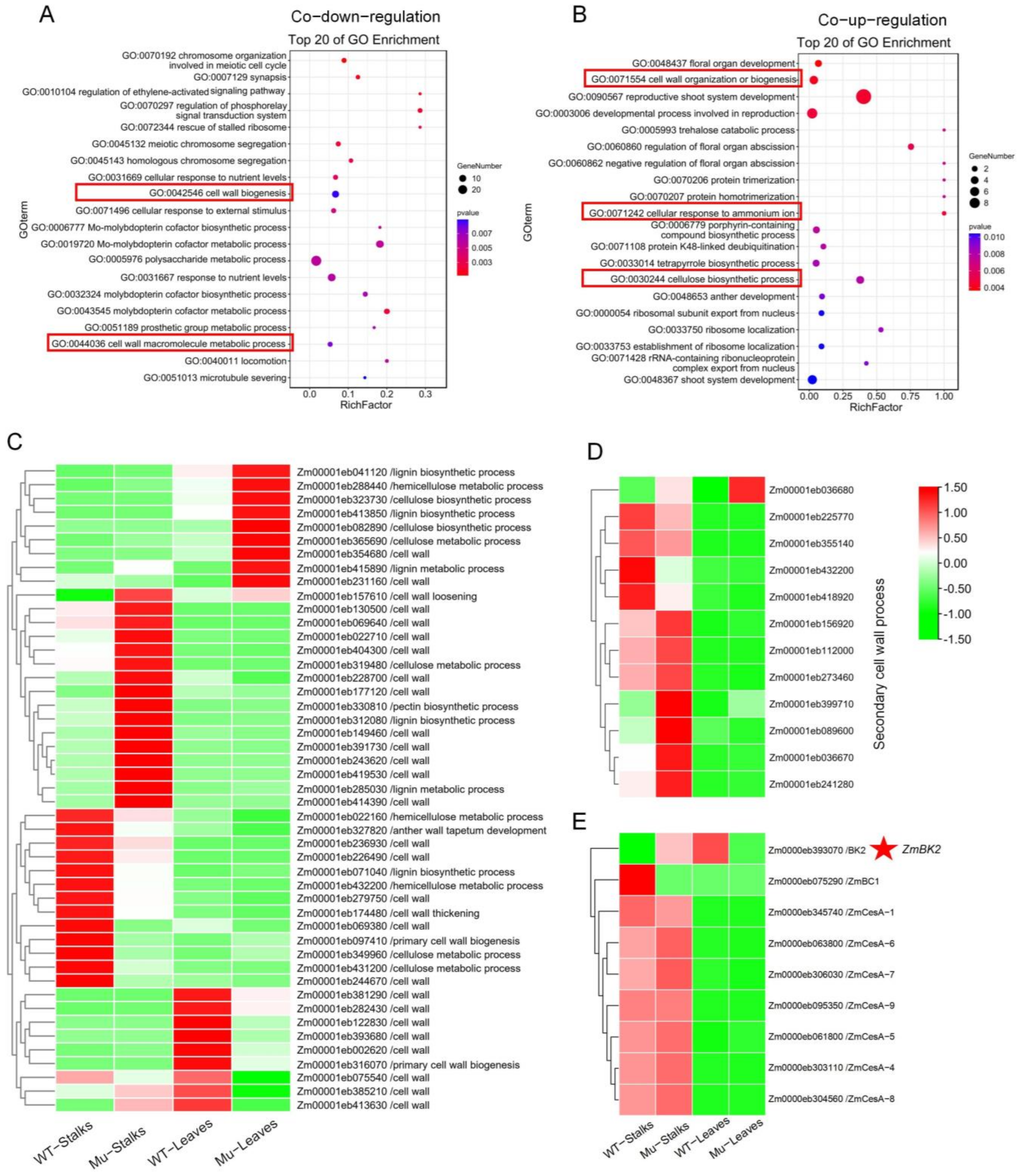
3.7. Transcriptome Alterations in mu-J1862 Stalks and Leaves

To examine the genome-wide effects of ZmBK2, we used RNA-seq to compare the transcriptome profiles of mu-J1862 and J1862. A total of 78.62 Gb original data was obtained from cDNA libraries. Approximately 79% of clean reads were unique and mapped to the maize B73 genome (Table S4).
In mutant stalks and leaves, 1694 and 1431 genes were up-regulated, and 2044 and 1729 genes were down-regulated (Figure 5A,B). Down-regulated genes in mutant stalks were mainly enriched in secondary metabolic processes, phenol-containing compound metabolic processes, metabolic processes, and phenylpropanoid biosynthesis (Figure S6A). Down-regulated genes in mutant leaves were enriched in secondary metabolite biosynthesis and cyanoamino acid metabolism (Figure S6C). Up-regulated genes in stalks were mainly enriched in riboflavin metabolism, phenylpropanoid metabolism, and secondary metabolic processes (Figure S6B). Up-regulated genes in leaves were enriched in secondary metabolite biosynthesis, metabolic pathways, and phenylpropanoid metabolism (Figure S6D). Up-regulated and down-regulated genes in stalks and leaves were co-enriched in secondary regulatory and phenylpropanoid metabolic pathways. Products of secondary regulatory pathways were essential for cell wall synthesis, and phenylalanine was the starting point for cellulose synthesis in the phenylpropanoid metabolic pathway. These results suggest that ZmBK2 may influence the expression of cell wall-related genes and that ZmBK2 may work together to regulate the development of plant cell walls.
We analyzed co-up-regulated and co-down-regulated genes in both stalks and leaves. Compared with wild-types, 376 genes were co-down-regulated and 276 genes were co-up-regulated in mutant leaves and stalks (Figure 5C). Co-down-regulated genes were mainly enriched in molybdopterin cofactor biosynthetic processes (GO:0019720), polysaccharide metabolic processes (GO:0005976), cell wall biogenesis (GO:0042546), and cell wall macromolecule metabolic processes (GO:0044036) (Figure 6A), while co-up-regulated genes were mainly enriched in reproductive shoot system development (GO:0090567), developmental processes involved in reproduction (GO:0003006), cell wall organization or biogenesis (GO:0071554), cellular response to ammonium ions (GO:0071242), and cellulose biosynthetic processes (GO:0030244) (Figure 6B). A large number of genes related to cell wall synthesis were enriched, which was consistent with abnormalities in mutant cell walls.
We discovered a substantial number of DEGs that were involved in cell wall synthesis in terms of starch and sucrose degradation and in processes connected to lignin biosynthesis, cellulose biosynthesis, cellulose metabolism, and primary cell walls (Figure 6C). With the exception of Zm00001eb036680, all genes associated with secondary cell wall synthesis were specifically expressed in stalks, and the majority were up-regulated in mutant plants (Figure 6D). Additionally, the majority of ZmBK2 homologous genes expressed in stalks and leaves were cellulose synthase genes, with higher expression levels in stalks (Figure 6E). Genes involved in cell wall development were differentially expressed, suggesting that ZmBK2 played a critical role in cell wall formation.

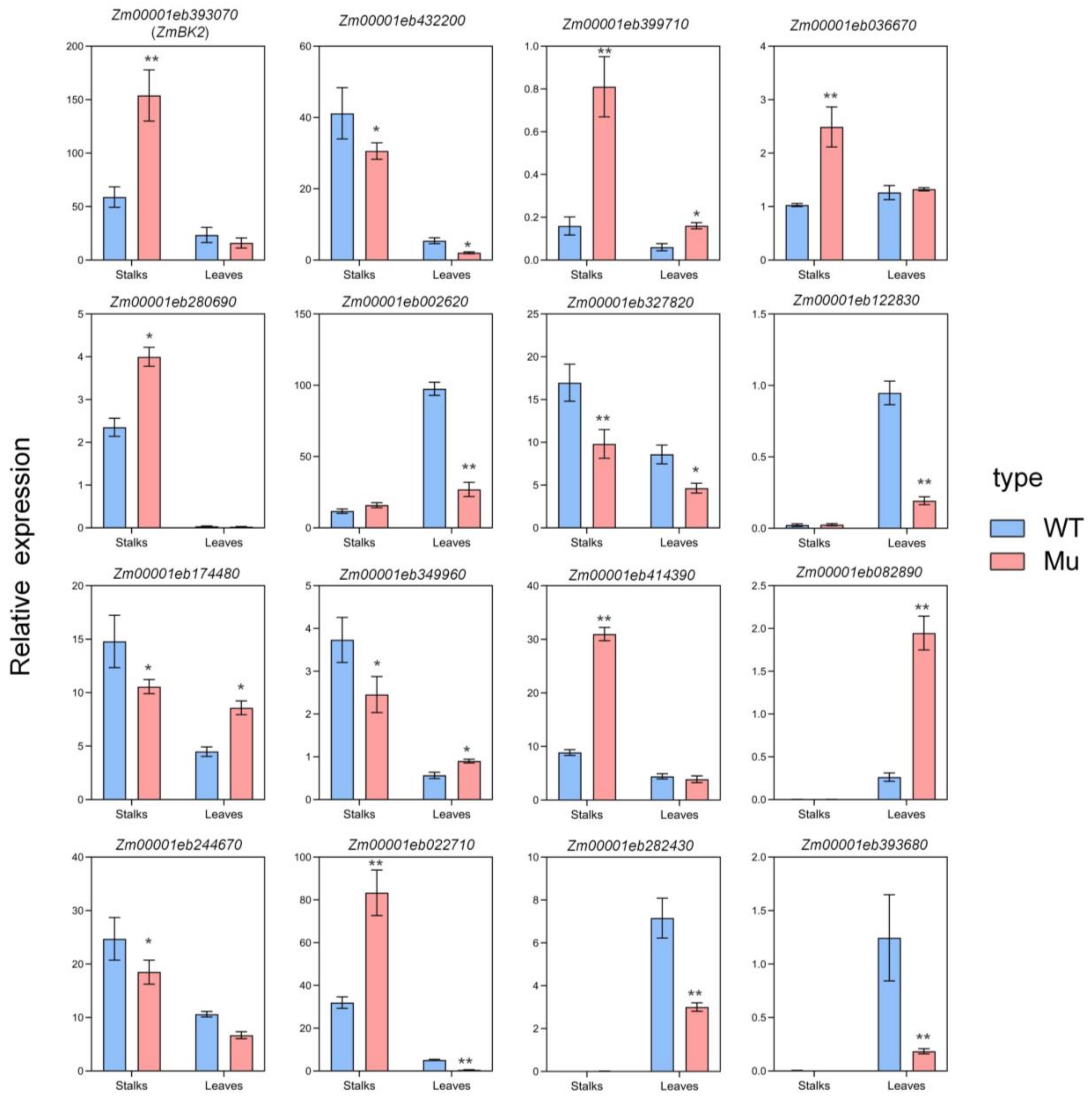
3.8. Validation of RNA-seq

We selected DEGs involved in cell wall formation for RT-qPCR validation. These included secondary cell wall synthesis genes, glycosyl hydrolase genes, and cellulase genes (Figure 7). Genes for RT-qPCR expression in the RNA-seq are shown in Table S5. RT-qPCR results also revealed that the expression levels of the selected genes were consistent with those determined by RNA-seq.

4. Discussion

4.1. Abnormal Cellulose Synthesis Affects Plant Brittleness

Cellulose has a very simple chemical nature, although its assembly may appear complex due to the presence of multi-scale cellulose fibers [32]. Crystalline cellulose elemental fibers composed of glucan chains are bundled into nanofibers, which are then aggregated into cellulosic fibrils. These multiscale cord-like structures constitute a fibrillar network that forms part of the cell wall nanostructure. Therefore, cellulose is a load-bearing polymer of the plant cell wall and can improve the mechanical strength of stalks [33,34]. Many brittle mutants have been identified to date, such as irregular xylem (irx1, irx2, irx3, and irx5) in Arabidopsis and brittle culm (bc3, bc5, bc6, bc7, bc10, bc11, bc12, bc13, bc14, bc15, bc17, bc18, and bc88) in rice [7,10,35,36]. The biochemical and molecular characteristics of these mutants suggest that the brittle phenotype is caused by defects in cellulose synthesis [37]. Cellulose synthase catalytic subunit genes are crucial proteins that directly catalyze glucan chain elongation at the plasma membrane (PM). The COBRA-like protein BC1 regulates cellulose crystallite size and acts as an auxiliary protein to stack nascent glucan chains into a crystalline structure, which is a crucial step in the assembly of cellulose [2,38]. Our RNA-seq results revealed altered CESA expression, demonstrating that catalysis of glucan chain elongation in these subunits is inhibited, which may thus not be normally stacked into a crystalline structure. Therefore, ZmBK2 may not modulate cellulose assembly by interacting specifically with crystalline cellulose, as the cellulose content was substantially reduced and stalk strength was decreased.

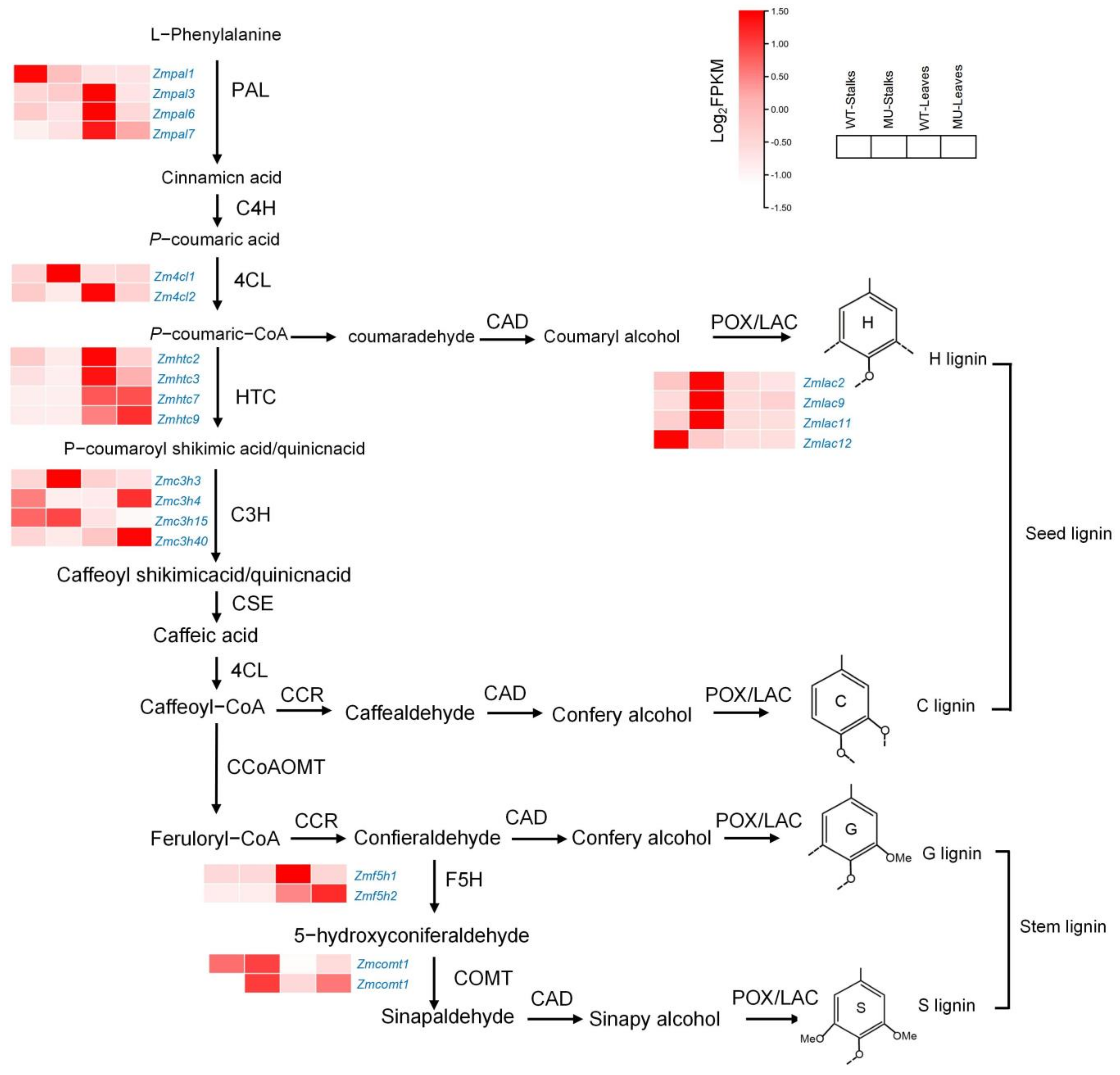
4.2. Lignin Synthesis Compensates for Cellulose Deficiency in Mutants

Recent studies have shown that 10 specific enzymes are required for lignin biosynthesis: phenylalanine ammonia-lyase (PAL), cinnamic acid 4-hydroxylase (C4H), 4-coumarate CoA ligase (4CL), cinnamyl alcohol dehydrogenase (CAD), p-hydroxycinnamoyl-CoA (HCT), p-coumarate 3-hydroxylase (C3H), caffeoyl-CoA O-methyltransferase (CCoAOMT), hydroxycinnamoyl-CoA reductase (CCR), ferulate 5-hydroxylase (F5H), and caffeic acid/5-hydroxyferulic acid O-methyltransferase (COMT) [39,40]. Phenylalanine ammonia-lyase is a key enzyme that connects primary and phenylalanine metabolism, catalyzes L-phenylalanine deamination to trans-cinnamic acid, and then converts it to p-coumaric acid through C4H [41]. Zmpal, Zm4cl, Zmhct, Zmc3h, Zmf5h, and Zmcomt are differentially expressed genes that may affect the lignin synthesis process (Figure 8) [42]. Similar to rice bc7, bc10, bc11, and sorghum bc1 [43], bk2 decreased cellulose content and slightly increased lignin content in stalks. Through trans-membrane sensor proteins, SBbc1 senses alterations to the cell wall structure and mechanical properties; in response, these proteins trigger compensatory mechanisms via activating TFs [44]. The compensatory mechanism of rice bc7 balances the decrease in cellulose content with an increase in hemicellulose, lignin, and silicon content. This dynamic may contribute to the restoration of normal cell wall integrity and maintaining plant growth [45]. Similarly, bk2 may have a compensatory mechanism in cell wall formation for cellulose deficiency. The synthesis of cellulose and lignin is a dynamically balanced process that involves many key enzymes in metabolism. Defects in any of these key enzymes may affect the abnormal synthesis of cellulose and lignin, ultimately affecting plant brittleness [46,47,48].

4.3. ZmBK2 Is a Member of the COBRA Family

Plant COBRA genes encode GPI-anchored proteins involved in biological processes related to cell wall biosynthesis and cell expansion [16]. The AtCOBL4 mutant is characterized by defective cellulose synthesis and decreased cellulose content in secondary cell walls [49]. As putative AtCOBL4 orthologs, rice bc1 and maize bk2 exhibit disrupted secondary cell wall biosynthesis [24]. Rice bc1 encodes a GPI-anchored protein and disrupts post-translational processing [38]. A 51-bp insertion in mu-J1862 and a 34-bp deletion in mu-J184 produced truncated proteins without the GPI attachment site. ZmBK2 may not properly encode COBRA-like proteins that influence secondary cell wall structure by blocking fibrillin deposition, which decreases cell wall thickness and cellulose content [24]. The bk2 mutant is derived from a transposon insertion [22], while mu-J1862 and mu-J184 are two novel spontaneous mutants that enrich the mutant type of the COBRA gene family and provide richer material for studying cell wall synthesis.

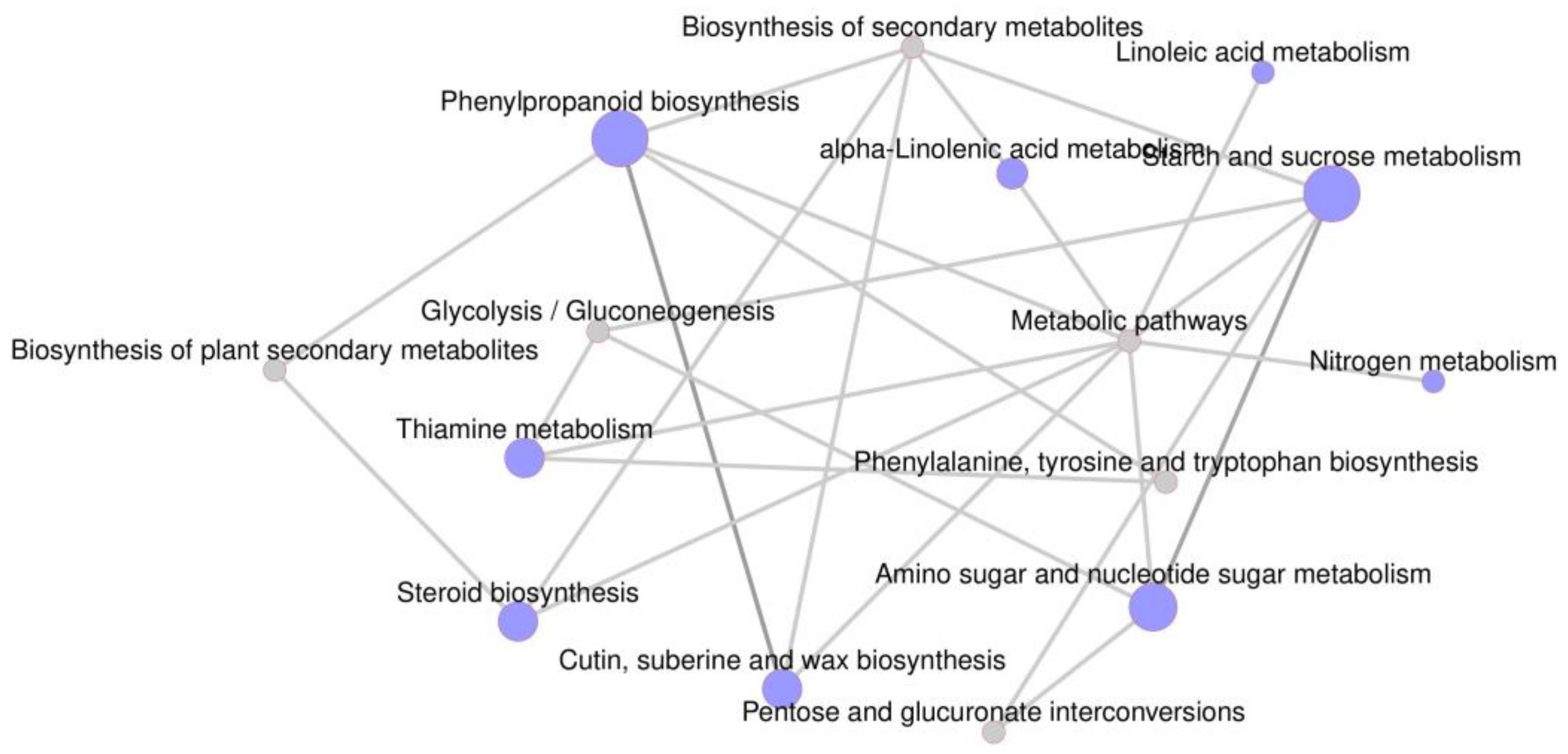
4.4. Regulatory Network for Cell Wall Synthesis

Plant cell walls are dynamic and complex networks that require strict coordination among cellulose, lignin, xylan, and other components to execute biosynthetic programs and complete proper cell wall assembly [50]. Specific TFs and other proteins are implicated in coordinating cell wall biosynthesis [51]. In this study, the important DEGs were used to build a regulatory network for cell wall synthesis, and wall-associated kinases (WAK) were at the center of the regulatory network (Figure 9). These protein kinases attach to the cell wall and are important for cell wall plasticity [52,53,54]. However, they are differentially expressed in multiple tissues; WAKs were implicated in cell wall signaling, gene regulation, and pathogen response [55,56]. Three WAK genes (Zm00001eb139070, Zm00001eb071980, and Zm00001eb335510) were down-regulated and may work with ZmBK2 to regulate cell wall extension and reduce cell wall thickness.
Recent Arabidopsis research has contributed to a thorough analysis of TFs involved in cell wall regulation, including subgroup members of the NAC, MYB, and SND [57,58,59]. The NAC TFs function as master switches for secondary cell wall biosynthesis and can activate downstream TFs such as MYB20, MYB42, MYB46, MYB83, MYB103, SND2, SND3, and KNAT7 [60,61]. Some MYB TFs act as second- or third-level master regulators to control the expression of secondary cell wall-related genes [62,63]. For example, MYB46 in Arabidopsis is a major switch for secondary cell wall formation and directly regulates the cellulose synthase genes: ATCESA4, ATCESA7, and ATCESA8 [64]. MYB46 and MYB83 inhibit the thinning of secondary cell walls by regulating the expression of SND1 and its homologs NST1, NST2, VND6, and VND7 [65]. The analysis of DEGs in stalks and leaves by transcriptome showed that 362 TFs were differentially expressed (Figure S7), and the majority belonged to the bHLH, C2H2, ERF, MYB, NAC, and WRKY gene families (Table S6). Twenty-four TFs, including two NAC genes and four MYB genes were co-down-regulated in stalks and leaves. Among these, NAC35 and NAC110 are likely to be master switches for cell wall biosynthesis to inhibit multiple TFs (MYB56, MYB59, MYB69, and MYB74). Cellulose synthesis-related genes (CESA1, CESA5, and CESA9) were down-regulated. Cellulose synthesis and assembly may be abnormal, which ultimately affects the synthesis of secondary cell walls. Similarly, bHLH152 and bHLH148 are activated as a class of TFs to participate in lignin synthesis.
In the cell wall regulatory network, multiple proteins act synergistically in signal transduction and the regulation of gene expression. The potential interacting proteins of ZmBK2 were analyzed using an online tool, STRING V11, and eight genes were predicted: Zm00001eb153340, Zm00001eb317090, Zm00001eb123640, Zm00001eb295810, Zm00001eb307070, Zm00001eb416400, Zm00001eb023530, and Zm00001eb421850 (Figure 10). Zm00001eb153340, Zm00001eb295810, Zm00001eb416400, and Zm00001eb307070 are closely related to cell wall synthesis and encode the catalytic subunits of cellulose synthase, NAC-type TFs, MYB DNA-binding domain superfamily proteins, and WAKs, respectively. These four genes function in the cell wall regulatory network and were differentially expressed in J1862 and mu-J1862 (Table S7). ZmBK2 may interact with them to participate in the expression of other genes that are regulated by TFs in the cell wall. Thus, the plant cell wall is regulated by a variety of physiological and biochemical activities, the regulation mechanisms of which still remain unclear.

4.5. ZmBK2 Can Potentially Reduce Residue Rate in Fresh Maize

Fresh maize is becoming an increasingly popular foodstuff for its desirable taste and unique qualities, including thin skin, low residue, moderate waxiness, and sweetness [66]. The bk2 strain has low cellulose content and thus great potential to reduce the residue rate. We measured the residue rate of wild-types and mutants using kernels after 24 days of pollination. The results were as expected: the residue rate of the wild-type was 14.4%, while the mutant had only 12.6% residues. ZmBK2 can therefore be considered a potentially valuable gene resource for a decrease in residue rate. By controlling ZmBK2 expression in different parts of the maize plant via genetic engineering, it may be possible to design good-tasting and lodging-resistant maize varieties.

5. Conclusions

This study identified a maize mutant with whole-plant brittleness and low cellulose content, leading to reduced lodging resistance. These characteristics were attributable to abnormal cell wall development. Through map cloning and allelic tests, we confirmed that Zm00001eb393070 encodes a COBRA-like protein and is a target gene for plant brittleness. Transcriptome analysis identified multiple genes related to cell wall development that were used to construct a regulatory network for cell wall synthesis. Our study thus provides a theoretical basis for the mechanisms underlying cell wall development.

Supplementary Materials

The following supporting information can be downloaded at: https://www.mdpi.com/article/10.3390/genes14061126/s1, Figure S1: Electrophoresis image of 12 RNA sample bands. Figure S2: Melting Curve analysis 12 pairs of primers. Figure S3: Determination of residue rate in bk2 and wild-types 24 days after pollination. Figure S4: BK2-J1862F/R and BK2-J184F/R were used as functional markers to detect the brittle phenotype. (A) BK2-J1862F/R was used to distinguish mu-J1862 and different inbred lines; (B) BK2-J184F/R was used to distinguish mu-J184 and different inbred lines; (C) Verification of BK2-J1862F/R marker in (mu-J1862/B73)/mu-J1862 backcross population. Figure S5: Mutant stalk thickness and 100-grain weight. Figure S6: Enrichment analysis of up-regulated and down-regulated genes in mutant leaves and stalks. Figure S7: Venn diagram of total TFs (Padj < 0.01) among genotypes. Table S1: RIN numbers of RNA samples. Table S2: Molecular markers for map-based cloning. Table S3: RT-qPCR primer information. Table S4: The detailed information of raw reads from different samples. Table S5: Homologous genes expression of ZmBK2. Table S6: Enrichment analysis of TFs. Table S7: Expression of reciprocal genes predicted using string.

Author Contributions

B.L. conceived and designed the experiments. W.X., Y.Z. (Yan Zhao), Q.L. and Y.D. performed the experiments. E.J. provided the mu-J1862 mutant. W.X. and Q.L. analyzed the data. Q.W. is responsible for data curation. W.X. wrote the article. Y.Z. (Yongzhong Zhang), Q.L., Y.D., J.Y. and Q.W. revised the article. All authors have read and agreed to the published version of the manuscript.

Funding

This work was supported by the Funds of Shandong improved variety Program (2022LZGC006), Breeding of New Maize Varieties with high yield, high quality and multi resistance and Germplasm Innovation (2021TSGC1210).

Institutional Review Board Statement

Not applicable.

Informed Consent Statement

Not applicable.

Data Availability Statement

Data are openly available in a public repository. The raw data of transcriptome sequencing in this study are provided at the NCBI short read archive (accession number: PRJNA896366).

Acknowledgments

We extend our appreciation to the anonymous reviewers for their valuable suggestions to help improve this article.

Conflicts of Interest

The authors declare no conflict of interest.

References

  1. Flint-Garcia, S.A.; Jampatong, C.; Darrah, L.L.; McMullen, M.D. Quantitative trait locus analysis of stalk strength in four maize populations. Crop Sci. 2003, 43, 13–22. [Google Scholar] [CrossRef]
  2. Li, Y.; Qian, Q.; Zhou, Y.; Yan, M.; Sun, L.; Zhang, M.; Fu, Z.; Wang, Y.; Han, B.; Pang, X.; et al. BRITTLE CULM1, which encodes a COBRA-like protein, affects the mechanical properties of rice plants. Plant Cell 2003, 15, 2020–2031. [Google Scholar] [CrossRef] [PubMed]
  3. Liu, H.; Wang, H.; Shao, C.; Han, Y.; He, Y.; Yin, Z. Genetic Architecture of Maize Stalk Diameter and Rind Penetrometer Resistance in a Recombinant Inbred Line Population. Genes 2022, 13, 579. [Google Scholar] [CrossRef] [PubMed]
  4. Keegstra, K. Plant cell walls. Plant Physiol. 2010, 154, 483–486. [Google Scholar] [CrossRef]
  5. Desprez, T.; Juraniec, M.; Crowell, E.F.; Jouy, H.; Pochylova, Z.; Parcy, F.; Höfte, H.; Gonneau, M.; Vernhettes, S. Organization of cellulose synthase complexes involved in primary cell wall synthesis in Arabidopsis thaliana. Proc. Natl. Acad. Sci. USA 2007, 104, 15572–15577. [Google Scholar] [CrossRef] [PubMed]
  6. Brown, D.M.; Zeef, L.A.H.; Ellis, J.; Goodacre, R.; Turner, S.R. Identification of novel genes in Arabidopsis involved in secondary cell wall formation using expression profiling and reverse genetics. Plant Cell 2005, 17, 2281–2295. [Google Scholar] [CrossRef] [PubMed]
  7. Tanaka, K.; Murata, K.; Yamazaki, M.; Onosato, K.; Miyao, A.; Hirochika, H. Three distinct rice cellulose synthase catalytic subunit genes required for cellulose synthesis in the secondary wall. Plant Physiol. 2003, 133, 73–83. [Google Scholar] [CrossRef]
  8. Atanassov, I.I.; Pittman, J.K.; Turner, S.R. Elucidating the Mechanisms of Assembly and Subunit Interaction of the Cellulose Synthase Complex of Arabidopsis Secondary Cell Walls. J. Biol. Chem. 2009, 284, 3833–3841. [Google Scholar] [CrossRef]
  9. Appenzeller, L.; Doblin, M.; Barreiro, R.; Wang, H.; Niu, X.; Kollipara, K.; Carrigan, L.; Tomes, D.; Chapman, M.; Dhugga, K.S. Cellulose synthesis in maize: Isolation and expression analysis of the cellulose synthase (CesA) gene family. Cellulose 2004, 11, 287–299. [Google Scholar] [CrossRef]
  10. Taylor, N.G.; Laurie, S.; Turner, S.R. Multiple cellulose synthase catalytic subunits are required for cellulose synthesis in Arabidopsis. Plant Cell 2000, 12, 2529–2539. [Google Scholar] [CrossRef]
  11. Hu, H.; Zhang, R.; Feng, S.; Wang, Y.; Wang, Y.; Fan, C.; Li, Y.; Liu, Z.; Schneider, R.; Xia, T.; et al. Three AtCesA6-like members enhance biomass production by distinctively promoting cell growth in Arabidopsis. Plant Biotechnol. J. 2018, 16, 976–988. [Google Scholar] [CrossRef]
  12. Zhang, B.; Gao, Y.; Zhang, L.; Zhou, Y. The Plant Cell Wall: Biosynthesis, construction, and functions. Integr. Plant Biol. 2020, 63, 251–272. [Google Scholar] [CrossRef]
  13. Zhang, B.; Deng, L.; Qian, Q.; Xiong, G.; Zeng, D.; Li, R.; Guo, L.; Li, J.; Zhou, Y. A missense mutation in the transmembrane domain of CESA4 affects protein abundance in the plasma membrane and results in abnormal cell wall biosynthesis in rice. Plant Mol. Biol. 2009, 71, 509–524. [Google Scholar] [CrossRef]
  14. Wang, L.; Guo, K.; Li, Y.; Tu, Y.; Hu, H.; Wang, B.; Cui, X.; Peng, L. Expression profiling and integrative analysis of the CESA/CSL superfamily in rice. BMC Plant Biol. 2010, 10, 282. [Google Scholar] [CrossRef]
  15. Holland, N.; Holland, D.; Helentjaris, T.; Dhugga, K.S.; Cazares, B.X.; Delmer, D.P. A comparative analysis of the plant cellulose synthase (CesA) gene family. Plant Physiol. 2000, 123, 1313–1324. [Google Scholar] [CrossRef]
  16. Roudier, F.; Schindelman, G.; DeSalle, R.; Benfey, P.N. The COBRA family of putative GPI-anchored proteins in Arabidopsis. A new fellowship in expansion. Plant Physiol. 2002, 130, 538–548. [Google Scholar] [CrossRef]
  17. Schindelman, G.; Morikami, A.; Jung, J.; Baskin, T.I.; Carpita, N.C.; Derbyshire, P.; McCann, M.C.; Benfey, P.N. COBRA encodes a putative GPI-anchored protein, which is polarly localized and necessary for oriented cell expansion in Arabidopsis. Genes Dev. 2001, 15, 1115–1127. [Google Scholar] [CrossRef]
  18. Pierleoni, A.; Martelli, P.L.; Casadio, R. PredGPI: A GPI-anchor predictor. BMC Bioinform. 2008, 9, 392. [Google Scholar] [CrossRef]
  19. Sorek, N.; Sorek, H.; Kijac, A.; Szemenyei, H.J.; Bauer, S.; Hématy, K.; Wemmer, D.E.; Somerville, C.R. The Arabidopsis COBRA protein facilitates cellulose crystallization at the plasma membrane. J. Biol. Chem. 2014, 289, 34911–34920. [Google Scholar] [CrossRef]
  20. Liu, L.; Shang-Guan, K.; Zhang, B.; Liu, X.; Yan, M.; Zhang, L.; Shi, Y.; Zhang, M.; Qian, Q.; Li, J.; et al. Brittle Culm1, a COBRA-like protein, functions in cellulose assembly through binding cellulose microfibrils. PLoS Genet. 2013, 9, e1003704. [Google Scholar] [CrossRef]
  21. Jiao, S.; Hazebroek, J.P.; Chamberlin, M.A.; Perkins, M.; Sandhu, A.S.; Gupta, R.; Simcox, K.D.; Li, Y.; Prall, A.; Heetland, L.; et al. Chitinase-like1 Plays a Role in Stalk Tensile Strength in Maize. Plant Physiol. 2019, 181, 1127–1147. [Google Scholar] [CrossRef] [PubMed]
  22. Ching, A.; Dhugga, K.S.; Appenzeller, L.; Meeley, R.; Bourett, T.M.; Howard, R.J.; Rafalski, A. Brittle stalk 2 encodes a putative glycosylphosphatidylinositol-anchored protein that affects mechanical strength of maize tissues by altering the composition and structure of secondary cell walls. Planta 2006, 224, 1174–1184. [Google Scholar] [CrossRef]
  23. Li, Q.; Nie, S.; Li, G.; Du, R.; Yang, X.; Liu, B.; Gao, X.; Liu, T.; Zhang, Z.; Zhao, X.; et al. Identification and Fine Mapping of the Recessive Gene BK-5, Which Affects Cell Wall Biosynthesis and Plant Brittleness in Maize. Int. J. Mol. Sci. 2022, 23, 814. [Google Scholar] [CrossRef] [PubMed]
  24. Sindhu, A.; Langewisch, T.; Olek, A.; Multani, D.S.; McCann, M.C.; Vermerris, W.; Carpita, N.C.; Johal, G. Maize Brittle stalk2 encodes a COBRA-like protein expressed in early organ development but required for tissue flexibility at maturity. Plant Physiol. 2007, 145, 1444–1459. [Google Scholar] [CrossRef] [PubMed]
  25. Sluiter, A.; Hames, B.; Ruiz, R.; Scarlata, C.; Sluiter, J.; Templeton, D.; Crocker, D. Determination of Structural Carbohydrates and Lignin in Biomass. NREL Laboratory Analytical Procedure, Issue Date: April 2008, Revision Date: 08-3-2012.
  26. Jin, X.; Chen, X.; Shi, C.; Li, M.; Guan, Y.; Yu, C.Y.; Yamada, T.; Sacks, E.J.; Peng, J. Determination of hemicellulose, cellulose and lignin content using visible and near infrared spectroscopy in Miscanthus sinensis. Bioresour. Technol. 2017, 241, 603–609. [Google Scholar] [CrossRef] [PubMed]
  27. Kim, D.; Langmead, B.; Salzberg, S.L. HISAT: A fast spliced aligner with low memory requirements. Nat. Methods. 2015, 12, 357–360. [Google Scholar] [CrossRef]
  28. Love, M.I.; Huber, W.; Anders, S. Moderated estimation of fold change and dispersion for RNA-seq data with deseq2 moderated estimation of fold change and dispersion for RNA-seq data with DESeq2. Genome Biol. 2014, 15, 550. [Google Scholar] [CrossRef]
  29. Young, M.D.; Wakefield, M.J.; Smyth, G.K.; Oshlack, A. Gene ontology analysis for RNA-seq: Accounting for selection bias. Genome Biol. 2010, 11, R14. [Google Scholar] [CrossRef]
  30. Livak, K.J.; Schmittgen, T.D. Analysis of relative gene expression data using real-time quantitative PCR and the 2(-Delta Delta C(T)) Method. Methods 2001, 25, 402–408. [Google Scholar] [CrossRef]
  31. Szklarczyk, D.; Gable, A.L.; Lyon, D.; Junge, A.; Wyder, S.; Huerta-Cepas, J.; Simonovic, M.; Doncheva, N.; Morris, J.H.; Bork, P.; et al. STRING v11: Protein-protein association networks with increased coverage, supporting functional discovery in genome-wide experimental datasets. Nucleic Acids Res. 2019, 47, D607–D613. [Google Scholar] [CrossRef]
  32. Ding, S.Y.; Liu, Y.S.; Zeng, Y.; Himmel, M.E.; Baker, J.O.; Bayer, E.A. How does plant cell wall nanoscale architecture correlate with enzymatic digestibility? Science 2012, 338, 1055–1060. [Google Scholar] [CrossRef]
  33. Hu, H.; Zhang, R.; Tao, Z.; Li, X.; Li, Y.; Huang, J.; Li, X.; Han, X.; Feng, S.; Zhang, G.; et al. Cellulose Synthase Mutants Distinctively Affect Cell Growth and Cell Wall Integrity for Plant Biomass Production in Arabidopsis. Plant Cell Physiol. 2018, 59, 1144–1157. [Google Scholar] [CrossRef]
  34. Watanabe, Y.; Meents, M.J.; Mcdonnell, L.M.; Barkwill, S.; Sampathkumar, A.; Cartwright, H.N.; Demura, T.; Ehrhardt, D.W.; Samuels, A.L.; Mansfield, S.D. Visualization of cellulose synthases in Arabidopsis secondary cell walls. Science 2015, 350, 198–203. [Google Scholar] [CrossRef]
  35. Desprez, T.; Vernhettes, S.; Fagard, M.; Refrégier, G.; Desnos, T.; Aletti, E.; Pelletier, N.P.S.; Höfte, H. Resistance against Herbicide Isoxaben and Cellulose Deficiency Caused by Distinct Mutations in Same Cellulose Synthase Isoform CESA6. Plant Physiol. 2002, 128, 482–490. [Google Scholar] [CrossRef]
  36. Fagard, M.; Desnos, T.; Desprez, T.; Goubet, F.; Refregier, G.; Mouille, G.; McCann, M.; Rayon, C.; Vernhettes, S.; Höfte, H. PROCUSTE1 Encodes a Cellulose Synthase Required for Normal Cell Elongation Specifically in Roots and Dark-Grown Hypocotyls of Arabidopsis. Plant Cell 2000, 12, 2409–2423. [Google Scholar] [CrossRef]
  37. Taylor, N.G.; Howells, R.M.; Huttly, A.K.; Vickers, K.; Turner, S.R. Interactions among three distinct CesA proteins essential for cellulose synthesis. Proc. Natl. Acad. Sci. USA 2003, 100, 1450–1455. [Google Scholar] [CrossRef]
  38. Sato, K.; Suzuki, R.; Nishikubo, N.; Takenouchi, S.; Ito, S.; Nakano, Y.; Nakaba, S.; Sano, Y.; Funada, R.; Kajita, S.; et al. Isolation of a novel cell wall architecture mutant of rice with defective Arabidopsis COBL4 ortholog BC1 required for regulated deposition of secondary cell wall components. Planta 2010, 232, 257–270. [Google Scholar] [CrossRef]
  39. Vanholme, R.; Demedts, B.; Morreel, K. Lignin biosynthesis and structure. Plant Biol. 2010, 15, 895–905. [Google Scholar] [CrossRef]
  40. Doorsselaere, J.V.; Baucher, M.; Chognot, E.; Chabbert, B.; Tollier, M.T.; Petit-Conil, M.; Leplé, J.C.; Pilate, G.; Cornu, D.; Monties, B. A novel lignin in poplar trees with a reduced caffeic acid/5 hydroxyferulic acid O-methyltransferase activity. Plant J. 2003, 8, 855–864. [Google Scholar] [CrossRef]
  41. Goujon, T.; Sibout, R.; Eudes, A.; MacKay, J.; Joulanin, L. Genes involved in the biosynthesis of lignin precursors in Arabidopsis thaliana. Plant Physiol. Biochem. 2003, 41, 677–687. [Google Scholar] [CrossRef]
  42. Fang, L.; Xu, X.; Li, J.; Zheng, F.; Li, M.; Yan, J.; Li, Y.; Zhang, X.; Li, L.; Ma, G.; et al. Transcriptome analysis provides insights into the non-methylated lignin synthesis in Paphiopedilum armeniacum seed. BMC Genom. 2020, 21, 524. [Google Scholar] [CrossRef] [PubMed]
  43. Zhang, M.; Wei, F.; Guo, K.; Hu, Z.; Li, Y.; Xie, G.; Wang, Y.; Cai, X.; Peng, L.; Wang, L. A novel FC116/BC10 mutation distinctively causes alteration in the expression of the genes for cell wall polymer synthesis in rice. Front. Plant Sci. 2016, 7, 1366. [Google Scholar] [CrossRef] [PubMed]
  44. Li, P.; Liu, Y.; Tan, W.; Chen, J.; Zhu, M.; Lv, Y.; Liu, Y.; Yu, S.; Zhang, W.; Cai, H. Brittle Culm 1 encodes a COBRA-like protein involved in secondary cell wall cellulose biosynthesis in sorghum. Plant Cell Physiol. 2019, 60, 788–801. [Google Scholar] [CrossRef] [PubMed]
  45. Wei, C.; Xie, P.; Chen, Y.; Chen, Y.; Yu, H.; Su, Y.; Gu, M.; Yan, C. Anatomical and chemical characteristics of culm of rice brittle mutant bc7 (t). Funct. Plant Biol. 2011, 38, 227–235. [Google Scholar] [CrossRef] [PubMed]
  46. Mewalal, R.; Mizrachi, E.; Mansfield, S.D.; Myburg, A.A. Cell wall-related proteins of unknown function: Missing links in plant cell wall development. Plant Cell Physiol. 2014, 55, 1031–1043. [Google Scholar] [CrossRef]
  47. Aohara, T.; Kotake, T.; Kaneko, Y.; Takatsuji, H.; Tsumuraya, Y.; Kawasaki, S. Rice BRITTLE CULM 5 (BRITTLE NODE) is involved in secondary cell wall formation in the sclerenchyma tissue of nodes. Plant Cell Physiol. 2009, 50, 1886–1897. [Google Scholar] [CrossRef]
  48. Hirano, K.; Kotake, T.; Kamihara, K.; Tsuna, K.; Aohara, T.; Kaneko, Y.; Takatsuji, H.; Tsumuraya, Y.; Kawasaki, S. Rice BRITTLE CULM 3 (BC3) encodes a classical dynamin OsDRP2B essential for proper secondary cell wall synthesis. Planta 2010, 232, 95–108. [Google Scholar] [CrossRef]
  49. Niu, E.; Fang, S.; Shang, X.; Guo, W. Ectopic expression of GhCOBL9A, a cotton glycosyl-phosphatidyl inositol-anchored protein encoding gene, promotes cell elongation, thickening and increased plant biomass in transgenic Arabidopsis. Mol. Genet. Genom. 2018, 293, 1191–1204. [Google Scholar] [CrossRef]
  50. Du, J.; Anderson, C.T.; Xiao, C. Dynamics of pectic homogalacturonan in cellular morphogenesis and adhesion, wall integrity sensing and plant development. Nat. Plants 2022, 8, 332–340. [Google Scholar] [CrossRef]
  51. Zhong, R.; Lee, C.; Zhou, J.; McCarthy, R.L.; Ye, Z.H. A battery of transcription factors involved in the regulation of secondary cell wall biosynthesis in Arabidopsis. Plant Cell 2008, 20, 2763–2782. [Google Scholar] [CrossRef]
  52. Kohorn, B.D. WAKs; cell wall associated kinases. Curr. Opin. Cell. Biol. 2001, 13, 529–533. [Google Scholar] [CrossRef]
  53. Lally, D.; Ingmire, P.; Tong, H.Y.; He, Z.H. Antisense expression of a cell wall-associated protein kinase WAK4, inhibits cell elongation and alters morphology. Plant Cell 2001, 13, 1317–1331. [Google Scholar]
  54. Park, A.R.; Cho, S.K.; Yun, U.J.; Jin, M.Y.; Lee, S.H.; Sachetto-Martins, G.; Park, O.K. Interaction of the Arabidopsis receptor protein kinase Wak1 with a glycine-rich protein, AtGRP3. J. Biol. Chem. 2001, 276, 26688–26693. [Google Scholar] [CrossRef]
  55. Cosgrove, D.J. Plant cell walls: Wall-associated kinases and cell expansion. Curr. Biol. 2001, 11, R558–R559. [Google Scholar] [CrossRef]
  56. Wagner, T.A.; Kohorn, B.D. Wall-associated kinases are expressed throughout plant development and are required for cell expansion. Plant Cell 2001, 13, 303–318. [Google Scholar] [CrossRef]
  57. Zhong, R.; Lee, C.; McCarthy, R.L.; Reeves, C.K.; Jones, E.G.; Ye, Z.H. Transcriptional activation of secondary wall biosynthesis by rice and maize NAC and MYB transcription factors. Plant Cell Physiol. 2011, 52, 1856–1871. [Google Scholar] [CrossRef]
  58. Zhong, R.; Yuan, Y.; Spiekerman, J.J.; Guley, J.T.; Egbosiuba, J.C.; Ye, Z.H. Functional characterization of NAC and MYB transcription factors involved in regulation of biomass production in switchgrass (Panicum virgatum). PLoS ONE 2015, 10, e0134611. [Google Scholar] [CrossRef]
  59. Mao, H.; Yu, L.; Han, R.; Li, Z.; Liu, H. ZmNAC55, a maize stress-responsive NAC transcription factor, confers drought resistance in transgenic Arabidopsis. Plant Physiol. Biochem. 2016, 105, 55–66. [Google Scholar] [CrossRef]
  60. Zhang, Q.; Luo, F.; Zhong, Y.; He, J.; Li, L. Modulation of NAC transcription factor NST1 activity by XYLEM NAC DOMAIN1 regulates secondary cell wall formation in Arabidopsis. J. Exp. Bot. 2020, 71, 1449–1458. [Google Scholar] [CrossRef]
  61. Kubo, M.; Udagawa, M.; Nishikubo, N.; Horiguchi, G.; Yamaguchi, M.; Ito, J.; Mimura, T.; Fukuda, H.; Demura, T. Transcription switches for protoxylem and metaxylem vessel formation. Genes Dev. 2005, 19, 1855–1860. [Google Scholar] [CrossRef]
  62. Geng, P.; Zhang, S.; Liu, J.; Zhao, C.; Wu, J.; Cao, Y.; Fu, C.; Han, X.; He, H.; Zhao, Q. MYB20, MYB42, MYB43, and MYB85 regulate phenylalanine and lignin biosynthesis during secondary cell wall formation. Plant Physiol. 2020, 182, 1272–1283. [Google Scholar] [CrossRef]
  63. Öhman, D.; Demedts, B.; Kumar, M.; Gerber, L.; Gorzsas, A.; Goeminne, G.; Hedenström, M.; Ellis, B.; Boerjan, W.; Sundberg, B. MYB103 is required for FERULATE-5-HYDROXYLASE expression and syringyl lignin biosynthesis in Arabidopsis stems. Plant J. 2013, 73, 63–76. [Google Scholar] [CrossRef] [PubMed]
  64. Kim, W.C.; Ko, J.H.; Kim, J.Y.; Kim, J.; Bae, H.J.; Han, K.H. MYB46 directly regulates the gene expression of secondary wall-associ-ated cellulose synthases in Arabidopsis. Plant J. 2013, 73, 26–36. [Google Scholar] [CrossRef] [PubMed]
  65. Ko, J.H.; Jeon, H.W.; Kim, W.C.; Kim, J.Y.; Han, K.H. The MYB46/MYB83-mediated transcriptional regulatory programme is a gatekeeper of secondary wall biosynthesis. Ann. Bot. 2014, 114, 1099–1107. [Google Scholar] [CrossRef] [PubMed]
  66. Sheng, X.; Yang, J.; Liu, J. Analysis of Agronomic Traits Combining Ability of Sweetwaxy Double Recessive Corn Inbred Lines. Bangladesh J. Bot. 2022, 51, 923–929. [Google Scholar] [CrossRef]
Figure 1. Phenotypes of two maize brittle mutants and map-based cloning of ZmBK2. (A,B) Phenotypes of mu-J1862 and mu-J184. (C) ZmBK2 was mapped to the long arm of chromosome 9 between markers Xwd6-21 and Xwd7-6. Bar indicates the genetic distance in cM. The localization interval contains four OFRs, and the target gene ZmBK2 is highlighted in red. (D) ZmBK2 structure and mutation sites of mu-J1862, mu-J184, and 916C. The white boxes represent the 5′ and 3′ UTR, the black boxes represent coding sequences, and the line graph line between boxes represents introns. The arrows indicate the mutation sites of mu-J1862, mu-J184, and 916C. (E) The sequencing peak map of mu-J1862 and mu-J184.
Genes 14 01126 g001
Figure 2. Allelic tests and composition measurements of stalks and leaves. (AC) Phenotypes of 916C, mu-J1862/916C, and mu-J1862/mu-J184. (D) Stiffness testing of the third internode above the ground for mu-J1862, mu-J184, 916C, mu-J1862/mu-J184, and mu-J1862/916C. (EG) Cellulose, lignin, and hemicellulose determination in leaves and stalks of mu-J1862, 916C and mu-J1862/916C after appearance of the brittle phenotype. The values are presented as means ± SD and were compared using Student’s t-test (* p < 0.05, ** p < 0.01). WT: wild-type, Mu: mutants.
Genes 14 01126 g002
Figure 3. Paraffin section observation of leaves and stalks (A) Paraffin sections of mu-J1862, mu-J184, and mu-J1862/mu-916C leaves. (B) Enlarged view of sclerenchyma cells marked by circles in (A). (C) Cell number count of sclerenchyma cells. The values are presented as means ± SD and were compared using Student’s t-test (** p < 0.01). (D) Paraffin sections of mu-J1862, mu-J184, and mu-J1862/mu-916C stalks. Vb: vascular bundle, St: sclerenchyma tissue, Ph: Phloem, Ve: vessel, Bs: bundle sheath, WT: wild-type, Mu: Mutants. The red arrows in (B) indicate example sclerenchyma cells.
Genes 14 01126 g003
Figure 4. Expression patterns of ZmBK2. (A) Expression of ZmBK2 in various types of tissues. The results are means ± SD of triplicate assays. (B) ZmBK2 was fused with eGFP, and the subcellular localization of ZmBK2 was analyzed by its transient expression in tobacco leaves. The bright green coloration indicates that the ZmBK2 protein is primarily localized in the cytoplasm (C) The maximum-likelihood tree based on 44 ZmBK2 homologs protein sequences is rooted in AtCOBL4. The amino acid sequences were obtained from a NCBI database search, and aligned using MEGAX. Scale bar represents 0.2 substitutions per site. Red, Zea mays; dark blue, Arabidopsis; black, Setaria viridis; sky blue, Setaria italica; green, Sorghum bicolor; yellow, Oryza sativa.
Genes 14 01126 g004
Figure 5. Overview of differentially expressed genes (DEGs) between mutants and wild-types. (A) Heatmap of 6942 DEGs between mutants and wild-types. The expression values were normalized by setting the mean of every row to zero and the standard deviation of every row to one. (B) Volcanic plot of differentially expressed genes in stalks and leaves. (C) Venn diagram of total DEGs (Padj < 0.01) between genotypes. WT: wild-type, Mu: mutants.
Genes 14 01126 g005
Figure 6. Differentially expressed genes analysis (DEGs). (A) Gene oncology (GO) enrichment analysis of co-down-regulation genes in leaves and stalks. (B) GO enrichment analysis of co-up-regulation genes in leaves and stalks. (CE) Heatmap of differential expression genes of the cell wall, secondary cell wall processes, and the homologous gene of ZmBK2. WT: wild-type, Mu: mutants.
Genes 14 01126 g006
Figure 7. Differentially expressed genes (DEGs) involved in cell wall metabolism were chosen for RT-qPCR validation. The relative expression of ZmBK2 genes in various tissues of mutant and wild-type plants is shown. The values are presented as means ± SD and were compared using Student’s t-test (* p < 0.05, ** p < 0.01). Mu (pink): mutants; WT (blue): wild-type.
Genes 14 01126 g007
Figure 8. Key differentially expressed genes (DEGs) in the lignin metabolic network.
Genes 14 01126 g008
Figure 9. Network graph showing GO terms in biological processes.
Genes 14 01126 g009
Figure 10. Interaction network graph between ZMBK2 gene and other factors/genes, as predicted by STRING.
Genes 14 01126 g010
Table 1. Genetic analysis of the brittle gene.
GroupTotal NumbersWild-TypeMutant Typeχ2(0.05) ≤ 3.84
(B73/mu-J1862) F26725111610.389
(Huang Zaosi/mu-J1862) F24573381190.26
Disclaimer/Publisher’s Note: The statements, opinions and data contained in all publications are solely those of the individual author(s) and contributor(s) and not of MDPI and/or the editor(s). MDPI and/or the editor(s) disclaim responsibility for any injury to people or property resulting from any ideas, methods, instructions or products referred to in the content.

