Article

Identification and Verification of Candidate miRNA Biomarkers with Application to Infection with Emiliania huxleyi Virus

College of Ocean Food and Biological Engineering, Jimei University, No. 43, Jiyuan Road, Xiamen 361021, China
*
Author to whom correspondence should be addressed.
Genes 2023, 14(9), 1716; https://doi.org/10.3390/genes14091716
Submission received: 30 July 2023 / Revised: 22 August 2023 / Accepted: 23 August 2023 / Published: 28 August 2023
(This article belongs to the Special Issue The Ins and Outs of miRNAs as Biomarkers, 2nd Edition)

Abstract

:
The interactions of Emiliania huxleyi and its specific lytic virus (EhV) have a profound influence on marine biogeochemical carbon–sulfur cycles and play a prominent role in global climate change. MicroRNAs (miRNAs) have emerged as promising candidates with extensive diagnostic potential due to their role in virus–host interactions. However, the application of miRNA signatures as diagnostic markers in marine viral infection has made limited progress. Based on our previous small-RNA sequencing data, one host miRNA biomarker that is upregulated in early infection and seven viral miRNA biomarkers that are upregulated in late infection were identified and verified using qRT-PCR and a receiver operating characteristic curve analysis in pure culture, mixed culture, and natural seawater culture. The host ehx-miR20-5p was able to significantly differentiate infection groups from the control in the middle (24 h post-infection, hpi) and late infection (48 hpi) phases, while seven virus-derived miRNA biomarkers could diagnose the early and late stages of EhV infection. Functional enrichment analysis showed that these miRNAs participated in numerous essential metabolic pathways, including gene transcription and translation, cell division-related pathways, protein-degradation-related processes, and lipid metabolism. Additionally, a dual-luciferase reporter assay confirmed the targeted relationship between a viral ehv-miR7-5p and the host dihydroceramide desaturase gene (hDCD). This finding suggests that the virus-derived miRNA has the ability to inhibit the host sphingolipid metabolism, which is a specific characteristic of EhV infection during the late stage. Our data revealed a cluster of potential miRNA biomarkers with significant regulatory functions that could be used to diagnose EhV infection, which has implications for assessing the infectious activity of EhV in a natural marine environment.

1. Introduction

Emiliania huxleyi (Haptophyta) is one of the most abundant coccolithophores, is widely distributed in the world’s oceans, and forms extensive blooms annually [1]. Importantly, E. huxleyi is also the key producer of calcite (CaCO3) [2,3] and dimethyl sulfide, a bioactive gas that significantly regulates climate by enhancing cloud formation [4,5]. Thus, the fate of E. huxleyi blooms may critically affect the carbon and sulfur cycles. It has been proven that the blooms formed by E. huxleyi are routinely terminated via programmed cell death (PCD) due to infection by a giant double-stranded DNA virus, E. huxleyi virus (EhV, ∼180 nm in diameter) [6,7]. Recent studies showed that infection with EhVs stimulated the release of transparent exopolymer particles in E. huxleyi [8], thereby facilitating the formation of sinking particles and enhancing the biological pump efficiency [9]. In addition, host cell lysis induces the release of dissolved organic matter into the water (the viral shunt) [10] and influences the efficiency of microbial carbon bumps [11]. Therefore, the interactions between E. huxleyi and the virus have a great impact on oceanic microscale ecosystems and biogeochemical cycles.
For diagnosing the level of active EhV infection in natural oceanic populations, it is necessary to identify functional biomarkers. Numerous studies demonstrated that EhV infection could lead to the reprogramming of glycerolipid and sphingolipid metabolism in host cells [12,13]; therefore, some lipid biomarkers were selected to diagnose the infection status, such as host sialic acid glycosphingolipids (sGSLs) and virus-specific GSLs (vGSLs) [14,15]. In addition, a set of chlorine–iodine-containing metabolites was detected in extracellular vesicles in virus-infected oceanic E. huxleyi blooms, which could be a distinct hallmark of EhV infection [16]. Most recently, our study revealed that both host and viral microRNAs (miRNAs) perform essential and diverse regulatory functions, especially in fatty acid and glycerolipid metabolism [17]. However, miRNA-related biomarkers have not been widely adopted as diagnostic tools for successful infection with EhVs.
miRNAs are single-stranded non-coding RNAs with a length of 19–24 nucleotides that complement the 3′ untranslated sequence of target mRNAs and regulate gene expression at the post-transcriptional level [18,19]. The interaction between miRNAs and mRNAs can lead to the degradation or inhibition of mRNA translation [20]. In addition to the traditional diagnostic marker molecules, such as mRNAs, proteins, and lipids, miRNAs have emerged as promising candidates with significant diagnostic potential due to their essential roles in post-transcriptional gene regulation, simplified structures, specific expression, and easily quantified and highly stable characteristics [21]. Recently, an increasing number of studies have indicated the application of miRNAs as novel diagnostic markers for viral infectious diseases [22]. Kawano et al. (2013) first reported an altered miRNA expression pattern in patients suffering from chronic active Epstein–Barr virus (CAEBV) infection and that the signature miRNAs could serve as potential biomarkers for assessing disease severity or prognosis [23]. Since then, the ability of microRNAs to serve as biomarkers of hepatitis C virus (HCV) [24], HIV [25], and influenza A virus infection [26], as well as numerous other infectious conditions, has been evaluated. Given the essential regulatory role of miRNAs in virus-infected E. huxleyi that we previously identified, we hypothesize that certain miRNAs may also serve as novel diagnostic signatures for EhV infection.
In the present study, significantly differentially expressed miRNAs (including both host and viral miRNAs) were screened from infected E. huxleyi cells during the entire infection process (0, 6, 12, 24, 48, and 60 h post-infection, hpi) and verified by qPCR. Then, these miRNAs were further validated by qPCR in pure culture, mixed culture (including seven strains of coccolithophores), and natural seawater culture. Finally, a dual-luciferase reporter assay was performed to confirm the potential target relationship between a viral miRNA and its target gene. Our data presented a cluster of potential miRNA biomarkers with important regulatory functions that could be used to diagnose EhV infection.

2. Materials and Methods

2.1. E. huxleyi Strain and Virus Preparation

E. huxleyi BOF92 was isolated from the North Sea at 48° N, 12° W, and the E. huxleyi virus (EhV) 99B1 was isolated from the Norwegian Fjords at 60° 24′ N, 5° 19′ E [27]. E. huxleyi was cultured in a f/2-Si medium at 16 ± 0.5 °C under a light intensity of approximately 100 µmol quanta m−2s−1 with a 14:10 h light–dark cycle. Exponentially growing cells were infected with EhV99B1. Once the host culture was cleared (5–6 days later), the lysate was passed through a GF/F with 0.45 μm and 0.22 μm filters. The filtrate was concentrated 50 times with a tangential ultrafiltration system (Prep/Scale TFF-1, PTQK50, Millipore, MA, USA). Virus enumerations were counted using flow cytometry. The concentrates were diluted from 1:10 to 1:1000 in a TE buffer (10 mM Tris, 1 mM EDTA, pH 8.0) and stained for 10 min at 80 °C with a commercial SYBR Green-1 solution (Molecular probes, Eugene, OR, USA) at a final concentration of 10−4 of the commercial solution. The samples were analyzed at a flow rate of 50–100 events per second, and the green fluorescence signal was recorded. The concentration of EhVs was up to ~1 × 10⁷ virions per mL, and the concentrates were stored at 4 °C.

2.2. miRNA Biomarker Candidate Identification

Based on our previous sequencing data [17], potential host miRNA biomarkers were screened using the following criteria: (1) compared with the control groups, the fold change of miRNA expression was greater than 3; (2) miRNA sequencing abundance was consistent with qRT-PCR results; (3) expression was detected in all samples; and (4) in biological replicates, the coefficient of variation of miRNA expression was less than 1. For viral miRNAs since viral miRNAs were only expressed in the infected samples, potential viral miRNA biomarkers could be identified if they were expressed in all biological replicates.

2.3. Culture System Setup and Sampling

To assess the potential application of virus-derived miRNAs as biomarkers for quantifying active infection in complex natural marine environments, multiple infection cultures were performed to verify the specific expression of candidate miRNA markers in the EhV-E. huxleyi systems. Exponentially growing cultures were infected with viral concentrates in a volume ratio of 1:50 (EhV:Eh). Different cultures were set up as follows: Pure cultures: The E. huxleyi BOF92 strain was cultured and infected with EhV99B1 (different batch with miRNA-Seq). Mixed cultures: Seven strains of coccolithophores, including E. huxleyi BOF92, E. huxleyi CCMP2090, E. huxleyi CCMP1516, E. huxleyi BO, E. huxleyi PMLB92/11, E. huxleyi CS369, and Pleurochrysis carterae LAMB143, were infected with EhV99B1. Natural seawater cultures: The E. huxleyi BOF92 strain was cultured and infected with EhV99B1, and the f/2-Si medium was prepared with unpasteurized natural seawaters (containing other microbes).
In the case of host miRNAs, control and infected samples were collected at 6 hpi (early stage), 24 hpi (middle stage), and 48 hpi (late stage) in the pure culture system. An additional 72 hpi (post-infection) sampling point was added to the mixed culture and natural seawater culture system. As for viral miRNAs, since there was none of the expression abundance of viral miRNAs in our sequencing data at 0–24 hpi [17], infected samples were collected only at 24 hpi, 48 hpi, and 72 hpi (or 60 hpi). Each sample was collected with 100 mL cultures and was set up in six biological replicates.

2.4. Quantitative Real-Time PCR and Receiver Operating Characteristic Curve (ROC) Analysis

The cDNA products of host miRNA ehx-miR20-5p were synthesized using a miRNA 1st Strand cDNA Synthesis Kit (by stem-loop) (Vazyme, Nanjing, China), while the cDNA products of viral miRNAs were synthesized by a miRcute Plus miRNA First-Strand cDNA Kit (TIANGEN, Beijing, China). These cDNA products were subsequently utilized as the templates for qPCR. All qPCR reactions were carried out using the Universal SYBR Green Supermix (Vazyme, Nanjing, China) in 96-well plates in a Roche LightCycler 480II/96 Real-time PCR System (Roche, Switzerland) according to the manufacturer’s recommendations. The relative abundance of miRNAs was normalized to U6 (small nuclear RNA) and calculated using the 2−ΔΔCT method [28]. All experiments were performed in triplicate. The primers used for qPCR are listed in Table 1.

2.5. Prediction and Function Enrichment Analysis of miRNA Targets

To predict the genes targeted by differentially expressed miRNAs, miRanda-3.3a [29] algorithms were used to predict the target sites of miRNAs and then searched for matches of miRNA seed region (nucleotides 2–8 from the 5′end of the miRNA) with a binding energy ≤ −20 kcal/mol and score ≥ 140. KEGG pathway enrichment analysis was performed for the predicted targets of the miRNAs using the DAVID online tool [30]. The p-value was calculated using a hypergeometric test and corrected by Q-value. A Q-value < 0.05 was considered a significant enrichment.

2.6. Dual-Luciferase Reporter Assay

Dihydroceramide desaturase (DCD) is one of the enzymes participating in ceramide synthesis via the de novo pathway and is a potential molecular target for regulating PCD. To investigate the potential targeting relationship between the viral miRNA ehv-miR7-5p and its predicted potential target gene, host DCD (hDCD), a dual-luciferase reporter assay was conducted to check ehv-miR7-5p binding to its downstream target. The partial 3′UTR of the hDCD gene (~400 nt) containing the predicted target site of ehv-miR7-5p was synthesized and cloned into the psiCHECK-2 vector (Promega, Madison, WI, USA). As a control, a sequence containing the mutated ehv-miR7-5p target sites was also cloned into the same vector. Ehv-miR7-5p mimics, mimics NC (negative control), hDCD 3′UTR, and mutated segments were synthesized by Sangon Bioteach (Shanghai, China). All recombinant plasmids were confirmed by sequencing. miRNA transfection experiments were performed in HepG2 cells. Briefly, the cells were seeded in 24-well plates with a 1 mL medium without antibiotics until 60–80% confluence and then co-transfected with 20 pmol ehv-miR7-5p mimics/mimics NC and 0.6 µg wild-type/mutated constructs using 3 µL Lipofectamine 2000 (Invitrogen, Carlsbad, CA, USA) per reaction. The luciferase activities in the cell lysates were measured forty-eight hours after transfection using the Dual-Luciferase Reporter Assay System (Promega, Madison, WI, USA), following the manufacturer’s instructions. The firefly luciferase activity was normalized to that of the Renilla luciferase.

2.7. qPCR of DCD Genes

Pure cultures: The E. huxleyi BOF92 strain was cultured and infected with EhV99B1. At 0, 6, 12, 24, 48, and 60 hpi, 100 mL of culture were collected by centrifugation at 2500× g for 5 min at 4 °C. The algal cell pellets were stored at −80 °C for subsequent total RNA extraction. Each sample was set up in three biological replicates. The TRIzol Reagent kit (Invitrogen, Carlsbad, CA, USA) was used for the total RNA extraction following the manufacturer’s instructions. First-strand cDNA was synthesized with 2 μg of total RNA using the GoScript Reversed Transcription reagent kit with gDNA Eraser (Promega, Madison, WI, USA) in a 20 μL reaction according to the manufacturer’s protocol. The cDNA samples were stored at −80 °C for subsequent analysis.
qPCR was performed to determine the changes in gene expression of hDCD and viral DCD (vDCD) genes. The primers used for qPCR were designed by Primer Premier 5 and listed as follows: hDCD: F-5′ CGGAGTGGCGGTCAAAGTA-3′, R-5′CGGCGACTTGAAGAAGAGGT-3′; vDCD: F-5′AAAGAACAACCGATAGACACCG-3′; R-5′GGGATTGAATGACGATTAGGAGT-3′. The gene templates of hDCD and vDCD used for primer design were obtained from our previous de novo transcriptome [17]. qPCR was performed using the Universal SYBR Green Supermix (Vazyme, Nanjing, China) in 96-well plates on a Roche LightCycler 480II/96 Real-time PCR System (Roche, Switzerland) according to the manufacturer’s instructions. The cyclin-dependent protein kinase A gene (CDKA) was used as an internal reference to calibrate the expression level of DCD genes in the EhV host system [31]. The comparative threshold (2−ΔΔCt) method was used to calibrate the relative gene abundance. All experiments were performed in triplicate.

2.8. Statistical Analysis

SPSS 25.0 software (SPSS Inc., Chicago, IL, USA) was used to perform statistical analysis. The triplicate qPCR data are shown as the mean ± SD, and a significant difference was evaluated by Student’s t-test. A difference at p < 0.05 was considered statistically significant.

3. Results

3.1. The Potential miRNA Biomarker Candidates

Based on our previous miRNA-Seq data [17] and the screening criteria for miRNA biomarkers described in the method section, one host miRNA and seven viral miRNAs were identified as potential biomarker candidates. The expression trends of these miRNAs are exhibited in Figure 1. Ehx-miR20-5p (derived from E. huxleyi) showed a relatively high expression during the early stage of viral infection (0–12 hpi), suggesting that it could be used as a biomarker for early EhV infection. On the contrary, all six viral miRNAs showed high expression levels at 48 and 60 hpi, indicating that these virus-derived miRNAs could be severed as biomarkers for late infection. The expression levels of these seven miRNAs were verified by qRT-PCR in our previous study. The results obtained from miRNA-Seq were found to be in agreement with the qRT-PCR results, as previously reported [17].

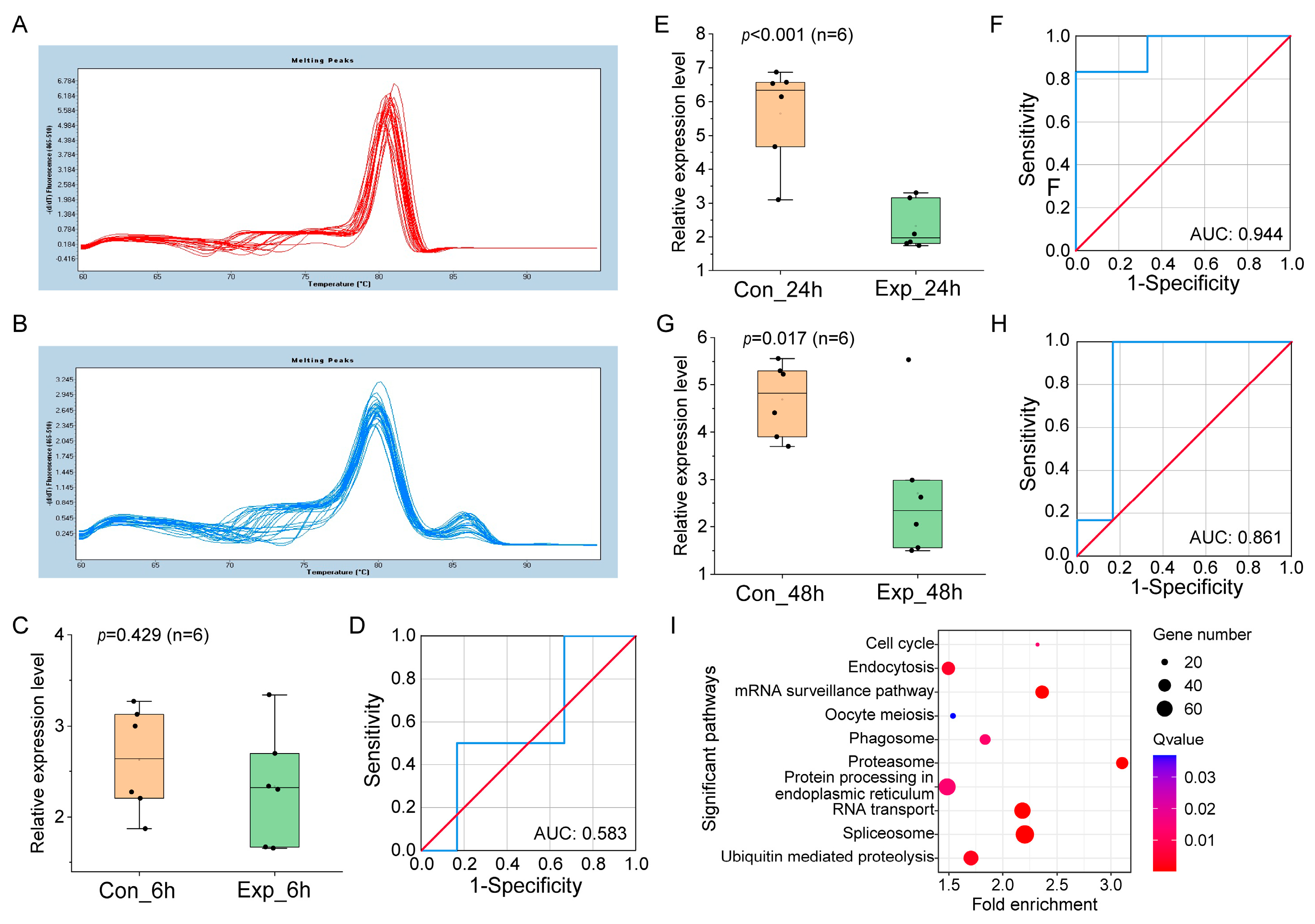
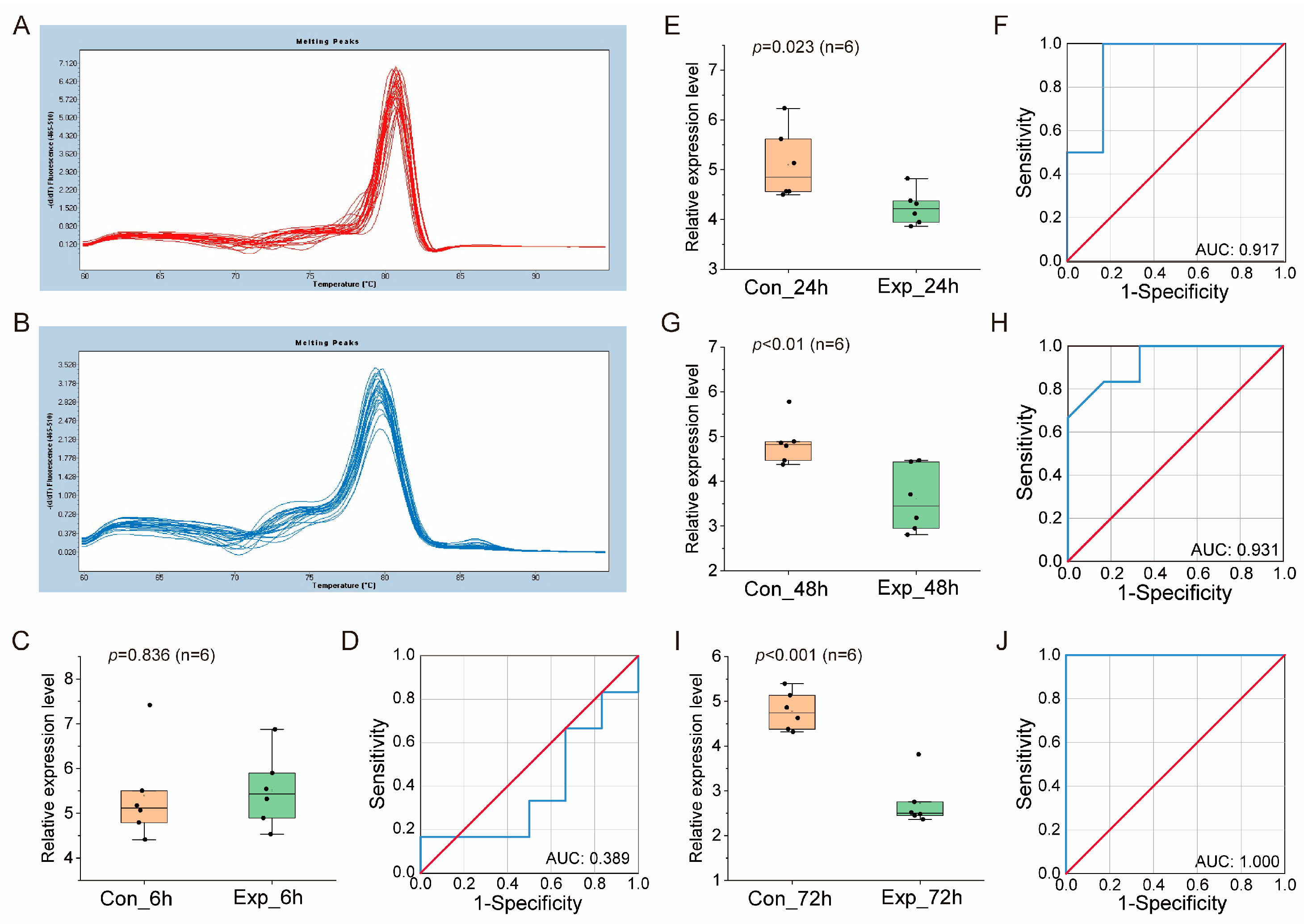
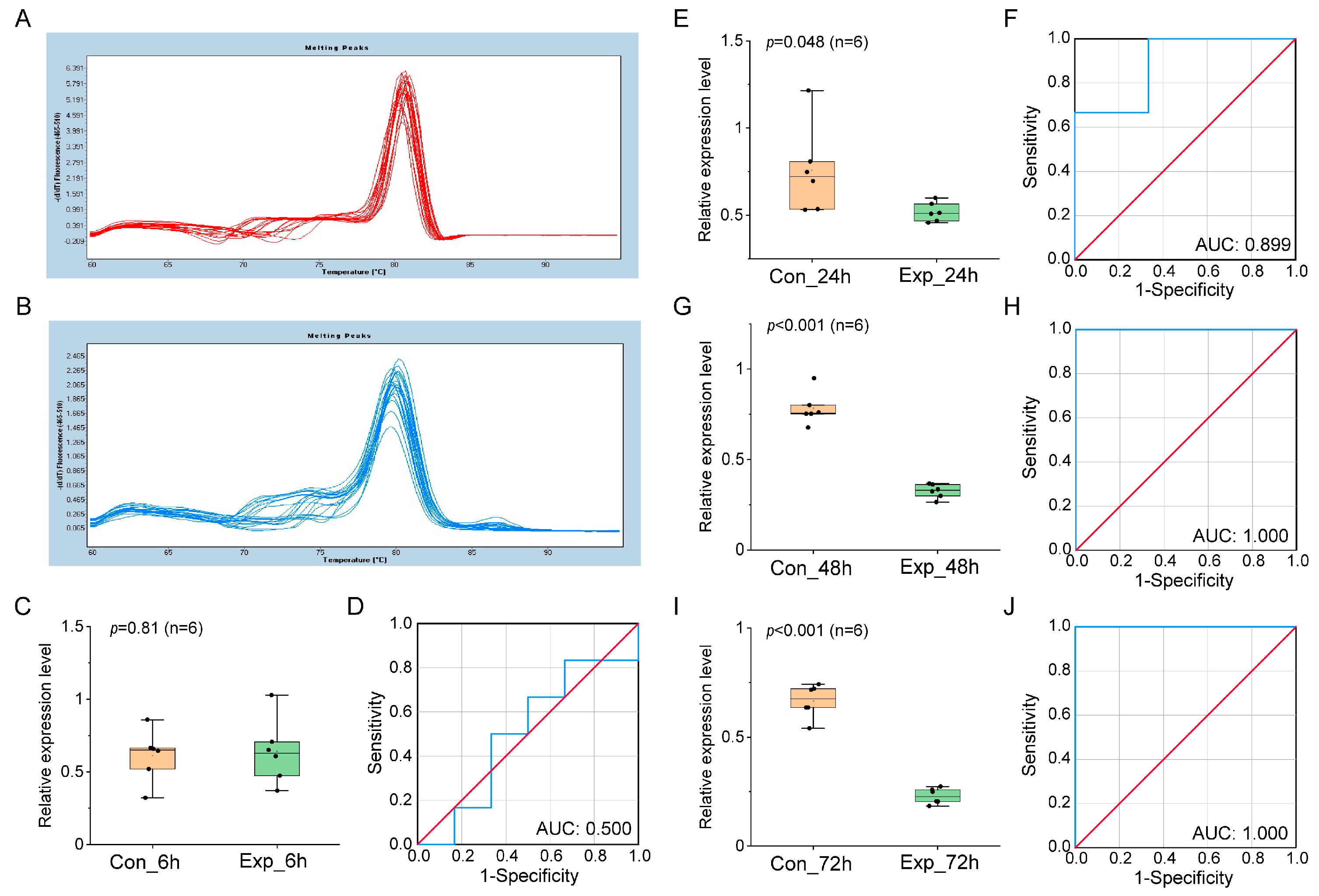
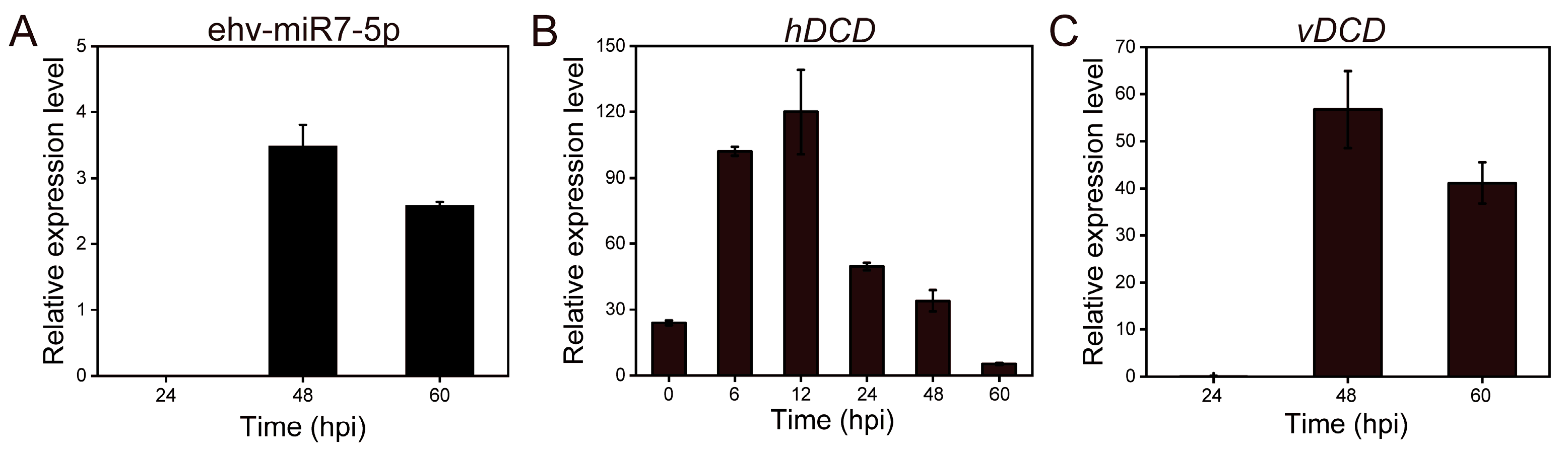
3.2. Verification of miRNA Biomarker Candidates in Different Culture Systems by qRT-PCR

Both host and viral miRNA biomarker candidates were verified in pure culture, mixed culture, and natural seawater culture using qRT-PCR. In the pure culture, the melting curves of the reference genes U6 and ehx-miR20-5p both displayed single peaks (Figure 2A,B), indicating the high amplification specificity of qPCR. Ehx-miR20-5p was not differentially expressed in infection groups at 6 hpi compared to control groups (p = 0.429, Figure 2C), and the area under the curve (AUC) value in ROC analysis was only 0.583 (Figure 2D). In contrast, ehx-miR20-5p were significantly downregulated at 24 hpi (p < 0.001, Figure 2E) with a high AUC value of 0.944 (Figure 2F). The expression of ehx-miR20-5p at 48 hpi was similar to that at 24 hpi (p = 0.017, AUC = 0.861, Figure 2G,H). These results indicate that ehx-miR20-5p can significantly distinguish control and infection groups at the middle (24 hpi) and late infection (48 hpi). The target gene enrichment results of ehx-miR20-5p are shown in Figure 2I. Significantly enriched pathways (Q < 0.05) included Endocytosis and Phagosome, indicating that viruses enter algal cells by Endocytosis at the late stage of infection. In addition, there were pathways involved in gene transcription and translation, such as spliceosome, the mRNA surveillance pathway, RNA transport, and protein processing in the endoplasmic reticulum, indicating that gene transcription and protein synthesis were active during and after viral infection, which is conducive to viral replication and assembly. Furthermore, cell division-related pathways such as cell cycle and oocyte meiosis were also significantly enriched, indicating that some algal cells might form haploids through meiosis to escape from viral infection [32]. Finally, there were several protein degradation-related processes, such as ubiquitin-mediated proteolysis and proteasome, which might be associated with programmed cell death [33,34].
The expression of ehx-miR20-5p was validated as well in mixed culture (Figure 3) and natural seawater culture (Figure 4), which was largely compatible with the results obtained in pure culture, further confirming the reliability of ehx-miR20-5p as a downregulated miRNA biomarker at the middle and late infection stage.
As for virus-derived miRNAs, compared to the expression levels at 24 hpi, the qRT-PCR analysis revealed that all seven of these miRNAs showed significant upregulation at 48 and 60 (or 72) hpi in pure (Figure 5A), mixed (Figure 5B), and natural seawater (Figure 5C) culture. The upregulation of viral miRNAs during late infection may be involved in the regulation of sphingolipid metabolism, steroid biosynthesis, terpenoid backbone biosynthesis, autophagy, ABC transporters, ubiquitin-mediated proteolysis, etc. [17].

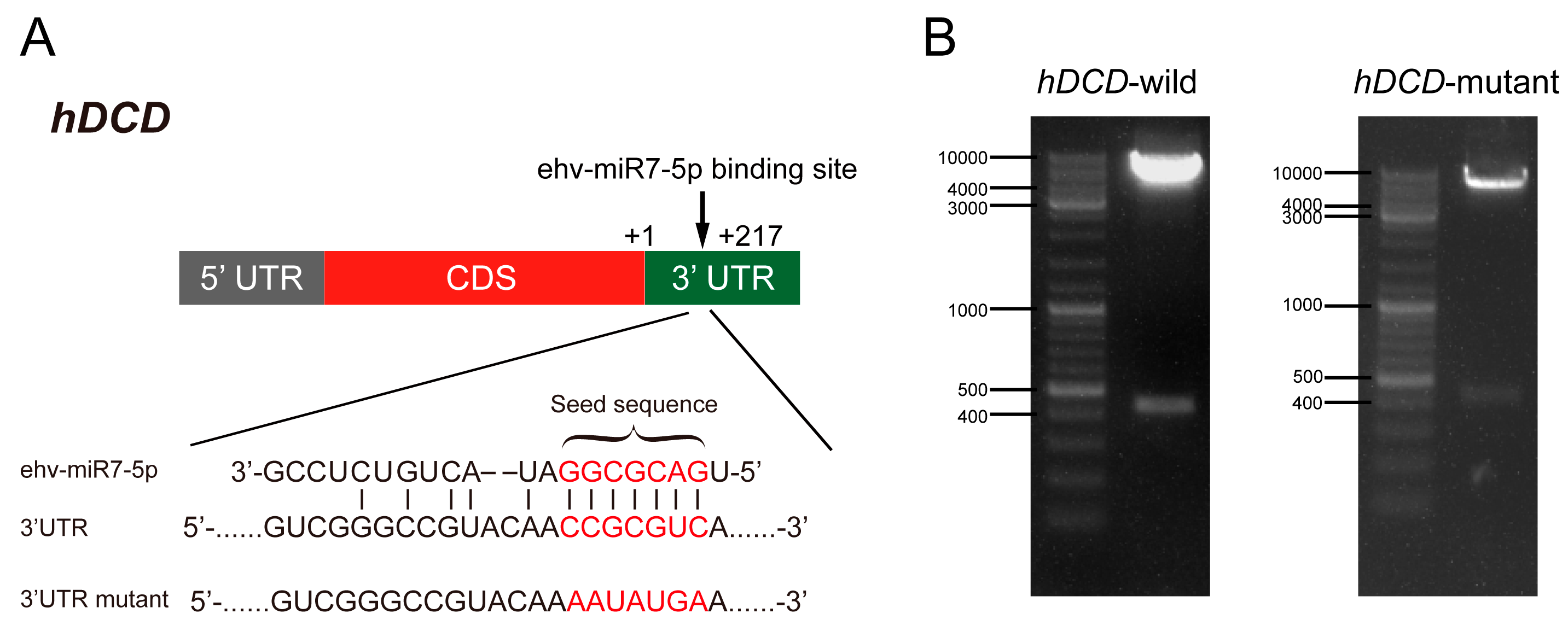
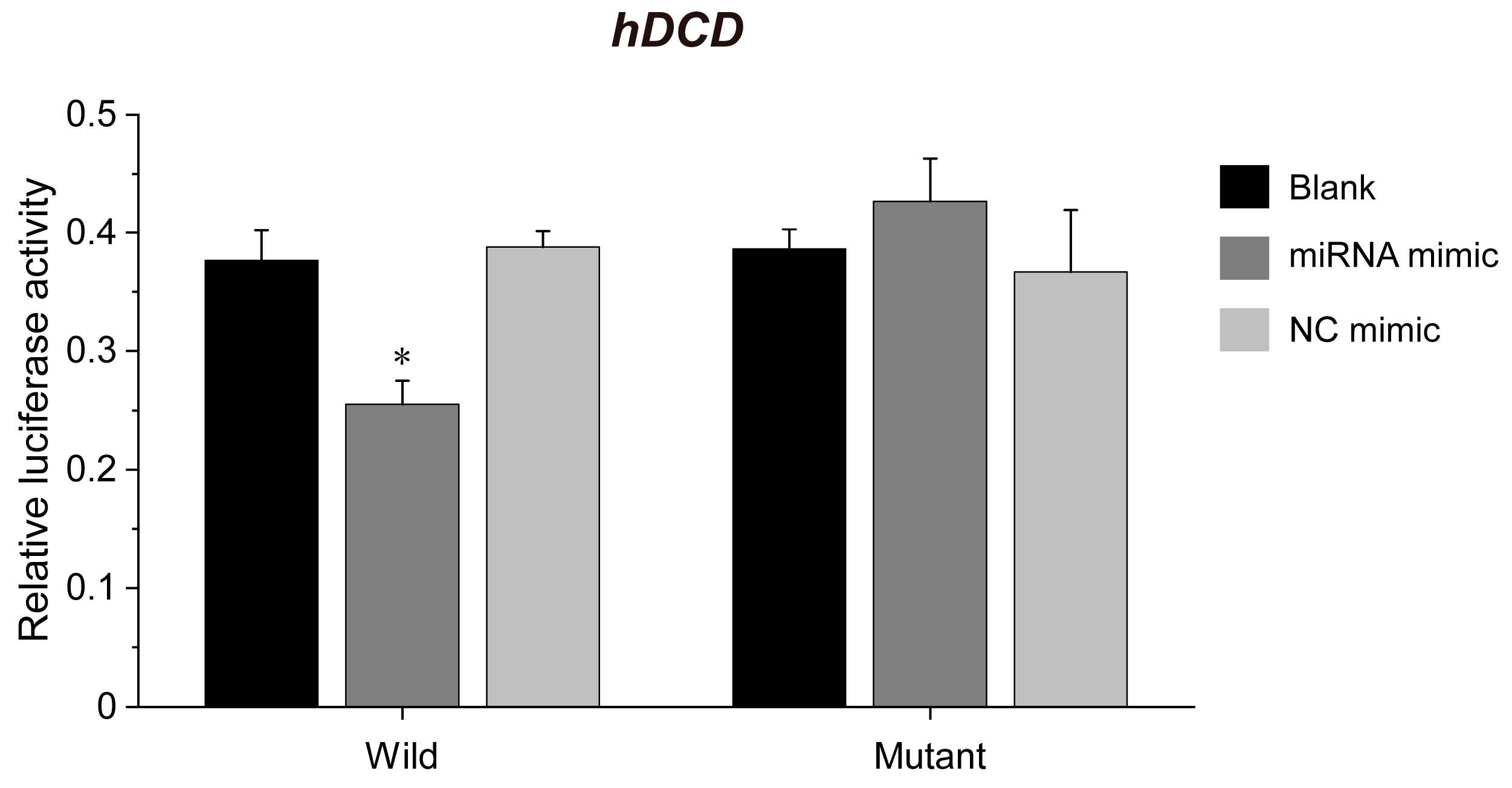
3.3. ehv-miR7-5p Could Target the hDCD Gene

Based on the bioinformatic prediction of differentially expressed miRNA target genes in virus-infected E. huxleyi cells, we found that viral ehv-miR7-5p targeted the hDCD gene with perfect pairing in the 3′-UTR [17]. The specific binding sites between ehv-miR7-5p and hDCD were further verified by the dual-luciferase reporter assay (Figure 6). As shown in Figure 6A, the seed sequence of ehv-miR7-5p is completely paired with the 3′-UTR of hDCD. The 3′-UTR sequences of hDCD were further inserted into the psiCHECK2 plasmid to construct a wild-type recombinant vector (WT vector). The 3′-UTR seed sequences were mutated to construct a mutant-type recombinant vector (MT vector) (Figure 6A). The wild-type and mutant psiCHECK2 recombinant plasmid constructs were then validated using restriction enzyme digestion (Figure 6B) and DNA sequence analysis. The results of the dual-luciferase reporter assay indicated a significant decrease of 34.3% in the psiCHECK2 hDCD WT vector and miRNA co-transfected group, compared to the hDCD MT vector and miRNA control mimic-transfected sample (p < 0.05). However, there was no significant difference observed in the results of transfection with the mutant vector (Figure 7). Moreover, the expression levels of hDCD and vDCD genes were further verified by qPCR (Figure 8), displaying a negative correlation between ehv-miR7-5p and hDCD (Figure 8A,B). It means that ehv-miR7-5p might inhibit the expression of hDCD during late infection. In contrast, vDCD upregulated significantly during late infection (Figure 8C), consistent with the metabolic switch toward viral sphingolipid biosynthesis. These results suggest that the hDCD gene might be a target of ehv-miR7-5p and that ehv-miR7-5p may be involved in the regulation of EhV-induced PCD by targeting hDCD.

4. Discussion

The interactions between E. huxleyi and EhV are characterized by a lipid-based coevolutionary “arms race” [1]. This process involves host lipid metabolism remodeling, viral sphingolipid production [12], host oxidative stress [35], programmed cell death [7], and so on. The entire viral infection process lasts for about 72 to 96 h [36]. Therefore, functional biomarkers that can distinguish viral infection status are needed to diagnose active infection levels in natural marine populations and quantitatively assess their ecosystem and biogeochemical impacts. At present, the screening of biomarkers for the characterization of EhV infection mainly focuses on the screening and identification of lipid metabolites. Significantly upregulated lipid biomarkers during viral infection include betaine-like lipids BLL (22:6/22:6) [14], viral sphingolipid vGSLs [37], host TG (57:8), TG (53:3), TG (64:17), FA (20:4) [15], etc. Significantly downregulated lipid biomarkers include sialic acid GSLs (sGSLs) [14], Cer (38:1; 2), Cer 40:2; 2_isomer1 [15], etc. In addition, host or virus-specific mRNA can also act as biomarkers, such as the host cytochrome oxidase C subunit 1 (COI) gene [9], the thylakoid membrane protein-coding gene psbA [38], and the viral capsid protein-coding gene MCP [38]. In this study, based on miRNA sequencing results, one host (ehx-miR20-5p) and seven viral miRNA biomarkers were screened, which could distinguish the early and late infection stages, respectively. As a kind of small non-coding RNA, the detection of miRNAs is more accurate, simple, and specific, which has become an emerging method to diagnose mammalian virus infection [21]. The miRNA biomarkers identified in this study further expand the biomarker repertoire of the E. huxleyi-EhV system and provide a scientific reference for quantifying the impact of marine viruses on microbial food webs, especially for assessing the contribution of E. huxleyi blooms to marine carbon export [39].
Functional enrichment analysis of ehx-miR20-5p target genes showed that the ehx-miR20-5p could target cell cycle and meiosis (Figure 2I). Previous studies have shown that oxidative stress induced by coccolithovirus infection can activate PCD to prevent virus replication [40] or induce a life cycle shift from the susceptible diploid to the resistant flagellated haploid life phase [32]. A recent study found that virus-infected E. huxleyi has the capability to secrete miRNAs through extracellular vesicles, which could also target the host cell cycle and meiosis processes [41]. Therefore, it is hypothesized that endogenous miRNAs induced by viral infection may participate in the regulation of the host’s transition from diploid to haploid by directly regulating intracellular-related target genes or by functioning as signal molecules in the form of extracellular vesicles.
The dual-luciferase reporter assay results indicated that ehv-miR7-5p could target hDCD through binding to its 3′-UTR and significantly inhibited hDCD gene expression (Figure 8), suggesting that virus-derived ehv-miR7-5p could inhibit host sphingolipid metabolism in late infection. DCD is an enzyme participating in ceramide synthesis via a de novo pathway. It can catalyze the insertion of a 4,5-trans-double bond to the sphingolipid backbone of dihydroceramide, resulting in the conversion of dihydroceramide into ceramide [42]. Ceramides are one of the most studied sphingolipids due to their complicated role in cell differentiation, death, and stress response [43]. Other previous work has highlighted that host sphingolipid biosynthesis was inhibited during EhV infection on both transcriptional and metabolic levels [12]. Recently, we further confirmed that viral infection led to a shift toward virus-specific sphingolipids, which is consistent with the downregulation of genes involved in the host de novo sphingolipid biosynthesis, such as serine palmitoyltransferase (SPT), ceramide synthetase (CERS), and DCD genes [44]. These results demonstrate that the sphingolipid biosynthesis process in virus-infected E. huxleyi is not only transcriptionally but also epigenetically regulated. Interestingly, we revealed the possible regulatory role of miRNA in glycerides and fatty acids metabolism through the PI3K-Akt-TOR signaling pathway in the EhV host system [17]. Thus, the EhV-mediated miRNA regulation might indicate that marine viruses have evolved to regulate the expression of their host genes by miRNA silencing to meet their lipid metabolic requirements.
Taken together, the eight miRNA biomarkers identified in this study as biomarkers of viral infection were able to characterize the entire process of viral infection (including early, middle, and late infection) and had potential applications for assessing the infectious activity of EhVs in a natural marine environment.

Author Contributions

E.Z. analyzed the small RNA-seq data, performed the experiments, and drafted the manuscript. S.Z. contributed to the algal culture, virus preparation, and experiment performance. G.L. carried out HepG2 cell culture and dual-luciferase reporter assay. Z.Z. participated in critical discussion, figure organization, English editing, and manuscript revision. J.L. designed the investigation, guided the study, and finished the final version of the manuscript. All authors contributed to the discussion, commented on the manuscript, and approved the final manuscript. All authors have read and agreed to the published version of the manuscript.

Funding

This research was funded by the National Natural Science Foundation of China (NSFC) project (grant number 42076086) and the Fujian Province Natural Science Foundation of China (grant number 2019J01696).

Institutional Review Board Statement

Not applicable.

Informed Consent Statement

Not applicable.

Data Availability Statement

All other data generated during this study are included in this published article.

Acknowledgments

We would like to thank Gunnar Bratbak (Department of Biology, University of Bergen) for providing the E. huxleyi BOF 92 strain and E. huxleyi 99B1 virus strain.

Conflicts of Interest

The authors declare no conflict of interest.

References

  1. Holligan, P.M.; Fernández, E.; Aiken, J.; Balch, W.M.; Boyd, P.; Burkill, P.H.; Finch, M.; Groom, S.B.; Malin, G.; Muller, K.; et al. A Biogeochemical Study of the Coccolithophore, Emiliania huxleyi, in the North Atlantic. Glob. Biogeochem. Cycles 1993, 7, 879–900. [Google Scholar] [CrossRef]
  2. Iglesias-Rodriguez, M.D.; Halloran, P.R.; Rickaby, R.E.M.; Hall, I.R.; Colmenero-Hidalgo, E.; Gittins, J.R.; Green, D.R.H.; Tyrrell, T.; Gibbs, S.J.; von Dassow, P.; et al. Phytoplankton Calcification in a High-CO 2 World. Science 2008, 320, 336–340. [Google Scholar] [CrossRef] [PubMed]
  3. Taylor, A.R.; Brownlee, C.; Wheeler, G. Coccolithophore Cell Biology: Chalking Up Progress. Ann. Rev. Mar. Sci. 2017, 9, 283–310. [Google Scholar] [CrossRef]
  4. Simó, R. Production of Atmospheric Sulfur by Oceanic Plankton: Biogeochemical, Ecological and Evolutionary Links. Trends Ecol. Evol. 2001, 16, 287–294. [Google Scholar] [CrossRef] [PubMed]
  5. Alcolombri, U.; Ben-Dor, S.; Feldmesser, E.; Levin, Y.; Tawfik, D.S.; Vardi, A. Identification of the Algal Dimethyl Sulfide–Releasing Enzyme: A Missing Link in the Marine Sulfur Cycle. Science 2015, 348, 1466–1469. [Google Scholar] [CrossRef] [PubMed]
  6. Bidle, K.D. The Molecular Ecophysiology of Programmed Cell Death in Marine Phytoplankton. Ann. Rev. Mar. Sci. 2015, 7, 341–375. [Google Scholar] [CrossRef] [PubMed]
  7. Liu, J.; Cai, W.; Fang, X.; Wang, X.; Li, G. Virus-Induced Apoptosis and Phosphorylation Form of Metacaspase in the Marine Coccolithophorid Emiliania huxleyi. Arch. Microbiol. 2018, 200, 413–422. [Google Scholar] [CrossRef]
  8. Nissimov, J.I.; Vandzura, R.; Johns, C.T.; Natale, F.; Haramaty, L.; Bidle, K.D. Dynamics of Transparent Exopolymer Particle Production and Aggregation during Viral Infection of the Coccolithophore, Emiliania huxleyi. Environ. Microbiol. 2018, 20, 2880–2897. [Google Scholar] [CrossRef]
  9. Laber, C.P.; Hunter, J.E.; Carvalho, F.; Collins, J.R.; Hunter, E.J.; Schieler, B.M.; Boss, E.; More, K.; Frada, M.; Thamatrakoln, K.; et al. Coccolithovirus Facilitation of Carbon Export in the North Atlantic. Nat. Microbiol. 2018, 3, 537–547. [Google Scholar] [CrossRef]
  10. Jover, L.F.; Effler, T.C.; Buchan, A.; Wilhelm, S.W.; Weitz, J.S. The Elemental Composition of Virus Particles: Implications for Marine Biogeochemical Cycles. Nat. Rev. Microbiol. 2014, 12, 519–528. [Google Scholar] [CrossRef]
  11. Zhang, R.; Wei, W.; Cai, L. The Fate and Biogeochemical Cycling of Viral Elements. Nat. Rev. Microbiol. 2014, 12, 850–851. [Google Scholar] [CrossRef] [PubMed]
  12. Rosenwasser, S.; Mausz, M.A.; Schatz, D.; Sheyn, U.; Malitsky, S.; Aharoni, A.; Weinstock, E.; Tzfadia, O.; Ben-Dor, S.; Feldmesser, E.; et al. Rewiring Host Lipid Metabolism by Large Viruses Determines the Fate of Emiliania huxleyi, a Bloom-Forming Alga in the Ocean. Plant Cell 2014, 26, 2689–2707. [Google Scholar] [CrossRef]
  13. Schleyer, G.; Shahaf, N.; Ziv, C.; Dong, Y.; Meoded, R.A.; Helfrich, E.J.N.; Schatz, D.; Rosenwasser, S.; Rogachev, I.; Aharoni, A.; et al. In Plaque-Mass Spectrometry Imaging of a Bloom-Forming Alga during Viral Infection Reveals a Metabolic Shift towards Odd-Chain Fatty Acid Lipids. Nat. Microbiol. 2019, 4, 527–538. [Google Scholar] [CrossRef] [PubMed]
  14. Hunter, J.E.; Frada, M.J.; Fredricks, H.F.; Vardi, A.; Van Mooy, B.A.S. Targeted and Untargeted Lipidomics of Emiliania huxleyi Viral Infection and Life Cycle Phases Highlights Molecular Biomarkers of Infection, Susceptibility, and Ploidy. Front. Mar. Sci. 2015, 2, 81. [Google Scholar] [CrossRef]
  15. Zeng, J.; Liu, S.; Cai, W.; Jiang, H.; Lu, X.; Li, G.; Li, J.; Liu, J. Emerging Lipidome Patterns Associated with Marine Emiliania huxleyi-Virus Model System. Sci. Total Environ. 2019, 688, 521–528. [Google Scholar] [CrossRef] [PubMed]
  16. Kuhlisch, C.; Schleyer, G.; Shahaf, N.; Vincent, F.; Schatz, D.; Vardi, A. Viral Infection of Algal Blooms Leaves a Unique Metabolic Footprint on the Dissolved Organic Matter in the Ocean. Sci. Adv. 2021, 7, eabf4680. [Google Scholar] [CrossRef]
  17. Zhang, E.; Gao, J.; Wei, Z.; Zeng, J.; Li, J.; Li, G.; Liu, J. MicroRNA-Mediated Regulation of Lipid Metabolism in Virus-Infected Emiliania huxleyi. ISME J. 2022, 16, 2457–2466. [Google Scholar] [CrossRef]
  18. Bartel, D.P. MicroRNAs: Genomics, Biogenesis, Mechanism, and Function. Cell 2004, 116, 281–297. [Google Scholar] [CrossRef]
  19. Lu, T.X.; Rothenberg, M.E. MicroRNA. J. Allergy Clin. Immunol. 2018, 141, 1202–1207. [Google Scholar] [CrossRef]
  20. Mockly, S.; Seitz, H. Inconsistencies and Limitations of Current MicroRNA Target Identification Methods; Laganà, A., Ed.; Springer: New York, NY, USA, 2019; pp. 291–314. ISBN 978-1-4939-9207-2. [Google Scholar]
  21. Pedersen, I.M.; Cheng, G.; Wieland, S.; Volinia, S.; Croce, C.M.; Chisari, F.V.; David, M. Interferon Modulation of Cellular MicroRNAs as an Antiviral Mechanism. Nature 2007, 449, 919–922. [Google Scholar] [CrossRef]
  22. Tribolet, L.; Kerr, E.; Cowled, C.; Bean, A.G.D.; Stewart, C.R.; Dearnley, M.; Farr, R.J. MicroRNA Biomarkers for Infectious Diseases: From Basic Research to Biosensing. Front. Microbiol. 2020, 11, 1197. [Google Scholar] [CrossRef]
  23. Kawano, Y.; Iwata, S.; Kawada, J.; Gotoh, K.; Suzuki, M.; Torii, Y.; Kojima, S.; Kimura, H.; Ito, Y. Plasma Viral MicroRNA Profiles Reveal Potential Biomarkers for Chronic Active Epstein–Barr Virus Infection. J. Infect. Dis. 2013, 208, 771–779. [Google Scholar] [CrossRef]
  24. EL-Abd, N.E.; Fawzy, N.A.; EL-Sheikh, S.M.; Soliman, M.E. Circulating MiRNA-122, MiRNA-199a, and MiRNA-16 as Biomarkers for Early Detection of Hepatocellular Carcinoma in Egyptian Patients with Chronic Hepatitis C Virus Infection. Mol. Diagn. Ther. 2015, 19, 213–220. [Google Scholar] [CrossRef]
  25. Biswas, S.; Haleyurgirisetty, M.; Lee, S.; Hewlett, I.; Devadas, K. Development and Validation of Plasma MiRNA Biomarker Signature Panel for the Detection of Early HIV-1 Infection. EBioMedicine 2019, 43, 307–316. [Google Scholar] [CrossRef] [PubMed]
  26. Lim, J.; Byun, J.; Guk, K.; Hwang, S.G.; Bae, P.K.; Jung, J.; Kang, T.; Lim, E.-K. Highly Sensitive in Vitro Diagnostic System of Pandemic Influenza A (H1N1) Virus Infection with Specific MicroRNA as a Biomarker. ACS Omega 2019, 4, 14560–14568. [Google Scholar] [CrossRef]
  27. Pagarete, A.; Lanzén, A.; Puntervoll, P.; Sandaa, R.A.; Larsen, A.; Larsen, J.B.; Allen, M.J.; Bratbak, G. Genomic Sequence and Analysis of EhV-99B1, a New Coccolithovirus from the Norwegian Fjords. Intervirology 2013, 56, 60–66. [Google Scholar] [CrossRef]
  28. Livak, K.J.; Schmittgen, T.D. Analysis of Relative Gene Expression Data Using Real-Time Quantitative PCR and the 2−ΔΔCT Method. Methods 2001, 25, 402–408. [Google Scholar] [CrossRef]
  29. Enright, A.J.; John, B.; Gaul, U.; Tuschl, T.; Sander, C.; Marks, D.S. MicroRNA Targets in Drosophila. Genome Biol. 2003, 5, R1. [Google Scholar] [CrossRef] [PubMed]
  30. Huang, D.W.; Sherman, B.T.; Lempicki, R.A. Systematic and Integrative Analysis of Large Gene Lists Using DAVID Bioinformatics Resources. Nat. Protoc. 2009, 4, 44–57. [Google Scholar] [CrossRef]
  31. Zhang, E.; Wu, S.; Cai, W.; Zeng, J.; Li, J.; Li, G.; Liu, J. Validation of Superior Reference Genes for QRT-PCR and Western Blot Analyses in Marine Emiliania huxleyi –Virus Model System. J. Appl. Microbiol. 2021, 131, 257–271. [Google Scholar] [CrossRef]
  32. Frada, M.; Probert, I.; Allen, M.J.; Wilson, W.H.; de Vargas, C. The “Cheshire Cat” Escape Strategy of the Coccolithophore Emiliania huxleyi in Response to Viral Infection. Proc. Natl. Acad. Sci. USA 2008, 105, 15944–15949. [Google Scholar] [CrossRef]
  33. Drexler, H.C.A. Programmed Cell Death and the Proteasome. Apoptosis 1998, 3, 1–7. [Google Scholar] [CrossRef]
  34. Kim, M.; Ahn, J.-W.; Jin, U.-H.; Choi, D.; Paek, K.-H.; Pai, H.-S. Activation of the Programmed Cell Death Pathway by Inhibition of Proteasome Function in Plants. J. Biol. Chem. 2003, 278, 19406–19415. [Google Scholar] [CrossRef]
  35. Sheyn, U.; Rosenwasser, S.; Ben-Dor, S.; Porat, Z.; Vardi, A. Modulation of Host ROS Metabolism Is Essential for Viral Infection of a Bloom-Forming Coccolithophore in the Ocean. ISME J. 2016, 10, 1742–1754. [Google Scholar] [CrossRef]
  36. Liu, J.; Bratbak, G.; Zheng, T.; Thyrhaug, R. Effects of Virus Infection on Expression of Cell Cycle Regulatory Proteins in the Unicellular Marine Algae Emiliania huxleyi. Acta Oceanol. Sin. 2011, 30, 89–95. [Google Scholar] [CrossRef]
  37. Malitsky, S.; Ziv, C.; Rosenwasser, S.; Zheng, S.; Schatz, D.; Porat, Z.; Ben-Dor, S.; Aharoni, A.; Vardi, A. Viral Infection of the Marine Alga Emiliania huxleyi Triggers Lipidome Remodeling and Induces the Production of Highly Saturated Triacylglycerol. New Phytol. 2016, 210, 88–96. [Google Scholar] [CrossRef]
  38. Vincent, F.; Sheyn, U.; Porat, Z.; Schatz, D.; Vardi, A. Visualizing Active Viral Infection Reveals Diverse Cell Fates in Synchronized Algal Bloom Demise. Proc. Natl. Acad. Sci. USA 2021, 118, e2021586118. [Google Scholar] [CrossRef]
  39. Sheyn, U.; Rosenwasser, S.; Lehahn, Y.; Barak-Gavish, N.; Rotkopf, R.; Bidle, K.D.; Koren, I.; Schatz, D.; Vardi, A. Expression Profiling of Host and Virus during a Coccolithophore Bloom Provides Insights into the Role of Viral Infection in Promoting Carbon Export. ISME J. 2018, 12, 704–713. [Google Scholar] [CrossRef]
  40. Evans, C.; Malin, G.; Mills, G.P.; Wilson, W.H. Viral Infection Of Emiliania huxleyi (Prymnesiophyceae) Leads To Elevated Production Of Reactive Oxygen Species. J. Phycol. 2006, 42, 1040–1047. [Google Scholar] [CrossRef]
  41. Schatz, D.; Rosenwasser, S.; Malitsky, S.; Wolf, S.G.; Feldmesser, E.; Vardi, A. Communication via Extracellular Vesicles Enhances Viral Infection of a Cosmopolitan Alga. Nat. Microbiol. 2017, 2, 1485–1492. [Google Scholar] [CrossRef]
  42. Ziv, C.; Malitsky, S.; Othman, A.; Ben-Dor, S.; Wei, Y.; Zheng, S.; Aharoni, A.; Hornemann, T.; Vardi, A. Viral Serine Palmitoyltransferase Induces Metabolic Switch in Sphingolipid Biosynthesis and Is Required for Infection of a Marine Alga. Proc. Natl. Acad. Sci. USA 2016, 113, E1907-16. [Google Scholar] [CrossRef] [PubMed]
  43. Mullen, T.D.; Obeid, L.M. Ceramide and Apoptosis: Exploring the Enigmatic Connections between Sphingolipid Metabolism and Programmed Cell Death. Anticancer Agents Med. Chem. 2012, 12, 340–363. [Google Scholar] [CrossRef] [PubMed]
  44. Liu, J.; Gao, J.; Zhang, E.; Jiang, H.; Li, G.; Li, J.; Zeng, J.; Wu, D. Characterization of the Sphingolipid Profiling of Emiliania huxleyi against Virus Infection. J. Oceanol. Limnol. 2023. [Google Scholar] [CrossRef]
Figure 1. The heatmap of seven miRNAs during virus infection (based on our previous miRNA-Seq data).
Genes 14 01716 g001
Figure 2. The validation of miRNA biomarkers in pure culture. (A,B) Melting curves of reference gene U6 and ehx-miR20-5p, respectively. (C,E,G) show the relative expression level of ehx-miR20-5p in control and infected groups at 6, 24, and 48 hpi. (D,F,H) show the ROC analysis of the results in (C,E,G), respectively. The blue line is the ROC curve. The red line is the random classifier line. (I) KEGG enrichment analysis of targets of ehx-miR20-5p.
Genes 14 01716 g002
Figure 3. The validation of miRNA biomarkers in mixed culture. (A,B) Melting curves of reference gene U6 and ehx-miR20-5p, respectively. (C,E,G,I) show the relative expression level of ehx-miR20-5p in control and infected groups at 6, 24, and 48 hpi. (D,F,H,J) show the ROC analysis of the results in (C,E,G,I), respectively. The blue line is the ROC curve. The red line is the random classifier line.
Genes 14 01716 g003
Figure 4. The validation of miRNA biomarker in natural seawater culture. (A,B) Melting curves of reference gene U6 and ehx-miR20-5p, respectively. (C,E,G,I) show the relative expression level of ehx-miR20-5p in control and infected groups at 6, 24, and 48 hpi. (D,F,H,J) show the ROC analysis of the results in (C,E,G,I), respectively. The blue line is the ROC curve. The red line is the random classifier line.
Genes 14 01716 g004
Figure 5. The validation of virus-specific miRNA biomarkers. (AC) show the relative expression level of seven viral miRNAs in pure culture, mixed culture, and natural seawater culture, respectively.
Genes 14 01716 g005
Figure 6. Interactions between miRNAs and their target genes. (A) Putative binding sites of ehv-miR7-5p on the 3′UTR region of hDCD and mutations were displayed in red. (B) The double enzyme digestion experiment results of recombinant vector psiCHECK2 (hDCD wild-type and mutant-type vectors).
Genes 14 01716 g006
Figure 7. The relative luciferase activity in hDCD-3′UTR luciferase reporter assay. Blank groups were used as controls. The results were analyzed by Student’s t-test. * p < 0.05.
Genes 14 01716 g007
Figure 8. qRT-PCR results of ehv-miR7-5p and DCD genes. (A) ehv-miR7-5p. (B) hDCD gene. (C) vDCD gene.
Genes 14 01716 g008
Table 1. Oligonucleotide primers used for miRNA qPCR.
miRNA IDPrimer Sequences
U6-FCTCGCTTCGGCAGCACA
U6-RAACGCTTCACGAATTTGCGT
ehx-miR20-5pCGGCGGTAGTCGGCGGTAAA
ehv-miR1-3pGGAATTGGTCGTCACGTTGTTGT
ehv-miR2-3pGGTGAGAGTGCATCGGATTGTGAA
ehv-miR3-3pGGATTTGGGCTGGCCCAAAA
ehv-miR4-3pGGATCTAGGAAAGATTGAGGCCAAA
ehv-miR5-3pGGCGAAGACACTGTGAATCAAGT
ehv-miR6-5pCGGGCGGAAAATATGATTCGTTA
ehv-miR7-5pGCCTGACGCGGATACTGTCTC
Disclaimer/Publisher’s Note: The statements, opinions and data contained in all publications are solely those of the individual author(s) and contributor(s) and not of MDPI and/or the editor(s). MDPI and/or the editor(s) disclaim responsibility for any injury to people or property resulting from any ideas, methods, instructions or products referred to in the content.

