Article

Molecular Genetic Analysis of Russian Patients with Coagulation Factor FVII Deficiency

by
Olesya Pshenichnikova
1,*,
Daria Selivanova
1,
Ekaterina Shchemeleva
1,
Tatiana Abramova
1,
Nadezhda Zozulya
2 and
Vadim Surin
1
1
Laboratory of Genetic Engineering of National Medical Research Center for Hematology, Novy Zykovski lane 4a, 125167 Moscow, Russia
2
Coagulopathies Department of National Medical Research Center for Hematology, Novy Zykovski lane 4a, 125167 Moscow, Russia
*
Author to whom correspondence should be addressed.
Genes 2023, 14(9), 1767; https://doi.org/10.3390/genes14091767
Submission received: 24 August 2023 / Revised: 4 September 2023 / Accepted: 6 September 2023 / Published: 6 September 2023
(This article belongs to the Special Issue Identifying the Molecular Basis of Rare Genetic Diseases)

Abstract

:
Coagulation factor VII (proconvertin) is one of the proteins starting the blood coagulation cascade. Plasma FVII concentration is regulated by different factors. A low level of FVII could also be a result of FVII deficiency (MIM# 227500), the rare autosomal recessive inherited disease caused by pathogenic variants in the F7 gene. The aim of this study was to describe a mutation spectrum of the F7 gene and genotype–phenotype relationship in patients with FVII deficiency in Russia for the first time. We studied the primary structure of the F7 gene of 54 unrelated patients with FVII deficiency by direct Sanger sequencing. Pathogenic variants in the F7 gene were detected in 37 (68.5%) of them. We identified 24 different mutations located mostly in the serine protease domain. Five pathogenic variants had never been reported before. A major mutation in the Russian population was c.1391delC (p. Pro464Hisfs*32), linked with rs36209567 and rs6046 functional polymorphisms, that is widely distributed in East Europe. As in other countries, the F7 genotypes poorly correlated with the severity of clinical manifestations but were quite well associated with FVII levels. Minor alleles of functional polymorphisms rs510335, rs5742910, rs561241, rs36209567, and rs6046 could also participate in the F7 genotype and influence FVII levels.

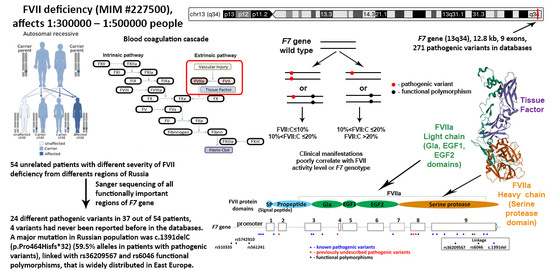
Graphical Abstract

1. Introduction

The blood coagulation process starts from the formation of the complex between the tissue factor (TF) and the activated coagulation factor FVII (FVIIa) when the vascular endothelium is damaged. A complete lack of any of these proteins is incompatible with life and leads to death in early childhood because of severe hemorrhages. However, even small amounts of them are enough to initiate a number of reactions on membrane surfaces during complex TF-FVIIa formation [1].
FVII is a zymogen of vitamin-K-dependent serine protease FVIIa, which is synthesized mostly in the liver and circulates in plasma. Plasma FVII concentration is regulated by different factors such as age, body mass index, dietary fat intake, plasma lipids (especially triglycerides), diabetes, and F7 polymorphisms [2,3].
The F7 gene (MIM# 613878) coding the FVII protein is located on chromosome 13 (13q34), consists of nine exons, and has a length of about 12.8 kbp. There are three transcripts of the gene resulting from alternative splicing. Two transcripts (NM_019616.4, NM_000131.4) encode identical mature proteins circulating in plasma, and in the third transcript (NM_001267554.1), terminal domains are absent, and its physiological role is unknown [1]. mRNA encodes 466 amino acids; the first twenty encode the signal peptide, the next forty—propeptide, providing biosynthesis/secretion of FVII, and these 60 amino acids are removed in the process of intracellular proteolysis. The mature FVII protein (50 kDa) consists of 406 amino acids. The 1–406 numeration is more widely used in protein description, while the 1–466 numeration is used mainly in molecular genetics; therefore, we will use the second one. The mature protein contains a light and a heavy chain. The light chain consists of a calcium-binding, vitamin-K-dependent Gla domain (61–142 amino acid residues) and two EGF-like domains (106–142 and 143–211 amino acid residues) interacting with membranes and other proteins of the coagulation cascade. The heavy chain contains a serine protease domain (212–466 amino acid residues) [4]. FVII activation occurs during proteolytic cleavage between Arg212-Ile213 amino acid residues [1,4].
Pathogenic variants in the F7 gene lead to the rare autosomal recessive inherited disease known as FVII deficiency (MIM# 227500) with a worldwide prevalence of 1:300,000–1:500,000. Nowadays, 271 unique variants in 1058 individuals are described in the international database EAHAD Factor VII Gene (F7) Variant Database (https://f7-db.eahad.org/ (accessed on 23 August 2023)). Patients with FVII deficiency demonstrate a high phenotypic variability, from asymptomatic to life-threatening gastrointestinal bleedings and central nervous system hemorrhages. Most patients have mucosal bleedings (epistaxis, gum bleeding, easy bruising), and women often present with menorrhagia. Despite active attempts to study the molecular basis of FVII deficiency, there has been no significant correlation between the F7 genotype, FVII factor activity (FVII:C), and a clinical phenotype found [5,6,7,8,9]. The genetic impact of F7 polymorphisms on FVII plasma levels is estimated at 57–63% [2]. Among the most studied single nucleotide polymorphisms (SNP), minor alleles of rs510335 (−401G/T), rs5742910 (−3230 bp/10 bp) in the promoter gene region, and rs6046 (Arg353Gln) in exon 9 are consistently associated with lower plasma FVII levels [3,10,11]. Nevertheless, because of the disease’s rarity, it is extremely important to collect information on patients with FVII deficiency from different populations, as it will allow for obtaining enough data for more detailed analysis and pattern findings.
In Russia, molecular genetic analysis of the F7 gene is conducted predominantly in our center. Therefore, the aim of this study was to describe a mutation spectrum of the gene in our country and the genotype–phenotype relationship in patients with FVII deficiency.

2. Materials and Methods

In this study, we included 54 unrelated patients (12 males, 42 females) with declined levels of FVII and a presumptive diagnosis of FVII deficiency (less than 70% FVII:C) with different types of severity. In our country, we have issues with diagnostics of rare diseases in regions. The majority of studied patients were from Moscow and the Moscow region (N = 35, 65%), while other 14 patients (26%) were from European part of Russia, and only 5 patients (9%) represented other regions. The collaborating physicians at our center who consulted and selected these patients were experienced in the diagnosis and treatment of hemophilia A, B, von Willebrand disease, and rare bleeding disorders. The study was carried out according to the Principles of the Declaration of Helsinki; informed consent was obtained from all participants.
Genomic DNA was isolated from EDTA-treated whole blood samples using phenol–chloroform extraction and ethanol precipitation [12]. Genomic DNA was dissolved in TE buffer (pH 8.0) and frozen at −20 °C until genotyping.
To conduct molecular genetic analysis, we amplified all functionally important regions of F7 gene (466 bp of promoter, exons 1–9, and exon-intron junctions) as 8 fragments with length of 261–775 bp with primers designed in our laboratory according to the reference genomic F7 sequence NG_009262 (Table 1, Figure 1).
PCR reactions were carried out on a Tercik programmable thermocycler (DNK-Technology, Moscow, Russia) with PCR Master Mix (Thermo Fisher Scientific, Waltham, MA, USA) using 10 pmol each oligonucleotide primer (Syntol, Moscow, Russia) and 50–100 ng template DNA in 25 μL of reaction mixture. Amplification conditions for these primer sets were 35 cycles of 94 °C for 1 min, 62 °C for 1 min, 72 °C for 3 min. We analyzed the obtained PCR fragments with electrophoresis in 6% polyacrylamide gel (PAAG) followed by staining with ethidium bromide (0.03 µg/mL) for 1 min and visualization in UV light. Amplified DNA fragments were purified using Wizard PCR Preps DNA Purification System (Promega Corporation, Madison, WI, USA) and subjected to direct Sanger sequencing using ABI PRISM BigDyeTM Terminator v.3.1 Cycle Sequencing Kit (Thermo Fisher Scientific, Waltham, MA, USA) on ABI PRISM 3100Avant genetic analyzer sequencer (Applied Biosystems, Foster City, CA, USA) at Genome CCU (Institute of Molecular Biology, Russian Academy of Sciences, Moscow, Russia).
The cDNA numbering system used is compliant with the Human Genome Variation Society recommendations ver. 15.11 (http://varnomen.hgvs.org (accessed on 23 August 2023)). The amino acid numbering is based upon the start methionine, codon +1; the reference sequence used was NG_009262 for genomic positioning and NM_000131.4 for cDNA numbering. As reference databases for pathogenic variants, we used Factor VII Variant Database (https://f7-db.eahad.org/ (accessed on 23 August 2023)), Human Gene Mutation Database (www.hgmd.cf.ac.uk (accessed on 23 August 2023)), and ClinVar (https://www.ncbi.nlm.nih.gov/clinvar/ (accessed on 23 August 2023)).
Missense variants unreported in databases were examined using deleteriousness prediction scoring programs: PolyPhen-2 v2.2.2 [13], MutationTaster2 (build NCBI 37/Ensembl 69 [14]), FATHMM (v2.3) [15]. Variant effects were then classified using the ACMG/AMP Variant Curation Guidelines [16].
All statistical analyses were performed using R Statistical Software (v.4.3.0 [17]). Differences in SNP frequencies between our and global populations were estimated with chi-square test with p-value < 0.05 considered statistically significant. Hardy–Weinberg equilibrium (HWE) was assessed with chi-square test. Pairwise linkage disequilibrium (LD) between SNPs was calculated from the genotype data and measured as D’. HWE and LD were calculated using the package “genetics” [18]).

3. Results

Molecular genetic analysis allowed us to identify pathogenic variants in the F7 gene in 37 out of 54 patients with a presumptive diagnosis of FVII deficiency (68.5%). We did not find severe disruption of the F7 gene despite a decreased level of FVII activity levels in 17 patients. However, in 11 (64.5%) of them, we could not exclude the influence of functional polymorphisms on both alleles. A summary of found F7 gene defects, functional polymorphisms, and clinical features of patients are presented in Table S1. Among all studied patients with a decreased level of FVII:C, 25 patients (46.3%) were homozygous or compound heterozygous for F7 mutations, 12 (22.2%) were heterozygous, whereas in 17 (31.5%), no F7 pathogenic variants were detected.

3.1. F7 Mutation Spectrum

Among 37 patients with identified F7 gene defects, we found 24 different pathogenic variants, five of which had never been reported before in the international databases (Table 2). The majority of gene defects were single nucleotide variations (21 variants, 87.5%); the other three variants were frameshifts. The most common defect type was missense mutation—there were 16 of them (66.7%), and two of them also had an effect on splicing; there were also three regulatory mutations, one frameshift, two inframe microdeletions, and two splicing mutations (besides the abovementioned two missense variants) (Table 2). Most mutations were found in exon 9, but it could be explained by the greatest size of this exon compared with others. Overall, all found defects were distributed uniformly along the gene (Figure 2). Nevertheless, it is characterized by the predominance of missense mutations located mainly in the serine protease domain.
A major mutation in our population was c.1391delC (p.Pro464Hisfs*32); it was found as 29 alleles in 22 patients (59.5% out of all with pathogenic variants), and six times it was in a homozygous state. The regulatory variant c. −55C>T occurred four times in four people in a heterozygous state. The missense mutation c.64G>A (p.Gly22Ser) appeared in two patients—once, it was homozygous, and in another patient, it was heterozygous. Five gene defects (c.291+1G>A, c.691_693del (p.Leu231del), c.817_831del (p.Leu273_Asp277del), c.911C>T (p.Ala304Val), c.1285G>A (p.Ala429Thr)) occurred twice in two people each. The other 16 pathogenic variants were found only once (Table 2).

3.2. Novel Mutations

Among previously undescribed variants, one was a splicing mutation disturbing a canonical splice site (c.347−1G>A), and per the ACMG/AMP Variant Curation Guidelines, it was evaluated as likely pathogenic (PM2, PVS1). The other four were missense variants evaluated as variants of unknown significance by the ACMG criteria. Two of them—c.65G>A (p.Gly22Asp) and c.713G>C (p.Cys238Ser)—affected positions with known pathogenic variants (PM2, PM5, PP3); the other two, c.728 T>C (p.Ile243Thr) and c.1175A>G (p.Tyr392Cys), were completely new (PM2, PP3). Pathogenicity prediction algorithms estimated all previously undescribed missense variants as probably pathogenic.

3.3. Functional Polymorphisms

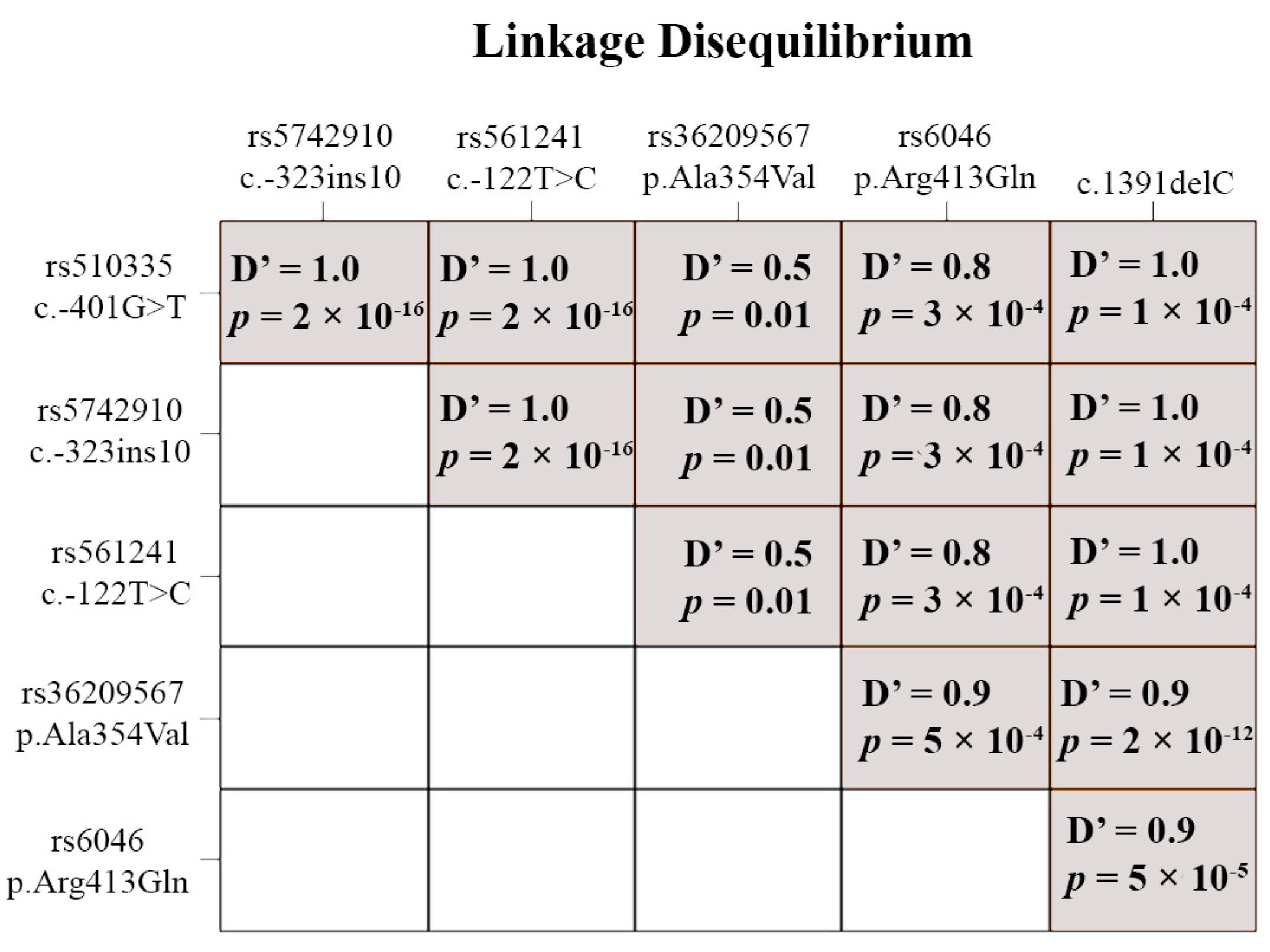
Except for obviously pathogenic variants, we also detected known functional polymorphisms: rs510335 (c. −401G>T), rs5742910 (c. −323ins10), rs561241 (c. −122T>C), rs36209567 (c.1061 C>T (p.Ala354Val)), and rs6046 (c.1238 G>A (p.Arg413Gln)). Although in Russian and global populations, their minor allele frequencies (MAF) are quite similar, the allele frequencies in our sample differed statistically significantly from them (Table 3). All polymorphisms were in Hardy–Weinberg equilibrium (p > 0.05) and in linkage disequilibrium (Figure 3), especially rs36209567 and rs6046, which were highly correlated with the pathogenic variant c.1391delC (p.Pro464Hisfs*32). We also noted that rs36209567, rs6046, and c.1391delC rather often were met with minor alleles of neutral polymorphisms rs6039 (c.64+9G>A), rs2774033 (c.291+71G>A), and rs6042 (c.525C>T (p.His175His)).
Polymorphisms rs510335 (c. −401G>T), rs5742910 (c. −323ins10), and rs561241 (c. −122T>C) had a complete allelic association and form a single haplotype (Figure 3).

3.4. Genotype and Phenotype Interaction

We estimated a clinical phenotype based on FVII level [29] and clinical manifestations [30].
Clinical phenotype was poorly correlated with the FVII level (Table 4) as well as with the F7 genotype (Table 5). In our sample, 50% of probands (27 patients) had an FVII:C ≤ 10%, 40.7% (22 patients) had an FVII:C > 20%, and only 9.3% (5 patients) had an intermediate level of FVII activity. As for clinical phenotype, probands with mild severity prevailed (42.6%, 23 patients), with such symptoms as epistaxis, gum bleeding, and easy bruising, although almost one-fifth of the sample was asymptomatic (Table 4, Table S1).
Proband’s genotype quite exactly reflected FVII level. All people with the homozygous pathogenic variant, almost all heterozygous compounds, and people with a combination of the heterozygous pathogenic variant and homozygosity by functional polymorphisms had a low FVII level. Probands with one heterozygous pathogenic variant but without homozygosity by functional polymorphisms or without a found pathogenic variant had a higher FVII level, mostly >20% (Table 5). The only exception was one proband with no mutation or functional polymorphisms found, almost an absence of symptoms, but a low factor activity level (FVII:C = 3.3%). In this patient, there could be either deep intronic mutations or FVII deficiency, in this case resulting from other mechanisms.
In our sample, females prevailed (42 vs. 12 males). Nevertheless, both sexes had quite similar distributions in FVII levels, the F7 genotype, and clinical symptoms.

4. Discussion

4.1. Mutation Spectrum of F7 Gene

The spectrum of pathogenic defects in the F7 gene in Russian patients with reduced FVII levels is similar to the spectra in other countries. It is characterized by the prevalence of missense mutations located predominantly in the serine protease domain, mainly because of the length of exon 9 but also because of its functional significance [5,6,7,9,31,32]. A major mutation in our population was c.1391delC (p.Pro464Hisfs*32), which is supposed to have its origin in Central Europe [6]. We found only one allele that was not linked with the rs36209567 and rs6046 functional polymorphisms. This combination of c.1391delC (p.Pro464Hisfs*32) with rs36209567 was previously described as common in East Europe (particularly in Slovakia), and it was also found in other European countries but with significantly less frequency [6]. Our sample, however, was biased because of a lack of diagnoses of rare diseases in regions, so we described mostly the mutation spectrum of the European part of the country. An extension of the sample to the Asian part of Russia could increase the variability of pathogenic variants or reveal mutations specific to certain regions.

4.2. Gene Variant c.1061 C>T (p.Ala354Val)

Variant rs36209567 (c.1061 C>T (p.Ala354Val)) should be discussed separately. In the literature, there are different opinions about its influence on the FVII protein. In most studies, it is referred to as a mutation [6,7,28,31,33]. Nevertheless, we suppose that it seems to be a functional polymorphism because its minor allele in our sample occurred with a frequency of 0.3889, which is too high for a pathogenic variant and, in most cases (in 31 probands out of 54), it was found in combination with two obviously pathogenic defects. Moreover, in patients homozygous by rs36209567 and without found pathogenic variants, we usually saw FVII:C > 20% (Table 5), indicating only a small reduction in FVII activity. However, combined with heterozygosity by other pathogenic variants, it led to a significant reduction in FVII levels (Table S1). It is according to the functional study of rs36209567 by Fromovich-Amit and colleagues, which demonstrated that this variant alone or in combination with rs6046 (c.1238 G>A (p.Arg413Gln)) led to the reduction of FVII to 41% and 55%, respectively [31].

4.3. Functional Polymorphisms

The influence of other functional polymorphisms—rs510335 (c. −401G>T), rs5742910 (c. −323ins10), rs561241 (c. −122T>C), and rs6046 (c.1238 G>A (p.Arg413Gln))—was also proved by functional studies [10,34,35]. The first three promoter variants have a complete allelic association and form a single haplotype, which reduces a basal level of FVII transcription [34,35]. We also showed that in a homozygous state without other F7 defects, they led to a reduction in FVII levels compared with the heterozygous influence of pathogenic variants (Table 5, Table S1).

4.4. Genotype and Phenotype Interaction

As in other studies [5,6,7,9,30,31,32], we showed a lack of relationships between the severity of clinical manifestations and genotypes (Table 4 and Table 5). Peyvandi and colleagues conducted a large study of the association between coagulant factor activity and clinical bleeding severity of rare bleeding disorders, which revealed a lack of it for FVII deficiency. They proposed to use the laboratory phenotype for this disease expressed by FVII levels [29]. Our results confirm that FVII levels correlate much better with the F7 genotype than clinical manifestations. Serious damage to both alleles resulting from a homozygous pathogenic defect, heterozygous compound, or heterozygous pathogenic defect combined with homozygosity by functional polymorphisms leads to a significant decrease in plasma FVII (FVII:C ≤ 10%). One allele damaged by a heterozygous pathogenic defect, or both alleles affected by homozygous functional polymorphisms, lead to much more mild consequences. However, even small amounts of FVII are sufficient to initiate the blood coagulation process [1], so most patients with a reduced level of FVII are asymptomatic or have only mild symptoms, such as epistaxis, gum bleeding, or easy bruising.

4.5. Patients without Found Pathogenic Variants

We did not find any pathogenic variants in 17 out of 54 patients with FVII deficiency (31.5%). It is a usual situation in FVII deficiency studies [7,9,36]. In 11 of them (64.7%), lower FVII levels could be explained by homozygous functional polymorphisms that partly reduced FVII. In one of the patients without pathogenic variants or homozygous functional polymorphisms, the diagnosis later was changed to the mild autoimmune acquired combined blood clotting factors FV, FVII, and FXII deficiency. The other five patients without pathogenic variants or homozygous functional polymorphisms needed to be studied further. Especially interesting among them is one proband with a low factor activity level (FVII:C = 3.3%) and almost asymptomatic clinical manifestation. In these patients, there could be deep intronic mutations (although one of a deep intronic hotspot, c.571 +78G>A, we checked in our sample but did not find it in anyone) [36] or large deletions that need the use of the MLPA method and are not accounted for in this study. There could also be other factors leading to FVII deficiency unrelated to the F7 gene.

5. Conclusions

We report herein the results of a molecular genetic analysis in Russian patients with coagulation factor FVII deficiency.
Overall, the mutation spectrum of the F7 gene reflects tendencies revealed in other populations. A major mutation in our population was c.1391delC (p.Pro464Hisfs*32), linked with rs36209567 and rs6046 functional polymorphisms. As in other countries, F7 genotypes were poorly correlated with the severity of clinical manifestations but were quite well associated with FVII levels.
Nevertheless, these data are the first attempt at an F7 genetic analysis in Russia, and they contribute to local and global knowledge of molecular mechanisms of congenital FVII deficiency, which could improve diagnostics of this disorder in our country. Undoubtedly, this research needs to be continued in order to increase coverage of patients with FVII deficiency, to include more patients from the eastern part of the country, and to study patients without found causal variants in more detail.

Supplementary Materials

The following supporting information can be downloaded at: https://www.mdpi.com/article/10.3390/genes14091767/s1, Table S1. Patients included in the study and pathogenic variants and functional polymorphisms found in them.

Author Contributions

Conceptualization, V.S. and N.Z.; methodology, V.S.; formal analysis, O.P. and D.S.; investigation, O.P., D.S., E.S. and T.A.; resources, V.S. and N.Z.; data curation, N.Z.; writing—original draft preparation, O.P., D.S. and V.S.; writing—review and editing, O.P., D.S., T.A. and V.S.; visualization, O.P. and D.S.; supervision, V.S. and N.Z.; project administration, V.S.; funding acquisition, V.S. All authors have read and agreed to the published version of the manuscript.

Funding

The research was funded by the state assignment of FASO of Russia No. AAAA-A18-118032290163-2.

Institutional Review Board Statement

The study was conducted in accordance with the Declaration of Helsinki and approved by the Ethics Committee of the National Medical Research Center for Hematology (protocol code 171, 27 April 2023).

Informed Consent Statement

Written informed consent was obtained from the patients to publish this paper.

Data Availability Statement

Data are available on request from the authors.

Conflicts of Interest

The authors declare no conflict of interest.

References

  1. Bernardi, F.; Mariani, G. Biochemical, molecular and clinical aspects of coagulation factor VII and its role in hemostasis and thrombosis. Haematologica 2021, 106, 351–362. [Google Scholar] [CrossRef] [PubMed]
  2. Ken-Dror, G.; Drenos, F.; Humphries, S.E.; Talmud, P.J.; Hingorani, A.D.; Kivimäki, M.; Kumari, M.; Bauer, K.A.; Morrissey, J.H.; Ireland, H.A. Haplotype and genotype effects of the F7 gene on circulating factor VII, coagulation activation markers and incident coronary heart disease in UK men. J. Thromb. Haemost. 2010, 8, 2394–2403. [Google Scholar] [CrossRef] [PubMed]
  3. Fang, J.; Yuan, Q.; Du, Z.; Liu, C.; Xu, H.; Yang, W.; Chen, L.; Zhao, J.; Xie, R.; Hu, J.; et al. Contribution of factor VII polymorphisms to coagulopathy in patients with isolated traumatic brain injury. Clin. Neurol. Neurosurg. 2021, 208, 106836. [Google Scholar] [CrossRef] [PubMed]
  4. Vadivel, K.; Bajaj, S.P. Structural biology of factor VIIa/tissue factor initiated coagulation. Front. Biosci. 2012, 17, 2476–2494. [Google Scholar] [CrossRef]
  5. Millar, D.S.; Kemball-Cook, G.; McVey, J.H.; Tuddenham, E.G.; Mumford, A.D.; Attock, G.B.; Reverter, J.C.; Lanir, N.; Parapia, L.A.; Reynaud, J.; et al. Molecular analysis of the genotype-phenotype relationship in factor VII deficiency. Hum. Genet. 2000, 107, 327–342. [Google Scholar] [CrossRef]
  6. Herrmann, F.H.; Wulff, K.; Auerswald, G.; Schulman, S.; Astermark, J.; Batorova, A.; Kreuz, W.; Pollmann, H.; Ruiz-Saez, A.; De Bosch, N.; et al. Factor VII deficiency: Clinical manifestation of 717 subjects from Europe and Latin America with mutations in the factor 7 gene. Haemophilia 2009, 15, 267–280. [Google Scholar] [CrossRef] [PubMed]
  7. Quintavalle, G.; Riccardi, F.; Rivolta, G.F.; Martorana, D.; Di Perna, C.; Percesepe, A.; Tagliaferri, A.; Ad-Hoc Study Group. F7 gene variants modulate protein levels in a large cohort of patients with factor VII deficiency. Results from a genotype-phenotype study. Thromb. Haemost. 2017, 117, 1455–1464. [Google Scholar] [CrossRef] [PubMed]
  8. Shahbazi, S.; Mahdian, R. Factor VII Gene Defects: Review of Functional Studies and Their Clinical Implications. Iran. Biomed. J. 2019, 23, 165–174. [Google Scholar] [CrossRef]
  9. Ouardani, C.; Elmahmoudi, H.; ElBorgi, W.; Gharbi, M.; Meriem, A.; Gouider, E. Clinical phenotype and F7 gene genotype in 40 Tunisian patients with congenital factor VII deficiency. Blood Coagul. Fibrinolysis 2022, 33, 280–284. [Google Scholar] [CrossRef] [PubMed]
  10. Hunault, M.; Arbini, A.A.; Lopaciuk, S.; Carew, J.A.; Bauer, K.A. The Arg353Gln polymorphism reduces the level of coagulation factor VII. In vivo and in vitro studies. Arterioscler. Thromb. Vasc. Biol. 1997, 17, 2825–2829. [Google Scholar] [CrossRef]
  11. Pollak, E.S.; Hung, H.L.; Godin, W.; Overton, G.C.; High, K.A. Functional characterization of the human factor VII 5′-flanking region. J. Biol. Chem. 1996, 271, 1738–1747. [Google Scholar] [CrossRef] [PubMed]
  12. Maniatis, T.; Fritsch, E.F.; Sambrook, J. Molecular Cloning: A Laboratory Manual; Cold Spring Harbor Laboratory: Cold Spring Harbor, NY, USA, 1982; ISBN 978-0-87969-136-3. [Google Scholar]
  13. Adzhubei, I.A.; Schmidt, S.; Peshkin, L.; Ramensky, V.E.; Gerasimova, A.; Bork, P.; Kondrashov, A.S.; Sunyaev, S.R. A Method and Server for Predicting Damaging Missense Mutations. Nat. Methods 2010, 7, 248–249. [Google Scholar] [CrossRef] [PubMed]
  14. Schwarz, J.M.; Cooper, D.N.; Schuelke, M.; Seelow, D. MutationTaster2: Mutation Prediction for the Deep-Sequencing Age. Nat. Methods 2014, 11, 361–362. [Google Scholar] [CrossRef]
  15. Shihab, H.A.; Gough, J.; Cooper, D.N.; Stenson, P.D.; Barker, G.L.A.; Edwards, K.J.; Day, I.N.M.; Gaunt, T.R. Predicting the Functional, Molecular and Phenotypic Consequences of Amino Acid Substitutions using Hidden Markov Models. Hum. Mutat. 2013, 34, 57–65. [Google Scholar] [CrossRef]
  16. Richards, S.; Aziz, N.; Bale, S.; Bick, D.; Das, S.; Gastier-Foster, J.; Grody, W.W.; Hegde, M.; Lyon, E.; Spector, E.; et al. Standards and Guidelines for the Interpretation of Sequence Variants: A Joint Consensus Recommendation of the American College of Medical Genetics and Genomics and the Association for Molecular Pathology. Genet. Med. 2015, 17, 405–424. [Google Scholar] [CrossRef] [PubMed]
  17. R Core Team. R: A Language and Environment for Statistical Computing; R Foundation for Statistical Computing: Vienna, Austria, 2023; Available online: https://www.R-project.org/ (accessed on 22 August 2023).
  18. Warnes, G.; Gorjanc, W.C.F.G.; Leisch, F.; Man, M. Genetics: Population Genetics. R package version 1.3.8.1.3. 2021. Available online: https://CRAN.R-project.org/package=genetics (accessed on 22 August 2023).
  19. Carew, J.A.; Pollak, E.S.; High, K.A.; Bauer, K.A. Severe factor VII deficiency due to a mutation disrupting an Sp1 binding site in the factor VII promoter. Blood 1998, 92, 1639–1645. [Google Scholar] [CrossRef] [PubMed]
  20. Kavlie, A.; Hiltunen, L.; Rasi, V.; Prydz, H. Two novel mutations in the human coagulation factor VII promoter. Thromb. Haemost. 2003, 90, 194–205. [Google Scholar] [CrossRef] [PubMed]
  21. Wulff, K.; Herrmann, F.H. Twenty two novel mutations of the factor VII gene in factor VII deficiency. Hum. Mutat. 2000, 15, 489–496. [Google Scholar] [CrossRef] [PubMed]
  22. Giansily-Blaizot, M.; Aguilar-Martinez, P.; Biron-Andreani, C.; Jeanjean, P.; Igual, H.; Schved, J.F.; Study Group of Factor Seven Deficiency. Analysis of the genotypes and phenotypes of 37 unrelated patients with inherited factor VII deficiency. Eur. J. Hum. Genet. 2001, 9, 105–112. [Google Scholar] [CrossRef]
  23. Peyvandi, F.; Jenkins, P.V.; Mannucci, P.M.; Billio, A.; Zeinali, S.; Perkins, S.J.; Perry, D.J. Molecular characterisation and three-dimensional structural analysis of mutations in 21 unrelated families with inherited factor VII deficiency. Thromb. Haemost. 2000, 84, 250–257. [Google Scholar] [CrossRef] [PubMed]
  24. Alshinawi, C.; Scerri, C.; Galdies, R.; Aquilina, A.; Felice, A.E. Two new missense mutations (P134T and A244V) in the coagulation factor VII gene. Hum. Mutat. 1998, 11 (Suppl. S1), S189–S191. [Google Scholar] [CrossRef]
  25. Bernardi, F.; Castaman, G.; Pinotti, M.; Ferraresi, P.; Di Iasio, M.G.; Lunghi, B.; Rodeghiero, F.; Marchetti, G. Mutation pattern in clinically asymptomatic coagulation factor VII deficiency. Hum. Mutat. 1996, 8, 108–115. [Google Scholar] [CrossRef]
  26. Tamary, H.; Fromovich, Y.; Shalmon, L.; Reich, Z.; Dym, O.; Lanir, N.; Brenner, B.; Paz, M.; Luder, A.S.; Blau, O.; et al. Ala244Val is a common, probably ancient mutation causing factor VII deficiency in Moroccan and Iranian Jews. Thromb. Haemost. 1996, 76, 283–291. [Google Scholar] [CrossRef]
  27. Marchetti, G.; Patracchini, P.; Gemmati, D.; DeRosa, V.; Pinotti, M.; Rodorigo, G.; Casonato, A.; Girolami, A.; Bernardi, F. Detection of two missense mutations and characterization of a repeat polymorphism in the factor VII gene (F7). Hum. Genet. 1992, 89, 497–502. [Google Scholar] [CrossRef] [PubMed]
  28. Arbini, A.A.; Bodkin, D.; Lopaciuk, S.; Bauer, K.A. Molecular analysis of Polish patients with factor VII deficiency. Blood 1994, 84, 2214–2220. [Google Scholar] [CrossRef] [PubMed]
  29. Peyvandi, F.; Di Michele, D.; Bolton-Maggs, P.H.; Lee, C.A.; Tripodi, A.; Srivastava, A. Project on Consensus Definitions in Rare Bleeeding Disorders of the Factor VIII/Factor IX Scientific and Standardisation Committee of the International Society on Thrombosis and Haemostasis. Classification of rare bleeding disorders (RBDs) based on the association between coagulant factor activity and clinical bleeding severity. J. Thromb. Haemost. 2012, 10, 1938–1943. [Google Scholar] [CrossRef] [PubMed]
  30. Mariani, G.; Herrmann, F.H.; Dolce, A.; Batorova, A.; Etro, D.; Peyvandi, F.; Wulff, K.; Schved, J.F.; Auerswald, G.; Ingerslev, J.; et al. Clinical phenotypes and factor VII genotype in congenital factor VII deficiency. Thromb. Haemost. 2005, 93, 481–487. [Google Scholar] [CrossRef]
  31. Fromovich-Amit, Y.; Zivelin, A.; Rosenberg, N.; Tamary, H.; Landau, M.; Seligsohn, U. Characterization of mutations causing factor VII deficiency in 61 unrelated Israeli patients. J. Thromb. Haemost. 2004, 2, 1774–1781. [Google Scholar] [CrossRef] [PubMed]
  32. Borhany, M.; Boijout, H.; Pellequer, J.L.; Shamsi, T.; Moulis, G.; Aguilar-Martinez, P.; Schved, J.F.; Giansily-Blaizot, M. Genotype and phenotype relationships in 10 Pakistani unrelated patients with inherited factor VII deficiency. Haemophilia 2013, 19, 893–897. [Google Scholar] [CrossRef]
  33. Mariani, G.; Bernardi, F. Factor VII Deficiency. Semin. Thromb. Hemost. 2009, 35, 400–406. [Google Scholar] [CrossRef]
  34. van ‘t Hooft, F.M.; Silveira, A.; Tornvall, P.; Iliadou, A.; Ehrenborg, E.; Eriksson, P.; Hamsten, A. Two common functional polymorphisms in the promoter region of the coagulation factor VII gene determining plasma factor VII activity and mass concentration. Blood 1999, 93, 3432–3441. [Google Scholar] [CrossRef] [PubMed]
  35. Sabater-Lleal, M.; Chillón, M.; Howard, T.E.; Gil, E.; Almasy, L.; Blangero, J.; Fontcuberta, J.; Soria, J.M. Functional analysis of the genetic variability in the F7 gene promoter. Atherosclerosis 2007, 195, 262–268. [Google Scholar] [CrossRef] [PubMed]
  36. Ferraresi, P.; Balestra, D.; Guittard, C.; Buthiau, D.; Pan-Petesh, B.; Maestri, I.; Farah, R.; Pinotti, M.; Giansily-Blaizot, M. Next-generation sequencing and recombinant expression characterized aberrant splicing mechanisms and provided correction strategies in factor VII deficiency. Haematologica 2020, 105, 829–837. [Google Scholar] [CrossRef] [PubMed]
Figure 1. Scheme of amplification system for F7 gene screening.
Genes 14 01767 g001
Figure 2. Distribution of different pathogenic variants found among 37 patients from Russia.
Genes 14 01767 g002
Figure 3. Linkage disequilibrium for functional polymorphisms and c.1391delC (p.Pro464Hisfs*32).
Genes 14 01767 g003
Table 1. Primers used in the study.
AmpliconPrimerNucleotide SequenceTargetFragment Length, bp
1F7F1xcct aag aaa cca gcc tcc cttPromoter; exon 1686
F7R1agc cgc cag aaa acc ctc ct
2F7F2cta gct cac agc atg gcc ttexon 2261
F7R2cgg tca ctt cct ctc gag ca
3F7F3agc gcc gct ccc ctc ctc caexon 3 1380
F7R3gcc gca gcc aaa gag acg ca
4F7F45gtc caa gtc ccc caa ccc caexon 4; exon 5439
F7R45cac cta gac caa ttt cca act
5F7F6aga aca cca ctg ctg acc caexon 6384
F7R6xtgc cca gat ccc acc tca ca
6F7F7tcc cat agc ctc ggc ctc aaexon 7315
F7R7cct gcc cat ttt ccc ttc ca
7F7F8xtgc aat gcc aga ggt tcc ttexon 8 1477
F7R8xgca cga agc cca gag cca ca
8F7DF9
F7R9
tgg cca cag ccc atc ccc atexon 9775
cct cct cta ccc cat taa ct
1 Amplification of these fragments was conducted with addition of betaine (5 M, 20% from PCR mix volume).
Table 2. Pathogenic variants revealed in the study.
#Pathogenic VariantExon/IntronDomainDefect TypeAllele CountReference
1c. −55C>TPromoterPromoterRegulatory4[19]
2c. −32A>CPromoterPromoterRegulatory1[20]
3c. −30A>CPromoterPromoterRegulatory1[5]
4c.64G>A
(p.Gly22Ser)
Exon 1PropeptideMissense/
splicing
3[21]
5c.65G>A
(p.Gly22Asp)
Exon 2PropeptideMissense/
splicing
1New
6c.218G>A
(p.Leu73Gln)
Exon 3GlaMissense1[5]
7c.226G>A (p.Glu76Lys) Exon 3GlaMissense1[22]
8c.291+1G>AIntron 3GlaSplicing2[23]
9c.347−1G>AIntron 3GlaSplicing1New
10c.580C>A (p.Pro194Thr) Exon 7EGF2Missense1[24]
11c.691_693del
(p.Leu231del)
Exon 8Serine proteaseInframe (microdeletion)2[6]
12c.713G>C (p.Cys238Ser) Exon 8Serine proteaseMissense1New
13c.728 T>C (p.Ile243Thr) Exon 8Serine proteaseMissense1New
14c.760G>C (p.Cys254Arg) Exon 8Serine proteaseMissense1[6]
15c.817_831del (p.Leu273_Asp277del) Exon 9Serine proteaseInframe (microdeletion)2[21]
16c.847C>T (p.Arg283Trp) Exon 9Serine proteaseMissense1[25]
17c.911C>T (p.Ala304Val) Exon 9Serine proteaseMissense2[26]
18c.1072A>G (p.Met358Val) Exon 9Serine proteaseMissense1[25]
19c.1109G>T (p.Cys370Phe) Exon 9Serine proteaseMissense1[27]
20c.1171G>A (p.Gly391Ser) Exon 9Serine proteaseMissense1[27]
21c.1175A>G (p.Tyr392Cys) Exon 9Serine proteaseMissense1New
22c.1285G>A (p.Ala429Thr) Exon 9Serine proteaseMissense2[22]
23c.1310A>T (p.Tyr437Phe) Exon 9Serine proteaseMissense1[6]
24c.1391delC (p.Pro464Hisfs*32) Exon 9Serine proteaseFrameshift (microdeletion)29[28]
Table 3. Functional polymorphisms revealed in the study, their minor allele frequencies (MAF), and results of chi-square test between global MAF and MAF in our sample.
rs IDFunctional PolymorphismMAF (Global)MAF (Russia, RUSeq Data)MAF
(Our Sample)
chi-Squarep-Value
rs510335c. −401G>T0.1622No data0.25937.440.006
rs5742910c. −323ins100.0523No data0.259392.9<0.00001
rs561241c. −122T>C0.11280.12390.259322.9<0.00001
rs36209567c.1061 C>T (p.Ala354Val)0.00060.00050.388923472.6<0.00001
rs6046c.1238 G>A (p.Arg413Gln)0.12650.12940.6111229.1<0.00001
Table 4. Relationship between clinical bleeding severity score based on Peyvandi [29] and Mariani [30].
Clinical Severity
Peyvandi (Vertically)/
Mariani (Horizontally)
Severe
N (%)
Moderate
N (%)
Mild
N (%)
Asymptomatic
N (%)
No Data
N (%)
Total
Severe
(FVII:C ≤ 10%)
3 (11.1%)9 (33.4%)10 (37%)4 (14.8%)1 (3.7%)27 (50%)
Moderate
(10% < FVII:C ≤ 20%)
02 (40%)2 (40%)1 (20%)05 (9.3%)
Mild
(FVII:C > 20%)
04 (18.2%)11 (50%)5 (22.7%)2 (9.1%)22 (40.7%)
Total3 (5.6%)15 (27.7%)23 (42.6%)10 (18.5%)3 (5.6%)
Table 5. Relationship between proband’s clinical bleeding severity score and genotype.
Pathogenic
Variant Status
TotalClinical Severity Peyvandi
[29]
Clinical Severity Mariani
[30]
Severe
(FVII:C ≤ 10%)
N (%)
Moderate
(10% < FVII:C ≤ 20%)
N (%)
Mild
(FVII:C > 20%)
N (%)
Severe
N (%)
Moderate
N (%)
Mild
N (%)
Asymptomatic
N (%)
No Data
N (%)
homozygous88 (100%)001 (12.5%)4 (50%)2 (25%)01 (12.5%)
heterozygous compound1715 (88.2%)2 (11.8%)02 (11.8%)3 (17.7%)8 (47%)4 (23.5%)0
heterozygous901 (11.1%)8 (88.9%)03 (33.3%)3 (33.3%)3 (33.4%)0
heterozygous
+
homozygous by functional polymorphisms
33 (100%)0002 (66.7%)01 (33.3%)0
no mutation found
+
homozygous by functional polymorphisms
1202 (16.7%)10 (83.3%)02 (16.7%)7 (58.3%)1 (8.3%)2 (16.7%)
no mutation found51 (20%)04 (80%)01 (20%)3 (60%)1 (20%)0
Total542752231523103
Disclaimer/Publisher’s Note: The statements, opinions and data contained in all publications are solely those of the individual author(s) and contributor(s) and not of MDPI and/or the editor(s). MDPI and/or the editor(s) disclaim responsibility for any injury to people or property resulting from any ideas, methods, instructions or products referred to in the content.