Next Article in Journal
RNAi Knockdown of EHMT2 in Maternal Expression of Prader–Willi Syndrome Genes
Previous Article in Journal
Reduced-Cost Genotyping by Resequencing in Peanut Breeding Programs Using Tecan Allegro Targeted Resequencing V2
 
 
genes-logo
Article Menu

Article Menu

Article

Unraveling the Complexity of Chikungunya Virus Infection Immunological and Genetic Insights in Acute and Chronic Patients

by
Hegger Fritsch
1,2,†,
Marta Giovanetti
3,4,5,†,
Luan Gaspar Clemente
6,
Gabriel da Rocha Fernandes
4,
Vagner Fonseca
7,8,
Maricelia Maia de Lima
9,10,
Melissa Falcão
10,
Neuza de Jesus
10,
Erenilde Marques de Cerqueira
9,
Rivaldo Venâncio da Cunha
11,
Marcos Vinicius Lima de Oliveira Francisco
12,
Isadora Cristina de Siqueira
12,
Carla de Oliveira
13,
Joilson Xavier
1,
Jorge Gomes Goulart Ferreira
14,
Fábio Ribeiro Queiroz
14,
Elise Smith
15,
Jennifer Tisoncik-Go
15,
Wesley C. Van Voorhis
16,
Peter M. Rabinowitz
17,
Judith N. Wasserheit
18,
Michael Gale, Jr.
15,
Ana Maria Bispo de Filippis
13,* and
Luiz Carlos Junior Alcantara
4,5,*
add Show full author list remove Hide full author list
1
Instituto de Ciências Biológicas, Universidade Federal de Minas Gerais, Belo Horizonte 31270-901, Brazil
2
Institut National de la Santé et de la Recherche Médicale, U1259—MAVIVHe, Université de Tours, 37032 Tours, France
3
Department of Science and Technologies for Sustainable Development and One Health, Università Campus Bio-Medico di Roma, 00128 Rome, Italy
4
Instituto René Rachou, Fundação Oswaldo Cruz, Belo Horizonte 30190-002, Brazil
5
Climate Amplified Diseases and Epidemics (CLIMADE)—CLIMADE Americas, Belo Horizonte 30190-002, Brazil
6
Escola Superior de Agricultura Luiz de Queiroz, Departamento de Zootecnia, Universidade de São Paulo, Piracicaba 13418-900, Brazil
7
Departamento de Ciências Exatas e da Terra, Universidade Estadual da Bahia, Salvador 41150-000, Brazil
8
Centre for Epidemic Response and Innovation (CERI), School of Data Science and Computational Thinking, Stellenbosch University, Stellenbosch 7600, South Africa
9
Departamento de Saúde, Universidade Estadual de Feira de Santana, Feira de Santana 44036-900, Brazil
10
Secretaria de Municipal de Saúde de Feira de Santana, Divisão de Vigilância Epidemiológica, Feira de Santana 44027-010, Brazil
11
Fundação Oswaldo Cruz, Bio-Manguinhos, Rio de Janeiro 21040-360, Brazil
12
Instituto Gonçalo Moniz, Fundação Oswaldo Cruz, Salvador 40296-710, Brazil
13
Laboratório de Arbovírus e Vírus Hemorrágicos, Instituto Oswaldo Cruz, Fundação Oswaldo Cruz, Rio de Janeiro 21040-360, Brazil
14
Núcleo de Ensino, Pesquisa e Inovação, Instituto Mário Penna, Belo Horizonte 30380-420, Brazil
15
Department of Immunology, University of Washington, Seattle, WA 98109, USA
16
Department of Medicine, University of Washington, Seattle, WA 98195, USA
17
Departments of Environmental and Occupational Health Sciences, University of Washington, Seattle, WA 98195, USA
18
Department of Global Health, University of Washington, Seattle, WA 98105, USA
*
Authors to whom correspondence should be addressed.
These authors contributed equally to this work.
Genes 2024, 15(11), 1365; https://doi.org/10.3390/genes15111365
Submission received: 1 October 2024 / Revised: 17 October 2024 / Accepted: 21 October 2024 / Published: 24 October 2024
(This article belongs to the Section Molecular Genetics and Genomics)

Abstract

:
Background: The chikungunya virus (CHIKV), transmitted by infected Aedes mosquitoes, has caused a significant number of infections worldwide. In Brazil, the emergence of the CHIKV-ECSA genotype in 2014 posed a major public health challenge due to its association with more severe symptoms. Objectives/Methods: This study aimed to shed new light on the host immune response by examining the whole-blood transcriptomic profile of both CHIKV-acute and chronically infected individuals from Feira de Santana, Bahia, Brazil, a region heavily affected by CHIKV, Dengue, and Zika virus epidemics. Results: Our data reveal complex symptomatology characterized by arthralgia and post-chikungunya neuropathy in individuals with chronic sequelae, particularly affecting women living in socially vulnerable situations. Analysis of gene modules suggests heightened metabolic processes, represented by an increase in NADH, COX5A, COA3, CYC1, and cap methylation in patients with acute disease. In contrast, individuals with chronic manifestations exhibit a distinct pattern of histone methylation, probably mediated by NCOA3 in the coactivation of different nuclear receptors, KMT2 genes, KDM3B and TET2, and with alterations in the immunological response, majorly led by IL-17RA, IL-6R, and STAT3 Th17 genes. Conclusion: Our results emphasize the complexity of CHIKV disease progression, demonstrating the heterogeneous gene expression and symptomatologic scenario across both acute and chronic phases. Moreover, the identification of specific gene modules associated with viral pathogenesis provides critical insights into the molecular mechanisms underlying these distinct clinical manifestations.

1. Introduction

Emerging and re-emerging viral infectious diseases impose a significant burden on public health and the global population, leading to devastating outbreaks and affecting a large number of individuals worldwide [1,2]. Among these viruses, arthropod-borne viruses (arboviruses) present particularly alarming scenarios due to their widespread distribution, posing a risk of infection to populations in more than 110 countries across Asia, Africa, Europe, and the Americas [3]. Chikungunya virus (CHIKV) is an arthrogenic alphavirus belonging to the Togaviridae family, transmitted by infected Aedes ssp. mosquitoes [4,5,6,7]. It can be classified into four distinct lineages or genotypes based on genomic differences: (i) the West African lineage; (ii) the East/Central/South African (ECSA) lineage; (iii) the Asian lineage; and (iv) the Indian Ocean lineage (IOL) [8,9,10]. Although there is no consensus in the literature regarding the impact of the viral lineage on the severity and prognosis of the disease, CHIKV-ECSA infections seem to be associated with more severe symptoms compared to other lineages [11]. Since its initial cases, reported in Tanzania in 1953, CHIKV infections have been reported worldwide [3,9,12,13], with almost 4 million probable cases in the Americas alone from 2013 to May 2024 [3,13]. In Brazil, the most affected country on the continent [3,13], the local introduction of the CHIKV-ECSA genotype was first detected in the municipality of Feira de Santana, Northeast region, in 2014. Since then, the ECSA genotype has been reported in several states across the country, posing a serious threat to public health [14,15,16,17,18,19,20,21,22,23], given the uncertain impact of CHIKV-ECSA infection in more severe cases [11].
The vast majority of Chikungunya-infected patients may experience an asymptomatic infection [24]. However, for those who show clinical manifestations, the acute phase may be marked by a sudden onset of fever, followed by cutaneous manifestations (rash), fatigue, and debilitating polyarthralgia. Although most symptoms resolve within a few weeks and the mortality rate for CHIKV is extremely low (estimated to be approximately 0.1%) [24], the persistence of joint symptoms for months or years in a significant proportion of the infected population presents a substantial global economic and public health challenge [25,26]. Presently, there is no highly effective drug or licensed vaccine available to prevent CHIKV complications. Treatment mainly involves the use of paracetamol (acetaminophen) and nonsteroidal anti-inflammatory drugs to alleviate rheumatological symptoms [27,28,29].
Even though several studies have focused on characterizing the immune response to CHIKV infection and describing the immunobiological mechanisms, the natural history of the disease and factors associated with chronicity remain poorly understood [2,30,31,32,33]. They suggest elevated host expression of proinflammatory mediators, such as interleukin 6 (IL-6), monocyte chemoattractant protein 1 (MCP-1), interferon (IFN) α, and IFN-γ, aimed at controlling the pathogen during the acute phase of CHIKV infection [34,35,36,37]. Additionally, IL-6 and MCP-1 have been associated with high viral load (HVL) [38,39], while markers such as interleukin 1β have been correlated with disease severity [40]. Furthermore, studies also suggest that the presence of IL-6 and granulocyte-macrophage colony-stimulating factor (GM-CSF) may be linked with persistent arthralgia [38]. However, further investigation is needed to fully characterize the mediators and pathways associated with the acute and chronic phases of CHIKV infection.
To further elucidate virus-host mechanisms, transcriptomic approaches offer a promising means of accessing molecular and immunological profiles in disease cases. With next-generation sequencing of transcripts, it is possible to investigate and characterize specific signatures in diseases, generating new insights into therapeutics and clinical management [41,42,43]. Recent studies have reported the use of peripheral blood as an alternative to tissue samples for transcriptome studies [42,44], offering the possibility of detecting pathological changes in different phases of the disease without invasive collection. In this study, we aimed to provide new insights into the molecular mechanisms and possible gene modules involved in different phases of Chikungunya infection. For that, we used transcriptomics to characterize acute and chronic CHIKV infection in individuals from Feira de Santana, Bahia, Brazil—the municipality where ECSA-CHIKV was introduced into the country and served as a source of transmission to several regions heavily impacted by the cocirculation of Dengue and Zika viruses.

2. Materials and Methods

2.1. Human Ethics Statement

This research and recruitment locations were approved by the University of Washington IRB Committee (protocol: STUDY00009300), the Ethical Committee of the Pan American World Health Organization (No. PAHO-2016-08-0029), the Oswaldo Cruz Foundation Ethics Committee (CAAE: 45279715.8.0000.0040), the Federal University of Minas Gerais Ethics Committee (CAAE: 32912820.6.1001.5149), and the Brazilian Ministry of Health (MoH) as part of the arbovirus genomic surveillance efforts. All patients consented to participate by signing a consent form.

2.2. Study Design and Sample Collection

To characterize the immunological response across distinct phases of Chikungunya virus infection, we selected 19 cases of chronic CHIKV infection and 13 individuals exhibiting acute symptoms. Individuals with chronic infection ranged in age from 42 to 86 years, while acute cases ranged from 18 to 84 years. The participants were recruited in collaboration with the epidemiological surveillance department of the Health Secretary of Feira de Santana-BA, Brazil, from April to September 2023.
Acute cases suspected of Chikungunya infection underwent nucleic acid extraction and purification using the Reliaprep Viral TNA kit (Promega, Madison, WI, USA) and subsequent laboratory confirmation through multiplex RT-qPCR assay targeting Zika, Dengue, and Chikungunya viruses (ZDC molecular kit, Bio-Manguinhos, Rio de Janeiro, Brazil). Individuals who tested positive for the Chikungunya virus up to 5 days after the onset of symptoms were invited to participate. Patients co-infected with other arbovirus pathogens in the arbovirus diagnosis panel were excluded from the analysis to mitigate potential biases in immunological response assessment.
As an inclusion criterion for the chronic group, all chronic individuals who demonstrated, by clinical report and physical exam, the persistence of symptoms for one or more years following CHIKV infection enrolled. Additionally, individuals with chronic diseases should have been under medical supervision since the convalescent phase (approximately 90 days post-symptom onset) in order to retrieve the medical report over the years. To control for genetic and behavioral biases, healthy individuals genetically related to chronic cases (members of the same family), who had a previous history of CHIKV infection but did not develop chronic disease, were selected as a control group.
A single blood sample was collected from all patients and control individuals in Tempus tubes (Thermo Fisher, Waltham, MA, USA) to stabilize RNA at the time of enrollment. Samples were stored at −20 degrees Celsius at the Experimental Pathology Laboratory (LAPEX)—Gonçalo Moniz Institute/Fiocruz Bahia, Salvador, Brazil, before being sent to the Laboratory of Mosquito Vectors and Endosymbionts at Instituto René Rachou/Fiocruz Minas Gerais, Belo Horizonte, Brazil. Additionally, all patients and control group members underwent clinical interviews and examinations by a physician, during which a clinical-epidemiological questionnaire was administered.

2.3. RNA Extraction and mRNA Library Preparation

For total RNA extraction, we used an in-house phenol-chloroform protocol, followed by purification with the RNeasy MinElute Clean-up kit (QIAGEN, Hilden, Germany). For that, 500 μL of the whole blood sample collected in Tempus Blood RNA Tube tubes (Thermo Fisher, Waltham, MA, USA) for RNA stabilization was used to carry out the extraction as described in Protocol S1 and eluted in 26 μL of nuclease-free water. The RNAs obtained were quantified using the Qubit® 3.0 fluorometer and Qubit RNA Quantification High Sensitivity (HS) kit, capable of quantifying within the range of 4–200 ng. After quantification, the RNA integrity score (RIN) was checked using the RNA ScreenTape TapeStation system kit (Agilent, Santa Clara, CA, USA).
After accessing the RNA integrity (RIN), the RNA library preparation procedure was carried out using the Illumina Stranded mRNA Prep Ligation kit (Illumina, San Diego, CA, USA). During the library preparation, the fraction corresponding to messenger RNAs (mRNA) contained in the total RNA fraction was segregated and used for cDNA synthesis with polyadenylated 5′ end tail specific primers. Each sample was identified using a distinct index from the Illumina RNA UD Indexes Ligation index kit (Illumina, San Diego, CA, USA), and the sequencing was performed on the NextSeq 2000 platform with a 300-cycle P3 cartridge for up to 72 h. The protocol was carried out on the Plataforma Genômica–Sequenciamento de Nova Geração–RPT01J (Rede de Plataformas Tecnológicas FIOCRUZ) in accordance with what is stated in the protocol.

2.4. Quality Control and Transcriptome Reconstruction

Quality control and adapter removal were performed using the Trim Galore program (version 0.6.7) [45]. Parameters were set to exclude reads below 70 base pairs in length and a Phred quality score (Q score) equal to or below 20. Following quality control, the trimmed reads were aligned to the reference genome using the STAR (Spliced Transcripts Alignment to a Reference) program (version 2.7.11) [46]. The alignment process utilized default parameters provided by the STAR program using the genome assembly GRCh38 retrieved from Ensemble (https://www.ensembl.org/Homo_sapiens/Info/Index, accessed on 15 January 2024) as a reference.
After reading the mapping, transcriptome reconstruction and quantification were performed using the StringTie software (version 2.2.1) [47]. The reconstruction process utilized the Gencode version 45 annotation file, which provided comprehensive gene annotations and transcript structures for the reference genome.
The gffcompare utility was employed to establish the transcriptome’s quality by comparing the reconstructed transcripts obtained in StringTie with the reference annotations provided in the Gencode v. 45 annotation file. This comparative analysis enabled the identification of novel transcripts and the assessment of transcript structure accuracy.

2.5. Differential Expression Analysis

Before performing the differential expression analysis, the StringTie output was processed using the prepDR.py3 script available on the StringTie website. To identify differentially expressed genes across various stages of Chikungunya infection, we utilized the edgeR package (version 4.0.16) [48] in the R statistical environment. The workflow included normalizing read counts, estimating dispersion parameters, and fitting statistical models to detect genes showing significant changes in expression between conditions.
Dispersion estimation in edgeR [48] was conducted in three stages. First, common dispersion was estimated using the estimateGLMCommonDisp function, which calculates a single dispersion value for all genes, assuming shared variability across samples. Next, trend dispersion was assessed using the estimateGLMTrendedDisp function to capture any relationship between gene abundance and variability. Finally, gene-specific (tagwise) dispersion was estimated using the estimateGLMTagwiseDisp function, providing individual dispersion estimates for each gene.
Statistical models were then fitted using the glmFit function, which applies a negative binomial generalized log-linear model (GLM) to the read count data. To identify significant differences in gene expression between conditions, we used the likelihood ratio test implemented in the glmLRT function. A contrast matrix was constructed to specifically compare gene expression between control versus acute and control versus chronic infection stages.
To ensure the reliability of the results, the false discovery rate (FDR) was controlled using the Benjamini–Hochberg correction. Genes were considered differentially expressed if they showed an FDR of <0.05 and a log fold-change (logFC) greater than |1|.

2.6. Gene Coexpression and Functional Enrichment Pathway Analysis

Following the differential expression analysis, we utilized the Weighted Gene Coexpression Network Analysis (WGCNA) package (version 1.72-5) [49] in R to identify gene coexpression modules. Prior to this, the gene expression data were converted to log2 counts per million (log2CPM) to ensure accurate comparisons. We employed the sft_fit function to determine the soft-thresholding power for network construction, utilizing a signed hybrid network type to capture both positive and negative correlations. Pearson correlation was used to calculate the correlation coefficients.
Subsequently, we correlated the identified modules with the various stages of Chikungunya infection using the module_trait_cor function. Modules exhibiting statistically significant correlations (p-value < 0.05) were prioritized for further investigation.
To elucidate the biological functions and pathways associated with the identified coexpression modules, we performed functional enrichment analysis using the gProfiler tool (available at https://biit.cs.ut.ee/gprofiler/, accessed on 15 February 2024) [50,51] and STRING (available at https://string-db.org/, accessed on 15 February 2024). Enriched terms were considered biologically relevant if they had a p-value < 0.05 and a significant number of genes involved in the pathway.
For further exploration of the significantly correlated modules, we applied the Markov Cluster Algorithm (MCL) [52] on STRING, using a graph clustering method based on stochastic flow simulation. An inflation parameter of 3 was used, and the resulting clusters within each module were visualized using the STRING platform (available on https://string-db.org/, accessed on 15 February 2024).

3. Results

3.1. Social Vulnerability Marks Chikungunya Infection in Chronic Patients

Among the sampled individuals, we observed a significant predominance of female patients, representing 89.6% of the chronic population (Table 1), while the control population was predominantly male. Both the chronic and control groups had a similar average age of 60.4 and 57.8 years, respectively, while individuals in the acute phase were younger with an average age of 44 years.
Regarding the socioeconomic factors, our analysis revealed that the chronic population predominantly (94.8%) (Table 1) consisted of individuals from families with a per capita income of up to twice the minimum monthly wage (minimum monthly wage of 1412.00 BRL). This result aligns with the observed low level of education within this population, with nine individuals having incomplete secondary education and only 10.6% possessing a higher education level (Table 1).

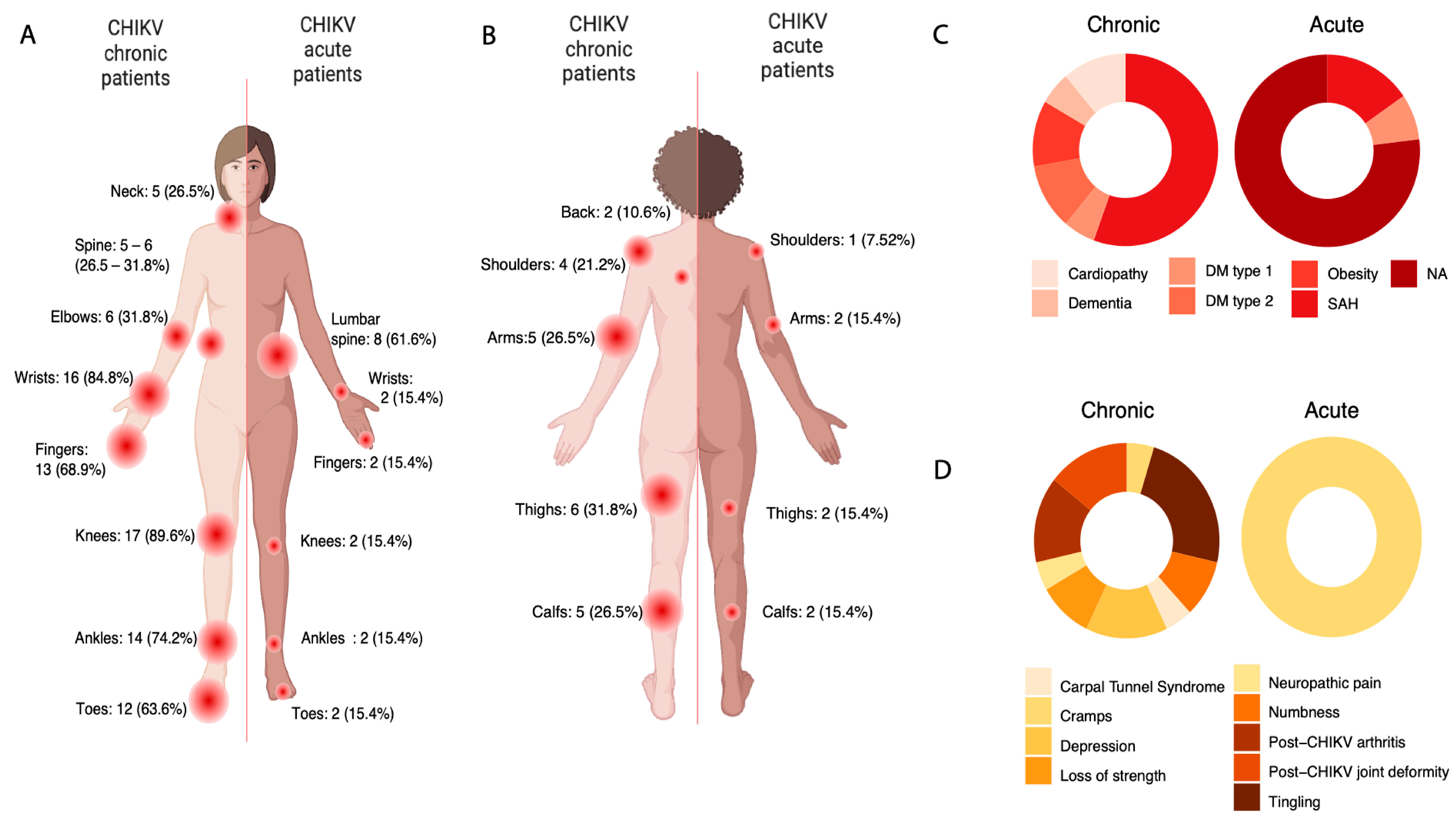
3.2. A Complex Symptomatological Scenario

The most prevalent symptoms, persisting into the chronic phase, included arthralgia (100%), with a notable occurrence of bilateral involvement (100%), edema (84.8%), in addition to myalgia (42.4%), transient arthralgia, and prostration (31.8% and 26.5%, respectively). The sites most affected by arthralgia were regions of the appendicular system, such as wrists (84.8%), fingers and toes (68.9% and 63.6%, respectively), ankles (74.2%), and knees (89.6%). Continued discomfort in axial skeleton regions, including the neck (26.5%) and lumbar spine (31.8%), was also reported (Figure 1A). Similar patterns were observed for myalgia, with 26.5% of individuals reporting muscular discomfort in the arms, shoulders, and calf regions, and 21.2% and 31.8% indicating the arms and thighs as the most affected regions, respectively (Figure 1B).
In acute cases, fever and myalgia were unanimously reported, followed by arthralgia (92.3%). Skin symptoms were reported in 53.8% of individuals, along with gastrointestinal symptoms such as nausea (61.6%). Additionally, 69.2% of acute cases reported migraine, while 53.8% presented retro-orbital pain. Focal points of myalgia and arthralgia were not adequately documented for ten individuals (76.9%).
Considering the potential impact of medications on immune response outcomes, medication intake at the time of collection and regular-use medications were recorded. Almost half (47.4%) of chronic patients reported using immunosuppressive or anti-inflammatory medication, while only 15.4% of individuals in the acute group reported use of these types of medication. The most commonly reported medications in the chronic group included opioids, corticosteroids, and antidepressants (Table S1). Additionally, some patients reported using muscle relaxants as complementary therapy.
Complementary therapies such as acupuncture, physical activity, and physiotherapy, aimed at improving joint mobility and recovery, were also reported in the chronic group (Table S1).

3.3. Clinical Data Suggests Possible Neuropathic Disorder in CHIKV Chronic

Pre-existing comorbidities and additional manifestations were also reported for both groups and are detailed in Figure 1C. Out of 19 chronic patients, 10 (50.3%) presented with high blood pressure (SAH). Obesity, type 2 diabetes mellitus, and heart disease were reported by 10.6% of patients. Interestingly, one patient (5.3%) reported a diagnosis of dementia. Concerning acute patients, the frequency of comorbidities was lower, with only two (15.4%) individuals having hypertension and one (7.52%) having type 1 diabetes, respectively (Figure 1C), probably due to the younger age among acute patients. No additional data on pre-existing diseases were available for 10 (76.9%) acute individuals.
Interestingly, additional manifestations were reported in the chronic group. After acute infection, five chronic patients (26.5%) developed tingling and symptoms compatible with neuropathic disease, such as numbness (10.6%), loss of strength (10.4%), and cramps (5.3%) (Figure 1D). Due to intense and constant pain, three patients (15.9%) developed depression, while 15.9% reported a diagnosis of postchikungunya arthritis and joint deformities, mainly affecting fingers and toes (Figure 1D).

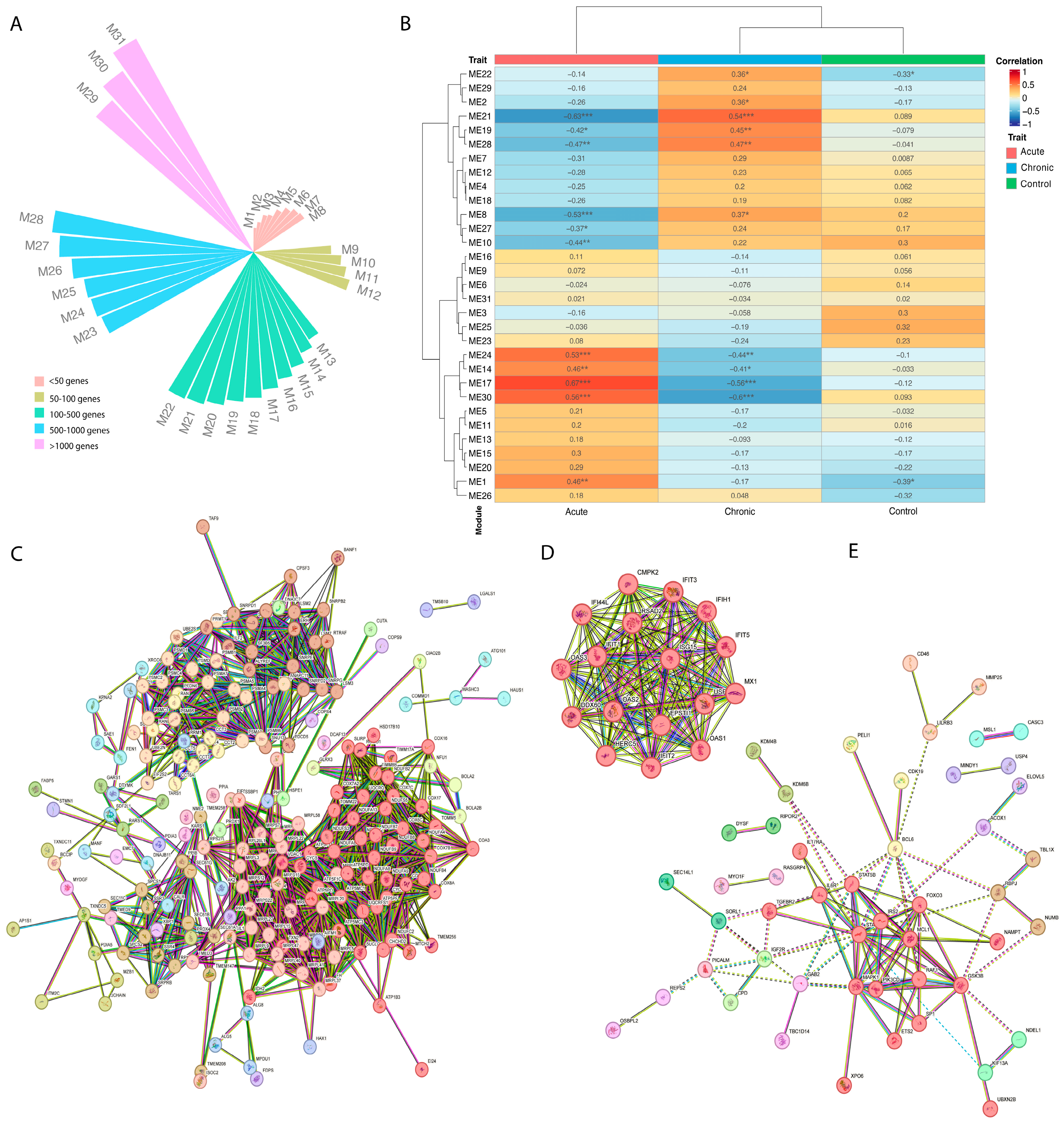
3.4. Expression Profile and Coexpression Analysis Identified Six Different Gene Sets

We used whole-blood RNA-seq to globally characterize the transcriptional changes occurring during the acute and chronic phases of chikungunya virus infection, focusing on the expression of proinflammatory mediators, such as cytokines and chemokines, as well as differential regulatory and immunological mechanisms correlated with different clinical stages. Detailed statistics of mapping and gene counts are available in Table S2.
When comparing the differential expression between chronic and acute patients against the control population, we found 2675 were found as differentially expressed (DE) (Figure S1A). For individuals with chronic disease, 2678 were differentially expressed, with 1727 (64.48%) showing elevated expression compared to controls (Table S3). In the acute group evaluation, 233 targets exhibited a positive LogFoldChange, indicating overexpression, while 130 (0.054%) showed reduced expression (Table S4 and Figure S1B). Interestingly, when comparing the differentially expressed transcripts between individuals in acute and chronic phases, 2676 genes were found to be either over- or underexpressed (Figure S1C and Table S5). Among these DE genes, 1725 genes (64.46%) were more abundant in the acute phase, while 951 (35.54%) were less expressed.
Although the difference in gene expression found between the groups was significant (p-value < 0.005), it was slightly associated with a small variation in the read count (Figure S1). In light of this, we carried out a more comprehensive evaluation seeking to describe the coexpression gene modules. Our aim was to understand better the relationship between these genes and the cellular and molecular processes they are involved in.
Using the Weighted Gene Coexpression Network Analysis (WGCNA) (version 1.72-5) [49], we identified 31 gene modules (M1–M31) that were differentially correlated with the Chikungunya infection stages investigated in the study (p-values < 0.005) (Figure 2A,B). Based on the p-values, six main modules were selected, namely M1, M17, M21-22, M28, and M30 (Figure 2B).
Modules M1, M17, and M30 were positively related to individuals in the acute phase and negatively associated with chronic patients or the control group, respectively (Figure 2B). Additionally, three modules suggest a greater correlation with individuals chronically impacted by the Chikungunya virus: modules 21, 22, and 28, with M21 and M28 negatively related to individuals in the acute phase. For both acute and chronic, the module sizes (higher than 500 and up to 1000 genes) (Figure 2A) suggest the complexity of disease stage scenarios.

3.5. CHIKV Acute Individuals Appear to Show Increase in Mitochondrial Respiratory Chain Genes, Besides Antiviral Expression Profile

The acute phase of Chikungunya virus infection is characterized by a considerable increase in proinflammatory cytokines and mediators responsible for initiating the antiviral response. However, CHIKV is also capable of promoting changes in basic energetic and transcriptional regulation processes, aiming to enhance virus replication and dissemination. In this study, we identified complex gene modules associated with hypermethylation, mitochondrial genes related to the electron transport chain and oxidative phosphorylation, as well as clusters associated with endosomes and methylosomes and splicing (Figure S2).
The M30 module consists of 2135 genes, of which 213 formed gene clusters (clusters 1–20) (Figure 2C). This module predominantly features genes involved in mitochondrial processes such as NADH to ubiquinone electron transport, proton-driven ATP synthesis, the aerobic electron transport chain, and oxidative phosphorylation (Table S9, Figure S2). Key genes like COX5A, COA3, and CYCS contribute to the cytochrome c oxidase complex, driving oxidative phosphorylation, while TXN2 regulates mitochondrial reactive oxygen species, apoptosis, and cell viability (Table S9). The M30 module also points to apoptosis regulation with genes like AIFM1, EIF5A, and LGALS1. AIFM1 may regulate caspase-independent apoptosis by relocating from the mitochondria to the nucleus, while EIF5A is linked to both p53/TP53-dependent and TNF-α-mediated apoptosis [53]. LGALS1 is a known regulator of T-cell apoptosis, cell proliferation, and differentiation [54,55]. However, despite the presence of apoptosis-related genes, enrichment analysis did not detect apoptosis pathways (Table S9).
The M17 module (Figure 2D) was found to be enriched in proteins and mediators associated with the response to interferons (Table S10). Enrichment analysis revealed increased biological processes such as the negative regulation of IP-1 production (2 of 3 genes, strength 2.89; FDR = 0.0024) and the negative regulation of chemokine (C-X-C motif) ligand 2 production (2 of 4 genes, strength 2.76; FDR = 0.0034). In acute phase individuals, we observed increases in antiviral pathways such as the OAS antiviral response (3 of 9 genes, strength 2.59; FDR ≤ 0.0001), interferon α/β signaling (11 of 71 genes, strength 2.25; FDR ≤ 0.0001), positive regulation of monocyte chemotactic protein-1 production (2 of 14 genes, strength 2.22; FDR = 0.02), negative regulation of viral genome replication (9 of 56 genes, strength 2.27; FDR ≤ 0.0001), and antiviral innate immune response pathway (3 of 22 genes, strength 2.2; FDR = 0.0005). Moreover, the gene EPSTI1 (epithelial-stromal interaction protein 1), important for M1 macrophage polarization, was also identified [40].
Of the 16 genes in this module, 13 perform antiviral functions mediated or induced by interferons, including the OAS family, which bind PPP-RNA to inhibit viral mRNA expression [56,57,58]. OAS2 and OAS3, activated by sRNA, also degrade cellular and viral RNA via ribonuclease L activation, halting viral replication. OAS3 may also influence apoptosis and gene regulation. An enriched pathway in our dataset is 7-methylguanosine cap hypermethylation (6 of 8 genes, strength: 1.85; FDR ≤ 0.0001) [56,57,58]. The module also includes IFIT1-3 and IFIT-5 genes, which complement OAS1-3 by inhibiting viral mRNA and inducing type I interferons and proinflammatory cytokines [59,60].

3.6. CHIKV Chronic Individuals Seem to Show Differentiation in Histones Methylation and Th-17 Regulation Genes

The factors that trigger the persistence of symptoms postinfection by Chikungunya virus in approximately 50% of the infected population and which can last months or years after the resolution of the acute phase remain unclear. In this study, we identified three overexpressed complex gene modules associated with chronic status: modules M21, M22, and M28.
Module M21, linked to erythrocyte development, showed a positive correlation with chronically affected CHIKV individuals and a negative correlation in acute-phase patients (Figure 2B). Pathways associated with erythrocyte development may reflect differences in cellularity (lymphocyte/erythrocyte ratio) between these phases. Notably, genes like RNF10 and TNS1 were identified (Supplementary Figure S3). RNF10, involved in MAG expression, may influence Schwann cell differentiation and myelination, potentially aiding nervous tissue repair in chronic individuals, consistent with neuropathic pain observed in acute patients. TNS1 may regulate cell migration and cartilage development, linking signal transduction pathways to the cytoskeleton [61,62].
Module M22 displayed enrichment in biological processes related to histone methylation, affecting transcriptional regulation (Table S11, Figure S4). Of 46 genes in this module, eight influence methylation on lysine residues, while four are involved in epigenetic marks. KMT2C and KMT2D methylate Lys-4 of histone H3, acting as nuclear receptor coactivators and marking for transcription [63,64]. KDM3B and TET2 remove methyl groups, with roles in active DNA demethylation [65]. Studies have shown viral infections, such as SARS-CoV-2 and arboviruses, can alter epigenetic profiles even after resolution [66,67].
In the M28 module (85 genes), pathways related to immune responses, including interleukin signaling (IL-2, 4, 6, 7, 9, 13, 15, 21) and CD46 receptor, were identified (Figure 2E, Table S12, Figure S4). Notably, three genes are linked to the regulation of the Th-17 immune profile (FDR = 0.018) and T-helper 17 cell lineage commitment (FDR = 0.0027), including IL-17RA, IL-6R, and STAT3, which regulate immune responses by inducing inflammatory chemokines and cytokines [56,57,68]. Chronic CHIKV patients also show upregulation of genes associated with neuronal death (FOXO3, MCL1, ZNF746, and PICALM) and T-cell differentiation, which may contribute to secondary neuropathy symptoms [69,70].

4. Discussion

Chikungunya fever, a disease caused by CHIKV infection, is characterized by an acute phase marked mainly by a high fever (>38.9 °C) of sudden onset. In this phase, classic symptoms of the infection, including arthralgia, become prominent and debilitating [71,72]. Additionally, as CHIKV viremia increases, the inflammatory condition becomes exacerbated, aiming to combat and restrict viral spread. Innate immunity cells are primarily responsible for attempting to suppress replication before an acquired immune response is triggered [73,74,75,76]. In this study, we aimed to identify differentially expressed transcripts in the peripheral blood of patients during distinct CHIKV infection phases.
The symptoms reported by our patients with acute infection align with those documented in the literature, especially when compared to those described in individuals infected by the CHIKV-ECSA genotype, the most prevalent genotype circulating in Brazil and in the municipality of Feira de Santana [14,15,16,17,18,19,20,21,22,23]. However, the persistence of post-infection joint symptoms, as well as the appearance of additional manifestations, lead to a daily pain sensation, resulting in the onset of depression in chronic individuals. Interestingly, a significant number of patients reported the use of amitriptyline and pregabalin (Table S1), which are among the first-line recommended drugs for neuropathic pain in addition to being used for depressive disorders [77,78].
We found that the spectrum of rheumatic and musculoskeletal disorders associated with post-chikungunya can include tunnel syndromes, modification of the joint axes, compromise of movement, and sometimes deformities [79,80,81,82]. This scenario has been reported in previous outbreaks in Réunion and India, indicating that the persistence of symptoms in a chronic postchikungunya stage impairs the individual’s quality of life [82,83,84]. Patients with postchikungunya rheumatoid arthritis may experience clinical improvement with methotrexate and hydroxychloroquine, which are used to treat classic rheumatoid arthritis [85]. Although methotrexate has been widely used in the clinic, it has not been reported in our population (Table S1).
The acute phase is marked by the overexpression of inflammatory mediators, such as IFN-α and γ, IL-2R, IL-6, and IL-9, as well as the presence of IL-8, IP-10, MCP-1, MIG, CXCL-9, CXCL10, MIP-1α and β chemokines, and growth factors G-CSF, GM-CSF, HGF, and VEGF-A [40,86,87,88,89,90,91]. In a recent study carried out in Brazil, acute patients have shown a similar expression profile, as reported here [92], for the same Chikungunya virus lineage (East-Central South African—ECSA). Additionally, due to the lack of information regarding the cycle threshold of CHIKV genome amplification, it was not possible to correlate the CHIKV viral load, the presence of multiple sites of arthralgia and myalgia, and the immune profiling.
After establishing an effective cellular infection, viral replication is initially limited by a rapid and robust production of proinflammatory mediators, such as Interferon-β and the downstream of signaling molecules. Despite rapid immune response against the virus triggered by interferons in permissive cells, CHIKV can effectively evade the cellular control mechanisms. One of the cytopathic effects caused by CHIKV is apoptosis, evidenced by the presence of numerous active caspase-3 and CHIKV double-positive HeLa cells [93]. Although the precise events leading to apoptosis are not yet fully understood, Jaffar-Bandjee et al. (2009) [93] suggest that both the intrinsic (mitochondrial) and extrinsic (Fas/TRAIL-like) apoptosis pathways are involved following CHIKV infection. Studies estimate that within 24 h postinfection, fibroblasts and stromal cells undergo apoptosis [94,95]. The intrinsic pathway, mediated by the release of cytochrome c in the mitochondria, activates caspase-9 and caspase-3 subsequentially; the extrinsic pathway seems to contribute, given the several upregulated death receptors and ligands. The increase in expression related to mitochondrial respiratory chain genes may be also related to the activation of the apoptosis intrinsic pathway found in the acute cases. The virus utilizes this cellular response to increase its rate of infection, and since the viral particles are sequestered within the apoptotic blebs, they escape recognition by the immune system. The engulfment of the apoptotic blebs by phagocytic cells, such as macrophages, can enhance infection silently [96]. Moreover, this mechanism triggers a heightened and dysregulated adaptive immune response, which may result in autoimmunity targeting self-antigens contained within apoptotic bodies [97,98], a process that requires further explanation as a potential trigger for an autoimmune response. Furthermore, the metabolic disturbances observed in these genes may be also associated with the exacerbated use of energy resources by host cells during cell infection [99,100,101,102].
In acute CHIKV patients, we observed an increase in the cap hypermethylation pathway, which involves the addition of the m7GMP cap structure (Cap0) to RNA. This structure is crucial for mRNA stability, processing, and translation [103,104,105] and is essential for viral protein synthesis. Additionally, capping helps viruses evade host immunity by preventing the recognition of terminal RNA phosphates by RIG-I and IFIT1 receptors [60,104,105,106]. In alphaviruses, the nsP1 protein adds cap0 structures to viral RNAs [106,107,108], but not all viral RNAs are capped, suggesting uncapped RNAs may modulate the immune response [109,110]. The increased expression of IFIT and RIG-1-like receptor genes (Table S5) and those involved in cap hypermethylation highlights an antiviral response aimed at controlling replication and viral protein translation. Further research on RNA capping in CHIKV infections is needed to develop treatments targeting this mechanism [104]. Overall, transcriptional changes during the acute phase rapidly engage immune and metabolic pathways to combat the infection.
Chronic joint involvement in Chikungunya virus infection shows similarities to rheumatoid arthritis, including shared symptoms and peripheral blood mononuclear cell profiles. Some studies attribute persistent symptoms to high levels of IL-6, GM-CSF, INF-α, and IL-17, a finding supported by our data. Other hypotheses suggest that viral RNA in the synovium and low-level replication in reservoir cells, like muscle satellite cells and synovial macrophages, drive chronic inflammation and autoimmunity [89,111,112]. More recently, epigenetic changes, such as DNA hypomethylation and histone modifications, have been described in rheumatoid arthritis and other viral infections like SARS-CoV-2 [66], though these mechanisms are less explored in Chikungunya [61].
Our findings also suggest that T-cell differentiation during the transition from acute to chronic infection may play a role in sequelae. Acute infection often involves lymphopenia and a predominance of CD8+ T cells expressing CD69, CD107a, granzyme B, and perforin, targeting virus-infected cells [36,37,113,114]. However, excessive antigenic exposure in the acute phase can exhaust these lymphocytes, reducing their efficacy in the chronic phase and potentially contributing to persistent symptoms [114,115]. Moreover, excessive cellular infiltrates, particularly from activated memory T-cells, can expose articular tissues to cytokines and chemokines like CXCL-9, CXCL-10, CCL2, IL-6, and IL-10, leading to tissue injury and edema [115,116,117]. While the role of T-cells in chronic Chikungunya infection remains unclear, an animal model study indicated that the absence of CD4+ T-cells reduces joint pathology [39]. Additionally, key lymphocyte subsets, such as Th17 and regulatory T cells (Tregs), may be involved in chronic infection. Th17 cells promote inflammation by producing IL-17, IL-22, and IL-23, while Tregs counterbalance by releasing anti-inflammatory cytokines like IL-10 and TGF-β, easing joint symptoms. Cytokine imbalance, particularly elevated IL-1b, IL-6, and IL-17, appears to drive persistent disease, as shown in multiple studies [37,40,118,119]. However, a study in Thailand during the 2009–2010 outbreak did not find a similar increase in IL-17, which could be due to methodological differences or a different CHIKV strain [120].
Even though our study highlights important candidates’ genes in the regulation and prognosis markers from different CHIKV infection phases, the small sample size may have reduced our ability to correctly identify these markers [121]. Furthermore, factors associated with the virus, such as strain and viral load during the acute phase, as well as factors associated with the sample type [122] and the study population [122], may add additional bias to the analyses. Above all, the use of immunosuppressive medication by the chronic population may affect gene expression and module identification [123], acting as a potential confounding factor for reproducibility and specificity of gene set analysis.
Taken together, our results highlight the high complexity of the distinct disease trajectories caused by the Chikungunya virus. They demonstrate the regulation of new genetic modules that can expand our understanding of the acute and chronic stages. However, further longitudinal studies on the role of immune regulation and different regulatory mechanisms are pivotal in order to provide a better understanding of the disease’s progression and to improve clinical management and patients’ quality of life.

Supplementary Materials

The following supporting information can be downloaded at: https://www.mdpi.com/article/10.3390/genes15111365/s1, Protocol S1: Procedure for extracting whole RNA from whole blood using Trizol LS and Chloroform; Figure S1: Differential gene expression among acute and chronic CHIKV individuals. A. Heatmap obtained from differentially expressed host transcripts between groups consisting of samples from acute and chronic individuals. Hierarchical clustering was applied to samples from the two clinical statuses (represented on the X-axis) and transcripts (Y-axis). B. Volcano plot of differentially expressed host genes between acute phase and control group samples, with average log CPM on the Y-axis and the log-fold change for each transcript on the Y-axis. Transcripts with p-value < 0.05 and log-fold change > |1.5| are colored red. C. Volcano plot of differentially expressed host genes between acute phase and chronic group samples, with average log CPM on the Y-axis and the log-fold change for each transcript on the Y-axis. Transcripts with p-value < 0.05 and log-fold change > |1.5| are colored red; Figure S2: Functional enrichment pathways for the genes positively correlated with CHIKV acute patients; Figure S3: Network of genes components of Module 21, negatively correlated with acute individuals and positively correlated with CHIKV chronic patients; Figure S4: Functional enrichments pathways for the genes positively correlated with CHIKV chronic patients; Table S1: Drug and complementary therapies among CHIKV-chronic and acute individuals; Table S2: Metrics and sequencing statistics regarding the read length, read counting, percentage of mapping, and gene count for all study individuals; Table S3: Differentially expressed genes found comparing chronic individuals and control group; Table S4: Differentially expressed genes found comparing acute individuals and control group; Table S5: Differentially expressed genes found comparing acute individuals and chronic group; Table S6: Coexpression module M30 MCL subclustering genes—positively correlated with CHIKV acute patients and negatively correlated with chronic individuals; Table S7: Coexpression module M17 MCL subclustering genes—positively correlated with CHIKV acute patients and negatively correlated with chronic individuals; Table S8: Coexpression module M28 MCL subclustering genes—negatively correlated with acute patients and positively correlated with CHIKV chronic individuals; Table S9: Functional enrichments for the genes belonging to Module 30—positively correlated with CHIKV acute patients and negatively correlated with chronic individuals; Table S10: Functional enrichments for the genes belonging to Module 17—positively correlated with CHIKV acute patients and negatively correlated with chronic individuals; Table S11: Functional enrichments for the genes belonging to Module 22—negatively correlated with the control group and positively correlated with CHIKV chronic; Table S12: Functional enrichments for the genes belonging to Module 28—negatively correlated with acute patients and positively correlated with CHIKV chronic individuals.

Author Contributions

Conceptualization, H.F., M.G., A.M.B.d.F. and L.C.J.A.; methodology, H.F., L.G.C., G.d.R.F., V.F., M.M.d.L., M.F., N.d.J., E.M.d.C., R.V.d.C., M.V.L.d.O.F., I.C.d.S., C.d.O., J.X., J.G.G.F., F.R.Q., E.S. and J.T.-G.; validation, H.F., M.G., L.G.C. and G.d.R.F.; formal analysis, H.F. and L.G.C.; investigation, H.F., M.G., M.M.d.L., M.F., N.d.J. and E.M.d.C.; resources, M.G., W.C.V.V., P.M.R., J.N.W., M.G.J., A.M.B.d.F. and L.C.J.A.; data curation, H.F., M.M.d.L. and M.F.; writing—original draft preparation, H.F., M.G. and L.G.C.; writing—review and editing, H.F., M.G., L.G.C., G.d.R.F., V.F., M.M.d.L., M.F., N.d.J., E.M.d.C., R.V.d.C., M.V.L.d.O.F., I.C.d.S., C.d.O., J.X., J.G.G.F., F.R.Q., E.S., J.T.-G., W.C.V.V., P.M.R., J.N.W., M.G.J., A.M.B.d.F. and L.C.J.A.; visualization, H.F.; supervision, M.G., G.d.R.F. and L.C.J.A.; project administration, M.G. and L.C.J.A.; funding acquisition, M.G., W.C.V.V., P.M.R., J.N.W., M.G.J., A.M.B.d.F. and L.C.J.A. All authors have read and agreed to the published version of the manuscript.

Funding

This work was supported in part through National Institutes of Health USA grant U01 AI151698 for the United World Arbovirus Research Network (UWARN), by the Brazilian Ministry of Health (SCON2021-00180), and by the Brazilian National Council for Scientific and Technological Development (CNPq-316456/2021-7). M.G. is funded by PON “Ricerca e Innovazione” 2014–2020 and by the CRP- ICGEB RESEARCH GRANT 2020 Project CRP/BRA20-03. H.F. is supported by the Brazilian National Council for Scientific and Technological Development (CNPq-141861/2020-7). ICS is supported by CNPq (316456/2021-7). LC is supported by CAPES (8887-705131/2022-00).

Institutional Review Board Statement

This research and recruitment locations were approved by the University of Washington IRB Committee (protocol: STUDY00009300), the Ethics Committee of the Pan American World Health Organization (No. PAHO-2016-08-0029), the Oswaldo Cruz Foundation Ethics Committee (CAAE: 45279715.8.0000.0040), the Federal University of Minas Gerais Ethics Committee (CAAE: 32912820.6.1001.5149), and the Brazilian Ministry of Health (MoH) as part of the arbovirus genomic surveillance efforts. All patients consented to participate by signing a consent form.

Informed Consent Statement

Informed consent was obtained from all subjects involved in the study.

Data Availability Statement

The original data presented in the study are openly available in the European Nucleotide Archive (ENA), submitted under accession code PRJEB74939. Further inquiries can be directed to the corresponding authors.

Acknowledgments

To patients and family members for participating and trusting in the research. To the health professionals, collection team, and laboratory workers of the Municipal Health Department of Feira de Santana for their tireless work in the municipality’s epidemiological surveillance. To all centers for chronic Chikungunya patients and the medical team, especially Melissa Falcão and Rivaldo Venâncio for monitoring patients and clinical management. To Maricelia Lima and Neuza de Jesus for their assistance in recruitment and collection and whose efforts made this project possible. The authors are grateful to the Plataforma Genômica—Sequenciamento de Nova Geração—RPT01J—Rede de Plataformas Tecnológicas FIOCRUZ. The Bioinformatics Platform at the René Rachou Institute was used, where the analyses were carried out. The Oswaldo Cruz Institute’s Next Generation Sequencing Platform team for carrying out the library preparation and sequencing. To UWARN and Michael Gale’s group for the study design and analysis assistance. The team from Mario Penna Instituto for providing the laboratory infrastructure for sample processing. The authors would also like to acknowledge the Global Consortium to Identify and Control Epidemics—CLIMADE, (L.C.J.A., M.G.) (https://climade.health/, accessed on 5 January 2020).

Conflicts of Interest

The authors declare no conflicts of interest.

References

  1. Weaver, S.C. Host Range, Amplification and Arboviral Disease Emergence. In Infectious Diseases from Nature: Mechanisms of Viral Emergence and Persistence; Peters, C.J., Calisher, C.H., Eds.; Springer-Verlag: Vienna, Austria, 2005; pp. 33–44. [Google Scholar] [CrossRef]
  2. Schwartz, O.; Albert, M.L. Biology and Pathogenesis of Chikungunya Virus. Nat. Rev. Microbiol. 2010, 8, 491–500. [Google Scholar] [CrossRef]
  3. PAHO, P.A.H. ORGANIZATION. Cases of Chikungunya Virus Disease. Available online: https://www3.paho.org/data/index.php/en/mnu-topics/chikv-en/550-chikv-weekly-en.html (accessed on 5 July 2022).
  4. Pialoux, G.; Gaüzère, B.-A.; Jauréguiberry, S.; Strobel, M. Chikungunya, an Epidemic Arbovirosis. Lancet Infect. Dis. 2007, 7, 319–327. [Google Scholar] [CrossRef] [PubMed]
  5. Cunha, M.S.; Costa, P.A.G.; Correa, I.A.; de Souza, M.R.M.; Calil, P.T.; da Silva, G.P.D.; Costa, S.M.; Fonseca, V.W.P.; da Costa, L.J. Chikungunya Virus: An Emergent Arbovirus to the South American Continent and a Continuous Threat to the World. Front. Microbiol. 2020, 11, 1297. [Google Scholar] [CrossRef] [PubMed]
  6. Weaver, S.C.; Lecuit, M. Chikungunya Virus and the Global Spread of a Mosquito-Borne Disease. N. Engl. J. Med. 2015, 372, 1231–1239. [Google Scholar] [CrossRef] [PubMed]
  7. Strauss, J.H.; Strauss, E.G. The Alphaviruses: Gene Expression, Replication, and Evolution. Microbiol. Rev. 1994, 58, 491–562. [Google Scholar] [CrossRef]
  8. Powers, A.M.; Brault, A.C.; Tesh, R.B.; Weaver, S.C. Re-Emergence of Chikungunya and O’nyong-Nyong Viruses: Evidence for Distinct Geographical Lineages and Distant Evolutionary Relationships. J. Gen. Virol. 2000, 81 Pt 2, 471–479. [Google Scholar] [CrossRef]
  9. Powers, A.M. Genomic Evolution and Phenotypic Distinctions of Chikungunya Viruses Causing the Indian Ocean Outbreak. Exp. Biol. Med. 2011, 236, 909–914. [Google Scholar] [CrossRef]
  10. Schuffenecker, I.; Iteman, I.; Michault, A.; Murri, S.; Frangeul, L.; Vaney, M.-C.; Lavenir, R.; Pardigon, N.; Reynes, J.-M.; Pettinelli, F.; et al. Genome Microevolution of Chikungunya Viruses Causing the Indian Ocean Outbreak. PLoS Med. 2006, 3, e263. [Google Scholar] [CrossRef]
  11. Bustos Carrillo, F.; Collado, D.; Sanchez, N.; Ojeda, S.; Lopez Mercado, B.; Burger-Calderon, R.; Gresh, L.; Gordon, A.; Balmaseda, A.; Kuan, G.; et al. Epidemiological Evidence for Lineage-Specific Differences in the Risk of Inapparent Chikungunya Virus Infection. J. Virol. 2019, 93. [Google Scholar] [CrossRef]
  12. Mascarenhas, M.; Garasia, S.; Berthiaume, P.; Corrin, T.; Greig, J.; Ng, V.; Young, I.; Waddell, L. A Scoping Review of Published Literature on Chikungunya Virus. PLoS ONE 2018, 13, e0207554. [Google Scholar] [CrossRef]
  13. Pan American Health Organization. PLISA Health Information Platform for the Americas—Pan American Health Organization: Cases of Chikungunya Virus Disease; Pan American Health Organization: Washington, DC, USA, 2021. [Google Scholar]
  14. Nunes, M.R.T.; Faria, N.R.; de Vasconcelos, J.M.; Golding, N.; Kraemer, M.U.G.; de Oliveira, L.F.; da Silva Azevedo, R.o.D.; da Silva, D.E.A.; da Silva, E.V.P.; da Silva, S.P.; et al. Emergence and Potential for Spread of Chikungunya Virus in Brazil. BMC Med. 2015, 13, 102. [Google Scholar] [CrossRef]
  15. de Souza, T.M.A.; Ribeiro, E.D.; Corrêa, V.C.E.; Damasco, P.V.; Santos, C.C.; de Bruycker-Nogueira, F.; Chouin-Carneiro, T.; Faria, N.R.; Nunes, P.C.G.; Heringer, M.; et al. Following in the Footsteps of the Chikungunya Virus in Brazil: The First Autochthonous Cases in Amapá in 2014 and Its Emergence in Rio de Janeiro during 2016. Viruses 2018, 10, 623. [Google Scholar] [CrossRef] [PubMed]
  16. Brito Ferreira, M.L.; de Fatima Pessoa Militão de Albuquerque, M.; de Brito, C.A.A.; de Oliveira França, R.F.; Porto Moreira, Á.J.; de Morais Machado, M.Í.; da Paz Melo, R.; Medialdea-Carrera, R.; Dornelas Mesquita, S.; Lopes Santos, M.; et al. Neurological Disease in Adults with Zika and Chikungunya Virus Infection in Northeast Brazil: A Prospective Observational Study. Lancet Neurol. 2020, 19, 826–839. [Google Scholar] [CrossRef] [PubMed]
  17. Xavier, J.; Alcantara, L.C.J.; Fonseca, V.; Lima, M.; Castro, E.; Fritsch, H.; Oliveira, C.; Guimarães, N.; Adelino, T.; Evaristo, M.; et al. Increased Interregional Virus Exchange and Nucleotide Diversity Outline the Expansion of Chikungunya Virus in Brazil. Nat. Commun. 2023, 14, 4413. [Google Scholar] [CrossRef] [PubMed]
  18. Souza, T.M.A.; Azeredo, E.L.; Badolato-Corrêa, J.; Damasco, P.V.; Santos, C.; Petitinga-Paiva, F.; Nunes, P.C.G.; Barbosa, L.S.; Cipitelli, M.C.; Chouin-Carneiro, T.; et al. First Report of the East-Central South African Genotype of Chikungunya Virus in Rio de Janeiro, Brazil. PLoS Curr. Influenza 2017, 9. [Google Scholar] [CrossRef]
  19. Xavier, J.; Fonseca, V.; Bezerra, J.F.; do Monte Alves, M.; Mares-Guia, M.A.; Claro, I.M.; de Jesus, R.; Adelino, T.; Araújo, E.; Cavalcante, K.R.L.J.; et al. Chikungunya Virus ECSA Lineage Reintroduction in the Northeasternmost Region of Brazil. Int. J. Infect. Dis. 2021, 105, 120–123. [Google Scholar] [CrossRef]
  20. Périssé, A.R.S.; Souza-Santos, R.; Duarte, R.; Santos, F.; de Andrade, C.R.; Rodrigues, N.C.P.; de Andrade Schramm, J.M.; da Silva, E.D.; da Silva Viana Jacobson, L.; Lemos, M.C.F.; et al. Zika, Dengue and Chikungunya Population Prevalence in Rio de Janeiro City, Brazil, and the Importance of Seroprevalence Studies to Estimate the Real Number of Infected Individuals. PLoS ONE 2020, 15, e0243239. [Google Scholar] [CrossRef]
  21. Monteiro, J.D.; Valverde, J.G.; Morais, I.C.; de Medeiros Souza, C.R.; Fagundes Neto, J.C.; de Melo, M.F.; Nascimento, Y.M.; Alves, B.E.B.; de Medeiros, L.G.; Pereira, H.W.B.; et al. Epidemiologic and Clinical Investigations during a Chikungunya Outbreak in Rio Grande Do Norte State, Brazil. PLoS ONE 2020, 15, e0241799. [Google Scholar] [CrossRef]
  22. Fritsch, H.; Giovanetti, M.; Xavier, J.; Adelino, T.E.R.; Fonseca, V.; de Jesus, J.G.; de Jesus, R.; Freitas, C.; Peterka, C.R.L.; Campelo de Albuquerque, C.F.; et al. Retrospective Genomic Surveillance of Chikungunya Transmission in Minas Gerais State, Southeast Brazil. Microbiol. Spectr. 2022, 10, e0128522. [Google Scholar] [CrossRef]
  23. de Souza, W.M.; de Lima, S.T.S.; Simões Mello, L.M.; Candido, D.S.; Buss, L.; Whittaker, C.; Claro, I.M.; Chandradeva, N.; Granja, F.; de Jesus, R.; et al. Spatiotemporal Dynamics and Recurrence of Chikungunya Virus in Brazil: An Epidemiological Study. Lancet Microbe 2023, 4, e319–e329. [Google Scholar] [CrossRef]
  24. Jaffar-Bandjee, M.C.; Ramful, D.; Gauzere, B.A.; Hoarau, J.J.; Krejbich-Trotot, P.; Robin, S.; Ribera, A.; Selambarom, J.; Gasque, P. Emergence and Clinical Insights into the Pathology of Chikungunya Virus Infection. Expert Rev. Anti Infect. Ther. 2010, 8, 987–996. [Google Scholar] [CrossRef]
  25. Bastos, M.L.A.; de Abreu, F.S.; da Silva Junior, G.B. Inability to Work Due to Chikungunya Virus Infection: Impact on Public Service during the First Epidemic in the State of Ceará, Northeastern Brazil. Braz. J. Infect. Dis. 2018, 22, 248–249. [Google Scholar] [CrossRef] [PubMed]
  26. Hossain, M.S.; Hasan, M.M.; Islam, M.S.; Islam, S.; Mozaffor, M.; Khan, M.A.S.; Ahmed, N.; Akhtar, W.; Chowdhury, S.; Arafat, S.M.Y.; et al. Chikungunya Outbreak (2017) in Bangladesh: Clinical Profile, Economic Impact and Quality of Life during the Acute Phase of the Disease. PLoS Negl. Trop. Dis. 2018, 12, e0006561. [Google Scholar] [CrossRef] [PubMed]
  27. Padmakumar, B.; Jayan, J.B.; Menon, R.M.; Krishnankutty, B.; Payippallil, R.; Nisha, R.S. Comparative Evaluation of Four Therapeutic Regimes in Chikungunya Arthritis: A Prospective Randomized Parallel-Group Study. Indian J. Rheumatol. 2009, 4, 94–101. [Google Scholar] [CrossRef]
  28. Guaraldo, L.; Wakimoto, M.D.; Ferreira, H.; Bressan, C.; Calvet, G.A.; Pinheiro, G.C.; Siqueira, A.M.; Brasil, P. Treatment of Chikungunya Musculoskeletal Disorders: A Systematic Review. Expert Rev. Anti Infect. Ther. 2018, 16, 333–344. [Google Scholar] [CrossRef] [PubMed]
  29. Suhrbier, A.; Jaffar-Bandjee, M.-C.; Gasque, P. Arthritogenic Alphaviruses—An Overview. Nat. Rev. Rheumatol. 2012, 8, 420–429. [Google Scholar] [CrossRef]
  30. Teng, T.-S.; Foo, S.-S.; Simamarta, D.; Lum, F.-M.; Teo, T.-H.; Lulla, A.; Yeo, N.K.W.; Koh, E.G.L.; Chow, A.; Leo, Y.-S.; et al. Viperin Restricts Chikungunya Virus Replication and Pathology. J. Clin. Investig. 2012, 122, 4447–4460. [Google Scholar] [CrossRef]
  31. Schilte, C.; Buckwalter, M.R.; Laird, M.E.; Diamond, M.S.; Schwartz, O.; Albert, M.L. Cutting Edge: Independent Roles for IRF-3 and IRF-7 in Hematopoietic and Nonhematopoietic Cells during Host Response to Chikungunya Infection. J. Immunol. 2012, 188, 2967–2971. [Google Scholar] [CrossRef]
  32. Joubert, P.-E.; Werneke, S.; de la Calle, C.; Guivel-Benhassine, F.; Giodini, A.; Peduto, L.; Levine, B.; Schwartz, O.; Lenschow, D.; Albert, M.L. Chikungunya Virus–Induced Autophagy Delays Caspase-Dependent Cell Death. J. Cell Biol. 2012, 197, i5. [Google Scholar] [CrossRef]
  33. Schilte, C.; Staikowsky, F.; Couderc, T.; Madec, Y.; Carpentier, F.; Kassab, S.; Albert, M.L.; Lecuit, M.; Michault, A. Chikungunya Virus-Associated Long-Term Arthralgia: A 36-Month Prospective Longitudinal Study. PLoS Negl. Trop. Dis. 2013, 7, e2137. [Google Scholar] [CrossRef]
  34. Venugopalan, A.; Ghorpade, R.P.; Chopra, A. Cytokines in Acute Chikungunya. PLoS ONE 2014, 9, e111305. [Google Scholar] [CrossRef] [PubMed]
  35. Kelvin, A.A.; Banner, D.; Silvi, G.; Moro, M.L.; Spataro, N.; Gaibani, P.; Cavrini, F.; Pierro, A.; Rossini, G.; Cameron, M.J.; et al. Inflammatory Cytokine Expression Is Associated with Chikungunya Virus Resolution and Symptom Severity. PLoS Negl. Trop. Dis. 2011, 5, e1279. [Google Scholar] [CrossRef] [PubMed]
  36. Hoarau, J.-J.; Jaffar Bandjee, M.-C.; Krejbich Trotot, P.; Das, T.; Li-Pat-Yuen, G.; Dassa, B.; Denizot, M.; Guichard, E.; Ribera, A.; Henni, T.; et al. Persistent Chronic Inflammation and Infection by Chikungunya Arthritogenic Alphavirus in Spite of a Robust Host Immune Response. J. Immunol. 2010, 184, 5914–5927. [Google Scholar] [CrossRef]
  37. Wauquier, N.; Becquart, P.; Nkoghe, D.; Padilla, C.; Ndjoyi-Mbiguino, A.; Leroy, E.M. The Acute Phase of Chikungunya Virus Infection in Humans Is Associated with Strong Innate Immunity and T CD8 Cell Activation. J. Infect. Dis. 2011, 204, 115–123. [Google Scholar] [CrossRef] [PubMed]
  38. Chow, A.; Her, Z.; Ong, E.K.S.; Chen, J.; Dimatatac, F.; Kwek, D.J.C.; Barkham, T.; Yang, H.; Rénia, L.; Leo, Y.-S.; et al. Persistent Arthralgia Induced by Chikungunya Virus Infection Is Associated with Interleukin-6 and Granulocyte Macrophage Colony-Stimulating Factor. J. Infect. Dis. 2011, 203, 149–157. [Google Scholar] [CrossRef]
  39. Reddy, V.; Mani, R.S.; Desai, A.; Ravi, V. Correlation of Plasma Viral Loads and Presence of Chikungunya IgM Antibodies with Cytokine/Chemokine Levels during Acute Chikungunya Virus Infection. J. Med. Virol. 2014, 86, 1393–1401. [Google Scholar] [CrossRef]
  40. Ng, L.F.P.; Chow, A.; Sun, Y.-J.; Kwek, D.J.C.; Lim, P.-L.; Dimatatac, F.; Ng, L.-C.; Ooi, E.-E.; Choo, K.-H.; Her, Z.; et al. IL-1beta, IL-6, and RANTES as Biomarkers of Chikungunya Severity. PLoS ONE 2009, 4, e4261. [Google Scholar] [CrossRef]
  41. Kittleson, M.M.; Hare, J.M. Molecular Signature Analysis: Using the Myocardial Transcriptome as a Biomarker in Cardiovascular Disease. Trends Cardiovasc. Med. 2005, 15, 130–138. [Google Scholar] [CrossRef]
  42. Mohr, S.; Liew, C.-C. The Peripheral-Blood Transcriptome: New Insights into Disease and Risk Assessment. Trends Mol. Med. 2007, 13, 422–432. [Google Scholar] [CrossRef]
  43. Liew, C.-C.; Dzau, V.J. Molecular Genetics and Genomics of Heart Failure. Nat. Rev. Genet. 2004, 5, 811–825. [Google Scholar] [CrossRef]
  44. Liew, C.-C.; Ma, J.; Tang, H.-C.; Zheng, R.; Dempsey, A.A. The Peripheral Blood Transcriptome Dynamically Reflects System Wide Biology: A Potential Diagnostic Tool. J. Lab. Clin. Med. 2006, 147, 126–132. [Google Scholar] [CrossRef] [PubMed]
  45. Martin, M. Cutadapt Removes Adapter Sequences from High-Throughput Sequencing Reads. EMBnet J. 2011, 17, 10. [Google Scholar] [CrossRef]
  46. Dobin, A.; Davis, C.A.; Schlesinger, F.; Drenkow, J.; Zaleski, C.; Jha, S.; Batut, P.; Chaisson, M.; Gingeras, T.R. STAR: Ultrafast Universal RNA-Seq Aligner. Bioinformatics 2013, 29, 15–21. [Google Scholar] [CrossRef] [PubMed]
  47. Pertea, M.; Pertea, G.M.; Antonescu, C.M.; Chang, T.-C.; Mendell, J.T.; Salzberg, S.L. StringTie Enables Improved Reconstruction of a Transcriptome from RNA-Seq Reads. Nat. Biotechnol. 2015, 33, 290–295. [Google Scholar] [CrossRef] [PubMed]
  48. Robinson, M.D.; McCarthy, D.J.; Smyth, G.K. EdgeR: A Bioconductor Package for Differential Expression Analysis of Digital Gene Expression Data. Bioinformatics 2010, 26, 139–140. [Google Scholar] [CrossRef] [PubMed]
  49. Langfelder, P.; Horvath, S. WGCNA: An R Package for Weighted Correlation Network Analysis. BMC Bioinform. 2008, 9, 559. [Google Scholar] [CrossRef]
  50. Reimand, J.; Kull, M.; Peterson, H.; Hansen, J.; Vilo, J. G:Profiler—A Web-Based Toolset for Functional Profiling of Gene Lists from Large-Scale Experiments. Nucleic Acids Res. 2007, 35, W193–W200. [Google Scholar] [CrossRef]
  51. Kolberg, L.; Raudvere, U.; Kuzmin, I.; Adler, P.; Vilo, J.; Peterson, H. G:Profiler-Interoperable Web Service for Functional Enrichment Analysis and Gene Identifier Mapping (2023 Update). Nucleic Acids Res. 2023, 51, W207–W212. [Google Scholar] [CrossRef]
  52. Van Dongen, S. Graph Clustering via a Discrete Uncoupling Process. SIAM. J. Matrix Anal. Appl. 2008, 30, 121–141. [Google Scholar] [CrossRef]
  53. Sfakianos, A.P.; Raven, R.M.; Willis, A.E. The Pleiotropic Roles of EIF5A in Cellular Life and Its Therapeutic Potential in Cancer. Biochem. Soc. Trans. 2022, 50, 1885–1895. [Google Scholar] [CrossRef]
  54. Wei, J.; Wu, Y.; Sun, Y.; Chen, D. Galectin-1 Regulates RNA Expression and Alternative Splicing of Angiogenic Genes in HUVECs. Front. Biosci. (Landmark Ed) 2023, 28, 74. [Google Scholar] [CrossRef] [PubMed]
  55. Hsu, D.K.; Yang, R.-Y.; Liu, F.-T. Galectins in Apoptosis. Meth. Enzymol. 2006, 417, 256–273. [Google Scholar] [CrossRef]
  56. Koul, A.; Deo, S.; Booy, E.P.; Orriss, G.L.; Genung, M.; McKenna, S.A. Impact of Double-Stranded RNA Characteristics on the Activation of Human 2′-5′-Oligoadenylate Synthetase 2 (OAS2). Biochem. Cell Biol. 2020, 98, 70–82. [Google Scholar] [CrossRef]
  57. Hornung, V.; Hartmann, R.; Ablasser, A.; Hopfner, K.-P. OAS Proteins and CGAS: Unifying Concepts in Sensing and Responding to Cytosolic Nucleic Acids. Nat. Rev. Immunol. 2014, 14, 521–528. [Google Scholar] [CrossRef]
  58. Huffman, J.E.; Butler-Laporte, G.; Khan, A.; Pairo-Castineira, E.; Drivas, T.G.; Peloso, G.M.; Nakanishi, T.; COVID-19 Host Genetics Initiative; Ganna, A.; Verma, A.; et al. Multi-Ancestry Fine Mapping Implicates OAS1 Splicing in Risk of Severe COVID-19. Nat. Genet. 2022, 54, 125–127. [Google Scholar] [CrossRef]
  59. Wu, Y.; Song, X.; Cui, D.; Zhang, T. IFIT3 and IFIT5 Play Potential Roles in Innate Immune Response of Porcine Pulmonary Microvascular Endothelial Cells to Highly Pathogenic Porcine Reproductive and Respiratory Syndrome Virus. Viruses 2022, 14. [Google Scholar] [CrossRef]
  60. Hyde, J.L.; Diamond, M.S. Innate Immune Restriction and Antagonism of Viral RNA Lacking 2′-O Methylation. Virology 2015, 479–480, 66–74. [Google Scholar] [CrossRef]
  61. Wang, Z.; Ye, J.; Dong, F.; Cao, L.; Wang, M.; Sun, G. TNS1: Emerging Insights into Its Domain Function, Biological Roles, and Tumors. Biology 2022, 11, 1571. [Google Scholar] [CrossRef]
  62. Hoshikawa, S.; Ogata, T.; Fujiwara, S.; Nakamura, K.; Tanaka, S. A Novel Function of RING Finger Protein 10 in Transcriptional Regulation of the Myelin-Associated Glycoprotein Gene and Myelin Formation in Schwann Cells. PLoS ONE 2008, 3, e3464. [Google Scholar] [CrossRef]
  63. Lavery, W.J.; Barski, A.; Wiley, S.; Schorry, E.K.; Lindsley, A.W. KMT2C/D COMPASS Complex-Associated Diseases [KCDCOM-ADs]: An Emerging Class of Congenital Regulopathies. Clin. Epigenetics 2020, 12, 10. [Google Scholar] [CrossRef]
  64. Froimchuk, E.; Jang, Y.; Ge, K. Histone H3 Lysine 4 Methyltransferase KMT2D. Gene 2017, 627, 337–342. [Google Scholar] [CrossRef] [PubMed]
  65. Rasmussen, K.D.; Helin, K. Role of TET Enzymes in DNA Methylation, Development, and Cancer. Genes Dev. 2016, 30, 733–750. [Google Scholar] [CrossRef]
  66. Huoman, J.; Sayyab, S.; Apostolou, E.; Karlsson, L.; Porcile, L.; Rizwan, M.; Sharma, S.; Das, J.; Rosén, A.; Lerm, M. Epigenetic Rewiring of Pathways Related to Odour Perception in Immune Cells Exposed to SARS-CoV-2 in Vivo and in Vitro. Epigenetics 2022, 17, 1875–1891. [Google Scholar] [CrossRef]
  67. de Aguiar, G.P.C.G.; da Silva Leite, C.M.G.; Dias, B.; Vasconcelos, S.M.M.; de Moraes, R.A.; de Moraes, M.E.A.; Vallinoto, A.C.R.; Macedo, D.S.; de Goes Cavalcanti, L.P.; Miyajima, F. Evidence for Host Epigenetic Signatures Arising from Arbovirus Infections: A Systematic Review. Front. Immunol. 2019, 10, 1207. [Google Scholar] [CrossRef]
  68. Platanias, L.C. Mechanisms of Type-I- and Type-II-Interferon-Mediated Signalling. Nat. Rev. Immunol. 2005, 5, 375–386. [Google Scholar] [CrossRef]
  69. Zhao, Y.; Liu, Y.-S. Longevity Factor FOXO3: A Key Regulator in Aging-Related Vascular Diseases. Front. Cardiovasc. Med. 2021, 8, 778674. [Google Scholar] [CrossRef]
  70. Widden, H.; Placzek, W.J. The Multiple Mechanisms of MCL1 in the Regulation of Cell Fate. Commun. Biol. 2021, 4, 1029. [Google Scholar] [CrossRef]
  71. Parola, P.; de Lamballerie, X.; Jourdan, J.; Rovery, C.; Vaillant, V.; Minodier, P.; Brouqui, P.; Flahault, A.; Raoult, D.; Charrel, R.N. Novel Chikungunya Virus Variant in Travelers Returning from Indian Ocean Islands. Emerging Infect. Dis. 2006, 12, 1493–1499. [Google Scholar] [CrossRef]
  72. Kam, Y.-W.; Ong, E.K.S.; Rénia, L.; Tong, J.-C.; Ng, L.F.P. Immuno-Biology of Chikungunya and Implications for Disease Intervention. Microbes Infect. 2009, 11, 1186–1196. [Google Scholar] [CrossRef]
  73. Rehermann, B. Hepatitis C Virus versus Innate and Adaptive Immune Responses: A Tale of Coevolution and Coexistence. J. Clin. Investig. 2009, 119, 1745–1754. [Google Scholar] [CrossRef]
  74. Grieder, F.B.; Nguyen, H.T. Virulent and Attenuated Mutant Venezuelan Equine Encephalitis Virus Show Marked Differences in Replication in Infection in Murine Macrophages. Microb. Pathog. 1996, 21, 85–95. [Google Scholar] [CrossRef] [PubMed]
  75. Coleman, C.M.; Wu, L. HIV Interactions with Monocytes and Dendritic Cells: Viral Latency and Reservoirs. Retrovirology 2009, 6, 51. [Google Scholar] [CrossRef] [PubMed]
  76. Kou, Z.; Quinn, M.; Chen, H.; Rodrigo, W.W.S.I.; Rose, R.C.; Schlesinger, J.J.; Jin, X. Monocytes, but Not T or B Cells, Are the Principal Target Cells for Dengue Virus (DV) Infection among Human Peripheral Blood Mononuclear Cells. J. Med. Virol. 2008, 80, 134–146. [Google Scholar] [CrossRef] [PubMed]
  77. Obata, H. Analgesic Mechanisms of Antidepressants for Neuropathic Pain. Int. J. Mol. Sci. 2017, 18, 2483. [Google Scholar] [CrossRef]
  78. Derry, S.; Bell, R.F.; Straube, S.; Wiffen, P.J.; Aldington, D.; Moore, R.A. Pregabalin for Neuropathic Pain in Adults. Cochrane Database Syst. Rev. 2019, 1, CD007076. [Google Scholar] [CrossRef]
  79. Foissac, M.; Javelle, E.; Ray, S.; Guérin, B.; Simon, F. Post-Chikungunya Rheumatoid Arthritis, Saint Martin. Emerging Infect. Dis. 2015, 21, 530–532. [Google Scholar] [CrossRef]
  80. Ribéra, A.; Degasne, I.; Jaffar Bandjee, M.C.; Gasque, P. Chronic Rheumatic Manifestations Following Chikungunya Virus Infection: Clinical Description and Therapeutic Considerations. Med. Trop. 2012, 72, 83–85. [Google Scholar]
  81. Bouquillard, E.; Combe, B. A Report of 21 Cases of Rheumatoid Arthritis Following Chikungunya Fever. A Mean Follow-up of Two Years. Jt. Bone Spine 2009, 76, 654–657. [Google Scholar] [CrossRef]
  82. Mathew, A.J.; Goyal, V.; George, E.; Thekkemuriyil, D.V.; Jayakumar, B.; Chopra, A.; Trivandrum COPCORD Study Group. Rheumatic-Musculoskeletal Pain and Disorders in a Naïve Group of Individuals 15 Months Following a Chikungunya Viral Epidemic in South India: A Population Based Observational Study. Int. J. Clin. Pract. 2011, 65, 1306–1312. [Google Scholar] [CrossRef]
  83. Chopra, A.; Anuradha, V.; Ghorpade, R.; Saluja, M. Acute Chikungunya and Persistent Musculoskeletal Pain Following the 2006 Indian Epidemic: A 2-Year Prospective Rural Community Study. Epidemiol. Infect. 2012, 140, 842–850. [Google Scholar] [CrossRef]
  84. Marimoutou, C.; Vivier, E.; Oliver, M.; Boutin, J.-P.; Simon, F. Morbidity and Impaired Quality of Life 30 Months after Chikungunya Infection: Comparative Cohort of Infected and Uninfected French Military Policemen in Reunion Island. Medicine 2012, 91, 212–219. [Google Scholar] [CrossRef] [PubMed]
  85. Saag, K.G.; Teng, G.G.; Patkar, N.M.; Anuntiyo, J.; Finney, C.; Curtis, J.R.; Paulus, H.E.; Mudano, A.; Pisu, M.; Elkins-Melton, M.; et al. American College of Rheumatology. American College of Rheumatology 2008 Recommendations for the Use of Nonbiologic and Biologic Disease-Modifying Antirheumatic Drugs in Rheumatoid Arthritis. Arthritis Rheum. 2008, 59, 762–784. [Google Scholar] [CrossRef] [PubMed]
  86. Babu, N.; Mahilkar, S.; Jayaram, A.; Ibemgbo, S.A.; Mathur, G.; Shetty, U.; Sudandiradas, R.; Kumar, P.S.; Singh, S.; Pani, S.S.; et al. Cytokine Profile, Neutralisation Potential and Viral Replication Dynamics in Sera of Chikungunya Patients in India: A Cross-Sectional Study. Lancet Reg. Health Southeast Asia 2023, 19, 100269. [Google Scholar] [CrossRef] [PubMed]
  87. Tang, B.L. The Cell Biology of Chikungunya Virus Infection. Cell. Microbiol. 2012, 14, 1354–1363. [Google Scholar] [CrossRef]
  88. Fox, J.M.; Diamond, M.S. Immune-Mediated Protection and Pathogenesis of Chikungunya Virus. J. Immunol. 2016, 197, 4210–4218. [Google Scholar] [CrossRef]
  89. Zaid, A.; Gérardin, P.; Taylor, A.; Mostafavi, H.; Malvy, D.; Mahalingam, S. Chikungunya Arthritis: Implications of Acute and Chronic Inflammation Mechanisms on Disease Management. Arthritis Rheumatol. 2018, 70, 484–495. [Google Scholar] [CrossRef]
  90. Ninla-Aesong, P.; Mitarnun, W.; Noipha, K. Proinflammatory Cytokines and Chemokines as Biomarkers of Persistent Arthralgia and Severe Disease After Chikungunya Virus Infection: A 5-Year Follow-Up Study in Southern Thailand. Viral Immunol. 2019, 32, 442–452. [Google Scholar] [CrossRef]
  91. Ferreira, A.S.; Baldoni, N.R.; Cardoso, C.S.; Oliveira, C.D.L. Biomarkers of Severity and Chronification in Chikungunya Fever: A Systematic Review and Meta-Analysis. Rev. Inst. Med. Trop. Sao Paulo 2021, 63, e16. [Google Scholar] [CrossRef]
  92. Soares-Schanoski, A.; Baptista Cruz, N.; de Castro-Jorge, L.A.; de Carvalho, R.V.H.; Santos, C.A.D.; da Rós, N.; Oliveira, Ú.; Costa, D.D.; Santos, C.L.S.D.; Cunha, M.D.P.; et al. Systems Analysis of Subjects Acutely Infected with the Chikungunya Virus. PLoS Pathog. 2019, 15, e1007880. [Google Scholar] [CrossRef]
  93. Jaffar-Bandjee, M.C.; Das, T.; Hoarau, J.J.; Krejbich Trotot, P.; Denizot, M.; Ribera, A.; Roques, P.; Gasque, P. Chikungunya Virus Takes Centre Stage in Virally Induced Arthritis: Possible Cellular and Molecular Mechanisms to Pathogenesis. Microbes Infect. 2009, 11, 1206–1218. [Google Scholar] [CrossRef]
  94. Sourisseau, M.; Schilte, C.; Casartelli, N.; Trouillet, C.; Guivel-Benhassine, F.; Rudnicka, D.; Sol-Foulon, N.; Le Roux, K.; Prevost, M.-C.; Fsihi, H.; et al. Characterization of Reemerging Chikungunya Virus. PLoS Pathog. 2007, 3, e89. [Google Scholar] [CrossRef] [PubMed]
  95. Wikan, N.; Sakoonwatanyoo, P.; Ubol, S.; Yoksan, S.; Smith, D.R. Chikungunya Virus Infection of Cell Lines: Analysis of the East, Central and South African Lineage. PLoS ONE 2012, 7, e31102. [Google Scholar] [CrossRef] [PubMed]
  96. Krejbich-Trotot, P.; Denizot, M.; Hoarau, J.-J.; Jaffar-Bandjee, M.-C.; Das, T.; Gasque, P. Chikungunya Virus Mobilizes the Apoptotic Machinery to Invade Host Cell Defenses. FASEB J. 2011, 25, 314–325. [Google Scholar] [CrossRef] [PubMed]
  97. Her, Z.; Malleret, B.; Chan, M.; Ong, E.K.S.; Wong, S.-C.; Kwek, D.J.C.; Tolou, H.; Lin, R.T.P.; Tambyah, P.A.; Rénia, L.; et al. Active Infection of Human Blood Monocytes by Chikungunya Virus Triggers an Innate Immune Response. J. Immunol. 2010, 184, 5903–5913. [Google Scholar] [CrossRef]
  98. de Lima Cavalcanti, T.Y.V.; Pereira, M.R.; de Paula, S.O.; Franca, R.F.d.O. A Review on Chikungunya Virus Epidemiology, Pathogenesis and Current Vaccine Development. Viruses 2022, 14, 969. [Google Scholar] [CrossRef]
  99. Shrinet, J.; Shastri, J.S.; Gaind, R.; Bhavesh, N.S.; Sunil, S. Serum Metabolomics Analysis of Patients with Chikungunya and Dengue Mono/Co-Infections Reveals Distinct Metabolite Signatures in the Three Disease Conditions. Sci. Rep. 2016, 6, 36833. [Google Scholar] [CrossRef]
  100. Chirathaworn, C.; Chansaenroj, J.; Poovorawan, Y. Cytokines and Chemokines in Chikungunya Virus Infection: Protection or Induction of Pathology. Pathogens 2020, 9, 415. [Google Scholar] [CrossRef]
  101. Chirathaworn, C.; Poovorawan, Y.; Lertmaharit, S.; Wuttirattanakowit, N. Cytokine Levels in Patients with Chikungunya Virus Infection. Asian Pac. J. Trop. Med. 2013, 6, 631–634. [Google Scholar] [CrossRef]
  102. Srivastava, P.; Chaudhary, S.; Malhotra, S.; Varma, B.; Sunil, S. Transcriptome Analysis of Human Macrophages upon Chikungunya Virus (CHIKV) Infection Reveals Regulation of Distinct Signaling and Metabolic Pathways during the Early and Late Stages of Infection. Heliyon 2023, 9, e17158. [Google Scholar] [CrossRef]
  103. Ramanathan, A.; Robb, G.B.; Chan, S.-H. MRNA Capping: Biological Functions and Applications. Nucleic Acids Res. 2016, 44, 7511–7526. [Google Scholar] [CrossRef]
  104. Tsukamoto, Y.; Igarashi, M.; Kato, H. Targeting Cap1 RNA Methyltransferases as an Antiviral Strategy. Cell Chem. Biol. 2024, 31, 86–99. [Google Scholar] [CrossRef] [PubMed]
  105. Jones, R.; Hons, M.; Rabah, N.; Zamarreño, N.; Arranz, R.; Reguera, J. Structural Basis and Dynamics of Chikungunya Alphavirus RNA Capping by NsP1 Capping Pores. Proc. Natl. Acad. Sci. USA 2023, 120, e2213934120. [Google Scholar] [CrossRef] [PubMed]
  106. Ahola, T.; Kääriäinen, L. Reaction in Alphavirus MRNA Capping: Formation of a Covalent Complex of Nonstructural Protein NsP1 with 7-Methyl-GMP. Proc. Natl. Acad. Sci. USA 1995, 92, 507–511. [Google Scholar] [CrossRef] [PubMed]
  107. Mi, S.; Stollar, V. Expression of Sindbis Virus NsP1 and Methyltransferase Activity in Escherichia Coli. Virology 1991, 184, 423–427. [Google Scholar] [CrossRef]
  108. Li, C.; Guillén, J.; Rabah, N.; Blanjoie, A.; Debart, F.; Vasseur, J.-J.; Canard, B.; Decroly, E.; Coutard, B. MRNA Capping by Venezuelan Equine Encephalitis Virus NsP1: Functional Characterization and Implications for Antiviral Research. J. Virol. 2015, 89, 8292–8303. [Google Scholar] [CrossRef]
  109. Sokoloski, K.J.; Haist, K.C.; Morrison, T.E.; Mukhopadhyay, S.; Hardy, R.W. Noncapped Alphavirus Genomic RNAs and Their Role during Infection. J. Virol. 2015, 89, 6080–6092. [Google Scholar] [CrossRef]
  110. Karpe, Y.A.; Aher, P.P.; Lole, K.S. NTPase and 5′-RNA Triphosphatase Activities of Chikungunya Virus NsP2 Protein. PLoS ONE 2011, 6, e22336. [Google Scholar] [CrossRef]
  111. Amaral, J.K.; Bilsborrow, J.B.; Schoen, R.T. Chronic Chikungunya Arthritis and Rheumatoid Arthritis: What They Have in Common. Am. J. Med. 2020, 133, e91–e97. [Google Scholar] [CrossRef]
  112. Ozden, S.; Huerre, M.; Riviere, J.-P.; Coffey, L.L.; Afonso, P.V.; Mouly, V.; de Monredon, J.; Roger, J.-C.; El Amrani, M.; Yvin, J.-L.; et al. Human Muscle Satellite Cells as Targets of Chikungunya Virus Infection. PLoS ONE 2007, 2, e527. [Google Scholar] [CrossRef]
  113. Poh, C.M.; Chan, Y.-H.; Ng, L.F.P. Role of T Cells in Chikungunya Virus Infection and Utilizing Their Potential in Anti-Viral Immunity. Front. Immunol. 2020, 11, 287. [Google Scholar] [CrossRef]
  114. Mapalagamage, M.; Weiskopf, D.; Sette, A.; De Silva, A.D. Current Understanding of the Role of T Cells in Chikungunya, Dengue and Zika Infections. Viruses 2022, 14, 242. [Google Scholar] [CrossRef] [PubMed]
  115. Mueller, S.N.; Ahmed, R. High Antigen Levels Are the Cause of T Cell Exhaustion during Chronic Viral Infection. Proc. Natl. Acad. Sci. USA 2009, 106, 8623–8628. [Google Scholar] [CrossRef] [PubMed]
  116. Teo, T.-H.; Chan, Y.-H.; Lee, W.W.L.; Lum, F.-M.; Amrun, S.N.; Her, Z.; Rajarethinam, R.; Merits, A.; Rötzschke, O.; Rénia, L.; et al. Fingolimod Treatment Abrogates Chikungunya Virus-Induced Arthralgia. Sci. Transl. Med. 2017, 9, eaal1333. [Google Scholar] [CrossRef] [PubMed]
  117. Petitdemange, C.; Wauquier, N.; Vieillard, V. Control of Immunopathology during Chikungunya Virus Infection. J. Allergy Clin. Immunol. 2015, 135, 846–855. [Google Scholar] [CrossRef] [PubMed]
  118. Gardner, C.L.; Burke, C.W.; Higgs, S.T.; Klimstra, W.B.; Ryman, K.D. Interferon-Alpha/Beta Deficiency Greatly Exacerbates Arthritogenic Disease in Mice Infected with Wild-Type Chikungunya Virus but Not with the Cell Culture-Adapted Live-Attenuated 181/25 Vaccine Candidate. Virology 2012, 425, 103–112. [Google Scholar] [CrossRef]
  119. Chaaitanya, I.K.; Muruganandam, N.; Sundaram, S.G.; Kawalekar, O.; Sugunan, A.P.; Manimunda, S.P.; Ghosal, S.R.; Muthumani, K.; Vijayachari, P. Role of Proinflammatory Cytokines and Chemokines in Chronic Arthropathy in CHIKV Infection. Viral Immunol. 2011, 24, 265–271. [Google Scholar] [CrossRef]
  120. Lohachanakul, J.; Phuklia, W.; Thannagith, M.; Thonsakulprasert, T.; Ubol, S. High Concentrations of Circulating Interleukin-6 and Monocyte Chemotactic Protein-1 with Low Concentrations of Interleukin-8 Were Associated with Severe Chikungunya Fever during the 2009-2010 Outbreak in Thailand. Microbiol. Immunol. 2012, 56, 134–138. [Google Scholar] [CrossRef]
  121. Maleki, F.; Ovens, K.; McQuillan, I.; Kusalik, A.J. Size Matters: How Sample Size Affects the Reproducibility and Specificity of Gene Set Analysis. Hum. Genom. 2019, 13 (Suppl. 1), 42. [Google Scholar] [CrossRef]
  122. Fryett, J.J.; Morris, A.P.; Cordell, H.J. Investigation of Prediction Accuracy and the Impact of Sample Size, Ancestry, and Tissue in Transcriptome-Wide Association Studies. Genet. Epidemiol. 2020, 44, 425–441. [Google Scholar] [CrossRef]
  123. Li, G.; Ruan, S.; Zhao, X.; Liu, Q.; Dou, Y.; Mao, F. Transcriptomic Signatures and Repurposing Drugs for COVID-19 Patients: Findings of Bioinformatics Analyses. Comput. Struct. Biotechnol. J. 2021, 19, 1–15. [Google Scholar] [CrossRef]
Figure 1. Symptoms and comorbidities panel reported by patients manifesting chronic and acute signs of Chikungunya virus infection. (A) Compromised joints in individuals with chronic and acute Chikungunya virus infection. For 10 acute individuals, it was not possible to retrieve data regarding the arthralgia sites. (B) Points of myalgia reported by chronic and acute individuals for Chikungunya virus infection. For 10 acute individuals, it was not possible to retrieve data about the myalgia points. (C) Pre-existing comorbidities described by individuals chronically affected by Chikungunya virus and individuals in the acute phase; (D) Secondary manifestations indicated by chronic and acute individuals for Chikungunya virus reported by clinical examination and medical record.
Genes 15 01365 g001
Figure 2. Weighted Gene Coexpression Network Analysis (WGCNA) for distinct Chikungunya virus infection stages and control group. (A) Composition of gene modules found differentially coexpressed in acute and chronic individuals for Chikungunya virus. In red are modules with a size of less than 50 genes; in olive modules comprising 50–100 genes; in green modules containing up to 500 genes; in blue gene size modules between 500 and 1000; and, in magenta, modules larger than 1000 genes. (B) Coexpression trait heatmap of the 31 modules was found to differentially coexpress in patients in the acute stage of infection and individuals in the chronic stage. The rows represent each of the 31 modules found, while the columns represent the different clinical stages of Chikungunya virus infection. Positive correlations are indicated in shades of red, while negative correlations are in shades of blue. *: p-values < 0.05; **: p-values < 0.01; ***: p-values < 0.001. (C) Coexpression module M30 positively correlated with CHIKV acute patients and negatively correlated with chronic individuals. Image obtained using the STRING software. Genes are colored based on subclustering identification using the MCL clustering algorithm (Table S6). (D) Coexpression module M17 positively correlated with CHIKV acute patients and negatively correlated with chronic individuals. Image obtained using the STRING software. Genes are colored based on subclustering identification using the MCL clustering algorithm (Table S7). (E) Coexpression module M28 negatively correlated with acute patients and positively correlated with CHIKV chronic individuals. Image obtained using the STRING software. Genes are colored based on subclustering identification using the MCL clustering algorithm (Table S8).
Genes 15 01365 g002
Table 1. Epidemiological and clinical description of chronic, acute, and control individuals for CHIKV infection.
Patients (n = 37)
Chronic (n = 19)Acute (n = 13)Control (n = 5)
Epidemiological
Mean age (SD)60.4 (±13.74)44.0 (±20.35)57.8(±20.57)
Gender
Female17 (89.6%)7 (54%)1 (20%)
Male2 (10.4%)6 (46%)4 (80%)
NA---
Onset of symptoms (SD)-5 days-
Schooling
NA1 (5.3%)11(84.1%)0 (0%)
Incomplete Elementary School2 (10.6%)0 (0%)0 (0%)
Complete Elementary School2 (10.6%)0 (0%)0 (0%)
Incomplete High School5 (26.3%)0 (0%)1 (20%)
Complete High School5 (26.3%)2 (15.9%)2 (40%)
Incomplete Higher Education2 (10.6%)0 (0%)0 (0%)
Complete Higher Education2 (10.6%)0 (0%)1 (20%)
Postgraduate studies0 (0%)0 (0%)1 (20%)
Household income
Less than minimum wage0 (0%)1 (7.52%)0 (0%)
From 1 to 2x minimum wages18 (94.8%)2 (15.4%)5 (100%)
From 3 to 4× minimum wages1 (5.3%)0 (0%)0 (0%)
More than 4× minimum wages0 (0%)0 (0%)0 (0%)
NA0 (0%)10 (77.08%)0 (0%)
Use of Immunosuppressive or Anti-inflammatory Medication
Yes9 (47.4%)2 (15.4%)0 (0%)
Disclaimer/Publisher’s Note: The statements, opinions and data contained in all publications are solely those of the individual author(s) and contributor(s) and not of MDPI and/or the editor(s). MDPI and/or the editor(s) disclaim responsibility for any injury to people or property resulting from any ideas, methods, instructions or products referred to in the content.

