
Estrogenic Prenylated Flavonoids in Sophora flavescens


Abstract
:1. Introduction
2. Materials and Methods
2.1. Materials
2.2. Sulforhodamine B (SRB) Assay
2.3. Western Blotting
2.4. Real-Time Reverse Transcription-Polymerase Chain Reaction (RT-PCR)
2.5. Statistical Analysis
3. Results
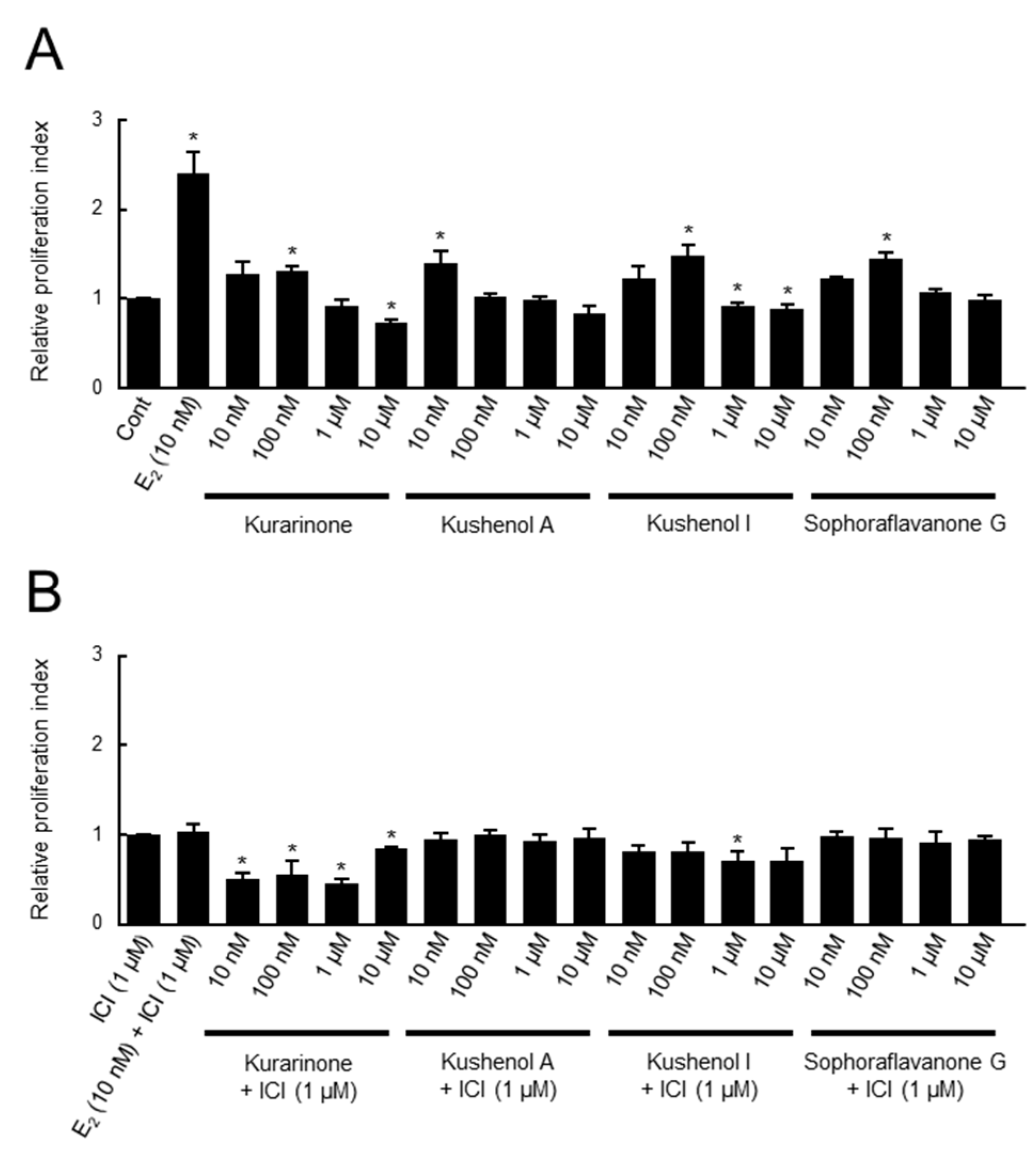
3.1. Evaluation of Estrogenic Activity by Cell Assay
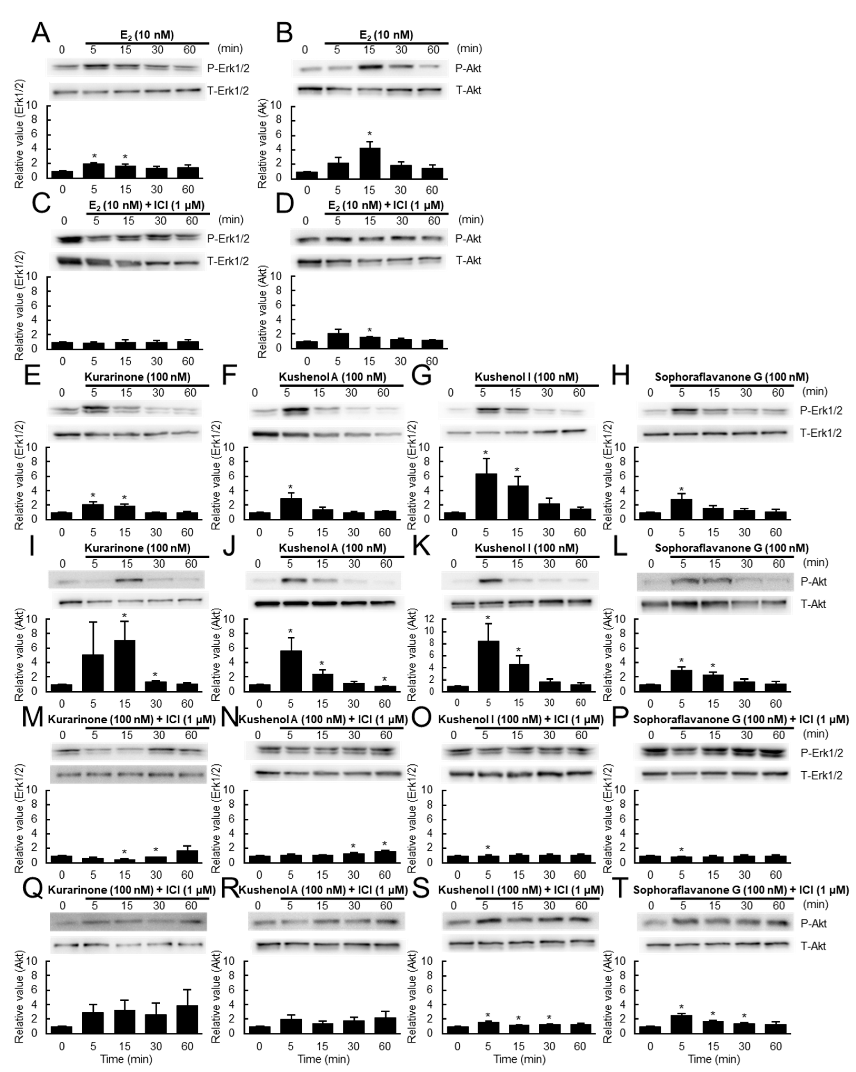
3.2. Evaluation of Estrogenic Activity by Protein Assay
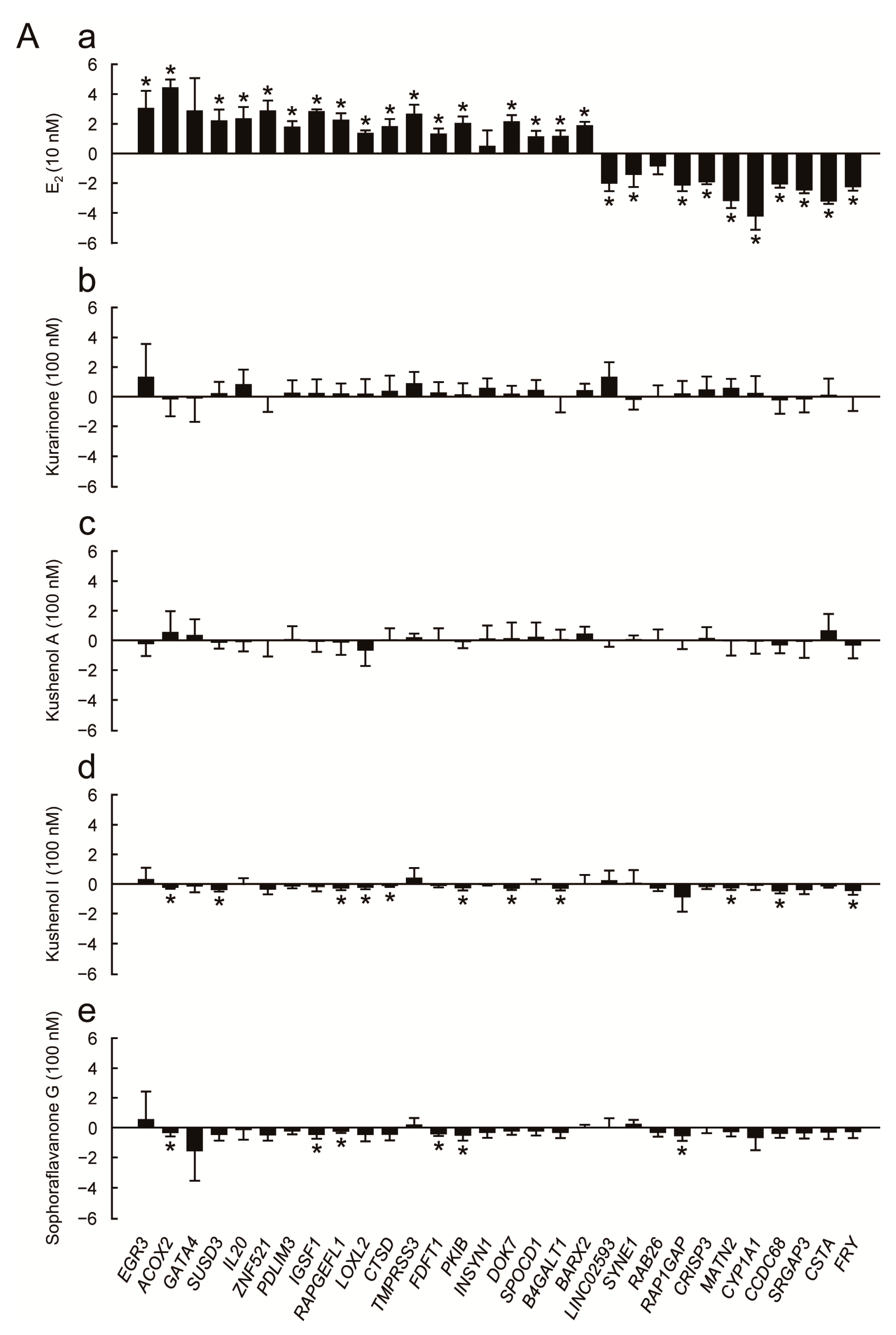
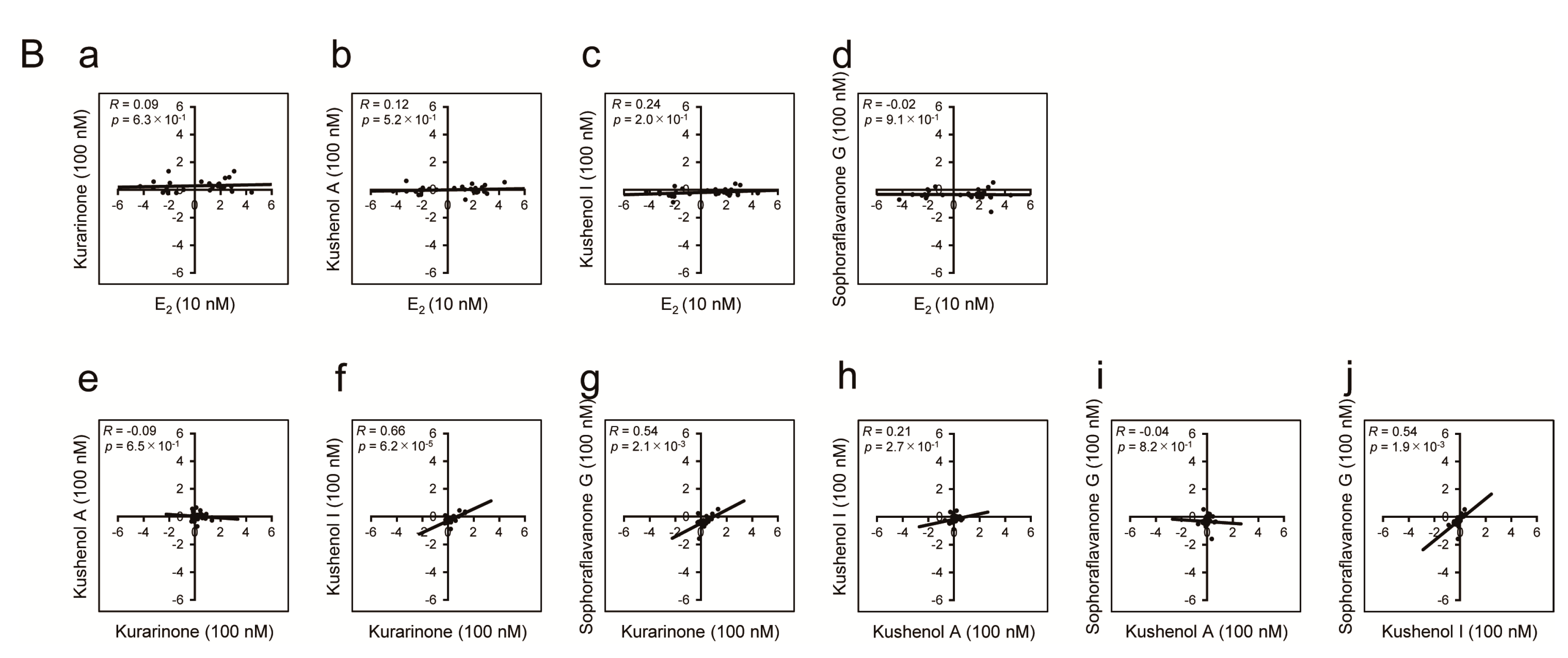
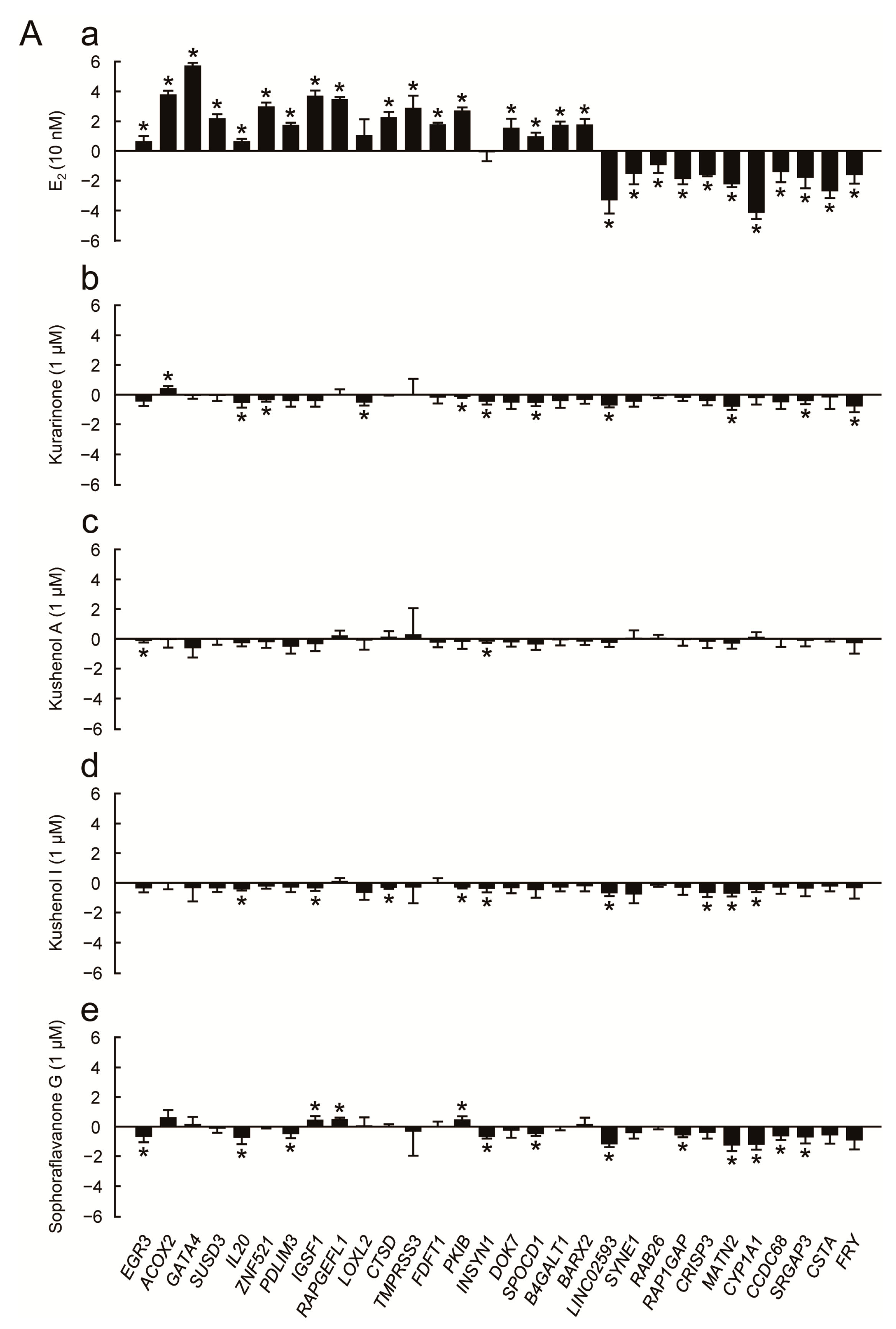
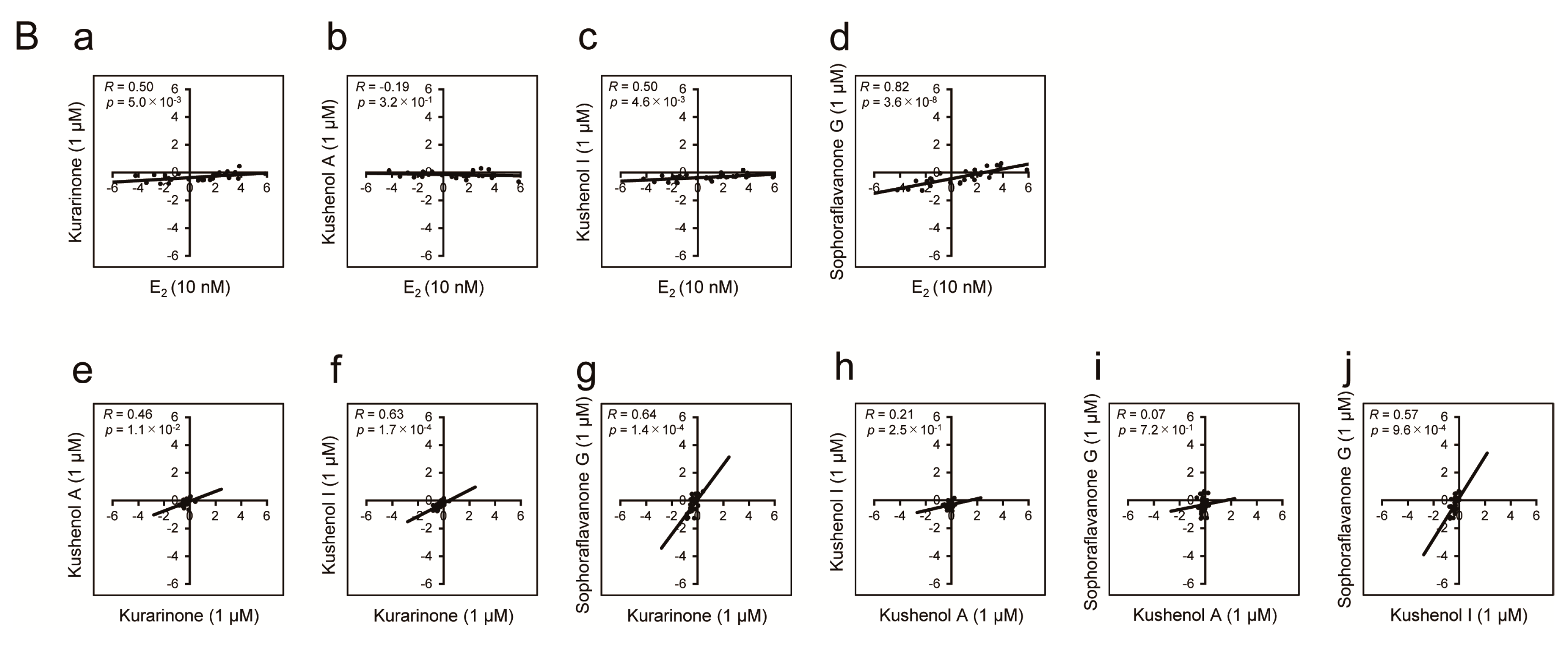
3.3. Evaluation of Estrogenic Activity by Transcription Assay
4. Discussion
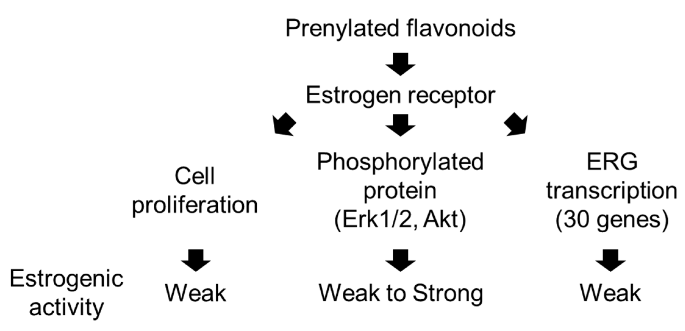
4.1. Estrogenic Response Induced by Prenylated Flavonoids in S. flavescens
4.2. Estrogenic Flavonoids in S. flavescens

| Chemical | Flavonoid Class | Estrogenicity a | Reference |
|---|---|---|---|
| Chalcone and Dihydrochalcone | |||
| Xanthohumol | Chalcone (prenylated) | A | Yoshimaru et al., 2014 [24] |
| Flavanone and Dihydroflavonol | |||
| Isoxanthohumol | Flavanone (prenylated) | E | Żołnierczyk et al., 2015 [26] |
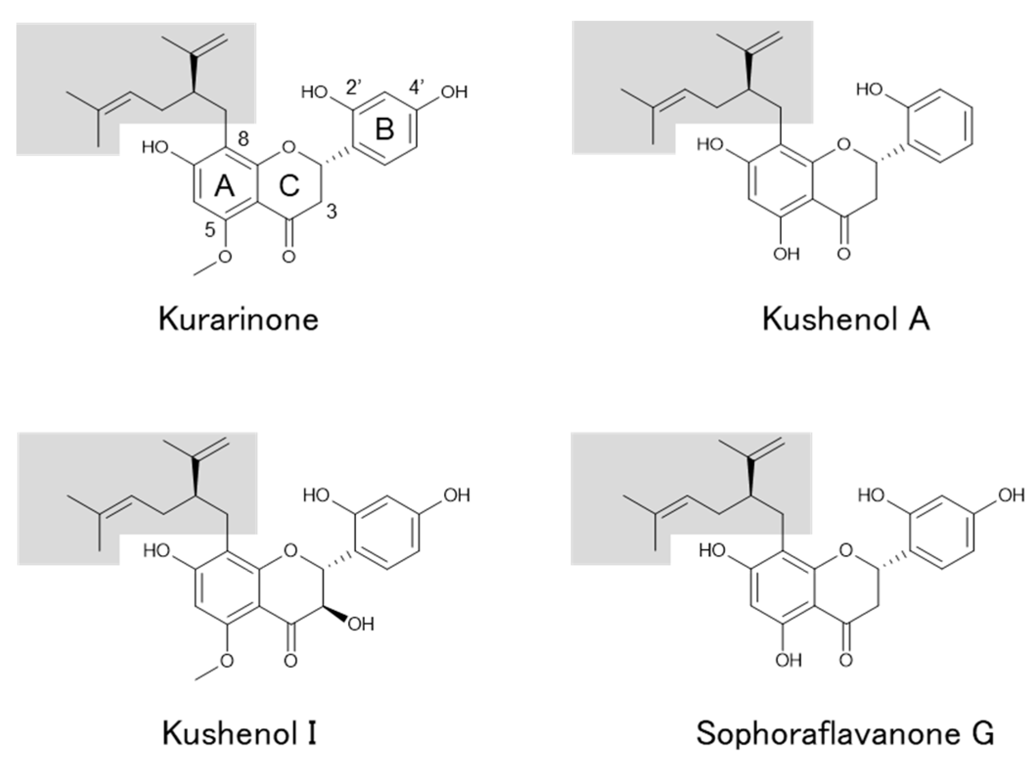
| Kurarinone | Flavanone (prenylated) | E | De Naeyer et al., 2004 [27]; |
| Wang et al., 2011 [28]; This study | |||
| Kushenol A | Flavanone (prenylated) | E | This study |
| Kushenol F | Flavanone (prenylated) | E | Wang et al., 2011 [28] |
| Kushenol I | Dihydroflavonol (prenylated) | E | This study |
| Kushenol X | Dihydroflavonol (prenylated) | L | Hillerns and Wink, 2005 [29] |
| Leachianone A | Flavanone (prenylated) | L | Hillerns and Wink, 2005 [29] |
| Naringenin | Flavanone | E/A/S/L/B | Kiyama, 2023 [19] |
| Sophoraflavanone G | Flavanone (prenylated) | L | Hillerns and Wink, 2005 [29]; |
| This study | |||
| Flavone and Flavonol | |||
| Kushenol C | Flavonol (prenylated) | L | Hillerns and Wink, 2005 [29] |
| Quercetin | Flavonol | E/A/B | Kiyama, 2023 [19] |
| Rutin | Flavonol (glycoside) | E | Kiyama, 2023 [19] |
| Isoflavone | |||
| Biochanin A | Isoflavone | E/A/S/B | Kiyama, 2023 [19] |
| Calycosin | Isoflavone | E/A/S/B | Kiyama, 2023 [19] |
| Daidzein | Isoflavone | E/A/S | Kiyama, 2023 [19] |
| Formononetin | Isoflavone | E/A | Kiyama, 2023 [19] |
4.3. Applications of Prenylated Flavonoids in S. flavescens
5. Conclusions
Supplementary Materials
Author Contributions
Funding
Institutional Review Board Statement
Informed Consent Statement
Data Availability Statement
Acknowledgments
Conflicts of Interest
References
- He, X.; Fang, J.; Huang, L.; Wang, J.; Huang, X. Sophora flavescens Ait.: Traditional usage, phytochemistry and pharmacology of an important traditional Chinese medicine. J. Ethnopharmacol. 2015, 172, 10–29. [Google Scholar] [CrossRef]
- Sun, P.; Zhao, W.; Wang, Q.; Chen, L.; Sun, K.; Zhan, Z.; Wang, J. Chemical diversity, biological activities and traditional uses of and important Chinese herb Sophora. Phytomedicine 2022, 100, 154054. [Google Scholar] [CrossRef]
- Chen, X.; Mukwaya, E.; Wong, M.S.; Zhang, Y. A systematic review on biological activities of prenylated flavonoids. Pharm. Biol. 2014, 52, 655–660. [Google Scholar] [CrossRef]
- Boozari, M.; Soltani, S.; Iranshahi, M. Biologically active prenylated flavonoids from the genus Sophora and their structure-activity relationship—A review. Phytother. Res. 2019, 33, 546–560. [Google Scholar] [CrossRef]
- Yang, J.; Wen, L.; Jiang, Y.; Yang, B. Natural estrogen receptor modulators and their heterologous biosynthesis. Trends Endocrinol. Metab. 2019, 30, 66–76. [Google Scholar] [CrossRef] [PubMed]
- Shi, S.; Li, J.; Zhao, X.; Liu, Q.; Song, S.J. A comprehensive review: Biological activity, modification and synthetic methodologies of prenylated flavonoids. Phytochemistry 2021, 191, 112895. [Google Scholar] [CrossRef] [PubMed]
- Wen, L.; Zhou, T.; Jiang, Y.; Chang, S.K.; Yang, B. Prenylated flavonoids in foods and their applications on cancer prevention. Crit. Rev. Food Sci. Nutr. 2022, 62, 5067–5080. [Google Scholar] [CrossRef]
- Li, J.J.; Zhang, X.; Shen, X.C.; Long, Q.D.; Xu, C.Y.; Tan, C.J.; Lin, Y. Phytochemistry and biological properties of isoprenoid flavonoids from Sophora flavescens Ait. Fitoterapia 2020, 143, 104556. [Google Scholar] [CrossRef]
- Kiyama, R. Nutritional implications of ginger: Chemistry, biological activities and signaling pathways. J. Nutr. Biochem. 2020, 86, 108486. [Google Scholar] [CrossRef]
- Kiyama, R.; Wada-Kiyama, Y. Estrogenic endocrine disruptors: Molecular mechanisms of action. Environ. Int. 2015, 83, 11–40. [Google Scholar] [CrossRef]
- Dong, S.; Terasaka, S.; Kiyama, R. Bisphenol A induces a rapid activation of Erk1/2 through GPR30 in human breast cancer cells. Environ. Pollut. 2011, 159, 212–218. [Google Scholar] [CrossRef]
- Nishi, K.; Fu, W.; Kiyama, R. Novel estrogen-responsive genes (ERGs) for the evaluation of estrogenic activity. PLoS ONE 2022, 17, e0273164. [Google Scholar] [CrossRef]
- Nishi, K.; Imamura, I.; Takemoto, T.; Iga, K.; Kiyama, R. Estrogenic activity of fermented soymilk extracts and soy compounds. Appl. Food Res. 2023, 3, 100341. [Google Scholar] [CrossRef]
- Yim, D.; Kim, M.J.; Shin, Y.; Lee, S.J.; Shin, J.G.; Kim, D.H. Inhibition of cytochrome P450 activities by Sophora flavescens extract and its prenylated flavonoids in human liver microsomes. Evid. Based Complement. Altern. Med. 2019, 2019, 2673769. [Google Scholar] [CrossRef]
- Chen, L.; Cheng, X.; Shi, W.; Lu, Q.; Go, V.L.; Heber, D.; Ma, L. Inhibition of growth of Streptococcus mutans, methicillin-resistant Staphylococcus aureus, and vancomycin-resistant enterococci by kurarinone, a bioactive flavonoid isolated from Sophora flavescens. J. Clin. Microbiol. 2005, 43, 3574–3575. [Google Scholar] [CrossRef]
- Anstead, G.M.; Kym, P.R. Benz[a]anthracene diols: Predicted carcinogenicity and structure-estrogen receptor binding affinity relationships. Steroids 1995, 60, 383–394. [Google Scholar] [CrossRef]
- Routledge, E.J.; Sumpter, J.P. Structural features of alkylphenolic chemicals associated with estrogenic activity. J. Biol. Chem. 1997, 272, 3280–3288. [Google Scholar] [CrossRef]
- Schultz, T.W.; Sinks, G.D.; Cronin, M.T. Structure-activity relationships for gene activation oestrogenicity: Evaluation of a diverse set of aromatic chemicals. Environ. Toxicol. 2002, 17, 14–23. [Google Scholar] [CrossRef]
- Kiyama, R. Estrogenic flavonoids and their molecular mechanisms of action. J. Nutr. Biochem. 2023, 114, 109250. [Google Scholar] [CrossRef]
- Barron, D.; Ibrahim, R.K. Isoprenylated flavonoids—A survey. Phytochemistry 1996, 43, 921–982. [Google Scholar] [CrossRef]
- Mukai, R. Prenylation enhances the biological activity of dietary flavonoids by altering their bioavailability. Biosci. Biotechnol. Biochem. 2018, 82, 207–215. [Google Scholar] [CrossRef]
- Kretzschmar, G.; Zierau, O.; Wober, J.; Tischer, S.; Metz, P.; Vollmer, G. Prenylation has a compound specific effect on the estrogenicity of naringenin and genistein. J. Steroid Biochem. Mol. Biol. 2010, 118, 1–6. [Google Scholar] [CrossRef]
- Mbachu, O.C.; Howell, C.; Simmler, C.; Malca Garcia, G.R.; Skowron, K.J.; Dong, H.; Ellis, S.G.; Hitzman, R.T.; Hajirahimkhan, A.; Chen, S.N.; et al. SAR Study on estrogen receptor α/β activity of (iso)flavonoids: Importance of prenylation, C-ring (un)saturation, and hydroxyl substituents. J. Agric. Food Chem. 2020, 68, 10651–10663. [Google Scholar] [CrossRef]
- Yoshimaru, T.; Komatsu, M.; Tashiro, E.; Imoto, M.; Osada, H.; Miyoshi, Y.; Honda, J.; Sasa, M.; Katagiri, T. Xanthohumol suppresses oestrogen-signalling in breast cancer through the inhibition of BIG3-PHB2 interactions. Sci. Rep. 2014, 4, 7355. [Google Scholar] [CrossRef]
- Hajirahimkhan, A.; Simmler, C.; Yuan, Y.; Anderson, J.R.; Chen, S.N.; Nikolić, D.; Dietz, B.M.; Pauli, G.F.; van Breemen, R.B.; Bolton, J.L. Evaluation of estrogenic activity of licorice species in comparison with hops used in botanicals for menopausal symptoms. PLoS ONE 2013, 8, e67947. [Google Scholar] [CrossRef]
- Żołnierczyk, A.K.; Mączka, W.K.; Grabarczyk, M.; Wińska, K.; Woźniak, E.; Anioł, M. Isoxanthohumol—Biologically active hop flavonoid. Fitoterapia 2015, 103, 71–82. [Google Scholar] [CrossRef]
- De Naeyer, A.; Vanden Berghe, W.; Pocock, V.; Milligan, S.; Haegeman, G.; De Keukeleire, D. Estrogenic and anticarcinogenic properties of kurarinone, a lavandulyl flavanone from the roots of Sophora flavescens. J. Nat. Prod. 2004, 67, 1829–1832. [Google Scholar] [CrossRef]
- Wang, X.; Zhen, L.; Zhang, G.; Wong, M.S.; Qin, L.; Yao, X. Osteogenic effects of flavonoid aglycones from an osteoprotective fraction of Drynaria fortune—An in vitro efficacy study. Phytomedicine 2011, 18, 868–872. [Google Scholar] [CrossRef]
- Hillerns, P.I.; Wink, M. Binding of flavonoids from Sophora flavescens to the rat uterine estrogen receptor. Planta Med. 2005, 71, 1065–1068. [Google Scholar] [CrossRef]
- Zhao, P.; Hamada, C.; Inoue, K.; Yamamoto, H. Efficient production and capture of 8-prenylnaringenin and leachianone G—biosynthetic intermediates of sophoraflavanone G—By the addition of cork tissue to cell suspension cultures of Sophora flavescens. Phytochemistry 2003, 62, 1093–1099. [Google Scholar] [CrossRef]
- Marín, M.; Máñez, S. Recent trends in the pharmacological activity of isoprenyl phenolics. Curr. Med. Chem. 2013, 20, 272–279. [Google Scholar] [CrossRef]
- Terao, J.; Mukai, R. Prenylation modulates the bioavailability and bioaccumulation of dietary flavonoids. Arch. Biochem. Biophys. 2014, 559, 12–16. [Google Scholar] [CrossRef]
- Kumar, S.; Prajapati, K.S.; Shuaib, M.; Kushwaha, P.P.; Tuli, H.S.; Singh, A.K. Five-decade update on chemopreventive and other pharmacological potential of kurarinone: A natural flavanone. Front. Pharmacol. 2021, 12, 737137. [Google Scholar] [CrossRef]
- Hwang, J.S.; Lee, S.A.; Hong, S.S.; Lee, K.S.; Lee, M.K.; Hwang, B.Y.; Ro, J.S. Monoamine oxidase inhibitory components from the roots of Sophora flavescens. Arch. Pharm. Res. 2005, 28, 190–194. [Google Scholar] [CrossRef]
- Hyun, S.K.; Lee, W.H.; Jeong, D.M.; Kim, Y.; Choi, J.S. Inhibitory effects of kurarinol, kuraridinol, and trifolirhizin from Sophora flavescens on tyrosinase and melanin synthesis. Biol. Pharm. Bull. 2008, 31, 154–158. [Google Scholar] [CrossRef]
- Kim, J.H.; Cho, I.S.; So, Y.K.; Kim, H.H.; Kim, Y.H. Kushenol A and 8-prenylkaempferol, tyrosinase inhibitors, derived from Sophora flavescens. J. Enzyme Inhib. Med. Chem. 2018, 33, 1048–1054. [Google Scholar] [CrossRef]
- Chen, H.; Yang, J.; Hao, J.; Lv, Y.; Chen, L.; Lin, Q.; Yuan, J.; Yang, X. A novel flavonoid kushenol Z from Sophora flavescens mediates mTOR pathway by inhibiting phosphodiesterase and Akt activity to induce apoptosis in non-small-cell lung cancer cells. Molecules 2019, 24, 4425. [Google Scholar] [CrossRef]
- Liu, T.; Gong, J.; Lai, G.; Yang, Y.; Wu, X.; Wu, X. Flavonoid extract Kushenol A exhibits anti-proliferative activity in breast cancer cells via suppression of PI3K/AKT/mTOR pathway. Cancer Med. 2023, 12, 1643–1654. [Google Scholar] [CrossRef]
- Zhang, Q.; Yu, J.; Wang, Y.; Su, W. Selective extraction of flavonoids from Sophora flavescens Ait. by mechanochemistry. Molecules 2016, 21, 989. [Google Scholar] [CrossRef]
- Wei, G.; Chen, Y.; Guo, X.; Wei, J.; Dong, L.; Chen, S. Biosyntheses characterization of alkaloids and flavonoids in Sophora flavescens by combining metabolome and transcriptome. Sci. Rep. 2021, 11, 7388. [Google Scholar] [CrossRef]
- Li, F.J.; Zhang, F.; He, X.D.; Liu, X.; Mu, J.K.; Yang, M.; Li, Y.Q.; Gu, W.; Yu, J.; Yang, X.X. A novel method for identifying parkin binding agents in complex preparations of herbal medicines. Oxid. Med. Cell Longev. 2022, 2022, 3260243. [Google Scholar] [CrossRef]
Disclaimer/Publisher’s Note: The statements, opinions and data contained in all publications are solely those of the individual author(s) and contributor(s) and not of MDPI and/or the editor(s). MDPI and/or the editor(s) disclaim responsibility for any injury to people or property resulting from any ideas, methods, instructions or products referred to in the content. |
© 2024 by the authors. Licensee MDPI, Basel, Switzerland. This article is an open access article distributed under the terms and conditions of the Creative Commons Attribution (CC BY) license (https://creativecommons.org/licenses/by/4.0/).
Share and Cite
Nishi, K.; Imamura, I.; Hoashi, K.; Kiyama, R.; Mitsuiki, S. Estrogenic Prenylated Flavonoids in Sophora flavescens. Genes 2024, 15, 204. https://doi.org/10.3390/genes15020204
Nishi K, Imamura I, Hoashi K, Kiyama R, Mitsuiki S. Estrogenic Prenylated Flavonoids in Sophora flavescens. Genes. 2024; 15(2):204. https://doi.org/10.3390/genes15020204
Chicago/Turabian StyleNishi, Kentaro, Ikumi Imamura, Kenichiro Hoashi, Ryoiti Kiyama, and Shinji Mitsuiki. 2024. "Estrogenic Prenylated Flavonoids in Sophora flavescens" Genes 15, no. 2: 204. https://doi.org/10.3390/genes15020204