Review

Genetic Basis of Breast and Ovarian Cancer: Approaches and Lessons Learnt from Three Decades of Inherited Predisposition Testing

by
Valeria Barili
1,†,
Enrico Ambrosini
2,†,
Beatrice Bortesi
3,
Roberta Minari
3,
Erika De Sensi
1,
Ilenia Rita Cannizzaro
1,
Antonietta Taiani
1,
Maria Michiara
3,4,
Angelica Sikokis
3,4,
Daniela Boggiani
3,4,
Chiara Tommasi
1,3,4,
Olga Serra
3,4,
Francesco Bonatti
3,
Alessia Adorni
3,
Anita Luberto
1,
Patrizia Caggiati
2,
Davide Martorana
2,
Vera Uliana
2,
Antonio Percesepe
1,2,*,
Antonino Musolino
1,3,4,* and
Benedetta Pellegrino
3,4
add Show full author list remove Hide full author list
1
Department of Medicine and Surgery, University of Parma, 43126 Parma, Italy
2
Medical Genetics, University Hospital of Parma, 43126 Parma, Italy
3
Medical Oncology Unit, University Hospital of Parma, 43126 Parma, Italy
4
Breast Unit, University Hospital of Parma, 43126 Parma, Italy
*
Authors to whom correspondence should be addressed.
These authors contributed equally to this work.
Genes 2024, 15(2), 219; https://doi.org/10.3390/genes15020219
Submission received: 30 December 2023 / Revised: 2 February 2024 / Accepted: 5 February 2024 / Published: 8 February 2024
(This article belongs to the Section Genetic Diagnosis)

Abstract

:
Germline variants occurring in BRCA1 and BRCA2 give rise to hereditary breast and ovarian cancer (HBOC) syndrome, predisposing to breast, ovarian, fallopian tube, and peritoneal cancers marked by elevated incidences of genomic aberrations that correspond to poor prognoses. These genes are in fact involved in genetic integrity, particularly in the process of homologous recombination (HR) DNA repair, a high-fidelity repair system for mending DNA double-strand breaks. In addition to its implication in HBOC pathogenesis, the impairment of HR has become a prime target for therapeutic intervention utilizing poly (ADP-ribose) polymerase (PARP) inhibitors. In the present review, we introduce the molecular roles of HR orchestrated by BRCA1 and BRCA2 within the framework of sensitivity to PARP inhibitors. We examine the genetic architecture underneath breast and ovarian cancer ranging from high- and mid- to low-penetrant predisposing genes and taking into account both germline and somatic variations. Finally, we consider higher levels of complexity of the genomic landscape such as polygenic risk scores and other approaches aiming to optimize therapeutic and preventive strategies for breast and ovarian cancer.

1. Introduction

Breast cancer is the most prevalent type of cancer in women worldwide and the existence of a familial predisposition has been known for at least 150 years [1]. The co-segregation of familial ovarian and breast cancer was described in the second part of the 20th century. In 1994, through extensive research on families with a history of early-onset breast and/or ovarian cancer, the discovery of the connection between BRCA1 and BRCA2 genes and the development of cancer risk was made [2,3]. Shortly after, BRCA1/2 screening became available for clinical use with an immediate effect on patient care [4].
In the last few years, the list of genes involved in cancer predisposition has increased, reflecting various consequences from the prognostic, preventive, and therapeutic point of view. These genes can be ranked in two categories according to their contribution to cancer risk: high penetrance, where the risk is more than fourfold higher than the one observed in the overall population, and intermediate penetrance, with a 2–4 times higher risk [5]. Among the high-penetrance hereditary cancer syndromes, BRCA1- and BRCA2-Associated Hereditary Breast and Ovarian Cancer (HBOC) still stands out as the most prevalent [6].
The acronym HBOC is used to define a genetic condition mainly characterized by an enhanced susceptibility to both female and male breast and ovarian tumors, such as fallopian tube and peritoneal malignancies. HBOC is historically linked to germline pathogenic variants in BRCA1 and BRCA2, which have vital functions in fixing double-stranded DNA error. Additionally, genetic variants associated with HBOC have been described to enhance risks for prostate cancer and pancreatic cancer.
The approximate lifetime probability of breast tumor development in the overall population is around 12%. In two extensive studies conducted prospectively within cancer genetics services, it was determined that by the age of 70, individuals carrying BRCA1 pathogenic variants faced a breast cancer risk ranging from 60% to 66%, while those with BRCA2 pathogenic variants exhibited a risk between 55% and 61%. The estimated incidence of ovarian cancer in the overall population is approximately 1–2%. Ovarian cancer risk was calculated to be between 41% and 58% for BRCA1 carriers and between 15% and 16.5% for BRCA2 carriers at age 70 [7,8].
The prevalence of HBOC is estimated to be around 1:400 individuals [9]. However, individuals with Ashkenazi Jewish ancestry have a significantly higher prevalence (1:40), attributable to three Jewish founder pathogenic changes, that eventually led to evaluation of a potential population screening [10].
Traditionally, HBOC risk is based on assessing the family history, particularly the presence of several individuals who have been diagnosed with breast and/or ovarian tumor occurring at young ages. The variability in the response to standard breast cancer treatment, together with faster and cheaper sequencing technologies, has prompted a directional shift, with the implementation of multigene panels and the use of genetic analysis also for therapeutic purposes.
This review covers the possible role of pathogenic variants in breast and ovarian cancer screening, prevention, and monitoring with a possible improvement in more-focused therapeutic interventions.

2. High-Penetrance Genes: BRCA1 and BRCA2

2.1. Pathogenic Mechanism

DNA repair encompasses various mechanisms, such as DNA single-strand break (dsDNA) repair or base mismatch repair (MMR). Notably, BRCA1 and BRCA2 stand as tumor-suppressor genes essential to the preservation of genomic integrity through the mending of dsDNA lesions employing homologous recombination (HR). In addition, they guide centrosome dynamics and the distribution of chromosome, cytokinesis, and genome stabilization throughout cell cycle progression [11].
A hormone-dependent carcinogenic environment that could potentially compromise genome stability through the disruption of BRCA functions has been hypothesized. This disruption might consequently lead to the expedited induction of pro-survival cues, thereby promoting the transformation of breast gland cells into transformed phenotypes [12]. Due to the correlation between the impairment of BRCA activity and defects in HR, the conceptual framework of “BRCAness” was introduced in 2004 [13]. This term denotes a phenocopy of BRCA1 or BRCA2 variants, a situation in which an HRR defect exists in a tumor in the absence of germline BRCA1 or BRCA2 changes. Several conceptually distinct approaches to detect non-BRCA1/2 gene variants could also result in the same consequences caused by those detected in BRCA1/2. Therefore, the concept of BRCAness could serve as a valuable therapeutic biomarker.
Moreover, it has been shown that tumors associated with BRCA1 pathogenic variants often exhibit a molecular profile reminiscent of the Triple-Negative Breast Cancer (TNBC) subtype, while tumors linked to BRCA2 variants tend to resemble the Luminal B subtype or, less commonly, the Luminal A subtype [14].

2.2. Types of Variants

Currently, the ClinVar database [15] has identified around 4300 different germline variants in BRCA1 and 5200 in BRCA2, classified as pathogenic or likely pathogenic (as considered in December 2023). About 80% of these variants are categorized as truncating modifications, which result in the formation of premature stop codons, including frameshift and nonsense changes. It must be noted that the removal of the terminal amino acids cannot necessarily impact on protein function, as stated by Nepomuceno et al. through functional evaluations of the impact of frameshift variants in the C-term of BRCA1, emphasizing the need for a comprehensive assessment of individual variants beyond their primary sequence alterations [16].
Missense variants constitute approximately 10% of the identified variants [17]. Their interpretation can be troublesome, as many of them are still categorized as variants of uncertain significance (VUSs) in line with the American College of Medical Genetics (ACMG) guidelines [18]. Pathogenic missense variants are typically localized within functionally crucial domains, such as the Really Interesting New Gene (RING) finger domain and the BRCA1 C terminus (BRCT) domains of BRCA1, as well as the regions spanning the oligosaccharide-binding folds (OBs) and helical domains of BRCA2. Intriguingly, by investigating the number of pathogenic missense variants in those domains with respect to the whole protein, BRCA1 includes approximately half of the variants with RING and BRCT domains (76/162), in contrast to BRCA2 which shows around 70% of missense changes (56/78) within the helicase and OB domains.
Moreover, amino acid modifications at distinct positions, e.g., first methionine or the regions near the exon–intron junctions, are usually considered pathogenic according to the ACMG classification.
Notably, about 10% of the variants are attributed to aberrant copy number variations (CNVs), ascertained through deletion or duplication analyses [19]. Moreover, a mechanism termed “secondary epimutation”, due to the presence of the BRCA1 deep intronic variant c.-107A > T, may lead to the methylation of the promoter and to a reduced expression of the gene [20].

2.3. Genotype–Phenotype Correlations

BRCA1 is positioned at locus 17q21.31 and comprises a total of 24 exons, encoding a sequence of 1863 amino acids (MIM #113705). The initiation of the exonic portion occurs with exon 2, wherein the primary transcript omits exon 4. Specific regions known as the breast cancer cluster regions (BCCRs) within BRCA1 are localized at coordinates c.179–505, c.4328–4945, and c.5261–5563. Likewise, the ovarian cancer cluster region (OCCR) resides at coordinates c.1380–4062 [21].
The genomic location of BRCA2 is on chromosome 13 q13.1 and it encompasses 27 exons, encoding a peptide sequence spanning 3418 amino acids. The protein-coding region starts within exon 2. The breast cancer cluster regions (BCCRs) within BRCA2 are identified at positions c.1–596, c.772–1806, and c.7394–8904. In a parallel manner, the ovarian cancer cluster regions (OCCRs) are positioned within the coordinates c.2831–6401 and c.6645–7471 [21]. Variants within these regions appear to trigger nonsense-mediated decay (NMD), resulting in the loss of BRCA2 expression and a markedly elevated susceptibility of ovarian cancer. In fact, the most prominent disparity observed between the risks observed in carriers with diverse ethnic backgrounds and those identified in individuals of Ashkenazi Jewish descent pertains to the heightened ovarian cancer risk associated with BRCA2 pathogenic variants in AJ populations. This discrepancy can be due to the data concerning the BRCA2 founder variant in AJ, which reflects the susceptibility linked to the 6174delT variant, which is situated within the OCCR of the BRCA2 gene [7,22]. Interestingly, BRCA2 showed a strong risk association with pathogenic variants and prostate and pancreatic cancer. Specifically, BRCA2 germline variants within the region c.6373–c.6492 exhibited a relative risk of 3.7 as compared with molecular changes outside this region [23], and pathogenic changes after the 3′ of c.7914 (prostate cancer cluster region or PrCCR) were significantly associated with an augmented risk of prostate cancer [24]. Additionally, a pancreatic cancer cluster region (PcCCR) was identified as associated with pancreatic cancer (between c.3515 and c.6787), a region which overlaps with BRCA2 OCCR but not BCCR and PrCCR, suggesting a possible common oncogenic mechanism between ovarian and pancreatic tumors [25]. On the other hand, specific variants seem to be associated with a better prognosis. For example, pathogenic variants within BRCA1 exon 11 have the potential to generate a partial BRCA1 protein due to the presence of known exon 11 splice variants, though the entire protein is subjected to degradation via NMD. Experimental evidence from murine embryos harboring the exon 11-deleted isoform indicates extended survival compared to embryos lacking BRCA1 entirely, suggesting that BRCA1 proteins lacking exon 11 may retain some degree of functional activity [26].
Another example is the pathogenic variant p.Lys3326Ter in BRCA2, correlated with a reduced possibility of breast and ovarian tumor when compared to other pathogenic BRCA2 variants [27]. However, the study reported an odds ratio (OR) of 1.28 for breast cancer and an OR of 1.26 for invasive ovarian cancer. Hence, in the absence of supplementary data, the utilization of genotype location and its correlation with the phenotype of cancer risk for the purpose of individual risk evaluation and management might be considered premature.
Considering that the genetic tests for BRCA1 and BRCA2 have more than doubled in the last decade, the importance of accessible and high-quality data becomes crucial.
Thus, the most extensive initiative named “BRCA Exchange project” (www.brcaexchange.org) started to create a comprehensive and global source of information on BRCA genetic variants. The project is dedicated to compiling and consolidating data related to BRCA1 and BRCA2 variants, defined for a high-penetrance phenotype, from diverse sources and populations worldwide. The goal is to establish a centralized and expansive repository of knowledge on BRCA genetic variations that can be accessed and utilized by researchers, clinicians, and professionals globally [28]

3. Other High-Penetrance Genes

The discovery of additional genes associated with breast and/or ovarian cancer risk has prompted their involvement in tumor-associated gene panels, and it has fostered the development of evolving frameworks aimed at optimizing the management of carriers. Across several studies involving high-risk HBOC patients, where individuals were not pre-selected for BRCA1/2 mutation status, the frequency of variants in non-BRCA1/2 genes ranges from 3.5% to 10.9% [29]. In other breast cancer patient cohorts with no selection bias, the rate of detection of variants in BRCA1/2 was approximately 4–5%, while the cumulative variant detection rate achieved through multigene panel analysis ranged from 6% to 9%, representing a 1.4- to 2-fold augmentation. Likewise, among ovarian cancer patients free from selection biases, the rate of detection of variants in BRCA1/BRCA2 was roughly 20%, whereas the rate for MGP analysis ranged from 26% to 31%, corresponding to a 1.5-fold elevation [30,31].
Recent comprehensive studies have highlighted that pathogenic variants (PVs) across eight distinct genes, such as ATM, BRCA1, BRCA2, CHEK2, PALB2 (FANCN), RAD51C, and RAD51D, exhibit a substantial correlation with breast cancer risk (Table 1). Notably, PVs within the BRCA1, BRCA2, and PALB2 genes are associated with a markedly elevated BC risk, with odds ratios spanning from 5.0 to 10.6. Conversely, PVs within the ATM and CHEK2 genes confer a moderately elevated BC risk, with odds ratios ranging from 2.1 to 2.5 [32,33]. It is pertinent to note, however, that variants in ATM and CHEK2 are comparatively more prevalent within the general population [34].
Ultimately, TP53 and CDH1 can be included among high-penetrance genes. Germline variants in BRCA1 and TP53 are primarily connected with invasive ductal carcinoma, whereas BRCA2 germline changes are linked to both ductal and lobular tumors. In contrast, variants in CDH1 are uniquely associated with lobular breast cancer, particularly the invasive type [35].

3.1. CDH1

Germline variants in CDH1 (E-cadherin) tumor suppressor protein are correlated with hereditary diffuse gastric cancer (HDGC) and a lifetime risk of 40–50% of developing lobular breast cancer [36,37]. Annual mammography and bilateral breast MRI with contrast are recommended beginning at age 30. Risk-reducing mastectomy depends on family history [38].
Inactivating germline variants in CDH1 underlie this high-penetrance cancer syndrome. These germline changes are identified in the 40% of families that meet the HDGC clinical criteria. The cumulative incidence of DGC by the age of 80 among CDH1 variants carriers is calculated as around 70% for males and 56% for females, while the likelihood of women developing LBC stands at 42%. Other clinical manifestations, including colorectal cancer (CRC), appendiceal signet ring cell carcinomas, and cleft lip/palate, have also been observed in HDGC families.
In the clinical context of HDGC, CDH1 variants are uniformly scattered across the gene, and no discernible genotype–phenotype correlation has been identified [39]. The predominance of documented germline modifications has been described to result in truncated proteins or in the absence of mRNA transcription, thereby facilitating a straightforward assessment of their pathogenicity. Missense variants constitute 16% of the described changes thus far: the clinical and functional implications of missense variants are debated among geneticists, in most instances, since a full-length protein is retained, and normal E-cadherin protein expression is typically maintained.
In 2013, a study involving 165 unrelated cases was conducted [37]. The selection criteria were based on personal and familial histories of gastric or breast cancers, and the findings revealed that 18 subjects (11%) were carriers of CDH1 variants, encompassing 18 distinct variants. Among these carriers, three women had personal histories of bilateral LBC diagnosed before the age of 50. None of these cases exhibited a family history of diffuse gastric cancer among first- and second-degree family members, and therefore did not satisfy the HDGC rules set forth in that period. Subsequently, two of these women were diagnosed with DGC, while the third woman underwent upper gastrointestinal endoscopy with several bioptic sampling. Significantly, in recent years, a growing body of research has emerged, documenting instances of early-onset LBC cases in individuals carrying CDH1 germline variants, even in the absence of any personal or family history of DGC. This highlights CDH1 as a newly identified susceptibility gene for LBC. Furthermore, the concept of hereditary lobular breast cancer (HLBC) has been suggested as a distinct clinical entity linked to CDH1 germline modifications. According to the consensus guidelines established for HDGC, women who meet specific criteria, including those diagnosed with bilateral LBC, regardless of family cases of LBC prior to the age of 50, or women diagnosed with unilateral LBC with other cases of LBC in the family, characterized below 45 years, meet the necessary criteria for CDH1 genetic testing [40].

3.2. PALB2

The PALB2 gene participates in the HR process as a binding partner of BRCA2. Biallelic modifications in PALB2, also known as FANCN, led to a distinct type of Fanconi anemia, while monoallelic PALB2 variants are linked to breast cancer and ovarian cancer with a 41–60% and 3–5% absolute risk, respectively [41,42]. The prevalence of these pathogenic variants ranges from 0.4% to 3.9%. PALB2 pathogenic variants are conducive to a 35% lifetime risk of developing breast tumor by the age of 70.
The NCCN guidelines recommend annual mammography and MRI with contrast starting at the age of 30 for women with the PALB2 pathogenetic variant [42]. Risk-reducing mastectomy (RRM) is another option to consider, whereas data about Risk-reducing Salpingo-oophorectomy (RRSO) are lacking, and the presence of other family cases should be considered [43].
Further increased risks have been observed for pancreatic cancer, ranging from 2% to 3%, and male breast cancer with a 1% risk [44].
A systematic review [45] revealed that the majority of the cataloged predicted pathogenic PALB2 variants, specifically 911 cases (92.5%), were documented in breast cancer patients, while 49 cases (5.0%) were observed in patients with ovarian tumors, and 24 cases (2.4%) were recorded in patients with pancreatic cancer. Notably, most frequent pathogenic PALB2 variants were c.509_510delGA, c.3113G > A, c.1592delT, c.172_175delTTGT, and c.1240C > T, collectively representing 57.3% of all cases. The “hotspots” exonic regions with the highest variant frequencies were in exons 2 (representing 6.7%), 1 (reflecting 6.3%), and 3 (5.8%).

3.3. PTEN

Tumor-suppressor gene PTEN pathogenic germline variants cause Cowden syndrome/PTEN Hamartoma Tumor Syndrome (PHTS), an autosomal dominant disease featured by multiple hamartomas and an increased risk of developing certain cancers in the thyroid, skin, endometrium, and breast [46]. The PHTS-estimated prevalence is currently at 1:200,000, with many cases which remain undetected [47]. The clinical presentation of PHTS is highly heterogeneous and encompasses an elevated risk for various cancer types, as well as macrocephaly, developmental delays, cutaneous manifestations, thyroid abnormalities, vascular malformations, and benign tumors [48].
Current population-based estimates for PHTS indicate that females exhibit a cancer risk of 85–90%, and males face a risk of 54–66% by the age of 60. Specifically, elevated risks have been noted for breast cancer (67–78%), endometrial cancer (19–28%), thyroid cancer (6–38%), colorectal cancer (9–20%), renal cancer (2–11%), and melanomas (0–6%) [49].
NCCN guidelines recommend 6–12-month breast inspections starting at the age of 25, and annual mammography and breast MRI screening with contrast starting at 30–35 years of age [42,43,46]. Thyroid cancer surveillance commences in childhood, at age 18, or upon PHTS diagnosis. Guidance for endometrial, colorectal, renal cancer, and melanoma surveillance varies, ranging from no recommended surveillance to annual monitoring [50].
Concerning the genotype–phenotype correlation, truncating variants exhibit a 2- to 3-fold higher risk of breast cancer (BC) when compared to missense variants [51]. Furthermore, variants located in the phosphatase domain are associated with an additional approximate 2-fold increased BC risk compared to those situated in the C2 domain. Although the precise underlying mechanisms driving these genotype-specific BC risks still need clarification, these statistically significant findings underscore the necessity of assessing genotype-related surveillance strategies. Particularly, it is imperative to consider the potential reduction in breast cancer monitoring for patients harboring missense variants in the C2 domain, as these changes are more frequently ascertained in individuals with (mild) developmental delay than in patients with high cancer risk [52].

3.4. TP53

The tumor suppressor gene TP53 is situated on chromosome 17p13, and its protein product, p53, assumes significant roles in the regulation of both the cell cycle and apoptosis. Li–Fraumeni syndrome (LFS) is a rare autosomal dominant disorder due to germline TP53 pathogenic variants and characterized by high cancer predisposition for a wide range of tumors, including brain cancer, sarcomas, acute leukemia, and early-onset breast cancer [41]. The occurrence of breast tumor in TP53-carrying women is remarkably high with a cumulative incidence at 60 years of 85%; screening suggests clinical breast surveillance every 6–12 months starting from 20 years of age, annual breast MRI with contrast from 20 to 75 years, and annual mammography from 30 to 75 years [42,46]. A clear association between pathogenetic variants in TP53 and ovarian cancer has not been established [42].
Soft tissue sarcoma, breast cancer, adrenocortical tumors, and specific types of brain tumors have been designated as the “core” cancers within the LFS spectrum [53]. The NCI Li–Fraumeni Syndrome Study, encompassing 5286 individuals, demonstrated cumulative incidence rates by the age of 70 in women, indicating 54% for breast cancer, 15% for soft tissue sarcoma, 6% for brain cancer, and 5% for osteosarcoma [54]. In a comprehensive case–control analysis involving 56,480 breast tumors, TP53 pathogenic variants exhibited a significant association with HER2-positive disease, irrespective of estrogen receptor status [55]. Collectively, these results imply that HER2 amplification may coincide with germline TP53 changes. This correlation demands additional exploration, as individuals with this dual characteristic may potentially derive benefits from chemoprevention strategies incorporating HER2-targeted agents.
Irrespective of familial medical histories, the prevalence of disease-causing germline TP53 changes stands between 3.8% and 7.7% in females diagnosed with breast carcinoma before the age of 31 years [56]. The occurrence of presentations without a familial cancer history can be attributed to two main factors: the contribution of de novo variants to the hTP53-related cancer syndrome, estimated to range from 7 to 20%, and the lacking in the penetrance of pathogenetic TP53 changes [57].
The absence of detectable TP53 variants on blood DNA analysis also through high-depth NGS does not ensure the absence of somatic alterations. In this context, systematically, a comprehensive screening should encompass the analysis of tumor tissue. Interestingly, blood detection of a low number of TP53 variant sequences may be due to circulating tumor DNA (e.g., metastatic tumors) or clonal hematopoiesis, which relies on somatic TP53 alterations which benefit hematopoietic stem cells, highlighting the complexity of mosaic variants in TP53 [58]. The detection of a TP53 variant in the bloodstream of a patient afflicted with metastatic high-grade serous ovarian carcinoma is more likely indicative of circulating tumor DNA. This presumption is based on the exceedingly high prevalence of somatic TP53 alterations in such malignancies, which surpasses 95%. Secondly, clonal hematopoiesis presents the second potential pitfall, encompassing the emergence of somatic TP53 alterations that confer a proliferative advantage within hematopoietic stem cells. Initially, clonal hematopoiesis was documented primarily in patients aged 70 and older, but it has been detected in individuals as young as 30 years of age. The prevalence of clonal hematopoiesis escalates with age and is influenced by factors such as tobacco use, as well as exposure to chemotherapy or radiotherapy [59]. Therefore, when a TP53 variant is identified within a minor fraction of sequencing reads derived from blood, it is imperative to consider the clinical presentation and relevant aspects of the medical history, including prior treatments and the presence of metastases, and to attest the presence of the variant within the tissue of origin of the tumor.
The penetrance of germline variants in TP53 which cause the disease exhibits variability, often determined by the type of molecular change. Dominant-negative missense TP53 changes are typically detected in families characterized by pediatric tumors and commonly display high penetrance. In contrast, truncating/frameshift variants are primarily found in families predominantly affected by adult tumors and show reduced disease penetrance. A notable case of a low-penetrance yet still pathogenic variant is the non-dominant negative missense p.Arg337His variant, which occurs in 0.3% of the Southern Brazilian population and is correlated with a founder genetic effect [60].
The observed phenotypic variability in disease expression within the same family strongly suggests the presence of genetic modifying factors, even environmental ones. Identifying these factors currently stands as a top priority in this genetic field. Germline TP53 variants can render p53 more permissive to oncogenic stress, highlighting the interplay between genetic and environmental factors in determining the ultimate clinical outcome.

4. Intermediate Penetrance Genes

4.1. ATM

The ataxia-telangiectasia-mutated (ATM) gene specifies a kinase engaged in the DNA double-strand break repair mechanism. ATM pathogenic variants, such as homozygous or compound heterozygous changes, cause ataxia telangiectasia, a syndromic disease characterized by progressive cerebellar ataxia, oculomotor apraxia, immunodeficiency, and enhanced cancer predisposition [61]. The severe and recessive syndromic form is rare; however, variant carrier frequency is not. ATM pathogenic variants are observed in heterozygosity in approximately 1–2% of Caucasian adults [62].
Women carrying a heterozygous pathogenetic variant in ATM have a 20–40% lifetime risk to develop breast cancer; thus, annual mammography is recommended from 40 years [42,46]. Data regarding RRM are not sufficient yet, so this option is considered based on family history [42]. Germline pathogenetic variants confer a 2-fold to 3-fold increase in the risk of ovarian cancer compared to the general population [63], but there are no available data to recommend RRSO [42].
Heterozygosity for loss-of-function variants in ATM has been correlated with an increased susceptibility to prostate, pancreatic, gastric, colorectal, and skin cancers too [64,65,66].
Recent outcomes from an international and a US-based investigation have provided evidence that both truncating and missense molecular changes of ATM associate with an augmented risk of ER-positive BC compared to ER-negative BC [32,33].
The available findings do not display a significantly elevated risk of contralateral breast cancer among carriers of ATM pathogenic variants in comparison to non-carriers. Currently, a few studies indicate a limited increase in risk among ATM PV carriers [67]. Further data are required to accurately evaluate the CBC risk in people with ATM variants.
Some genotype–phenotype correlations have been demonstrated. The ATM missense variant c.7271T > G, initially associated with a milder phenotype of ataxia-telangiectasia (A-T), has been found to carry a substantial breast cancer risk comparable to that of BRCA2 [68,69].
The interplay between radiation exposure and breast cancer risk is intricate for patients harboring ATM PVs. Ataxia-telangiectasia-affected patients exhibit a heightened sensitivity to ionizing radiation. However, available data generally do not contraindicate for patients with heterozygous ATM PVs and radiation therapy. The Women’s Environmental Cancer and Radiation Epidemiology (WECARE) study examines the interaction between radiation exposure, genetic predisposition, and breast cancer, specifically radiation-induced CBC. Women carrying a common ATM variant may experience a protective effect, reducing CBC risk. Conversely, women with rare, likely deleterious ATM missense variants face an elevated dose-dependent CBC risk compared to ATM PV carriers who did not undergo RT [67,70]. The efficacy of PARPi in breast cancer (BC) patients with ATM pathogenic variants (PVs) is currently under examination within the metastatic setting. The TBCRC048 phase II study revealed no notable activity among ATM carriers; however, the trial cohort included only a limited number of ATM carriers [71]. Olaparib obtained approval for metastatic prostate cancer patients carrying pathogenic variants in DNA repair genes, including ATM, primarily based on the De Bono et al. study. It is worth noting that specific activity in ATM carriers was not studied in that trial [72].

4.2. BARD1

The BARD1 gene (BRCA1-associated RING domain 1) possesses both structural and functional resemblances with the BRCA1 protein, playing pivotal roles in DNA repair and apoptosis functions [73]. Germline variants in BARD1 exhibited a higher risk of 2- to 4-fold to develop breast cancer as compared to population-based risk [74], but there is insufficient evidence for RRM [42]. A cancer predisposition due to BARD1 pathogenic variants is in fact relatively rare, occurring in fewer than 1 in 500 BC patients [75].
Shimelis et al. conducted comprehensive tests on triple-negative BC, demonstrating a significant association between germline PVs in BARD1 and a notably elevated risk of TNBC. Additionally, the risk for BC over a lifetime exceeded 20% [76].
Among a cohort of 222 patients diagnosed with aggressive neuroblastoma, two germline BARD1-truncating variants were identified [77]. This discovery prompts an inquiry into the potential involvement of BARD1 variants in the context of high-risk neuroblastoma. Investigations have been conducted to explore the plausible role of BARD1 in ovarian cancer (OC); however, the current body of evidence is insufficient to establish a definitive association with increased OC risk [78].

4.3. BRIP1

The BRCA1-interacting protein 1 (BRIP1) gene specifies for a helicase protein, which interacts with the C-terminal domain of BRCA1 and plays a role in the BRCA1-dependent DNA repair process, particularly concerning interstrand cross-link (ICL) damage repair.
At present, BRIP1 germline modifications are not clearly associated with an increased risk for breast cancer; conversely, there is a strong association with ovarian cancer with an absolute lifetime risk up to 15% and risk-reducing surgery is recommended at 45–50 years of age [42]. BRIP1 has been observed as the third-most commonly associated gene with ovarian cancer susceptibility, with nearly 0.9% to 2.5% of all ovarian cancer patients carrying a variant.
Germline BRIP1 variants, either in homozygosity or compound heterozygosity, are correlated with a subtype of Fanconi anemia (FANCJ) [79].
Its role as a BC-predisposing gene was first reported in 2006 through a gene-based method for families with hereditary BC that could not be attributed to pathogenic variants in BRCA1 and BRCA2 [80]. However, subsequent independent studies have not consistently shown an association between PVs in BRIP1 and BC, leaving the role of BRIP1 in BC risk uncertain [81].
These BRIP1 variants are found in fewer than 1–5% of familial and sporadic OC or BC cases, a significantly lower prevalence compared to pathogenic BRCA1 and BRCA2 variants [82].
While pathogenic variants in BRIP1 have been characterized, genetic testing often reveals missense variants with uncertain effects on molecular function and cancer risk. Germline analysis from 2160 early-onset breast tumor patients and 1199 ovarian cancer patients displayed that nearly 2% of these individuals carry very rare missense variants in BRIP1 (allele frequency < 0.0001), with a frequency that is three times higher than that of all rare BRIP1 missense variants found in over 60,000 subjects from general individuals (p < 0.0001). Moreover, Moyer et al. functionally characterized 20 missense variants, within the helicase domain, using the CRISPR/Cas9 strategy and rescue assays, evaluating the impaired protein’s stability and affecting the repair of ICL damage. Notably, 75% of the studied variants were found to render the protein hypomorphic or nonfunctional. In a clinical gene-panel-tested cohort comprising over 117,000 BC- and OC-affected patients, the combined odds ratio associated with carriers of BRIP1 hypomorphic or nonfunctional missense variants compared to the general population was 2.30 (1.60–3.30; p < 0.0001), highlighting the importance of functional testing to assess the impact of such variants [83].

4.4. CHEK2

The CHEK2 (Checkpoint Kinase 2) gene encodes for a tumor-suppressor protein involved in DNA repair, cell cycle arrest, and apoptosis as a response to DNA damage. It is classified as a moderate-penetrance breast cancer risk gene. Heterozygosity for CHEK2 pathogenic variants is observed in approximately 1% of European descendants. Different alterations in CHEK2 have been documented, including 1100delC (the most extensively analyzed), I157T, R117G, I160 M, and G167R. By studying Dutch and Finnish people, CHEK2 1100delC is prevalent, whereas p.S428F is more common in Ashkenazi Jews and less commonly found in women from Asia [33]. Notably, the most prevalent CHEK2 truncating molecular changes (1100delC and del5395) are associated with over a 2-fold increased BC risk with a lifetime risk ranging from 28 to 37%, depending on the family history [84]. Women carrying these variants should undergo annual mammography and MRI starting from age 40, and RRM may be considered based on family history [42,46]. The association with CHEK2 variants and ovarian cancer is not clear, so, at the moment, there are no specific protocols or recommendations for RRSO [43]. Biallelic 1100delC was linked to a more than two-fold augmented risk of BC compared to heterozygotes, suggesting the need for intensive breast cancer surveillance [85].
The missense change I157T, predominantly in Finland and Poland, correlates with a 1.4-fold BC risk [86].
Recent case–control studies showed that CHEK2 PVs related to protein-truncating changes are highly associated with ER-positive BC compared to ER-negative ones [32]. Additionally, truncating CHEK2 variant carriers exhibited a positive familial history and higher incidence of bilateral BC [87]. In another study from England, a significant correlation between the CHEK2*1100delC PVs and pure ductal carcinoma in situ (DCIS) was reported, whereas no such association was observed with missense variants [88].
Muranen et al. conducted an analysis to assess the potential combined effect of the CHEK2 1100delC variant and 77 common germline changes related to the polygenic risk score (PRS) on breast cancer risk. Their study demonstrated that the PRS could effectively establish a high-risk subgroup of CHEK2 1100delC PV carriers who may have an advantage from clinical interventions [89].
Besides BC, CHEK2 pathogenic variants have been linked to various tumor types, including colorectal cancer, prostate cancer [90], renal cell carcinoma [91], thyroid cancer [92], testicular germ cell tumors [93], and male breast cancer [94]. The potential association for melanoma and CHEK2 PVs is currently under examination [95]. The missense change I157T has been linked to ovarian tumors of benign, borderline, and low-grade malignancy [96].

4.5. RAD51C and RAD51D

The RAD51 gene paralogs are involved in DSB repair, HR, and NHEJ [46]. Pathogenetic variants in RAD51C and RAD51D are linked to a lifetime risk up to 20–40% for BC and management consists of annual mammography and contrast MRI starting at age 40. The risk of ovarian cancer for RAD51C and RAD51D pathogenic variants carriers is up to 10–20% and RRSO is recommended at 45–50 years or earlier based on other family cases [42].
Within a Finnish study, a recurring PV in RAD51D (c.576+1G>A) was identified among patients affected with both BC and OC [97].
Notably, Shimelis et al. highlighted a novel association between TNBC and RAD51D. Within their study, the authors recognized five TNBC predisposing genes, such as RAD51D, characterized by a cumulative lifetime risk exceeding 20% for breast cancer [76].
Another study identified the c.270_271dupTA as a recurring variant and revealed that RAD51D pathogenic variant carriers with TNBC exhibited characteristics such as positive axillary lymph nodes and high-grade tumors. Furthermore, these carriers displayed an aggressive type and early onset BC, with an average age comparable to that of patients with BRCA pathogenic variants [98].

5. Variants of Uncertain Significance

A primary limitation of multigene panel implementation is the risk of finding a higher number of variants of uncertain significance (VUSs). These are usually missense variants, uncommon in the population (not defined as polymorphisms) and lacking clear evidence of altering protein function. Due to their uncertain clinical significance, VUSs are not “actionable”, and clinical management typically relies on family history. However, identifying VUSs can lead to significant anxiety and raise the risk of clinical misinformation, including the improper execution of management approaches exclusively for individuals with known pathogenic variants.
As the number of genes included in panel testing increases, the number of VUSs also rises [99]. Moreover, the number of VUSs can be influenced by the calling and variant algorithms employed by the testing laboratory. Due to the variability in variant annotations used by different panel studies, it is challenging to rigorously calculate an overall VUS rate for multigene panel testing. Some earlier estimates have suggested that up to 88.4% of patients may have at least one VUS, but further research is required to refine this understanding [100]. Importantly, the American College of Medical Genetics and Genomics (ACMG) and the Association for Molecular Pathology (AMP) in 2015 realized a guideline for the interpretation of germline variants [18] relevant for genes, inheritance patterns, and diseases. The 28 defined criteria categorize the type of variant evidence toward a benign or pathogenic effect and the level of strength such as strong, moderate, or supporting, based mainly on population frequency data, variant type, location in the protein, segregation, functional, and computational information [18]. In this context, a VUS becomes etiopathogenic in relation to the disease with a probabilistic range which varies from 0.10 to 0.90. To determine the effective association, the ACMG/AMP settled strong evidence for functional validation of the protein activity associated with that variant, determined by the PS3 and BS3 criteria. However, discordances between laboratories in the application of this criteria were evident depending on the tested assays. Thus, in 2020, The Clinical Genome Resource (ClinGen) Sequence Variant Interpretation (SVI) Working Group highlighted specific definitions for the use of PS3/BS3 by calculating a predictive value (OddsPath) on validated controls to provide robust evidence on the various functional assays [101]. Not of lesser importance, a pathogenicity parameter to apply for germline variant evaluation relies on in silico functional prediction, based mainly on the knowledge obtained by computer simulation/model analysis. The algorithms evaluate sequence phylogenetic conservation in both evolutionary and interspecific contexts, biochemical/structural variables, splice-site, and unsupervised machine learning. Some of the most important available tools are SIFT, which analyzes all the possible amino acid changes to compromise protein function [102]; CADD (https://cadd.gs.washington.edu/) [103]; PolyPhen-2 (http://genetics.bwh.harvard.edu/pph2/index.shtml) [104]; Mutation Taster (https://spliceailookup.broadinstitute.org/) [105]; AlphaFold2 [106,107]; Alphamissense [108]; REVEL [109]; and SpliceAI (https://spliceailookup.broadinstitute.org/) [105].
In recent decades, the advent of high-throughput multiplexed assays for variant effect (MAVEs), which collected a significant number of germline variant genotypes, helped in directly associating the molecular alteration with its effect through functional assays [110]. The MAVE project aims to assess and characterize the functional effects of genetic variants in the BRCA genes. The initiative employs a multiplexed assay approach, involving a series of simultaneous functional tests, to analyze and better understand the impacts of genetic variants. This involves using a series of tests that evaluate different aspects of the gene’s function, with the goal of gaining a more comprehensive understanding of the effects of genetic variants. The outcomes of MAVE have significantly contributed to the classification of genetic variants in BRCA genes, providing crucial information for a more accurate assessment of the associated risk with these variants. MAVEs are a set of techniques encompassing deep mutational scanning experiments on proteins and massively parallel reporter assays on gene regulatory sequences. MAVE experiments have collected sequence–function associations with a base-pair resolution through deep mutational scans and for regulatory elements via massively parallel reporter assays.
With the advent of CRISPR/Cas9 technology and other gene-editing tools, novel methods are emerging that can directly introduce variants into endogenous gene loci. This idea was first shown by Findlay et al. (2014), who mutagenized specific codons in a single exon of BRCA1, measured transcript abundance by targeted RNA sequencing, or analyzed reduced cellular fitness by targeted DNA sequencing [111]. In 2018, Findlay et al. applied genome editing to assess the functionality of every potential SNV in crucial regions of BRCA1, irrespective of prior human observations. They use the saturation genome editing of BRCA1 by coupling multiplex-homology-directed repair with CRISPR/Cas9 RNA-guided cleavage using a composite library of donor templates to show the saturation editing of genomic regions.
The functional effects of nearly 4000 SNVs were found to be in perfect concordance with established assessments of pathogenicity. Furthermore, they identified over 400 non-functional missense SNVs, as well as approximately 300 SNVs that disrupt expression [112].
Using a similar approach, Sahu et al. classified 599 BRCA2 variants, including 93 SNVs across 11 codons, 28 of which are documented in ClinVar. Additionally, they functionally classified 252 SNVs from exon 13 into 188 functional variants and 4 intermediate and 60 non-functional variants [113]. Recently, a functional characterization, based on saturation genome editing (SGE), allowed 97% of all putative SNVs in the BRCA2 DNA-binding domain (DBD) to be evaluated for pathogenic missense variants. Thanks to this study, the number of SNVs in BRCA2 DBD, previously classified by ClinVar as Pathogenic/Likely Pathogenic (n = 417) or Benign/Likely Benign (n = 993), increases from 1410 to 5818 SNVs classified [114]. Moreover, Fayer et al. discovered that approximately half of the BRCA1 VUSs tested through SGE could be clinically reclassified as “likely pathogenic” or “likely benign” [115].
Similar to BRCA1 and BRCA2, numerous other variants can be deciphered in tumor suppressor genes (e.g., PALB2, RAD51D, RAD51C). Indeed, ongoing efforts are being made to perform SGE on many other genes, and this information is accessible through the online platform MaveDB, a public repository for datasets derived from MAVEs (https://www.mavedb.org/#/) [116].
Currently, additional functional assays have been conducted to assess the impact of certain VUSs in these genes. For instance, Boonen et al. employed a combination of functional tests in PALB2 and CHEK2. Their evaluation demonstrated that having VUSs in the Coiled-Coil domain of PALB2 can disrupt the binding with BRCA1, while VUSs in the VD40 region interfere with the protein’s inherent stability [117,118].
Concerning CHEK2, the authors employed a system based on mouse embryonic stem cells to quantitatively determine the functional impact of 50 missense VUSs. Assessing the activity of human CHEK2 in phosphorylating one of its key targets, it was discovered that 31 missense VUSs in CHEK2 compromise protein function to a similar extent as truncating variants, while 9 missense VUSs yielded intermediate results. Furthermore, this study reveals that most VUSs impair the function of the CHK2 kinase by causing protein instability or compromising activation through (auto)phosphorylation. Quantitative results demonstrated that the CHK2 kinase dysfunction severity correlates with an increased risk of breast cancer [118].
What emerges from these studies is that SGE is a viable strategy for functionally classifying thousands of variants in a clinically relevant gene. The use of a scoring system to assess hundreds of observed variants can provide immediate functional evaluations for newly observed ones.
The issue of VUSs is pertinent for the racial and ethnic minority community, in which a higher proportion of VUSs are determined in multigene panels compared to non-Caucasians [99]. This higher VUS rate is partly attributed to the higher representation of Caucasians in reference databases, which results from a well-documented referral bias, leading to less testing of minority populations. Additionally, populations with higher genetic variability, such as Africans and African Americans, can contribute to an increased rate of VUSs due to their lack of a historical population bottleneck [119]. These challenges in variant classification point out the value of executing genetic testing in collaboration with experienced genetics professionals.
To improve consistency, numerous private laboratories contribute to peer-reviewed databases like ClinVar [15] and collaborate in shared genomics data projects, facilitated by consortia like ClinGen [120], the Global Alliance for Genomics and Health (http://genomicsandhealth.org), and Atlas of Variants Alliance (https://www.varianteffect.org/) [121]. Moreover, the Evidence-based Network for the Interpretation of Germline Mutant Alleles (ENIGMA) transnational alliance was instituted to address the BRCA1-2 VUS issue (https://enigmaconsortium.org/). ENIGMA stands as a research-driven, globally collaborative entity recognized as an authoritative expert panel by ClinVar. Its principal objective is to enhance research endeavors and methodologies for the classification of variants in BRCA1, BRCA2, and additional genes associated with breast and ovarian cancer susceptibility. The consortium has devised criteria for the classification of variants that encompass both statistics-based quantitative and rules-based qualitative methodologies, tailored to evaluate the clinical evaluation of variants in BRCA1 and BRCA2. The quantitative variants grouping is grounded in multifactorial probability models encompassing parameters such as population data, clinical assessments, and predictions derived from bioinformatics. Notably, the consortium advances the practice of sharing data from expansive-scale projects complemented by variant annotations [122,123].
The study of Fayer et al. explores the outcomes of a comprehensive multiplexed functional data curation initiative, a rigorous assessment for helping in resolving VUSs for clinical value. The evaluation encompasses genetic variants in BRCA1, TP53, and PTEN genes, employing a variety of assays for assessment. In the context of BRCA1, the functional data effectively differentiated between pathogenic and benign variants, providing robust evidence in favor of both classifications. TP53 variants underwent evaluation using a classifier trained on four distinct assays, yielding accurate predictions and supporting evidence codes that contributed to the overall strength of interpretation. Despite limitations in sensitivity, the PTEN data played a role in reclassifying certain variants. Thus, this study extends its focus to variant reinterpretation, demonstrating the substantial impact of incorporating multiplexed functional data into the classification process. This impact is particularly pronounced in cases where conflicting evidence exists, emphasizing the value of such data in refining variant interpretations. The research underscores the potential utility of integrating multiplexed functional data with a machine-learning approach into clinical assessments, offering a nuanced and comprehensive understanding of genetic variant effects for improved clinical decision making [115].
Lastly, to fully validate VUSs in BRCA genes, functional assays should be employed by detecting homologous recombination activity in patient-derived primary fibroblasts and/or by in vitro overexpression of the variants. Particularly, treatment with X-rays, UV radiation, PARP inhibitors, mitomycin C, cisplatin, topoisomerase inhibitors, and alkylating agents induces DNA recombination, and the various events may be evaluated, such as phosphorylated histone H2AX and RAD51 foci, and the detection of DNA repair synthesis with BrdU incorporation. Nonetheless, ambiguity persists regarding the extent to which the functional assessment of HR activity can serve as a predictor for the susceptibility of tumor cells harboring BRCA1 or BRCA2 variants to the effects of PARP inhibitors and DNA-damaging agents.
The complexities surrounding VUSs have emerged as a significant argument against the implementation of genetic screening. The incidence of VUSs tends to escalate with the inclusion of a greater number of genes in a testing panel. Furthermore, they are more prevalent in populations that have been less extensively studied and in genes that have only recently come under investigation. We contend that this concern can be effectively addressed through a governance decision not to report VUSs in the context of screening. This guideline is acceptable because the primary purpose of screening is not to identify every individual at risk. In fact, the nonreporting of VUSs is already a standard practice in preconception carrier analysis, the reporting of other incidental findings in genomic tests, and the disclosure of data in clinical biobanks. This approach may limit sensitivity, leading to a specific degree of incorrect negatives, and it offers two pivotal advantages: (a) it enhances specificity, thereby reducing incorrect positives, and (b) in the case of BRCA screening, it may empower policymakers to increase testing efforts and improve the currently inadequate proportion of carrier identification [124].

Functional Assays of Specific HRR Genes (Table 2)

To discriminate between pathogenic variants and VUSs in HRR genes, several functional assays have been developed that evaluate how the variant impacts on the gene activity.
BRCA1 functional assays [125]
Different functional assays have been used to assess the impact of genetic variants of uncertain significance (VUSs) in the BRCA1 gene. These techniques aim to classify VUSs based on their impact on protein conformation or function.
-
Ubiquitin ligase activity and protein interaction: this combines ubiquitin ligase activity and yeast two-hybrid assays to assess the variant impact on the BRCA1 RING domain in mediating interactions.
-
Transcription Activation (TA) assay: a quantitative assay that measures the impact of variants on transactivation by the acidic C-terminal region of BRCA1 on reporter genes.
Other functional assays:
-
Protease sensitivity assay: this can be used to detect VUSs that affect protein folding.
-
Phosphopeptide binding assays: this can be used to study the interaction of BRCA1 BRCT domains with phosphorylated peptides.
-
Small-Colony Phenotype (SCP) assay: this reveals how BRCA1 expression impacts on yeast growth.
-
Yeast Localization Phenotype (YLP) assay: this can be used to investigate the cellular localization of BRCA1 in yeast.
-
ESC-based functional assay: this can be used to study the impact of VUSs by mouse embryonic stem cells.
-
Restoration of radiation resistance: this can be used to investigate if BRCA1 variants are able to restore radiation resistance.
-
Homology-Directed Recombination (HDR) assay: this can be used to evaluate how VUSs impact on the correct functionality of the Homologous Recombination Repair (HRR) pathway.
-
Centrosome amplification: this can be used to study how VUSs impact on centrosome amplification.
-
Yeast recombination assay: by studying the yeast HRR pathway, this can be used to evaluate the effect of BRCA1 missense VUSs.
-
Subcellular localization assay: this can be used to observe how BRCA1 subcellular localization varies under the influence of VUS.
BRCA2 functional assays [126]
Several functional assays are available to analyze the effect of VUSs on the BRCA2 protein:
-
Homology-Directed Recombination (HDR) assay: see previous paragraph.
-
HRR assay in human cells: this can be used to evaluate the effect of transient overexpression of BRCA2 VUSs on a recombination reporter substrate in HeLa G1 cells. As certain pathogenic variants exhibited effects similar to non-pathogenic ones, its specificity is uncertain.
-
Yeast recombination assay: human full-length BRCA2 is expressed in the yeast strain to measure HRR. HRR is increased by neutral variants, while it is decreased/is stable by pathogenic variants.
-
Centrosome-amplification assay: as pathogenic BRCA2 mutations induce increased centrosomes, while neutral variants and WT do not, this exhibits high specificity and reasonable sensitivity.
-
Mitomycin C (MMC) survival assay: this can be used to evaluate the activity of MMC on cells harboring BRCA2 VUSs (cell lines with pathogenic mutations are more sensitive to MMC).
-
Embryonic stem cell (ESC)-based functional assay: this can be used to study the ability of human BRCA2 VUSs to rescue ES cell viability.
-
Syngeneic human cancer BRCA2 knockout cell line model (SyVal model): this can be used to evaluate RAD51 foci formation and sensitivity to DNA-damaging agents introducing BRCA2 VUSs into a p53-deficient human epithelial colorectal cancer cell line.
-
Nuclear localization assay: as pathogenic variants exhibit cytoplasmic localization, while non-pathogenic variants remain nuclear, this can be used to observe the subcellular localization of GFP-tagged BRCA2 variants to discriminate between VUSs and pathogenic mutations.
-
BRCA2 protein–protein-interaction-based assays: this can be used to study how BRCA2 VUSs modify the interactions with other proteins, such as PALB2.
-
Analysis of variants that affect RNA splicing.
-
Phenotype in heterozygous carriers: cells from BRCA2 heterozygous variant carriers and healthy controls seem to behave differently upon DNA damage. Available data are still not robust to routinely use this assay to validate BRCA2 VUSs.
CHK2 functional assays
VUSs in CHEK2 gene have been extensively studied in order to discriminate between pathogenic and non-pathogenic variants, supporting their clinical interpretation and clarifying their role in cancer predisposition. Here is a list of the main functional assays employed:
-
In vitro kinase assays;
-
Yeast strains expressing human CHEK2;
-
Knockout of breast cell lines for CHEK2.
Using these tools, researchers have been able to classify almost 179 CHEK2 VUSs as pathogenic or not. Nonetheless, a mechanistic follow-up is needed to confirm the functional evaluation [127].
TP53 functional assays
The following assays are currently used to validate TP53 variants:
-
Apoptotic pathway activation upon DNA damage: this evaluates the impact on the function of TP53 variants by verifying the capacity of the protein in activating the apoptotic pathway upon DNA damage. The cell lines used to test the apoptotic response are peripheral blood lymphocytes administered with ionizing radiation.
-
FASAY (Functional Assay for the Separation of Alleles in Yeast): this involves testing the ability of TP53 proteins to transactivate the ADE2 gene in yeast, providing a complementary approach to the apoptotic assay [128].
Table 2. Functional assays for the classification of variants of uncertain significance.
AssayDescriptionMain Findings
BRCA1 Functional Assays
Ubiquitin Ligase Activity and Protein InteractionThis combines ubiquitin ligase activity and yeast two-hybrid assays to assess variant impact on BRCA1 RING domain in mediating interactions.VUSs influence on protein conformation and interactions.
Transcription Activation (TA) AssayThis is a quantitative assay that measures the impact of variants on transactivation by the acidic C-terminal region of BRCA1 on the reporter gene.VUSs affect BRCA1 transcription activation.
Other Functional AssaysProtease sensitivity, phosphopeptide binding, SCP, YLP, ESC-based, restoration of radiation resistance, HDR, centrosome amplification, yeast recombination, subcellular localization.VUSs impact on protein structure, cell cycle, and response to treatment.
BRCA2 Functional Assays
Homology-directed repair (HDR) assayThis can be used to evaluate how VUSs impact on the correct functionality of Homologous Recombination Repair (HRR) pathway.VUSs impact on HRR pathway.
Homologous recombination assay in human cells This can be used to evaluate the effect of the transient overexpression of BRCA2 VUS on a recombination reporter substrate in HeLa G1 cells.VUSs affect intra-chromosomal recombination.
Yeast recombination assayHuman full-length BRCA2 is expressed in the yeast strain to measure HRR.HRR is increased by neutral variants, while it is decreased/is stable by pathogenic variants.
Centrosome-amplification assay Pathogenic BRCA2 mutations induce increased centrosomes, while neutral variants and WT do not.It exhibits high specificity and reasonable sensitivity.
Mitomycin C (MMC) survival assayThis can be used to evaluate the activity of MMC on cells harboring BRCA2 VUSs.Cell lines with pathogenic mutations are more sensitive to MMC.
Embryonic stem cell (ESC)-based functional assayThis can be used to study the ability of human BRCA2 VUSs to rescue ES cell viability.High specificity.
Syngeneic human cancer BRCA2 knockout cell line modelThis can be used to evaluate RAD51 foci formation and sensitivity to DNA-damaging agents, introducing BRCA2 VUSs into a TP53-deficient human epithelial colorectal cancer cell line.VUSs impact on DNA damage response.
Nuclear localization assayThis can be used to observe the subcellular localization of GFP-tagged BRCA2 variants.Pathogenic variants exhibit cytoplasmic localization, while non-pathogenic variants remain nuclear.
BRCA2 protein–protein-interaction-based assaysThis can be used to study how BRCA2 VUSs modify the interactions with other proteins.The readout is the interaction with other proteins, such as PALB2.
Analysis of variants that affect RNA splicing This can be used to investigate the presence of aberrant RNA splicing.VUSs may influence RNA splicing.
Phenotype in heterozygous carriers Cells from BRCA2 heterozygous mutation carriers and healthy controls seem to behave differently upon DNA damage.Available data are still not robust to routinely use this assay to validate BRCA2 VUSs.
CHEK2 Functional Assays
Various in vitro kinase assays, budding yeast strains, and mammalian cell linesThese can be used to evaluate how VUSs impact on protein structure and activity upon DNA damage.Using these tools, researchers have been able to classify almost 179 CHEK2 VUSs as pathogenic or not. Nonetheless, a mechanistic follow-up is needed to confirm the functional evaluation.
TP53 Functional Assays
Apoptotic assayThis can be used to evaluate the impact on the function of TP53 variants by verifying the capacity of the protein to activate the apoptotic pathway upon DNA damage.The cell lines used to test the apoptotic response are peripheral blood lymphocytes administered with ionizing radiation.
FASAY (Functional Assay for the Separation of Alleles in Yeast)This involves testing the ability of TP53 proteins to transactivate the ADE2 gene in yeast.This provides a complementary approach to the apoptotic assay.

6. Mainstreaming or Direct Genetic Testing

Two alternative models that may be relevant for unaffected subjects are mainstreaming and direct genetic testing. Mainstreaming involves enlisting healthcare professionals who are not specialized in genetics to initiate genetic testing, typically with the support of genetics specialists [129]. In accordance with the model, patients are conveyed to genetic counseling only after the completion of gene analysis, and specifically in the case of a positive or undetermined result. Mainstreaming has primarily been explored and applied within the oncology domain, specifically with oncologists directly referring OC patients for genetic analysis. Research on mainstreaming in OC patients has revealed significantly higher referral rates and uptake (ranging from 89% to 100%) compared to traditional GC (15% to 31%). Patient waiting times have been notably reduced too [130,131]. In this framework, it is intended that all carriers receive post-test GC from a specialist in genetics.
In theory, mainstreaming may be employed for unaffected subjects, where general practitioners, gynecologists, or breast surgeons may provide genetic analysis throughout surveillance or periodic consultations. Nevertheless, mainstreaming heavily relies on healthcare providers who are not specialized in genetics and has not been extended to the vast majority of non-cancer specialists. For instance, while gynecological and oncological surgeons in UK have embraced mainstreaming, mammary surgeons expressed concerns about their expertise in delivering GC and supporting patients in making testing decisions, along with reservations about the time commitment involved [132].
The prospect of providing genetic counseling as part of regular primary care has also been examined [133]. Primary-care providers have identified several barriers, primarily rooted in their limited knowledge and abilities to provide patient counseling regarding genetic risks and the proper stewardship. Related problems, inclusive of those related to ethical, social, and legal issues, have been posed particularly concerning the offering of cancer genetics care [134]. Moreover, an improvement in providers’ knowledge and confidence following educational interventions [135] or employing appropriate electronic instruments has been shown in different studies [136]. Nonetheless, mainstreaming to non-genetics specialists necessitates retraining to attain the requisite expertise. It would entail a substantial transfer of responsibilities from genetic counselors to other clinical professionals, necessitating a resolution of logistical challenges, particularly those related to staff resources and time allocation.

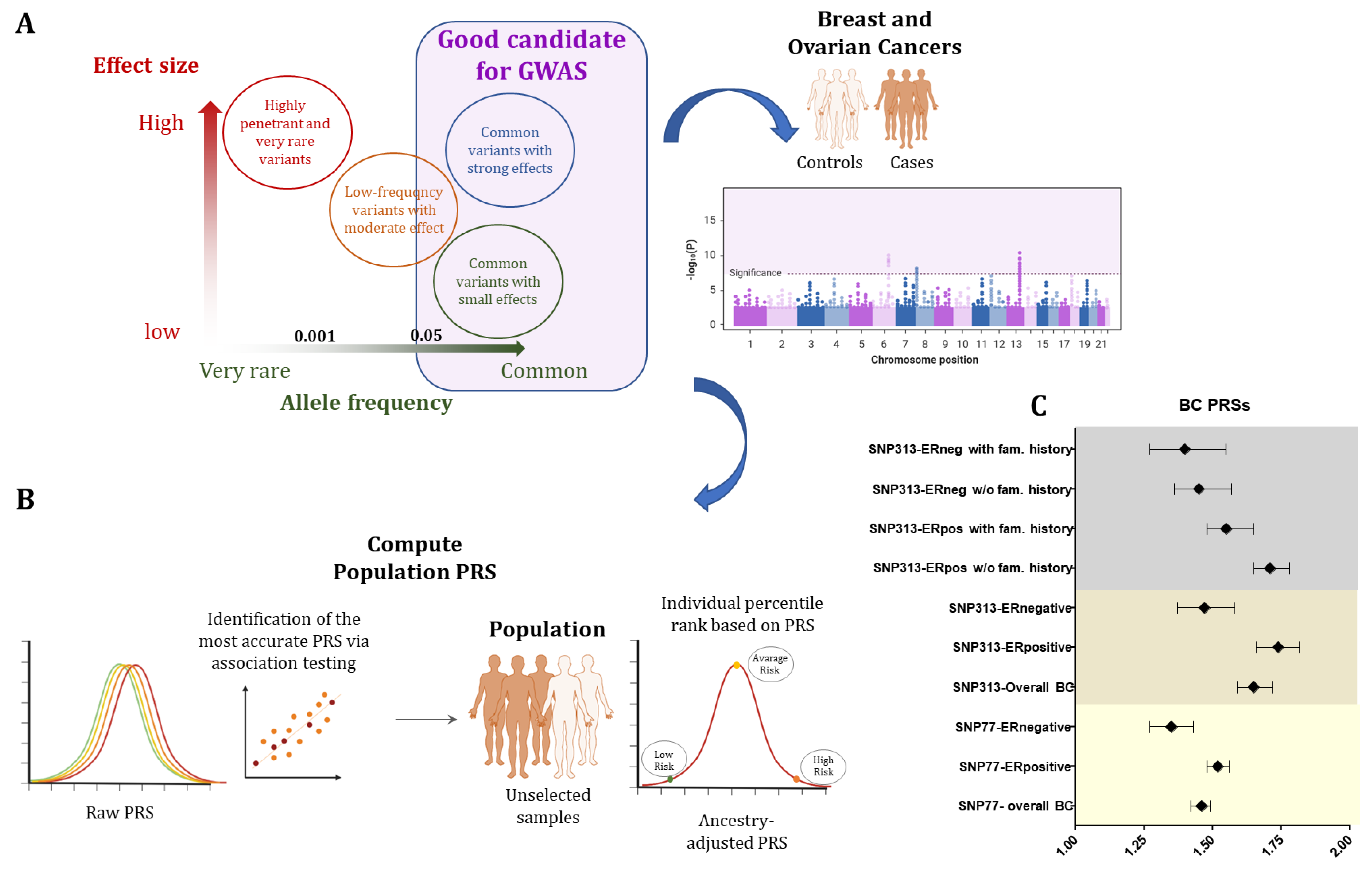
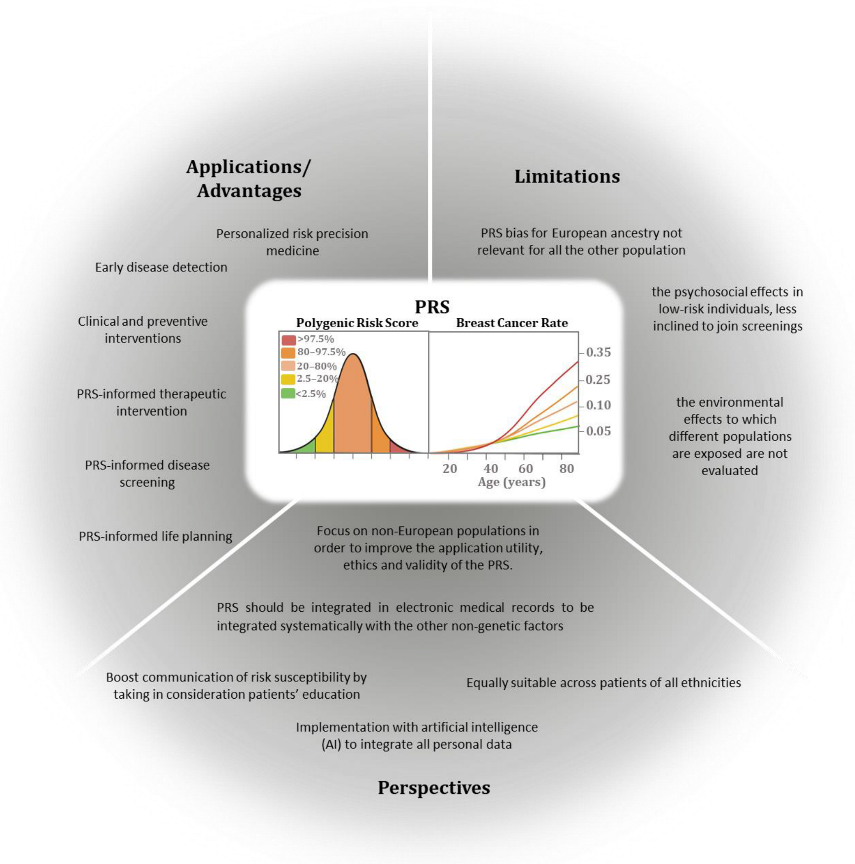
7. Polygenic Risk Score

Mendelian inheritance with higher or lower penetrance, however, accounts for only a small share of the whole breast cancer burden, leaving the majority of women, even with a positive family history for the disease, without the possibility of a specific risk prediction. However, over the past 10 years, significant growth has been made in determining germline variants or single-nucleotide polymorphisms (SNPs), which can be associated with the risk of developing breast cancer and can be used for the risk prediction. Over the last decade, genome-wide association studies (GWASs) have demonstrated that common single-nucleotide polymorphisms (SNPs) are involved in determining the susceptibility to common complex disorders, uncovering variants, which occur in disease-affected vs. unaffected controls at a significantly higher frequency (typically with p-values lower than 5 × 10⁻⁷). In a polygenic condition, if considered individually, most of these SNPs have a moderate effect on disease association and are not valuable for estimating disease risk; on the contrary, when combined together, they show differences in their frequencies in affected and non-affected breast cancer patients [137]. Fundamental to unmasking the relationship between the individual genetic variants and the disease development is understanding the disease genetic architecture, i.e., in the case of breast and ovarian cancers, the number and frequency of genetic variants possibly associated with the disease risk and the relative weight of each of them. A source of confusion on genetic architecture relies on the categorization of a disease as monogenic as opposed to polygenic, meaning that the occurrence of the disease depends on a single- or many-gene changes. From a genetic point of view, common breast cancers likely represent a continuum among common low-risk and rare high-risk genetic variants, which cumulatively contribute to the overall risk for each single person. For breast cancer, the rare high-risk genetic variants displaying a minor allele frequency (MAF) lower than 0.5% account for nearly 1–10% of disease incidence, allowing the identification of very high-risk individuals who could benefit from timely medical interventions. The remaining population does not show evidence of any familial disease (i.e., small families) or may have de novo genetic changes or a more complex genetic architecture. In fact, many high-(MAF > 5%) and low-frequency (MAF between 0.5% and 5%) genetic variants, which per se contribute small additive effects, may account for the nonfamilial risk of the disease. By combining together risk alleles, which are selected on the basis of the evidence derived from genome-wide association studies and weighted according to their effect size [138], polygenic risk scores (PRSs) have been established [137]. The PRS clinical validity is defined by the accuracy in the discrimination of the population under study into categories of different degrees of absolute risk, also in combination with clinical risk factors. In order to be used in clinical and preventive interventions, it is necessary that the PRS can be applied individually with a good balance between specificity (false-positive rate) and sensitivity (true-positive rate). The risk model’s discriminatory accuracy is assessed by calculating the area under the receiver operating characteristic curve (AUC), which represents the general probability that the predicted risk is higher for cases than for the control. Practically, the individual-level PRS values group the population based on risk percentiles (such as top 1%, top 10%), giving a different level of absolute probabilistic risk in each tier [139,140].
In breast and ovarian cancer, the first case–control study predicting BC [141] in women without any pathogenic variants in BRCA1/2 genes developed an SNP18 PRS with an OR of 1.55 (95% CI 1.29 to 1.87) and an AUC of 0.59, demonstrating the PRS feasibility. SNP77 was the next and well-approved BC PRS, based on around 33,000 BC cases and 33,000 European controls, which was able to stratify women with and without a family history of BC [139] with an OR of 1.52 (95% CI 1.45–1.59) and an AUC of 0.62.
The largest study of the Breast Cancer Association Consortium (about 11,000 cases vs. 18,000 controls) defined the most predictive SNP313 PRS for breast cancer risk development with an OR of 1.61 (95% CI 1.57–1.65) and an AUC of 0.63 in the European population [142]. Moreover, the BC lifetime risk in the top centile of the PRS was as high as 32.6% and women had 4.37- and 2.78-fold risks of developing both ER-positive and ER-negative diseases as compared to those in the intermediate quintile. Interestingly, both SNP77 and SNP313 are good and reliable predictors of ER-positive BC (subtype-specific PRSs), with more significant ORs than those for ER-negative tumors, possibly reflecting a higher statistical power of the cases with ER-positive tumors (Figure 1). Finally, when combined with the cancer family history, the PRS showed higher values in women without a family history (OR = 1.71, 95% CI 1.65–1.78) as compared to those with a positive family history (OR = 1.55 (95% CI 1.48–1.65) in the ER-positive subgroup [142].
It has also been demonstrated that 94-SNP and 18-SNP PRSs can predict absolute risks of developing BC and OC for BRCA1/2 carriers [143]. The PRS for ER-negative women exhibited the highest correlation with BC risk in BRCA1 patients with an HR of 1.27 (95% CI = ¼ 1.23 to 1.31); also, BRCA2 carriers showed PRS for the risk of BC with HR = 1.22 (95% CI = 1.17 to 1.28), showing differences in absolute risks (more than 10% in each case) between the upper and lower deciles of the PRS curve [143].
Moreover, the following studies focused on implementing the breast and ovarian cancer SNP313 PRS for BRCA1/2 carriers, to evaluate how its application may influence cancer risk guidance for women with pathogenic variants in these genes. Barnes et al. (2020) studied the largest cohort of BRCA1/2-positive women and further validated the associations in a prospective sample of carriers by applying population-based PRSs. BRCA1 carriers showed the highest PRSs for ER-negative BC with a hazard ratio of 1.29 (95% CI 1.25–1.33), reflecting that those at the 5th and 95th percentiles of the PRSER distribution have predicted breast cancer risks to 80 years of age of 59% and 83%, respectively. Alongside this, BRCA2 patients display the strongest correlation with the overall BC PRS and an HR of 1.31 (95% CI 1.27–1.36), which corresponds to risks of 57% and 81% for the BRCA2 carriers in the 5th and 95th percentiles of the PRS. When evaluating ovarian cancer risk by the age of 80 years for BRCA1 and BRCA2 carriers, the PRSHGS (22 SNPs predicting high-grade serous OC) at the 5th and 95th percentiles showed risks of 30% and 59%, and 10% and 28%, respectively [144].
The risk stratification models have been further developed by integrating family history information, such as the age of disease onset and family breast cancer cases and other factors, such as endocrine and anthropomorphic factors, breast imaging density, lifestyle, and cases of previous benign proliferative disorders [145]. Several statistical models have included these factors, such as the Gail model, Breast and Ovarian Analysis of Disease Incidence and Carrier Estimation Algorithm (BOADICEA), Tyrer–Cuzick (TC), BRCAPRO, and Breast Cancer Surveillance Consortium (BCSC).
The first BOADICEA prediction model integrated the risk due to the rare loss-of-function variants in the “major genes” (BRCA1 and 2, PALB2, CHEK2 and ATM) with tumor pathology (receptors status), basic demographic element (e.g., year and country of birth and family ethnicity), and family history [146]. The authors also incorporated the SNP313 PRS, the mammographic density and questionnaire-based risk factors, to generate the first comprehensive model for the prediction of personal BC risk in unaffected women [146]. Thus, the integration of the effects of all known elements in a single model allows a more coherent method for risk stratification both in general individuals and in women with a family history of BC, with the major limitation represented by the reference population bias, due to the assessment of an only-European population in the computation of the PRS.
More recently, PRS has improved also via artificial intelligence technologies and a deep neural network (DNN), and other computational models (logistic regression, decision tree, random forest, AdaBoots, gradient boosting, support vector machine (SVM), and Gaussian naïve Bayes) have been tested to estimate breast cancer PRS starting from 5273 SNPs as input [147]. For this study, a breast cancer GWAS dataset containing 49,111 individuals of the DRIVE (Discovery, Biology, and Risk of Inherited Variants in Breast Cancer) project obtained from the NIH dbGaP database (accession number: phs001265.v1.p1.) was employed [148]. The top-100 relevant SNPs identified the individuals with the highest PRS (>0.67 cutoff) with a 90% precision and, among those SNPs, 8 belonged to genes with a functional role in BC pathogenesis [147]. AI tools based on PRS have also been used to reduce screening costs, as emerged in some recent studies [149,150,151]. Mital et al.’s [149] economic evaluation using a hybrid decision tree/microsimulation model for the comparison of costs and effectiveness found that AI-based risk prediction and the subsequent lack of screening for women with low susceptibility was the most efficient and budget-conscious approach. Thus, AI was more accurate in identifying high-risk women than family history when used in conjunction with PRS, achieving fewer false-positive diagnoses from not screening low-risk women.
In conclusion, BC PRS is an increasingly successful attempt to establish a personalized medicine for risk prediction (Figure 2) in unaffected subjects as compared to classic risk factors, although further studies are still needed to understand the potential contribution of other novel technologies (e.g., AI) and to improve the predictive power beyond the ancestry-dependent genetic background.

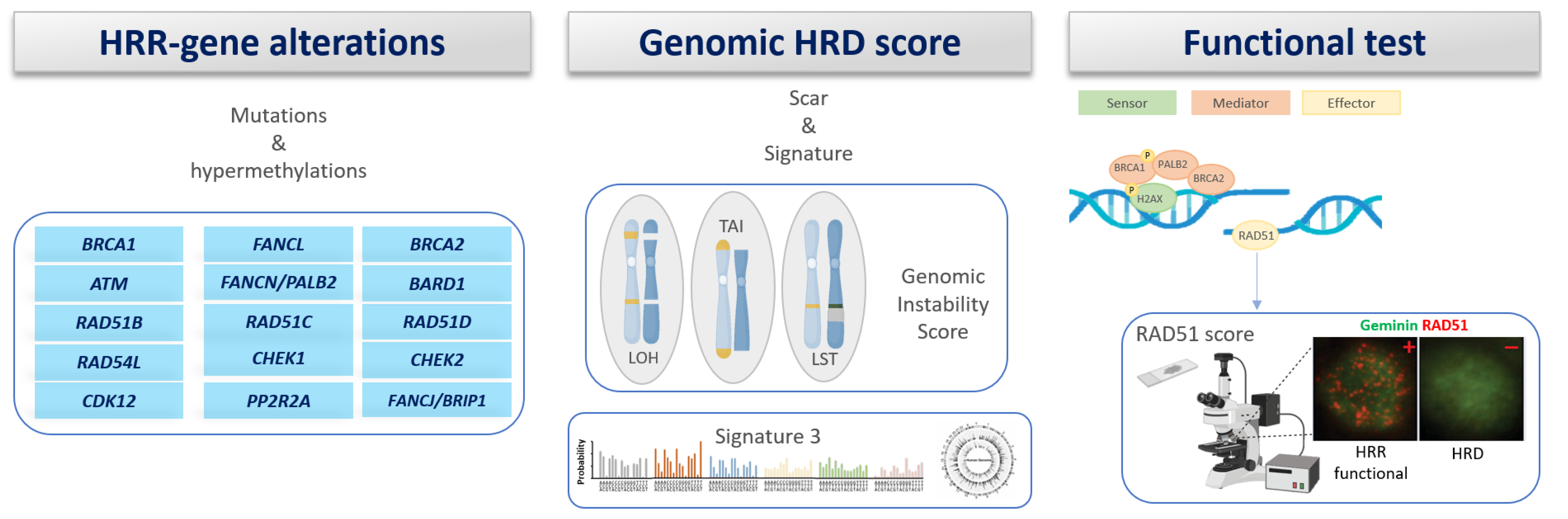
8. Homologous Recombination Deficiency (HRD)

The homologous recombination deficiency (HRD), which is intrinsic to the HBOC-associated cancers, has been exploited both in therapy (Table 3), which is based on the suppression of alternative repair pathways (PARP inhibition), and in diagnostics (Figure 3), by tracing the genomic lesions attributable to the diminished activity in the Homologous Recombination Repair pathway, called genomic scars.

8.1. Gene Scar/Signature

Cancer cells and cells with BRCA1/2 variants are characterized by genomic instability: they exhibit copy number modifications and numerous somatic changes in the genome, including single-nucleotide polymorphisms and structural aberrancies (structural variants, SVs). The evaluation of these genomic characteristics allows the identification of tumors with a history of HRD, regardless of the underlying etiology.
Genomic HRD tests have been developed using SNP microarrays, measuring somatic CNVs. In 2012, three papers reported SNP-based CNV tests that calculated BRCA status by quantifying large-scale transitions (LSTs), loss of heterozygosity (LOH), and the number of sub-chromosomal regions with an allelic imbalance up to the telomere (TAI/telomeric allelic imbalance) [152].
Two commercial tests related to genomic scars have been approved by the FDA and used to identify tumors with HRD: (1) the “myChoice HRD” kit by Myriad, which inquires the LOH, the TAI, and the LST throughout the genome [153], and (2) the “FoundationFocus™ CDx BRCA LOH”, which is conceived to identify the occurrence of variants in BRCA1/2 and the amount of the genome impacted by LOH in tumor-derived DNA of patients with OC [154]. Finally, the non-commercial test HRDetect [155] includes additional mutational profiles and characteristics derived by each mutational mechanism on the tumor genome: for instance, HRD has been correlated with the “signature 3” characterized by Alexandrov et al. [156,157].

8.2. Functional Assays of HRD

A key constraint of the gene scar tests is the lack of evaluation of the processes of tumor progression, such as a recovery of the HRR process in response to treatment-driven pressure. Instead, it might be advantageous to integrate functional biomarkers developed on assays capable of estimating the functionality of a repair pathway in a dynamic way. A crucial step of HRR is mediated by the RAD51 protein, a nucleoprotein filament, which is accomplished to execute the strand exchange step of HRR [158]. Despite some limitations yet to be solved, several approaches, both in vivo and in vitro, underpinned the highly sensitive and specific predictive value of the absence of nuclear RAD51 foci in response to PARPi therapy (Table 4) [159,160,161,162]. One such drawback is that the RAD51 test might fail to identify ATM-variant-associated tumors that can benefit from PARPi according to their specific mechanisms of sensitivity [163,164,165,166].

8.3. RAD51 Assay as Functional Biomarker of HRD in Early BC

A retrospective study of the accuracy of the RAD51 assay, which was based on 133 tumors from TNBC patients enrolled in the GeparSixto trial, has shown that in BRCA1-mutated tumors, the nuclear foci level (BRCA1 score) does not occur in 43% of tumors, which were also with low RAD51 levels. The RAD51 prognostic marker detected 93% (76–99%, 95% CI) of tBRCA1/2-mutated cancer and 45% (34–56%, 95% CI) of the non-tBRCA1/2 mutants with active HRD. RAD51 detected 86% of cancers with HRD and 90% with HRR proficiency (HRP). Overall, RAD51 and genomic HRD were concordant in 87% (79–93%), demonstrating the feasibility of the RAD51 assay in its application on FFPE samples derived from untreated TNBC and its high degree of congruence with tBRCA1/2 mutations and HRD results [167].
When used for monitoring the response to chemotherapy, the RAD51 assay can significantly distinguish cancers responsive to carboplatin (p = 0.02): the benefit of its administration was maintained also in the multivariate analysis after adjustment for predefined clinical–pathological parameters (OR = 7.52, 95% CI 2.21–25.61, p = 0.001). Moreover, in terms of DFS (disease-free survival), a similar response was observed in RAD51-high and RAD51-low groups, whereas in OS, no evident correlation was reached [167].
The RAD51 assay was also performed on samples from early TNBC patients enrolled in the PETREMAC trial and treated with PARPi Olaparib in a neoadjuvant setting. Functional HRD, with low RAD51 scores, is associated with HR mutations/BRCA1 methylation profiles, as well as Olaparib response [168].
Table 3. Efficacy of PARPi according to HRD status in ovarian cancer.
Clinical TrialDrugSettingStudy PopulationHRD Role
ARIEL-2RucaparibMonotherapyRelapsed, platinum-sensitive ovarian cancerHigher performance in gBRCA1/2-variant-associated and/or LOH-high compared to LOH-low tumors. Not effective in manifesting a difference between LOH-high and LOH-low tumors
ARIEL-3RucaparibMaintenance therapyPlatinum-sensitive ovarian cancerEfficacy regardless of LOH status.
NOVA-trialNiraparibMaintenance therapyPlatinum-sensitive ovarian cancerEfficacy regardless of HRD status.
Figure 3. Homologous recombination repair deficiency (HRD) detection is based on various methods based on germline or somatic analysis.
Genes 15 00219 g003
Table 4. Efficacy of PARPi according to HRD status in breast cancer.
Clinical TrialDrugSettingStudy PopulationHRD Role
PrECOG 0105
Cisplatin-1 trial
Cisplatin-2 trial
Platinum saltsNeoadjuvant settingUntreated patientsHRD-positive patients had higher complete pathologic response
Gepar-Sixto trialCarboplatinNeoadjuvant settingUntreated patientsHRD-positive patients have a better prognosis by comparing with HRD-negative ones. No robust evidence can be reached about the predictive role of HRD regarding carboplatin
TBCRC009 trialPlatinum saltsAdvanced settingFirst- or second-line treatmentHigher HRD scores were reported in responding patients, independent of BRCA1/2 variant status.
TNT trialCarboplatinumAdvanced settingFirst-line treatmentORR is not associated with HRD levels in the primary tumors.

9. Conclusions and Perspectives

Once established, the contribution of high-penetrance mendelian genes to the pathogenesis of HBOC, however, contributes only to a small share of the whole breast cancer burden, and the paradigm shifts to the study of the genome in search of variants to be used for prognosis or therapy. Still, there is a long way to go, and even genes which have been tested for diagnostic purposes in millions of women worldwide are still frequently (~5%) burdened by the presence of variants of uncertain significance, which deprive the result of the test of its clinical significance and may result in inappropriate clinical management [169,170]. Crucially, genetic findings related to the overall BRCA1/2 variants were conducted mainly on Caucasian individuals. This fundamental fact is primarily due to the significant absence of knowledge in the genetic variation for all other ethnic populations, highlighting the issue of understanding the full spectrum of pathogenicity of the current variant information related to BRCA genes, which may not be applicable to other ethnicities. In this perspective, future studies might decipher the proper genetic landscape underneath BRCA genes only after inclusion of a broader ethnicity in the gene analysis with the final aim of giving more focused clinical interventions. Moreover, the HBOC has contributed to reduce the borders between mendelian and multifactorial disorder categories, also showing the deep interaction between the two factors: for example, the probability of BC occurrence by age 75 spans from 12.7% to 75.7% among BRCA1 and BRCA2 pathogenic variant carriers, while the risk of non-carriers oscillates between 3.3% and 29.6%, showing a high overlap between carriers and non-carriers of high-risk variants, which can be explained only in terms of the polygenic risk of other low-penetrance variants. HBOC has always been ahead of all the other hereditary cancer predisposition syndromes in terms of knowledge, protocols, and specific therapeutic options available: replicating the efforts and successes achieved for HBOC should inspire future research for other cancer predisposition syndromes and for their sporadic counterparts.

Author Contributions

V.B., E.A., B.B., R.M., E.D.S., I.R.C., A.T., M.M., A.S., D.B., C.T., O.S., F.B., A.A., A.L., D.M., P.C. and B.P., data curation and writing—original draft preparation; A.P., V.U., B.P. and A.M., writing—original draft preparation, supervision, and project administration. BioRender.com for figure creation. All authors have read and agreed to the published version of the manuscript.

Funding

This study was partially supported by the “Fondazione Emma ed Ernesto Rulfo per la Genetica Medica” and by the “BANDO DI ATENEO PER LA RICERCA 2022 MUR_DM737_2022” from the University of Parma.

Acknowledgments

This work is generated within the European Reference Network on Genetic Tumour Risk Syndromes (ERN GENTURIS). ERN GENTURIS is funded by the European Union.

Conflicts of Interest

The authors declare no conflicts of interest.

References

  1. Broca, P. Traité des Tumeurs; P. Asselin: Paris, France, 1866. [Google Scholar]
  2. Miki, Y.; Swensen, J.; Shattuck-Eidens, D.; Futreal, P.A.; Harshman, K.; Tavtigian, S.; Liu, Q.; Cochran, C.; Bennett, L.M.; Ding, W.; et al. A Strong Candidate for the Breast and Ovarian Cancer Susceptibility Gene BRCA1. Science 1994, 266, 66–71. [Google Scholar] [CrossRef] [PubMed]
  3. Wooster, R.; Neuhausen, S.L.; Mangion, J.; Quirk, Y.; Ford, D.; Collins, N.; Nguyen, K.; Seal, S.; Tran, T.; Averill, D.; et al. Localization of a Breast Cancer Susceptibility Gene, BRCA2, to Chromosome 13q12-13. Science 1994, 265, 2088–2090. [Google Scholar] [CrossRef] [PubMed]
  4. Joly, Y.; Tonin, P.N. Social, Ethical and Legal Considerations Raised by the Discovery and Patenting of the BRCA1 and BRCA2 Genes. New Genet. Soc. 2014, 33, 167–180. [Google Scholar] [CrossRef]
  5. Easton, D.F.; Pharoah, P.D.P.; Antoniou, A.C.; Tischkowitz, M.; Tavtigian, S.V.; Nathanson, K.L.; Devilee, P.; Meindl, A.; Couch, F.J.; Southey, M.; et al. Gene-Panel Sequencing and the Prediction of Breast-Cancer Risk. N. Engl. J. Med. 2015, 372, 2243–2257. [Google Scholar] [CrossRef] [PubMed]
  6. Petrucelli, N.; Daly, M.B.; Pal, T. BRCA1- and BRCA2-Associated Hereditary Breast and Ovarian Cancer; University of Washington: Seattle, WA, USA, 1998. [Google Scholar]
  7. Kuchenbaecker, K.B.; Hopper, J.L.; Barnes, D.R.; Phillips, K.-A.; Mooij, T.M.; Roos-Blom, M.-J.; Jervis, S.; van Leeuwen, F.E.; Milne, R.L.; Andrieu, N.; et al. Risks of Breast, Ovarian, and Contralateral Breast Cancer for BRCA1 and BRCA2 Mutation Carriers. JAMA 2017, 317, 2402. [Google Scholar] [CrossRef] [PubMed]
  8. Mavaddat, N.; Peock, S.; Frost, D.; Ellis, S.; Platte, R.; Fineberg, E.; Evans, D.G.; Izatt, L.; Eeles, R.A.; Adlard, J.; et al. Cancer Risks for BRCA1 and BRCA2 Mutation Carriers: Results from Prospective Analysis of EMBRACE. JNCI J. Natl. Cancer Inst. 2013, 105, 812–822. [Google Scholar] [CrossRef] [PubMed]
  9. Whittemore, A.S.; Gong, G.; John, E.M.; McGuire, V.; Li, F.P.; Ostrow, K.L.; Dicioccio, R.; Felberg, A.; West, D.W. Prevalence of BRCA1 Mutation Carriers among U.S. Non-Hispanic Whites. Cancer Epidemiol. Biomark. Prev. 2004, 13, 2078–2083. [Google Scholar] [CrossRef]
  10. Manchanda, R.; Legood, R.; Burnell, M.; McGuire, A.; Raikou, M.; Loggenberg, K.; Wardle, J.; Sanderson, S.; Gessler, S.; Side, L.; et al. Cost-Effectiveness of Population Screening for BRCA Mutations in Ashkenazi Jewish Women Compared with Family History–Based Testing. JNCI J. Natl. Cancer Inst. 2015, 107, 380. [Google Scholar] [CrossRef]
  11. Venkitaraman, A.R. Cancer Suppression by the Chromosome Custodians, BRCA1 and BRCA2. Science 2014, 343, 1470–1475. [Google Scholar] [CrossRef]
  12. Sasanuma, H.; Tsuda, M.; Morimoto, S.; Saha, L.K.; Rahman, M.M.; Kiyooka, Y.; Fujiike, H.; Cherniack, A.D.; Itou, J.; Callen Moreu, E.; et al. BRCA1 Ensures Genome Integrity by Eliminating Estrogen-Induced Pathological Topoisomerase II–DNA Complexes. Proc. Natl. Acad. Sci. USA 2018, 115, E10642–E10651. [Google Scholar] [CrossRef]
  13. Turner, N.; Tutt, A.; Ashworth, A. Hallmarks of “BRCAness” in Sporadic Cancers. Nat. Rev. Cancer 2004, 4, 814–819. [Google Scholar] [CrossRef]
  14. Toss, A.; Molinaro, E.; Venturelli, M.; Domati, F.; Marcheselli, L.; Piana, S.; Barbieri, E.; Grandi, G.; Piombino, C.; Marchi, I.; et al. BRCA Detection Rate in an Italian Cohort of Luminal Early-Onset and Triple-Negative Breast Cancer Patients without Family History: When Biology Overcomes Genealogy. Cancers 2020, 12, 1252. [Google Scholar] [CrossRef]
  15. Landrum, M.J.; Lee, J.M.; Benson, M.; Brown, G.R.; Chao, C.; Chitipiralla, S.; Gu, B.; Hart, J.; Hoffman, D.; Jang, W.; et al. ClinVar: Improving Access to Variant Interpretations and Supporting Evidence. Nucleic Acids Res. 2018, 46, D1062–D1067. [Google Scholar] [CrossRef]
  16. Nepomuceno, T.C.; Foo, T.K.; Richardson, M.E.; Ranola, J.M.O.; Weyandt, J.; Varga, M.J.; Alarcon, A.; Gutierrez, D.; von Wachenfeldt, A.; Eriksson, D.; et al. BRCA1 Frameshift Variants Leading to Extended Incorrect Protein C Termini. Hum. Genet. Genom. Adv. 2023, 4, 100240. [Google Scholar] [CrossRef] [PubMed]
  17. Corso, G.; Feroce, I.; Intra, M.; Toesca, A.; Magnoni, F.; Sargenti, M.; Naninato, P.; Caldarella, P.; Pagani, G.; Vento, A.; et al. BRCA1/2 Germline Missense Mutations: A Systematic Review. Eur. J. Cancer Prev. 2018, 27, 279–286. [Google Scholar] [CrossRef] [PubMed]
  18. Richards, S.; Aziz, N.; Bale, S.; Bick, D.; Das, S.; Gastier-Foster, J.; Grody, W.W.; Hegde, M.; Lyon, E.; Spector, E.; et al. Standards and Guidelines for the Interpretation of Sequence Variants: A Joint Consensus Recommendation of the American College of Medical Genetics and Genomics and the Association for Molecular Pathology. Genet. Med. 2015, 17, 405–424. [Google Scholar] [CrossRef] [PubMed]
  19. Venkitaraman, A.R. How Do Mutations Affecting the Breast Cancer Genes BRCA1 and BRCA2 Cause Cancer Susceptibility? DNA Repair 2019, 81, 102668. [Google Scholar] [CrossRef] [PubMed]
  20. Evans, D.G.R.; van Veen, E.M.; Byers, H.J.; Wallace, A.J.; Ellingford, J.M.; Beaman, G.; Santoyo-Lopez, J.; Aitman, T.J.; Eccles, D.M.; Lalloo, F.I.; et al. A Dominantly Inherited 5′ UTR Variant Causing Methylation-Associated Silencing of BRCA1 as a Cause of Breast and Ovarian Cancer. Am. J. Hum. Genet. 2018, 103, 213–220. [Google Scholar] [CrossRef] [PubMed]
  21. Rebbeck, T.R.; Mitra, N.; Wan, F.; Sinilnikova, O.M.; Healey, S.; McGuffog, L.; Mazoyer, S.; Chenevix-Trench, G.; Easton, D.F.; Antoniou, A.C.; et al. Association of Type and Location of BRCA1 and BRCA2 Mutations with Risk of Breast and Ovarian Cancer. JAMA 2015, 313, 1347. [Google Scholar] [CrossRef]
  22. Gayther, S.A.; Mangion, J.; Russell, P.; Seal, S.; Barfoot, R.; Ponder, B.A.J.; Stratton, M.R.; Easton, D. Variation of Risks of Breast and Ovarian Cancer Associated with Different Germline Mutations of the BRCA2 Gene. Nat. Genet. 1997, 15, 103–105. [Google Scholar] [CrossRef]
  23. Roed Nielsen, H.; Petersen, J.; Therkildsen, C.; Skytte, A.-B.; Nilbert, M. Increased Risk of Male Cancer and Identification of a Potential Prostate Cancer Cluster Region in BRCA2. Acta Oncol. 2016, 55, 38–44. [Google Scholar] [CrossRef]
  24. Patel, V.L.; Busch, E.L.; Friebel, T.M.; Cronin, A.; Leslie, G.; McGuffog, L.; Adlard, J.; Agata, S.; Agnarsson, B.A.; Ahmed, M.; et al. Association of Genomic Domains in BRCA1 and BRCA2 with Prostate Cancer Risk and Aggressiveness. Cancer Res. 2020, 80, 624–638. [Google Scholar] [CrossRef]
  25. Chian, J.S.; Xu, W.; Wang, S.M. Pancreatic Cancer Cluster Region Identified in BRCA2. J. Med. Genet. 2023, 60, 1052–1056. [Google Scholar] [CrossRef]
  26. de Garibay, G.R.; Fernandez-Garcia, I.; Mazoyer, S.; Leme de Calais, F.; Ameri, P.; Vijayakumar, S.; Martinez-Ruiz, H.; Damiola, F.; Barjhoux, L.; Thomassen, M.; et al. Altered Regulation of BRCA1 Exon 11 Splicing Is Associated with Breast Cancer Risk in Carriers of BRCA1 Pathogenic Variants. Hum. Mutat. 2021, 42, 1488–1502. [Google Scholar] [CrossRef] [PubMed]
  27. Meeks, H.D.; Song, H.; Michailidou, K.; Bolla, M.K.; Dennis, J.; Wang, Q.; Barrowdale, D.; Frost, D.; McGuffog, L.; Ellis, S.; et al. BRCA2 Polymorphic Stop Codon K3326X and the Risk of Breast, Prostate, and Ovarian Cancers. J. Natl. Cancer Inst. 2016, 108, djv315. [Google Scholar] [CrossRef]
  28. Cline, M.S.; Liao, R.G.; Parsons, M.T.; Paten, B.; Alquaddoomi, F.; Antoniou, A.; Baxter, S.; Brody, L.; Cook-Deegan, R.; Coffin, A.; et al. BRCA Challenge: BRCA Exchange as a Global Resource for Variants in BRCA1 and BRCA2. PLoS Genet. 2018, 14, e1007752. [Google Scholar] [CrossRef] [PubMed]
  29. LaDuca, H.; Stuenkel, A.J.; Dolinsky, J.S.; Keiles, S.; Tandy, S.; Pesaran, T.; Chen, E.; Gau, C.-L.; Palmaer, E.; Shoaepour, K.; et al. Utilization of Multigene Panels in Hereditary Cancer Predisposition Testing: Analysis of More than 2,000 Patients. Genet. Med. 2014, 16, 830–837. [Google Scholar] [CrossRef]
  30. Buys, S.S.; Sandbach, J.F.; Gammon, A.; Patel, G.; Kidd, J.; Brown, K.L.; Sharma, L.; Saam, J.; Lancaster, J.; Daly, M.B. A Study of over 35,000 Women with Breast Cancer Tested with a 25-gene Panel of Hereditary Cancer Genes. Cancer 2017, 123, 1721–1730. [Google Scholar] [CrossRef] [PubMed]
  31. Sun, J.; Meng, H.; Yao, L.; Lv, M.; Bai, J.; Zhang, J.; Wang, L.; Ouyang, T.; Li, J.; Wang, T.; et al. Germline Mutations in Cancer Susceptibility Genes in a Large Series of Unselected Breast Cancer Patients. Clin. Cancer Res. 2017, 23, 6113–6119. [Google Scholar] [CrossRef]
  32. Hu, C.; Hart, S.N.; Gnanaolivu, R.; Huang, H.; Lee, K.Y.; Na, J.; Gao, C.; Lilyquist, J.; Yadav, S.; Boddicker, N.J.; et al. A Population-Based Study of Genes Previously Implicated in Breast Cancer. N. Engl. J. Med. 2021, 384, 440–451. [Google Scholar] [CrossRef]
  33. Breast Cancer Association, C.; Dorling, L.; Carvalho, S.; Allen, J.; González-Neira, A.; Luccarini, C.; Wahlström, C.; Pooley, K.A.; Parsons, M.T.; Fortuno, C.; et al. Breast Cancer Risk Genes—Association Analysis in More than 113,000 Women. N. Engl. J. Med. 2021, 384, 428–439. [Google Scholar] [CrossRef]
  34. Narod, S.A. Which Genes for Hereditary Breast Cancer? N. Engl. J. Med. 2021, 384, 471–473. [Google Scholar] [CrossRef]
  35. Figueiredo, J.; Melo, S.; Carneiro, P.; Moreira, A.M.; Fernandes, M.S.; Ribeiro, A.S.; Guilford, P.; Paredes, J.; Seruca, R. Clinical Spectrum and Pleiotropic Nature of CDH1 Germline Mutations. J. Med. Genet. 2019, 56, 199–208. [Google Scholar] [CrossRef]
  36. Kaurah, P.; MacMillan, A.; Boyd, N.; Senz, J.; De Luca, A.; Chun, N.; Suriano, G.; Zaor, S.; Van Manen, L.; Gilpin, C.; et al. Founder and Recurrent CDH1 Mutations in Families with Hereditary Diffuse Gastric Cancer. JAMA 2007, 297, 2360. [Google Scholar] [CrossRef] [PubMed]
  37. Benusiglio, P.R.; Malka, D.; Rouleau, E.; De Pauw, A.; Buecher, B.; Noguès, C.; Fourme, E.; Colas, C.; Coulet, F.; Warcoin, M.; et al. CDH1 Germline Mutations and the Hereditary Diffuse Gastric and Lobular Breast Cancer Syndrome: A Multicentre Study. J. Med. Genet. 2013, 50, 486–489. [Google Scholar] [CrossRef] [PubMed]
  38. Blair, V.R.; McLeod, M.; Carneiro, F.; Coit, D.G.; D’Addario, J.L.; van Dieren, J.M.; Harris, K.L.; Hoogerbrugge, N.; Oliveira, C.; van der Post, R.S.; et al. Hereditary Diffuse Gastric Cancer: Updated Clinical Practice Guidelines. Lancet Oncol. 2020, 21, e386–e397. [Google Scholar] [CrossRef] [PubMed]
  39. Oliveira, C.; Pinheiro, H.; Figueiredo, J.; Seruca, R.; Carneiro, F. E-Cadherin Alterations in Hereditary Disorders with Emphasis on Hereditary Diffuse Gastric Cancer. Prog. Mol. Biol. Transl. Sci. 2013, 116, 337–359. [Google Scholar] [PubMed]
  40. Corso, G.; Figueiredo, J.; La Vecchia, C.; Veronesi, P.; Pravettoni, G.; Macis, D.; Karam, R.; Lo Gullo, R.; Provenzano, E.; Toesca, A.; et al. Hereditary Lobular Breast Cancer with an Emphasis on E-Cadherin Genetic Defect. J. Med. Genet. 2018, 55, 431–441. [Google Scholar] [CrossRef]
  41. Fairoosa, P.; Witharana, C. Gene Mutations in Hereditary Breast Cancer—A Review. Eur. J. Med. Health Sci. 2020, 2. [Google Scholar] [CrossRef]
  42. Daly, M.B.; Pal, T.; Maxwell, K.N.; Churpek, J.; Kohlmann, W.; AlHilli, Z.; Arun, B.; Buys, S.S.; Cheng, H.; Domchek, S.M.; et al. NCCN Guidelines® Insights: Genetic/Familial High-Risk Assessment: Breast, Ovarian, and Pancreatic, Version 2.2024. J. Natl. Compr. Canc. Netw. 2023, 21, 1000–1010. [Google Scholar] [CrossRef]
  43. Angeli, D.; Salvi, S.; Tedaldi, G. Genetic Predisposition to Breast and Ovarian Cancers: How Many and Which Genes to Test? Int. J. Mol. Sci. 2020, 21, 1128. [Google Scholar] [CrossRef] [PubMed]
  44. Yang, X.; Leslie, G.; Doroszuk, A.; Schneider, S.; Allen, J.; Decker, B.; Dunning, A.M.; Redman, J.; Scarth, J.; Plaskocinska, I.; et al. Cancer Risks Associated with Germline PALB2 Pathogenic Variants: An International Study of 524 Families. J. Clin. Oncol. 2020, 38, 674–685. [Google Scholar] [CrossRef] [PubMed]
  45. Janssen, B.; Bellis, S.; Koller, T.; Tischkowitz, M.; Liau, S.-S. A Systematic Review of Predicted Pathogenic PALB2 Variants: An Analysis of Mutational Overlap between Epithelial Cancers. J. Hum. Genet. 2020, 65, 199–205. [Google Scholar] [CrossRef] [PubMed]
  46. Piombino, C.; Cortesi, L.; Lambertini, M.; Punie, K.; Grandi, G.; Toss, A. Secondary Prevention in Hereditary Breast and/or Ovarian Cancer Syndromes Other Than BRCA. J. Oncol. 2020, 2020, 6384190. [Google Scholar] [CrossRef] [PubMed]
  47. Nelen, M.R.; Kremer, H.; Konings, I.B.; Schoute, F.; van Essen, A.J.; Koch, R.; Woods, C.G.; Fryns, J.-P.; Hamel, B.; Hoefsloot, L.H.; et al. Novel PTEN Mutations in Patients with Cowden Disease: Absence of Clear Genotype–Phenotype Correlations. Eur. J. Hum. Genet. 1999, 7, 267–273. [Google Scholar] [CrossRef] [PubMed]
  48. Pilarski, R. PTEN Hamartoma Tumor Syndrome: A Clinical Overview. Cancers 2019, 11, 844. [Google Scholar] [CrossRef]
  49. Hendricks, L.A.J.; Hoogerbrugge, N.; Schuurs-Hoeijmakers, J.H.M.; Vos, J.R. A Review on Age-related Cancer Risks in PTEN Hamartoma Tumor Syndrome. Clin. Genet. 2021, 99, 219–225. [Google Scholar] [CrossRef]
  50. Tischkowitz, M.; Colas, C.; Pouwels, S.; Hoogerbrugge, N.; Bisseling, T.; Bubien, V.; Caux, F.; Chabbert-Buffet, N.; Colas, C.; Da Mota Gomes, S.; et al. Cancer Surveillance Guideline for Individuals with PTEN Hamartoma Tumour Syndrome. Eur. J. Hum. Genet. 2020, 28, 1387–1393. [Google Scholar] [CrossRef]
  51. Hendricks, L.A.J.; Hoogerbrugge, N.; Mensenkamp, A.R.; Brunet, J.; Lleuger-Pujol, R.; Høberg-Vetti, H.; Tveit Haavind, M.; Innella, G.; Turchetti, D.; Aretz, S.; et al. Cancer Risks by Sex and Variant Type in PTEN Hamartoma Tumor Syndrome. JNCI J. Natl. Cancer Inst. 2023, 115, 93–103. [Google Scholar] [CrossRef]
  52. Frazier, T.W.; Embacher, R.; Tilot, A.K.; Koenig, K.; Mester, J.; Eng, C. Molecular and Phenotypic Abnormalities in Individuals with Germline Heterozygous PTEN Mutations and Autism. Mol. Psychiatry 2015, 20, 1132–1138. [Google Scholar] [CrossRef]
  53. Gonzalez, K.D.; Noltner, K.A.; Buzin, C.H.; Gu, D.; Wen-Fong, C.Y.; Nguyen, V.Q.; Han, J.H.; Lowstuter, K.; Longmate, J.; Sommer, S.S.; et al. Beyond Li Fraumeni Syndrome: Clinical Characteristics of Families with P53 Germline Mutations. J. Clin. Oncol. 2009, 27, 1250–1256. [Google Scholar] [CrossRef] [PubMed]
  54. Mai, P.L.; Best, A.F.; Peters, J.A.; DeCastro, R.M.; Khincha, P.P.; Loud, J.T.; Bremer, R.C.; Rosenberg, P.S.; Savage, S.A. Risks of First and Subsequent Cancers among TP53 Mutation Carriers in the National Cancer Institute Li-Fraumeni Syndrome Cohort. Cancer 2016, 122, 3673–3681. [Google Scholar] [CrossRef] [PubMed]
  55. Melhem-Bertrandt, A.; Bojadzieva, J.; Ready, K.J.; Obeid, E.; Liu, D.D.; Gutierrez-Barrera, A.M.; Litton, J.K.; Olopade, O.I.; Hortobagyi, G.N.; Strong, L.C.; et al. Early Onset HER2-Positive Breast Cancer Is Associated with Germline TP53 Mutations. Cancer 2012, 118, 908–913. [Google Scholar] [CrossRef]
  56. Frebourg, T.; Bajalica Lagercrantz, S.; Oliveira, C.; Magenheim, R.; Evans, D.G. Guidelines for the Li–Fraumeni and Heritable TP53-Related Cancer Syndromes. Eur. J. Hum. Genet. 2020, 28, 1379–1386. [Google Scholar] [CrossRef]
  57. Renaux-Petel, M.; Charbonnier, F.; Théry, J.-C.; Fermey, P.; Lienard, G.; Bou, J.; Coutant, S.; Vezain, M.; Kasper, E.; Fourneaux, S.; et al. Contribution of de Novo and Mosaic TP53 Mutations to Li-Fraumeni Syndrome. J. Med. Genet. 2018, 55, 173–180. [Google Scholar] [CrossRef]
  58. Weitzel, J.N.; Chao, E.C.; Nehoray, B.; Van Tongeren, L.R.; LaDuca, H.; Blazer, K.R.; Slavin, T.; Pesaran, T.; Rybak, C.; Solomon, I.; et al. Somatic TP53 Variants Frequently Confound Germ-Line Testing Results. Genet. Med. 2018, 20, 809–816. [Google Scholar] [CrossRef]
  59. Chen, S.; Liu, Y. P53 Involvement in Clonal Hematopoiesis of Indeterminate Potential. Curr. Opin. Hematol. 2019, 26, 235–240. [Google Scholar] [CrossRef] [PubMed]
  60. Achatz, M.I.W.; Olivier, M.; Le Calvez, F.; Martel-Planche, G.; Lopes, A.; Rossi, B.M.; Ashton-Prolla, P.; Giugliani, R.; Palmero, E.I.; Vargas, F.R.; et al. The TP53 Mutation, R337H, Is Associated with Li-Fraumeni and Li-Fraumeni-like Syndromes in Brazilian Families. Cancer Lett. 2007, 245, 96–102. [Google Scholar] [CrossRef]
  61. Choi, M.; Kipps, T.; Kurzrock, R. ATM Mutations in Cancer: Therapeutic Implications. Mol. Cancer Ther. 2016, 15, 1781–1791. [Google Scholar] [CrossRef]
  62. Swift, M.; Morrell, D.; Cromartie, E.; Chamberlin, A.R.; Skolnick, M.H.; Bishop, D.T. The Incidence and Gene Frequency of Ataxia-Telangiectasia in the United States. Am. J. Hum. Genet. 1986, 39, 573–583. [Google Scholar]
  63. Lu, H.-M.; Li, S.; Black, M.H.; Lee, S.; Hoiness, R.; Wu, S.; Mu, W.; Huether, R.; Chen, J.; Sridhar, S.; et al. Association of Breast and Ovarian Cancers with Predisposition Genes Identified by Large-Scale Sequencing. JAMA Oncol. 2019, 5, 51. [Google Scholar] [CrossRef] [PubMed]
  64. Pritchard, C.C.; Mateo, J.; Walsh, M.F.; De Sarkar, N.; Abida, W.; Beltran, H.; Garofalo, A.; Gulati, R.; Carreira, S.; Eeles, R.; et al. Inherited DNA-Repair Gene Mutations in Men with Metastatic Prostate Cancer. N. Engl. J. Med. 2016, 375, 443–453. [Google Scholar] [CrossRef] [PubMed]
  65. Roberts, N.J.; Jiao, Y.; Yu, J.; Kopelovich, L.; Petersen, G.M.; Bondy, M.L.; Gallinger, S.; Schwartz, A.G.; Syngal, S.; Cote, M.L.; et al. ATM Mutations in Patients with Hereditary Pancreatic Cancer. Cancer Discov. 2012, 2, 41–46. [Google Scholar] [CrossRef] [PubMed]
  66. Helgason, H.; Rafnar, T.; Olafsdottir, H.S.; Jonasson, J.G.; Sigurdsson, A.; Stacey, S.N.; Jonasdottir, A.; Tryggvadottir, L.; Alexiusdottir, K.; Haraldsson, A.; et al. Loss-of-Function Variants in ATM Confer Risk of Gastric Cancer. Nat. Genet. 2015, 47, 906–910. [Google Scholar] [CrossRef] [PubMed]
  67. Bernstein, J.L.; Concannon, P. ATM, Radiation, and the Risk of Second Primary Breast Cancer. Int. J. Radiat. Biol. 2017, 93, 1121–1127. [Google Scholar] [CrossRef] [PubMed]
  68. Goldgar, D.E.; Healey, S.; Dowty, J.G.; Da Silva, L.; Chen, X.; Spurdle, A.B.; Terry, M.B.; Daly, M.J.; Buys, S.M.; Southey, M.C.; et al. Rare Variants in the ATMgene and Risk of Breast Cancer. Breast Cancer Res. 2011, 13, R73. [Google Scholar] [CrossRef]
  69. Hall, M.J.; Bernhisel, R.; Hughes, E.; Larson, K.; Rosenthal, E.T.; Singh, N.A.; Lancaster, J.M.; Kurian, A.W. Germline Pathogenic Variants in the Ataxia Telangiectasia Mutated (ATM) Gene Are Associated with High and Moderate Risks for Multiple Cancers. Cancer Prev. Res. 2021, 14, 433–440. [Google Scholar] [CrossRef]
  70. Tung, N.M.; Boughey, J.C.; Pierce, L.J.; Robson, M.E.; Bedrosian, I.; Dietz, J.R.; Dragun, A.; Gelpi, J.B.; Hofstatter, E.W.; Isaacs, C.J.; et al. Management of Hereditary Breast Cancer: American Society of Clinical Oncology, American Society for Radiation Oncology, and Society of Surgical Oncology Guideline. J. Clin. Oncol. 2020, 38, 2080–2106. [Google Scholar] [CrossRef]
  71. Tung, N.M.; Robson, M.E.; Ventz, S.; Santa-Maria, C.A.; Nanda, R.; Marcom, P.K.; Shah, P.D.; Ballinger, T.J.; Yang, E.S.; Vinayak, S.; et al. TBCRC 048: Phase II Study of Olaparib for Metastatic Breast Cancer and Mutations in Homologous Recombination-Related Genes. J. Clin. Oncol. 2020, 38, 4274–4282. [Google Scholar] [CrossRef]
  72. de Bono, J.; Mateo, J.; Fizazi, K.; Saad, F.; Shore, N.; Sandhu, S.; Chi, K.N.; Sartor, O.; Agarwal, N.; Olmos, D.; et al. Olaparib for Metastatic Castration-Resistant Prostate Cancer. N. Engl. J. Med. 2020, 382, 2091–2102. [Google Scholar] [CrossRef]
  73. Wu, L.C.; Wang, Z.W.; Tsan, J.T.; Spillman, M.A.; Phung, A.; Xu, X.L.; Yang, M.-C.W.; Hwang, L.-Y.; Bowcock, A.M.; Baer, R. Identification of a RING Protein That Can Interact in Vivo with the BRCA1 Gene Product. Nat. Genet. 1996, 14, 430–440. [Google Scholar] [CrossRef] [PubMed]
  74. Śniadecki, M.; Brzeziński, M.; Darecka, K.; Klasa-Mazurkiewicz, D.; Poniewierza, P.; Krzeszowiec, M.; Kmieć, N.; Wydra, D. BARD1 and Breast Cancer: The Possibility of Creating Screening Tests and New Preventive and Therapeutic Pathways for Predisposed Women. Genes 2020, 11, 1251. [Google Scholar] [CrossRef]
  75. Couch, F.J.; Shimelis, H.; Hu, C.; Hart, S.N.; Polley, E.C.; Na, J.; Hallberg, E.; Moore, R.; Thomas, A.; Lilyquist, J.; et al. Associations Between Cancer Predisposition Testing Panel Genes and Breast Cancer. JAMA Oncol. 2017, 3, 1190. [Google Scholar] [CrossRef] [PubMed]
  76. Shimelis, H.; LaDuca, H.; Hu, C.; Hart, S.N.; Na, J.; Thomas, A.; Akinhanmi, M.; Moore, R.M.; Brauch, H.; Cox, A.; et al. Triple-Negative Breast Cancer Risk Genes Identified by Multigene Hereditary Cancer Panel Testing. JNCI J. Natl. Cancer Inst. 2018, 110, 855–862. [Google Scholar] [CrossRef] [PubMed]
  77. Capasso, M.; Devoto, M.; Hou, C.; Asgharzadeh, S.; Glessner, J.T.; Attiyeh, E.F.; Mosse, Y.P.; Kim, C.; Diskin, S.J.; Cole, K.A.; et al. Common Variations in BARD1 Influence Susceptibility to High-Risk Neuroblastoma. Nat. Genet. 2009, 41, 718–723. [Google Scholar] [CrossRef] [PubMed]
  78. Weber-Lassalle, N.; Borde, J.; Weber-Lassalle, K.; Horváth, J.; Niederacher, D.; Arnold, N.; Kaulfuß, S.; Ernst, C.; Paul, V.G.; Honisch, E.; et al. Germline Loss-of-Function Variants in the BARD1 Gene Are Associated with Early-Onset Familial Breast Cancer but Not Ovarian Cancer. Breast Cancer Res. 2019, 21, 55. [Google Scholar] [CrossRef]
  79. Nalepa, G.; Clapp, D.W. Fanconi Anaemia and Cancer: An Intricate Relationship. Nat. Rev. Cancer 2018, 18, 168–185. [Google Scholar] [CrossRef]
  80. Seal, S.; Thompson, D.; Renwick, A.; Elliott, A.; Kelly, P.; Barfoot, R.; Chagtai, T.; Jayatilake, H.; Ahmed, M.; Spanova, K.; et al. Truncating Mutations in the Fanconi Anemia J Gene BRIP1 Are Low-Penetrance Breast Cancer Susceptibility Alleles. Nat. Genet. 2006, 38, 1239–1241. [Google Scholar] [CrossRef]
  81. Easton, D.F.; Lesueur, F.; Decker, B.; Michailidou, K.; Li, J.; Allen, J.; Luccarini, C.; Pooley, K.A.; Shah, M.; Bolla, M.K.; et al. No Evidence That Protein Truncating Variants in BRIP1 Are Associated with Breast Cancer Risk: Implications for Gene Panel Testing. J. Med. Genet. 2016, 53, 298–309. [Google Scholar] [CrossRef] [PubMed]
  82. Suszynska, M.; Ratajska, M.; Kozlowski, P. BRIP1, RAD51C, and RAD51D Mutations Are Associated with High Susceptibility to Ovarian Cancer: Mutation Prevalence and Precise Risk Estimates Based on a Pooled Analysis of ~30,000 Cases. J. Ovarian Res. 2020, 13, 50. [Google Scholar] [CrossRef] [PubMed]
  83. Moyer, C.L.; Ivanovich, J.; Gillespie, J.L.; Doberstein, R.; Radke, M.R.; Richardson, M.E.; Kaufmann, S.H.; Swisher, E.M.; Goodfellow, P.J. Rare BRIP1 Missense Alleles Confer Risk for Ovarian and Breast Cancer. Cancer Res. 2020, 80, 857–867. [Google Scholar] [CrossRef] [PubMed]
  84. Cybulski, C.; Wokołorczyk, D.; Jakubowska, A.; Huzarski, T.; Byrski, T.; Gronwald, J.; Masojć, B.; Dębniak, T.; Górski, B.; Blecharz, P.; et al. Risk of Breast Cancer in Women with a CHEK2 Mutation with and without a Family History of Breast Cancer. J. Clin. Oncol. 2011, 29, 3747–3752. [Google Scholar] [CrossRef] [PubMed]
  85. Adank, M.A.; Jonker, M.A.; Kluijt, I.; van Mil, S.E.; Oldenburg, R.A.; Mooi, W.J.; Hogervorst, F.B.L.; van den Ouweland, A.M.W.; Gille, J.J.P.; Schmidt, M.K.; et al. CHEK2*1100delC Homozygosity Is Associated with a High Breast Cancer Risk in Women. J. Med. Genet. 2011, 48, 860–863. [Google Scholar] [CrossRef] [PubMed]
  86. Kilpivaara, O.; Vahteristo, P.; Falck, J.; Syrjäkoski, K.; Eerola, H.; Easton, D.; Bartkova, J.; Lukas, J.; Heikkilä, P.; Aittomäki, K.; et al. CHEK2 Variant I157T May Be Associated with Increased Breast Cancer Risk. Int. J. Cancer 2004, 111, 543–547. [Google Scholar] [CrossRef] [PubMed]
  87. Decker, B.; Allen, J.; Luccarini, C.; Pooley, K.A.; Shah, M.; Bolla, M.K.; Wang, Q.; Ahmed, S.; Baynes, C.; Conroy, D.M.; et al. Rare, Protein-Truncating Variants in ATM, CHEK2 and PALB2, but Not XRCC2, Are Associated with Increased Breast Cancer Risks. J. Med. Genet. 2017, 54, 732–741. [Google Scholar] [CrossRef] [PubMed]
  88. Petridis, C.; Arora, I.; Shah, V.; Moss, C.L.; Mera, A.; Clifford, A.; Gillett, C.; Pinder, S.E.; Tomlinson, I.; Roylance, R.; et al. Frequency of Pathogenic Germline Variants in CDH1, BRCA2, CHEK2, PALB2, BRCA1, and TP53 in Sporadic Lobular Breast Cancer. Cancer Epidemiol. Biomark. Prev. 2019, 28, 1162–1168. [Google Scholar] [CrossRef]
  89. Muranen, T.A.; Greco, D.; Blomqvist, C.; Aittomäki, K.; Khan, S.; Hogervorst, F.; Verhoef, S.; Pharoah, P.D.P.; Dunning, A.M.; Shah, M.; et al. Genetic Modifiers of CHEK2*1100delC-Associated Breast Cancer Risk. Genet. Med. 2017, 19, 599–603. [Google Scholar] [CrossRef]
  90. Katona, B.W.; Yurgelun, M.B.; Garber, J.E.; Offit, K.; Domchek, S.M.; Robson, M.E.; Stadler, Z.K. A Counseling Framework for Moderate-Penetrance Colorectal Cancer Susceptibility Genes. Genet. Med. 2018, 20, 1324–1327. [Google Scholar] [CrossRef]
  91. Carlo, M.I.; Mukherjee, S.; Mandelker, D.; Vijai, J.; Kemel, Y.; Zhang, L.; Knezevic, A.; Patil, S.; Ceyhan-Birsoy, O.; Huang, K.-C.; et al. Prevalence of Germline Mutations in Cancer Susceptibility Genes in Patients with Advanced Renal Cell Carcinoma. JAMA Oncol. 2018, 4, 1228. [Google Scholar] [CrossRef]
  92. Siołek, M.; Cybulski, C.; Gąsior-Perczak, D.; Kowalik, A.; Kozak-Klonowska, B.; Kowalska, A.; Chłopek, M.; Kluźniak, W.; Wokołorczyk, D.; Pałyga, I.; et al. CHEK2 Mutations and the Risk of Papillary Thyroid Cancer. Int. J. Cancer 2015, 137, 548–552. [Google Scholar] [CrossRef]
  93. AlDubayan, S.H.; Pyle, L.C.; Gamulin, M.; Kulis, T.; Moore, N.D.; Taylor-Weiner, A.; Hamid, A.A.; Reardon, B.; Wubbenhorst, B.; Godse, R.; et al. Association of Inherited Pathogenic Variants in Checkpoint Kinase 2 (CHEK2) with Susceptibility to Testicular Germ Cell Tumors. JAMA Oncol. 2019, 5, 514. [Google Scholar] [CrossRef] [PubMed]
  94. Pritzlaff, M.; Summerour, P.; McFarland, R.; Li, S.; Reineke, P.; Dolinsky, J.S.; Goldgar, D.E.; Shimelis, H.; Couch, F.J.; Chao, E.C.; et al. Male Breast Cancer in a Multi-Gene Panel Testing Cohort: Insights and Unexpected Results. Breast Cancer Res. Treat. 2017, 161, 575–586. [Google Scholar] [CrossRef] [PubMed]
  95. LaDuca, H.; Polley, E.C.; Yussuf, A.; Hoang, L.; Gutierrez, S.; Hart, S.N.; Yadav, S.; Hu, C.; Na, J.; Goldgar, D.E.; et al. A Clinical Guide to Hereditary Cancer Panel Testing: Evaluation of Gene-Specific Cancer Associations and Sensitivity of Genetic Testing Criteria in a Cohort of 165,000 High-Risk Patients. Genet. Med. 2020, 22, 407–415. [Google Scholar] [CrossRef] [PubMed]
  96. Szymanska-Pasternak, J.; Szymanska, A.; Medrek, K.; Imyanitov, E.N.; Cybulski, C.; Gorski, B.; Magnowski, P.; Dziuba, I.; Gugala, K.; Debniak, B.; et al. CHEK2 Variants Predispose to Benign, Borderline and Low-Grade Invasive Ovarian Tumors. Gynecol. Oncol. 2006, 102, 429–431. [Google Scholar] [CrossRef] [PubMed]
  97. Pelttari, L.M.; Kiiski, J.; Nurminen, R.; Kallioniemi, A.; Schleutker, J.; Gylfe, A.; Aaltonen, L.A.; Leminen, A.; Heikkilä, P.; Blomqvist, C.; et al. A Finnish Founder Mutation in RAD51D: Analysis in Breast, Ovarian, Prostate, and Colorectal Cancer: Table 1. J. Med. Genet. 2012, 49, 429–432. [Google Scholar] [CrossRef] [PubMed]
  98. Chen, X.; Li, Y.; Ouyang, T.; Li, J.; Wang, T.; Fan, Z.; Fan, T.; Lin, B.; Xie, Y. Associations between RAD51D Germline Mutations and Breast Cancer Risk and Survival in BRCA1/2-Negative Breast Cancers. Ann. Oncol. 2018, 29, 2046–2051. [Google Scholar] [CrossRef]
  99. Ricker, C.; Culver, J.O.; Lowstuter, K.; Sturgeon, D.; Sturgeon, J.D.; Chanock, C.R.; Gauderman, W.J.; McDonnell, K.J.; Idos, G.E.; Gruber, S.B. Increased Yield of Actionable Mutations Using Multi-Gene Panels to Assess Hereditary Cancer Susceptibility in an Ethnically Diverse Clinical Cohort. Cancer Genet. 2016, 209, 130–137. [Google Scholar] [CrossRef]
  100. Kurian, A.W.; Hare, E.E.; Mills, M.A.; Kingham, K.E.; McPherson, L.; Whittemore, A.S.; McGuire, V.; Ladabaum, U.; Kobayashi, Y.; Lincoln, S.E.; et al. Clinical Evaluation of a Multiple-Gene Sequencing Panel for Hereditary Cancer Risk Assessment. J. Clin. Oncol. 2014, 32, 2001–2009. [Google Scholar] [CrossRef]
  101. Brnich, S.E.; Abou Tayoun, A.N.; Couch, F.J.; Cutting, G.R.; Greenblatt, M.S.; Heinen, C.D.; Kanavy, D.M.; Luo, X.; McNulty, S.M.; Starita, L.M.; et al. Recommendations for Application of the Functional Evidence PS3/BS3 Criterion Using the ACMG/AMP Sequence Variant Interpretation Framework. Genome Med. 2020, 12, 3. [Google Scholar] [CrossRef]
  102. Ng, P.C. SIFT: Predicting Amino Acid Changes That Affect Protein Function. Nucleic Acids Res. 2003, 31, 3812–3814. [Google Scholar] [CrossRef]
  103. Kircher, M.; Witten, D.M.; Jain, P.; O’Roak, B.J.; Cooper, G.M.; Shendure, J. A General Framework for Estimating the Relative Pathogenicity of Human Genetic Variants. Nat. Genet. 2014, 46, 310–315. [Google Scholar] [CrossRef]
  104. Adzhubei, I.; Jordan, D.M.; Sunyaev, S.R. Predicting Functional Effect of Human Missense Mutations Using PolyPhen-2. Curr. Protoc. Hum. Genet. 2013, 76, 7.20.1–7.20.41. [Google Scholar] [CrossRef] [PubMed]
  105. Jaganathan, K.; Kyriazopoulou Panagiotopoulou, S.; McRae, J.F.; Darbandi, S.F.; Knowles, D.; Li, Y.I.; Kosmicki, J.A.; Arbelaez, J.; Cui, W.; Schwartz, G.B.; et al. Predicting Splicing from Primary Sequence with Deep Learning. Cell 2019, 176, 535–548.e24. [Google Scholar] [CrossRef] [PubMed]
  106. Jumper, J.; Evans, R.; Pritzel, A.; Green, T.; Figurnov, M.; Ronneberger, O.; Tunyasuvunakool, K.; Bates, R.; Žídek, A.; Potapenko, A.; et al. Highly Accurate Protein Structure Prediction with AlphaFold. Nature 2021, 596, 583–589. [Google Scholar] [CrossRef] [PubMed]
  107. Schmidt, A.; Röner, S.; Mai, K.; Klinkhammer, H.; Kircher, M.; Ludwig, K.U. Predicting the Pathogenicity of Missense Variants Using Features Derived from AlphaFold2. Bioinformatics 2023, 39, btad280. [Google Scholar] [CrossRef] [PubMed]
  108. Cheng, J.; Novati, G.; Pan, J.; Bycroft, C.; Žemgulytė, A.; Applebaum, T.; Pritzel, A.; Wong, L.H.; Zielinski, M.; Sargeant, T.; et al. Accurate Proteome-Wide Missense Variant Effect Prediction with AlphaMissense. Science 2023, 381, eadg7492. [Google Scholar] [CrossRef] [PubMed]
  109. Ioannidis, N.M.; Rothstein, J.H.; Pejaver, V.; Middha, S.; McDonnell, S.K.; Baheti, S.; Musolf, A.; Li, Q.; Holzinger, E.; Karyadi, D.; et al. REVEL: An Ensemble Method for Predicting the Pathogenicity of Rare Missense Variants. Am. J. Hum. Genet. 2016, 99, 877–885. [Google Scholar] [CrossRef] [PubMed]
  110. Starita, L.M.; Ahituv, N.; Dunham, M.J.; Kitzman, J.O.; Roth, F.P.; Seelig, G.; Shendure, J.; Fowler, D.M. Variant Interpretation: Functional Assays to the Rescue. Am. J. Hum. Genet. 2017, 101, 315–325. [Google Scholar] [CrossRef]
  111. Findlay, G.M.; Boyle, E.A.; Hause, R.J.; Klein, J.C.; Shendure, J. Saturation Editing of Genomic Regions by Multiplex Homology-Directed Repair. Nature 2014, 513, 120–123. [Google Scholar] [CrossRef]
  112. Findlay, G.M.; Daza, R.M.; Martin, B.; Zhang, M.D.; Leith, A.P.; Gasperini, M.; Janizek, J.D.; Huang, X.; Starita, L.M.; Shendure, J. Accurate Classification of BRCA1 Variants with Saturation Genome Editing. Nature 2018, 562, 217–222. [Google Scholar] [CrossRef]
  113. Sahu, S.; Sullivan, T.L.; Mitrophanov, A.Y.; Galloux, M.; Nousome, D.; Southon, E.; Caylor, D.; Mishra, A.P.; Evans, C.N.; Clapp, M.E.; et al. Saturation Genome Editing of 11 Codons and Exon 13 of BRCA2 Coupled with Chemotherapeutic Drug Response Accurately Determines Pathogenicity of Variants. PLoS Genet. 2023, 19, e1010940. [Google Scholar] [CrossRef] [PubMed]
  114. Huang, H.; Hu, C.; Na, J.; Hart, S.N.; David Gnanaolivu, R.; Abozaid, M.; Rao, T.; Tecleab, Y.A.; Pesaran, T.; Cilas Morais Lyra, P.; et al. Saturation Genome Editing-Based Functional Evaluation and Clinical 2 Classification of BRCA2 Single Nucleotide Variants. bioRxiv 2023. [Google Scholar] [CrossRef]
  115. Fayer, S.; Horton, C.; Dines, J.N.; Rubin, A.F.; Richardson, M.E.; McGoldrick, K.; Hernandez, F.; Pesaran, T.; Karam, R.; Shirts, B.H.; et al. Closing the Gap: Systematic Integration of Multiplexed Functional Data Resolves Variants of Uncertain Significance in BRCA1, TP53, and PTEN. Am. J. Hum. Genet. 2021, 108, 2248–2258. [Google Scholar] [CrossRef] [PubMed]
  116. Esposito, D.; Weile, J.; Shendure, J.; Starita, L.M.; Papenfuss, A.T.; Roth, F.P.; Fowler, D.M.; Rubin, A.F. MaveDB: An Open-Source Platform to Distribute and Interpret Data from Multiplexed Assays of Variant Effect. Genome Biol. 2019, 20, 223. [Google Scholar] [CrossRef] [PubMed]
  117. Boonen, R.A.C.M.; Vreeswijk, M.P.G.; van Attikum, H. Functional Characterization of PALB2 Variants of Uncertain Significance: Toward Cancer Risk and Therapy Response Prediction. Front. Mol. Biosci. 2020, 7, 169. [Google Scholar] [CrossRef] [PubMed]
  118. Boonen, R.A.C.M.; Wiegant, W.W.; Celosse, N.; Vroling, B.; Heijl, S.; Kote-Jarai, Z.; Mijuskovic, M.; Cristea, S.; Solleveld-Westerink, N.; van Wezel, T.; et al. Functional Analysis Identifies Damaging CHEK2 Missense Variants Associated with Increased Cancer Risk. Cancer Res. 2022, 82, 615–631. [Google Scholar] [CrossRef] [PubMed]
  119. Lohmueller, K.E.; Indap, A.R.; Schmidt, S.; Boyko, A.R.; Hernandez, R.D.; Hubisz, M.J.; Sninsky, J.J.; White, T.J.; Sunyaev, S.R.; Nielsen, R.; et al. Proportionally More Deleterious Genetic Variation in European than in African Populations. Nature 2008, 451, 994–997. [Google Scholar] [CrossRef]
  120. Rehm, H.L.; Berg, J.S.; Brooks, L.D.; Bustamante, C.D.; Evans, J.P.; Landrum, M.J.; Ledbetter, D.H.; Maglott, D.R.; Martin, C.L.; Nussbaum, R.L.; et al. ClinGen—The Clinical Genome Resource. N. Engl. J. Med. 2015, 372, 2235–2242. [Google Scholar] [CrossRef]
  121. Fowler, D.M.; Adams, D.J.; Gloyn, A.L.; Hahn, W.C.; Marks, D.S.; Muffley, L.A.; Neal, J.T.; Roth, F.P.; Rubin, A.F.; Starita, L.M.; et al. An Atlas of Variant Effects to Understand the Genome at Nucleotide Resolution. Genome Biol. 2023, 24, 147. [Google Scholar] [CrossRef]
  122. Parsons, M.T.; Tudini, E.; Li, H.; Hahnen, E.; Wappenschmidt, B.; Feliubadaló, L.; Aalfs, C.M.; Agata, S.; Aittomäki, K.; Alducci, E.; et al. Large Scale Multifactorial Likelihood Quantitative Analysis of BRCA1and BRCA2 Variants: An ENIGMA Resource to Support Clinical Variant Classification. Hum. Mutat. 2019, 40, 1557–1578. [Google Scholar] [CrossRef]
  123. Spurdle, A.B.; Healey, S.; Devereau, A.; Hogervorst, F.B.L.; Monteiro, A.N.A.; Nathanson, K.L.; Radice, P.; Stoppa-Lyonnet, D.; Tavtigian, S.; Wappenschmidt, B.; et al. ENIGMA-Evidence-Based Network for the Interpretation of Germline Mutant Alleles: An International Initiative to Evaluate Risk and Clinical Significance Associated with Sequence Variation in BRCA1 and BRCA2 Genes. Hum. Mutat. 2012, 33, 2–7. [Google Scholar] [CrossRef]
  124. Prince, A.E.R.; Berg, J.S.; Evans, J.P.; Jonas, D.E.; Henderson, G. Genomic Screening of the General Adult Population: Key Concepts for Assessing Net Benefit with Systematic Evidence Reviews. Genet. Med. 2015, 17, 441–443. [Google Scholar] [CrossRef]
  125. Millot, G.A.; Carvalho, M.A.; Caputo, S.M.; Vreeswijk, M.P.G.; Brown, M.A.; Webb, M.; Rouleau, E.; Neuhausen, S.L.; Hansen, T.v.O.; Galli, A.; et al. A Guide for Functional Analysis of BRCA1 Variants of Uncertain Significance. Hum. Mutat. 2012, 33, 1526–1537. [Google Scholar] [CrossRef]
  126. Guidugli, L.; Carreira, A.; Caputo, S.M.; Ehlen, A.; Galli, A.; Monteiro, A.N.A.; Neuhausen, S.L.; Hansen, T.V.O.; Couch, F.J.; Vreeswijk, M.P.G. Functional Assays for Analysis of Variants of Uncertain Significance in BRCA2. Hum. Mutat. 2014, 35, 151–164. [Google Scholar] [CrossRef]
  127. Boonen, R.A.C.M.; Vreeswijk, M.P.G.; van Attikum, H. CHEK2 Variants: Linking Functional Impact to Cancer Risk. Trends Cancer 2022, 8, 759–770. [Google Scholar] [CrossRef]
  128. Camplejohn, R.S.; Rutherford, J. P53 Functional Assays: Detecting P53 Mutations in Both the Germline and in Sporadic Tumours. Cell Prolif. 2001, 34, 1–14. [Google Scholar] [CrossRef]
  129. McCuaig, J.; Armel, S.; Care, M.; Volenik, A.; Kim, R.; Metcalfe, K. Next-Generation Service Delivery: A Scoping Review of Patient Outcomes Associated with Alternative Models of Genetic Counseling and Genetic Testing for Hereditary Cancer. Cancers 2018, 10, 435. [Google Scholar] [CrossRef] [PubMed]
  130. George, A.; Riddell, D.; Seal, S.; Talukdar, S.; Mahamdallie, S.; Ruark, E.; Cloke, V.; Slade, I.; Kemp, Z.; Gore, M.; et al. Implementing Rapid, Robust, Cost-Effective, Patient-Centred, Routine Genetic Testing in Ovarian Cancer Patients. Sci. Rep. 2016, 6, 29506. [Google Scholar] [CrossRef]
  131. Rahman, B.; Lanceley, A.; Kristeleit, R.S.; Ledermann, J.A.; Lockley, M.; McCormack, M.; Mould, T.; Side, L. Mainstreamed Genetic Testing for Women with Ovarian Cancer: First-Year Experience. J. Med. Genet. 2019, 56, 195–198. [Google Scholar] [CrossRef] [PubMed]
  132. Hallowell, N.; Wright, S.; Stirling, D.; Gourley, C.; Young, O.; Porteous, M. Moving into the Mainstream: Healthcare Professionals’ Views of Implementing Treatment Focussed Genetic Testing in Breast Cancer Care. Fam. Cancer 2019, 18, 293–301. [Google Scholar] [CrossRef] [PubMed]
  133. Mikat-Stevens, N.A.; Larson, I.A.; Tarini, B.A. Primary-Care Providers’ Perceived Barriers to Integration of Genetics Services: A Systematic Review of the Literature. Genet. Med. 2015, 17, 169–176. [Google Scholar] [CrossRef]
  134. Hamilton, J.G.; Abdiwahab, E.; Edwards, H.M.; Fang, M.-L.; Jdayani, A.; Breslau, E.S. Primary Care Providers’ Cancer Genetic Testing-Related Knowledge, Attitudes, and Communication Behaviors: A Systematic Review and Research Agenda. J. Gen. Intern. Med. 2017, 32, 315–324. [Google Scholar] [CrossRef]
  135. Wilkes, M.S.; Day, F.C.; Fancher, T.L.; McDermott, H.; Lehman, E.; Bell, R.A.; Green, M.J. Increasing Confidence and Changing Behaviors in Primary Care Providers Engaged in Genetic Counselling. BMC Med. Educ. 2017, 17, 163. [Google Scholar] [CrossRef]
  136. Scheuner, M.T.; Hamilton, A.B.; Peredo, J.; Sale, T.J.; Austin, C.; Gilman, S.C.; Bowen, M.S.; Goldzweig, C.L.; Lee, M.; Mittman, B.S.; et al. A Cancer Genetics Toolkit Improves Access to Genetic Services through Documentation and Use of the Family History by Primary-Care Clinicians. Genet. Med. 2014, 16, 60–69. [Google Scholar] [CrossRef]
  137. Chatterjee, N.; Shi, J.; García-Closas, M. Developing and Evaluating Polygenic Risk Prediction Models for Stratified Disease Prevention. Nat. Rev. Genet. 2016, 17, 392–406. [Google Scholar] [CrossRef]
  138. Torkamani, A.; Wineinger, N.E.; Topol, E.J. The Personal and Clinical Utility of Polygenic Risk Scores. Nat. Rev. Genet. 2018, 19, 581–590. [Google Scholar] [CrossRef] [PubMed]
  139. Mavaddat, N.; Pharoah, P.D.P.; Michailidou, K.; Tyrer, J.; Brook, M.N.; Bolla, M.K.; Wang, Q.; Dennis, J.; Dunning, A.M.; Shah, M.; et al. Prediction of Breast Cancer Risk Based on Profiling with Common Genetic Variants. JNCI J. Natl. Cancer Inst. 2015, 107, djv036. [Google Scholar] [CrossRef] [PubMed]
  140. Yang, X.; Leslie, G.; Gentry-Maharaj, A.; Ryan, A.; Intermaggio, M.; Lee, A.; Kalsi, J.K.; Tyrer, J.; Gaba, F.; Manchanda, R.; et al. Evaluation of Polygenic Risk Scores for Ovarian Cancer Risk Prediction in a Prospective Cohort Study. J. Med. Genet. 2018, 55, 546–554. [Google Scholar] [CrossRef] [PubMed]
  141. Evans, D.G.; Brentnall, A.; Byers, H.; Harkness, E.; Stavrinos, P.; Howell, A.; Newman, W.G.; Cuzick, J. The Impact of a Panel of 18 SNPs on Breast Cancer Risk in Women Attending a UK Familial Screening Clinic: A Case–Control Study. J. Med. Genet. 2017, 54, 111–113. [Google Scholar] [CrossRef] [PubMed]
  142. Mavaddat, N.; Michailidou, K.; Dennis, J.; Lush, M.; Fachal, L.; Lee, A.; Tyrer, J.P.; Chen, T.-H.; Wang, Q.; Bolla, M.K.; et al. Polygenic Risk Scores for Prediction of Breast Cancer and Breast Cancer Subtypes. Am. J. Hum. Genet. 2019, 104, 21–34. [Google Scholar] [CrossRef] [PubMed]
  143. Kuchenbaecker, K.B.; McGuffog, L.; Barrowdale, D.; Lee, A.; Soucy, P.; Dennis, J.; Domchek, S.M.; Robson, M.; Spurdle, A.B.; Ramus, S.J.; et al. Evaluation of Polygenic Risk Scores for Breast and Ovarian Cancer Risk Prediction in BRCA1 and BRCA2 Mutation Carriers. JNCI J. Natl. Cancer Inst. 2017, 10, djw302. [Google Scholar] [CrossRef] [PubMed]
  144. Barnes, D.R.; Rookus, M.A.; McGuffog, L.; Leslie, G.; Mooij, T.M.; Dennis, J.; Mavaddat, N.; Adlard, J.; Ahmed, M.; Aittomäki, K.; et al. Polygenic Risk Scores and Breast and Epithelial Ovarian Cancer Risks for Carriers of BRCA1 and BRCA2 Pathogenic Variants. Genet. Med. 2020, 22, 1653–1666. [Google Scholar] [CrossRef] [PubMed]
  145. Amir, E.; Freedman, O.C.; Seruga, B.; Evans, D.G. Assessing Women at High Risk of Breast Cancer: A Review of Risk Assessment Models. JNCI J. Natl. Cancer Inst. 2010, 102, 680–691. [Google Scholar] [CrossRef] [PubMed]
  146. Lee, A.; Mavaddat, N.; Wilcox, A.N.; Cunningham, A.P.; Carver, T.; Hartley, S.; Babb de Villiers, C.; Izquierdo, A.; Simard, J.; Schmidt, M.K.; et al. BOADICEA: A Comprehensive Breast Cancer Risk Prediction Model Incorporating Genetic and Nongenetic Risk Factors. Genet. Med. 2019, 21, 1708–1718. [Google Scholar] [CrossRef]
  147. Badré, A.; Zhang, L.; Muchero, W.; Reynolds, J.C.; Pan, C. Deep Neural Network Improves the Estimation of Polygenic Risk Scores for Breast Cancer. J. Hum. Genet. 2021, 66, 359–369. [Google Scholar] [CrossRef]
  148. Amos, C.I.; Dennis, J.; Wang, Z.; Byun, J.; Schumacher, F.R.; Gayther, S.A.; Casey, G.; Hunter, D.J.; Sellers, T.A.; Gruber, S.B.; et al. The OncoArray Consortium: A Network for Understanding the Genetic Architecture of Common Cancers. Cancer Epidemiol. Biomark. Prev. 2017, 26, 126–135. [Google Scholar] [CrossRef]
  149. Mital, S.; Nguyen, H.V. Cost-Effectiveness of Using Artificial Intelligence versus Polygenic Risk Score to Guide Breast Cancer Screening. BMC Cancer 2022, 22, 501. [Google Scholar] [CrossRef]
  150. Areia, M.; Mori, Y.; Correale, L.; Repici, A.; Bretthauer, M.; Sharma, P.; Taveira, F.; Spadaccini, M.; Antonelli, G.; Ebigbo, A.; et al. Cost-Effectiveness of Artificial Intelligence for Screening Colonoscopy: A Modelling Study. Lancet Digit. Health 2022, 4, e436–e444. [Google Scholar] [CrossRef]
  151. Morrison, S.L.; Dukhovny, D.; Chan, R.V.P.; Chiang, M.F.; Campbell, J.P. Cost-Effectiveness of Artificial Intelligence–Based Retinopathy of Prematurity Screening. JAMA Ophthalmol. 2022, 140, 401. [Google Scholar] [CrossRef]
  152. Telli, M.L.; Timms, K.M.; Reid, J.; Hennessy, B.; Mills, G.B.; Jensen, K.C.; Szallasi, Z.; Barry, W.T.; Winer, E.P.; Tung, N.M.; et al. Homologous Recombination Deficiency (HRD) Score Predicts Response to Platinum-Containing Neoadjuvant Chemotherapy in Patients with Triple-Negative Breast Cancer. Clin. Cancer Res. 2016, 22, 3764–3773. [Google Scholar] [CrossRef]
  153. Watkins, J.A.; Irshad, S.; Grigoriadis, A.; Tutt, A.N. Genomic Scars as Biomarkers of Homologous Recombination Deficiency and Drug Response in Breast and Ovarian Cancers. Breast Cancer Res. 2014, 16, 211. [Google Scholar] [CrossRef]
  154. Coleman, R.L.; Oza, A.M.; Lorusso, D.; Aghajanian, C.; Oaknin, A.; Dean, A.; Colombo, N.; Weberpals, J.I.; Clamp, A.; Scambia, G.; et al. Rucaparib Maintenance Treatment for Recurrent Ovarian Carcinoma after Response to Platinum Therapy (ARIEL3): A Randomised, Double-Blind, Placebo-Controlled, Phase 3 Trial. Lancet 2017, 390, 1949–1961. [Google Scholar] [CrossRef]
  155. Chopra, N.; Tovey, H.; Pearson, A.; Cutts, R.; Toms, C.; Proszek, P.; Hubank, M.; Dowsett, M.; Dodson, A.; Daley, F.; et al. Homologous Recombination DNA Repair Deficiency and PARP Inhibition Activity in Primary Triple Negative Breast Cancer. Nat. Commun. 2020, 11, 2662. [Google Scholar] [CrossRef] [PubMed]
  156. Alexandrov, L.B.; Nik-Zainal, S.; Wedge, D.C.; Aparicio, S.A.J.R.; Behjati, S.; Biankin, A.V.; Bignell, G.R.; Bolli, N.; Borg, A.; Børresen-Dale, A.-L.; et al. Signatures of Mutational Processes in Human Cancer. Nature 2013, 500, 415–421. [Google Scholar] [CrossRef] [PubMed]
  157. Garg, G.; Shah, J.P.; Kumar, S.; Bryant, C.S.; Munkarah, A.; Morris, R.T. Ovarian and Uterine Carcinosarcomas: A Comparative Analysis of Prognostic Variables and Survival Outcomes. Int. J. Gynecol. Cancer 2010, 20, 888–894. [Google Scholar] [CrossRef] [PubMed]
  158. Ciccia, A.; Elledge, S.J. The DNA Damage Response: Making It Safe to Play with Knives. Mol. Cell 2010, 40, 179–204. [Google Scholar] [CrossRef] [PubMed]
  159. Graeser, M.; McCarthy, A.; Lord, C.J.; Savage, K.; Hills, M.; Salter, J.; Orr, N.; Parton, M.; Smith, I.E.; Reis-Filho, J.S.; et al. A Marker of Homologous Recombination Predicts Pathologic Complete Response to Neoadjuvant Chemotherapy in Primary Breast Cancer. Clin. Cancer Res. 2010, 16, 6159–6168. [Google Scholar] [CrossRef] [PubMed]
  160. Naipal, K.A.T.; Verkaik, N.S.; Ameziane, N.; van Deurzen, C.H.M.; ter Brugge, P.; Meijers, M.; Sieuwerts, A.M.; Martens, J.W.; O’Connor, M.J.; Vrieling, H.; et al. Functional Ex Vivo Assay to Select Homologous Recombination–Deficient Breast Tumors for PARP Inhibitor Treatment. Clin. Cancer Res. 2014, 20, 4816–4826. [Google Scholar] [CrossRef]
  161. Cruz, C.; Castroviejo-Bermejo, M.; Gutiérrez-Enríquez, S.; Llop-Guevara, A.; Ibrahim, Y.H.; Gris-Oliver, A.; Bonache, S.; Morancho, B.; Bruna, A.; Rueda, O.M.; et al. RAD51 Foci as a Functional Biomarker of Homologous Recombination Repair and PARP Inhibitor Resistance in Germline BRCA-Mutated Breast Cancer. Ann. Oncol. 2018, 29, 1203–1210. [Google Scholar] [CrossRef]
  162. Castroviejo-Bermejo, M.; Cruz, C.; Llop-Guevara, A.; Gutiérrez-Enríquez, S.; Ducy, M.; Ibrahim, Y.H.; Gris-Oliver, A.; Pellegrino, B.; Bruna, A.; Guzmán, M.; et al. A RAD51 Assay Feasible in Routine Tumor Samples Calls PARP Inhibitor Response beyond BRCA Mutation. EMBO Mol. Med. 2018, 10, e9172. [Google Scholar] [CrossRef]
  163. Mateo, J.; Carreira, S.; Sandhu, S.; Miranda, S.; Mossop, H.; Perez-Lopez, R.; Nava Rodrigues, D.; Robinson, D.; Omlin, A.; Tunariu, N.; et al. DNA-Repair Defects and Olaparib in Metastatic Prostate Cancer. N. Engl. J. Med. 2015, 373, 1697–1708. [Google Scholar] [CrossRef] [PubMed]
  164. Goodall, J.; Mateo, J.; Yuan, W.; Mossop, H.; Porta, N.; Miranda, S.; Perez-Lopez, R.; Dolling, D.; Robinson, D.R.; Sandhu, S.; et al. Circulating Cell-Free DNA to Guide Prostate Cancer Treatment with PARP Inhibition. Cancer Discov. 2017, 7, 1006–1017. [Google Scholar] [CrossRef] [PubMed]
  165. Bakr, A.; Oing, C.; Köcher, S.; Borgmann, K.; Dornreiter, I.; Petersen, C.; Dikomey, E.; Mansour, W.Y. Involvement of ATM in Homologous Recombination after End Resection and RAD51 Nucleofilament Formation. Nucleic Acids Res. 2015, 43, 3154–3166. [Google Scholar] [CrossRef] [PubMed]
  166. McCabe, N.; Turner, N.C.; Lord, C.J.; Kluzek, K.; Białkowska, A.; Swift, S.; Giavara, S.; O’Connor, M.J.; Tutt, A.N.; Zdzienicka, M.Z.; et al. Deficiency in the Repair of DNA Damage by Homologous Recombination and Sensitivity to Poly(ADP-Ribose) Polymerase Inhibition. Cancer Res. 2006, 66, 8109–8115. [Google Scholar] [CrossRef] [PubMed]
  167. Llop-Guevara, A.; Loibl, S.; Villacampa, G.; Vladimirova, V.; Schneeweiss, A.; Karn, T.; Zahm, D.-M.; Herencia-Ropero, A.; Jank, P.; van Mackelenbergh, M.; et al. Association of RAD51 with Homologous Recombination Deficiency (HRD) and Clinical Outcomes in Untreated Triple-Negative Breast Cancer (TNBC): Analysis of the GeparSixto Randomized Clinical Trial. Ann. Oncol. 2021, 32, 1590–1596. [Google Scholar] [CrossRef] [PubMed]
  168. Eikesdal, H.P.; Yndestad, S.; Elzawahry, A.; Llop-Guevara, A.; Gilje, B.; Blix, E.S.; Espelid, H.; Lundgren, S.; Geisler, J.; Vagstad, G.; et al. Olaparib Monotherapy as Primary Treatment in Unselected Triple Negative Breast Cancer. Ann. Oncol. 2021, 32, 240–249. [Google Scholar] [CrossRef]
  169. So, M.-K.; Jeong, T.-D.; Lim, W.; Moon, B.-I.; Paik, N.S.; Kim, S.C.; Huh, J. Reinterpretation of BRCA1 and BRCA2 Variants of Uncertain Significance in Patients with Hereditary Breast/Ovarian Cancer Using the ACMG/AMP 2015 Guidelines. Breast Cancer 2019, 26, 510–519. [Google Scholar] [CrossRef]
  170. Martorana, D.; Barili, V.; Uliana, V.; Ambrosini, E.; Riva, M.; De Sensi, E.; Luppi, E.; Messina, C.; Caleffi, E.; Pisani, F.; et al. Reassessment of the NF1 Variants of Unknown Significance Found during the 20-Year Activity of a Genetics Diagnostic Laboratory. Eur. J. Med. Genet. 2023, 66, 104847. [Google Scholar] [CrossRef]
Figure 1. Polygenic risk scores (PRSs) in breast cancer risk. (A) Generation of PRSs by genome-wide association studies (GWASs) which identify SNV with various effect sizes on disease penetrance, to create numerous PRSs. (B) The various PRSs are then examined through different correlation testing, and the most accurate PRS is identified and then implemented in an independent-subject cohort. (C) Forest plot showing BC risk for specific breast cancer subtypes associated with 313- and 77-SNP PRSs from Mavaddat et al., 2019 [142].
Genes 15 00219 g001
Figure 2. Benefits, limitations, and future perspectives of PRS in breast cancer management.
Genes 15 00219 g002
Table 1. Genes associated with breast and ovarian cancers. * lifetime risk.
GeneBC Risk *OC Risk *Other Cancer Risk
BRCA160–66%41–58%Pancreatic cancer
BRCA255–61%15–16%Pancreatic and Prostate cancer
ATM20–40%2–3%Pancreatic, Prostate cancer
BARD120–40%Not AssessedInsufficient Evidence
BRIP1Not Assessed5–15%Insufficient Evidence
CDH141–60%Not AssessedHereditary diffuse gastric cancer
CHEK220–40%Not AssessedColorectal, kidney, thyroid cancer
PALB241–60%3–5%Pancreatic cancer
PTEN40–60%Not AssessedColorectal, renal, thyroid cancer
RAD51C20–40%10–15%Insufficient Evidence
RAD51D20–40%10–20%Insufficient Evidence
TP5360%Not AssessedBrain tumors, sarcoma, acute leukemia, adrenocortical tumors
Disclaimer/Publisher’s Note: The statements, opinions and data contained in all publications are solely those of the individual author(s) and contributor(s) and not of MDPI and/or the editor(s). MDPI and/or the editor(s) disclaim responsibility for any injury to people or property resulting from any ideas, methods, instructions or products referred to in the content.

