Article

Karyotype Description and Comparative Chromosomal Mapping of 5S rDNA in 42 Species

1
National Forestry and Grassland Administration Key Laboratory of Forest Resources Conservation and Ecological Safety on the Upper Reaches of the Yangtze River, College of Forestry, Sichuan Agricultural University, Chengdu 611130, China
2
Chengdu Academy of Agriculture and Forestry Sciences, Nongke Road 200, Wenjiang District, Chengdu 611130, China
*
Author to whom correspondence should be addressed.
These authors contributed equally to this work.
Genes 2024, 15(5), 647; https://doi.org/10.3390/genes15050647
Submission received: 29 February 2024 / Revised: 29 April 2024 / Accepted: 17 May 2024 / Published: 20 May 2024
(This article belongs to the Topic Vegetable Breeding, Genetics and Genomics)

Abstract

:
This study was conducted to evaluate the 5S rDNA site number, position, and origin of signal pattern diversity in 42 plant species using fluorescence in situ hybridization. The species were selected based on the discovery of karyotype rearrangement, or because 5S rDNA had not yet been explored the species. The chromosome number varied from 14 to 160, and the chromosome length ranged from 0.63 to 6.88 μm, with 21 species having small chromosomes (<3 μm). The chromosome numbers of three species and the 5S rDNA loci of nineteen species are reported for the first time. Six 5S rDNA signal pattern types were identified. The 5S rDNA varied and was abundant in signal site numbers (2–18), positions (distal, proximal, outside of chromosome arms), and even in signal intensity. Variation in the numbers and locations of 5S rDNA was observed in 20 species, whereas an extensive stable number and location of 5S rDNA was found in 22 species. The potential origin of the signal pattern diversity was proposed and discussed. These data characterized the variability of 5S rDNA within the karyotypes of the 42 species that exhibited chromosomal rearrangements and provided anchor points for genetic physical maps.

1. Introduction

Ribosomal DNA (rDNA) is essential to all cell types that code rRNA. rDNAs are detected by exercising considerable parts of chromosomes, including 45S and 5S rDNA [1]. There are numerous copies of 5S rDNA in genomes and this rDNA has been used in hundreds of cytogenetic investigations as an important marker for fluorescence in situ hybridization (FISH) analysis [2,3]. The 5S rDNA is not only for essential crops [4], but also for woody plants, such as walnuts [5], the Chinese pepper [6,7,8], and wintersweet [9].
The sequence lengths of 5S rDNA range from 48 bp [10] to 854 bp [11] according to the copies and variations available when searching for “5S rDNA” in the Nucleotide database of the National Center for Biotechnology Information (NCBI). Furthermore, the lengths of 5S rDNA as a FISH probe in the NCBI PubMed database varied considerably, at 41–1193 bp [12,13].
In previous research, the number of 5S rDNA FISH signal sites ranged from 1 to 71 [3]. Some species conserve 5S rDNA, including only two stable chromosomes with two 5S rDNA signal sites [14,15,16,17,18]. However, a few species occupy visible 5S rDNA FISH signals, including the numbers of both major and visible dispersed sites. There are four 5S rDNA signal sites in six in Sinapidendron frutescens (Aiton) Lowe [19], and up to 38 sites in Paphiopedilum gigantifolium Braem, M.L.Baker & C.O.Baker [20]. In addition, two 5S rDNA sites were found on the same chromosome of certain species, such as for P. concolor [12], Brassica oleracea L. [19], and Prunus cerasus L. [21].
Usually, the 5S rDNA FISH signal position is diverse. The 5S rDNA signal has been found in the chromosomal interstitial position of Macroptilium bracteatum (Nees & Mart.) Maréchal & Baudet [22], Deschampsia antarctica E. Desv [23], Polemonium caeruleum Linnaeus [24], in the chromosome distal position from Pinus koraiensis Siebold et Zuccarini [25], Cannabis sativa L. [16], Pseudotsuga menziesii (Mlrb.) Franco [26], in the chromosome proximal position for Brassica rapa L. [27], and even far from the chromosome, as reported for Hedysarum setigerum Turcz. [18] and P. cerasus [21].
Consequently, 5S rDNA as a FISH probe is an excellent marker for labeling plant chromosomes and distinguishing closely related species among more than 300 species (listed in Table S1), including more than 20 woodies species (bold type in Table S1). Examples of taxa distinguished using 5S rDNA as a FISH probe include six species of Fabaceae [12,28], five Pinus L. species of Pinaceae [25], four species of Oleaceae [29], two Berberis L. species of Berberidaceae [30], two species of Malvaceae [31,32], and Zanthoxylum armatum DC. of Rutaceae of [6,7]. However, the use of 5S rDNA as a FISH probe lacked discrimination (only stable two chromosomes with signal) in more than 40 species, including Chimonanthus campanulatus R.H. Chang & C.S. Ding [9], Hippophaë rhamnoides L. [33], Juglans regia L., and Juglans sigillata Dodeof [5].
In summary, 5S rDNA is a valuable cytogenetic marker owing to the tandem repeats and multiple copies with unusual chromosomal locations. These discoveries may provide a method for surveying the locations and number of rDNA diversifications among species and their respective relational accessions. Such findings will enhance understanding of the developmental and phylogenetic links of studied species. Patent oligonucleotide FISH (oligo-FISH) technology is claimed to be problematic in related cytogenetic investigations compared to conventional FISH analysis because of its low cost [34]. Moreover, oligonucleotide probes (oligo-probes) have been developed and successfully applied to many plants based on available DNA sequencing data [7,33,35,36,37]. This study aimed to analyze and compare the polymorphism of the signal patterns of 5S rDNA among 42 plant species, examining the 5S rDNA signal number, position, and intensity, and the chromosome number of each plant. Furthermore, several contributing factors caused by the diversity of the 5S rDNA signal pattern remain to be addressed.

2. Materials and Methods

The species used for this experiment were chosen due to the discovery of karyotype reshuffling [5,6,7,9,12,28,29,30,32,33,37,38,39]. In addition, the 5S rDNA of the species had not been explored previously. Information on the seeds or seedlings of these 64 plants (52 woody plants and 12 herbaceous plants belonging to 42 species, 30 genera, and 20 families) is provided in Table 1. All specimens of the 64 plants were collected from 23 counties or districts in six Chinese provinces.

2.1. Chromosome and Oligonucleotide Probe Preparation

The seeds of the analyzed species were germinated in Petri dishes with moistened filter paper. These seeds were maintained at 25 °C during the day and 18 °C at night until their roots reached approximately 2 cm in length. Subsequently, the roots of the germinated seeds were removed and the collected seedlings were grown in soil at room temperature (15–25 °C) until they produced new roots, which were cut again. The cut root tips were incubated in nitrous oxide (N2O) gas for 3–5 h, with a processing time according to cell wall lignification and chromosome size. The root tips were then soaked in glacial acetic acid for 5 min and in 75% ethyl alcohol afterward. Chromosome preparation was based on a study by Luo et al. [14]. Briefly, an enzymolysis procedure was performed at 1 mm of the meristematic zone of the root tip (cut root cap) at 37 °C by 45 min using pectinase and cellulase. There was a total of 50 mL (buffer was covered with 0.4324 g citric acid and 0.5707 g trisodium citrate, 1 mL buffer, 0.02 g pectinase, and 0.04 g cellulase). The enzymes were purchased from Kyowa Chemical Products Co., Ltd. (Osaka, Japan) and Yakult Pharmaceutical Ind. Co., Ltd. (Tokyo, Japan). The enzymes were then blended into suspension for dropping onto clean slides. These slides were air dried at room temperature and examined using an Olympus CX23 microscope (Olympus, Tokyo, Japan).
The oligoprobe of 5′ TCAGAACTCCGAAGTTAAGCGTGCTTGGGCGAGAGTAGTAC 3′ (41 bp) was initially observed in P. nepalensis (P. concolor, former name in Flora of China) of Fabaceae [12]. Subsequently, this oligoprobe was steadily executed in two Berberis species of Berberidaceae [30], Chimonanthus campanulatus of Calycanthaceae [9], cultural/wild H. rhamnoides ssp. sinensis, three H. rhamnoides cultivars of Elaeagnaceae [33], A. fruticose and S. japonicum of Fabaceae and three Robinia species [28], two Juglans species of Juglandaceae [5], A. digitata and H. mutabilis of Malvaceae [31,32]), F. pennsylvanica of Oleaceae, two species of Ligustrum and S. oblata [29], three Bletilla species of Orchidaceae [40], two Zanthoxylum species of Rutaceae [6,7], and five Taxus species of Taxaceae [38]. This oligoprobe was produced by Sangon Biotech Co., Ltd. (Shanghai, China) and conducted simultaneously in a single round of FISH. The oligoprobe was labeled with 6-carboxy-fluorescein (6-FAM; absorption/emission wavelengths 494 nm/518 nm; green).

2.2. FISH

Slides with well-spread chromosomes were used to hybridize the above oligo-probe. Chromosome samples were fixed (4% paraformaldehyde at room temperature, for 10 min), dehydration (75%, 95%, and 100% ethanol at room temperature for 5 min), denatured (deionized formamide at 80 °C for 2 min), and dehydrated again (75%, 95%, and 100% ethanol, at −20 °C for 5 min), Subsequently the chromosome samples were hybridized (0.375 μL of 5S rDNA, 4.675 μL of 2× SSC, and 4.95 μL of ddH2O in a total of 10 μL hybridization mixture) for 2 h in an incubator at 37 °C. The hybridized slides were then rinsed with 2× SSC and ddH2O twice for 5 min at room temperature and air dried before counterstaining with 4,6-diamidino-2-phenylindole (DAPI, Vector Laboratories, Inc., Burlingame, CA, USA) for 5 min according to the step described by Luo et al. [12]. Chromosomes were traced using an Olympus BX-63 microscope (Olympus Corporation, Tokyo, Japan), and FISH photographs were obtained using a DP-70 CCD camera allocated to the microscope.

2.3. Karyotype Analysis

Karyotype data were executed using Photoshop CC 2015 (Adobe Systems Inc., San Jose, CA, USA) and DP Manager (Olympus Corporation, Tokyo, Japan). More than eight slides of each plant were observed, and at least 15 well-spread cells were selected to determine the chromosome number and length. All examined chromosomes were assembled from longest to shortest. The chromosome ratio was controlled by the length of the longest chromosome to that of the shortest chromosome. Exhaustive and deep karyotype analysis could not be conducted because of the obscure centromere position and small chromosome size of many of the analyzed species.

3. Results

3.1. Karyotype Analysis Revealed Differences among 42 Species

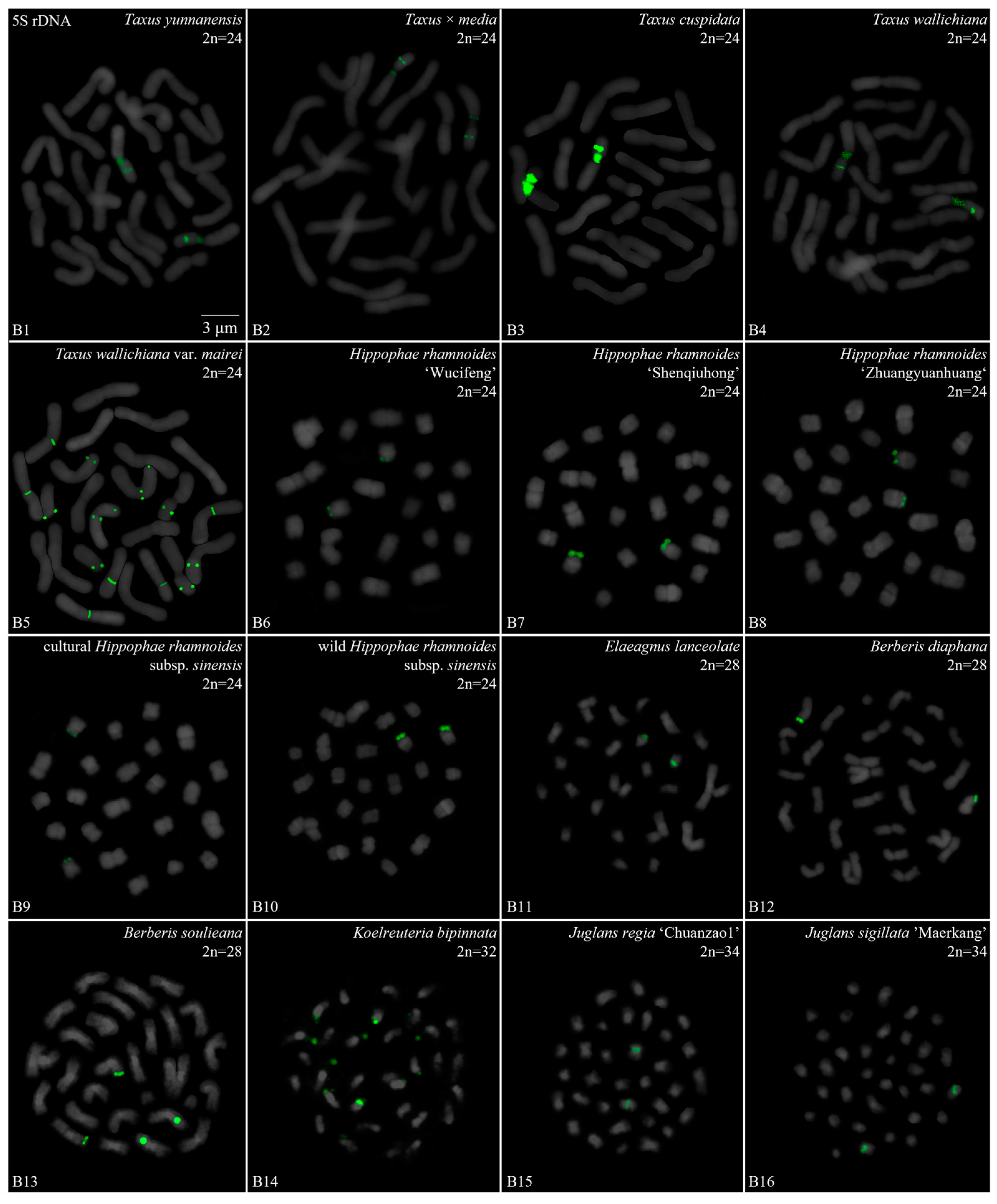
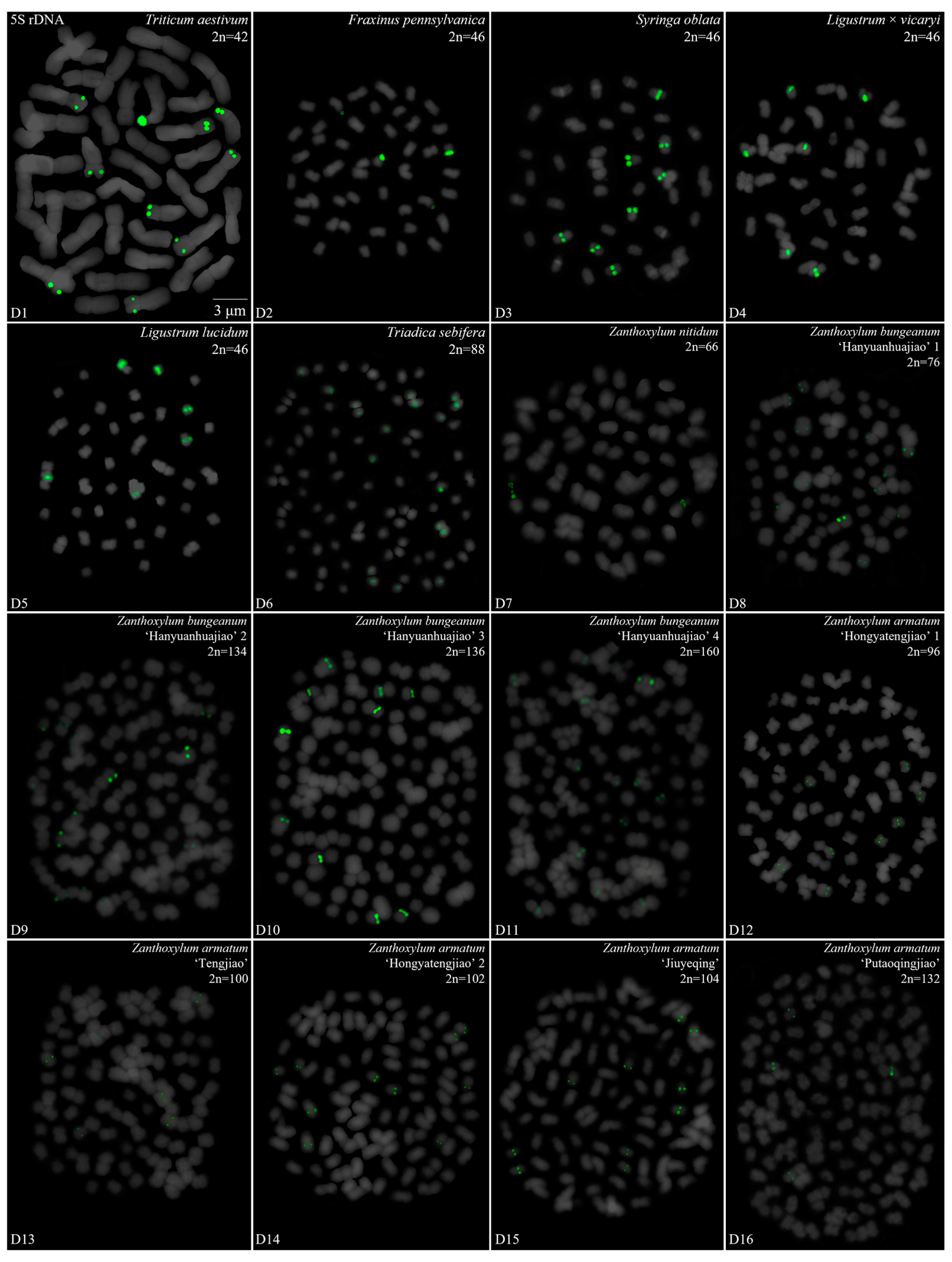
We performed a FISH analysis to visualize the chromosomal distribution of 5S rDNA in 42 plants species, as shown in Figure 1 (A1–A16), Figure 2 (B1–B16), Figure 3 (C1–C16) and Figure 4 (D1–D16). We cut each chromosome distribution of the 5S rDNA in Figure 1, Figure 2, Figure 3 and Figure 4 and aligned them for display, as shown in Figures S1 (A1–A16), S2 (B1–B16), S3 (C1–C16) and S4 (D1–D16). This is the first time that 5S rDNA testing has been performed and analyzed for 19 species from 13 families (A1, A8–A11, A13–A16, B11, B14, C12, C14–C16, D6–D12, and D14–D15).
The chromosome numbers and lengths for the examined species are sorted in Table 2. The chromosome numbers in the 42 species ranged from 14 (C. chinensis, A1) to 160 (Z. bungeanum ‘Hanyuanhuajiao’ 3, D11). Eight species had 24 chromosomes (19%), whereas five species (12%) had 34 chromosomes. The chromosome number in three species was determined for the first time (asterisk in Table 2): P. sibiricum (2n = 18), I. chinensis (2n = 40), and T. sebifera (2n = 88). The longest chromosome length of each plant ranged from 1.23 μm (T. sebifera, D6) to 6.88 μm (T. × media, B2; T. aestivum, D1), whereas the shortest chromosome length of each plant ranged from 0.63 μm (J. sigillata ‘Maerkang’ B16) to 3.85 μm (T. aestivum, D1). A total of 21 species (50%) had a chromosome length of less than 3 μm, thus were in the small chromosome category. Detailed and deep karyotype analysis was not conducted because of the unclear position of centromeres and the chromosomes in many of the examined plants, such as long/short arm length and karyotype formula.
Karyotype asymmetry (Table 2) was assessed using the longest to shortest chromosome length ratio. The largest ratio was 4.61 for P. sibiricum (A10), whereas the smallest was 0.74 for R. pseudoacacia ‘Idaho’ (A5). The ratio for 22 species ranged from 2 to 3 (52%), while that for 11 species ranged from 1 to 2 (26%), and seven plants ranged from 3 to 4 (17%). The ratio was >4 for two species—P. sibiricum (A10) and B. striata f. ‘Dujiangyan’ (C4)—whereas the ratio was <1 for R. pseudoacacia ‘Idaho’ (A5). These results indicate various differences in the karyotypes of the 42 examined in this study.

3.2. Diverse Signal Patterns of 5S rDNA Reveal a Complex Genome Architecture

To further investigate the diversity of 5S rDNA, different types of ideograms for the 42 species were drawn (Figures S5–S8) based on the FISH karyograms shown in Figures S1–S4. The first diversity of 5S rDNA was the signal location. Proximal signals were observed at several chromosomes in 31 species (74%), while distal signals were observed at several chromosome terminus in 28 species (67%). Interstitial signals were observed at several chromosomes in 19 species (45%), whereas distal signals deviated from the chromosome in four species (10%, the fourth class): C. chinensis (A1), S. japonicum (A6), K. paniculata (B14), and Z. nitidum (D7). The second diversity was the signal number. The largest number of chromosomes with a 5S rDNA signal was 18 in T. sebifera (D6), while the smallest number was two in 22 species (52%). Seven species (17%) contained 10–16 chromosomes with a 5S rDNA signal and 18 species (43%) had 4–8 chromosomes with a 5S rDNA signal. The ratio of chromosomes with a 5S rDNA signal to the total chromosome was assessed to signal cover. The largest ratio was 0.89 in P. nepalensis (A2), and the smallest ratio was 0.03 in Z. nitidum (D7) and Z. armatum ‘Putaoqingjiao’ (D16). The ratio for 31 species (74%) ranged from 0.03 to 0.20, while the ratio for eight species (19%) ranged from 0.20 to 0.50. Only two species had ratios ranging from 0.50 to 0.89: R. pseudoacacia (A3) and T. wallichiana var. mairei (B5).
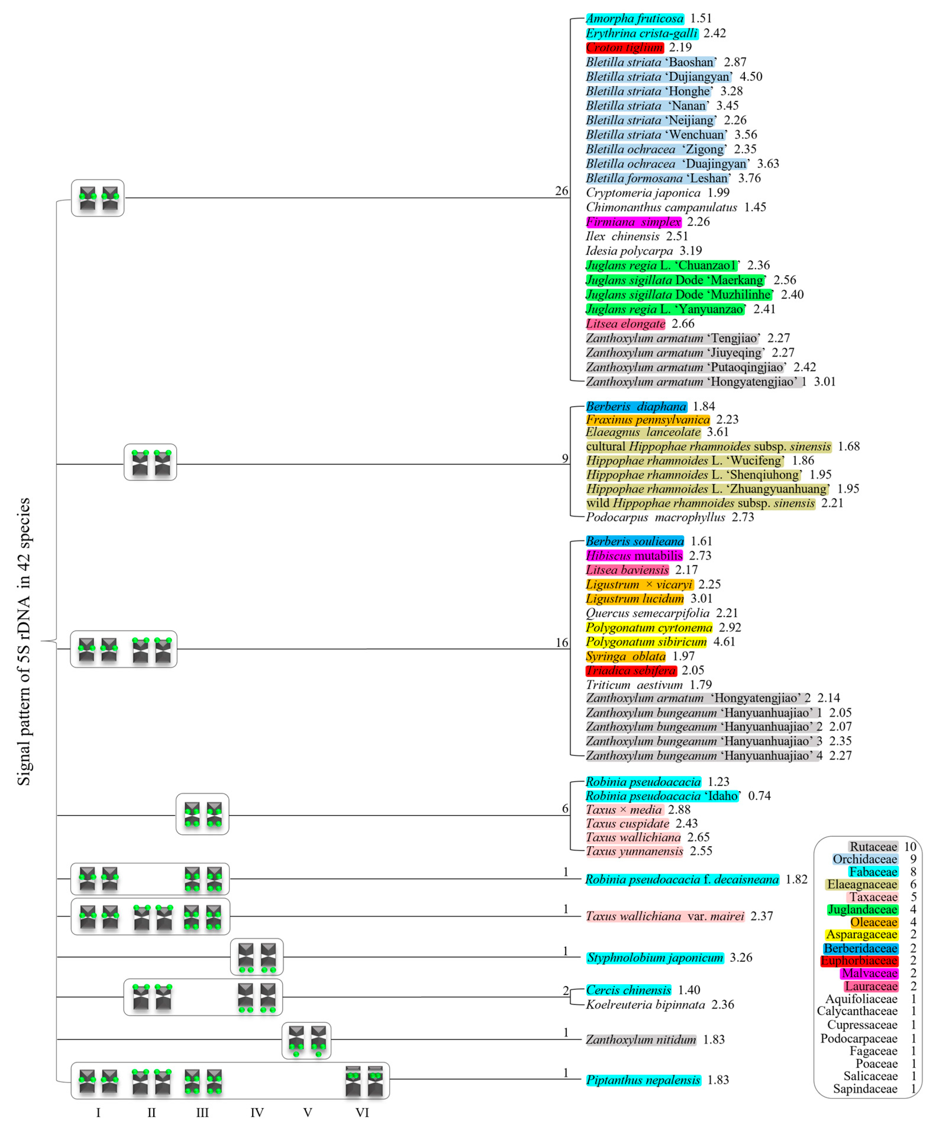
We summarized the results in Figures S5–S8 to produce the 5S rDNA signal pattern in Figure 5. The results for 42 species belonging to 20 families are shown in different colors. There were six 5S rDNA signal pattern types: type I, chromosome includes the proximal signal location; type II, chromosome consists of the distal signal location; type III, chromosome consists of the proximal and distal signal locations; type IV, chromosome only consists of signal outside of the chromosome; type V, chromosome consists of the distal signal location and signal outside of the chromosome; and type VI, satellite chromosome consists of distal signal location. These types of signal patterns indicate that there is various diversity in the 5S rDNA signal arrangement.
In the 42 species examined in this study, 10 signal pattern types or type combinations were present (Figure 5). Twenty six plants only possessed signal pattern type I; nine plants only possessed signal pattern type II; sixteen plants had a combination of type I + type II; six plants only had type III; R. pseudoacacia f. decaisneana had a combination of type I + type III; T. wallichiana var. mairei had a combination of type I + type II + type III; S. japonicum only had type IV; C. chinensis and K. bipinnata had a combination of type II + type IV; Z. nitidum only had type V; and P. nepalensis had a combination of type I + type II + type III + type VI.
There were diverse signal patterns of 5S rDNA among the 42 species, indicating a complex genome architecture. In the family Rutaceae, four varieties of Zanthoxylum had type I, but five varieties of Zanthoxylum had a combination of type I + type II, and Z. nitidum had type V. In Fabaceae, A. fruticose and E. crista-galli had type I, but R. pseudoacacia and R. pseudoacacia ‘Idaho’ had type III, R. pseudoacacia f. decaisneana had combination of type I + type III, S. japonicum only had type IV, C. chinensis had a combination of type II + type IV, and P. nepalensis had a combination of type I + type II + type III + type VI. In the family Taxaceae, four species of Taxus had type III, but T. wallichiana var. mairei had a combination of type I + type II + type III. In Oleaceae, F. pennsylvanica had type II, but L. lucidum, L. × vicaryi and S. oblata had a combination of type I + type III. In Berberidaceae, B. diaphana had type II, but B. soulieana had a combination of type I + type II. In the family Malvaceae, F. simplex had type I, but H. mutabilis had a combination of type I + type II. In Lauraceae, L. baviensis had type I, but L. baviensis had a combination of type I + type II.
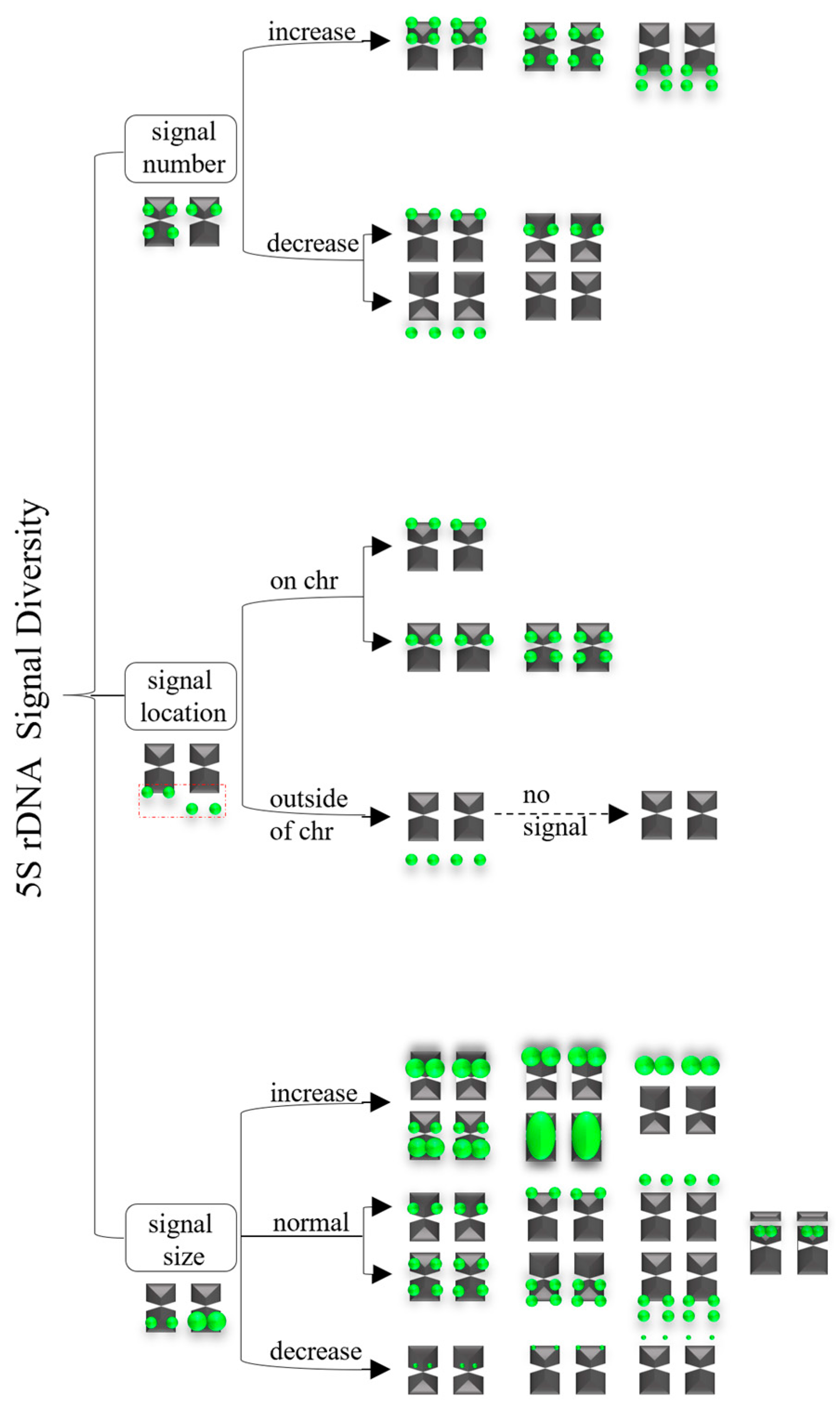
A summarized 5S rDNA signal diversity is illustrated in Figure 6. There were three major groups contributing to this diversity: (i) signal number: increases or decreases; (ii) signal location: on distal chromosome or proximal chromosome, or deviation from the chromosome; (iii) signal size: increases or decreases, or the normal signal size.

4. Discussion

4.1. Karyotype Analysis of the 42 Species

Traditional karyotype analysis involves counting chromosome numbers, determining centromere location, and measuring chromosome length and long/short arm ratio. Based on this, the karyotype formula and cytotype are obtained to compare whether the species is evolving [41]. The chromosome numbers of the 42 species in this study ranged from 2n = 14 to 160. Most chromosome numbers were consistent with previous studies [12,28,29,30,38,39,42,43,44,45,46], with only nine species having different chromosome numbers. In addition, the chromosome number of three species was analyzed for the first time in this study: P. sibiricum (2n = 18), I. chinensis (2n = 40), and T. sebifera (2n = 88). More information related to T. sebifera was found in the chromosome numbers of a previous research study. A stable chromosome number was found in the genus Ilex L.: Ilex crenata Thunb. (2n = 40), I. makinoi ‘Hara’ (2n = 40), I. leucoclada (2n = 40), and I. yunnanensis var. gentilis Franch. (2n = 160) [47]. Conversely, chromosome numbers varied in four genera: (i) Polygonatum anhuiense D. C. Zhang et J. Z. Shao (2n = 24), Polygonatum langyaense D. C. Zhang et J. Z. Shao (2n = 18), Polygonatum odoratum (Mill.) Druce (2n = 18), Polygonatum zanlanscianense Pamp. (2n = 28), P. cyrtonema (2n = 22) [43,44]; (ii) Zanthoxylum acanthopodium Candelle (2n = 64), Zanthoxylum dimorphophyllum Hemsley (2n = 36/68), Zanthoxylum scandens Blume (2n = 68), Zanthoxylum oxyphyllum Edgeworth (2n = 72), Zanthoxylum tomentellum J.D. Hance (2n = 72), Zanthoxylum simulans Hance (2n = ~132, Z. nitidum (2n = 68), Z. armatum (2n = 66/98/128/132/136), Z. bungeanum (2n = 132/136) [6,7,8,37,48,49,50]; (iii) Bletilla formosana (2n = 32/36), B. striata (2n = 32/34/36/48/51/64/76), B. ochracea (2n = 34/36) [39,40,46,51]; (iv) J. regia and J. sigillata (2n = 34) [5], Juglans (2n = 32) [52,53,54,55,56]. However, in this study, the results for P. cyrtonema (2n = 18), Z. bungeanum (2n = 76/134/136/160), Z. armatum (2n = 96/100/102/104/132), Z. nitidum (2n = 66), B. formosana, B. striata, B. ochracea, J. regia, and J. sigillata (all 2n = 34) contradicted those of previous studies. The possible causes of inconsistency may be related to (i) improper chromosome count in the small and high chromosomes, (ii) root lignification limiting their chromosome preparation of chromosomes from these species, (iii) hybridization between closely related species, (iv) natural or artificial polyploidization, and (v) apomixis (polyembryo).
Intraspecific chromosome number variation, even in the entire population, has also been found in species such as Cuscuta epithymum (L.) L. and Cuscuta planiflora Ten. [57], where most variation was attributable to auto- or allopolyploidy. The additional numbers can be explained by ascending or descending dysploidy. Thus, the accumulation of repetitive DNA can lead to an increase in chromosomes and, consequently, to an increase in genome size, especially in subgenus Monogynella [58]. In our study, chromosome numbers varied in the interspecific and intraspecific populations of the genus Zanthoxylum. The cause of the variation was probably similar to that of Cuscuta and Monogynella. Furthermore, the stable differentiation in the 5S rDNA FISH pattern between the subgenera suggests that chromosomal rearrangements played a role in splitting the two subgenera. Rather than major structural changes, transpositional events are responsible for the variable rDNA distribution patterns among species of the same subgenus with conserved karyotypes [25]. Zanthoxylum genomes have complex chromosome rearrangements, such as chromosomal fission, reversal, and translocations [8], which also explains the chromosome number variation in this genus. Chromosome polymorphisms within species in natural populations of vertebrates are far less common and are believed to be temporary transitions during chromosomal evolution [59,60]. Likewise, the genus Zanthoxylum may be experiencing chromosomal evolution.
The longest chromosome length of the 64 plants evaluated in this study ranged from 1.23 to 6.88 μm. In contrast, the shortest chromosome length of each plant ranged from 0.63 to 3.85 μm, exhibiting striking differences among the examined species. Previous research has shown the accumulated chromosome lengths of hundreds of plant species [6,9,12,28,30,32,33,37,38,61,62,63]. Analyzing these data, it is not difficult to find slight differences in chromosome length, even for the same accession of the same species. For example, R. pseudoacacia has reported chromosome lengths of 1.12–1.74 μm [37] and 0.94–1.67 μm [28]. Nonetheless, these two chromosome lengths were small (<3 μm). Hence, chromosome length was more suitable for qualitative rather than for quantitative analyses. Thirty-seven plant species analyzed (more than half) had chromosome lengths < 3 μm, placing them in the small chromosome rank. Owing to the hazy centromere mark and tiny chromosomes in many of the investigated plants, the chromosome size was determined by metaphase and the measurement method. A more delicate karyotype analysis (e.g., arm length, karyotype, and cytotype) was unavailable and limited.

4.2. Occurrence and Diversity of 5S rDNA in Plants

The 5S rDNA, which occurs in all cellular life forms, is a highly stable tandem repeat sequence that ubiquitously exists in plants [64]. With the evolution and development of the plant, 5S rDNA also underwent simultaneous changes. The length of 5S rDNA in the NCBI Nucleotide database ranged from 48 to 854 bp [10,11], while its length as a FISH probe in the PubMed database of NCBI ranged from 41 to 1193 bp [12,13,31]. This study was the first time that the 41-bp oligoprobe had been used to analyze 5S rDNA for 19 species from 13 families. Overall, 5S rDNA occurred in at least two chromosomes in all 42 species. With advances in science and technology, 5S rDNA has been confirmed in an increasing number of species [3,22,65,66]. However, whether the reported length of 5S rDNA is a complete or partial sequence, there are no doubt considerable differences among these 5S rDNAs, including the length and base pair [17,67,68].
Accordingly, it is reasonable that the chromosomally diverse distribution of 5S rDNA is visualized by FISH. Previous studies have shown that the numbers of 5S rDNA FISH signal sites range from 1 to 71 [3,12,19,20,69]. Such a signal position was found in the chromosome interstitial position, distal position, proximal position, and far away from the chromosome [3,21,25,26,27]. In this study, 5S rDNA was diverse and abundant in signal site number (2–18), position (e.g., interstitial, distal, proximal position, and occasionally, outside the chromosome), and even in intensity (e.g., strong, weak, slight). These findings are consistent with previous studies of the 5S rDNA signal pattern of A. fruticose, B. formosana ‘Leshan’, C. campanulatus, H. mutabilis, P. nepalensis, S. oblata, two species of Berberis, two varieties of J. regia, two varieties of J. sigillata, two species of Ligustrum, two varieties of Robinia, and two varieties of Z. armatum, wild/cultural H. rhamnoides ssp. sinensis, three varieties of H. rhamnoides, four species of Taxus, and six types of B. striata [6,9,12,29,30,33,37]. In contrast, the 5S rDNA signal pattern determined in this study differed from those previously reported for F. pennsylvanica, R. pseudoacacia, S. japonicum, T. wallichiana var. mairei, Z. bungeanum ‘Hanyuanhuajiao’, two types of B. ochracea, and two varieties of Z. armatum [7,29,40,44]. Possible causes of the 5S rDNA signal pattern discrepancy are lost satellite chromosomes with signals, and variation in different batches of materials in the same species (i.e., intraspecific variation).

4.3. Potential Origin of 5S rDNA Diversity in Plants

The diversity of signal patterns means 5S rDNA has been used as an excellent and dynamic marker in the species of F. pennsylvanica, Iris versicolor L., L. vicaryi, L. lucidum, P. nepalensis (P. concolor, former name in Flora of China), R. pseudoacacia, and S. oblata [12,28,29,70]. After comparing the 5S rDNA obtained in previous studies with ours, both perfectly reflect the extensive diversity in P. nepalensis, in which all 18 chromosomes can be distinguished according to the signal position, signal intensity, and signal number. Nevertheless, 5S rDNA was quite conserved and dormant in numerous species, such as C. campanulatus, C. sativa, H. rhamnoides, J. regia, M. atropurpureum, P. stratiotes, P. trichocarpa, and Sanguisorba L. [9,16,22,33,36,71,72]; leaving a question about the big difference in 5S rDNA among the above species. There are several plausible hypotheses under investigation, such as (i) chromosome rearrangement (e.g., deletion, duplication, inversion, translocation), (ii) polyploidization, (iii) self-incompatibility, and (iv) chromosome satellites.
The 5S rDNA position is a hot topic for chromosomal reshuffling because of its system into long reaches of the standpat tandem repetition unit and its active transcription. This feature implies that they are impressionable to chromosomal destruction or non-allelic homologous recombination, thus raising the feasibility of chromosomal reorganization, such as fissions, inversions, and fusions [73,74,75,76,77]. The 5S rDNA diversification was regarded as variable genomic areas, compliant with double-strand break and chromosomal reorganization, facilitating karyotypic reconstruction [77,78,79]. The 5S rDNA position was also diversified by translocation or transposition events of repeats in those chromosomes [80]. An interstitial 5S rDNA position with a diverse location is presumed to indicate a thin inversion. A plus 5S rDNA implies the presence of a replication [81]. In addition, the 5S rDNA site of one parent was either excluded from the chromosome or shifted into gene silencing and then disappeared, which could decrease the 5S rDNA site number [82,83]. These studies demonstrate that chromosome rearrangement causes variations in 5S rDNA diversity.
The 5S rDNA has a polyploidization-relevant preference to the distal from a proximal position but keeps a stable loci number [14]. The 5S rDNA sites are proximal, a highly transparent direction in chromosomes with a single site [84]. Consequently, the presence of the 5S rDNA site in the distal chromosomes and the abundance of microsatellites in adjacent areas provide conducive conditions for additional reshuffling. These results emphasize the effect of variable chromosomal 5S rDNA loci in generating assignments [85]. There are no associations between the number of 5S loci and chromosome number, but there is correspondence with ploidy level and genome size [20,84,86], although anomalies still occur [64,87,88], such as the genus Cuscuta L. [84]. These studies demonstrate that polyploidization causes variation in 5S rDNA diversity.
In summary, the variations 5S rDNA in signal number, location, and size were caused by chromosome rearrangement (e.g., deletion, duplication, inversion, translocation), recombination, self-incompatibility, as well as chromosome satellites.

5. Conclusions

In this study, we conducted FISH-based chromosomal mapping of 5S rDNA markers to provide valid karyotype landmarks for revealing the chromosome number and 5S rDNA signal pattern distribution in 42 plant species. Furthermore, we established chromosome physical mapping of each species. Finally, we discussed the proposed origin of 5S rDNA diversity in plants. We are devoted to developing universal oligo sequence markers (GAA)6, (TTG)6, (ACT)6, 45S, ITS, and combinatory analysis of additional plant species, particularly woody plants. Collectively, our results enhance the assumption that cytogenetic characteristics (conventional and molecular) are excellent markers for chromosome distinction and the presentation and profile of existing biodiversity in woody plants.

Supplementary Materials

The following supporting information can be downloaded at: https://www.mdpi.com/article/10.3390/genes15050647/s1, Figure S1: FISH karyograms of 16 plants from Figure 1. Oligo-probe 5S rDNA is exhibited by green signals. The up/lower numbers (1–14) represent the chromosome pairs. The first/last chromosome of each plant were used to calculate the chromosome length: (A1) C. chinensis, 1.58–2.21 μm; (A2) P. nepalensis, 1.65–3.02 μm; (A3) R. pseudoacacia, 1.37–1.68 μm; (A4) R. pseudoacacia f. decaisneana, 0.77–1.40 μm; (A5) R. pseudoacacia ‘Idaho’, 1.09–1.47 μm; (A6) S. japonicum, 0.98–3.19 μm; (A7) A. fruticosa, 1.44–2.18 μm; (A8) E. crista-galli, 1.37–3.32 μm; (A9) P. cyrtonema, 1.54–4.49 μm; (A10) P. sibiricum, 1.47–6.77 μm; (A11) C. tiglium, 1.65–3.61 μm; (A12) C. campanulatus, 1.33–1.93 μm; (A13) C. japonica, 3.09–6.14 μm; (A14) Q. semecarpifolia, 1.47–3.10 μm; (A15) L. baviensis, 1.86–4.04 μm; (A16) L. elongate, 1.58–4.21 μm; Figure S2: FISH karyograms of 16 plants from Figure 2. Oligo-probe 5S rDNA is exhibited by green signals. The up/lower numbers (1–14) represent the chromosome pairs. The first/last chromosome of each plant were used to calculate the chromosome length: (B1) T. yunnanensis, 2.46–6.28 μm; (B2) T. media, 2.39–6.88 μm; (B3) T. cuspidata, 2.60–6.32 μm; (B4) T. wallichiana, 2.42–6.42 μm; (B5) T. wallichiana var. mairei, 2.81–6.67 μm; (B6) H. rhamnoides ‘Wucifeng’, 1.40–2.60 μm; (B7) H. rhamnoides ‘Shengqiuhong’, 1.26–2.46 μm; (B8) H. rhamnoides ‘Zhuangyuanhuang’, 1.44–2.81 μm; (B9) cultural H. rhamnoides ssp. sinensis, 1.30–2.18 μm; (B10) wild H. rhamnoides ssp. sinensis, 1.05–2.32 μm; (B11) E. lanceolata, 1.02–3.68 μm; (B12) B. diaphana, 1.54–2.84 μm; (B13) B. soulieana, 2.18–3.51 μm; (B14) K. paniculata, 0.77–1.82 μm; (B15) J. regia ‘Chuanzao1’, 0.70–1.65 μm; (B16) J. sigillata ‘Maerkang’, 0.63–1.61 μm; Figure S3: FISH karyograms of 16 plants from Figure 3. Oligo-probe 5S rDNA is exhibited by green signals. The up/lower numbers (1–20) represent the chromosome pairs. The first/last chromosome of each plant were used to calculate the chromosome length: (C1) J. sigillata ‘Muzhilinhe’, 0.95–2.28 μm; (C2) J. regia ‘Yanyuanzao’, 0.91–2.49 μm; (C3) B. formosana ‘Leshan’, 0.98–3.68 μm; (C4) B. striata ‘Dujiangyan’, 0.88–3.96 μm; (C5) B. striata ‘Wenchuan’, 1.33–4.74 μm; (C6) B. striata ‘Neijiang’, 1.40–3.16 μm; (C7) B. striata ‘Baoshan’, 1.26–3.21 μm; (C8) B. striata ‘Nanan’, 1.12–3.86 μm; (C9) B. striata ‘Honghe’, 1.23–4.04 μm; (C10) B. ochracea ‘Zigong’, 1.12–2.63 μm; (C11) B. ochracea ‘Dujiangyan’, 1.19–3.02 μm; (C12) F. simplex, 0.98–2.21 μm; B. soulieana, 2.18–3.51 μm; (C13) H. mutabilis, 1.09–2.98; (C14) P. macrophyllus, 1.93–5.26 μm; (C15) I. polycarpa, 0.77–2.46 μm; (C16) I. chinensis, 0.63–1.58 μm; Figure S4: FISH karyograms of 16 plants from Figure 4. Oligo-probe 5S rDNA is exhibited by green signals. The up/lower numbers (1–20) represent the chromosome pairs. The first/last chromosome of each plant were used to calculate the chromosome length: (D1) T. aestivum, 3.85–6.88 μm; (D2) F. pennsylvanica, 0.88–1.96 μm; (D3) S. oblata, 0.98–1.93 μm; (D4) L. × vicaryi, 0.95–2.14 μm; (D5) L. lucidum, 0.70–2.11 μm; (C6) (D6) T. sebifera, 0.60–1.23 μm; (D7) Z. nitidum, 1.40–2.56 μm; (D8) Z. bungeanum ‘Hanyuanhuajiao’ 4, 0.84–1.72 μm; (D9) Z. bungeanum ‘Hanyuanhuajiao’ 1, 0.88–1.82 μm; (D10) Z. bungeanum ‘Hanyuanhuajiao’ 2, 0.91–2.14 μm; (D11) Z. bungeanum ‘Hanyuanhuajiao’ 3, 0.74–1.68 μm; (D12) Z. armatum ‘Hongyatengjiao’ 1, 0.70–1.47 μm; (D13) Z. armaum ‘Tengjiao’, 0.88–2.00; (D14) Z. armatum ‘Hongyatengjiao’ 2, 0.95–2.04 μm; (D15) Z. armatum ‘Jiuyeqing’, 0.88–2.00 μm; (D16) Z. armatum ‘Putaoqingjiao’, 0.74–1.79 μm; Figure S5: Ideograms of 16 plants based on the signal patterns in the FISH karyograms shown in Figure S1: (A1) C. chinensis; (A2) P. nepalensis; (A3) R. pseudoacacia; (A4) R. pseudoacacia f. decaisneana; (A5) R. pseudoacacia ‘Idaho’; (A6) S. japonicum; (A7) A. fruticosa; (A8) E. crista-galli; (A9) P. cyrtonema; (A10) P. sibiricum; (A11) C. tiglium; (A12) C. campanulatus; (A13) C. japonica; (A14) Q. semecarpifolia; (A15) L. baviensis and (A16) L. elongate. The numbers (1–10, 11, 15) above the karyograms are the numbers of the chromosome pairs. Oligo-probe 5S rDNA is exhibited by green signals. Distal signals were observed at several chromosome terminus in (A1, A2, A6, A9, A10, A14–A15), while proximal signals were observed at several chromosome in (A2, A7–A9, A11, A13–A16), interstitial signals were observed at several chromosome in (A2–A5, A10, A12), while distal signals deviated from the chromosome in (A1, A6); Figure S6: Ideograms of 16 plants based on the signal patterns in the FISH karyograms shown in Figure S2: (B1) T. yunnanensis; (B2) T. media; (B3) T. cuspidata; (B4) T. wallichiana; (B5) T. wallichiana var. mairei; (B6) H. rhamnoides ‘Wucifeng’; (B7) H. rhamnoides ‘Shengqiuhong’; (B8) H. rhamnoides ‘Zhuangyuanhuang’; (B9) cultural H. rhamnoides ssp. sinensis; (B10) wild H. rhamnoides ssp. sinensis; (B11) E. lanceolata; (B12) B. diaphana; (B13) B. soulieana; (B14) K. paniculata; (B15) J. regia ‘Chuanzao1’ and (B16) J. sigillata ‘Maerkang’. The numbers (1, 3–9, 11, 12) above the karyograms are the numbers of the chromosome pairs. Oligo-probe 5S rDNA is exhibited by green signals. Distal signals were observed at several chromosome terminus in (B1, B5–B14), while proximal signals were observed at several chromosome in (B1–B5, B15). Interstitial signals were observed at several chromosome termini in (B1–B5, B13, B16), while distal signals deviated from the chromosome in B14; Figure S7: Ideograms of 16 plants based on the signal patterns in the FISH karyograms shown in Figure S3: (C1) J. sigillata ‘Muzhilinhe’; (C2) J. regia ‘Yanyuanzao’; (C3) B. formosana ‘Leshan’; (C4) B. striata ‘Dujiangyan’; (C5) B. striata ‘Wenchuan’; (C6) B. striata ‘Neijiang’; (C7) B. striata ‘Baoshan’; (C8) B. striata ‘Nanan’; (C9) B. striata ‘Honghe’; (C10) B. ochracea ‘Zigong’; (C11) B. ochracea ‘Dujiangyan’; (C12) F. simplex; (C13) H. mutabilis; (C14) P. macrophyllus; (C15) I. polycarpa; (C16) I. chinensis. The numbers (1–3, 5–9, 12–15, 18) above the karyograms are the numbers of the chromosome pairs. Oligo-probe 5S rDNA is exhibited by green signals. Distal signals were observed at several chromosome terminus in C14, while proximal signals were observed at several chromosome in (C1–C3, C5–C13, C15). Interstitial signals were observed at several chromosome termini in (C2, C4, C12); Figure S8: Ideograms of 16 plants based on the signal patterns in the FISH karyograms shown in Figure S4: (D1) T. aestivum; (D2) F. pennsylvanica; (D3) S. oblata; (D4) L. × vicaryi; (D5) L. lucidum; (C6) (D6) T. sebifera; (D7) Z. nitidum; (D8) Z. bungeanum ‘Hanyuanhuajiao’ 4; (D9) Z. bungeanum ‘Hanyuanhuajiao’ 1; (D10) Z. bungeanum ‘Hanyuanhuajiao’ 2; (D11) Z. bungeanum ‘Hanyuanhuajiao’ 3; (D12) Z. armatum ‘Hongyatengjiao’ 1; (D13) Z. armatum ‘Tengjiao’; (D14) Z. armatum ‘Hongyatengjiao’ 2; (D15) Z. armatum ‘Jiuyeqing’; (D16) Z. armatum ‘Putaoqingjiao’. The numbers (1–4, 8–13, 17–19, 21–22, 25–26, 29–30, 32, 34–35, 38, 40, 42, 44–48, 51–52, 54–56, 63, 65, 69, 71–75) above the karyograms are the numbers of the chromosome pairs. Oligo-probe 5S rDNA is exhibited by green signals. Distal signals were observed at several chromosome terminus in (D1–D6, D8–D11, D14), while proximal signals were observed at several chromosome in (D3–D6, D8, D10–D16). Interstitial signals were observed at several chromosome termini in (D1, D9–D10, B13, B16), while distal signals deviated from the chromosome in D7. Table S1: 5S rDNA as a FISH probe was an excellent marker to label plant chromosome in previous reaches.

Author Contributions

X.L.: conceptualization; investigation; methodology; writing—original draft, review, and editing; supervision; and funding acquisition. Y.L.: data curation; formal analysis; investigation; funding acquisition; and writing—original draft, review, and editing. X.G.: data curation; project administration; and writing—original draft. M.Y.: conceptualization; funding acquisition; resources; and supervision. Q.X.: conceptualization and funding acquisition. Z.Z.: investigation and funding acquisition. All authors have read and agreed to the published version of the manuscript.

Funding

This work was supported by the Research and Application Demonstration of Industrial Integrated Technology for Characteristic Edible Woody Oil Crops (2021-YF09-00014-SN) and the Natural Science Foundation of China (31500993).

Institutional Review Board Statement

Not applicable.

Informed Consent Statement

Not applicable.

Data Availability Statement

The original contributions presented in the study are included in the article/Supplementary Material, further inquiries can be directed to the corresponding author.

Acknowledgments

We thank Zhou Yonghong for laboratory equipment support.

Conflicts of Interest

The authors declare no conflicts of interest.

References

  1. Slidler, C. Chapter 29—Genome stability and Aging: Causes and Consequences. In Genome Stability: From Virus to Human Application; Kovalchuk, I., Kovalchuk, O., Eds.; Academic Press: Cambridge, MA, USA, 2016; pp. 511–525. ISBN 9780128033098. [Google Scholar] [CrossRef]
  2. Heslop-Harrison, J.S. Comparative genome organization in plants: From sequence and markers to chromatin and chromosomes. Plant Cell 2000, 12, 617–636. [Google Scholar] [CrossRef] [PubMed]
  3. Rodríguez-González, R.; Gutiérrez, M.L.; Fuentes, I.; Gálvez-Prada, F.; Sochorová, J.; Kovařík, A.; Garcia, S. Release 4.0 of the plant rDNA database: A database on plant ribosomal DNA loci number, their position, and organization: An information source for comparative cytogenetics. Methods Mol. Biol. 2023, 2703, 237–245. [Google Scholar] [CrossRef] [PubMed]
  4. Kamisugi, Y.; Nakayama, S.; Nakajima, R.; Ohtsubo, H.; Ohtsubo, E.; Fukui, K. Physical mapping of the 5S ribosomal RNA genes on rice chromosome 11. Mol. Gen. Genet. 1994, 245, 133–138. [Google Scholar] [CrossRef] [PubMed]
  5. Luo, X.; Chen, J. Distinguishing Sichuan walnut cultivars and examining their relationships with Juglans regia and J. sigillata by FISH, early-fruiting gene analysis, and SSR analysis. Front. Plant Sci. 2020, 11, 27. [Google Scholar] [CrossRef] [PubMed]
  6. Luo, X.; Liu, J.; Wang, J.; Gong, W.; Chen, L.; Wan, W. FISH analysis of Zanthoxylum armatum based on oligonucleotides for 5S rDNA and (GAA)6. Genome 2018, 61, 699–702. [Google Scholar] [CrossRef] [PubMed]
  7. He, Z.; Lei, Y.; Gong, W.; Ye, M.; Luo, X. Karyotype and phylogenetic relationship analysis of five varieties and cultivars of Zanthoxylum armatum based on oligo-FISH. Genes 2023, 14, 1459. [Google Scholar] [CrossRef] [PubMed]
  8. Hu, L.; Xu, Z.; Fan, R.; Wang, G.; Wang, F.; Qin, X.; Yan, L.; Ji, X.; Meng, M.; Sim, S.; et al. The complex genome and adaptive evolution of polyploid Chinese pepper (Zanthoxylum armatum and Zanthoxylum bungeanum). Plant Biotechnol. J. 2023, 21, 78–96. [Google Scholar] [CrossRef] [PubMed]
  9. Luo, X.; Chen, J. Physical map of FISH 5S rDNA and (AG3T3)3 signals displays Chimonanthus campanulatus R.H. Chang & C.S. Ding chromosomes, reproduces its metaphase dynamics and distinguishes its chromosomes. Genes 2019, 10, 904. [Google Scholar] [CrossRef] [PubMed]
  10. Turner, D.J.; Brown, T.A. Abridged 5S rDNA units in sea beet (Beta vulgaris subsp. maritima). Genome 2005, 48, 352–354. [Google Scholar] [CrossRef] [PubMed]
  11. Liu, T.; Song, M.; Xia, Y.; Zeng, X. A tandemly arranged pattern of two 5S rDNA arrays in Amolops mantzorum (Anura, Ranidae). Cytogenet. Genome Res. 2017, 151, 161–170. [Google Scholar] [CrossRef] [PubMed]
  12. Luo, X.; Liu, J.; Zhao, A.; Chen, X.; Wan, W.; Chen, L. Karyotype analysis of Piptanthus concolor based on FISH with an oligonucleotide for rDNA 5S. Sci. Hortic. 2017, 226, 361–365. [Google Scholar] [CrossRef]
  13. Glugoski, L.; Deon, G.; Schott, S.; Vicari, M.R.; Nogaroto, V.; Moreira-Filho, O. Comparative cytogenetic analyses in Ancistrus species (Siluriformes: Loricariidae). Neotrop. Ichthyol. 2020, 18, e200013. [Google Scholar] [CrossRef]
  14. Zhang, Z.T.; Yang, S.Q.; Li, Z.A.; Zhang, Y.X.; Wang, Y.Z.; Cheng, C.Y.; Li, J.; Chen, J.F.; Lou, Q.F. Comparative chromosomal localization of 45S and 5S rDNAs and implications for genome evolution in Cucumis. Genome 2016, 59, 449–457. [Google Scholar] [CrossRef] [PubMed]
  15. Khensuwan, S.; Sassi, F.M.C.; Moraes, R.L.R.; Jantarat, S.; Seetapan, K.; Phintong, K.; Thongnetr, W.; Kaewsri, S.; Jumrusthanasan, S.; Supiwong, W.; et al. Chromosomes of Asian cyprinid fishes: Genomic differences in conserved karyotypes of ‘Poropuntiinae’ (Teleostei, Cyprinidae). Animals 2023, 13, 1415. [Google Scholar] [CrossRef] [PubMed]
  16. Alexandrov, O.S.; Romanov, D.V.; Divashuk, M.G.; Razumova, O.V.; Ulyanov, D.S.; Karlov, G.I. Study and physical mapping of the species-specific tandem repeat CS-237 linked with 45S ribosomal DNA intergenic spacer in Cannabis sativa L. Plants 2022, 11, 1396. [Google Scholar] [CrossRef] [PubMed]
  17. Moraes, J.N.; Viana, P.F.; Favarato, R.M.; Pinheiro-Figliuolo, V.S.; Feldberg, E. Karyotype variability in six Amazonian species of the family Curimatidae (Characiformes) revealed by repetitive sequence mapping. Genet. Mol. Biol. 2022, 45, e20210125. [Google Scholar] [CrossRef] [PubMed]
  18. Yurkevich, O.Y.; Samatadze, T.E.; Selyutina, I.Y.; Suprun, N.A.; Suslina, S.N.; Zoshchuk, S.A.; Amosova, A.V.; Muravenko, O.V. Integration of genomic and cytogenetic data on tandem DNAs for analyzing the genome diversity within the genus Hedysarum L. (Fabaceae). Front. Plant Sci. 2022, 13, 865958. [Google Scholar] [CrossRef] [PubMed]
  19. Ali, H.B.; Lysak, M.A.; Schubert, I. Chromosomal localization of rDNA in the Brassicaceae. Genome 2005, 48, 341–346. [Google Scholar] [CrossRef] [PubMed]
  20. Lan, T.; Albert, V.A. Dynamic distribution patterns of ribosomal DNA and chromosomal evolution in Paphiopedilum, a lady’s slipper orchid. BMC Plant Biol. 2011, 11, 126. [Google Scholar] [CrossRef] [PubMed]
  21. Wang, L.; Feng, Y.; Wang, Y.; Zhang, J.; Chen, Q.; Liu, Z.; Liu, C.; He, W.; Wang, H.; Yang, S.; et al. accurate chromosome identification in the Prunus subgenus Cerasus (Prunus pseudocerasus) and its relatives by oligo-FISH. Int. J. Mol. Sci. 2022, 23, 13213. [Google Scholar] [CrossRef] [PubMed]
  22. de Barros, D.; Montenegro, C.; Gomes, M.; Ferraz, M.E.; Miotto, S.T.S.; Pedrosa-Harand, A. Cytogenetic characterization and karyotype evolution in six Macroptilium species (Leguminosae). Genome 2023, 66, 165–174. [Google Scholar] [CrossRef] [PubMed]
  23. Amosova, A.V.; Yurkevich, O.Y.; Bolsheva, N.L.; Samatadze, T.E.; Zoshchuk, S.A.; Muravenko, O.V. Repeatome analyses and satellite DNA chromosome patterns in Deschampsia sukatschewii, D. cespitosa, and D. antarctica (Poaceae). Genes 2022, 13, 762. [Google Scholar] [CrossRef] [PubMed]
  24. Samatadze, T.E.; Yurkevich, O.Y.; Khazieva, F.M.; Basalaeva, I.V.; Konyaeva, E.A.; Burova, A.E.; Zoshchuk, S.A.; Morozov, A.I.; Amosova, A.V.; Muravenko, O.V. Agro-morphological and cytogenetic characterization of colchicine-induced tetraploid plants of Polemonium caeruleum L. (Polemoniaceae). Plants 2022, 11, 2585. [Google Scholar] [CrossRef] [PubMed]
  25. Cai, Q.; Zhang, D.; Liu, Z.L.; Wang, X.R. Chromosomal localization of 5S and 18S rDNA in five species of subgenus Strobus and their implications for genome evolution of Pinus. Ann. Bot. 2006, 97, 715–722. [Google Scholar] [CrossRef] [PubMed]
  26. Amarasinghe, V.; Carlson, J.E. Physical mapping and characterization of 5S rRNA genes in Douglas-fir. J. Hered. 1998, 89, 495–500. [Google Scholar] [CrossRef] [PubMed]
  27. Campomayor, N.B.; Waminal, N.E.; Kang, B.Y.; Nguyen, T.H.; Lee, S.S.; Huh, J.H.; Kim, H.H. Subgenome discrimination in Brassica and raphanus allopolyploids using microsatellites. Cells 2021, 10, 2358. [Google Scholar] [CrossRef] [PubMed]
  28. He, Z.; Zhang, W.; Luo, X.; Huan, J. Five Fabaceae karyotype and phylogenetic relationship analysis based on oligo-FISH for 5S rDNA and (AG3T3)3. Genes 2022, 13, 768. [Google Scholar] [CrossRef] [PubMed]
  29. Luo, X.; Liu, J. Fluorescence In Situ Hybridization (FISH) analysis of the locations of the oligonucleotides 5S rDNA, (AGGGTTT)3, and (TTG)6 in three genera of Oleaceae and their phylogenetic framework. Genes 2019, 10, 375. [Google Scholar] [CrossRef] [PubMed]
  30. Liu, J.; Luo, X. First report of bicolour FISH of Berberis diaphana and B. soulieana reveals interspecific differences and co-localization of (AGGGTTT)3 and rDNA 5S in B. diaphana. Hereditas 2019, 156, 13. [Google Scholar] [CrossRef] [PubMed]
  31. Islam-Faridi, N.; Sakhanokho, H.F.; Dana, N.C. New chromosome number and cyto-molecular characterization of the African Baobab (Adansonia digitata L.)—“The Tree of Life”. Sci. Rep. 2020, 10, 13174. [Google Scholar] [CrossRef] [PubMed]
  32. Luo, X.; He, Z. Distribution of FISH oligo-5S rDNA and oligo-(AGGGTTT)3 in Hibiscus mutabilis L. Genome 2021, 64, 655–664. [Google Scholar] [CrossRef] [PubMed]
  33. Luo, X.; Liu, J.; He, Z. Oligo-FISH can identify chromosomes and distinguish Hippophaë rhamnoides L. taxa. Genes 2022, 13, 195. [Google Scholar] [CrossRef] [PubMed]
  34. Beliveau, B.J.; Joyce, E.F.; Apostolopoulos, N.; Yilmaz, F.; Fonseka, C.Y.; McCole, R.B.; Chang, Y.; Li, J.B.; Senaratne, T.N.; Williams, B.R.; et al. Versatile design and synthesis platform for visualizing genomes with oligopaint FISH probes. Proc. Natl. Acad. Sci. USA 2012, 109, 21301–21306. [Google Scholar] [CrossRef] [PubMed]
  35. He, L.; Zhao, H.; He, J.; Yang, Z.; Guan, B.; Chen, K.; Hong, Q.; Wang, J.; Liu, J.; Jiang, J. Extraordinarily conserved chromosomal synteny of Citrus species revealed by chromosome-specific painting. Plant J. 2020, 103, 2225–2235. [Google Scholar] [CrossRef] [PubMed]
  36. Xin, H.; Zhang, T.; Wu, Y.; Zhang, W.; Zhang, P.; Xi, M.; Jiang, J. An extraordinarily stable karyotype of the woody Populus species revealed by chromosome painting. Plant J. 2020, 101, 253–264. [Google Scholar] [CrossRef] [PubMed]
  37. Luo, X.; He, Z.; Liu, J.; Wu, H.; Gong, X. FISH mapping of telomeric and non-telomeric (AG3T3)3 reveal the chromosome numbers and chromosome rearrangements of 41 Woody plants. Genes 2022, 13, 1239. [Google Scholar] [CrossRef] [PubMed]
  38. He, Z.; Luo, X.; Lei, Y.; Zhang, W. Five species of Taxus karyotype based on oligo-FISH for 5S rDNA and (AG3T3)3. Genes 2022, 13, 2209. [Google Scholar] [CrossRef] [PubMed]
  39. He, Y.; Yang, L.; Zhang, Y.; Liang, Q. Ploidy level, karyotype, and genome size of Bletilla species (Orchidaceae) from China. HortScience 2022, 57, 48–55. [Google Scholar] [CrossRef]
  40. Huan, J.; He, Z.; Lei, Y.; Li, W.; Jiang, L.; Luo, X. The genetic diversity of Bletilla spp. based on SLAF-seq and oligo-FISH. Genes 2022, 13, 1118. [Google Scholar] [CrossRef] [PubMed]
  41. Alberts, B.; Johnson, A.; Lewis, J.; Raff, M.; Roberts, K.; Walter, P. Molecular Biology of the Cell, 4th ed.; Garland Science: New York, NY, USA, 2002. Available online: https://www.ncbi.nlm.nih.gov/books/NBK21054/ (accessed on 2 February 2024).
  42. Sergeeva, E.M.; Shcherban, A.B.; Adonina, I.G.; Nesterov, M.A.; Beletsky, A.V.; Rakitin, A.L.; Mardanov, A.V.; Ravin, N.V.; Salina, E.A. Fine organization of genomic regions tagged to the 5S rDNA locus of the bread wheat 5B chromosome. BMC Plant Biol. 2017, 17 (Suppl. S1), 183. [Google Scholar] [CrossRef] [PubMed]
  43. Sun, Y.G. Preliminary study of the kayotype of Polygonatum anhuiense and P. langaense. J. Anhui Nor. Univ. (Nat. Sci.) 1996, 2, 144–150. [Google Scholar] [CrossRef]
  44. Chen, C.W.; Chen, S.B. Karyotype analysis of three species of Polygonatum Mill from Tiantangzhai, West Anhui. J. Anhui Nor. Univ. (Nat. Sci.) 2005, 28, 324–327. [Google Scholar]
  45. Wang, Z.; Du, F.; Zhang, L.; Wang, S.; Wei, S. Advances in classification and identification of the plants of Polygonati rhizoma and its adulterants. North Hortic. 2019, 24, 130–136. Available online: http://qikan.cqvip.com/Qikan/Article/Detail?id=7100712585 (accessed on 2 February 2024).
  46. Yang, Y.; Huang, M.; Wang, D.; Ruan, B.; Yang, Q.; Yang, Y.; Luo, Y.; Tan, Y. Analysis of genome size and characteristics of Bletilla striata based on flow cytometry and genomic survey. Chin. Tradit. Pat. Med. 2023, 45, 3677–3682. Available online: https://kns.cnki.net/KCMS/detail/detail.aspx?dbname=cjfdtemp&filename=zcya202311028 (accessed on 2 February 2024).
  47. Geukens, E.; Haegeman, A.; van Meulder, J.; van Laere, K.; Smolders, E.; Ruttink, T.; Leus, L. Exploring genetic diversity in an Ilex crenata breeding germplasm. Horticulturae 2023, 9, 485. [Google Scholar] [CrossRef]
  48. Zhang, D.X.; Hartley, G.T. Zanthoxylum Linnaeus. In Flora of China; Flora of China Editorial Committee, Ed.; Science Press: Beijing, China; Missouri Botanical Garden Press: St. Louis, MO, USA, 2008; pp. 53–66. [Google Scholar]
  49. Chen, R.Y.; Chen, C.B.; Song, W.Q.; Liang, G.L.; Li, X.L.; Chen, L. Chromosome atlas of major economic plants genome in China (Tomus V). In Chromosome Atlas of Medicinal Plants in China; Li, S.W., Wang, J., Eds.; Science Press: Beijing, China, 2009; p. 636. ISBN 978-7-03-022915-1. Available online: https://www.hceis.com/home/book_view.aspx?id=6683 (accessed on 2 February 2024).
  50. Yu, L.Y.; Tan, X.M.; Zhou, Y.Q. Karyotype analysis of Zanthoxylum nitidum. Lishizhen Med. Mater. Med. Res. 2010, 21, 3284–3285. Available online: http://qikan.cqvip.com/Qikan/Article/Detail?id=2000017695 (accessed on 2 February 2024).
  51. Miduno, T. Chromosome studies an Orchidaceae V. The study of cytology between Bletilla striata (n = 16) and Bl. formosana (n = 18). Cytologia 1954, 19, 239–248. [Google Scholar] [CrossRef]
  52. Woodworth, R.H. Meiosis of micro-sporogenesis in the Juglandaceae. Am. J. Bot. 1930, 17, 863–869. [Google Scholar] [CrossRef]
  53. Hans, A.S. Chromosomal numbers in Juglandeceae. J. Arnold. Abroretum. 1970, 51, 534–539. Available online: https://www.jstor.org/stable/43781708 (accessed on 2 February 2024). [CrossRef]
  54. Tulecke, W.; McGranahan, G.; Ahmadi, H. Regeneration by somatic embryogenesis of triploid plants from endosperm of walnut, Juglans regia L. cv manregian. Plant Cell Rep. 1988, 7, 301–314. [Google Scholar] [CrossRef] [PubMed]
  55. Mu, Y.L.; Xi, R.T. Microsporogenesis studying and karyotype analysis of Juglans regia L. and J. hopeiensis Hu. J. Agric. Univ. Hebei 1988, 11, 48–55. [Google Scholar]
  56. Mu, Y.L.; Xi, R.T.; Lv, Z.S. Microsporogenesis observation and karyotype analysis of some species in genus Juglans L. J. Wuhan Bot. Res. 1990, 8, 301–310. Available online: https://plantscience.cn/cn/article/id/1255 (accessed on 2 February 2024).
  57. García, M.A.; Castroviejo, S. Estudios citotaxonómicos en las especies Ibéricas del género Cuscuta (Convolvulaceae). An. Jard. Bot. Madr. 2003, 60, 33–44. [Google Scholar]
  58. Ibiapino, A.; García, M.A.; Amorim, B.; Baez, M.; Costea, M.; Stefanović, S.; Pedrosa-Harand, A. The evolution of cytogenetic traits in Cuscuta (Convolvulaceae), the genus with the most diverse chromosomes in angiosperms. Front. Plant Sci. 2022, 13, 842260. [Google Scholar] [CrossRef] [PubMed]
  59. Damas, J.; Corbo, M.; Kim, J.; Turner-Maier, J.; Farré, M.; Larkin, D.M.; Ryder, O.A.; Steiner, C.; Houck, M.L.; Hall, S.; et al. Evolution of the ancestral mammalian karyotype and syntenic regions. Proc. Natl. Acad. Sci. USA 2022, 119, e2209139119. [Google Scholar] [CrossRef] [PubMed]
  60. Damas, J.; Corbo, M.; Lewin, H.A. Vertebrate chromosome evolution. Annu. Rev. Anim. Biosci. 2021, 9, 1–27. [Google Scholar] [CrossRef] [PubMed]
  61. He, J.; Zhao, Y.; Zhang, S.; He, Y.; Jiang, J.; Chen, S.; Fang, W.; Guan, Z.; Liao, Y.; Wang, Z.; et al. Uneven levels of 5S and 45S rDNA site number and loci variations across wild Chrysanthemum accessions. Genes 2022, 13, 894. [Google Scholar] [CrossRef] [PubMed]
  62. Xing, M.S.; Xue, C.J.; Li, R.P. Karyotype analysis of sea buckthorn. J. Shanxi Univ. (Nat. Sci. Ed.) 1989, 12, 323–330. Available online: https://kns.cnki.net/KCMS/detail/detail.aspx?dbname=cjfd1989&filename=sxdr198903014&dbcode=cjfq (accessed on 2 February 2024).
  63. Liu, H.Z.; Sheng, L.X. Studies on karyotype of three sea buckthorn. J. Jilin Agr. Univ. 2011, 33, 628–631, 636. Available online: http://qikan.cqvip.com/Qikan/Article/Detail?id=39921155 (accessed on 2 February 2024).
  64. Said, M.; Hřibová, E.; Danilova, T.V.; Karafiátová, M.; Čížková, J.; Friebe, B.; Doležel, J.; Gill, B.S.; Vrána, J. The Agropyron cristatum karyotype, chromosome structure and cross-genome homoeology as revealed by fluorescence in situ hybridization with tandem repeats and wheat single-gene probes. Theor. Appl. Genet. 2018, 131, 2213–2227. [Google Scholar] [CrossRef] [PubMed]
  65. de Moraes, R.L.R.; Sassi, F.M.C.; Marinho, M.M.F.; Ráb, P.; Porto, J.I.R.; Feldberg, E.; Cioffi, M.B. Small body, large chromosomes: Centric fusions shaped the karyotype of the Amazonian miniature fish Nannostomus anduzei (Characiformes, Lebiasinidae). Genes 2023, 14, 192. [Google Scholar] [CrossRef] [PubMed]
  66. Kroupin, P.Y.; Ulyanov, D.S.; Karlov, G.I.; Divashuk, M.G. The launch of satellite: DNA repeats as a cytogenetic tool in discovering the chromosomal universe of wild Triticeae. Chromosoma 2023, 132, 65–88. [Google Scholar] [CrossRef] [PubMed]
  67. Röser, M.; Winterfeld, G.; Grebenstein, B.; Hemleben, V. Molecular diversity and physical mapping of 5S rDNA in wild and cultivated oat grasses (Poaceae: Aveneae). Mol. Phylogenet. Evol. 2001, 21, n198–n217. [Google Scholar] [CrossRef] [PubMed]
  68. Kulak, S.; Hasterok, R.; Maluszynska, J. Karyotyping of Brassica amphidiploids using 5S and 25S rDNA as chromosome markers. Hereditas 2002, 136, 144–150, Erratum in Hereditas 2002, 137, 79–80. [Google Scholar] [CrossRef] [PubMed]
  69. Kovács, Z.; Mlinarec, J.; Höhn, M. Living on the edge: Morphological, karyological and genetic diversity studies of the Hungarian Plantago maxima populations and established ex situ collection. Bot. Stud. 2023, 64, 2. [Google Scholar] [CrossRef] [PubMed]
  70. Lim, K.Y.; Matyasek, R.; Kovarik, A.; Leitch, A. Parental origin and genome evolution in the allopolyploid Iris versicolor. Ann. Bot. 2007, 100, 219–224. [Google Scholar] [CrossRef] [PubMed]
  71. Stepanenko, A.; Chen, G.; Hoang, P.T.N.; Fuchs, J.; Schubert, I.; Borisjuk, N. The ribosomal DNA loci of the ancient monocot Pistia stratiotes L. (Araceae) contain different variants of the 35S and 5S ribosomal RNA gene units. Front. Plant Sci. 2022, 13, 819750. [Google Scholar] [CrossRef] [PubMed]
  72. Mishima, M.; Ohmido, N.; Fukui, K.; Yahara, T. Trends in site-number change of rDNA loci during polyploid evolution in Sanguisorba (Rosaceae). Chromosoma 2002, 110, 550–558. [Google Scholar] [CrossRef] [PubMed]
  73. Rosa, K.O.; Ziemniczak, K.; de Barros, A.V.; Nogaroto, V.; Almeida, M.C.; Cestari, M.M.; Artoni, R.F.; Vicari, M.R. Numeric and structural chromosome polymorphism in Rineloricaria lima (Siluriformes: Loricariidae): Fusion points carrying 5S rDNA or telomere sequence vestiges. Rev. Fish Biol. Fish. 2012, 22, 739–749. [Google Scholar] [CrossRef]
  74. Barros, A.V.; Wolski, M.A.; Nogaroto, V.; Almeida, M.C.; Moreira-Filho, O.; Vicari, M.R. Fragile sites, dysfunctional telomere and chromosome fusions: What is 5S rDNA role? Gene 2017, 608, 20–27. [Google Scholar] [CrossRef] [PubMed]
  75. Potapova, T.A.; Gerton, J.L. Ribosomal DNA and the nucleolus in the context of genome organization. Chromosome Res. 2019, 27, 109–127. [Google Scholar] [CrossRef] [PubMed]
  76. Warmerdam, D.O.; Wolthuis, R.M.F. Keeping ribosomal DNA intact: A repeating challenge. Chromosome Res. 2019, 27, 57–72. [Google Scholar] [CrossRef] [PubMed]
  77. Deon, G.A.; Glugoski, L.; Hatanaka, T.; Sassi, F.M.C.; Nogaroto, V.; Bertollo, L.A.C.; Liehr, T.; Al-Rikabi, A.; Moreira Filho, O.; Cioffi, M.B.; et al. Evolutionary breakpoint regions and chromosomal remodeling in Harttia (Siluriformes: Loricariidae) species diversification. Genet. Mol. Biol. 2022, 45, e20210170. [Google Scholar] [CrossRef] [PubMed]
  78. Deon, G.A.; Glugoski, L.; Vicari, M.R.; Nogaroto, V.; Sassi, F.M.C.; Cioffi, M.B.; Liehr, T.; Bertollo, L.A.C.; Moreira-Filho, O. Highly rearranged karyotypes and multiple sex chromosome systems in armored catfishes from the genus Harttia (Teleostei, Siluriformes). Genes 2020, 11, 1366. [Google Scholar] [CrossRef] [PubMed]
  79. Glugoski, L.; Giuliano-Caetano, L.; Moreira-Filho, O.; Vicari, M.R.; Nogaroto, V. Co-located hAT transposable element and 5S rDNA in an interstitial telomeric sequence suggest the formation of Robertsonian fusion in armored catfish. Gene 2018, 650, 49–54. [Google Scholar] [CrossRef] [PubMed]
  80. Venancio, N.S.; Noleto, R.B.; Azambuja, M.; Gazolla, C.B.; Santos, B.R.; Nogaroto, V.; Vicari, M.R. Comparative cytogenetics among Boana species (Anura, Hylidae): Focus on evolutionary variability of repetitive DNA. Genet. Mol. Biol. 2023, 45, e20220203. [Google Scholar] [CrossRef] [PubMed]
  81. Coluccia, E.; Deidda, F.; Lobina, C.; Melis, R.; Porcu, C.; Agus, B.; Salvadori, S. Chromosome mapping of 5S ribosomal genes in indo-pacific and atlantic Muraenidae: Comparative analysis by dual colour Fluorescence In Situ Hybridisation. Genes 2020, 11, 1319. [Google Scholar] [CrossRef] [PubMed]
  82. de Melo, N.F.; Guerra, M. Variability of the 5S and 45S rDNA sites in Passiflora L. species with distinct base chromosome numbers. Ann. Bot. 2003, 92, 309–316. [Google Scholar] [CrossRef] [PubMed]
  83. Volkov, R.A.; Panchuk, I.I.; Borisjuk, N.V.; Hosiawa-Baranska, M.; Maluszynska, J.; Hemleben, V. Evolutional dynamics of 45S and 5S ribosomal DNA in ancient allohexaploid Atropa belladonna. BMC Plant Biol. 2017, 17, 21. [Google Scholar] [CrossRef] [PubMed]
  84. Garcia, S.; Kovařík, A.; Leitch, A.R.; Garnatje, T. Cytogenetic features of rRNA genes across land plants: Analysis of the Plant rDNA database. Plant J. 2017, 89, 1020–1030. [Google Scholar] [CrossRef] [PubMed]
  85. Glugoski, L.; Deon, G.A.; Nogaroto, V.; Moreira-Filho, O.; Vicari, M.R. Robertsonian fusion site in Rineloricaria pentamaculata (Siluriformes: Loricariidae): Involvement of 5S Ribosomal DNA and satellite sequences. Cytogenet. Genome Res. 2022, 162, 657–664. [Google Scholar] [CrossRef] [PubMed]
  86. Adams, S.P.; Leitch, I.J.; Bennett, M.D.; Chase, M.W.; Leitch, A.R. Ribosomal DNA evolution and phylogeny in Aloe (Asphodelaceae). Am. J. Bot. 2000, 87, 1578–1583. [Google Scholar] [CrossRef] [PubMed]
  87. Hasterok, R.; Wolny, E.; Hosiawa, M.; Kowalczyk, M.; Kulak-Ksiazczyk, S.; Ksiazczyk, T.; Heneen, W.K.; Maluszynska, J. Comparative analysis of rDNA distribution in chromosomes of various species of Brassicaceae. Ann. Bot. 2006, 97, 205–216. [Google Scholar] [CrossRef] [PubMed]
  88. Chai, J.; Luo, L.; Yu, Z.; Lei, J.; Zhang, M.; Deng, Z. Repetitive sequence barcode probe for karyotype analysis in Tripidium arundinaceum. Int. J. Mol. Sci. 2022, 23, 6726. [Google Scholar] [CrossRef] [PubMed]
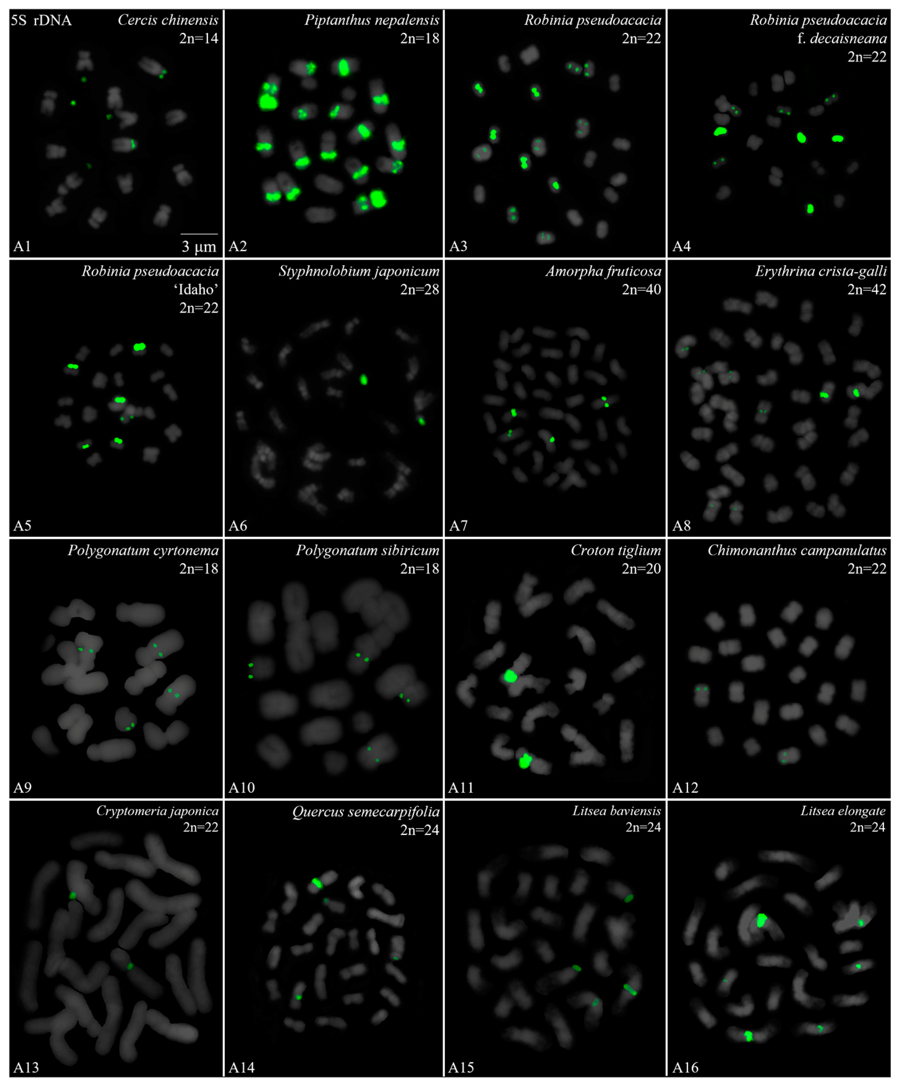
Figure 1. Oligo-FISH depicting the 5S rDNA present in 16 plants. Chromosomes in (A1A16) are all from metaphase. Oligo-probe 5S rDNA is exhibited by green signals: (A1) C. chinensis, 2n = 14; (A2) P. nepalensis, 2n = 18; (A3) R. pseudoacacia, 2n = 22; (A4) R. pseudoacacia f. decaisneana, 2n = 22; (A5) R. pseudoacacia ‘Idaho’, 2n = 22; (A6) S. japonicum, 2n = 22; (A7) A. fruticosa, 2n = 22; (A8) E. crista-galli, 2n = 42; (A9) P. cyrtonema, 2n = 18; (A10) P. sibiricum, 2n = 18; (A11) C. tiglium, 2n = 20; (A12) C. campanulatus, 2n = 22; (A13) C. japonica, 2n = 22; (A14) Q. semecarpifolia, 2n = 24; (A15) L. baviensis, 2n = 24; (A16) L. elongate, 2n = 24. Bar: 3 μm. (A1,A8A11,A13A16) these are the first time that 5S rDNA testing has been reported.
Genes 15 00647 g001
Figure 2. Oligo-FISH depicting the 5S rDNA present in 16 plants. Chromosomes in (B1B16) are all from metaphase. Oligo-probe 5S rDNA is exhibited by green signals: (B1) T. yunnanensis, 2n = 24; (B2) T. media, 2n = 24; (B3) T. cuspidata, 2n = 24; (B4) T. wallichiana, 2n = 24; (B5) T. wallichiana var. mairei, 2n = 24; (B6) H. rhamnoides ‘Wucifeng’, 2n = 24; (B7) H. rhamnoides ‘Shengqiuhong’, 2n = 24; (B8) H. rhamnoides ‘Zhuangyuanhuang’, 2n = 24; (B9) cultural H. rhamnoides ssp. sinensis, 2n = 24; (B10) wild H. rhamnoides ssp. sinensis, 2n = 24; (B11) E. lanceolata, 2n = 28; (B12) B. diaphana, 2n = 28; (B13) B. soulieana, 2n = 28; (B14) K. paniculata, 2n = 32; (B15) J. regia ‘Chuanzao1’, 2n = 34; (B16) J. sigillata ‘Maerkang’, 2n = 34. Bar: 3 μm. (B11,B14) these are the first time that 5S rDNA testing has been reported.
Genes 15 00647 g002
Figure 3. Oligo-FISH depicting the 5S rDNA present in 16 plants. Chromosomes in (C1C16) are all from metaphase. Oligo-probe 5S rDNA is exhibited by green signals: (C1) J. sigillata ‘Muzhilinhe’, 2n = 34; (C2) J. regia ‘Yanyuanzao’, 2n = 34; (C3) B. formosana ‘Leshan’, 2n = 34; (C4) B. striata ‘Dujiangyan’, 2n = 34; (C5) B. striata ‘Wenchuan’, 2n = 34; (C6) B. striata ‘Neijiang’, 2n = 34; (C7) B. striata ‘Baoshan’, 2n = 34; (C8) B. striata ‘Nanan’, 2n = 34; (C9) B. striata ‘Honghe’, 2n = 34; (C10) B. ochracea ‘Zigong’, 2n = 34; (C11) B. ochracea ‘Dujiangyan’, 2n = 34; (C12) F. simplex, 2n = 36; (C13) H. mutabilis, 2n = 90; (C14) P. macrophyllus, 2n = 36; (C15) I. polycarpa, 2n = 38; (C16) I. chinensis. 2n = 40. Bar: 3 μm. (C12,C14C16) these are the first time that 5S rDNA testing has been reported.
Genes 15 00647 g003
Figure 4. Oligo-FISH depicting the 5S rDNA present in 16 plants. Chromosomes in (D1D16) are all from metaphase. Oligo-probe 5S rDNA is exhibited by green signals: (D1) T. aestivum, 2n = 42; (D2) F. pennsylvanica, 2n = 46; (D3) S. oblata, 2n = 46; (D4) L. × vicaryi, 2n = 46; (D5) L. lucidum, 2n = 46; (D6) T. sebifera, 2n = 88; (D7) Z. nitidum, 2n = 66; (D8) Z. bungeanum ‘Hanyuanhuajiao’ 4, 2n = 76; (D9) Z. bungeanum ‘Hanyuanhuajiao’ 1, 2n = 134; (D10) Z. bungeanum ‘Hanyuanhuajiao’ 2, 2n = 136; (D11) Z. bungeanum ‘Hanyuanhuajiao’ 3, 2n = 160; (D12) Z. armatum ‘Hongyatengjiao’ 1, 2n = 96; (D13) Z. armatum ‘Tengjiao’, 2n = 100; (D14) Z. armatum ‘Hongyatengjiao’ 2, 2n = 102; (D15) Z. armatum ‘Jiuyeqing’, 2n = 104; (D16) Z. armatum ‘Putaoqingjiao’, 2n = 132. Bar: 3 μm. (D6D12,D14,D15) these are the first time that 5S rDNA testing has been reported.
Genes 15 00647 g004
Figure 5. Signal pattern of 5S rDNA in 42 species. Signal patterns type I–VI are summarized based on Figures S5–S8. The number between the signal pattern and the species represents the species number. The number after the species represents the ratio of longest to shortest chromosome length. Rutaceae includes 10 plants (grey); Orchidaceae includes nine plants (light blue); Fabaceae includes eight plants (cyan); Elaeagnaceae includes six plants (light yellow); Taxaceae includes five plants (light pink); Juglandaceae and Oleaceae both include four plants (green and orange); Asparagaceae, Berberidaceae, Euphorbiaceae, Malvaceae, and Lauraceae all include two plants (yellow, blue, red, magenta, pink); Aquifoliaceae, Calycanthaceae, Cupressaceae, Podocarpaceae, Fagaceae, Poaceae, Salicaceae, and Sapindaceae each include one plant, respectively.
Genes 15 00647 g005
Figure 6. The 5S rDNA signal diversity.
Genes 15 00647 g006
Table 1. Details of all 64 plants used in this work.
Family No.SpeciesCollection Location
Rutaceae 1Zanthoxylum nitidum (Roxb.) DC.Hanyuan, Sichuan
2Zanthoxylum bungeanum Maxim. ‘Hanyuanhuajiao’ 1Hanyuan, Sichuan
3Zanthoxylum bungeanum Maxim. ‘Hanyuanhuajiao’ 2Hanyuan, Sichuan
4Zanthoxylum bungeanum Maxim. ‘Hanyuanhuajiao’ 3Hanyuan, Sichuan
5Zanthoxylum bungeanum Maxim. ‘Hanyuanhuajiao’ 4Hanyuan, Sichuan
6Zanthoxylum armatum DC. ‘Hongyatengjiao’ 1Hongya, Sichuan
7Zanthoxylum armatum DC. ‘Hongyatengjiao’ 2Hongya, Sichuan
8Zanthoxylum armatum DC. ‘Jiuyeqing’Jiangjin, Chongqing
9Zanthoxylum armatum DC. ‘Putaoqingjiao’Hanyuan, Sichuan
10Zanthoxylum armatum DC. ‘Tengjiao’Hanyuan, Sichuan
Orchidaceae11Bletilla striata (Thunb. ex Murray) Rchb. f. ‘Dujiangyan’Dujiangyan, Sichuan
12Bletilla striata (Thunb. ex Murray) Rchb. f. ‘Neijiang’Neijiang, Sichuan
13Bletilla striata (Thunb. ex Murray) Rchb. f. ‘Wenchuan’Wenchuan, Sichuan
14Bletilla striata (Thunb. ex Murray) Rchb. f. ‘Baoshan’Baoshan, Yunnan
15Bletilla striata (Thunb. ex Murray) Rchb. f. ‘Honghe’Honghe, Yunnan
16Bletilla striata (Thunb. ex Murray) Rchb. f. ‘Nanan’Nanan, Chongqing
17Bletilla ochracea Schltr. ‘Dujiangyan’Dujiangyan, Sichuan
18Bletilla ochracea Schltr. ‘Zigong’Zigong, Sichuan
19Bletilla formosana (Hayata) Schltr. ‘Leshan’Leshan, Sichuan
Fabaceae20Cercis chinensis BungeWenjiang, Sichuan
21Piptanthus nepalensis (Hook.) D. DonQingbaijiang, Sichuan
22Robinia pseudoacacia L.Wenjiang, Sichuan
23Robinia pseudoacacia f. decaisneana (Carr.) VossSuqian, Jiangsu
24Robinia pseudoacacia ‘Idaho’Suqian, Jiangsu
25Styphnolobium japonicum (L.) SchottSuqian, Jiangsu
26Amorpha fruticosa L.Suqian, Jiangsu
27Erythrina crista-galli L.Wenjiang, Sichuan
Elaeagnaceae28Hippophae rhamnoides L. ‘Shenqiuhong’Huai’an, Hebei
29Hippophae rhamnoides L. ‘Wucifeng’Huai’an, Hebei
30wild Hippophae rhamnoides subsp. sinensis RousiHuai’an, Hebei
31cultural Hippophae rhamnoides subsp. sinensis RousiTieling, Liaoning
32Hippophae rhamnoides L. ‘Zhuangyuanhuang’Wenchuan, Sichuan
33Elaeagnus lanceolate Warb. apud DielsWenchuan, Sichuan
Taxaceae34Taxus cuspidata Siebold & Zucc.Suqian, Jiangsu
35Taxus × media RehderYa’an, Sichuan
36Taxus wallichiana var. mairei (Lemee & H. Léveillé) L. K. Fu & N. LiYa’an, Sichuan
37Taxus wallichiana Zucc.Xichuang, Sichuan
38Taxus yunnanensis W.C. Cheng & L.K. FuYa’an, Sichuan
Juglandaceae 39Juglans regia L. ‘Chuanzao1’Qingbaijiang, Sichuan
40Juglans sigillata Dode ‘Maerkang’Maerkang, Sichuan
41Juglans sigillata Dode ‘Muzhilinhe’Gulin, Sichuan
42Juglans regia L. ‘Yanyuanzao’Yanyuan, Sichuan
Oleaceae43Fraxinus pennsylvanica Marsh.Wenjiang, Sichuan
44Syringa oblata Lindl.Wenjiang, Sichuan
45Ligustrum × vicaryi RehderWenjiang, Sichuan
46Ligustrum lucidum Ait.Wenjiang, Sichuan
Asparagaceae47Polygonatum cyrtonema HuaWenchuan, Sichuan
48Polygonatum sibiricum Delar. ex RedouteDujiangyan, Sichuan
Berberidaceae49Berberis diaphana Maxim.Wenchuan, Sichuan
50Berberis soulieana Schneid.Wenchuan, Sichuan
Lauraceae51Litsea baviensis Lec.Jinniu, Sichuan
52Litsea elongate (Wall. ex Nees) Benth. et Hook. f.Jinniu, Sichuan
Malvaceae53Firmiana simplex (L.) W. WightWenjiang, Sichuan
54Hibiscus mutabilis L.Jinniu, Sichuan
Euphorbiaceae55Croton tiglium L.Ya’an, Sichuan
56Triadica sebifera (Linnaeus) SmallJiangyou, Sichuan
Cupressaceae57Cryptomeria japonica (L.f.) D. DonWenjiang, Sichuan
Aquifoliaceae58Ilex chinensis SimsDujiangyan, Sichuan
Calycanthaceae59Chimonanthus campanulatus R.H. Chang & C.S. DingJinniu, Sichuan
Fagaceae60Quercus semecarpifolia SmithWenchuan, Sichuan
Poaceae61Triticum aestivum L.Wenjiang, Sichuan
Podocarpaceae62Podocarpus macrophyllus (Thunb.) SweetWenjiang, Sichuan
Salicaceae63Idesia polycarpa Maxim.Wenjiang, Sichuan
Sapindaceae64Koelreuteria bipinnata Franch.Wenjiang, Sichuan
Table 2. Chromosome number and length of the 42 species used in this work.
AccessionSpeciesChromosome
Number
Chromosome
Length
Karyotype
Asymmetry
A1Cercis chinensis2n = 141.58–2.21 μm1.40
A2Piptanthus nepalensis2n = 181.65–3.02 μm1.83
A3Robinia pseudoacacia2n = 221.37–1.68 μm1.23
A4Robinia pseudoacacia f. decaisneana2n = 220.77–1.40 μm1.82
A5Robinia pseudoacacia ‘Idaho’2n = 221.09–1.47 μm0.74
A6Styphnolobium japonicum2n = 280.98–3.19 μm3.26
A7Amorpha fruticosa2n = 401.44–2.18 μm1.51
A8Erythrina crista-galli2n = 421.37–3.32 μm2.42
A9Polygonatum cyrtonema2n = 181.54–4.49 μm2.92
A10 *Polygonatum sibiricum2n = 181.47–6.77 μm4.61
A11Croton tiglium2n = 201.65–3.61 μm2.19
A12Chimonanthus campanulatus2n = 221.33–1.93 μm1.45
A13Cryptomeria japonica2n = 223.09–6.14 μm1.99
A14Quercus semecarpifolia2n = 241.47–3.10 μm3.11
A15Litsea baviensis2n = 241.86–4.04 μm2.17
A16Litsea elongate2n = 241.58–4.21 μm2.66
B1Taxus yunnanensis2n = 242.46–6.28 μm2.55
B2Taxus × media2n = 242.39–6.88 μm2.88
B3Taxus cuspidata2n = 242.60–6.32 μm2.43
B4Taxus wallichiana2n = 242.42–6.42 μm2.65
B5Taxus wallichiana var. mairei2n = 242.81–6.67 μm2.37
B6Hippophae rhamnoides ‘Wucifeng’2n = 241.40–2.60 μm1.86
B7Hippophae rhamnoides ‘Shenqiuhong’2n = 241.26–2.46 μm1.95
B8Hippophae rhamnoides ‘Zhuangyuanhuang’2n = 241.44–2.81 μm1.95
B9cultural Hippophae rhamnoides subsp. sinensis2n = 241.30–2.18 μm1.68
B10wild Hippophae rhamnoides subsp. sinensis2n = 241.05–2.32 μm2.21
B11Elaeagnus lanceolate2n = 281.02–3.68 μm3.61
B12Berberis diaphana2n = 281.54–2.84 μm1.84
B13Berberis soulieana2n = 282.18–3.51 μm1.61
B14Koelreuteria bipinnata2n = 320.77–1.82 μm2.36
B15Juglans regia ‘Chuanzao1’2n = 340.70–1.65 μm2.36
B16Juglans sigillata ‘Maerkang’2n = 340.63–1.61 μm2.56
C1Juglans sigillata ‘Muzhilinhe’2n = 340.95–2.28 μm2.40
C2Juglans regia ‘Yanyuanzao’2n = 340.91–2.19 μm2.41
C3Bletilla formosana ‘Leshan’2n = 340.98–3.68 μm3.76
C4Bletilla striata f. ‘Dujiangyan’2n = 340.88–3.96 μm4.50
C5Bletilla striata f. ‘Wenchuan’2n = 341.33–4.74 μm3.56
C6Bletilla striata f. ‘Neijiang’2n = 341.40–3.16 μm2.26
C7Bletilla striata f. ‘Baoshan’2n = 341.26–3.61 μm2.87
C8Bletilla striata f. ‘Nanan’2n = 341.12–3.86 μm3.45
C9Bletilla striata f. ‘Honghe’2n = 341.23–4.04 μm3.28
C10Bletilla ochracea ‘Zigong’2n = 341.12–2.63 μm2.35
C11Bletilla ochracea ‘Dujiangyan’2n = 341.19–3.02 μm2.63
C12Firmiana simplex2n = 360.98–2.21 μm2.26
C13Hibiscus mutabilis2n = 901.09–2.98 μm2.73
C14Podocarpus macrophyllus2n = 361.93–5.26 μm2.73
C15Idesia polycarpa2n = 380.77–2.46 μm3.19
C16 *Ilex chinensis2n = 400.63–1.58 μm2.51
D1Triticum aestivum2n = 423.85–6.88 μm1.79
D2Fraxinus pennsylvanica2n = 460.88–1.96 μm2.23
D3Syringa oblata2n = 460.98–1.93 μm1.97
D4Ligustrum × vicaryi 2n = 460.95–2.14 μm2.25
D5Ligustrum lucidum2n = 460.70–2.11 μm3.01
D6 *Triadica sebifera2n = 880.60–1.23 μm2.05
D7Zanthoxylum nitidum2n = 661.40–2.56 μm1.83
D8Zanthoxylum bungeanum ‘Hanyuanhuajiao’ 12n = 760.84–1.72 μm2.05
D9Zanthoxylum bungeanum ‘Hanyuanhuajiao’ 22n = 1340.88–1.82 μm2.07
D10Zanthoxylum bungeanum ‘Hanyuanhuajiao’ 32n = 1360.91–2.14 μm2.35
D11Zanthoxylum bungeanum ‘Hanyuanhuajiao’ 42n = 1600.74–1.68 μm2.27
D12Zanthoxylum armatum ‘Hongyatengjiao’ 12n = 960.70–1.47 μm3.01
D13Zanthoxylum armatum ‘Tengjiao’2n = 1000.88–2.00 μm2.27
D14Zanthoxylum armatum ‘Hongyatengjiao’ 22n = 1020.95–2.04 μm2.14
D15Zanthoxylum armatum ‘Jiuyeqing’2n = 1040.88–2.00 μm2.27
D16Zanthoxylum armatum ‘Putaoqingjiao’2n = 1320.74–1.79 μm2.42
Note: asterisk (*) in Table 2 indicates chromosome number of three species are first reported.
Disclaimer/Publisher’s Note: The statements, opinions and data contained in all publications are solely those of the individual author(s) and contributor(s) and not of MDPI and/or the editor(s). MDPI and/or the editor(s) disclaim responsibility for any injury to people or property resulting from any ideas, methods, instructions or products referred to in the content.