Article

Gut Microbiota, Human Blood Metabolites, and Esophageal Cancer: A Mendelian Randomization Study

1
State Key Laboratory of Oncology in South China, Department of Thoracic Surgery, Sun Yat-sen University Cancer Center, Guangdong Provincial Clinical Research Center for Cancer, Collaborative Innovation Center for Cancer Medicine, Guangzhou 510060, China
2
State Key Laboratory of Oncology in South China, Department of Minimally Invasive Intervention, Sun Yat-sen University Cancer Center, Guangdong Provincial Clinical Research Center for Cancer, Collaborative Innovation Center for Cancer Medicine, Guangzhou 510060, China
3
State Key Laboratory of Oncology in South China, Department of Thoracic Oncology, Sun Yat-sen University Cancer Center, Guangdong Provincial Clinical Research Center for Cancer, Collaborative Innovation Center for Cancer Medicine, Guangzhou 510060, China
*
Authors to whom correspondence should be addressed.
These authors contributed equally to this work.
Genes 2024, 15(6), 729; https://doi.org/10.3390/genes15060729
Submission received: 30 April 2024 / Revised: 23 May 2024 / Accepted: 30 May 2024 / Published: 2 June 2024
(This article belongs to the Section Microbial Genetics and Genomics)

Abstract

:
Background: Unbalances in the gut microbiota have been proposed as a possible cause of esophageal cancer (ESCA), yet the exact causal relationship remains unclear. Purpose: To investigate the potential causal relationship between the gut microbiota and ESCA with Mendelian randomization (MR) analysis. Methods: Genome-wide association studies (GWASs) of 207 gut microbial taxa (5 phyla, 10 classes, 13 orders, 26 families, 48 genera, and 105 species) and 205 gut microbiota metabolic pathways conducted by the Dutch Microbiome Project (DMP) and a FinnGen cohort GWAS of esophageal cancer specified the summary statistics. To investigate the possibility of a mediation effect between the gut microbiota and ESCA, mediation MR analyses were performed for 1091 blood metabolites and 309 metabolite ratios. Results: MR analysis indicated that the relative abundance of 10 gut microbial taxa was associated with ESCA but all the 12 gut microbiota metabolic pathways with ESCA indicated no statistically significant association existing. Two blood metabolites and a metabolite ratio were discovered to be mediating factors in the pathway from gut microbiota to ESCA. Conclusion: This research indicated the potential mediating effects of blood metabolites and offered genetic evidence in favor of a causal correlation between gut microbiota and ESCA.

1. Introduction

A major global disease, esophageal cancer (ESCA), ranks sixth among all cancers in terms of mortality based on global cancer statistics [1]. Individuals with early-stage ESCA may not recognize the symptoms of obstruction or stricture due to the dilated and muscular nature of the esophagus. Symptoms only appear after the tumor has progressed locally or even metastatically [2]. The majority of patients with ESCA in the United States and Europe are diagnosed with locally advanced or metastatic disease, which is ineligible for curative treatment [3]. Over 70% of patients in the UK receive a diagnosis of distant metastases or lymph node metastases, of which distant metastases account for about 40% [3]. Patients with advanced ESCA typically encounter an unfavorable prognosis. Due to etiological, molecular, and histological heterogeneity, advanced ESCA patients often acquire innate resistance to systemic therapy, significantly reducing treatment efficacy [4]. Surgical intervention remains the most effective way to treat ESAC, but even after surgery, less than 25% of individuals with advanced ESCA survive for five years [5]. Early adoption of preventative strategies and a detailed understanding of the etiology of ESCA is critical in reducing the incidence of ESCA.
With approximately 1014 species of microorganisms, the intestinal microbiota is regarded as the largest microbial reservoir in our body [6]. As an important regulator of human health, the gut microbiota plays an integral role in the development of the human immune system and the maintenance of intestinal homeostasis [7]. An increasing corpus of studies has demonstrated the intricate relationships between ESCA and the human gut flora in recent years [8,9]. The composition and abundance of fecal microorganisms in ESCA patients are closely related to the severity of the disease [10]. In addition, the genome-wide methylation level of ESCA can be regulated by the gut microbiota, which affects the occurrence, development, and metastasis of ESCA. The intestinal microbiota can function through various bioactive metabolites that systematically affect the internal microenvironment, including bile acids, short-chain fatty acids, and lipopolysaccharides, to regulate the function of the corresponding target organs [11,12]. Deficiency or disorder of intestinal flora significantly affects polysaccharide decomposition and lipid absorption, resulting in liver and adipose tissue dysfunction, leading to cardiovascular and cerebrovascular diseases, type 2 diabetes, and obesity, among other metabolic-related diseases [13]. Recent studies have shown that there were notable variations in the concentration of amino acids such as tryptophan and tyrosine, as well as lipids such as oleic acid and palmitoleic acid, between ESCA patients and healthy controls [14,15]. The evidence suggests that human circulating metabolites are integral components in the development of ESCA. Tumor cells can disrupt the entire metabolism in the process of continuously adapting to the dynamic metabolic microenvironment, thus affecting the distribution and content of metabolites in blood circulation, such as upregulating the glycolytic pathway under adequate oxygen conditions, resulting in rapid growth [16].
In recent years, for determining possible causal associations between various exposures and clinical outcomes, Mendelian randomization (MR) analysis has become widely applied, which is a method for determining the causal relationship between genetically predicted exposure and genetically predicted outcome, particularly single nucleotide polymorphisms (SNPs) [17]. Most epidemiologic investigations that have been accomplished on the causality between ESCA, human blood metabolites, and gut microbiota are based on conventional approaches (e.g., cross-sectional, case-control, cohort). Nevertheless, the estimates of effect may be impacted by a number of constraints, including confounding and reverse causation bias [18]. In recent years, for determining possible causal associations between various exposures and clinical outcomes, MR analysis has become widely applied. By virtue of the fact that allelic randomization occurs prior to the onset of disease, MR analysis has an advantage over conventional observational studies in reducing reverse causation bias. Furthermore, the independent assortment and random segregation of genetic polymorphisms at conception mitigate the confounding bias, as genetic markers are used as instrumental variables (IVs) in MR analysis [19].
Given the absence of research examining the causal correlation between gut microbiota and ESCA mediated by human blood metabolites, we undertook a two-sample, two-step MR analysis to reveal the relationship.

2. Method

2.1. Study Design

Figure 1 illustrates the study design. More than 647,920 participants were selected from summary level, publicly available datasets to conduct a large two-step, two-sample MR study using a two-step strategy to assess the association between the genetic prediction of gut microbiota and esophageal carcinoma and to determine whether plasma metabolites could mediate this association. A two-sample MR analysis utilizing different datasets is performed to assess correlations of the same genetic variants with exposure (e.g., gut microbiota) and outcome (e.g., esophageal carcinoma). Initially, the causal effects of genetic prediction of 412 gut microbiota with a genetic disposition to esophageal carcinoma were analyzed, and the two-step approach utilized in mediating analysis examined the association between genetically predicted gut microbiota and each potential mediator. Subsequently, we investigate and quantify the mediation effects of potential mediators in the pathway from the 412 gut microbiota to esophageal carcinoma.

2.2. Data Sources

2.2.1. Genetic Instrumental Variables for Gut Microbiome

A large-scale genome-wide association study (GWAS) carried out by the Dutch Microbiome Project (DMP) provided the species-level dataset for the gut microbiota [20]. 7738 participants of European descent were involved in the analysis of this dataset, which is the hitherto largest species-level genomics study on the human gut microbiota. An analysis of stool samples was performed utilizing shotgun metagenomic sequencing to determine the gut microbiome, ultimately identifying 207 taxonomies (105 species, 48 genera, 26 families, 13 orders, 10 classes, 5 phyla) and 205 gut microbiota metabolic pathways related to microbial functions. This GWAS dataset is described in more detail in its original publication [20]. The GWAS data are publicly available at https://mibiogen.gcc.rug.nl, accessed on 5 January 2024.
SNPs with genome-wide significance (p < 1 × 10⁻⁵) and clumping at a linkage disequilibrium (LD) threshold of r2 < 0.001 (clumping distance: 10,000 kb) were used in the analyses as instrumental variables (IVs) for gut microbiota. The estimated F-statistics for exposure, which were used to quantify the IVs, ranged from 19 to 57, which is consistent with the notion of F > 10 for MR studies and enables us to remove weak instrumental variable bias [21].

2.2.2. Genetic Instrumental Variables for Potential Mediators

A recent GWAS carried out on the Canadian Longitudinal Study on Aging (CLSA) cohort, involving a total of 8299 individuals and approximately 15.4 million SNPs, explored the association of SNPs with human metabolite levels. A genome-wide association study of 1091 blood metabolites and 309 metabolite ratios was performed in this research [22]. The results of the present investigation identified correlations with 248 loci containing 690 metabolites and 69 loci containing 143 metabolite ratios. Kyoto Encyclopedia of Genes and Genomes database is the basis for the classification of these known metabolites into categories such as peptide, nucleotide, amino acid, carbohydrates, cofactors, and vitamin, energy, lipid, and xenobiotics metabolism.

2.2.3. Genetic Instrumental Variables for Esophageal Carcinoma

Esophageal carcinoma GWAS summary data were derived from the tenth version of the FinnGen consortium (https://r10.finngen.fi/, accessed on 5 January 2024). Esophageal carcinoma was identified using International Classification of Diseases (ICD) diagnosis codes in this prospective cohort study, involving 619 cases and 314,193 controls originating from European ancestry. FinnGen has correlated genetic variation with data on healthy individuals to uncover disease mechanisms and genetic predispositions [23].

2.3. Statistical Analyses

We examined the potential association between genetically predicted gut microbiota and genetically predicted esophageal carcinoma by applying a bidirectional two-sample MR analysis. Additionally, to examine the possible mediation effects of human blood metabolites in the causal relationship, a two-step MR analysis was carried out using summary statistical data.
In both forward and reverse directions of MR analyses, the inverse-variance weighted (IVW) method was utilized as the primary analytical approach to estimate odds ratios and p-values, widely recognized as the most robust methodology for generating reliable causal estimates in MR studies.
The IVW method, analyzing the causal effects of exposure SNPs on outcome data, was employed as the primary approach [24]. p-values of IVW less than 0.05 and consistent directions for both IVW and MR–Egger demonstrated statistical significance in the results. A two-sided p-value that was accepted after the Bonferroni adjustment p-values of 0.0001 (0.05/412) for gut microbiota and 0.00004 (0.05/1400) for metabolites were deemed statistically significant, while p < 0.05 was regarded as indicating a suggestively significant association. In the absence of effective instruments, the weighted median method was employed, as it is capable of offering reliable causal effect estimates even if less than fifty percent of the information is derived from valid instruments [25]. To discover the anomalies in the analysis due to the large horizontal pleiotropy during the MR analysis and to account for the weak effects and uncertainties of the weak horizontal pleiotropy, we performed further analysis using Bayesian weighted Mendelian randomization (BWMR) [26].

2.4. Sensitivity Analyses

An assessment of the heterogeneity between SNPs was carried out using Cochran’s Q statistics [27]. Unless evidence of substantial heterogeneity (p < 0.05), fixed-effects models were employed; otherwise, random-effects models were applied. As well as determining whether instrumental SNPs are multi-effect, we used the MR–Egger method to identify the multi-effects. The p value of its intercept was calculated as part of an MR–Egger regression analysis for uncovering possible horizontal pleiotropy [28]. Furthermore, we also performed MR pleiotropy residual sum and outlier (MR-PRESSO), thus removing possible outliers from multi-effects estimates [29] and rectifying potential confounding factors [30]. The odds ratio (OR) and 95% confidence interval (CI) per standard deviation were calculated as the result. The mediation proportions were determined based on the formula: (beta1 × beta2) / beta_all. beta_all represents the total causal effects of gut microbiota on esophageal carcinoma derived from the main analysis, beta1 represents the estimated effect of gut microbiota-related traits on potential blood metabolites mediators, and beta2 represents the causal effects of blood metabolites mediators on esophageal carcinoma.

3. Results

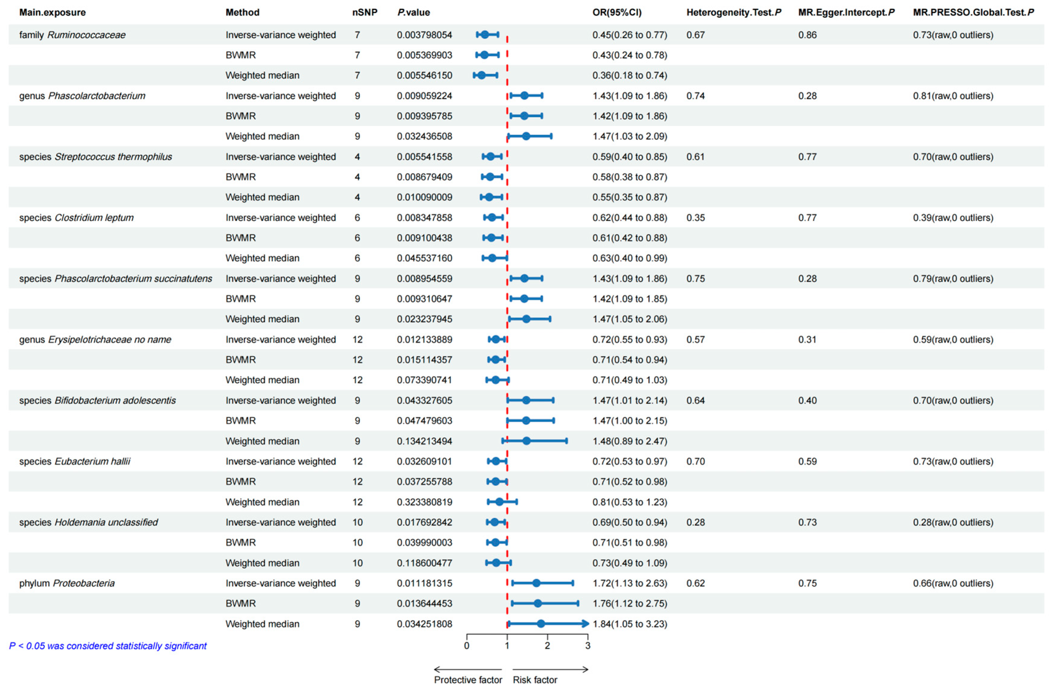
3.1. Bidirectional Two-Sample MR Analyses between Gut Microbiota and Esophageal Carcinoma

In total, ten gut microbiota (including one phylum, one family, two genera, and six species) and twelve gut microbiota metabolic pathways were associated with esophageal carcinoma. In Tables S2 and S3, 85 SNPs for 10 gut microbiota and 117 SNPs for 12 gut microbiota metabolic pathways are presented in detail.
Based on the MR analyses, Figure 2 illustrates the correlation of four gut microbiota with the increased risk of esophageal carcinoma. The genus Phascolarctobacterium (OR = 1.426, 95%CI = 1.092 ~ 1.862, p = 0.009), species Phascolarctobacterium succinatutens (OR = 1.426, 95%CI = 1.093 ~ 1.861, p = 0.009), species Bifidobacterium adolescentis (OR = 1.426, 95%CI = 1.012 ~ 2.139, p = 0.043) and phylum Proteobacteria (OR =  1.724, 95%CI = 1.132 ~ 2.626, p = 0.011) significantly increased the risk of esophageal carcinoma. Six genetically predicted gut microbiotas were associated with the decreased risk of esophageal carcinoma. The family Ruminococcaceae (OR = 0.446, 95%CI = 0.258 ~ 0.770, p = 0.004), species Streptococcus thermophilus (OR = 0.586, 95%CI = 0.402 ~ 0.855, p = 0.006), species Clostridium leptum (OR = 0.621, 95%CI = 0.436 ~ 0.885, p = 0.008), genus Erysipelotrichaceae no name (OR = 0.716, 95%CI = 0.552 ~ 0.930, p = 0.012), species Eubacterium hallii (OR = 0.719, 95%CI = 0.532 ~ 0.973, p = 0.033), and species Holdemania unclassified (OR = 0.687, 95%CI = 0.504 ~ 0.937, p = 0.018) remarkably decreased the risk of esophageal carcinoma. The BWMR method yielded consistent conclusions in the causal association analysis of these ten gut microbiota. However, the results of the weighted median method with genus Erysipelotrichaceae no name (p = 0.073), species B. adolescentis (p = 0.134), species Eubacterium hallii (p = 0.323), and species Holdemania unclassified (p = 0.119) were negative, and no mediating metabolite was found to be associated with species S. thermophilus and phylum Proteobacteria, leading to their exclusion. The reverse MR analysis revealed no significant causal effects of genetic prediction of esophageal carcinoma on the 4 gut microbiota as mentioned above with the p value higher than 0.05 shown in the IVW method, indicating the absence of a reverse causal relationship between them (Table S8). Ultimately, the family Ruminococcaceae, genus Phascolarctobacterium, species C. leptum, and species P. succinatutens were chosen as the exposure variables (Table S4).
Only the causal association between the PANTOSYN.PWY..pantothenate.and. coenzyme.A.biosynthesis.I pathway and ESCA was validated by both the IVW approach and the weighted median method among the 12 gut microbiota metabolic pathways; however, these pathways were excluded due to the absence of any significant mediated metabolites (Table S5).

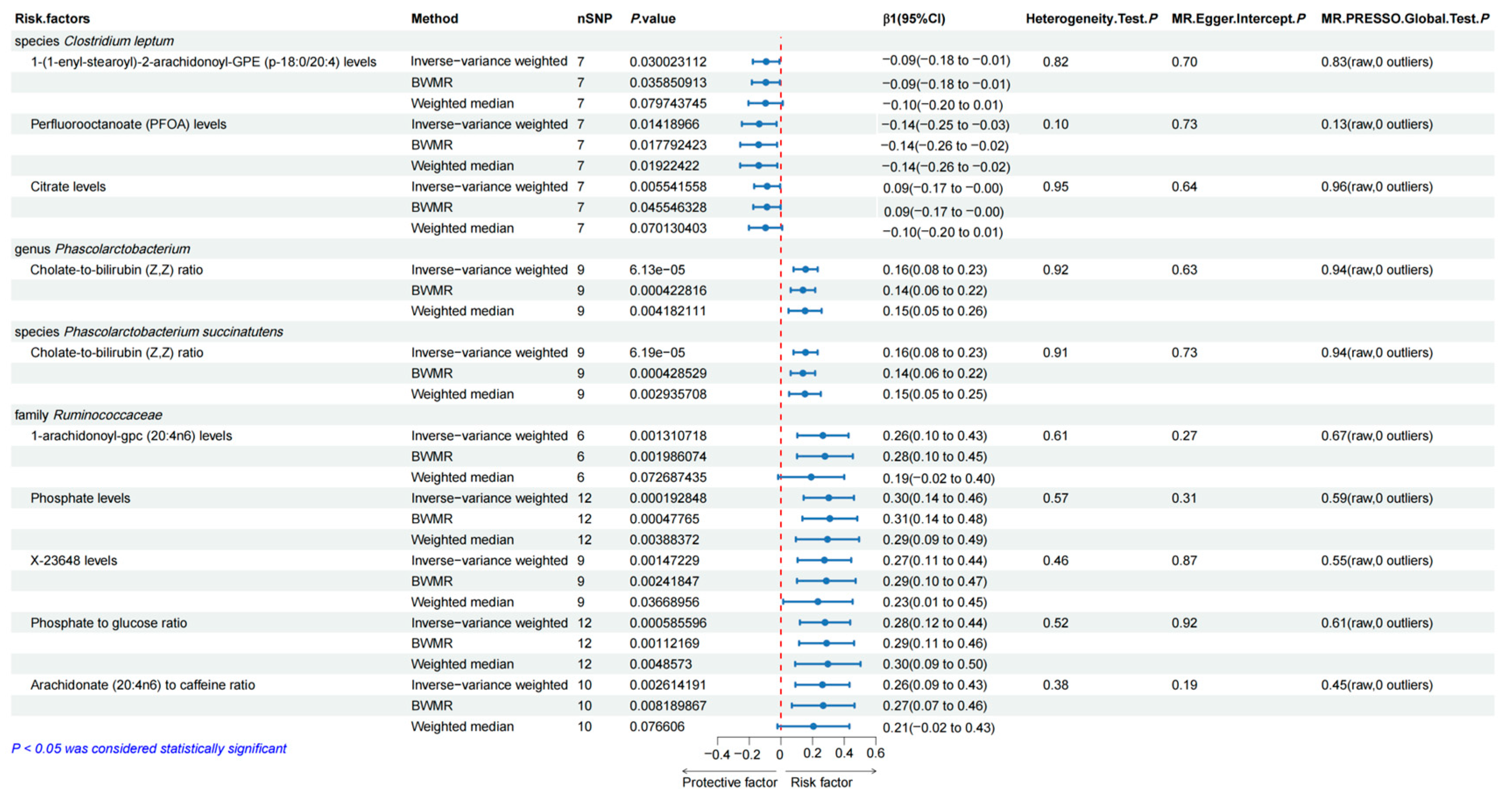
3.2. Causal Effects of the Selected Gut Microbiota on the Human Blood Metabolites

Figure 3 identifies that family Ruminococcaceae was causally associated with higher 1-arachidonoyl-gpc (20:4n6) levels (β = 0.265, 95%CI = 0.103 ~ 0.427, p = 0.001), phosphate levels (β = 0.302, 95%CI = 0.143 ~ 0.461, p = 0.0002), X-23648 levels (β = 0.274, 95%CI = 0.105 ~ 0.443, p = 0.001), phosphate-to-glucose ratio (β = 0.278, 95%CI = 0.120 ~ 0.437, p = 0.0006), and Arachidonate (20:4n6)-to-caffeine ratio (β = 0.262, 95%CI = 0.091 ~ 0.433, p = 0.003).
We identified that the family Ruminococcaceae, genus Phascolarctobacterium, species C. leptum, and species P. succinatutens were causally associated with 69, 59, 47, and 61 metabolites, respectively, primarily applying the IVW approach (Supplementary Table S3). The weighted median method and BWMR supported the robustness of the results.

3.3. Bidirectional Two-Sample MR Analyses between Human Blood Metabolites and Esophageal Carcinoma

After examining the causal association between gut microbiota and the above significant metabolites through the weighted median method and BWMR, we found that perfluorooctanoate (PFOA) levels (OR = 0.713, 95%CI = 0.508 ~ 1.000, p = 0.0498) were a significant risk factor in the causal pathway from species C. leptum to esophageal carcinoma; the cholate-to-bilirubin (Z, Z) ratio (OR = 1.298, 95%CI = 1.010 ~ 1.668, p = 0.041) was a significant risk factor in the causal pathway from genus Phascolarctobacterium to esophageal carcinoma; the cholate-to-bilirubin (Z, Z) ratio (OR = 1.298, 95%CI = 1.010 ~ 1.668, p = 0.041) was also a significant risk factor in the causal pathway from species P. succinatutens to esophageal carcinoma. Genetic prediction of 1-arachidonoyl-gpc (20:4n6) levels (OR = 0.814, 95%CI = 0.664 ~ 0.997, p = 0.047) was a significant risk factor in the causal pathway from family Ruminococcaceae to esophageal carcinoma as shown in Figure 4. However, the causal relationship between family Ruminococcaceae and 1-arachidonoyl-gpc (20:4n6) levels was not identified by the weighted median method with a p value higher than 0.05. MR analysis of the rest of the four metabolites with esophageal carcinoma indicated no statistically significant association existing. The detailed information on the results is presented in Table S4. Reverse MR analysis indicated no reverse causal association between the 3 blood metabolites and esophageal carcinoma (Table S9).

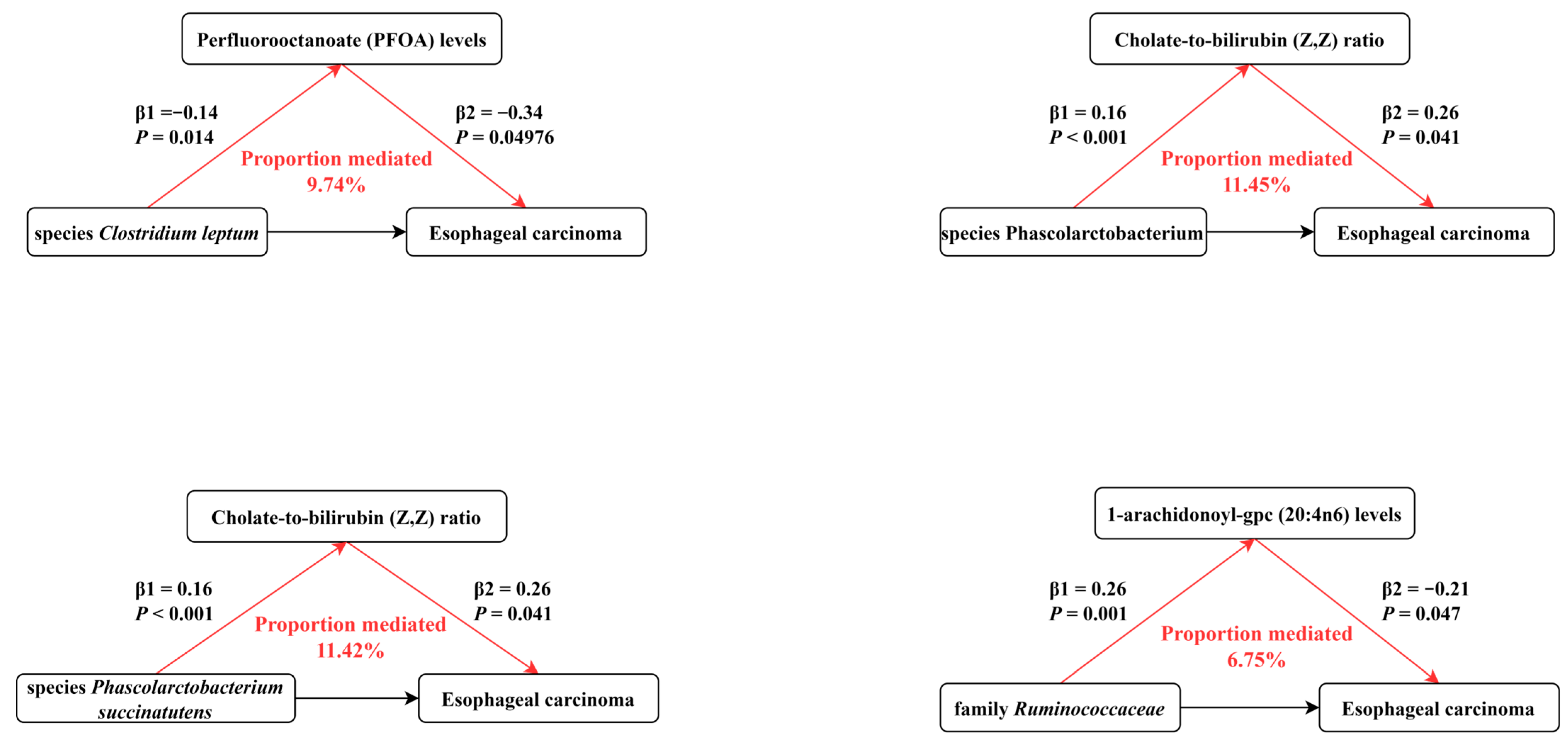
3.4. Mediation Effects of the Selected Human Blood Metabolites on Esophageal Carcinoma

For the mediation analysis illustrated in Figure 5, we excluded mediating factors that were not causally affected by gut microbiota and those that did not causally influence esophageal carcinoma. Finally, our results indicated that PFOA levels, cholate-to-bilirubin (Z, Z) ratio, and 1-arachidonoyl-gpc (20:4n6) levels were significant risk factors mediating the correlation of gut microbiota-related traits with esophageal carcinoma. The overall effect can be separated into direct effect (via mediators) and indirect effect (without mediators). Our results demonstrated that PFOA levels accounted for 9.74% in the causal pathway from species C. leptum to esophageal carcinoma; the cholate-to-bilirubin (Z, Z) ratio accounted for 11.45% in the causal pathway from genus Phascolarctobacterium to esophageal carcinoma; the cholate-to-bilirubin (Z, Z) ratio accounted for 11.42% in the causal pathway from species P. succinatutens to esophageal carcinoma; and 1-arachidonoyl-gpc (20:4n6) levels accounted for 6.75% in the causal pathway from family Ruminococcaceae to esophageal carcinoma.

3.5. Sensitivity Analyses

There is a low likelihood of weak instrument bias for these instrumental factors, as shown by the F-statistics for the selected SNPs, which are all over 10 (Table S2). There is no LD and the SNPs are randomly dispersed, according to the r2 values, which range from zero to one (Table S1). To assess the heterogeneity of our estimates, we calculated Cochrane’s Q and p values derived from Cochrane’s Q test (Tables S4, S6, and S7). No evidence of significant heterogeneity was found in our analysis. To test and correct for the directional pleiotropy in causal estimates, a series of sensitivity analyses were carried out. The null results of the directional pleiotropy were indicated by the other MR analyses stated above, which did not find any significant intercept. Furthermore, a leave-one-out analysis was carried out to ascertain whether a particular SNP substantially deviated from the causal estimate, evaluating the effect of each SNP on the overall causal estimate (Figures S1–S4). All of our positive results were consistent after removing the outliers in the original MR-PRESSO global test, which was utilized to ascertain and exclude outliers, as well as decrease heterogeneity in our analysis (Tables S4, S6, and S7).

4. Discussion

To the best of our knowledge, based on statistical approaches that account for directional pleiotropy, this is the first study to investigate the likelihood of metabolite traits mediating a causal path between gut microbiota and ESCA. In this comprehensive and large-scale MR analysis, we affirmed that PFOA levels and 1-arachidonoyl-gpc (20:4n6) levels, respectively, mediate the causal influence of species C. leptum and family Ruminococcaceae on ESCA, while the cholate-to-bilirubin (Z, Z) ratio mediates the pathway from genus Phascolarctobacterium and species P. succinatutens to ESCA.
There is a large microbial population in the stool of adults, of which Cluster IV (C. leptum group) occupies a dominant position, and its abundance is generally higher than 15%. These bacteria tend to be associated with multiple metabolic pathways in the body. These bacteria are involved in a variety of metabolic pathways that maintain the balance of the intestinal microecological environment. One of the main sources of energy for colonic epithelial cells to regulate intestinal epithelial function is short-chain fatty acids (SCFAs) produced by C. leptum [31,32]. C. leptum ferments polysaccharides through acetyl-CoA and pyruvate pathways to produce propionate and butyrate [33], thereby controlling glucose concentration in the intestinal microenvironment. Recent studies have shown that the intestinal flora produces some metabolites with weight loss effects in the process of fermenting polysaccharides [34]. Li et al. found that C. leptum can alleviate obesity by fermenting metabolites produced by FP [35], which could lower the risk factors of ESCA [36]. It is plausible to speculate that this may be one of the mechanisms by which C. leptum can lower the risk of ESCA.
PFOA, one of the four types of polyfluoroalkyl substances (PFASs), is a newly discovered environmental contaminant that can cause health problems as an endocrine disruptor [37]. Four PFAS (PFOA, perfluorooctane sulfonic acid [PFOS], perfluorohexane sulfonic acid [PFHxS], and perfluorononanoic acid [PFNA]) have been reported to be detected in the serum among individuals over 12 in the United States, with a positive rate of more than 98%, indicating the prevalence of PFOA exposure [38]. Several studies have elucidated the mechanism of activation of PFOA in the development of ESCA [39].
High structural similarity exists between SCFAs and PFAS, which have been demonstrated to interfere with hepatic lipid metabolism through interactions with a variety of nuclear receptors, including peroxisome proliferator-activated receptors (PPARs) [40]. Additionally, glucose metabolism pathways may be affected [41]. High levels of PFOA exposure have been correlated to an unbalance in Clostridium abundance, according to recent studies [42]. These findings imply that PFOA may be employed as a potential mediator to affect Clostridium’s causal effect on ESCA.
Phascolarctobacterium is a fecal-phase intestinal bacterium extracted from koala excrement by Del Dot et al. [43]. This bacterium is classified as Gram-negative, pleomorphic rod-shaped cells composed of P. faecium and P. succinatutens [44]. A recent study has shown that Phascolarctobacterium is widespread in the human gastrointestinal tract and can produce SCFAs, including acetic acid, propionic acid, isobutyric acid, butyric acid, and isovaleric acid [45]. Phascolarctobacterium stimulates growth by succinic acid and decomposes it into propionic acid [46], which is involved in significant metabolic pathways, such as hepatic gluconeogenesis [47]. Clinically substantial reductions in Phascolarctobacterium abundance are observed in patients with head and neck cancer [48], while pancreatic and prostate cancer patients exhibit significantly larger abundances compared to healthy controls [49,50].
A bile acid receptor, the G-protein coupled bile acid receptor Gpbar1 (TGR5), is widely distributed in muscles, adipose tissue, immune systems, enteric nervous systems, etc., which can modulate the expression of TGR5 in the EAC FLO cell line and the BE BAR-T cell line [51], and possibly affect the progression from BE to EAC [52,53]. One of the bile acid receptors known as the vitamin D receptor (VDR) is overexpressed in precancerous lesions and EAC [54]. These findings imply that bile acids may be involved in the early carcinogenesis process through TGR5 and VDR. Bilirubin levels in serum reveal liver dysfunction as a result of chronic viral hepatitis, alcohol consumption, and chemoradiotherapy. The albumin–bilirubin (ALBI) score was proved to be a predictive prognostic factor in patients with EACC, as the 5 years survival rate in the albumin–bilirubin ratio low group was significantly higher than that in the albumin-bilirubin ratio high group [55,56].
New insights into the relationship between the bile acid and gut microbiota have highlighted the interplay between intestinal bacteria and bile acids in controlling digestive health [57]. A slight variation in bile acids can cause a significant shift in the composition of the bacterial community, which advantageously aids in the host’s defense against infections [58]. Phascolarctobacterium abundance was discovered to vary in post-cholecystectomy diarrhea (PCD) patients, and Xu et al. verified that there is a positive correlation between Phascolarctobacterium and taurolithocholic acid (TLCA) [59].
In healthy individuals, the colonic mucosal biofilm contains Ruminococcaceae bacteria which are strictly anaerobic [60]. SCFAs generated by Ruminococcaceae metabolism can stabilize the homeostasis of the intestinal microenvironment [45]. Dysfunction of the colonic mucosa frequently coexists with osmotic diarrhea and is generally brought on by a deficiency in SCFAs [61]. Butyrate exerts anti-inflammatory effects by upregulating the tight junctions between colonocytes to strengthen the intestinal barrier and prevent lipopolysaccharides (LPSs) from being transported into the systemic circulation [62]. Therefore, the abundance of Ruminococcaceae typically declines in patients with inflammatory bowel diseases such as Crohn’s disease or ulcerative colitis [32,63,64]. As an essential lysophosphatidylcholine, 1-Arachidonoyl-GPC hinders the migration of CXCR3+ T cells to the inflammatory microenvironment by inhibiting autoimmunity [65,66]. According to the Phe-MR analysis of 655 diseases conducted by Jia et al., 1-arachidonol-gpc supplementation may lead to an increased risk of benign neoplasm of the colon and impaired thyroid function [67]. The mechanism of 1-arachidonol-GPC in the prevention of ESCA is still poorly understood, and research on it should be further conducted.
Overall, despite some evidence supporting the association of species C. leptum, genus Phascolarctobacterium, species P. succinatutens, and family Ruminococcaceae with ESCA, the evidence is apparently inadequate and of relatively low quality. Hence, clinical trials with larger sample sizes, as well as studies of cellular mechanisms are necessary to confirm the health effects and mechanisms underlying these bacteria.
Several crucial strengths have been identified in our study. First, our study closes a knowledge vacuum in this area by examining whether gut microbiota are causally associated with ESCA through metabolite traits, as no other study has done so yet. Second, to move forward with animal experiments and mechanism research, we analyzed gut microbiota at the species level. Finally, as part of our attempt to uncover the possible mechanisms responsible for the association between gut microbiota and ESCA, we employed a mediating analysis.
However, we must acknowledge certain limitations in our research. First, since our analysis was carried out mostly among European populations, the results should be extrapolated with caution since the correlation between the gut microbiota and the host genomes may vary based on ethnicity. Second, as the gut microbiota of different populations vary considerably in terms of their gut microbiota composition, the sample size of the GWAS summary data may not have been adequate for all potential causal relationships to be revealed. Third, our study identified independent variants associated with gut microbiota traits at the genome-wide significance of p < 1 × 10⁻⁵, as the criteria applied in the primary GWAS and MR analysis of gut microbiota was found in the other literature. However, as indicated by large F-statistics, genetic instruments were significantly correlated to exposure in this study.

5. Conclusions

We found four gut microbiota in our MR analysis that may be causally related to ESCA. Our research offers genetic evidence that alterations in the gut flora could be an essential risk factor for the progression of ESCA, which could be mediated by several human blood metabolites. These findings provide novel perspectives on the pathogenesis of ESCA and propose possible EACA intervention targets. To validate these results and comprehend the underlying mechanisms involved, further investigation is required.

Supplementary Materials

The following supporting information can be downloaded at: https://www.mdpi.com/article/10.3390/genes15060729/s1, Figure S1: MR leave-one-out sensitivity analysis for Gut microbiota on esophageal cancer; Figure S2: Scatter plots for the effects of Gut microbiota on esophageal cancer; Figure S3: Funnel plots for the effects of Gut microbiota on esophageal cancer; Figure S4: Forest plots for the effects of Gut microbiota on esophageal cancer; Table S1: Overview of the source of summary data; Table S2: The characteristics of 85 SNPs analyzing the causal effects of the 10 gut microbial taxas on esophageal cancer; Table S3: The characteristics of 117 SNPs analyzing the causal effects of the 12 gut microbial pathways on esophageal cancer; Table S4: The causal effects of 10 gut microbial taxas on esophageal cancer; Table S5: The causal effects of gut microbial pathways on esophageal cancer; Table S6: The causal effects of 4 gut microbial taxas on blood metabolites; Table S7: The causal effects of blood metabolites on esophageal cancer; Table S8: The causal effects of esophageal cancer on 4 gut microbial taxas; Table S9: The causal effects of esophageal cancer on 3 blood metabolites.

Author Contributions

H.Y. and Z.Z. contributed to the study’s conception and design. Data collection and analysis were performed by Z.Z. The first draft of the manuscript was written by X.L., and B.X. commented on previous versions of the manuscript. All authors have read and agreed to the published version of the manuscript.

Funding

This work received financial support from by the Science and Technology Program of Guangzhou (Grant Number 202102080084).

Institutional Review Board Statement

This research has been conducted using published studies and consortia providing publicly available summary statistics. All original studies have been approved by the corresponding ethical review board, and the participants have provided informed consent. In addition, no individual-level data was used in this study. Therefore, no new ethical review board approval was required.

Informed Consent Statement

Not applicable.

Data Availability Statement

GWAS summary data for gut microbiota from the Dutch Microbiome project are available at https://dutchmicrobiomeproject.molgeniscloud.org/, accessed on 5 January 2024; the GWAS summary statistics of human blood metabolites was obtained from the GWAS Catalog (https://www.ebi.ac.uk/gwas/, accessed on 5 January 2024); GWAS summary statistics for esophageal cancer is available at (https://www.finngen.fi/en/, accessed on 5 January 2024) access results.

Conflicts of Interest

The authors have no relevant financial or non-financial interests to disclose.

References

  1. Sung, H.; Ferlay, J.; Siegel, R.L.; Laversanne, M.; Soerjomataram, I.; Jemal, A.; Bray, F. Global Cancer Statistics 2020: GLOBOCAN Estimates of Incidence and Mortality Worldwide for 36 Cancers in 185 Countries. CA Cancer J. Clin. 2021, 71, 209–249. [Google Scholar] [CrossRef] [PubMed]
  2. Morita, F.H.; Bernardo, W.M.; Ide, E.; Rocha, R.S.; Aquino, J.C.; Minata, M.K.; Yamazaki, K.; Marques, S.B.; Sakai, P.; de Moura, E.G. Narrow band imaging versus lugol chromoendoscopy to diagnose squamous cell carcinoma of the esophagus: A systematic review and meta-analysis. BMC Cancer 2017, 17, 54. [Google Scholar] [CrossRef] [PubMed]
  3. NCCN Esophageal Cancer Guidelines. Available online: https://www.nccn.org/patients/guidelines/content/PDF/esophageal-patient.pdf (accessed on 5 January 2024).
  4. Kelly, R.J. Emerging Multimodality Approaches to Treat Localized Esophageal Cancer. J. Natl. Compr. Cancer Netw. 2019, 17, 1009–1014. [Google Scholar] [CrossRef] [PubMed]
  5. Oppedijk, V.; van der Gaast, A.; van Lanschot, J.J.; van Hagen, P.; van Os, R.; van Rij, C.M.; van der Sangen, M.J.; Beukema, J.C.; Rütten, H.; Spruit, P.H.; et al. Patterns of recurrence after surgery alone versus preoperative chemoradiotherapy and surgery in the CROSS trials. J. Clin. Oncol. 2014, 32, 385–391. [Google Scholar] [CrossRef] [PubMed]
  6. Alkasir, R.; Li, J.; Li, X.; Jin, M.; Zhu, B. Human gut microbiota: The links with dementia development. Protein Cell 2017, 8, 90–102. [Google Scholar] [CrossRef]
  7. O’Hara, A.M.; Shanahan, F. The gut flora as a forgotten organ. EMBO Rep. 2006, 7, 688–693. [Google Scholar] [CrossRef]
  8. Muszyński, D.; Kudra, A.; Sobocki, B.K.; Folwarski, M.; Vitale, E.; Filetti, V.; Dudzic, W.; Kaźmierczak-Siedlecka, K.; Połom, K. Esophageal cancer and bacterial part of gut microbiota—A multidisciplinary point of view. Front. Cell Infect. Microbiol. 2022, 12, 1057668. [Google Scholar] [CrossRef]
  9. Cheung, M.K.; Yue, G.G.L.; Lauw, S.; Li, C.S.Y.; Yung, M.Y.; Ng, S.C.; Yip, H.C.; Kwan, H.S.; Chiu, P.W.Y.; Lau, C.B.S. Alterations in gut microbiota of esophageal squamous cell carcinoma patients. J. Gastroenterol. Hepatol. 2022, 37, 1919–1927. [Google Scholar] [CrossRef]
  10. Lin, M.Q.; Wu, Y.H.; Yang, J.; Lin, H.C.; Liu, L.Y.; Yu, Y.L.; Yao, Q.W.; Li, J.C. Gut Microbiota Characteristics Are Associated with Severity of Acute Radiation-Induced Esophagitis. Front. Microbiol. 2022, 13, 883650. [Google Scholar] [CrossRef]
  11. Fang, C.; Zuo, K.; Liu, Z.; Liu, Y.; Liu, L.; Wang, Y.; Yin, X.; Li, J.; Liu, X.; Chen, M.; et al. Disordered gut microbiota promotes atrial fibrillation by aggravated conduction disturbance and unbalanced linoleic acid/SIRT1 signaling. Biochem. Pharmacol. 2023, 213, 115599. [Google Scholar] [CrossRef]
  12. Zeng, Y.; Cao, S.; Yang, H. Roles of gut microbiome in epilepsy risk: A Mendelian randomization study. Front. Microbiol. 2023, 14, 1115014. [Google Scholar] [CrossRef] [PubMed]
  13. Holmes, D. Gut microbiota: Antidiabetic drug treatment confounds gut dysbiosis associated with type 2 diabetes mellitus. Nat. Rev. Endocrinol. 2016, 12, 61. [Google Scholar] [CrossRef] [PubMed]
  14. Sanchez-Espiridion, B.; Liang, D.; Ajani, J.A.; Liang, S.; Ye, Y.; Hildebrandt, M.A.; Gu, J.; Wu, X. Identification of Serum Markers of Esophageal Adenocarcinoma by Global and Targeted Metabolic Profiling. Clin. Gastroenterol. Hepatol. 2015, 13, 1730–1737.e9. [Google Scholar] [CrossRef] [PubMed]
  15. Zhu, X.; Wang, K.; Liu, G.; Wang, Y.; Xu, J.; Liu, L.; Li, M.; Shi, J.; Aa, J.; Yu, L. Metabolic Perturbation and Potential Markers in Patients with Esophageal Cancer. Gastroenterol. Res. Pract. 2017, 2017, 5469597. [Google Scholar] [CrossRef] [PubMed]
  16. Granja, S.; Pinheiro, C.; Reis, R.M.; Martinho, O.; Baltazar, F. Glucose Addiction in Cancer Therapy: Advances and Drawbacks. Curr. Drug Metab. 2015, 16, 221–242. [Google Scholar] [CrossRef] [PubMed]
  17. Smith, G.D.; Ebrahim, S. ‘Mendelian randomization’: Can genetic epidemiology contribute to understanding environmental determinants of disease? Int. J. Epidemiol. 2003, 32, 1–22. [Google Scholar] [CrossRef] [PubMed]
  18. Smith, G.D.; Ebrahim, S. Mendelian randomization: Prospects, potentials, and limitations. Int. J. Epidemiol. 2004, 33, 30–42. [Google Scholar] [CrossRef] [PubMed]
  19. Davey Smith, G.; Ebrahim, S. What can mendelian randomisation tell us about modifiable behavioural and environmental exposures? BMJ (Clin. Res. Ed.) 2005, 330, 1076–1079. [Google Scholar] [CrossRef] [PubMed]
  20. Lopera-Maya, E.A.; Kurilshikov, A.; van der Graaf, A.; Hu, S.; Andreu-Sanchez, S.; Chen, L.; Vila, A.V.; Gacesa, R.; Sinha, T.; Collij, V.; et al. Effect of host genetics on the gut microbiome in 7,738 participants of the Dutch Microbiome Project. Nat. Genet. 2022, 54, 143–151. [Google Scholar] [CrossRef]
  21. Palmer, T.M.; Lawlor, D.A.; Harbord, R.M.; Sheehan, N.A.; Tobias, J.H.; Timpson, N.J.; Smith, G.D.; Sterne, J.A. Using multiple genetic variants as instrumental variables for modifiable risk factors. Stat. Methods Med. Res. 2012, 21, 223–242. [Google Scholar] [CrossRef]
  22. Chen, Y.; Lu, T.; Pettersson-Kymmer, U.; Stewart, I.D.; Butler-Laporte, G.; Nakanishi, T.; Cerani, A.; Liang, K.Y.H.; Yoshiji, S.; Willett, J.D.S.; et al. Genomic atlas of the plasma metabolome prioritizes metabolites implicated in human diseases. Nat. Genet. 2023, 55, 44–53. [Google Scholar] [CrossRef] [PubMed]
  23. Kurki, M.I.; Karjalainen, J.; Palta, P.; Sipilä, T.P.; Kristiansson, K.; Donner, K.M.; Reeve, M.P.; Laivuori, H.; Aavikko, M.; Kaunisto, M.A.; et al. FinnGen provides genetic insights from a well-phenotyped isolated population. Nature 2023, 613, 508–518. [Google Scholar] [CrossRef] [PubMed]
  24. Burgess, S.; Dudbridge, F.; Thompson, S.G. Combining information on multiple instrumental variables in Mendelian randomization: Comparison of allele score and summarized data methods. Stat. Med. 2016, 35, 1880–1906. [Google Scholar] [CrossRef] [PubMed]
  25. Bowden, J.; Davey Smith, G.; Haycock, P.C.; Burgess, S. Consistent Estimation in Mendelian Randomization with Some Invalid Instruments Using a Weighted Median Estimator. Genet. Epidemiol. 2016, 40, 304–314. [Google Scholar] [CrossRef] [PubMed]
  26. Zhao, J.; Ming, J.; Hu, X.; Chen, G.; Liu, J.; Yang, C. Bayesian weighted Mendelian randomization for causal inference based on summary statistics. Bioinformatics 2020, 36, 1501–1508. [Google Scholar] [CrossRef] [PubMed]
  27. Hemani, G.; Bowden, J.; Davey Smith, G. Evaluating the potential role of pleiotropy in Mendelian randomization studies. Hum. Mol. Genet. 2018, 27, R195–R208. [Google Scholar] [CrossRef] [PubMed]
  28. Zhu, Z.; Zhang, F.; Hu, H.; Bakshi, A.; Robinson, M.R.; Powell, J.E.; Montgomery, G.W.; Goddard, M.E.; Wray, N.R.; Visscher, P.M.; et al. Integration of summary data from GWAS and eQTL studies predicts complex trait gene targets. Nat. Genet. 2016, 48, 481–487. [Google Scholar] [CrossRef] [PubMed]
  29. Burgess, S.; Butterworth, A.; Thompson, S.G. Mendelian randomization analysis with multiple genetic variants using summarized data. Genet. Epidemiol. 2013, 37, 658–665. [Google Scholar] [CrossRef] [PubMed]
  30. Verbanck, M.; Chen, C.Y.; Neale, B.; Do, R. Detection of widespread horizontal pleiotropy in causal relationships inferred from Mendelian randomization between complex traits and diseases. Nat. Genet. 2018, 50, 693–698. [Google Scholar] [CrossRef]
  31. Mondot, S.; Kang, S.; Furet, J.P.; Aguirre de Carcer, D.; McSweeney, C.; Morrison, M.; Marteau, P.; Doré, J.; Leclerc, M. Highlighting new phylogenetic specificities of Crohn’s disease microbiota. Inflamm. Bowel Dis. 2011, 17, 185–192. [Google Scholar] [CrossRef]
  32. Sokol, H.; Pigneur, B.; Watterlot, L.; Lakhdari, O.; Bermúdez-Humarán, L.G.; Gratadoux, J.J.; Blugeon, S.; Bridonneau, C.; Furet, J.P.; Corthier, G.; et al. Faecalibacterium prausnitzii is an anti-inflammatory commensal bacterium identified by gut microbiota analysis of Crohn disease patients. Proc. Natl. Acad. Sci. USA 2008, 105, 16731–16736. [Google Scholar] [CrossRef] [PubMed]
  33. Guevara-Ramirez, P.; Cadena-Ullauri, S.; Paz-Cruz, E.; Tamayo-Trujillo, R.; Ruiz-Pozo, V.A.; Zambrano, A.K. Role of the gut microbiota in hematologic cancer. Front. Microbiol. 2023, 14, 1185787. [Google Scholar] [CrossRef] [PubMed]
  34. Petersen, C.; Bell, R.; Klag, K.A.; Lee, S.H.; Soto, R.; Ghazaryan, A.; Buhrke, K.; Ekiz, H.A.; Ost, K.S.; Boudina, S.; et al. T cell-mediated regulation of the microbiota protects against obesity. Science 2019, 365, eaat9351. [Google Scholar] [CrossRef] [PubMed]
  35. Li, T.; Liang, M.; Luo, J.; Peng, X. Metabolites of Clostridium leptum fermenting flaxseed polysaccharide alleviate obesity in rats. Int. J. Biol. Macromol. 2024, 264, 129907. [Google Scholar] [CrossRef]
  36. Gerson, L.B.; Triadafilopoulos, G. Screening for esophageal adenocarcinoma: An evidence-based approach. Am. J. Med. 2002, 113, 499–505. [Google Scholar] [CrossRef] [PubMed]
  37. Tsai, M.S.; Chang, S.H.; Kuo, W.H.; Kuo, C.H.; Li, S.Y.; Wang, M.Y.; Chang, D.Y.; Lu, Y.S.; Huang, C.S.; Cheng, A.L.; et al. A case-control study of perfluoroalkyl substances and the risk of breast cancer in Taiwanese women. Environ. Int. 2020, 142, 105850. [Google Scholar] [CrossRef]
  38. Calafat, A.M.; Wong, L.Y.; Kuklenyik, Z.; Reidy, J.A.; Needham, L.L. Polyfluoroalkyl chemicals in the U.S. population: Data from the National Health and Nutrition Examination Survey (NHANES) 2003–2004 and comparisons with NHANES 1999–2000. Environ. Health Perspect. 2007, 115, 1596–1602. [Google Scholar] [CrossRef]
  39. Moon, J.; Mun, Y. The association between per- and polyfluoroalkyl substances (PFASs) and brain, esophageal, melanomatous skin, prostate, and lung cancer using the 2003–2018 US National Health and Nutrition Examination Survey (NHANES) datasets. Heliyon 2024, 10, e24337. [Google Scholar] [CrossRef] [PubMed]
  40. Behr, A.C.; Plinsch, C.; Braeuning, A.; Buhrke, T. Activation of human nuclear receptors by perfluoroalkylated substances (PFAS). Toxicol. In Vitro 2020, 62, 104700. [Google Scholar] [CrossRef]
  41. Deierlein, A.L.; Rock, S.; Park, S. Persistent Endocrine-Disrupting Chemicals and Fatty Liver Disease. Curr. Environ. Health Rep. 2017, 4, 439–449. [Google Scholar] [CrossRef]
  42. Sen, P.; Fan, Y.; Schlezinger, J.J.; Ehrlich, S.D.; Webster, T.F.; Hyötyläinen, T.; Pedersen, O.; Orešič, M. Exposure to environmental toxicants is associated with gut microbiome dysbiosis, insulin resistance and obesity. Environ. Int. 2024, 186, 108569. [Google Scholar] [CrossRef]
  43. Dot, T.D.; Osawa, R.; Stackebrandt, E.J.S.; Microbiology, A. Phascolarctobacterium faecium gen. nov, spec. nov., a Novel Taxon of the Sporomusa Group of Bacteria. Syst. Appl. Microbiol. 1993, 16, 380–384. [Google Scholar] [CrossRef]
  44. Watanabe, Y.; Nagai, F.; Morotomi, M. Characterization of Phascolarctobacterium succinatutens sp. nov., an asaccharolytic, succinate-utilizing bacterium isolated from human feces. Appl. Environ. Microbiol. 2012, 78, 511–518. [Google Scholar] [CrossRef] [PubMed]
  45. Wong, J.M.; de Souza, R.; Kendall, C.W.; Emam, A.; Jenkins, D.J. Colonic health: Fermentation and short chain fatty acids. J. Clin. Gastroenterol. 2006, 40, 235–243. [Google Scholar] [CrossRef] [PubMed]
  46. Yachida, S.; Mizutani, S.; Shiroma, H.; Shiba, S.; Nakajima, T.; Sakamoto, T.; Watanabe, H.; Masuda, K.; Nishimoto, Y.; Kubo, M.; et al. Metagenomic and metabolomic analyses reveal distinct stage-specific phenotypes of the gut microbiota in colorectal cancer. Nat. Med. 2019, 25, 968–976. [Google Scholar] [CrossRef] [PubMed]
  47. Louis, P.; Flint, H.J. Formation of propionate and butyrate by the human colonic microbiota. Environ. Microbiol. 2017, 19, 29–41. [Google Scholar] [CrossRef] [PubMed]
  48. Al-Qadami, G.; Bowen, J.; Van Sebille, Y.; Secombe, K.; Dorraki, M.; Verjans, J.; Wardill, H.; Le, H. Baseline gut microbiota composition is associated with oral mucositis and tumour recurrence in patients with head and neck cancer: A pilot study. Support. Care Cancer 2023, 31, 98. [Google Scholar] [CrossRef] [PubMed]
  49. Genton, L.; Lazarevic, V.; Stojanovic, O.; Spiljar, M.; Djaafar, S.; Koessler, T.; Dutoit, V.; Gaïa, N.; Mareschal, J.; Macpherson, A.J.; et al. Metataxonomic and Metabolic Impact of Fecal Microbiota Transplantation From Patients with Pancreatic Cancer into Germ-Free Mice: A Pilot Study. Front. Cell Infect. Microbiol. 2021, 11, 752889. [Google Scholar] [CrossRef] [PubMed]
  50. Liu, Y.; Jiang, H. Compositional differences of gut microbiome in matched hormone-sensitive and castration-resistant prostate cancer. Transl. Androl. Urol. 2020, 9, 1937–1944. [Google Scholar] [CrossRef]
  51. Hong, J.; Behar, J.; Wands, J.; Resnick, M.; Wang, L.J.; DeLellis, R.A.; Lambeth, D.; Souza, R.F.; Spechler, S.J.; Cao, W. Role of a novel bile acid receptor TGR5 in the development of oesophageal adenocarcinoma. Gut 2010, 59, 170–180. [Google Scholar] [CrossRef]
  52. Morrow, D.J.; Avissar, N.E.; Toia, L.; Redmond, E.M.; Watson, T.J.; Jones, C.; Raymond, D.P.; Litle, V.; Peters, J.H. Pathogenesis of Barrett’s esophagus: Bile acids inhibit the Notch signaling pathway with induction of CDX2 gene expression in human esophageal cells. Surgery 2009, 146, 714–721; discussion 721–722. [Google Scholar] [CrossRef]
  53. Tamagawa, Y.; Ishimura, N.; Uno, G.; Yuki, T.; Kazumori, H.; Ishihara, S.; Amano, Y.; Kinoshita, Y. Notch signaling pathway and Cdx2 expression in the development of Barrett’s esophagus. Lab. Investig. 2012, 92, 896–909. [Google Scholar] [CrossRef]
  54. Zhou, Z.; Xia, Y.; Bandla, S.; Zakharov, V.; Wu, S.; Peters, J.; Godfrey, T.E.; Sun, J. Vitamin D receptor is highly expressed in precancerous lesions and esophageal adenocarcinoma with significant sex difference. Hum. Pathol. 2014, 45, 1744–1751. [Google Scholar] [CrossRef]
  55. Aoyama, T.; Ju, M.; Machida, D.; Komori, K.; Tamagawa, H.; Tamagawa, A.; Maezawa, Y.; Kano, K.; Hara, K.; Segami, K.; et al. Clinical Impact of Preoperative Albumin-Bilirubin Status in Esophageal Cancer Patients Who Receive Curative Treatment. In Vivo 2022, 36, 1424–1431. [Google Scholar] [CrossRef]
  56. Kitahama, T.; Ishii, K.; Haneda, R.; Inoue, M.; Mayanagi, S.; Tsubosa, Y. Clinical Significance of Albumin-Bilirubin Grade in Thoracic Esophageal Squamous Cell Carcinoma. J. Surg. Res. 2023, 295, 673–682. [Google Scholar] [CrossRef]
  57. Jia, W.; Xie, G.; Jia, W. Bile acid-microbiota crosstalk in gastrointestinal inflammation and carcinogenesis. Nat. Rev. Gastroenterol. Hepatol. 2018, 15, 111–128. [Google Scholar] [CrossRef]
  58. Buffie, C.G.; Bucci, V.; Stein, R.R.; McKenney, P.T.; Ling, L.; Gobourne, A.; No, D.; Liu, H.; Kinnebrew, M.; Viale, A.; et al. Precision microbiome reconstitution restores bile acid mediated resistance to Clostridium difficile. Nature 2015, 517, 205–208. [Google Scholar] [CrossRef]
  59. Xu, Y.; Jing, H.; Wang, J.; Zhang, S.; Chang, Q.; Li, Z.; Wu, X.; Zhang, Z. Disordered Gut Microbiota Correlates with Altered Fecal Bile Acid Metabolism and Post-cholecystectomy Diarrhea. Front. Microbiol. 2022, 13, 800604. [Google Scholar] [CrossRef]
  60. De Weirdt, R.; Van de Wiele, T. Micromanagement in the gut: Microenvironmental factors govern colon mucosal biofilm structure and functionality. NPJ Biofilms Microbiomes 2015, 1, 15026. [Google Scholar] [CrossRef]
  61. Young, V.B.; Schmidt, T.M. Antibiotic-associated diarrhea accompanied by large-scale alterations in the composition of the fecal microbiota. J. Clin. Microbiol. 2004, 42, 1203–1206. [Google Scholar] [CrossRef]
  62. Bultman, S.J. Bacterial butyrate prevents atherosclerosis. Nat. Microbiol. 2018, 3, 1332–1333. [Google Scholar] [CrossRef]
  63. Morgan, X.C.; Tickle, T.L.; Sokol, H.; Gevers, D.; Devaney, K.L.; Ward, D.V.; Reyes, J.A.; Shah, S.A.; LeLeiko, N.; Snapper, S.B.; et al. Dysfunction of the intestinal microbiome in inflammatory bowel disease and treatment. Genome Biol. 2012, 13, R79. [Google Scholar] [CrossRef]
  64. Joossens, M.; Huys, G.; Cnockaert, M.; De Preter, V.; Verbeke, K.; Rutgeerts, P.; Vandamme, P.; Vermeire, S. Dysbiosis of the faecal microbiota in patients with Crohn’s disease and their unaffected relatives. Gut 2011, 60, 631–637. [Google Scholar] [CrossRef]
  65. Kurachi, M.; Kurachi, J.; Suenaga, F.; Tsukui, T.; Abe, J.; Ueha, S.; Tomura, M.; Sugihara, K.; Takamura, S.; Kakimi, K.; et al. Chemokine receptor CXCR3 facilitates CD8(+) T cell differentiation into short-lived effector cells leading to memory degeneration. J. Exp. Med. 2011, 208, 1605–1620. [Google Scholar] [CrossRef]
  66. Alcover, A.; Alarcón, B.; Di Bartolo, V. Cell Biology of T Cell Receptor Expression and Regulation. Annu. Rev. Immunol. 2018, 36, 103–125. [Google Scholar] [CrossRef]
  67. Jia, Y.; Hui, L.; Sun, L.; Guo, D.; Shi, M.; Zhang, K.; Yang, P.; Wang, Y.; Liu, F.; Shen, O.; et al. Association Between Human Blood Metabolome and the Risk of Psychiatric Disorders. Schizophr. Bull. 2023, 49, 428–443. [Google Scholar] [CrossRef]
Figure 1. Study Design. The overview of our two-stage study design is displayed in the diagram. First, to discover putative causal gut microbiota of esophageal cancer, we employed a bidirectional two-sample Mendelian randomization (MR) study, along with several sensitivity analyses. Second, an MR analysis of mediation was carried out. We assessed the causal association of several human blood metabolites on gut microbiota, as well as the degree to which these blood metabolites modulate the influence of gut microbiota on esophageal cancer.
Genes 15 00729 g001
Figure 2. Mendelian randomization analysis of causal effects between vital gut microbiota and esophageal cancer.
Genes 15 00729 g002
Figure 3. Mendelian randomization analysis of causal effects between vital gut microbiota and mediated blood metabolites.
Genes 15 00729 g003
Figure 4. Mendelian randomization analysis of causal effects between mediated blood metabolites and esophageal cancer.
Genes 15 00729 g004
Figure 5. The proportions of each significant blood metabolite mediating from corresponding gut microbiota to esophageal cancer.
Genes 15 00729 g005
Disclaimer/Publisher’s Note: The statements, opinions and data contained in all publications are solely those of the individual author(s) and contributor(s) and not of MDPI and/or the editor(s). MDPI and/or the editor(s) disclaim responsibility for any injury to people or property resulting from any ideas, methods, instructions or products referred to in the content.

