Article

Characterization and Phylogenetic Analysis of the Chloroplast Genomes of Stephania japonica var. timoriensis and Stephania japonica var. discolor

College of Chinese Materia Medica, Yunnan University of Chinese Medicine, Kunming 650500, China
*
Author to whom correspondence should be addressed.
Genes 2024, 15(7), 877; https://doi.org/10.3390/genes15070877
Submission received: 29 May 2024 / Revised: 30 June 2024 / Accepted: 1 July 2024 / Published: 3 July 2024
(This article belongs to the Section Plant Genetics and Genomics)

Abstract

:
This study sequenced the complete chloroplast genomes of Stephania japonica var. timoriensis and Stephania japonica var. discolor using the Illumina NovaSeq and PacBio RSII platforms. Following sequencing, the genomes were assembled, annotated, comparatively analyzed, and used to construct a phylogenetic tree to explore their phylogenetic positions. Results indicated that the chloroplast genomes of S. japonica var. timoriensis and S. japonica var. discolor both displayed a typical double-stranded circular tetrameric structure, measuring 157,609 and 157,748 bp in length, respectively. Each genome contained 130 annotated genes, with similar total GC content and relative codon usage patterns, showing a distinct preference for A/U at the third codon position. Simple sequence repeat analysis identified 207 and 211 repeats in S. japonica var. timoriensis and S. japonica var. discolor, respectively, primarily the A/T type. Boundary condition analysis indicated no significant expansion or contraction in the inverted repeat regions with consistent gene types and locations across both varieties. Nucleotide polymorphism analysis highlighted greater variation in the intergenic regions than in the coding sequences of Stephania chloroplast genomes. Phylogenetic analyses demonstrated that the species Stephania clustered into a distinct, well-supported clade. Notably, Stephania japonica, along with S. japonica var. discolor and S. japonica var. timoriensis, established a monophyletic lineage. Within this lineage, S. japonica and S. japonica var. discolor were closely related, with S. japonica var. timoriensis serving as their sister taxon.

1. Introduction

The genus Stephania, mainly consisting of herbaceous or woody vines, belongs to the family Menispermaceae, renowned for its medicinal properties [1]. This genus has long been used medicinally in China, first recorded in the Supplement to Materia Medica from the Dynasty Tang [2]. Currently, there are approximately 60 known species of Stephania worldwide, including 39 species and varieties in China alone [1]. This genus is primarily distributed in the Yangtze River basin and Southern China, particularly in the Guangxi and Yunnan province, and is commonly used as traditional Chinese medicine in its local area [3,4]. The challenge of the Stephania genus arises from the morphological similarities and overlapping habitats of its species, complicating accurate identification and increasing the risk of unintentional mixing during medicinal preparation [5,6,7,8]. Consequently, collecting genetic data for the genus is essential to addressing these issues.
In China, the genus Stephania is classified into three subgenera, Botryodiscia, Stephania, and Tuberiphania [9]. Notably, Stephania tetrandra S. Moore is recognized as a distinct subgenus due to its unique biological characteristics and chemical properties, as documented in Part I of the Chinese Pharmacopoeia [10,11]. Tuberiphania includes the largest number of species within the genus [12,13]. To date, research efforts have predominantly focused on the subgenera Botryodiscia and Tuberiphania, including their morphology, chemical composition, and pharmacological effects [14,15,16,17,18,19]. In contrast, the subgenus Stephania has received less attention, with four species and varieties classified under Sect. Stephania, including S. japonica var. timoriensis and S. japonica var. discolor, varieties of S. japonica [1]. While the chloroplast genome of S. japonica has been characterized [20], the complete chloroplast genome of S. japonica var. timoriensis and S. japonica var. discolor have yet to be reported. Given this knowledge gap, it is crucial to augment the chloroplast genome data for these variants to better understand the interspecific relationships within the Stephania genus.
Chloroplasts, the primary sites for photosynthesis, possess their own genetic system, commonly referred to as chloroplast genomes [21]. Given the recent advancements in sequencing technology, the chloroplast genome has emerged as a vital tool for delineating species differences and addressing phylogenetic issues, especially in medicinal plants, becoming increasing prevalent in both plant taxonomy and medicinal plant research [22,23,24,25].
Chloroplast genome sequencing has evolved to incorporate third-generation technologies, which, while slightly more costly, offer greater accuracy and completeness compared to the second-generation technologies known for their shorter read lengths and reduced costs [26]. Currently, a hybrid approach combining both second-generation and third-generation sequencing technologies is often employed in chloroplast genome research [27]. In the present study, the chloroplast genomes of S. japonica var. timoriensis and S. japonica var. discolor were sequenced using a combination of second- and third-generation sequencing technologies. Furthermore, the genome characteristics of Stephania were compared, and phylogenetic analyses were conducted. This research is expected to provide valuable reference data for genetic diversity and phylogenetic research within the genus Stephania.

2. Materials and Methods

2.1. Sample Collection, DNA Extraction, and Sequencing

Specimens of S. japonica var. timoriensis and S. japonica var. discolor were collected from the Gaoligong Mountain in Yunnan, China (25°5′9″ N, 98°48′46″ E; 25°18′2″ N, 98°48′07″ E), while additional samples were downloaded from GenBank (Table 1). Fresh leaves with good growth status were collected, frozen with liquid nitrogen, and stored at −80 °C until further use. Genomic DNA was extracted using a plant DNA extraction kit (Beijing TransGen Biotech Co., Ltd., Beijing, China). For sequencing, a library with 300 bp–500 bp insertion fragments and 150 bp read lengths was prepared using the Illumina NovaSeq platform. A separate library with a 10 kb insertion fragment was created using the PacBio RS II platform. Trimmomatic v0.39 [28] was used to trim the Illumina raw data, and Pacbio data were filtered to obtain clean reads.

2.2. CpDNA Assembly and Annotation

The Illumina datasets were initially assembled using GetOrganelle v1.7.5, followed by the assembly of the second- and third-generation data using SPAdes v3.14.1 [29,30]. The chloroplast genomes were annotated using GeSeq (https://chlorobox.mpimp-golm.mpg.de/geseq.html (accessed on 4 December 2023)) and verified manually [31]. The chloroplast genomes of S. japonica var. timoriensis and S. japonica var. discolor were submitted to the National Center for Biotechnology Information (NCBI) database under the accession numbers PP175340 and PP175339. Visualization of the chloroplast genome maps was accomplished using the OGDRAW online tool (https://chlorobox.mpimp-golm.mpg.de/OGDraw.html (accessed on 3 February 2024)).

2.3. Codon Preference Analysis and Repeat Sequence Analysis

CodonW v1.4.2 was used to calculate the relative synonymous codon usage (RSCU), total GC content, and the GC content of each codon within the chloroplast genomes [32]. This analysis followed an initial screening of the chloroplast genome protein-coding sequences based on specific criteria as follows: (1) start codon of ATG and stop codon of TAA, TAG, or TGA; (2) sequence length of at least 300 bp; and (3) deletion of any repetitive sequences [33]. Analysis of simple sequence repeats (SSRs) within the chloroplast genome sequences of the two species was conducted using MISA [34], with parameters of repeat units set to ≥10 for mononucleotides, ≥5 for dinucleotides, ≥4 for trinucleotides, and ≥3 for tetranucleotides, pentanucleotides, and hexanucleotides. The minimum distance between two SSRs was set to 100 bp.

2.4. Comparative Genomics Analysis of the Chloroplast Genome

Boundary regions of the chloroplast genomes from eight species of Stephania, including S. japonica var. timoriensis and S. japonica var. discolor, were examined using IRscope [35], assessing the contraction and expansion of these regions by comparing boundary genes. Comparative genomic analysis across these species was conducted using mVISTA with the Shuffle–LAGAN model [36]. Nucleotide polymorphisms (Pi) across common genes and intergenic regions of the chloroplast genomes were calculated using DnaSP v.6.12.03 [37].

2.5. Phylogenetic Analysis

To investigate the phylogenetic positions and affinities of S. japonica var. timoriensis and S. japonica var. discolor, chloroplast genome sequences from six species of Stephania, five species of the family Menispermaceae, two species of the family Lardizabalaceae, two species of the family Ranunculaceae, and five species of the family Berberidaceae were downloaded from GenBank for phylogenetic tree construction. Sequence alignment was performed using MAFFT, and the phylogenetic tree was constructed using IQ-TREE, based on the maximum likelihood (ML) method with the following parameters: −m MFP −bb 1000 −alrt 1000 −nt 4 [38,39].

3. Results

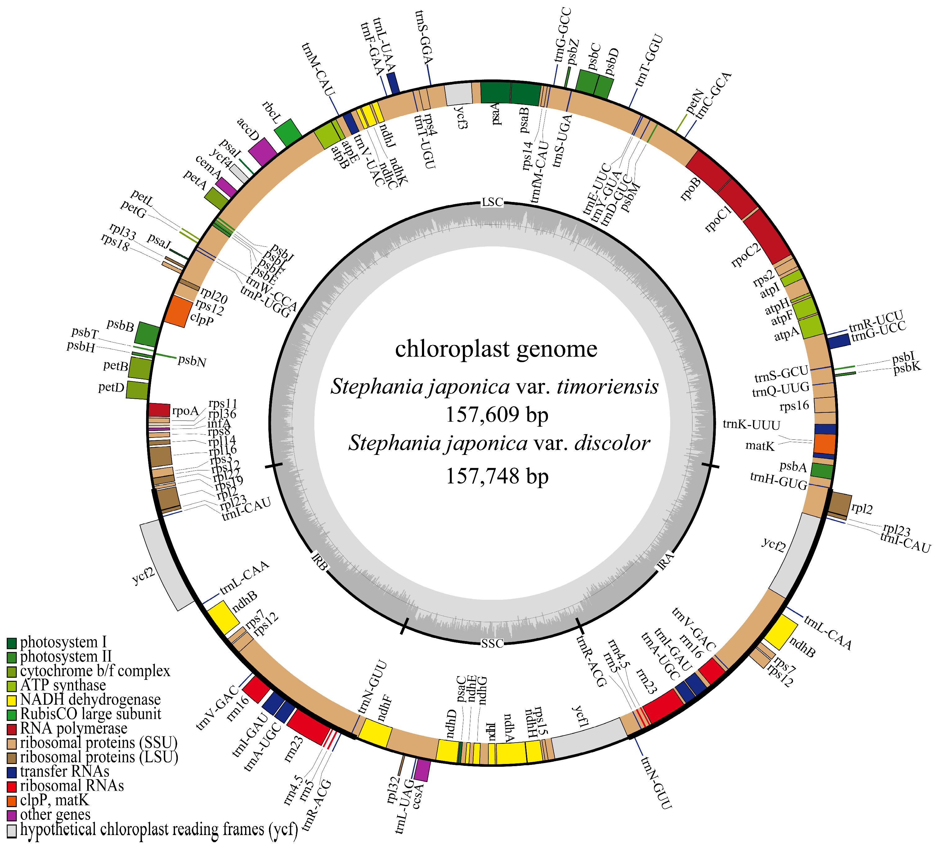
3.1. Analysis of Characteristics of Chloroplast Genomes

A total of 8449 Mb and 6862 Mb of Illumina raw sequencing data, as well as 2245 Mb and 630 Mb PacBio raw data, were obtained for S. japonica var. timoriensis and S. japonica var. discolor, respectively. The assembled chloroplast genomes of S. japonica var. timoriensis and S. japonica var. discolor were 157,609 bp and 157,748 bp in length, respectively (Table 2), composed of double-stranded circular DNA with typical tetrameric structures, including one large single copy (LSC), one small single copy (SSC) and two inverted repeats (IR) (Figure 1). The LSC regions of S. japonica var. timoriensis and S. japonica var. discolor were 88,477 bp and 88,581 bp, respectively, while the SSC regions were 20,346 bp and 20,365 bp and the IR regions were 24,393 bp and 24,401 bp (Table 2). Both genomes exhibited a total GC content of 38.25% and 38.26% (Table 2), with highest GC content observed in the IR region (43.72% and 43.73%), followed by the LSC (36.46% and 36.46%) and SSC regions (32.94% and 32.97%).
Annotation of the chloroplast genomes of S. japonica var. timoriensis and S. japonica var. discolor yielded 130 genes, including 85 protein-coding genes (PCGs), 37 transfer RNA genes (tRNAs), and 8 ribosomal RNA genes (rRNAs) (Table 3). Among them, six PCGs (ndhB, rpl2, rpl23, rps7, rps12, and ycf2), seven tRNAs (trnA-UGC, trnI-CAU, trnI-GAU, trnL-CAA, trnN-GUU, trnR-ACG, and trnV-GAC), and four rRNAs (rrn4.5S, rrn5S, rrn16S, and rrn23S) were located in the IR region, each with two copies. All other genes were present as single copies. The genes in the chloroplast genome were categorized into four functional groups. The first group consisted of 45 genes related to photosynthesis, including 5 photosystem I genes, 15 photosystem II genes, 6 cytochrome b/f complex genes, 12 NADH dehydrogenase genes, 6 ATP synthase genes, and 1 Rubisco subunit gene. The second group consisted of 74 genes related to self-replication, including 11 ribosomal large subunit genes, 14 ribosomal small subunit genes, 4 RNA polymerase genes, in addition to tRNA genes and rRNA genes. The third group consisted of six genes for various proteins, while the fourth group consisted of five genes with unknown functions.

3.2. Codon Usage Bias

A total of 50 protein-coding sequences from the S. japonica var. timoriensis and S. japonica var. discolor chloroplast genomes were analyzed for codon usage preferences (Figure 2). The codon usage patterns of chloroplast genome across both varieties were largely similar, exhibiting only minor variations. Codon analysis revealed that each genome contained 64 codons, with total usage frequencies of 20,002 and 22,022, respectively. Excluding the 3 termination codons, the remaining 61 codons encoded 20 amino acids. Notably, codons for leucine (Leu) were the most abundant in the two chloroplast genomes, accounting for 10.15% (2031) and 10.23% (2252) of total codons, respectively, while the codons for cysteine (Cys) were the least abundant, accounting for 1.16% (233) and 1.17% (258), respectively. Most amino acids were encoded by 2–6 synonymous codons, except for methionine (Met) and tryptophan (Trp), which were uniquely encoded by the single codons AUG and UGG, respectively, both with an RSCU value of one, indicating no preference in usage. When RSCU is larger than one, it indicates a stronger preference for using this codon to encode the same amino acid as compared to other synonymous codons. When RSCU is less than one, it is lower than that of other synonymous codons in usage. In the chloroplast genomes of these two varieties, except two codons with an RSCU of one, there were 62 remaining codons, including 31 codons with an RSCU greater than one, suggesting a preference for these codons, and 31 codons with an RSCU less than one, indicating lesser usage. Among these, 29 preferred codons ending in A or U, with only 3 not preferring codons ending in A or U, suggesting that high-frequency codons tend to end in A/U and low-frequency codons in G/C. The highest RSCU values in the chloroplast genomes of S. japonica var. timoriensis and S. japonica var. discolor were for UUA encoding Leu and AGA encoding arginine (Arg), respectively. Total GC content in the codons encoding amino acids was 38.7% for both varieties, with the third codon showing GC content of 27.9% and 28.6%, respectively. This pattern indicates a bias towards A/U-rich codons, especially at the third codon position in both varieties.

3.3. Repeat Sequences and SSR Analysis

A total of 207 and 211 SSRs were identified in the chloroplast genomes of S. japonica var. timoriensis and S. japonica var. discolor, respectively. These SSRs comprised five types, including mononucleotide, dinucleotide, trinucleotide, tetranucleotide, and pentanucleotide repeat sequences, with no hexanucleotide repeats detected. As shown in Table 4, the SSRs were primarily concentrated in the LSC region, followed by the SSC region, with the fewest found in the IR region. Specifically, the LSC region contained 142 and 143 SSRs, the SSC region contained 45 and 48 SSRs, and the IR region contained 20 and 20 SSRs in S. japonica var. timoriensis and S. japonica var. discolor, respectively. Mononucleotide repeats were the most common, accounting for 81.2% (168) and 83.4% (176) of all SSRs, mostly the A/T type, followed by dinucleotide repeats (18), mainly the AT/AT type. The remaining sequences accounted for a relatively small proportion, including seven and six trinucleotide repeats, nine and seven tetranucleotide repeats, and five and four pentanucleotide repeats in S. japonica var. timoriensis and S. japonica var. discolor, respectively.

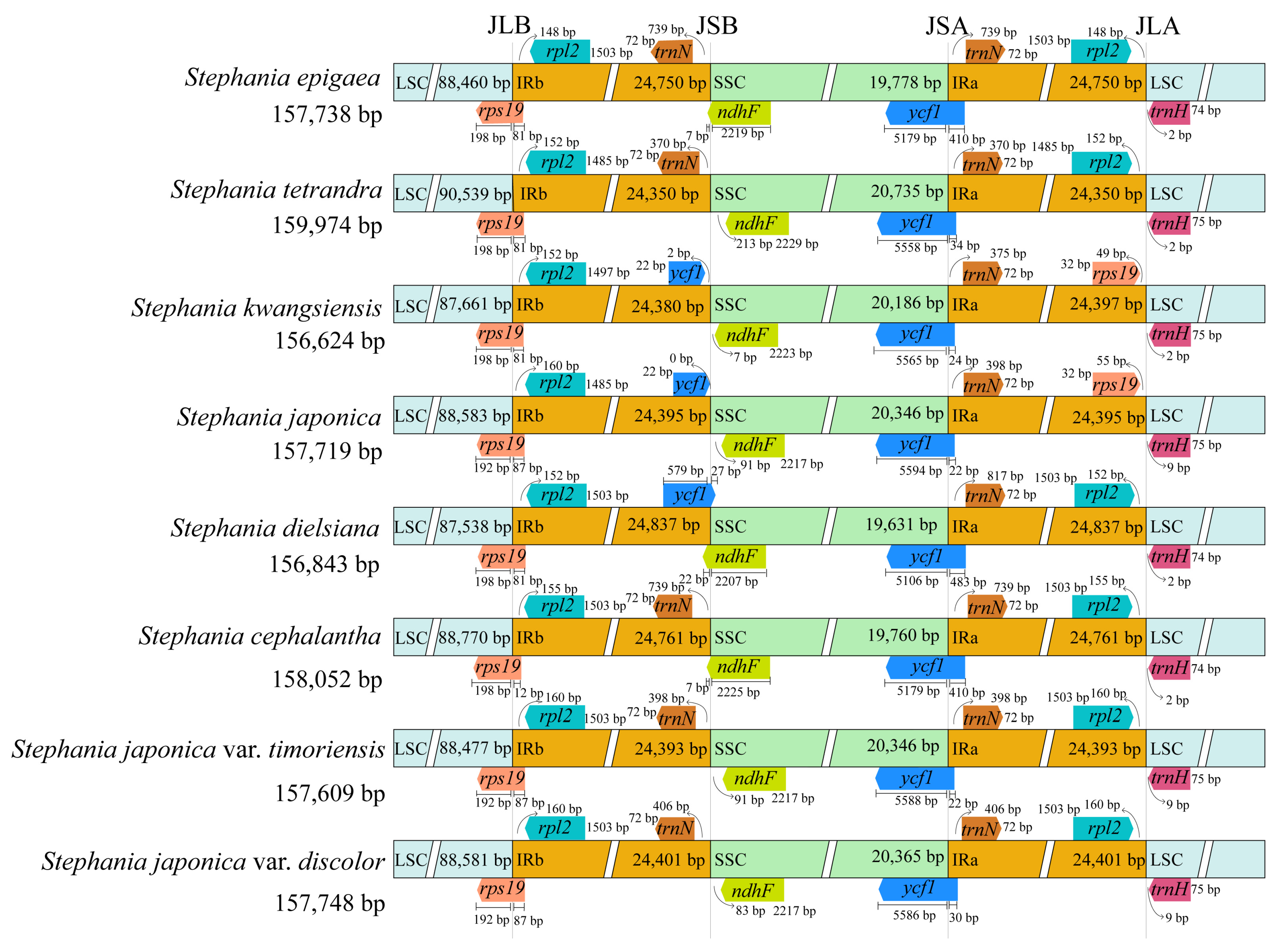
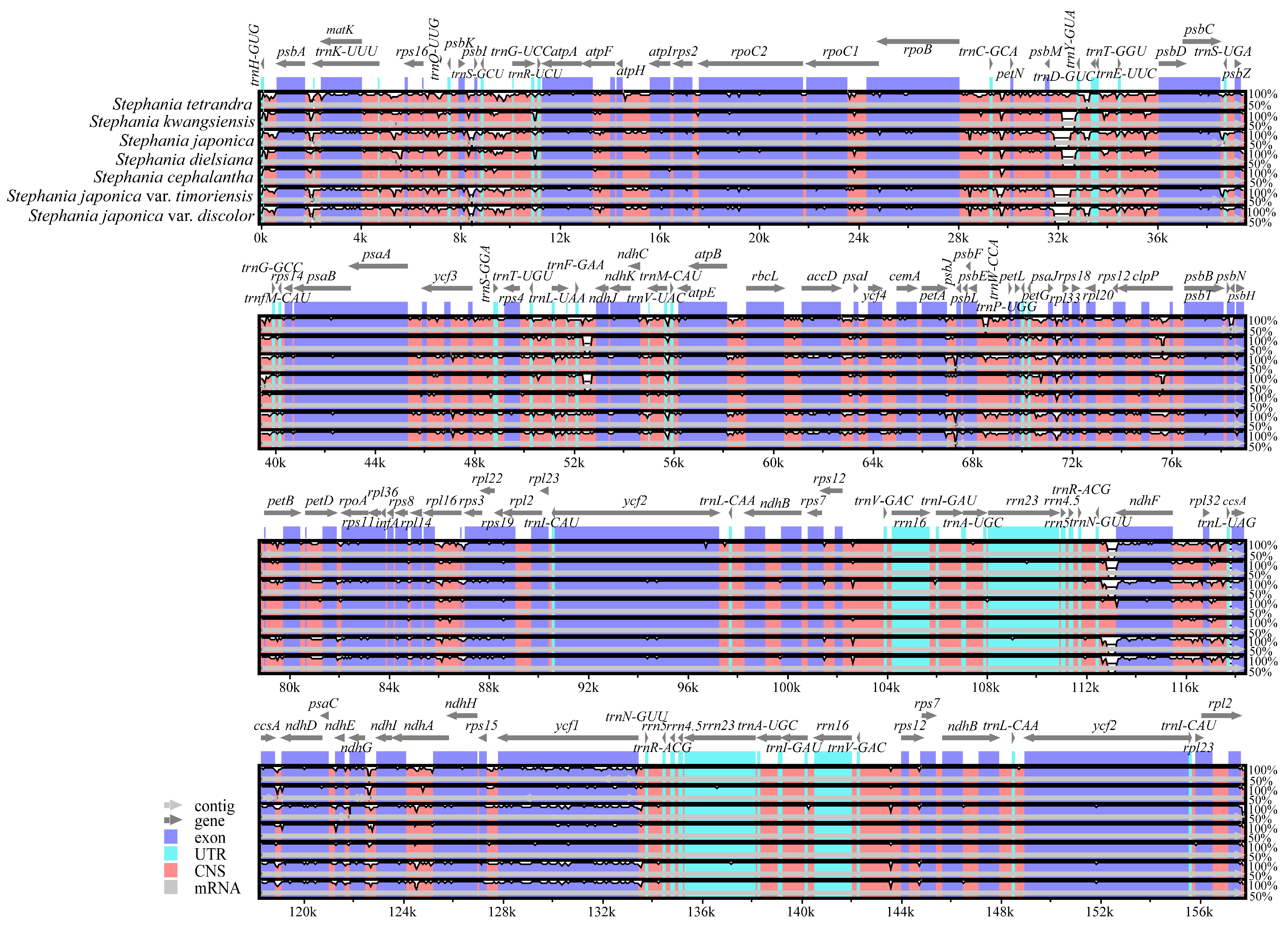
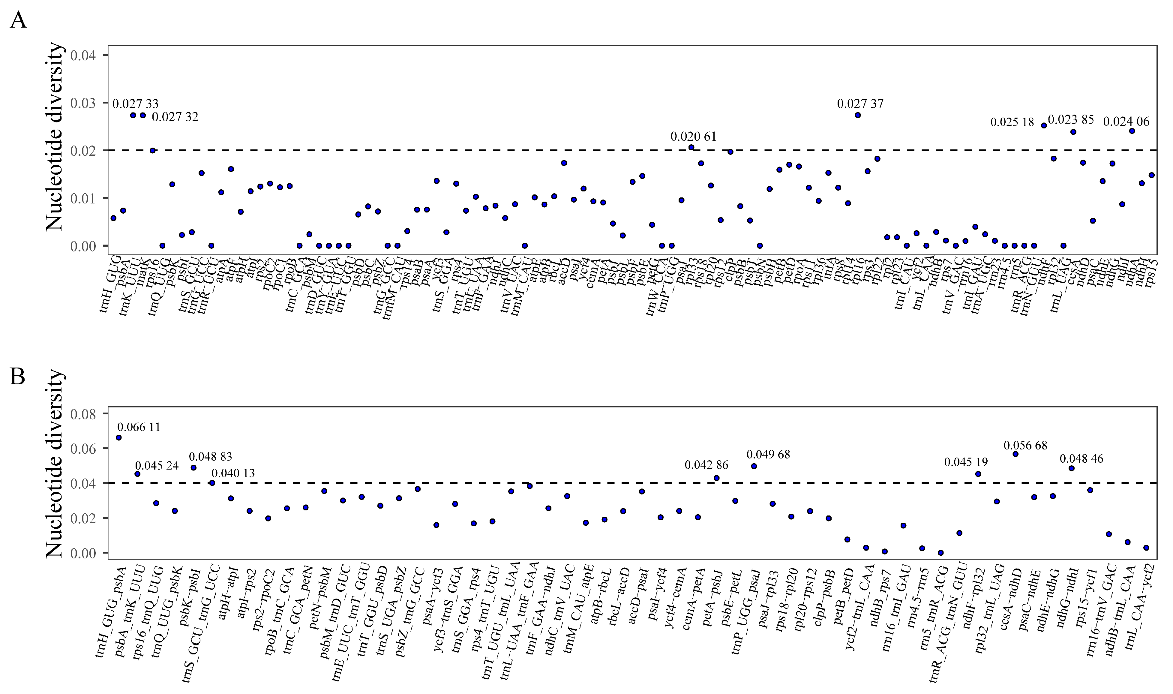
3.4. Comparative Genome Analysis

The chloroplast genomes of the eight Stephania species exhibited minimal differences in total length and within their four zones, indicating a high level of conservation (Figure 3). However, the genes at the boundaries were not completely identical, displaying some expansion and contraction. The boundaries between two adjacent single copy regions and the IR region were designated as JLB, JSB, JSA, and JLA, respectively. The JLB boundaries of all eight species consistently fell within the rps19 gene, to the left of the rpl2 gene. The JSA boundaries consistently occurred within the ycf1 gene, to the left of the trnN gene. There were slight differences in the degree of expansion between these eight species. The genes at the JSB boundary of S. japonica var. timoriensis, S. japonica var. discolor, Stephania epigaea, S. tetrandra, and Stephania cephalantha were trnN and ndhF, while the genes at the JSB boundary of the remaining species were ycf1 and ndhF. At the JLA boundary, Stephania kwangsiensis and S. japonica had the same flanking genes, rps19 and trnH, whereas the other six species, including S. japonica var. timoriensis and S. japonica var. discolor, contained the flanking genes rpl2 (in the IRa region) and trnH (in the LSC region), with a distance of 148–160 bp from the end of rpl2 and the JLA boundary. In this study, the boundary gene types of S. japonica var. timoriensis and S. japonica var. discolor were completely identical, and the distances between the flanking genes and boundaries were similar, with no significant contraction or expansion observed.
To assess the extent of chloroplast genome variation within the genus, comprehensive sequence alignment was performed on the chloroplast genomes of seven other Stephania species, using S. epigaea as a reference. Overall, the Stephania chloroplast genomes exhibited high similarity and conservation. The LSC and SSC regions showed greater variability compared to the IR region, with non-coding regions displaying significantly higher variation than coding regions. The tRNA and rRNA sequences within the IR region were highly conserved (Figure 4).
A total of 107 common gene sequences and 56 common spacer sequences were extracted from the chloroplast genomes of Stephania. The Pi values for the common spacer sequences were generally higher than those for the common gene sequences. Among the shared gene sequences, the Pi values for rpl33, ccsA, ndhA, ndhF, matK, trnK-UUU, and rpl16 exceeded 0.020. Among the shared spacer sequences, the Pi values of trnS-GCU-trnG-UCC, petA-psbJ, ndhF-rpl32, psbA-rnK-UUUU, ndhG-ndhI, psbK-psbI, trnP-UGG-psaJ, ccsA-ndhD, and trnH-GUG-psbA exceeded 0.040 (Figure 5). These highly mutated regions could serve as potential molecular markers for species identification within the genus Stephania.

3.5. Phylogenetic Analysis

Phylogenetic analysis indicated that the support rate for all lineages was greater than 85%, confirming the reliability of the results. All species within the family Menispermaceae formed a monophyletic branch. Within this framework, the eight Stephania species clustered into one lineage with 100% support. The genus was further divided into three lineages. S. tetrandra formed an independent lineage, while S. japonica, S. japonica var. discolor and S. japonica var. timoriensis formed a monophyletic lineage with 100% support, and S. japonica var. discolor and S. japonica formed a lineage as sister taxon to S. japonica var. timoriensis. Furthermore, S. cephalantha and S. epigaea formed a distinct lineage which was the closest relative to the lineage formed by Stephania dielsiana and S. kwangsiensis. These four species collectively constituted the third lineage within the genus Stephania (Figure 6).

4. Discussion

In this study, we assembled the chloroplast genomes of S. japonica var. timoriensis and S. japonica var. discolor. Consistent with other angiosperms, the chloroplast genomes of these two species exhibited a typical four-part structure, including the LSC, SSC, IRa, and IRb regions. The chloroplast genomes of the two species showed remarkable similarity in length, total GC content, and GC content of each partition. Additionally, the number, type, and arrangement of genes were highly consistent with previously reported Stephania species [40,41]. The Stephania chloroplast genomes exhibited a degree of conservation; however, they differed from other genera within Menispermaceae, such as Menispermum, Sinomenium, and Fibraurea, which had longer chloroplast genomes (exceeding 160,000 bp) and lower total GC content [42,43,44].
Our analysis also showed that the boundary wing gene types in the chloroplast genomes of the two varieties were identical. However, the genotypes at JSB and JLA differed among the eight Stephania species, particularly at the location of the ndhF gene, which occurred in either the SSC region or at the IRb/SSC junction. These differences are likely due to varying degrees of IR region constriction or expansion, which are primary factors contributing to variations in chloroplast genomes [45]. The preference for codon usage is important for understanding species evolution and gene expression. Our analysis revealed a high degree of similarity in codon usage between the two varieties, with both showing a preference for A/U, likely due to the high content of the A and T bases, resulting in a bias towards A or T ending codons. The RSCU values indicated that the codons for Leu and Arg had the highest frequency of occurrence. These codon preference results are consistent with previously reported findings for S. tetrandra [40].
The identification of SSRs is crucial for investigating and identifying genetic diversity at the molecular level. In this study, 207 and 211 SSRs were identified in the chloroplast genomes of S. japonica var. timoriensis and S. japonica var. discolor, respectively. These SSRs were diverse in type and primarily distributed in the LSC region, consistent with other angiosperms [23,24,46]. These SSR loci are promising candidates for future molecular identification of Stephania species. By examining the sequence variations in the chloroplast genome of the genus Stephania, we identified seven common gene sequences with Pi values greater than 0.020 and nine common inter-region sequences with Pi values of greater than 0.040 in the highly variable region. In previous studies, Wang et al. identified one of the six candidate DNA barcodes identified in our results (matk), while Dong et al. identified two of the five high Pi value regions consistent with our findings (trnH-psbA, ndhA) [40,47]. In addition to these three reported molecular markers, we screened 13 mutation hotspots, including rpl33, ccsA, ndhF, trnK, rpl16, trnS-trnG, petA-psbJ, ndhF-rpl32, psbA-trnK, ndhG-ndhI, psbK-psbI, trnP-psaJ, and ccsA-ndhD. These highly variable sequences could serve as a potential tool for identifying different species within the genus Stephania.
The phylogenetic tree constructed from the chloroplast genomes of S. japonica var. timoriensis, S. japonica var. discolor, and 20 related species demonstrated that all Stephania species formed a monophyletic group, clearly divided into three lineages, corresponding to the subgenera Botryodiscia, Stephania, and Tuberiphania. Stephania and Tuberiphania diverged from the same node, indicating a close relationship. Based on phylogenetic analysis of the nuclear transcribed spacer (ITS) and chloroplast transcribed spacer (trnL-F) sequences, Xie et al. also divided Stephania into three major lineages [48], consistent with our findings, but did not fully resolve the interrelationships among the three lineages due to low support values [48]. In our study, S. japonica, S. japonica var. discolor, and S. japonica var. timoriensis were clustered within the subgenus Stephania. Specifically, S. japonica and S. japonica var. discolor formed a lineage that was the sister taxon to S. japonica var. timoriensis. These findings align with the phylogenetic tree constructed based on ITS+psbA-trnH in Wang et al. [47]. However, our internal branching results of the subgenus Tuberiphania differ slightly from those of Wang et al. [47], particularly regarding the relationship between S. epigaea and S. cephalantha. These preliminary phylogenetic results indicate that the taxonomic status of the two varieties needs to be further examined. Further research including more species is necessary to determine the phylogenetic positions within Stephania. Additionally, expanding the chloroplast genome data for other species in this genus is essential.

5. Conclusions

In this study, the chloroplast genomes of S. japonica var. timoriensis and S. japonica var. discolor were obtained using second- and third-generation sequencing techniques. This study enriches the chloroplast genome data within the genus Stephania and provides valuable molecular data for the precise identification of similar species, as well as for studying genetic diversity in the genus Stephania.

Author Contributions

L.-P.Z. conceived and designed the study; L.-P.Z. and Y.-M.G. collected the samples; L.-L.W., Y.-M.G. and L.-P.Z. performed the experiments and analysis; L.-L.W. wrote the manuscript; and L.-P.Z. revised the manuscript. All authors have read and agreed to the published version of the manuscript.

Funding

This research was funded by the Department of Science and Technology of Yunnan Province Joint Special Project of Traditional Chinese Medicine (202101AZ070001-056), Yunnan Fundamental Research Project (202301AT070254), and National Natural Science Foundation of China (31960103).

Institutional Review Board Statement

Not applicable.

Informed Consent Statement

Not applicable.

Data Availability Statement

The genome sequence data generated in this study are openly available in GenBank of NCBI at (https://www.ncbi.nlm.nih.gov/) under the accession no. PP175339-PP175340.

Conflicts of Interest

The authors declare no conflicts of interest.

References

  1. Committee for the Flora of China, Chinese Academy of Sciences. Flora of China; Beijing Science Press: Beijing, China, 1996; Volume 30, Part 2; pp. 40–42. [Google Scholar]
  2. Chen, Z.Q. Supplement to Materia Medica; Wannan Medical College: Wuhu, China, 1983; p. 86. [Google Scholar]
  3. Dai, Z.H.; Wei, W.; Fan, L.Y. Microscopic identification of Stephaniae Longae Herba of Zhuang medicine. Res. Zhuang Yao Ethn. Med. 2020, 4, 63–67. (In Chinese) [Google Scholar]
  4. Li, L.; Zuo, A.X.; Rao, G.X. Studies on alkaloids from the Stephania epigaea of Dai traditional medicine. J. Yunnan Univ. Chin. Med. 2012, 35, 14–19. (In Chinese) [Google Scholar]
  5. An, C.; Chen, M.; Yang, C.Z.; Meng, J.; Zhuang, Y.X. Pharmacognostic Examination and Nomenclature Organization of ‘Jinbuhuan’. J. Chin. Med. Mater. 2019, 42, 2449–2452. (In Chinese) [Google Scholar]
  6. Luo, X.R. A preliminary study on the genus Stephania in China. Acta Phytotaxon. Sin. 1978, 16, 10–40. (In Chinese) [Google Scholar]
  7. Feng, Y.X.; Zhu, Z.Y.; Chen, H. Comparative Morphological and Histological Study of Medicinal Plants in the Genus Stephania. Acta Pharm. Sin. 1983, 11, 849–861. (In Chinese) [Google Scholar]
  8. Tang, C.F.; Song, S.Q. Woody lianas of the genus Stephania. Life World 2022, 11, 70–71. (In Chinese) [Google Scholar]
  9. Luo, X.R. An Outline of the Classification of the Genus Stephania in China. Bull. Bot. Res. 1982, 1, 33–59. (In Chinese) [Google Scholar]
  10. Zhu, Z.Y.; Feng, Y.X.; He, L.Y.; Wang, Y.C. Studies on the utilization of medicinal plant resources of the genus Stephania of the family Menispermaceae in China. Acta Pharm. Sin. 1983, 6, 460–467. (In Chinese) [Google Scholar]
  11. National Pharmacopoeia Committee. Pharmacopoeia of the People’s Republic of China; China Medical Science and Technology Press: Beijing, China, 2020; Volume I, p. 155. [Google Scholar]
  12. Yang, H.M.; Luo, X.R. Research on the ‘shanwugui’. Acta Pharm. Sin. 1980, 11, 674–683+705–707. (In Chinese) [Google Scholar]
  13. Yang, H.M.; Chen, Y. An overview of the chemical classification of plants of the genus Stephania in China. J. Integr. Plant Biol. 1994, 11, 14–22. (In Chinese) [Google Scholar]
  14. Li, X.J.; Liu, L.; Li, G.J.; Dong, J.W. Progress in chemical constituents, pharmacology and fermentation modification of radix Stephaniae Epigaeae. Shandong Chem. Ind. 2023, 52, 89–94. (In Chinese) [Google Scholar]
  15. Li, S.Z.; Wu, F. A review on the identification of radix Stephania Tetrandrae and the fake. Chin. J. Ethnomed. Ethnopharm. 2020, 29, 57–62. (In Chinese) [Google Scholar]
  16. Guo, M.; Zhang, R.Y.; Cai, F. Research progress on pharmacological activities of tetrandrine. China Mod. Med. 2018, 25, 30–33. (In Chinese) [Google Scholar]
  17. He, D.H.; Liu, J.; Fang, D.M.; Wang, X.L.; Li, L.M. Study on alkaloid constituents of Stephania tetrandra S. Moore. Acta Pharm. Sin. 2021, 56, 3503–3510. (In Chinese) [Google Scholar]
  18. Mo, L.Y.; Zhang, F.; Hao, E.W.; Deng, J.G.; Chen, F.; Li, R.L.; Mo, Y.M.; Zhang, L.Q.; Hou, X.T.; Du, Z.C. Predictive analysis of quality markers (Q-Marker) of Stephania tetrandra based on chemical components and pharmacological and network pharmacology. Chin. Tradit. Herb. Drugs 2022, 53, 6283–6295. (In Chinese) [Google Scholar]
  19. Shen, X.J.; Dong, J.W.; Mei, R.F.; Cai, L.; Ding, Z.T. Study on alkaloids of Stephania epigaea. Chem. Res. Appl. 2016, 28, 1730–1734. (In Chinese) [Google Scholar]
  20. Sun, Y.; Moore, M.J.; Zhang, S.; Soltis, P.S.; Soltis, D.E.; Zhao, T.; Meng, A.; Li, X.; Li, J.; Wang, H. Phylogenomic and structural analyses of 18 complete plastomes across nearly all families of early-diverging eudicots, including an angiosperm-wide analysis of IR gene content evolution. Mol. Phylogenet. Evol. 2016, 96, 93–101. [Google Scholar] [CrossRef]
  21. Wang, L.; Dong, W.P.; Zhou, S.L. Structural mutations and reorganizations in chloroplast genomes of flowering plants. Acta Bot. Boreali-Occident. Sin. 2012, 32, 1282–1288. (In Chinese) [Google Scholar]
  22. Freudenthal, J.A.; Pfaff, S.; Terhoeven, N.; Korte, A.; Ankenbrand, M.J.; Förster, F. A systematic comparison of chloroplast genome assembly tools. Genome Biol. 2020, 21, 254. [Google Scholar] [CrossRef] [PubMed]
  23. Zhang, Y.; Du, C.H.; Zhan, H.X.; Shang, C.L.; Li, R.F.; Yuan, S.J. Comparative and phylogeny analysis of Platycodon grandiflorus complete chloroplast genomes. Chin. Tradit. Herb. Drugs 2023, 54, 4981–4991. (In Chinese) [Google Scholar]
  24. Zhang, W.X.; Liu, H.; Wang, L.; Zhang, S.H. Chloroplast genome comparison and phylogenetic analysis of four medicinal plants of evodia. Guid. J. Tradit. Chin. Med. Pharm. 2023, 29, 44–50+65. (In Chinese) [Google Scholar]
  25. Yang, X.Y.; Zhang, M.J.; Yin, T.; Han, P.C.; Du, C.J.; Zhang, H.Y. Chloroplast genome characteristics and codon preferences analysis in Syzygium. Southwest China J. Agric. Sci. 2023, 36, 1869–1880. (In Chinese) [Google Scholar]
  26. Schloss, P.D.; Jenior, M.L.; Koumpouras, C.C.; Westcott, S.L.; Highlander, S.K. Sequencing 16S rRNA gene fragments using the PacBio SMRT DNA sequencing system. PeerJ 2016, 4, e1869. [Google Scholar] [CrossRef] [PubMed]
  27. Lian, Y.; Huang, F.; Zhu, W.T.; Liu, X.F.; Wu, H.; Jiang, G.H.; Yin, X.M. Chloroplast genome structure of Stemona tuberosa and phylogenetic analysis based on PacBio sequencing. Chin. J. Exp. Tradit. Med. Formulae 2023, 29, 123–132. (In Chinese) [Google Scholar]
  28. Bolger, A.M.; Lohse, M.; Usadel, B. Trimmomatic: A flexible trimmer for Illumina sequence data. Bioinformatics 2014, 30, 2114–2120. [Google Scholar] [CrossRef] [PubMed]
  29. Jin, J.J.; Yu, W.B.; Yang, J.B.; Song, Y.; de Pamphilis, C.W.; Yi, T.S.; Li, D.Z. GetOrganelle: A fast and versatile toolkit for accurate de novo assembly of organelle genomes. Genome Biol. 2020, 21, 241. [Google Scholar] [CrossRef]
  30. Bankevich, A.; Nurk, S.; Antipov, D.; Gurevich, A.A.; Dvorkin, M.; Kulikov, A.S.; Lesin, V.M.; Nikolenko, S.I.; Pham, S.; Prjibelski, A.D. SPAdes: A new genome assembly algorithm and its applications to single-cell sequencing. J. Comput. Biol. 2012, 19, 455–477. [Google Scholar] [CrossRef]
  31. Kearse, M.; Moir, R.; Wilson, A.; Stones-Havas, S.; Cheung, M.; Sturrock, S.; Buxton, S.; Cooper, A.; Markowitz, S.; Duran, C. Geneious Basic: An integrated and extendable desktop software platform for the organization and analysis of sequence data. Bioinformatics 2012, 28, 1647–1649. [Google Scholar] [CrossRef]
  32. Sharp, P.M.; Li, W.H. Codon usage in regulatory genes in Escherichia coli does not reflect selection for rare codons. Nucleic Acids Res. 1986, 14, 7737–7749. [Google Scholar] [CrossRef]
  33. Dai, J.P.; Cai, Y.M.; Liu, Q.Z.; Gao, X.X.; Zhu, S. Analysis of codon usage bias of chloroplast genome in seven Glycyrrhiza species. Chin. Tradit. Herb. Drugs 2023, 54, 2907–2916. (In Chinese) [Google Scholar]
  34. Beier, S.; Thiel, T.; Münch, T.; Scholz, U.; Mascher, M. MISA-web: A web server for microsatellite prediction. Bioinformatics 2017, 33, 2583–2585. [Google Scholar] [CrossRef] [PubMed]
  35. Amiryousefi, A.; Hyvönen, J.; Poczai, P. IRscope: An online program to visualize the junction sites of chloroplast genomes. Bioinformatics 2018, 34, 3030–3031. [Google Scholar] [CrossRef] [PubMed]
  36. Frazer, K.A.; Pachter, L.; Poliakov, A.; Rubin, E.M.; Dubchak, I. VISTA: Computational tools for comparative genomics. Nucleic Acids Res. 2004, 32, W273–W279. [Google Scholar] [CrossRef] [PubMed]
  37. Rozas, J.; Ferrer-Mata, A.; Sánchez-DelBarrio, J.C.; Guirao-Rico, S.; Librado, P.; Ramos-Onsins, S.E.; Sánchez-Gracia, A. DnaSP 6: DNA sequence polymorphism analysis of large data sets. Mol. Biol. Evol. 2017, 34, 3299–3302. [Google Scholar] [CrossRef] [PubMed]
  38. Katoh, K.; Standley, D.M. MAFFT multiple sequence alignment software version 7: Improvements in performance and usability. Mol. Biol. Evol. 2013, 30, 772–780. [Google Scholar] [CrossRef] [PubMed]
  39. Nguyen, L.T.; Schmidt, H.A.; von Haeseler, A.; Quang Minh, B. IQ-TREE: A fast and effective stochastic algorithm for estimating maximum-likelihood phylogenies. Mol. Biol. Evol. 2015, 32, 268–274. [Google Scholar] [CrossRef] [PubMed]
  40. Dong, S.J.; Ying, Z.Q.; Yu, S.S.; Wang, Q.R.; Liao, G.H.; Ge, Y. Complete chloroplast genome of Stephania tetrandra (Menispermaceae) from Zhejiang Province: Insights into molecular structures, comparative genome analysis, mutational hotspots and phylogenetic relationships. BMC Genom. 2021, 22, 880. [Google Scholar] [CrossRef]
  41. Guan, Q.; Feng, D.; Fan, M. The complete chloroplast genome sequence of the medicinal plant Stephania epigaea H. S. Lo, 1978 (Menispermaceae) from Yunnan, China. Mitochondrial DNA B Resour. 2022, 7, 1403–1405. [Google Scholar] [CrossRef] [PubMed]
  42. Song, S.; Cameron, K.M.; Wang, Y.; Wang, S.; Jin, X.; Hina, F.; Yang, Z.; Li, P. Phylogenomics and phylogeography of Menispermum (Menispermaceae). Front. Plant Sci. 2023, 14, 1116300. [Google Scholar] [CrossRef]
  43. Kim, J.; Lee, J.; Um, S.; Choi, S.; Kim, H.; Chun, H.S.; Nah, G. The complete chloroplast genome of Sinomenium acutum (Menispermaceae). Mitochondrial DNA Part B Resour. 2020, 5, 2992–2993. [Google Scholar] [CrossRef]
  44. Zheng, L.P.; Feng, R. Characterization of the complete chloroplast genome of Fibraurea recisa Pierre 1885 (Menispermaceae), an important medicinal herb from Yunnan, China. Mitochondrial DNA Part B Resour. 2022, 7, 501–502. [Google Scholar] [CrossRef] [PubMed]
  45. Ni, Z.; Ye, Y.; Bai, T.; Xu, M.; Xu, L.A. Complete Chloroplast Genome of Pinus massoniana (Pinaceae): Gene Rearrangements, Loss of ndh Genes, and Short Inverted Repeats Contraction, Expansion. Molecules 2017, 22, 1528. [Google Scholar] [CrossRef] [PubMed]
  46. Zhao, P.Y.; Zhao, W.; Hou, Z.Y.; Li, X.; Liu, B.K.; Du, Y.; Xie, R.Q.; Cao, Z.L.; Wang, K.K. Comparison of chloroplast genome characteristics and phylogenetic analysis between Themeda japonica and two species of Themeda. Chin. Tradit. Herb. Drugs 2023, 54, 3261–3272. (In Chinese) [Google Scholar]
  47. Wang, X.L.; Xue, J.Y.; Zhang, Y.Y.; Xie, H.; Wang, Y.Q.; Weng, W.; Kang, Y.; Huang, J. DNA barcodes for the identification of Stephania (Menispermaceae) species. Mol. Biol. Rep. 2020, 47, 2197–2203. [Google Scholar] [CrossRef]
  48. Xie, D.T.; He, J.Y.; Huang, J.M.; Xie, H.; Wang, Y.Q.; Kang, Y.; Jabbour, F.; Guo, J. Molecular phylogeny of Chinese Stephania (Menispermaceae) and reassessment of the subgeneric and sectional classifications. Aust. Syst. Bot. 2015, 28, 246–255. [Google Scholar] [CrossRef]
Figure 1. Chloroplast genomes of S. japonica var. timoriensis and S. japonica var. discolor.
Genes 15 00877 g001
Figure 2. Relative synonymous codon usage of chloroplast genomes of S. japonica var. timoriensis and S. japonica var. discolor. Histogram of each amino acid from left to right is S. japonica var. timoriensis and S. japonica var. discolor.
Genes 15 00877 g002
Figure 3. Comparison of LSC, IRs, and SSC border regions of chloroplast genomes of Stephania.
Genes 15 00877 g003
Figure 4. Comparative analysis of chloroplast genomes of Stephania.
Genes 15 00877 g004
Figure 5. Nucleic acid polymorphism analysis of chloroplast genomes of Stephania. (A) Nucleic acid polymorphism analysis of common genes. (B) Nucleic acid polymorphism analysis of common intergenic regions.
Genes 15 00877 g005
Figure 6. Maximum likelihood (ML) phylogenetic tree of analysis based on chloroplast genome sequences of 22 species. The species highlighted in pink font color are newly sequenced in this study.
Genes 15 00877 g006
Table 1. Samples used in this study.
No.SpeciesFromGenBank Accession No.
1Stephania japonica var. timoriensisThis studyPP175340
2Stephania japonica var. discolorThis studyPP175339
3Stephania cephalanthaGenBankNC_067079
4Stephania dielsianaGenBankNC_054337
5Stephania epigaeaGenBankNC_058988
6Stephania japonicaGenBankNC_029432
7Stephania kwangsiensisGenBankNC_048524
8Stephania tetrandraGenBankNC_050924
9Pericampylus glaucusGenBankNC_046846
10Menispermum canadenseGenBankNC_048451
11Menispermum dauricumGenBankNC_042371
12Sinomenium acutumGenBankMT040976
13Fibraurea recisaGenBankNC_060536
14Akebia quinataGenBankKX611091
15Decaisnea insignisGenBankKY200671
16Thalictrum baicalenseGenBankMW133265
17Thalictrum minusGenBankNC_041544
18Berberis amurensisGenBankKM057374
19Nandina domesticaGenBankDQ923117
20Sinopodophyllum hexandrumGenBankKR779994
21Epimedium lishihcheniiGenBankKU522472
22Vancouveria planipetalaGenBankMH337373
Table 2. Composition and characteristics of chloroplast genomes of S. japonica var. timoriensis and S. japonica var. discolor.
SpeciesGenomeLSCIRSSC
Length/bpGC/%Length/bpGC/%Length/bpGC/%Length/bpGC/%
S. japonica var. timoriensis157,60938.2588,47736.4624,39343.7220,34632.94
S. japonica var. discolor 157,748 38.2688,581 36.4624,401 43.7320,365 32.97
Table 3. Functional annotation and classification of chloroplast genomes of S. japonica var. timoriensis and S. japonica var. discolor.
Gene FunctionGene GroupGene NameNumber
Gene for photosynthesisPhotosystem IpsaA, psaB, psaC, psaI, psaJ5
Photosystem IIpsbA, psbB, psbC, psbD, psbE, psbF, psbH, psbI, psbJ, psbK, psbL, psbM, psbN, psbT, psbZ15
Cytochrome b/f complexpetA, petB, petD, petG, petL, petN6
NADH dehydrogenasendhA, ndhB a, ndhC, ndhD, ndhE, ndhF, ndhG, ndhH, ndhI, ndhJ, ndhK12
Subunits of ATP synthaseatpA, atpB, atpE, atpF, atpH, atpI6
Rubisco large subunitsrbcL1
Self-replicationLange subunits of ribosomerpl2 a, rpl14, rpl16, rpl20, rpl22, rpl23 a, rpl32, rpl33, rpl3611
Small subunits of ribosomerps2, rps3, rps4, rps7 a, rps8, rps11, rps12 a, rps14, rps15, rps16, rps18, rps1914
RNA polymeraserpoA, rpoB, rpoC1, rpoC24
Ribosomal RNA genesrrn4.5S a, rrn5S a, rrn16S a, rrn23S a8
Transfer RNA genestrnA-UGC a, trnC-GCA, trnD-GUC, trnE-UUC, trnF-GAA, trnfM-CAU, trnG-GCC, trnG-UCC, trnH-GUG, trnI-CAU a, trnI-GAU a, trnK-UUU, trnL-CAA a, trnL-UAA, trnL-UAG, trnM-CAU, trnN-GUU a, trnP-UGG, trnQ-UUG, trnR-ACG a, trnR-UCU, trnS-GCU, trnS-GGA, trnS-UGA, trnT-GGU, trnT-UGU, trnV-GAC a, trnV-UAC, trnW-CCA, trnY-GUA37
Other genesProteaseclpP1
MaturasematK1
Translation initiation factorinfA1
Envelop membrane proteincemA1
Subunits of acety-CoA-carboxylaseaccD1
C-type cytochrome synthesisccsA1
Unknow geneHypothetical chloroplast reading framesycf1, ycf2 a, ycf3, ycf45
Total 130
Note: a indicates that it contains two copies of genes.
Table 4. SSR statistics of chloroplast genomes of S. japonica var. timoriensis and S. japonica var. discolor.
Types and RegionsNameS. japonica var. timoriensisS. japonica var. discolor
Typesmononucleotide168176
dinucleotide1818
trinucleotide76
tetranucleotide97
pentanucleotide54
RegionsLSC142143
SSC4548
IR2020
Total 207211
Disclaimer/Publisher’s Note: The statements, opinions and data contained in all publications are solely those of the individual author(s) and contributor(s) and not of MDPI and/or the editor(s). MDPI and/or the editor(s) disclaim responsibility for any injury to people or property resulting from any ideas, methods, instructions or products referred to in the content.

